Embed Size (px)
Citation preview

Coordination Chemistry Reviews 393 (2019) 79–117
Contents lists available at ScienceDirect
Coordination Chemistry Reviews
journal homepage: www.elsevier .com/ locate/ccr
Review
Organometallic and coordination rhenium compounds and theirpotential in cancer therapy
https://doi.org/10.1016/j.ccr.2019.04.0140010-8545/� 2019 Elsevier B.V. All rights reserved.
Abbreviations: AChE, acetylcholinesterase; BiPy, bipyridine; BSA, bovine serum albumin; Cp, cyclopentadienyl; CQR, chloroquine-resistant; CQS, chloroquine-senmagna, Daphnia magna; DCA, dichloroacetate; DFT, density functional theory; dppf, 1,10-bis(diphenylphosphino)-ferrocene; EC50, half maximal effective concentratihalf maximal effective dose; EGFR, epidermal growth factor receptor; ER, estrogen receptor; GFAAS, graphite furnace atomic absorption spectroscopy; GLUTtransporter; GnRH, gonadotropin-releasing hormone; h, hour; HDAC, histone deactylase; hMAB, humanized monoclonal antibody; HPLC, high-performancchromatography; HSAB, Hard and Soft Acid and Base; IC50, half-maximal inhibitor concentration; L. minor, Lemna minor; LD50/LC50, half-maximal lethal dose/conceMAG3, mercapto acetyl triglycine; min, minute; MTT, 3-(4,5-dimehtylthiazol-2-yl)-2,5-diphenyltetrazolium bromide; n.a., not active; NAD, nicotinamidedinucleotide; NADH, reduced NAD; NIS, sodium iodide symporter; NIS US, National Institute of Health of the United States; n.d., not determined; MAB, monoclonal aOTf, triflate; PACT, photoactivated chemotherapy; PBR, peripheral benzodiazepine receptor; PDK, pyruvate dehydrogenase kinase; PDT, photodynamic therapolyethylene glycol; PI, propidium iodide; R. subcapitata, Raphidocelis subcapitata; ROS, reactive oxygen species; SAHA, suberanilohydroxamic acid (also known as VorSERM, Selective Estrogen Receptor Modulator; SPECT, single photon emission computed tomography; t, ton (metric); t1/2, half-life; TFA, trifluoroacetic acid; TSPO, tranprotein; V. fischeri, Vibro fischeri.⇑ Corresponding authors.
E-mail address: [email protected] (F.E. Kühn).
Elisabeth B. Bauer a, Allison A. Haase b, Robert M. Reich a, Debbie C. Crans b,c,⇑, Fritz E. Kühn a,⇑aMolecular Catalysis, Catalysis Research Center and Department of Chemistry, Technische Universität München, Lichtenbergstr. 4, 85747 Garching bei München, GermanybDepartment of Chemistry, Colorado State University, Fort Collins, CO 80523-1872, United StatescCell and Molecular Biology Program, Colorado State University, Fort Collins, CO 80523-1872, United States
a r t i c l e i n f o
Article history:Received 30 November 2018Received in revised form 29 April 2019Accepted 30 April 2019Available online 22 May 2019
This manuscript is dedicated to our friendand colleague Armando Pombeiro on theoccasion of his 70th birthday.
Keywords:Rhenium complexesAnticancerTherapeutic applicationsCytotoxicityIC50 valuesClinical applications of Re compounds
a b s t r a c t
This review focuses on cytotoxic rhenium compounds in terms of IC50 values, their mode of action in bio-logical systems and their status in clinical trials. Biological studies of different rhenium compoundsordered by the oxidation state of rhenium are presented. Numerous rhenium complexes are reported,with the greatest number of compounds containing a Re(I)(CO)3+ core. A wide range of complexes hasbeen designed using a combination of organometallic ligands, N- or S-based ligands, peptides, multiden-tate ligands and oxo groups. Design concepts based on membrane permeability and lipophilicity, mem-brane receptor targets and specific enzyme targets are presented. The cytotoxicity parameter IC50 isshown for organometallic compounds, coordination complexes, clusters and Re(oxo) complexes. In addi-tion, a brief summary of in vivo studies is given. A further summary of rhenium compounds subjected toclinical trials is presented to provide information about the classes of rhenium compounds that have beentested in human beings and the approaches used in these studies. Moreover, the comparability of the IC50
values among the cytotoxicity studies is critically assessed to provide the basis for a summary of the mostpotent rhenium compounds according to their reported IC50 values for each type of cancer. The summaryof the structures for the most cytotoxic complexes allows the identification of structural similarities andbasic features that could lead to their cytotoxicity and might be useful for future investigations. Finally,the information from the analysis of the rhenium compounds subjected to cellular studies is compared todata on the rhenium compounds that have been involved in in vivo evaluation and clinical trials.
� 2019 Elsevier B.V. All rights reserved.
Contents
1. Introduction . . . . . . . . . . . . . . . . . . . . . . . . . . . . . . . . . . . . . . . . . . . . . . . . . . . . . . . . . . . . . . . . . . . . . . . . . . . . . . . . . . . . . . . . . . . . . . . . . . . . . . . . . . 802. Basics of bioinorganic rhenium anticancer studies: rhenium chemistry and compounds. . . . . . . . . . . . . . . . . . . . . . . . . . . . . . . . . . . . . . . . . . . . . 80
2.1. Elemental rhenium’s occurrence and application . . . . . . . . . . . . . . . . . . . . . . . . . . . . . . . . . . . . . . . . . . . . . . . . . . . . . . . . . . . . . . . . . . . . . . . 802.2. Methods of evaluating cytotoxicity in cell culture . . . . . . . . . . . . . . . . . . . . . . . . . . . . . . . . . . . . . . . . . . . . . . . . . . . . . . . . . . . . . . . . . . . . . . 81
3. Organometallic Re(I) compounds . . . . . . . . . . . . . . . . . . . . . . . . . . . . . . . . . . . . . . . . . . . . . . . . . . . . . . . . . . . . . . . . . . . . . . . . . . . . . . . . . . . . . . . . . 82
sitive; D.on; ED50,, glucosee liquidntration;adeninentibody;py; PEG,inostat);slocator

80 E.B. Bauer et al. / Coordination Chemistry Reviews 393 (2019) 79–117
3.1. Fundamental chemical properties of organometallic Re(I) compounds . . . . . . . . . . . . . . . . . . . . . . . . . . . . . . . . . . . . . . . . . . . . . . . . . . . . . . 823.2. (CO)3Re(I)Cp derivatives . . . . . . . . . . . . . . . . . . . . . . . . . . . . . . . . . . . . . . . . . . . . . . . . . . . . . . . . . . . . . . . . . . . . . . . . . . . . . . . . . . . . . . . . . . . 823.3. (CO)3Re complexes with N-donor ligands . . . . . . . . . . . . . . . . . . . . . . . . . . . . . . . . . . . . . . . . . . . . . . . . . . . . . . . . . . . . . . . . . . . . . . . . . . . . . 853.4. (CO)3Re complexes with pyridine ligands . . . . . . . . . . . . . . . . . . . . . . . . . . . . . . . . . . . . . . . . . . . . . . . . . . . . . . . . . . . . . . . . . . . . . . . . . . . . . 913.5. (CO)3Re complexes with mixed donor ligands . . . . . . . . . . . . . . . . . . . . . . . . . . . . . . . . . . . . . . . . . . . . . . . . . . . . . . . . . . . . . . . . . . . . . . . . . 93
4. Re coordination compounds . . . . . . . . . . . . . . . . . . . . . . . . . . . . . . . . . . . . . . . . . . . . . . . . . . . . . . . . . . . . . . . . . . . . . . . . . . . . . . . . . . . . . . . . . . . . . 98
4.1. Coordination compounds of Re(III), Re(IV) and Re(V) . . . . . . . . . . . . . . . . . . . . . . . . . . . . . . . . . . . . . . . . . . . . . . . . . . . . . . . . . . . . . . . . . . . . 985. Re clusters, Re(VII) trioxo compounds and perrhenates . . . . . . . . . . . . . . . . . . . . . . . . . . . . . . . . . . . . . . . . . . . . . . . . . . . . . . . . . . . . . . . . . . . . . . 101
5.1. Re cluster compounds. . . . . . . . . . . . . . . . . . . . . . . . . . . . . . . . . . . . . . . . . . . . . . . . . . . . . . . . . . . . . . . . . . . . . . . . . . . . . . . . . . . . . . . . . . . . 1015.2. Re(VII) trioxo compounds and perrhenates . . . . . . . . . . . . . . . . . . . . . . . . . . . . . . . . . . . . . . . . . . . . . . . . . . . . . . . . . . . . . . . . . . . . . . . . . . . 1056. In vivo studies of Re compounds . . . . . . . . . . . . . . . . . . . . . . . . . . . . . . . . . . . . . . . . . . . . . . . . . . . . . . . . . . . . . . . . . . . . . . . . . . . . . . . . . . . . . . . . . 105
6.1. (CO)3Re(I) based compounds . . . . . . . . . . . . . . . . . . . . . . . . . . . . . . . . . . . . . . . . . . . . . . . . . . . . . . . . . . . . . . . . . . . . . . . . . . . . . . . . . . . . . . 1056.2. Dirhenium(III) based compounds . . . . . . . . . . . . . . . . . . . . . . . . . . . . . . . . . . . . . . . . . . . . . . . . . . . . . . . . . . . . . . . . . . . . . . . . . . . . . . . . . . . 1066.3. Re(V) based compounds . . . . . . . . . . . . . . . . . . . . . . . . . . . . . . . . . . . . . . . . . . . . . . . . . . . . . . . . . . . . . . . . . . . . . . . . . . . . . . . . . . . . . . . . . . 1076.4. Re cluster and Re(VII) based compounds. . . . . . . . . . . . . . . . . . . . . . . . . . . . . . . . . . . . . . . . . . . . . . . . . . . . . . . . . . . . . . . . . . . . . . . . . . . . . 1087. Clinical trials. . . . . . . . . . . . . . . . . . . . . . . . . . . . . . . . . . . . . . . . . . . . . . . . . . . . . . . . . . . . . . . . . . . . . . . . . . . . . . . . . . . . . . . . . . . . . . . . . . . . . . . . . 1098. Summary of most effective compounds in various cell lines . . . . . . . . . . . . . . . . . . . . . . . . . . . . . . . . . . . . . . . . . . . . . . . . . . . . . . . . . . . . . . . . . . 1129. Conclusions. . . . . . . . . . . . . . . . . . . . . . . . . . . . . . . . . . . . . . . . . . . . . . . . . . . . . . . . . . . . . . . . . . . . . . . . . . . . . . . . . . . . . . . . . . . . . . . . . . . . . . . . . . 115
Acknowledgments . . . . . . . . . . . . . . . . . . . . . . . . . . . . . . . . . . . . . . . . . . . . . . . . . . . . . . . . . . . . . . . . . . . . . . . . . . . . . . . . . . . . . . . . . . . . . . . . . . . . 115Declaration of interest . . . . . . . . . . . . . . . . . . . . . . . . . . . . . . . . . . . . . . . . . . . . . . . . . . . . . . . . . . . . . . . . . . . . . . . . . . . . . . . . . . . . . . . . . . . . . . . . . 115References . . . . . . . . . . . . . . . . . . . . . . . . . . . . . . . . . . . . . . . . . . . . . . . . . . . . . . . . . . . . . . . . . . . . . . . . . . . . . . . . . . . . . . . . . . . . . . . . . . . . . . . . . . 115
1. Introduction
Although a vast number of compounds with anticancer effectshave been identified, cancer is still among the leading causes fordeath worldwide [1]. Cancer treatment options depend on type,stage and location of the cancer. Often, all malignant cells areremoved by surgery. Alternatively, either with or without surgery,chemotherapeutic agents or hormonal agents are used to treat thecancerous cells followed by continued treatments such as radio-therapy [2]. Increasingly, the biological targets of the chemothera-peutic drugs are identified leading to a better understanding oftheir mechanism of action. This growing knowledge allows thedevelopment of new and more powerful target-specific drugs. Thisis important, as cells can develop immunity against commonlyused, rather unspecific anticancer drugs, like cisplatin [3,4]. It iscritical that scientists continue to identify innovative approachesto combat cancer cells, and consequently information on themolecular structures for active compounds are essential for anystructural based approaches to drug development.
Diagnosis at an early stage of cancer is also a crucial factor forsuccessful treatment and therefore survival of patients. Thus, diag-nostic nuclear medicine using radiolabeled cancer-targeting com-pounds represents a powerful, non-invasive tool. One of the mostwidely applied radioactive isotopes in this field is technetium-99m, which emits c-radiation with an energy of 140 keV and ahalf-life of 6 h which is ideal for diagnostic imaging. Its congenerrhenium has both stable isotopes and radioactive isotopes suitablefor therapeutic applications. Due to similar physicochemical prop-erties, Tc and Re are often applied to combine diagnostic and ther-apeutic (‘theranostic’) approaches, and such applications havefrequently been reviewed [5–12]. Furthermore, Re compoundsare promising theranostic candidates due the diagnostic applicablec-emission of 186Re and 188Re isotopes or by replacing the Re coreby 99mTc. Therapeutic possibilities are provided by cytotoxic Recompounds, based on either non-radioactive Re isotopes or b-emitting 186Re and 188Re. In addition, non-radioactive Re com-pounds are studied for their medicinal benefits. For example, lumi-nescent Re carbonyl complexes and their application as imagingagents as well as (photo)cytotoxic and photosensitizing agentsare summarized in some recent reviews [13–15]. However, thereported anticancer effects of Re and its ability to be used for ther-apeutic purposes makes this element particularly attractive as adrug component.
Already clinically approved drugs like cisplatin for treatment ofcancer and chloroquine for treatment of malaria have been widelyapplied for decades and resistances to these drugs have been doc-umented and increasingly becoming a problem. In the case of cis-platin, the observed resistance is presumably a consequence ofgenetic and epigenetic changes of different cellular pathways aswell as inherent cellular defense mechanisms activated in responseto external toxins [4]. P. falciparum, a parasite that causes malariain humans and is treated with chloroquine, develops resistanceover the years of chloroquine treatment by changes in the para-site’s chromosome [16]. Therefore, many studies focus on over-coming resistance, and replacing these older drugs.
Thus, in-depth biological studies on Re(I) tricarbonyl complexesare performed by several groups [17,18] and an excellent reviewreported in 2014 by Gasser et al. [19] summarizes the effects ofcytotoxic Re(I) carbonyl complexes. In addition covering recentresults, this review describes cytotoxic Re complexes in variousoxidation states and coordination modes, the limited number ofRe compounds in in vivo studies, and highlights the class of com-pounds having advanced to clinical trials. Additionally, whereinvestigated, the studies on the mechanism of action of the com-plexes and their biological targets are discussed. Thus, the aim ofthis review is to identify basic concepts and structural featuresfor future investigations on Re based pharmaceuticals.
2. Basics of bioinorganic rhenium anticancer studies: rheniumchemistry and compounds
2.1. Elemental rhenium’s occurrence and application
Rhenium is one of the rarest earth metals and the last of thestable elements that has been identified [20,21]. In nature, rhe-nium is only found in association with other, more commonmetalssuch as molybdenum in ores and not as an element [22].Comparatively high amounts of Re sulfide are associated withmolybdenum sulfide ores where Re is obtained by roasting andreduction processes. The most stable natural Re isotopes are185Re (37.4%, stable) and 187Re (62.6%, b�-radiation,t1/2 = 4.3 � 1010 years). Additionally, the most prominent artificialisotopes are 186Re (91%, b�-radiation, t1/2 = 89.3 h, Emax = 1.07 MeV;9% c-radiation, 137 keV) and 188Re (85%, b�-radiation,t1/2 = 17.021 h, Emax = 2.12 MeV; 15%, c-radiation, 155 keV) [23].The global annual Re metal production was about 49.4 tons in

E.B. Bauer et al. / Coordination Chemistry Reviews 393 (2019) 79–117 81
2015, which is an increase of 5% compared to 47.1 tons in 2014[24]. The most common industrial application of Re is in superalloys found in turbine blades, combustion chambers and theexhaust nozzles of jet engines. The second most common applica-tion is in industrial catalysis, e.g. the ‘‘Rheniforming process” forproduction of lead-free gasoline [25]. Further industrially relevantcatalytic reactions of Re compounds include hydrogenations, dehy-drogenations, oxidations, olefin metathesis, aldehyde olefinationsand epoxidation reactions [26–28].
Re and its radioactive congener Tc have similar physical proper-ties such as ionic radii, shape, dipole moment and lipophilicity dueto lanthanide contraction [29]. However, their chemical propertiesdiffer specifically in terms of their thermodynamic stability in highoxidation states and ligand substitution on the metal center, whichis responsible for the differences when comparing Re and Tc com-pounds [11,29]. Nevertheless, the similarity of Tc and Re can beexploited for analysis of Tc compounds with methods requiringnon-radioactive compounds as well as for combining therapeuticand diagnostic approaches for medicinal purposes without struc-tural modifications of the compound [8]. Non-radioactive Re com-plexes are also applied in biological studies, which is a topicthoroughly discussed in this review. In the following, different bio-logically active Re complexes are described and classified based onthe oxidation states of Re.
2.2. Methods of evaluating cytotoxicity in cell culture
To obtain insight into the degree of the compounds’ cytotoxicityand/or antiproliferative effects, in vitro studies with various cancercell lines are performed [30]. The most commonly applied assaymethod to evaluate anticancer effects is the MTT assay. Its nameis derived from the yellow reagent 3-(4,5-dimehtylthiazol-2-yl)-2,5-diphenyltetrazolium bromide (MTT), which is converted to pur-ple formazan upon transformation by mitochondrial reductasewhich only occurs in healthy, living cells. The amount of formazan
Fig. 1. Schematic representation
can be quantified through changes in absorbance at 570 nm, whichis proportional to the number of viable cells [31].
An alternative assay, the resazurin assay, makes use of the blue,non-fluorescent 7-hydroxy-10-oxidophenoxazin-10-ium-3-one(resazurin), which is also reduced in living cells to yield resofurin,a pink fluorescent dye. This reduction is also proportional to theamount of living cells and can be quantified by either measuringthe absorbance or the fluorescence of a cell suspension [32,33].Resazurin is also used in the alamar blue assay where supplementspreventing the formation of resofurin are added to avoid reductionof the dye prior to its application in cell culture. Thus, a longerincubation period is required [32,33]. Another colorimetric methodto determine the quantity of living cells in an assay is the crystalviolet stain. Crystal violet has a strong affinity to the external sur-face of the DNA double helix and can stain DNA by intercalation[34]. In the lactate dehydrogenase assay, lactate is oxidized topyruvate at the same time NAD is reduced to NADH [35,36]. Theseassays are readily monitored using the absorbance of NADH at340 nm and belongs to a class of enzyme assays that takes advan-tage of the chromophore of the NADH/NAD couple. Such assays areused to display many different reactions that either use the NADH/NAD or NADPH/NADP directly or are connected to such a reaction.The reader is referred to other publications to obtain more infor-mation regarding assays [32,35,37].
The observed growth inhibition in these cytotoxicity assays ismeasured by calculating the number of living cells and plottingthem against the concentration of the applied drug. For evaluation,a descending sigmoidal dose–response curve will be generated. Inthere, the concentration of the substance that is needed to inhibitcell growth by 50% is represented by the IC50 value. The identifica-tion of IC50 values is mostly used to determine the cytotoxicity of adrug. In contrast, the half-maximal effective concentration (EC50) isthe concentration of the substance that gives half-maximalresponse/effect, whereas the curve is ascending (see Fig. 1). Thedifference between IC50 (where ‘I’ stands for inhibition) and EC50
of EC50, IC50 and LD50 values.
82 E.B. Bauer et al. / Coordination Chemistry Reviews 393 (2019) 79–117
(where ‘E’ stands for effect) is just the definition of the startingpoint and the end of the curve. Accordingly, IC50 values can be cal-culated for antagonists and EC50 for agonists. Furthermore, LD50 isthe concentration of the compound that is lethal to half of the pop-ulation of the organisms that are being studied. A schematic repre-sentation of how these values can be obtained is given in Fig. 1. Forcalculation of IC/EC50 values, different software packages can beused, for example Origin�, SigmaPlot�, Excel� or GraphPad�. Theyare able to calculate dose–response curves using the ‘dose’ of theapplied substrate in the experimental cell assay (usually the loga-rithmic scale of the compound concentration) as the x-axis and the‘response’ measured by spectrophotometry as the y-axis.
A standard dose–response curve can be calculated by non-linearregression using different calculation approaches. One is the four-parameter logistic equation (see Eq. (1)). The four parameters arethe top and the bottom plateaus of the curve, the IC/EC50 and theslope factor (Hill slope), which is �1.0.
Y ¼ minþ max�minð Þ= 1þ 10 X� logIC50ð Þð Þ ð1ÞThis standard assumption is the easiest one for a data set with
few data points; a variable slope is only useful with many datapoints (Eq. (2)).
Y ¼ 100= 1þ 10 logIC50 � Xð Þslopeð Þ ð2ÞIdeally, the curve matches the experimental data points [38].Accordingly, the lower the IC50 value, the more cytotoxic the
tested compound. Compounds with IC50 values >100 mM are ter-med ‘non-toxic’. Compounds with an IC50 value between 5 and100 mM are named ‘moderately cytotoxic’ and cytotoxic com-pounds have IC50 values <5 mM. Moreover, incubation times of24 h represent the acute toxicity of the compound, whereas anincubation time of 72 h rather represents systemic influence ofthe test compound on cell growth, namely the antiproliferativeproperties of the compound. Some researchers will provide oneor more times depending on the assay.
IC50 values provide preliminary insights into biological effectsand the biological potency of the molecules in certain cell lines.In such assays, different biochemical processes occur and variousreceptors are presented by the cells. Hence, determination andcomparison of IC50 values are measures for biological characteris-tics and profiles of potential drugs. Many issues complicate theobservation and interpretation of cell growth studies, because itis usually assumed that the compound investigated readily pene-trates the membrane and the compound reaches the active sitein the cell. However, if the drug is not soluble in water, a DMSOsolution is used for in vitro studies to administer the drug. If precip-itation takes place when mixing the test solution with cell med-ium, the drug uptake will be delayed/reduced and thus the effecton cell growth will not be as large as anticipated. Therefore, drugprecipitation will result in lower measurement of drug toxicitycompared to a soluble drug. Nevertheless, cell culture evaluationshave been used for many years, providing a well-known tool forcomparing various compounds. However, caution must be usedwhen comparing the values in various publications, as reportedby Di et al. [30] and Sebaugh et al. [39]. It is every researcher’sresponsibility to consider published IC50 values when evaluatingproperties of compounds as potential drugs. Since many drugsare hydrophobic, they have limited cell culture media solubilityand this fact may delay cellular uptake, which will result inreduced measurement of toxicity. Considering that many com-pounds under evaluation as potential drugs are very hydrophobic,such systematic errors in measurements are a widespread problemthat should not be neglected.
Many studies include control drugs to provide a reference fortheir assays. Examples of control drugs that are used are cisplatin,chloroquine or, for specific drug-conjugated metal complexes, the
corresponding ligand. These benchmark systems are well knownwith regard to their mechanism of action and their biologicaleffects and therefore provide a valuable baseline to evaluate thepotential of new compounds. Appropriate study designs willinclude growth studies in diseased, normal, and drug-resistant celllines with the potential drug under investigation and the propercontrols.
3. Organometallic Re(I) compounds
3.1. Fundamental chemical properties of organometallic Re(I)compounds
Synthesis and characterization of organometallic Re carbonylcomplexes started in the early 1940’s when Walter Hieber et al.studied the substitution of halogen Re pentacarbonyl complexes[40]. The Re(I)(CO)3+ core is very stable even in the presence of coor-dinating solvents or in solutions of dilute hydrochloric acid. Onlysulfuric acid and nitric acid are capable of oxidizing the Re coreand consequently decomposing these compounds [40].
The basic Re carbonyl chemistry and the synthetic proceduresare well established. Depending on the nature of the ligands, thekinetically inert Re carbonyl complexes can exhibit distinct phos-phorescence/luminescence properties and are therefore increas-ingly applied as photosensitizers and bioimaging agents [15].Accordingly, investigations are ongoing to develop non-toxic imag-ing agents. Two reviews focusing on bioimaging, photocytotoxicityand photophysical properties of Re complexes were recently pub-lished [14,15].
Moreover, numerous cytotoxic Re(I) tricarbonyl complexeshave been synthesized and evaluated for their anticancer proper-ties since the first cytotoxic Re(I)(CO)3 complexes, namely[Re2(l-OH)3(CO)6], [Re2(l-OH)(l-OPh)2(CO)6], [Re2(l-OMe)2(l-dppf)2(CO)6], and [Re2(l-OPh)2(l-dppf)2(CO)6] (dppf = 1,10-bis(diphenylphosphino)-ferrocene) were reported in 2000 by Yan et al.[41]. A review by Gasser et al. published in 2014 summarizes thebiological evaluation of different Re(I)(CO)3 based complexes[19]. The general structural feature of the rhenium complexes isthe octahedral coordination geometry of the rhenium core provid-ing three vacant coordination sites besides the three coordinatedcarbonyls. Considering the ‘Hard and Soft Acid and Base’ (HSAB)concept developed in 1963 by Pearson [42], the Re(I) carbonyl coreis recognized as a hard Lewis acid. Accordingly, DFT calculations aswell as experimental data determined the stability of Re(I) car-bonyl complexes resulting in complex stabilities in the orderN > S > O for donor atoms of the coordinating ligands stabilizingthe Re(I)(CO)3+ core [43]. Therefore, predominantly Re(I) tricarbonylcomplexes of pyridine, bipyridine and phenanthroline basedligands are found in literature, as the following sections show.However, convenient structural modifications of the Re complexesare possible, creating task-specific compounds for desired applica-tion, e.g. by changing the solubility and/or introducing certainfunctionalities. For biological applications, the relatively smalland compact size of the Re carbonyl moiety compared to chelatesand coordination complexes can be advantageous as well as thekinetic stability and inertness of the Re(I)(CO)3 core [5].
In the following biomedical studies with different classes of Re(I) complexes will be summarized.
3.2. (CO)3Re(I)Cp derivatives
Cyrhetrene [(CO)3Re(ƞ5-C5H5)] based compounds are increas-ingly applied in biological studies due to their photo-physicaland electrochemical properties, high stability in water and air,lipophilicity and specific biological activities [44,45]. This class of

Fig. 2. Structures of (CO)3Re(I)Cp based compounds 1 to 5 (corresponding IC50 values are given in Table 1) and 6 and 7 (corresponding IC50 values are given in Table 2).Additionally, the structures of the reference drugs ferroquine and chloroquine are given.
Table 1Antimalarial activity given as IC50 values in mM for different (CO)3Re(I)Cp basedcomplexes with diamine appendages, 1 to 5 and the reference drugs ferroquine andchloroquine.
Compound [ref.] Anti-malarial activity(P. falciparum)
Chloroquine-sensitive Chloroquine-resistantStrain 3D7 W2
1a 0.064 ± 0.014 2.563 ± 0.772[46]2a 0.497 ± 0.042 0.267 ± 0.083[46]3a 0.085 ± 0.017 2.167 ± 0.306[46]4a 5.500 ± 0.557 2.533 ± 0.153[46]Ferroquinea 0.0034 ± 0.0006 0.0068 ± 0.0007[46]Chloroquinea 0.023 ± 0.005 0.563 ± 0.085[46]
Strain D10 Dd2
5b 0.27 ± 0.05 0.37 ± 0.06[47]
a IC50 determination using microdilution radioisotope assay of Dejardins [46].b IC50 determination using lactate dehydrogenase assay [47].
E.B. Bauer et al. / Coordination Chemistry Reviews 393 (2019) 79–117 83
compounds is often studied as antimalarial [46,47] and anticanceragents [44,48–52]. The compounds shown in Fig. 2 are conjugatedto a chloroquine moiety, which was a very successful organic anti-malarial agent for decades until resistances to it were developed[47]. Therefore, research focused on chloroquine derivatives, likeferroquine, a ferrocene-chloroquine conjugate (structure given inFig. 2), which is currently in phase II clinical trials [53].Cyrhetrene-chloroquine conjugates 1 to 5 (Fig. 2) were studiedfor their biologic activity. The IC50 values against chloroquine-sensitive (CQS) and chloroquine-resistant (CQR) strains of Plasmod-ium falciparum are given in Table 1.
Table 2IC50 values [mM] determined for (CO)3Re(I)Cp based complexes with imine appendages,incubation time of 72 h.
Compound [ref.] Human cancer cell lines
MCF-7 MDA-MB-231
6 >30 >30[44]7 12 ± 5.2 7.4 ± 1.5[44]Cisplatin 12 ± 2.8 13 ± 1.8[44]
Klahn et al. published antimalarial studies on this class of com-pounds in 2010 with different linkers between the chloroquineentity and cyrhetrene (Fig. 2, compounds 1–4). Additionally, theinfluence of metallocene substitution on antimalarial activity wasstudied by modification of the cyclopentadienyl ring with anelectron-withdrawing group. The highest activities are shown by1 and 3, respectively, against the CQS strain (Table 1). Compound2 displays higher cytotoxicity in the CQR strain exceeding evenchloroquine. Attempts to correlate the lipophilicity with growthinhibition did not yield an obvious association [46].
Nordlander et al. synthesized compound 5 in order to comparethe influence of the metal center on the antimalarial activity. How-ever, a comprehensive evaluation of this study is difficult as theexperimental section contains too little information about theassay procedure [47]. Even though the IC50 values are reported inthe low micromolar range, the full potential of these compoundsremains unclear due to solubility issues and the difficulties in car-rying out an evaluation with such compound properties [47].
Further studies from the Klahn group focus on investigations offerrocene-cyrhetrene derivatives 6 and 7 and their anticanceractivity (Fig. 2 and Table 2) [44]. Compound 7 is both more stableand more active in cancer cell lines than 6, although like cisplatin,is more cytotoxic to normal cells. Due to structural differences, thecomparison of the IC50 values of 6 and 7 to 1–4 are based on func-tion and show that 1 to 4 have significantly lower IC50 values andtherefore are more potent than 6 and 7 [44].
(CO)3Re(I)Cp compounds 8 and 10 conjugated to the cell-penetrating peptide sC18 were synthesized, tested in vitro andcompared to the corresponding Mn complexes 9 and 11 bySchatzschneider et al. [48]. The corresponding IC50 values are givenin Table 3, the structures in Fig. 3. The results indicate that theexchange of the metal center has no influence on the cytotoxicity.However, the change of the linker from keto (8 and 9) to aliphatic(10 and 11) slightly decreases the IC50 value from about 60 mM toabout 40 mM [48]. The peptide itself is non-toxic [54].
6 and 7, in cancer and non-cancer cell lines applying a colorimetric assay with an
Human non-cancer cell line
HCT-116 BJ
>30 71 ± 2
7.8 ± 3.3 21 ± 2
14 ± 1.0 23 ± 2
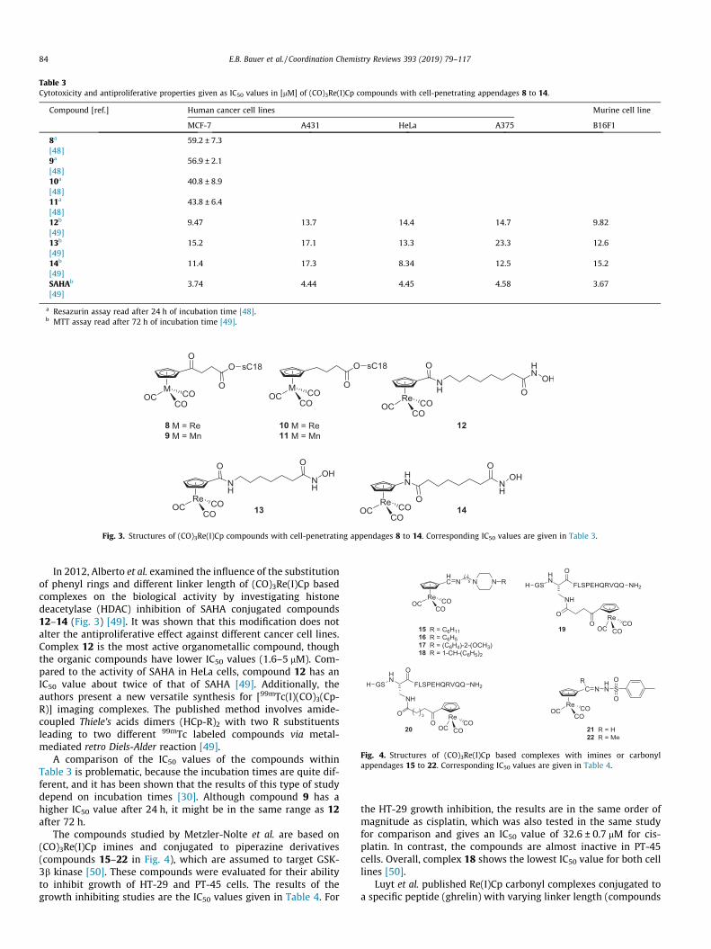
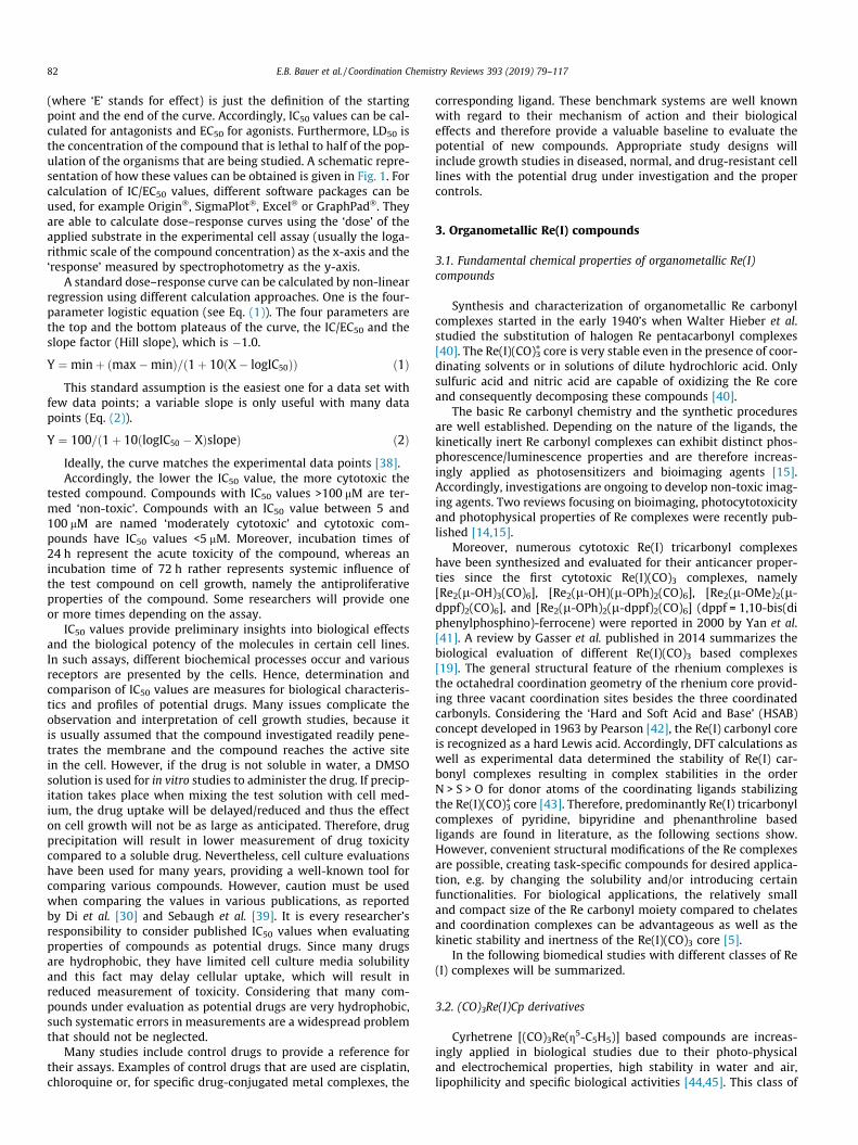
Table 3Cytotoxicity and antiproliferative properties given as IC50 values in [mM] of (CO)3Re(I)Cp compounds with cell-penetrating appendages 8 to 14.
Compound [ref.] Human cancer cell lines Murine cell line
MCF-7 A431 HeLa A375 B16F1
8a 59.2 ± 7.3[48]9a 56.9 ± 2.1[48]10a 40.8 ± 8.9[48]11a 43.8 ± 6.4[48]12b 9.47 13.7 14.4 14.7 9.82[49]13b 15.2 17.1 13.3 23.3 12.6[49]14b 11.4 17.3 8.34 12.5 15.2[49]SAHAb 3.74 4.44 4.45 4.58 3.67[49]
a Resazurin assay read after 24 h of incubation time [48].b MTT assay read after 72 h of incubation time [49].
Fig. 3. Structures of (CO)3Re(I)Cp compounds with cell-penetrating appendages 8 to 14. Corresponding IC50 values are given in Table 3.
Fig. 4. Structures of (CO)3Re(I)Cp based complexes with imines or carbonylappendages 15 to 22. Corresponding IC50 values are given in Table 4.
84 E.B. Bauer et al. / Coordination Chemistry Reviews 393 (2019) 79–117
In 2012, Alberto et al. examined the influence of the substitutionof phenyl rings and different linker length of (CO)3Re(I)Cp basedcomplexes on the biological activity by investigating histonedeacetylase (HDAC) inhibition of SAHA conjugated compounds12–14 (Fig. 3) [49]. It was shown that this modification does notalter the antiproliferative effect against different cancer cell lines.Complex 12 is the most active organometallic compound, thoughthe organic compounds have lower IC50 values (1.6–5 mM). Com-pared to the activity of SAHA in HeLa cells, compound 12 has anIC50 value about twice of that of SAHA [49]. Additionally, theauthors present a new versatile synthesis for [99mTc(I)(CO)3(Cp-R)] imaging complexes. The published method involves amide-coupled Thiele’s acids dimers (HCp-R)2 with two R substituentsleading to two different 99mTc labeled compounds via metal-mediated retro Diels-Alder reaction [49].
A comparison of the IC50 values of the compounds withinTable 3 is problematic, because the incubation times are quite dif-ferent, and it has been shown that the results of this type of studydepend on incubation times [30]. Although compound 9 has ahigher IC50 value after 24 h, it might be in the same range as 12after 72 h.
The compounds studied by Metzler-Nolte et al. are based on(CO)3Re(I)Cp imines and conjugated to piperazine derivatives(compounds 15–22 in Fig. 4), which are assumed to target GSK-3b kinase [50]. These compounds were evaluated for their abilityto inhibit growth of HT-29 and PT-45 cells. The results of thegrowth inhibiting studies are the IC50 values given in Table 4. For
the HT-29 growth inhibition, the results are in the same order ofmagnitude as cisplatin, which was also tested in the same studyfor comparison and gives an IC50 value of 32.6 ± 0.7 mM for cis-platin. In contrast, the compounds are almost inactive in PT-45cells. Overall, complex 18 shows the lowest IC50 value for both celllines [50].
Luyt et al. published Re(I)Cp carbonyl complexes conjugated toa specific peptide (ghrelin) with varying linker length (compounds

Table 4IC50 values in [mM] of (CO)3Re(I)Cp based complexes with imines or carbonyl appendages 15 to 22.
Compound [ref.] Human cancer cell lines Compound [ref.] Chinese hamster cell line Human cancer cell line
HT-29 PT-45 CHO-K1 H1299
15a 97.27 ± 0.06 na 19b 0.035[50] [51]16a 72.78 ± 0.04 na 20b 0.174[50] [51]17a 76.38 ± 0.05 90.16 ± 0.05 21c 37.5 ± 6.6[50] [52]18a 30.48 ± 0.03 25.82 ± 0.03 22c 24.3 ± 8.3[50] [52]Cisplatina 32.6 ± 0.7 2.2 ± 0.3 Cisplatinc 12.8 ± 5.6[50] [52]
(na = not active).a MTT assay read after 72 h of incubation time [50].b Radio ligand binding assay; no incubation time given [51].c MTT assay read after 24 h of incubation time [52].
Table 5Antiproliferative activity of compound 23 and 4-hydroxytamoxifen on MVLN (highERa expression) and MDA-MB-231 (no ERa expression, supposedly ERb expression)breast cancer cell lines in % of luciferase induction after 24 h of culture [56].
Compound [ref.] Molarity Human cancer cell line
MVLN MDA-MB-231
23 1 � 10�6 55.5 n.d.[56]23 1 � 10�7 51.5 91[56]4-Hydroxytamoxifen 1 � 10�7 51.5 88[56]
n.d. = not determined.
E.B. Bauer et al. / Coordination Chemistry Reviews 393 (2019) 79–117 85
19 and 20 in Fig. 4) [51]. Compound 19, with a shorter linker, has asignificantly lower IC50 value than complexes with a longer linker(see Table 4). Thus, the linker length clearly influences the bindingaffinity to the receptor [51].
Concha et al. investigated the biological behavior of (CO)3Re(I)Cp tosylhydrazone complexes 21 and 22 (Fig. 4) [52]. These com-pounds were tested in MTT assays in comparison to the corre-sponding Mn and FeCp complexes as well as cisplatin. Theresulting IC50 values are higher than that for cisplatin. The resultsof the study indicate that the electronic effect of the hydrazonesubstituent has much more influence on the biological activitythan the metal center. This shows the importance of the appliedligand system [52].
Jaouen et al. modified Tamoxifen, one of the best-evaluatedSelective Estrogen Receptor Modulators (SERMs), which are usedto treat hormone dependent breast cancer, with organometallicmoieties [55–57]. For a description on the mechanism of actionof SERMs, the reader is referred to the publication by Jaouenet al. [55]. Modifications of Tamoxifen with cyclopentadienyl moi-eties of Fe, Ti, Re or 99mTc were studied to improve the therapeuticefficacy of this class of drugs [55]. Re(I) carbonyl derivatives ofTamoxifen (isomers of compound 23 in Fig. 5) are active in ERapositive breast cancer cells and inactive in ERa negative cells, sim-ilar to the parent drug. In brief, two different sub-types of estrogenreceptor (ER) were discovered to play an important role in manyphysiological functions in the human body as well as in the evolu-tion of breast cancer, namely ERa and ERb. ERa is considered to beresponsible for increased proliferation in breast tumors and in con-trast, ERb is thought to prevent proliferation. Accordingly, treat-ment of breast cancer should be possible applying ERa-antagonists or ERb-agonists, respectively [58]. To evaluate whethera compound is acting as an agonist or antagonist, different cellmodels were applied. In the present study, the MVLN cell line withhigh ERa expression and the MDA-MB-231 cell line without ERa
Fig. 5. Structure of (CO)3Re(I)Cp based Tamoxifen derivative 23 and 4-hydroxytamoxifen.
expression (but supposedly ERb expression) were used. The resultsshown in Table 5 display no effect of incubating an isomeric mix-ture of Re complex 23 as well as Tamoxifen on MDA-MB-231 cells,however both tested compounds show antiproliferative effects inMVLN cells. This indicates an anti-oestrogenic effect (control isset at 100%; a value above 100% indicates an oestrogenic effectand a value lower than 100% an anti-oestogenic effect) [56].
Replacing the non-radioactive Re core by either 188Re or 186Refor therapeutic purposes and by 99mTc for diagnostic applicationsprovides theranostic use of these Tamoxifen derivatives [59].
3.3. (CO)3Re complexes with N-donor ligands
Efforts have been made to design complexes with improvedcytotoxic activity for treatment of cancer. One approach taken isto add a ligand, which will provide additional toxicity to the com-plex. For example, the ligand is derived from organic drugs withknown anticancer properties like doxorubicin or the ligand system,can be tuned resulting in increased cytotoxicity upon photolysis.Examples for such systems are shown in Fig. 6.
The (CO)3Re(I) core is known to have photosensitizing proper-ties and it was shown that the cytotoxicity increased upon irradi-ation. Patra et al. studied (CO)3Re(I) based complexes withiminedipyridyl ligands 24 to 27 for their cytotoxicity in healthyand cancerous cell lines (Fig. 6 and Table 6) [60]. Moderate growthinhibition was observed with the lowest IC50 value of 7.8 ± 1.9 mMfor complex 24 against HeLa cells, which is even lower than thatdetermined for cisplatin in this cell line (see Table 6). However,no toxicity in HepG2 and healthy cells was observed, indicatingthat these compounds possess the potential to have a good andsafe activity profile suitable for treatment of cervical cancer [60].
Paulo et al. studied the (photo)cytotoxicity of a series of (CO)3Re(I) based complexes with iminedipyridyl ligands including a heter-

Fig. 7. Structures of a series of (CO)3Re(I) based complexes with iminedipyridyl ligandsThe corresponding IC50 values are given in Table 7.
Fig. 6. Structures of (CO)3Re(I) based complexes with iminedipyridyl ligands 24 to 27. Corresponding IC50 values are given in Table 6.
Table 6IC50 values [mM] of different (CO)3Re(I) based complexes with iminedipyridyl ligandsdetermined in different human cell lines.
Compound [ref.] Human cancer cell lines Human non-cancer cell line
HepG2 HeLa MRC-5
24a >100 7.8 ± 1.9 >100[60]25a >100 10.2 ± 2.0 22.8 ± 3.1[60]26a >100 8.0 ± 1.0 n.d.[60]27a 52.7 ± 9.7 26.3 ± 0.8 36.9 ± 5.0[60]Cisplatina 5.5 ± 0.5 11.5 ± 2.9 7.9 ± 1.2[60]
n.d. = not determined.a MTT assay read after 48 h of incubation time [60].
86 E.B. Bauer et al. / Coordination Chemistry Reviews 393 (2019) 79–117
obimetallic complex 29 shown in Fig. 7. The Re complexes 28 and29 are cytotoxic in the dark as well as upon irradiation against cis-platin resistant cells. For both complexes, the IC50 values decreasedupon irradiation to less than half compared to the IC50 values in thedark. The IC50 value in healthy cells in the dark is slightly higherthan the values obtained by irradiation (Table 7). For cisplatin, acontradictory effect was observed leading to higher IC50 valuesupon irradiation, which is explainable by the shorter incubationtime [61].
Superior cytotoxicity has been found for (CO)3Re(I) complexes(and include two (CO)3Re(I)Cp complexes) with doxorubicin conju-gates 30–34 reported by Alberto et al. One of these complexes, 33,has an IC50 value of 0.34 mM in HeLa cells (Table 7). Moreover, itwas shown by confocal microscopy and ICP-MS that in contrastto the parent drug and compounds 30–32, complexes 33 and 34accumulate to a much higher extent in mitochondria (2% and
(28 to 32) and two (CO)3Re(I)Cp complexes with doxorubicin conjugates (33 to 34).

Table 7IC50 values in [mM] determined for (CO)3Re(I) based complexes with iminedipyridyl ligands (28 to 32) and two (CO)3Re(I)Cp complexes with doxorubicin conjugates (33 and 34).
Compound [ref.] Human non-cancer cell line Human cancer cell line Murine cell line
MRC-5 RPE1-hTERT HeLa A2780 A2780R B16F1
28a 121 ± 10.1 155 ± 22/20.1 ± 6.5 46.1 ± 6.5/7.8 ± 1.6 198 ± 20.5/19.3 ± 2.1[61]29a 22.0 ± 5.3 42.8 ± 4.7/13.5 ± 4.1 28.7 ± 4.2/18.4 ± 5.2 27.8 ± 4.7/16.5 ± 2.7[61]Cisplatina 8.4 ± 2.1 10.2 ± 2.3/35.2 ± 4.6 1.7 ± 0.5/9.3 ± 2.1 9.5 ± 2.3/27.5 ± 3.1[61]30b 19.7 ± 2.1 11.4 ± 4.9[62]31b 6.2 ± 2.3[63]32b 12.2 ± 2.4[63]33b 1.82 ± 0.54 0.34 ± 0.03[64]34b 1.27 ± 0.53 1.65 ± 0.26[64]Doxorubicin 0.093 ± 0.02 0.095 ± 0.01[62]
a Resazurin assay in the dark, read after 48 h of incubation/4 h incubation with complex followed by 10 min irradiation with wavelength of 350 nm (2.58 J cm�1) [61].b Resazurin assay read after 48 h of incubation time [62–64].
E.B. Bauer et al. / Coordination Chemistry Reviews 393 (2019) 79–117 87
30%, respectively). This is important and suggests that some cyto-toxic action may involve the critical energy production in the mito-chondria. All these compounds were found to be inhibitors ofhuman topoisomerase with a comparable affinity to the parentdrug. Comparison of in vivo bio-distribution of doxorubicin andits conjugates with Re (complex 32) and 99mTc showed that themetal coordination does not significantly alter the bio-distribution [62–64].
Gasser et al. reported two (CO)3Re(I)N,N-bis(quinolinoyl) com-plexes (35 and 36, Fig. 8) and their potential application in photo-dynamic therapy (PDT). However, PDT can only treat surfacecancers or those accessible by endoscopy. The investigated com-plexes contain two different targeting peptides (NLS and bombe-sin) for an increased selectivity towards cancer cells, which isconnected via a photolinker (36-NLS/bombesin to 38 in Fig. 8).Upon irradiation, the bio-conjugates are separated from the Remoiety and singlet oxygen is generated which results in IC50 val-ues � 9 mM, which is lower than the value determined for cisplatin(Table 8). Notably, precursors 28 and 35 are similar except for theirlength (one CH2 group) and have comparable IC50 values againstMRC-5 and HeLa cells (see Tables 7 and 8, respectively). The mostcytotoxic complexes against HeLa and PC3 cancer cell lines werefound to be the bombesin conjugates 36-bombesin and 38, whichadditionally have the highest IC50 values in healthy cells. Theinvestigation of the mode of action of 37 reveals that thiscompound induces a combination of apoptosis and necrosis[19,65–67].
Furthermore, vitamin B12 conjugates with (CO)3Re(I)phenan-throline 40 and 41 were studied in PC3 cells by Santoro et al.[68]. Table 8 shows the IC50 values for complex 40 and the Re(I)starting material 39. Due to the instability of 40, it can be assumedthat both IC50 values represent the toxicity of 39 with differentaxial ligands, because MeOH is not strongly bound to Re(I) andmay be replaced by water or chlorine when dissolved in the cellmedium [68]. Similar observations were noted in a study publishedby Wilson et al. [69]. Further investigations concerning the axialligand and IC50 determination of additional cell lines might bepromising for 39 and 41 [68].
Luengo et al. published heterometallic Re(I)/Au(I) complexes42–47 (Fig. 9) in 2017. Their cytotoxic properties in A549 cellsshow double the activity for trimetallic complexes 45–47 than
for bimetallic compounds (see Table 9). This finding may be attrib-uted to an increased cellular uptake resulting from a beneficial bal-ance of the charge and lipophilicity. These compounds can betracked and further studied for their location/accumulation in cellsby fluorescence microscopy for a better understanding of their bio-logical behavior and targets [70].
A549 cells were also used by Maislus et al. for evaluation of thebiologic properties of b-carboline compounds 48–51 (Fig. 9), whichhave suitable photochemical properties for investigations usingfluorescence microscopy. It was shown that 51 exhibits the lowestIC50 value (10 mM) compared to all compounds given in Table 9,which is comparable to cisplatin (8 mM). Therefore, further evalua-tion of this compound in cisplatin-resistant and healthy cellswould be of interest [71].
Ye et al. studied the inhibitory effect of histone deactylase(HDAC) inhibitor conjugated to Re(I) carbonyl complex 52(Fig. 10 and Table 10). The inhibition of HDAC was measured innuclear extracts from HeLa cells as well as on the isolated enzymehuman recombinant HDAC7. The corresponding IC50 values aregiven in Table 10 and are in high nanomolar range, which is com-parable to the parent drug SAHA. Studies were performed with thiscompound to determine its mode of action. According to theresults, the mechanism could not be identified, however, the bio-logical response is not identical to the one resulting from SAHAor cisplatin treatment. SAHA is an organic small molecule causingcell death by inhibiting HDAC, and cisplatin is a small inorganicdrug causing cell death by binding to DNA and inhibiting its repli-cation. Further studies of 52 in cisplatin/SAHA resistant cell lineswere performed to verify the different mode of action [72]. InTable 3, results are summarized with the (CO)3Re(I)Cp based com-plexes with cell-penetrating appendages, 12–14, also modifiedwith the SAHA moiety. These IC50 values are in the range of 8.3to 23.3 mM, which is significantly higher than the nanomolar valuesdetermined for 52. However, the nanomolar values are obtained onnuclear extracts, which are not directly comparable to intact cellmeasurements. Accordingly, values for SAHA are 0.078 mM (cell-based measurement) and 4.45 mM (isolated enzyme) [49,72].
Water-soluble porphyrin conjugates 53 and 54 were studiedin vitro for their photocytotoxicity. Complex 54 shows no toxicityin the dark; however, it exhibits moderate activity upon irradia-tion, which is also observed for the corresponding organic deriva-

Table 8IC50 values in [mM] determined for (CO)3Re(I) complexes with NLS and bombesin ligands 35 to 41.
Compound [ref.] Human non-cancer cell line Human cancer cell lines
3T3 MRC-5 HeLa PC3
35a >100/40.3 ± 5.4* 187.1 ± 17.9/17.3 ± 2.9* >100/>100*
[66]36a >100 >100/9.3 ± 2.2*
[65]36-NLSa 17.8 ± 1.8/13.0 ± 2.5* 35.1 ± 1.8/18.3 ± 1.4* n.d.[66]37a 36.2 ± 0.6/20.5 ± 5.5* 14.5 ± 5.2/9.3 ± 0.8* n.d.[66]36-bombesina 44.1 ± 9.9/41.6 ± 15.9* >100/5.3 ± 1.0* >100 /13.6 ± 1.7*
[66]38a 72.3 ± 3.6/23.3 ± 0.6* >100/9.7 ± 4.4* >100/19.2 ± 2.4*
[66]Cisplatin 10.5 ± 2.8/47.8 ± 1.5* 9.2 ± 0.6/26.8 ± 1.7*
[66]39b 45 ± 3 4 ± 2[68]40b 47 ± 7 7 ± 1[68]41b 200 15 ± 2[68]
n.d. = not determined.* 10 min irradiation with UV-A light (350 nm, 2.58 J cm�1) [66].a Resazurin assay read after 48 h of incubation time [65,66].b Trypan blue assay read after 48 h of incubation time [68].
Fig. 8. Structures of (CO)3Re(I) complexes with NLS and bombesin ligands to form complexes 35 to 41. Corresponding IC50 values are given in Table 8.
88 E.B. Bauer et al. / Coordination Chemistry Reviews 393 (2019) 79–117

Fig. 9. Structures of (CO)3Re(I) complexes with (bi)pyridine ligands 42–51. Corresponding IC50 values are given in Table 9.
Table 9IC50 values in [mM] determined for (CO)3Re(I) complexeswith (bi)pyridine ligands 42 to 51 using the MTT assayand read after 24 h of incubation.
Compound [ref.] Human cancer cell lineA549
42 75.25 ± 10.67[70]43 67.80 ± 4.11[70]44 64.69 ± 3.32[70]45 42.44 ± 4.03[70]46 36.09 ± 16.99[70]47 35.82 ± 1.82[70]48 85 ± 1[71]49 88 ± 1[71]50 65 ± 1[71]51 10 ± 1[71]
E.B. Bauer et al. / Coordination Chemistry Reviews 393 (2019) 79–117 89
tive. Thus the observed activity is at least in part due to the ligandof the Re(I) complex. The cytotoxicity of compounds 53 and 54 issignificantly higher upon irradiation both in cancerous and healthy
cells (see IC50 values in Table 10). The influence of further metalcomplexation of the porphyrin moiety remains to be determined,but would be of interest for understanding the potential modula-tion of the activity of this system [73].
(CO)3Re(I) based b-elemene conjugates 55–57were synthesizedand tested in vitro by Ren et al. [74]. These systems showed compa-rable cytotoxic activity in the cancer cell lines (see Table 10). Fur-thermore, the radioactive 188Re analogues were synthesized [74]although no further evaluation of their biological activity has beenreported so far.
Policar et al. evaluated the influence of the length of the sidechain on lipophilicity, cellular uptake and antiproliferative effectof the luminescent compounds 58 to 60. The results reveal thatthe higher the lipophilicity, the greater the cell uptake and there-fore the highest antiproliferative effect with the lowest IC50 (of3.3 mM) is found for the compounds with the C12 side chain 60(see Fig. 11 and Table 11) [75]. A purity of 95% in HPLC analysisusing water/acetonitrile indicates that these are relatively stablecoordination complexes with regard to ligand exchange comparedto other Re coordination complexes [69,75].
Complexes 61 to 63 published by Lo et al. are shown to selec-tively react with azide-functionalized proteins making them usefulfor imaging of azide-labeled biomolecules. However, these com-plexes exhibit a cytotoxicity of 3–10 mM in the Chinese hamstercell line CHO and therefore displays higher cytotoxicity than cis-platin (Table 11). Additionally, an increased cellular uptake is pos-sible when the cells are pretreated with an azide-modifiedglycoprotein-labeling probe [76].

Fig. 10. Structures of (CO)3Re(I) complexes with long tethers 52–57. Corresponding IC50 values are given in Table 10.
Table 10IC50 values in [mM] determined for (CO)3Re(I) complexes with long tethers 52 to 57.
Compound [ref.] Human cancer cell lines Human non-cancer cell line
HeLa HDAC7 H460M2 LLC HBL-100
52a 0.106 ± 0.007 0.682 ± 0.060[72]SAHAa 0.0786 ± 0.006 0.529 ± 0.048[72]53* >100/1.4 ± 1.3* 7.4 ± 2.0/0.5 ± 0.2* 33.7 ± 14.5/0.5 ± 0.1*
[73]54* >100/73 ± 19* >100/12 ± 5* >100/42.8 ± 5.3*
[73]55b 10.9 ± 1.2 5.0 ± 1.9[74]56b 11.2 ± 1.5 5.1 ± 1.3[74]57b 10.5 ± 2.9 4.8 ± 2.3[74]
a MTT assay; no incubation time given [72].* Irradiation with wavelength of 650 nm with a total light dose of 10 J cm�1; MTT assay read 24 h post-irradiation [73].b WST-1 assay read after 24 h of incubation time [74].
90 E.B. Bauer et al. / Coordination Chemistry Reviews 393 (2019) 79–117

Fig. 11. Structures of (CO)3Re(I) complexes with lipophilic appendages 58–65. Corresponding IC50 values are given in Table 11.
Table 11IC50 values in [nM] determined for (CO)3Re(I) complexes with lipophilic appendages58 to 65.
Compound[ref.]
Human cancercell lines
Chinese hamstercell line
Human non-cancercell lines
MDA-MB-231 CHO HEK293
58a 19000 ± 300[75]59a 4000 ± 200[75]60a 3300 ± 100[75]61b 9550 ± 1550[76]62b 3500 ± 30[76]63b 2940 ± 20[76]Cisplatinb 25490 ± 54[76]64c 0.076 ± 0.001[77]65c 4.4 ± 0.4[77]
a Methylene blue staining read after 5 d of incubation time [75].b MTT assay read after 48 h of incubation time [76].c Forskolin-stimulated cAMP accumulation assay [77].
E.B. Bauer et al. / Coordination Chemistry Reviews 393 (2019) 79–117 91
Metzler-Nolte et al. reported complexes 64 and 65 combiningthe opioid peptide dermorphin with a Re carbonyl core. By evalu-ating their biological effects, the authors discovered in a blockinggroup scan that not only the N-terminal domain is a ligand bindingsite but also the C-terminal domain. The binding affinities of thesecomplexes to the opioid receptor give IC50 values in the nano- tosubnanomolar range (see Fig. 11 and Table 11). Moreover, adose-dependent analgesic effect was observed in the preliminaryin vivo studies which potentially paves the way to complexes thatbinds as substrates for opioid receptors [77].
3.4. (CO)3Re complexes with pyridine ligands
The following section covers Re(I) carbonyl complexes coordi-nated to ligands derived from the fundamental pyridine structure,starting with Mao’s publications from 2016 to 2018 [78–80]. Thesephosphorescent compounds can be divided into two structuralgroups: the phenanthroline (66a-f) and diphenyl-phenanthroline(67a-j) coordinated complexes (Fig. 13). Comparison of the IC50
values given in Table 12 shows that diphenyl-phenanthroline coor-dinated complexes are more active than the phenanthroline basedones. These complexes have a higher lipophilicity and thereforeshow an increased cell uptake leading to increased cytotoxicity.The increased cell uptake was confirmed by confocal microscopy.In addition, the IC50 values are lower in cisplatin-sensitive andcisplatin-resistant cell lines than the values determined for cis-platin (21.5 and 65.6 mM, respectively). Complex 67e displays alow IC50 value (0.52 mM) in HeLa cells and shows toxicity againstother cancer cell lines, particularly when compared to healthy cells(see Table 12). The selectivity towards cancer cells is further veri-fied by co-incubation and imaging experiments (Fig. 12). The highcytotoxicity of compounds 67a-j is caused by selectively targetingthe mitochondria, inducing mitochondrial damage and caspase-dependent apoptosis [78,79]. Moreover, evidence for selectiveapoptotic cell death was observed for complexes 67d and 67e.Therefore, A549 cells were stained with Hoechst, a cell-permeable blue dye, which readily stains the nucleus of living cells(see Fig. 12, ‘Hoechst’ column). These pre-stained cancerous A549cells were co-cultured with healthy, non-stained LO2 cells fol-lowed by treatment of these co-cultured A549/LO2 cells with theRe complexes. Finally, annexin V/PI double staining was performedon these co-cultured cells. Annexin V is a protein with high affinityto phosphatidylserine, another protein that is only present on thesurface of apoptotic cells. By modification of annexin V with a flu-orescent tag (a green fluorescent dye shown in Fig. 12), apoptotic

Fig. 12. Confocal microscopic images of A549 cells pre-stained with Hoechst following treatment and co-incubation of pre-stained A549 and LO2 cells with 5 mM of eithercomplex 67d or 67e. Final annexin V/PI staining shows the capability of complex 67e to selectively induce apoptosis in cancer cells. Reprinted with permissions from ACSAppl. Mater. Interfaces, 9 (2017) 13900–13912. Copyright 2018 American Chemical Society.
92 E.B. Bauer et al. / Coordination Chemistry Reviews 393 (2019) 79–117
cells can be displayed using fluorescence microscopy. Propidiumiodide (PI) is a cell impermeable, red fluorescent dye that interca-lates with DNA. PI only stains apoptotic and/or necrotic cells,because apoptotic and necrotic cells have a decreased nuclearand plasma membrane integrity and thus PI is able to pass throughthese membranes and intercalate with the DNA of these cells andstain them. This means that PI cannot pass the membranes of livingor early apoptotic cells and therefore are not stained. Accordingly,these fluorescent markers are applied in fluorescence microscopicexperiments to distinguish between living and dying/dead cells.
In Fig. 12, the confocal microscopic images of A549 (bluenuclei)/LO2 co-cultured cells treated with complexes 67d (refer-ring to 2b in the figure) and 67e (referring to 3b in the figure)and stained with different fluorescent labels are presented. Consid-ering the different staining properties of the applied labels, 67ddisplays lower selectivity of killing healthy and cancerous cellscompared to complex 67e, because not all three labels (Hoechstin blue, annexin V in green and PI in red) are always located inthe same cells when looking at the overlay. Nevertheless, thisexperiment reveals a good selectivity of compound 67e in killingcancer cells rather than healthy cells [78].
In addition, a recent study of Mao et al. shows submicromolarIC50 values for the mitochondria-targeting compound 67j, whichis coordinated to dichloroacetate (DCA), a metabolic modulator.This complex is shown to inhibit the pyruvate dehydrogenasekinase (PDK) and therefore selectively target and kill the PDK-overexpressing cells NCI-H1229 and RKO as indicated by theIC50 values (see Table 12). This compound also shows high anti-cancer and anti-angiogenetic activity compared to its DCA-freeanalogue. Therefore, this study shows that targeting the metabo-lism of cancer cells is a highly effective and selective method forcancer treatment as well as understanding the mechanism ofaction [80].
Gasser et al. studied the fluorescent complex 68 in detail for itsanticancer activity, showing an activity comparable to 66 and 67
(Fig. 13) [81]. However, the selectivity towards cancer cells overhealthy cells was not determined. Experiments on a biosensor-chip micro bioreactor reveals that this complex is also targetingthe mitochondria and inhibits the mitochondrial respiration [81].
Red light emitting, CF3 functionalized complexes 69 and 70(Fig. 13) were biologically evaluated by Mueller et al. [82]. TheIC50 values in Table 12 show a certain selectivity for different celllines and activity in a range of 4–84 mM. Additionally, the positionof CF3 is shown to strongly influence the cytotoxicity, thus, the IC50
values are comparable to the structurally related complexes 67a-j[78–80].
In a further publication, Gasser et al. showed an enhanced cyto-toxicity of luminescent Re carbonyl complexes 71 (Fig. 14), whencoupled to a lipid-modified peptide, which is known to increasecell permeability. The IC50 values of precursor 71 and thepeptide-conjugate 72 are given in Table 13, which is, for 72, inthe range of cisplatin (9.1 ± 2.8 mM) [83].
Lo et al. reported a series of Re(I) carbonyl complexes 73–78(Fig. 14). Their study shows that the glucose-free compounds(73–75, for IC50 values see Table 13) have lower IC50 values com-pared to the D-glucose conjugated (CO)3Re-complexes (76–78).Moreover, the cytotoxicity of the compounds can be related tothe respective lipophilicities and thus the cellular uptake of thesecomplexes. Additional biological evaluation of glucose-conjugatesindicates a strong affinity for the GLUT receptor [84]. A furtherpublication by Lo et al. describes trifunctional complexes 79–81.These luminescent fluorine-containing Re(I) carbonyl complexescontain different functional groups for conjugation with biomole-cules, like bovine serum albumin (BSA) or glutathione, and there-fore are precursors rather than bioactive molecules. However,compound 81 is not stable in aqueous solution and was thereforenot submitted for cytotoxicity evaluation [85].
Table 13 shows how different results can be obtained in thesame cell line for the same compound, like cisplatin. Therefore,these values just represent a basis for evaluating cytotoxic effects,
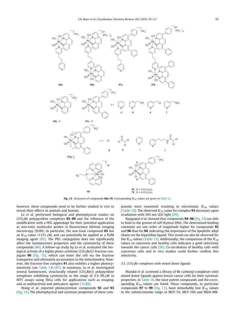
Fig. 13. Structures of compounds 66a-70. Corresponding IC50 values are given in Table 12.
E.B. Bauer et al. / Coordination Chemistry Reviews 393 (2019) 79–117 93
however, these compounds need to be further studied in vivo toreveal their effects in animals and human.
Lo et al. performed biological and photophysical studies on(CO)3Re polypyridine complexes 82–89 and the influence of themodification with a PEG appendage for their potential applicationas non-toxic multicolor probes in fluorescence lifetime imagingmicroscopy (FLIM). In particular, the non-toxic compound 85 hasan IC50 value >1151 mM, and can potentially be applied as a FLIMimaging agent [85]. The PEG conjugation does not significantlyaffect the luminescence properties and the cytotoxicity of thesecompounds [86]. A follow-up study by Lo et al. evaluated the bio-logical activity of a highly photo-cytotoxic (CO)3Re(I)-fructose con-jugate 90 (Fig. 15), which can enter the cell via the fructosetransporter and ultimately accumulates in the mitochondria. How-ever, the fructose-free complex 91 also exhibits a higher photocy-totoxicity (see Table 14) [87]. In summary, Lo et al. investigatedseveral luminescent, structurally related (CO)3Re(I) polypyridinecomplexes exhibiting cytotoxicity in the range of 3.5–90 mM inMTT assays using HeLa cells for applications such as imaging,and as antibacterial and anticancer agents [15,88].
Wang et al. reported photocytotoxic compounds 92 and 93(Fig. 15). The photophysical and cytotoxic properties of these com-
pounds were examined resulting in micromolar IC50 values(Table 14). The observed IC50 value for complex 93 decreases uponirradiation with 365 nm LED light [89].
Rajagopal et al. showed that compounds 94–96 (Fig. 16) are ableto bind to the groove of calf thymus DNA. The determined bindingconstants are one order of magnitude higher for compounds 95and 96 than for 94, indicating the importance of the lipophilic alkylchains on the bipyridine ligand. This trend can also be observed forthe IC50 values (Table 15). Additionally, the comparison of the IC50
values in cancerous and healthy cells indicates a good selectivitytowards the cancer cells [90]. Co-incubation of healthy cells withcancerous cells and in vivo studies could further confirm thisselectivity.
3.5. (CO)3Re complexes with mixed donor ligands
Mandal et al. screened a library of Re carbonyl complexes withmixed donor ligands against breast cancer cells for their cytotoxicproperties. In Table 16, the most potent compounds and the corre-sponding IC50 values are listed. These compounds, in particularcompounds 97 to 99 (Fig. 17), have remarkably low IC50 valuesin the submicromolar range in MCF-7A, MCF-10A and MDA-MB-

Table 12IC50 values [mM] of (CO)3Re(I) based (poly)pyridine compounds tested in different cell lines.
Compound [ref.] Human non-cancer cell lines Human cancer cell lines
LO2 HeLa A549 A549cisR HepG2 MCF-7 U2OS THP-1 RKO NCI-H1229
66aa >100 44.7 ± 4.0 >100 >100 39.8 ± 3.7[79]66ba 12.4 ± 1.2 2.0 ± 0.2 4.6 ± 0.3 1.6 ± 0.1 3.1 ± 0.3[79]66ca >100 >100 >100 >100[78]66da >100 >100 >100 >100[78]66ea >100 64.6 ± 2.2 75.8 ± 2.3 37.3 ± 1.1[78]66fa 47.9 ± 1.5 52.5 ± 3.0 39.8 ± 0.7 36.5 ± 1.8[78]67aa 56.2 ± 4.7 9.1 ± 0.8 11.7 ± 4.1 11.5 ± 1.0 12.0 ± 1.1[79]67ba 10.2 ± 1.0 1.7 ± 0.1 2.0 ± 0.2 1.9 ± 0.1 2.1 ± 0.2[79]67ca 3.1 ± 0.5 0.95 ± 0.11 3.9 ± 0.7 1.2 ± 0.5[78]67da 7.6 ± 0.9 1.7 ± 0.4 5.5 ± 0.6 2.7 ± 0.5[78]67ea 18.7 ± 1.1 0.52 ± 0.07 3.4 ± 0.6 0.75 ± 0.12[78]67fa 6.4 ± 0.7 5.9 ± 0.9 22.4 ± 1.2 8.5 ± 1.1[78]67gb 30.0 ± 1.1 18.1 ± 2.3 17.8 ± 2.2 32.1 ± 2.1 14.2 ± 1.7 12.4 ± 1.4[80]67hb 21.0 ± 3.2 15.0 ± 1.4 15.9 ± 2.1 14.4 ± 1.2 9.7 ± 1.1 3.7 ± 0.2[80]67ib 20.0 ± 3.5 11.0 ± 1.2 13.0 ± 3.4 7.6 ± 1.1 9.1 ± 0.8 2.9 ± 0.1[80]67jb 22.0 ± 2.1 4.1 ± 0.9 4.3 ± 0.7 4.0 ± 1.2 2.2 ± 0.2 0.8 ± 0.1[80]Cisplatina 29.9 ± 2.1 8.9 ± 1.0 21.5 ± 2.5 65.6 ± 1.6[78]Re(CO)5Brc >100 76.9 ± 3.8 >100[81]68c 29.8 ± 1.0 8.6 ± 0.2 16.9 ± 1.5[81]Cisplatinc 3.9 ± 1.0 2.7 ± 0.1 8.2 ± 1.5[81]69d 4.56 ± 3 3.62 ± 0.8 33 ± 16[82]70d 83 ± 80 4.76 ± 2.5 14 ± 4.1[82]
a MTT assay read after 48 h of incubation time [78,79].b MTT assay read after 24 h of incubation in the dark [80].c Resazurin assay read after 48 h of incubation time [81].d MTS assay read after 72 h of incubation time [82].
94 E.B. Bauer et al. / Coordination Chemistry Reviews 393 (2019) 79–117
231 cell lines. Such high cytotoxic activity of the (CO)3Re(I)-phenyl-phenanthroline compounds was also observed for the(CO)3Re(I) complexes previously discussed, like 67j. As statedbefore, the authors attribute the effect to the lipophilicity of thesecomplexes, which is important for cell membrane penetration andtheir ability to intercalate with DNA [91]. However, this is not thecase for all of these compounds, e.g. the diphenyl-phenanthrolinecoordinated complex 97 is the most active one in MCF-7 cells,however, the diphenyl-phenanthroline based complex 111 is theleast active (see Table 16). For MCF-10A cells, thephenanthroline-based complex 98 has the lowest IC50 value andthe diphenyl-phenanthroline coordinated 104 displays the highestIC50 value. Overall, the exchange of the axial ligand seems to haveless influence on the activity than the changes on the phenanthro-line moiety. This can be seen by looking at complexes 102 and 103(additional benzyl moiety on the axial ligand), which shows lesssignificant changes in IC50 values in all cell lines, as compared to103 (diphenyl-phenanthroline) and 109 (phenanthroline).
Yan et al. studied Re phosphine compounds 112–118, [NBu4][ReO4] and [NEt4]2[ReBr3(CO)3 (Fig. 18) for their cytotoxic effectsin cultured cell line suspensions (Table 17) and in solid tumor cul-tures (Table 18). However, the authors determined ED50 values(mean effective dose of the compound) in mg per ml and not themolar concentration. Recalculation of these values to EC50 values(molar concentration) using the molecular weight of the com-pounds results in values that are more comparable. Since theEC50 values are easier to compare, the presented discussion isbased on the recalculated EC50 values. However, both values areincluded in Tables 17 and 18 in the order ED50/EC50 value to showthe difference in the values and the importance of using the molarconcentration and not the dose of a test compound.
The recalculated EC50 values range from 0.93 to 18.00 mM. Com-pound 116 displays the lowest EC50 values in almost all tested celllines except for A549, 1-A9 and T-molt4, where compound 115shows the lowest values (see Tables 17 and 18). In cultured cellline suspensions, the lowest observed EC50 value is 0.93 mM for
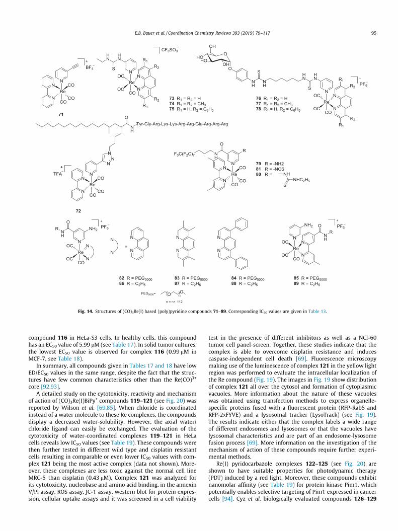
Fig. 14. Structures of (CO)3Re(I) based (poly)pyridine compounds 71–89. Corresponding IC50 values are given in Table 13.
E.B. Bauer et al. / Coordination Chemistry Reviews 393 (2019) 79–117 95
compound 116 in HeLa-S3 cells. In healthy cells, this compoundhas an EC50 value of 5.99 mM (see Table 17). In solid tumor cultures,the lowest EC50 value is observed for complex 116 (0.99 mM inMCF-7, see Table 18).
In summary, all compounds given in Tables 17 and 18 have lowED/EC50 values in the same range, despite the fact that the struc-tures have few common characteristics other than the Re(CO)3+
core [92,93].A detailed study on the cytotoxicity, reactivity and mechanism
of action of (CO)3Re(I)BiPy+ compounds 119–121 (see Fig. 20) wasreported by Wilson et al. [69,85]. When chloride is coordinatedinstead of a water molecule to these Re complexes, the compoundsdisplay a decreased water-solubility. However, the axial water/chloride ligand can easily be exchanged. The evaluation of thecytotoxicity of water-coordinated complexes 119–121 in HeLacells reveals low IC50 values (see Table 19). These compounds werethen further tested in different wild type and cisplatin resistantcells resulting in comparable or even lower IC50 values with com-plex 121 being the most active complex (data not shown). More-over, these complexes are less toxic against the normal cell lineMRC-5 than cisplatin (0.43 mM). Complex 121 was analyzed forits cytotoxicity, nucleobase and amino acid binding, in the annexinV/PI assay, ROS assay, JC-1 assay, western blot for protein expres-sion, cellular uptake assays and it was screened in a cell viability
test in the presence of different inhibitors as well as a NCI-60tumor cell panel-screen. Together, these studies indicate that thecomplex is able to overcome cisplatin resistance and inducescaspase-independent cell death [69]. Fluorescence microscopymaking use of the luminescence of complex 121 in the yellow lightregion was performed to evaluate the intracellular localization ofthe Re compound (Fig. 19). The images in Fig. 19 show distributionof complex 121 all over the cytosol and formation of cytoplasmicvacuoles. More information about the nature of these vacuoleswas obtained using transfection methods to express organelle-specific proteins fused with a fluorescent protein (RFP-Rab5 andRFP-2XFYVE) and a lysosomal tracker (LysoTrack) (see Fig. 19).The results indicate either that the complex labels a wide rangeof different endosomes and lysosomes or that the vacuoles havelysosomal characteristics and are part of an endosome-lysosomefusion process [69]. More information on the investigation of themechanism of action of these compounds require further experi-mental methods.
Re(I) pyridocarbazole complexes 122–125 (see Fig. 20) areshown to have suitable properties for photodynamic therapy(PDT) induced by a red light. Moreover, these compounds exhibitnanomolar affinity (see Table 19) for protein kinase Pim1, whichpotentially enables selective targeting of Pim1 expressed in cancercells [94]. Cyz et al. biologically evaluated compounds 126–129

Table 13IC50 values [mM] of (CO)3Re(I) based (poly)pyridine compounds tested in HeLa cells.
Compound [ref.] Human cancer cell linesHeLa
71a 29.9 ± 6.1[83]72a 13.0 ± 2.0[83]Cisplatina 9.1 ± 2.8[83]73b 22.8 ± 5.2[84]74b 7.7 ± 0.6[84]75b 2.8 ± 0.4[84]76b >150[84]77b 90.0 ± 7.6[84]78b 68.9 ± 2.3[84]Cisplatinb 27.6 ± 1.8[84]79b 8.70[85]80b 17.02[85]82b 26.3 ± 1.6[86]83b 11.9 ± 1.6[86]84b 6.6 ± 0.4[86]85b >1151.7[86]86b 15.0 ± 4.8[86]87b 5.0 ± 0.4[86]88b 3.6 ± 0.4[86]89b 159.1 ± 8.0[86]
a Resazurin assay read after 48 h of incubation time [83].b MTT assay read after 48 h of incubation time [84–86].
Fig. 15. Structures of compounds 90–93. Corresponding IC50 values are given inTable 14.
96 E.B. Bauer et al. / Coordination Chemistry Reviews 393 (2019) 79–117
(Fig. 20, Table 19). Comparing the IC50 values, compounds 126 and127 are slightly more active than 128 and 129. Additionally, thesecompounds were shown to initiate apoptosis [95].
The EGFR inhibiting compound 130 was evaluated in A431 cellsby Permettis et al. showing a slightly lower IC50 value (2.0 mM, seeTable 19) than the parent compound (4.8 mM) [96].
Mieczkowski et al. studied the cytotoxicity of complexes 131–136 against ovarian cancer cell lines and healthy cells [97]. Theresults against wild type and cisplatin resistant cell lines indicatethat complex 133 has low IC50 values in both cancer cell linesand a high value in healthy cells (see Table 20). Interestingly, thedifference between compounds 131–133 is the substitution of acoordinated halogen resulting in a dramatically different cytotoxi-city [97].
An additional publication by Wilson et al. studied the photo-cytoxicity of structurally comparable compounds 137 and 138(Fig. 21) to their previous publication (compounds 119–121 inFig. 20) which are highly cytotoxic (1.2 mM) when exposing thecells to visible light. Importantly, complexes 137 and 138 are nottoxic in the dark, but after irradiation at 365 nm, IC50 values of2.2 mM in wild type cells and 3.2 mM in cisplatin resistant cellsare observed for complex 138 (see Table 20). Interestingly, thecomplexes 137 and 138 are shown to release CO upon irradiationwith UV light and are therefore useful for photodynamic therapy(PDT) or photoactivated chemotherapy (PACT) [98].
Wong et al. reported a study on the cytotoxicity of 139 (Fig. 21)and its interaction with calf thymus DNA. The IC50 values obtainedby MTT assays in HepG2, HeLa and KB-3–1 cells are 30–50 mMwithless toxicity towards normal cells (Table 20). The observed changesin pH in solid tumor cell lines was shown to have no influence onthe effects of these compounds. For multi-drug resistant KB-V-1cells, the IC50 value is about 4 times higher, but considering the celllines resistance to other drugs, the observed effects are stillpromising [99]. Moreover, in Table 20 the varying IC50 values forcisplatin evaluated in the same cell line is observed. For A2780cisRthis value ranges from 5 to 30 mM.
Natile et al. evaluated bimetallic Re(I) carbonyl complexes 140and 141 conjugated to a translocator protein (TSPO) (Fig. 21 andTable 20). Structurally, 141 has two Re(CO)3 moieties, whereas140 has one Re(CO)3 and one PtCl moiety. Interestingly, for thesecomplexes the biological evaluation indicates a slightly highercytotoxicity for the ligand (9.0 mM), than for the Re complexes.However, the heterobimetallic complex 140 exhibits only a slightlyhigher IC50 value than the Re-free analogue, and both maintainantiproliferative activity in cisplatin resistant cells [101]. Of thesecompounds, the ligand is the most active [100]. Overall, of thecompounds in Table 20, the most active compounds are the irradi-ated photoactivated complexes 133, 137 and 138 with IC50 valuesof 2–4.6 mM in A2780 and A2780cisR cell lines.
Manimaran et al. reported three studies on Re(I) metallacycles142–151 (Fig. 22) [102–104]. These complexes were tested in dif-ferent cancer cell lines as well as in normal blood cells for their IC50
values (see Table 21). A great selectivity towards cancer cell lineswas observed since the complexes are inactive (IC50 val-ues > 100 mM) in normal blood cells. Overall, the tested compoundshave a moderate activity comparable to cisplatin (26 mM in A549cells), which was included in the assay studies. The lowest IC50
value is observed for complex 151 in MCF-7 cells. Morphologicalobservations on cells treated with complexes 142 and 146 indicatethat these compounds induce apoptosis [102–104].
Desmaële et al. synthesized four Re carbonyl complexes withdiseleno-ether ligands. Due to water insolubility and limited solu-bility in DMSO only complex 152 was tested in vitro. The complexshows cytotoxicity in MCF-7 cells (see Table 21) and moderate tono activity in the other tested cell lines [105]. Therefore, this com-plex was further evaluated in breast-cancer-bearing mice showing
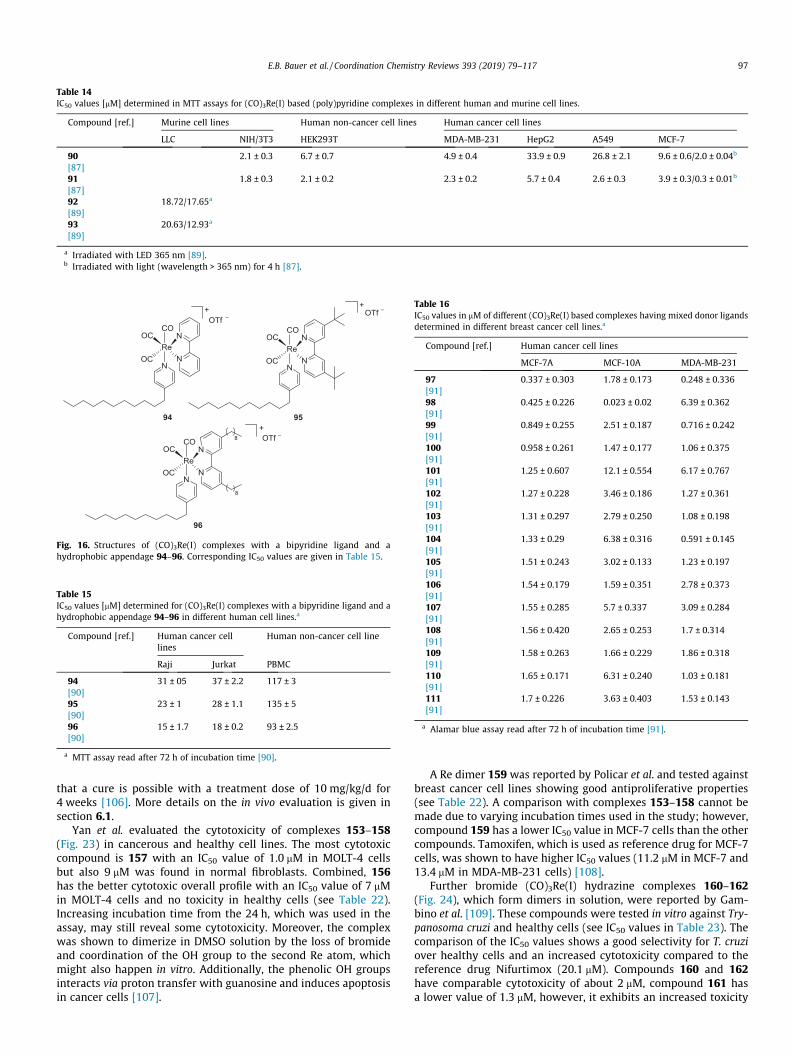
Table 14IC50 values [mM] determined in MTT assays for (CO)3Re(I) based (poly)pyridine complexes in different human and murine cell lines.
Compound [ref.] Murine cell lines Human non-cancer cell lines Human cancer cell lines
LLC NIH/3T3 HEK293T MDA-MB-231 HepG2 A549 MCF-7
90 2.1 ± 0.3 6.7 ± 0.7 4.9 ± 0.4 33.9 ± 0.9 26.8 ± 2.1 9.6 ± 0.6/2.0 ± 0.04b
[87]91 1.8 ± 0.3 2.1 ± 0.2 2.3 ± 0.2 5.7 ± 0.4 2.6 ± 0.3 3.9 ± 0.3/0.3 ± 0.01b
[87]92 18.72/17.65a
[89]93 20.63/12.93a
[89]
a Irradiated with LED 365 nm [89].b Irradiated with light (wavelength > 365 nm) for 4 h [87].
Fig. 16. Structures of (CO)3Re(I) complexes with a bipyridine ligand and ahydrophobic appendage 94–96. Corresponding IC50 values are given in Table 15.
Table 15IC50 values [mM] determined for (CO)3Re(I) complexes with a bipyridine ligand and ahydrophobic appendage 94–96 in different human cell lines.a
Compound [ref.] Human cancer celllines
Human non-cancer cell line
Raji Jurkat PBMC
94 31 ± 05 37 ± 2.2 117 ± 3[90]95 23 ± 1 28 ± 1.1 135 ± 5[90]96 15 ± 1.7 18 ± 0.2 93 ± 2.5[90]
a MTT assay read after 72 h of incubation time [90].
Table 16IC50 values in mM of different (CO)3Re(I) based complexes having mixed donor ligandsdetermined in different breast cancer cell lines.a
Compound [ref.] Human cancer cell lines
MCF-7A MCF-10A MDA-MB-231
97 0.337 ± 0.303 1.78 ± 0.173 0.248 ± 0.336[91]98 0.425 ± 0.226 0.023 ± 0.02 6.39 ± 0.362[91]99 0.849 ± 0.255 2.51 ± 0.187 0.716 ± 0.242[91]100 0.958 ± 0.261 1.47 ± 0.177 1.06 ± 0.375[91]101 1.25 ± 0.607 12.1 ± 0.554 6.17 ± 0.767[91]102 1.27 ± 0.228 3.46 ± 0.186 1.27 ± 0.361[91]103 1.31 ± 0.297 2.79 ± 0.250 1.08 ± 0.198[91]104 1.33 ± 0.29 6.38 ± 0.316 0.591 ± 0.145[91]105 1.51 ± 0.243 3.02 ± 0.133 1.23 ± 0.197[91]106 1.54 ± 0.179 1.59 ± 0.351 2.78 ± 0.373[91]107 1.55 ± 0.285 5.7 ± 0.337 3.09 ± 0.284[91]108 1.56 ± 0.420 2.65 ± 0.253 1.7 ± 0.314[91]109 1.58 ± 0.263 1.66 ± 0.229 1.86 ± 0.318[91]110 1.65 ± 0.171 6.31 ± 0.240 1.03 ± 0.181[91]111 1.7 ± 0.226 3.63 ± 0.403 1.53 ± 0.143[91]
a Alamar blue assay read after 72 h of incubation time [91].
E.B. Bauer et al. / Coordination Chemistry Reviews 393 (2019) 79–117 97
that a cure is possible with a treatment dose of 10 mg/kg/d for4 weeks [106]. More details on the in vivo evaluation is given insection 6.1.
Yan et al. evaluated the cytotoxicity of complexes 153–158(Fig. 23) in cancerous and healthy cell lines. The most cytotoxiccompound is 157 with an IC50 value of 1.0 mM in MOLT-4 cellsbut also 9 mM was found in normal fibroblasts. Combined, 156has the better cytotoxic overall profile with an IC50 value of 7 mMin MOLT-4 cells and no toxicity in healthy cells (see Table 22).Increasing incubation time from the 24 h, which was used in theassay, may still reveal some cytotoxicity. Moreover, the complexwas shown to dimerize in DMSO solution by the loss of bromideand coordination of the OH group to the second Re atom, whichmight also happen in vitro. Additionally, the phenolic OH groupsinteracts via proton transfer with guanosine and induces apoptosisin cancer cells [107].
A Re dimer 159 was reported by Policar et al. and tested againstbreast cancer cell lines showing good antiproliferative properties(see Table 22). A comparison with complexes 153–158 cannot bemade due to varying incubation times used in the study; however,compound 159 has a lower IC50 value in MCF-7 cells than the othercompounds. Tamoxifen, which is used as reference drug for MCF-7cells, was shown to have higher IC50 values (11.2 mM in MCF-7 and13.4 mM in MDA-MB-231 cells) [108].
Further bromide (CO)3Re(I) hydrazine complexes 160–162(Fig. 24), which form dimers in solution, were reported by Gam-bino et al. [109]. These compounds were tested in vitro against Try-panosoma cruzi and healthy cells (see IC50 values in Table 23). Thecomparison of the IC50 values shows a good selectivity for T. cruziover healthy cells and an increased cytotoxicity compared to thereference drug Nifurtimox (20.1 mM). Compounds 160 and 162have comparable cytotoxicity of about 2 mM, compound 161 hasa lower value of 1.3 mM, however, it exhibits an increased toxicity

Fig. 17. Structures of different (CO)3Re(I) based complexes 97–111 having mixed donor ligands. Corresponding IC50 values are given in Table 16.
Fig. 18. Structures of (CO)3Re(I) compounds 112–118. Corresponding ED/EC50
values are given in Table 17 (cultured cell line suspensions) and 18 (solid tumorcultures).
98 E.B. Bauer et al. / Coordination Chemistry Reviews 393 (2019) 79–117
against healthy cells. Therefore, the biological profile of 160 and162 is better. Additionally, the authors were able to show thatthese compounds inhibit the respiration system of mitochondria[109]. Targeting the mitochondria was also shown to increase cyto-toxicity by Mao et al., who introduced two groups of (CO)3Re(I)containing phenanthroline (66a-f) and diphenyl-phenanthrolinecompounds (67a-j) discussed in Table 12 [78–80].
Massi et al. published a very detailed biological study on (CO)3-Re(I) carbene complexes 163–165, which are modified with indo-methacin, an anti-inflammatory drug, and tested theirantiproliferative activity against different pancreatic cancer celllines and healthy HEK cells (see Table 23). In the same setup,ruthenium complexes with the same indomethacin-based ligandsystem were also examined and showed no activity. The deter-
mined IC50 values for the (CO)3Re(I) carbene based complexes arein the low micromolar range (similar to cisplatin) for all three can-cer cell lines and are slightly higher for healthy cells. Further bio-logical studies were performed to investigate the origin of thevarying biological effects observed for these compounds. A seriesof these complexes were subjected to an anticancer screen, theresults of which showed that the most influential structural varia-tion involves the lability of the ancillary bromide ligand. Further-more, it was shown that complexes 163–165 inhibitphosphorylation of Aurora-A kinase and thus induces partial cellcycle arrest at the G2/M phase (in about 26% of the cells) [110].Wilson et al. similarly observed cell cycle arrest of the G2/M phaseinduced by compound 121 reaching about 50% of the cells [69].
4. Re coordination compounds
In coordination compounds, Re is mostly reported in oxidationstates III, IV and V [111–113] as well as Re(VII) in trioxo complexes[114]. It was initially assumed that Re(IV) and (V) complexes weredisproportionating in aqueous solution, until Davison et al.reported highly stable Re(V) and Tc(V) oxobis(dithiolato) com-plexes [115]. The increased complex stability opened up the appli-cations of Re coordination complexes in medicinal chemistry andunderscore the importance of the ligand system as a crucial drugcomponent for medical application as evidenced by the com-pounds that are in clinical trials (see below).
4.1. Coordination compounds of Re(III), Re(IV) and Re(V)
The following paragraph describes the cytotoxic effects of Recoordination complexes, where Re exists in oxidation states III, IVor V. A detailed biological study on this class of compounds wasreported by Lippard et al. [116]. The structures of the studied Re(V)oxo phenanthroline complexes 166 and 167 are given inFig. 26. The cytotoxic properties of complexes 166 and 167 werecompared to cisplatin, which was evaluated in the same assay,showing that 166 and 167 exhibit lower IC50 values in most ofthe cell lines (see Table 24). In A2780 cisplatin resistant cells, bothcomplexes have low IC50 values compared to cisplatin

Table 17ED/EC50 values of different (CO)3Re(I) based complexes having mixed donor ligands determined in different cell line suspensions ([mg/ml]/[mM]; EC50 values are recalculated byconsidering the molecular weight of the respective compound).a
Compound [ref.] Human cancer cell lines Murine cell lines Human non-cancer cell lines
HL-60 T molt3 T molt4 HuT-78 THP-1 HeLa-S3 L1210 P388 RMPI 1788
112 3.42/4.07 3.27/3.89 4.52/5.38 2.60/3.09 2.10/2.50 2.69/3.20 2.36/2.81 2.72/3.24 6.78/8.07[92]113 3.33/4.12 4.70/5.82 4.36/5.40 2.96/3.67 3.28/4.06 2.32/2.87 1.72/2.13 2.91/3.60 7.02/8.70[92]114 2.14/2.35 5.17/5.67 4.32/4.74 2.55/2.80 3.30/3.62 2.68/2.94 1.92/2.11 2.30/2.52 7.60/8.34[92]115 3.37/3.48 3.01/3.11 3.64/3.76 3.53/3.64 5.21/5.38 2.85/2.94 2.80/2.89 3.61/3.73 6.86/7.08[92]116 2.05/1.32 3.06/1.97 6.16/3.97 2.70/1.74 3.87/2.49 1.45/0.93 2.90/1.87 2.02/1.30 9.31/5.99[92]117 3.01/4.09 3.17/4.31 5.44/7.39 3.48/4.73 4.93/6.70 3.02/4.10 3.09/4.20 2.49/3.38 8.50/11.55[93]118 5.97/6.28 3.74/3.93 5.64/5.93 3.58/3.76 2.86/3.01 2.75/2.89 3.15/3.31 2.48/2.61 6.70/7.05[93][NBu4][ReO4] 3.56/7.23 3.59/7.29 4.96/10.07 2.54/5.16 2.10/4.26 3.12/6.33 3.65/7.41 2.69/5.46 7.18/14.57[93][NEt4]2ReBr3(CO)3] 3.83/4.97 3.96/5.14 4.60/5.97 3.17/4.11 2.85/3.70 2.42/3.14 2.65/3.44 4.55/5.91 8.00/10.38[93]
a Measurements were carried out using the Trypan blue exclusion method.
Table 18ED/EC50 values of different (CO)3Re(I) based complexes having mixed donor ligands determined in different solid tumor cultures in [mg/ml]/mM using crystal violet/MeOH (EC50
values are recalculated by considering the molecular weight of the respective compound).
Compound [ref.] Human cancer cell line
KB A549 1-A9 MCF-7 UM 86 HCT-8 HSO SK2 PL HepG2
112 6.88/8.19 8.02/9.55 7.71/9.18 3.52/4.19 8.99/10.71 5.45/6.49 4.59/5.47 6.11/7.28 3.90/4.64 4.76/5.67[92]113 4.88/6.04 4.02/4.98 3.39/4.20 2.01/2.49 6.49/8.04 10.69/13.24 5.32/6.59 4.18/5.18 7.30/9.04 5.07/6.28[92]114 4.34/4.76 8.51/9.33 8.92/9.78 2.37/2.60 9.05/9.93 3.95/4.33 5.86/6.43 3.45/3.78 5.06/5.55 4.02/4.41[92]115 6.45/6.66 3.24/3.34 3.72/3.84 2.42/2.50 6.67/6.88 4.55/4.70 7.72/7.97 13.3/13.73 8.61/8.89 4.49/4.63[92]116 5.49/3.53 6.82/4.39 7.39/4.76 1.53/0.99 7.78/5.01 5.65/3.64 5.00/3.22 2.87/1.85 4.95/3.19 3.74/2.41[92]117 3.71/5.04 5.53/7.52 6.07/8.25 3.60/4.89 4.89/6.65 3.51/4.77 7.11/9.66 2.85/3.87[93]118 6.74/7.09 5.76/6.06 2.48/2.61 3.11/3.27 7.28/7.66 6.33/6.66 13.12/13.80 4.15/4.36[93][NBu4][ReO4] 7.79/15.81 8.87/18.00 4.18/8.48 7.99/16.22 8.26/16.77 8.04/16.32 12.48/25.33 3.82/7.75[93][NEt4]2ReBr3(CO)3] 7.27/9.44 8.35/10.84 5.37/6.97 2.17/2.82 8.15/10.58 10.92/14.17 6.74/8.75 4.46/5.79[93]
E.B. Bauer et al. / Coordination Chemistry Reviews 393 (2019) 79–117 99
(IC50 = 0.042 mM and 8.42 mM, respectively, see Table 24). However,166 and 167 show lower IC50 values in healthy MRC-5 cells thancisplatin.
Studies investigating the mechanism of action of these com-pounds were performed. The mechanism of cell death induced bythe test compounds was investigated by staining A549 cells withthe fluorescent dyes Hoechst, a cell-permeable bisbenzimide dyestaining the cell nucleus, and propidium iodide (PI), a cell imper-meable DNA intercalating dye. The differences in cell permeabilityof these dyes are used in fluorescence microscopy experiments todistinguish between intact living cells and dying, necrotic or late-apoptotic cells with a damaged plasma membrane. Furthermore,necrostatin-1 (5-(indol-3-ylmethyl)-3-methyl-2-thio-hydantoin),a compound inhibiting apoptosis, is used to further investigatethe cell death caused by compounds 166 and 167.
In Fig. 25, the fluorescence microscopy images of Hoechst/PIstained A549 cells incubated with different concentrations of 166and 167 and necrostatin-1 are shown [116].
In summary, the images suggest a necrotic cell death inducedby complexes 166 and 167, which can be blocked with
necrostatin-1. In combination with further investigations, theresults show that these complexes induce mitochondrial damageand necrosis as evidenced by RIP1-RIP3-dependent ROS produc-tion. Cell cycle analysis shows an arrest in the G1 phase [116].
Abram et al. evaluated the biological activity of complexes 168–176 (Fig. 27) in MCF-7 breast cancer cells [117]. The activities ofthese ‘3 + 1’ compounds were found to be strongly influenced bythe lability of the monodentate ancillary ligand Cl, CN or SCN;the more labile the monodentate ligand, the more cytotoxic thecomplex. Thus, the chloride and cyanide coordinated complexes168 and 172, 173 have the lowest IC50 values (0.41 mM (168) and0.6 mM (172 and 173); see Table 25). Coordination of chelatingligands significantly increases the IC50 value to 376 mM for complex176 [117].
Complexes 177–186 (Fig. 27) were examined in vitro and in vivofor the binding affinity towards the 5-HT2 serotonin receptor [118].The most potent complexes are 182 and 183 (see Table 25). Thecorresponding 99mTc compounds exhibit comparable activity andbio-distribution with high liver uptake, renal excretion and lowbrain uptake. The bio-distribution of the Re compounds was not

Table 19IC50 values [mM] determined for (CO)3Re(I) complexes 119–130 in different human cell lines.
Compound [ref.] Human cancer cell lines Human non-cancer cell line
A549 A549cisR HeLa KinasePim 1 A375 K562 A431 MRC-5
119a 5.2 ± 4.0 3.9 ± 4.6 5.8 ± 3.4 10.7 ± 0.5[69]120a 9.7 ± 4.1 5.7 ± 1.8 8.8 ± 2.4 6.0 ± 1.9[69]121a 6.7 ± 4.9 5.4 ± 1.8 1.2 ± 0.2 4.1 ± 0.9[69]Cisplatina 3.0 ± 1.8 12.4 ± 8.5 3.0 ± 1.2 0.43 ± 0.14[69]122b 0.084[94]123b 0.673[94]124b 0.058[94]125b 10.0/0.03c 0.075[94]126d 0.9 3.4[95]127d 0.7 3.0[95]128d 1.3 7.5[95]129d 1.8 7.8[95]130a 2.0 ± 0.98[96]
a MTT assay read after 72 h of incubation time [69,96].b radioactive assay: measured by the degree of phosphorylated substrate peptide P70 S6 after 30 min of incubation at room temperature with an ATP concentration of
10 mM [94].c EC50 value; dark/ irradiated with 580 nm LED light; determined after 24 h incubation with MTT assay [94].d MTT assay read after 48 h of incubation time [95].
100 E.B. Bauer et al. / Coordination Chemistry Reviews 393 (2019) 79–117
evaluated. However, compounds targeting the brain receptorsrequire the ability to cross the blood–brain barrier, which limitsthe application of these complexes [118].
In Table 26, some examples of theranostic Re/99mTc approachesare presented. Jurisson et al. evaluated the binding affinity ofBombesin-conjugates 187 and 188 (Fig. 28) in vitro. The ligand sys-tem was studied for complexation of non-radioactive Re, 99mTc andcarrier-added as well as non-carrier-added 186Re showing ade-quate complexation properties and stabilities for these metals.The very low IC50 values (see Table 26) and the biological profileof the tested compounds are promising for the development of acombination of imaging and therapeutic agent for prostate cancer[119,120].
Cyclometalated complexes 189 and 190 (Fig. 28) were evalu-ated for potential theranostic applications and biological activityby Gilon et al. [121]. These complexes were designed to addressGonadotropin-releasing hormone (GnRH) receptors. This receptoris overexpressed on different cancer types, for example on breast,prostate and ovarian cancers. In order to evaluate the potential ofthese compounds, the IC50 values on rat pituitary cortex membranewere determined. The highest affinity of 50 nM (see Table 26) wasfound for 189. For comparison a higher affinity was found fornative GnRH (10 nM) [121].
Vomero et al. evaluated the biological application of peripheralbenzodiazepine receptor (PBR) targeting complexes 191 and 192(Fig. 28). Although these complexes have very low IC50 values(see Table 26), the bio-distribution of the corresponding 99mTccomplex show no distinct accumulation of these complexes. There-fore, the properties of these complexes must be further optimizedto maintain this high affinity towards the receptor and simultane-ously show a better bio-distribution [122].
Pirmettis et al. studied quinazoline coordinated complexes 193and 194 (Fig. 29) for their ability to target epidermal growth factorreceptor (EGFR) [123]. The EGFR is known to be overexpressed oncertain solid tumors. The authors report that the Re compoundssignificantly inhibit autophosphorylation of EGFR and have lowIC50 values (see Table 27). Another interesting aspect mentionedin the paper is the substitution of the chloride and one fluoridein complex 193 with bromide and a proton, respectively. Thisresults in complex 194 and lead to significantly increased antipro-liferative activity (see Table 27) [123]. It would be very interestingto determine if the single exchange of the chloride by bromidewould similarly influence the biological activity.
The cytotoxicity of a metal-cyclized peptide has been presentedby Jurisson et al. [124]. The Re-core is coordinated to the thioaceticacid modified terminal amino acid and a cysteine residue as deter-mined by NMR studies with the stability of the complex being sup-ported by quantum chemical calculations [124]. The authorssynthesized four different isomers of the Re(V) octreotide ana-logues, but were only able to separate three of them, namely195–197 (Fig. 29). Thus, one of the evaluated compounds is a mix-ture of two isomers, where one of them is complex 196. The otherisomer could not be structurally characterized in similar detail asthe others due to overlapping signals in the NMR studies. Regard-less, the compounds were evaluated for their binding affinity to thesomatostatin receptor overexpressing rat cells. The determinedIC50 values are in the high nanomolar range with the lowest valuebeing 130 nM for complex 196. The benchmark system In-DOTA-Tyr3-octreotide, when tested in the same assay, has an IC50 valueof 8 nM. Importantly, these complexes have one of the lowestIC50 values reported for cyclized Re(V) octreotide analogues andother Re compounds presented in this review. Furthermore, these

Fig. 19. Confocal microscopic images of HeLa cells treated with differentconcentrations of compound 121 and stained or transfected with the specifieddye or plasmid. Reprinted with permissions from J. Am. Chem. Soc., 139 (2017)14302–14314. Copyright 2018 American Chemical Society.
Fig. 20. Structures of (CO)3Re(I) compounds 119–130 bearing dinitrogen basedligands. Corresponding IC50 values are given in Table 19.
E.B. Bauer et al. / Coordination Chemistry Reviews 393 (2019) 79–117 101
complexes are more stable than non-cyclized metal-coordinatedpeptides, and thus provide a good basis for theranostic approacheswith cyclized Re-peptides [124].
5. Re clusters, Re(VII) trioxo compounds and perrhenates
Finally, octahedral water-soluble hexarhenium cluster com-pounds have been assumed applicable for medicinal purposesdue to their favorable properties. Based on their luminescenceand X-ray contrast properties, these clusters may be applied asimaging agents in the future [125]. Moreover, Re clusters werefound to generate reactive singlet oxygen upon irradiation withUV-light and therefore are studied for their potential in photody-namic therapy of actinic keratosis or basal cell carcinoma [126].
Biological studies involving Re(VII) trioxo compounds havebeen performed to evaluate potential (eco)toxic effects of methyl-
trioxorhenium (MTO), a widely applied homogenous epoxidationcatalyst [127]. In addition, perrhenate based ionic liquids (ILs),which are promising catalysts for oxidation and epoxidation reac-tions, have been studied in vitro to get insight in to potential (eco)-toxic effects [128]. It had turned out, however, that in perrhenatecontaining ILs the toxicity depends largely on the cations andMTO is converted to perrhenate in aqueous, particularly basic envi-ronments, reducing its toxicity relatively quickly [127,128]. Thearea of ecotoxicity determines the influence of chemicals pollutingthe environment and ecosystem. Chemical pollution is of big con-cern in modern environmental risk management, as ecosystemsare still exposed to steadily increasing amounts of hazardouschemicals eventually leading to chronic effects. First, pollutantinteractions can be observed on the cellular level e.g. by gene acti-vation, activation of different signaling pathways or stress-specificresponses. Additionally, organisms and populations of differentspecies show different, complex reactions upon exposure to differ-ent pollutants. To get insight into the effect of a compound that isfrequently exposed to an ecosystem by the industry, different spe-cies and microorganisms are studied in in vitro tests. Additionally,these results may give insights into the mechanism of action ofthese compounds [129]. Furthermore, cancer cells are included inthese studies, since compounds previously declared as ‘toxic’ werefound to have anticancer properties depending on the applied dose[130].
5.1. Re cluster compounds
Re clusters are increasingly investigated for their potential bio-logical application in blue-light PDT (light irradiation up to500 nm) due to their photosensitizing properties and the abilityto generate singlet oxygen. Blue-light PDT is widely used in treat-ment of cancer affecting the skin like basal cell carcinoma andsquamous cell carcinoma [126]. For application in blue-light PDT,cluster compounds should be stable, water-soluble, and lumines-cent, have a high cellular uptake, show photo-induced cytotoxicity,and low dark toxicity. The compounds described in the followingwill be evaluated for such properties.
Studies on the cluster compounds Na4[{Re6S8}(CN)6], Na4[{Re6-Se8}(CN)6] and Na4[{Re6Te8}(CN)6] studied by Shestopalov et al.[125,126] showed that the cytotoxicity and the cellular uptakekinetics of the compounds are strongly dependent on the chalco-

Table 20IC50 values [mM] determined for (CO)3Re(I) heteroatom containing complexes 131–141 in different cell lines.
Compound [ref.] Human cancer cell lines Rat Human non-cancer celllines
HeLa KB-31 KB-V-1 A2780 A2780cisR HepG2 C6b CCD-19Lu HEK293T
131a 7.75 ± 0.07 8.91 ± 0.17 6.61 ± 0.48[97]132a 19.04 ± 1.12 26.61 ± 2.37 15.76 ± 4.74[97]133a 2.02 ± 0.19 4.29 ± 0.27 22.24 ± 1.07[97]134a 18.34 ± 1.36 24.97 ± 0.43 16.67 ± 1.34[97]135a 13.74 ± 0.53 20.40 ± 2.55 15.84 ± 3.31[97]136a 18.09 ± 0.77 48.45 ± 2.02 63.36 ± 1.17[97]Cisplatina 8.32 ± 0.88 30.2 ± 0.94 3.44 ± 0.48[97]137a >200/26.4 ± 9.2* >200/4.6 ± 1.4* >200/29.9 ± 7.7*
[98]138a >200/5.9 ± 1.4* >200/2.2 ± 1.1* >200/3.2 ± 0.7*
[98]Cisplatina 0.18 ± 0.07 5.14 ± 1.1[98]139a 50.3 ± 0.2 43.5 ± 2.2 195 ± 1 30.9 ± 1.1 112 ± 3[99]Cisplatina 11.6 ± 0.2 22.1 ± 3.6 39.1 ± 1.7 10.5 ± 0.5 129 ± 1[99]140c 10.6 ± 0.3 19.1 ± 0.6 4.5 ± 0.1[100]141c n.d. n.d. 19.2 ± 0.6[100]Cisplatinc 2.9 ± 0.4 9.2 ± 0.4 0.7 ± 0.2[100]
a MTT assay read after 24 h of incubation time [97,99].* 1h irradiation with light of wavelength 365 nm, MTT assay [98].b Cell line with high expression of TSPO.c MTT assay read after 72 h of incubation time [100].
Fig. 21. Structures of (CO)3Re(I) heteroatom containing compounds 131–141. Corresponding IC50 values are given in Table 20.
102 E.B. Bauer et al. / Coordination Chemistry Reviews 393 (2019) 79–117
gen atoms. The cytotoxicity was found to decrease in the orderS > Se > Te whereas the cellular accumulation in contrast increasesin the order S < Se < Te in Hep-2 cells. However, photocytotoxicityand reactive oxygen species (ROS) production increases in theorder Te < S < Se and thus breaks the periodic table trend. Never-theless, the Se complex was able to reduce cell viability to 63% ata concentration of 100 mM [125,126]. The hexarhenium clusterNa4[{Re6Te8}(CN)6], studied by Mironov et al., was found to becompletely non-toxic in vitro (see Table 28) and in vivo it wasnot taken up by cells. The low toxicity of this cluster represents a
good start for the development of X-ray contrast agents based onhexarhenium cluster complexes [131] although evaluating strate-gies to get the compound into cells should be considered a highpriority. In conclusion, the reported data form a good basis for fur-ther development of biological applicable cluster compounds[125,126,131].
Other hexarhenium clusters K2H8[{Re6Se8}(P(CH2CH2CONH2)(CH2CH2COO)2)6] and K4[{Re6(l3-Se8}(BTA)6] having benzotriazo-late apical ligands were photochemically and biologically evalu-ated by Brylev et al. [132]. The compounds display red

Fig. 22. Schematic illustration of structures of binuclear (CO)3Re(I) compounds and two appended monomeric complexes 142–152. Corresponding IC50 values are given inTable 21.
Table 21IC50 values [mM] determined for (CO)3Re(I) complexes 142–152 in different human cancer cell lines.
Compound [ref.] Human cancer cell lines
HeLa A549 HCT-15 HT-29 MCF-7 HepG2 K562
142a >100 95.72 ± 8.2 20.80 ± 1.9[104]143a 54.19 ± 2.4 56.15 ± 2.9 n.a.[104]144a 19.81 ± 2.5 88.49 ± 1.2 n.a.[104]145a 40.49 ± 1.9 65.23 ± 7.2 n.a.[104]146a 64.50 ± 1.9 29.65 ± 1.4 30.53 ± 1.3[104]Cisplatina 39.84 ± 6.4 25.89 ± 1.8 23.93 ± 2.8[104]147a 23.4 ± 1.4 >100 48.2 ± 3.4 30.1 ± 1.6 27.3 ± 1.3[102]148a 63.4 ± 3.1 41.4 ± 2.2 42.2 ± 3.1 23.3 ± 1.2 >100[102]149a 22.8 ± 2.5 >100 46.9 ± 3.1 29.4 ± 2.8 26.4 ± 1.9[102]150a 21.2 ± 2.7 24.7 ± 2.9 22.1 ± 2.3 27.3 ± 2.6 24.1 ± 1.7[102]Cisplatina 20.94 ± 2.1 18.63 ± 3.8 17.92 ± 2.9 21.91 ± 3.4 23.65 ± 2.2[102]151a 17.56 ± 5.7 18.2 ± 5.7 n.a. 11.9 ± 1.4 20.2 ± 9.2 n.a.[103]152b 75.1 131.5 >500 4.75[105]
(n.a. = not active).a MTT assay read after 48 h of incubation time [102–104].b No incubation time given; MTT assay [105].
E.B. Bauer et al. / Coordination Chemistry Reviews 393 (2019) 79–117 103

Fig. 23. Structures of an asymmetric binuclear Re(I) compounds and phenol containing monomeric complexes 153–159. Corresponding IC50 values are given in Table 22.
Table 22IC50 values [mM] determined for asymmetric binuclear Re(I) compounds and phenol containing monomeric complexes 153–159 in different human cell lines.
Compound [ref.] Human cancer cell lines Human non-cancer cell line
MOLT-4 MCF-7 MDA-MB-231 Fibroblast
153a 24 ± 6 >125 >125[107]154a 15 ± 1 >125 >125[107]155a 15 ± 1 >125 >125[107]156a 7.3 ± 0.4 24 ± 4 >125[107]157a 1.0 ± 0.1 35 ± 4 9 ± 1[107]158a 22 ± 2 >125 >125[107]Cisplatina 18 ± 1 71 ± 8 28 ± 2[107]159b 19.41 8.61[108]Tamoxifenb 11.20 13.40[108]
a MTT assay read after 24 h of incubation time [107].b MTT assay read after 72 h of incubation time [108].
Fig. 24. Structures of (CO)3Re(I) carbene or hydrazine compounds 160–165.Corresponding IC50 values are given in Table 23.
Table 23IC50 values [mM] determined for (CO)3Re(I) carbene or hydrazine complexes 160–165 in d
Compound [ref.] Human non-cancer cell line Antimicrobial Comp
EA.hy926 T. cruzi (Dm28c strain)
160a 36.9 ± 0.7 2.4 ± 0.2 163b
[109] [110]161a 14.3 ± 0.6 1.3 ± 0.3 164b
[109] [110]162a 43.6 ± 1.1 2.01 ± 0.03 165b
[109] [110]Nifurtimoxa 20.1 ± 0.8 – Carbo[109] [110]
a MTT assay read after 24 h of incubation time [109].b Resazurin assay read after 72 h of incubation time [110].
104 E.B. Bauer et al. / Coordination Chemistry Reviews 393 (2019) 79–117
phosphorescence and high quantum yields, which is beneficial forimaging applications. In addition, the compounds taken up by thecells accumulate in the cytoplasm. The IC50 indicate no acute cyto-toxicity although long-time toxicity of the complexes are possible.Compared to Na4[{Re6S8}(CN)6], Na4[{Re6Te8}(CN)6], K2H8[{Re6Se8}(P(CH2CH2CONH2)(CH2CH2COO)2)6] and K4[{Re6(l3-Se8}(BTA)6]these clusters are 4–7 fold more toxic [132].
Ramirez-Tagle et al. evaluated the biological activity of clusterRe6Se8I63� [133]. The authors determined the IC50 and the ICmax
value in healthy and cancer cells. Results show that the concentra-tion, at which all cancer cells are killed (ICmax see Table 28), is inthe same concentration range, where half of the healthy cells are
ifferent biological systems.
ound [ref.] Human cancer cell lines Human non-cancer cell line
HPAF-II ASPC1 CFPAC HEK293T
6.0 ± 2.0 7.9 ± 1.4 6.0 ± 1.8 11.8 ± 2.3
4.8 ± 0.8 9.4 ± 3.5 5.4 ± 1.4 8.6 ± 0.3
5.6 ± 0.6 4.0 ± 1.2 5.7 ± 2.8 14.8 ± 2.4
platinb 8.7 ± 4.3 6.8 ± 2.0 7.4 ± 1.2 45
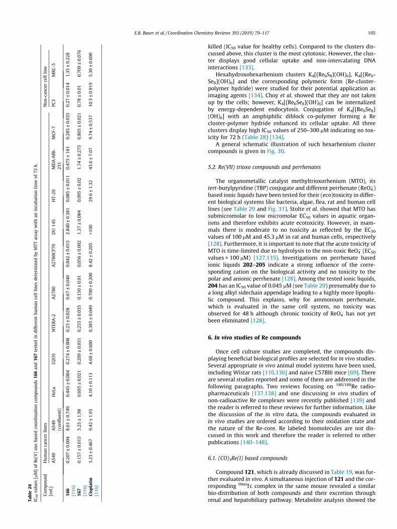
Table24
IC50va
lues
[mM]of
Re(V
)ox
oba
sedco
ordina
tion
compo
unds
166an
d16
7tested
indifferen
thu
man
celllin
esde
term
ined
byMTT
assaywithan
incu
bation
timeof
72h.
Com
pound
[ref.]
Human
cancerlines
Non
-can
cercellline
A54
9A54
9(con
fluen
t)HeL
aU2O
SNTE
RA-2
A27
80A27
80CP7
0DU
145
HT-29
MDA-M
B-
231
MCF-7
PC3
MRC-5
166
0.20
7±0.00
48.61
±0.74
90.44
5±0.00
40.27
4±0.00
60.23
±0.02
80.67
±0.04
00.04
2±0.01
52.84
0±0.38
10.08
5±0.01
10.47
5±16
10.28
5±0.03
50.27
±0.01
41.35
±0.22
8[116
]16
70.15
7±0.01
55.25
±1.98
0.69
5±0.02
10.20
9±0.03
10.25
5±0.03
50.15
0±0.01
0.05
6±0.00
21.37
±0.08
40.09
5±0.02
1.74
±0.27
50.80
5±0.02
10.78
±0.01
0.70
9±0.07
6[116
]Cisplatin
3.23
±0.46
79.42
±1.93
4.10
±0.11
34.60
±0.60
00.38
5±0.04
90.70
0±0.20
08.42
±0.20
5>1
0029
.6±1.32
43.6
±7.07
9.74
±0.53
710
.3±0.91
95.30
±0.60
0[116
]
E.B. Bauer et al. / Coordination Chemistry Reviews 393 (2019) 79–117 105
killed (IC50 value for healthy cells). Compared to the clusters dis-cussed above, this cluster is the most cytotoxic. However, the clus-ter displays good cellular uptake and non-intercalating DNAinteractions [133].
Hexahydroxohexarhenium clusters K4[{Re6S8}(OH)6], K4[{Re6-Se8}(OH)6] and the corresponding polymeric form (Re-cluster-polymer hydride) were studied for their potential application asimaging agents [134]. Choy et al. showed that they are not takenup by the cells; however, K4[{Re6Se8}(OH)6] can be internalizedby energy-dependent endocytosis. Conjugation of K4[{Re6Se8}(OH)6] with an amphiphilic diblock co-polymer forming a Recluster-polymer hydride enhanced its cellular uptake. All threeclusters display high IC50 values of 250–300 mM indicating no tox-icity for 72 h (Table 28) [134].
A general schematic illustration of such hexarhenium clustercompounds is given in Fig. 30.
5.2. Re(VII) trioxo compounds and perrhenates
The organometallic catalyst methyltrioxorhenium (MTO), itstert-butylpyridine (TBP) conjugate and different perrhenate (ReO4
�)based ionic liquids have been tested for their (eco)toxicity in differ-ent biological systems like bacteria, algae, flea, rat and human celllines (see Table 29 and Fig. 31). Stolte et al. showed that MTO hassubmicromolar to low micromolar EC50 values in aquatic organ-isms and therefore exhibits acute ecotoxicity. However, in mam-mals there is moderate to no toxicity as reflected by the EC50
values of 100 mM and 45.3 mM in rat and human cells, respectively[128]. Furthermore, it is important to note that the acute toxicity ofMTO is time-limited due to hydrolysis to the non-toxic ReO4
� (EC50
values > 100 mM) [127,135]. Investigations on perrhenate basedionic liquids 202–205 indicate a strong influence of the corre-sponding cation on the biological activity and no toxicity to thepolar and anionic perrhenate [128]. Among the tested ionic liquids,204 has an IC50 value of 0.045 mM (see Table 29) presumably due toa long alkyl sidechain appendage leading to a highly more lipophi-lic compound. This explains, why for ammonium perrhenate,which is evaluated in the same cell system, no toxicity wasobserved for 48 h although chronic toxicity of ReO4
� has not yetbeen eliminated [128].
6. In vivo studies of Re compounds
Once cell culture studies are completed, the compounds dis-playing beneficial biological profiles are selected for in vivo studies.Several appropriate in vivo animal model systems have been used,including Wistar rats [116,136] and naive C57Bl6 mice [69]. Thereare several studies reported and some of them are addressed in thefollowing paragraphs. Two reviews focusing on 186/188Re radio-pharmaceuticals [137,138] and one discussing in vivo studies ofnon-radioactive Re complexes were recently published [139] andthe reader is referred to these reviews for further information. Likethe discussion of the in vitro data, the compounds evaluated inin vivo studies are ordered according to their oxidation state andthe nature of the Re-core. Re labeled biomolecules are not dis-cussed in this work and therefore the reader is referred to otherpublications [140–148].
6.1. (CO)3Re(I) based compounds
Compound 121, which is already discussed in Table 19, was fur-ther evaluated in vivo. A simultaneous injection of 121 and the cor-responding 99mTc complex in the same mouse revealed a similarbio-distribution of both compounds and their excretion throughrenal and hepatobiliary pathway. Metabolite analysis showed the

Fig. 25. Fluorescence microscopy images of: (A) untreated A549 cells, (B) cells treated with 20 mM 166 for 12 h, (C) cells treated with 20 mM 167 for 12 h, (D) cells treated withnecrostatin-1, (E) cells co-incubated with 166 and necrostatin-1, (F) cells co-incubated with 167 and necrostatin-1. Reprinted with permissions from J. Am. Chem. Soc., 137(2015) 2967–2974. Copyright 2018 American Chemical Society.
Fig. 26. Structures of Re(V) oxo based coordination compounds 166 and 167.Corresponding IC50 values are given in Table 24.
106 E.B. Bauer et al. / Coordination Chemistry Reviews 393 (2019) 79–117
presence of 121 (and its chloride analogue, where the axial waterligand is substituted by chloride) up to 120 min in blood and urine.Nevertheless, also two distinct metabolites formed, which werenot further analyzed. Overall, compound 121 displays sufficientstability, fast blood clearance and no non-specific accumulationin any organs, which makes this compound a promising candidatefor further in vivo tumor targeting studies [69].
Collery et al. extensively studied the in vivo effects of Re(I) dis-elenoether complex 152 [106]. As shown in Table 21, complex 152shows cytotoxicity in MCF-7 cells with an IC50 value of 4.75 mM,however, is non-toxic in A549 and HT-29 cells. Additionally, itwas shown that the compound is able to inhibit cell proliferationof MDA-MB-231 cells at a concentration of 10 mM over a periodof 48 h and the effect is retained even after removal of the Re com-plex. Therefore, the complex was evaluated in vivo in MDA-MB-231bearing mice resulting in remarkably reduced tumor volume com-pared to mice treated with cisplatin. After one month of treatment,a complete cure of the mice was observed. The investigation on themechanism of action indicate a beneficial interaction of the redoxchemistry of the Se ligand, which is suggested to be liberated in thecells. The Re core is proposed to interact with the DNA in a methodsimilar to cisplatin and therefore is able to induce cell death.Experiments show that treatment with 152 (reducing the tumorvolume) resulted in significantly increased tumor growth uponadditional administration of a single dose of cisplatin at day 41of the experiment. Therefore, it is assumed that the Re complexis potentially replaced by cisplatin [106,149,150]. However, further
studies confirming this assumption are needed. Overall, the studiesshow that 152 is a very promising candidate for breast-tumortreatment.
6.2. Dirhenium(III) based compounds
Shtemenko et al. evaluated the biological activity of quadruple-bonded dirhenium(III) carboxylates (see Fig. 32) [136,151–155]. Arecent review summarizes these findings in detail [156]. Neverthe-less, dirhenium compounds are rarely studied in anticancer studiesand need to be mentioned in this review. Its absence from biolog-ical studies is mostly due to low solubility and instability in aque-ous solution. However, encapsulation of these clusters inliposomes stabilizes the quadruple bond and therefore makes itpossible to evaluate these compounds in biological studies [156].Shtemenko et al. were able to show that these dirhenium clustersexhibit antihemolytic properties shifting the maximum rate ofhemolysis in vitro from about 1.5 min to 6 min [153]. Additionally,co-encapsulation of dirhenium cluster and cisplatin in a ratio of 1:4displays the highest antitumor activity compared to cisplatin andthe liposomal form of the rhenium cluster. During a 21 day exper-iment in tumor-bearing mice, the rhenium-platinum liposome wasable to reduce the total tumor volume from 60�100 mm3 to 0–2 mm3 indicating its strong anticancer effect [136,154,156].Increasing antitumor efficiency was shown by introducing differ-ent substituents for the dicarboxylates in the order methyl (206)< ethyl (207) < propyl (208) < butyl (209) [156]. Additionally, notoxic effects were observed in kidneys, liver and on the productionin morphology of red blood cells during in vivo experiments. More-over, it was shown that the dirhenium compounds were able tobind covalently to DNA, like cisplatin and the investigations doneby Collery et al. [106] on Re(I) diselenoether complexes. However,the Re cluster compounds display different redox behavior leadingto antioxidative properties in vivo in contrast to pro-oxidativeeffect of cisplatin [156]. Furthermore, due to the d-component ofthe quadruple bond, the cluster is able to scavenge an unpairedelectron and therefore reduce oxidative stress in cells. In summary,the liposomal encapsulated quadruple-bonded Re cluster showssynergistic effects with cisplatin leading to antihemolytic, antirad-

Table 25IC50 values [mM] determined for complexes 168–186 in different human cell lines.
Compound[ref]
Human cancer cellline
Compound[ref]
Frontal cortical tissuefor 5-HT2 receptorbindingMCF-7
168a 0.41 ± 0.02 177b >100[117] [118]169a 1.51 ± 0.17 178b 1.289[117] [118]170a 1.7 ± 0.3 179b 0.845[117] [118]171a 2.0 ± 0.2 180b >100[117] [118]172a 0.6 ± 0.3 181b 0.029[117] [118]173a 0.6 ± 0.3 182b 0.019[117] [118]174a 2.3 ± 0.3 183b 0.014[117] [118]175a 22.4 ± 0.3 184b 0.023[117] [118]176a 376 ± 2 185b 0.378[117] [118]Cisplatina 1.6 ± 0.1 186b 0.352[117] [118]
a No incubation time given; CV staining assay [117].b Competition binding assay using [3H]ketanserine [118].
Fig. 27. Structures of compounds 168–186. Corr
E.B. Bauer et al. / Coordination Chemistry Reviews 393 (2019) 79–117 107
ical, antioxidative and antitumor properties representing a newperspective for the development of new potent anticancer agents[156].
6.3. Re(V) based compounds
Re(V) oxo compounds 166 and 167 (Table 24), were testedin vivo in C57BL/6 mice to evaluate toxicity and the side effectsof these compounds. After the administration of a single dose, micewere monitored for further 6 days showing no acute toxicity orweight loss. Although IC50 values in MRC-5 cells indicate a highercytotoxicity compared to cisplatin (1.35 mM and 0.71 mM, respec-tively; cisplatin 5.3 mM), the in vivo data indicates a lower toxicity.Therefore, this study shows that IC50 values can be seen as indictorof in vivo behavior, however, they do not display the complete tox-icity profile of a compound [116]. The stability of 166 in wholeblood was evaluated at different time points over a period of 6 hby analysis of octanol extracts from aliquots of blood incubatedwith 166 at 37 �C using GFAAS. The determined half-life is29.1 min, which is comparable to cisplatin (21.6 min). However,decomposition of the investigated Re complex to water-solubleperrhenate is not covered using this method and is a possibleexplanation for the observed tolerance in mice.
esponding IC50 values are given in Table 25.

Table 26IC50 values [mM] determined for Re(oxo)-N,S-donor ligands coordination compounds 187–192 in different cell lines.
Compound [ref.] Human cancer cell line Compound [ref.] Rat Compound [ref.] RatPC3 [nM] Pituitary membrane [mM] Cerebral cortex membrane suspension [mM]
187a 2.0 ± 0.7 189b 0.05 191c 0.127 ± 0.015[119] [121] [122]188a 1.0 ± 0.2 190b 1.00 192c 0.187 ± 0.019[120] [121] [122]
a Competition assay with [125I]-Tyr-BBN(NH2) [119].b Competition assay with [125I]-GnRH [121].c Competition assay with [3H]-PK11195 [122]
Fig. 28. Structures of Re(oxo)-N,S-donor ligands coordination compounds 187–192.Corresponding IC50 values are given in Table 26.
108 E.B. Bauer et al. / Coordination Chemistry Reviews 393 (2019) 79–117
6.4. Re cluster and Re(VII) based compounds
The sodium iodide symporter (NIS), a membrane glycoprotein,transfers iodide from blood to cells and is mainly expressedin the thyroid. NIS-mediated uptake is extensively studied for
Fig. 29. Structures of Re(III) and Re(V) based coordination compou
99mTcO4�, 125I and 131I for diagnosis and therapy of NIS expressing
tumors. A study by Li et al. published in 2016 used NIS transfectedHeLa cells and studied 188ReO4
� uptake in vitro and in vivo [157]. Itwas shown that 188ReO4
� displays a better distribution and excre-tion profile compared to 125I. 188ReO4
� reached a maximum uptakelevel in NIS transfected tumor cells after 0.5 h and decreased fasterthan 125I, however transferring a higher energy dose to the tumor.Furthermore, 188ReO4
� provides c-radiation for imaging andb-radiation for therapy displaying a new possibility for NIS genetherapy [157].
Non-toxic hexarhenium cluster (IC50 values given in Table 28;about 400 mM) Na4[{Re6S8}(CN)6], Na4[{Re6Se8}(CN)6] and Na4[{-Re6Te8}(CN)6] were further evaluated for their toxicity in vivo inBALB/C mice [158,159]. After applying a dose of 500 mg (Re)/kgof the Te or S containing cluster, one mouse treated with the Tecontaining cluster died whereas the others showed no sign ofintoxication. The Se containing cluster administered in the samedose led to the deaths of two mice. The surviving mice did notshow signs of weight loss or intoxication. Morphological analysisof the liver, kidneys, spleen, lungs and heart of the mice aftertwo weeks of injection showed adverse morphological changes inthe liver for the animals treated with Na4[{Re6S8}(CN)6] and Na4[{-Re6Se8}(CN)6]. This is contrary to the very high IC50 values of about400 mM for these cluster compounds (see Table 28). For the Te con-taining cluster, no morphological changes could be observed and
nds 193–197. Corresponding IC50 values are given in Table 27.

Table 27IC50 values [mM] of Re(III) and Re(V) based coordination compounds tested in differenthuman cancer cell lines.
Compound [ref.] Human cancer cell lines RatA431 AR42J
193a 6.4 ± 1.8[123]194a 2.9 ± 0.3[123]195b 1.5 ± 0.7[124]196b 0.13 ± 0.08[124]197b 0.4 ± 0.1[124]196 + Isomerb 0.5 ± 0.2[124]natIn-DOTA-Tyr3-octreotideb 0.008 ± 0.003[124]
a MTT assay read after 72 h of incubation time [123].b 1 h incubation time using competition assay with [125I]-Tyr-somatostatin-14
[124].
Fig. 30. General scheme of hexarhenium Re6 chalcogenide cluster compoundsdiscussed in Table 28. (Q = S/Se/Te; L = ligand) [132]
E.B. Bauer et al. / Coordination Chemistry Reviews 393 (2019) 79–117 109
therefore this compound is considered safe in application [159].This low toxicity is also displayed by the higher IC50 values of4500 mM in Hep-2 cells (Table 28). Bio-distribution studies in ratusing the K4[{Re6S8}(CN)6] cluster showed the highest accumula-tion in liver and kidney medulla. Moreover, accumulation in thespleen was observed suggesting this compound is a suitable imag-ing (e.g. as an X-ray contrast agent) and therapeutic agent for lym-phoproliferative disorders [158].
7. Clinical trials
The ultimate goal of developing new Re-based anticancer com-pounds is their application in therapeutic/clinical treatment. Cellculture studies of anticancer Re compounds are often used asimportant initial pre-screening steps ahead of animal and humanstudies. Many steps of translational research are necessary beforestudies in humans are possible. Although the field of Re based anti-cancer agents may be in its infancy, some Re compounds have beeninvestigated in human beings and these will be summarized below.
After extensive studies in cell culture and animal model sys-tems, some compounds show varying degrees of effectiveness
Table 28IC50 values in [mM] of Re based cluster compounds in different human cell lines.
Compound [ref.] Human tumor cell lines
HeLa Hep-2 H
Na4[{Re6S8}(CN)6]a 401 ± 7 417 ± 4[126]Na4[{Re6Se8}(CN)6]a 410 ± 5 478 ± 8[126]Na4[{Re6Te8}(CN)6]a 4800 ± 0.3[131]K2H8[{Re6Se8}(P(CH2CH2CONH2)(CH2CH2COO)2)6]b 123.7 ± 0.8[132]K4[{Re6(l3-Se8}(BTA)6]b 122.6 ± 2.1[132]Re6Se8I63�b 5[133]K4[{Re6S8}(OH)6]b 262.3 ± 8.1[134]K4[{Re6Se8}(OH)6]b 297.8 ± 9.5[134]Re cluster- polymer hydrideb 251.7 ± 5.2[134]
a MTT assay read after 48 h of incubation [126,131].b MTT assay read after 72 h of incubation time [132–134].c Determined ICmax.
and selectivity in killing cancer cells and only a few compoundsare continued to human studies. Information on potential progresson compounds being evaluated in human beings are thus relevantto the topic of the current review. Studies in humans are referred toas clinical trials beginning with Phase I and progressing to Phase IIand III trials. Phase I clinical trials focus on determining the safetyof the compounds in normal subjects, Phase II focuses on deter-mining the proper dose to use in patients and involves a limitednumber of subjects and a short time period. Phase III studies focuson the treatment of patients for a specific clinical condition (e.g.melanoma) and involves a larger number of subjects for longerperiods. The summary of Re compounds in clinical trials (Table 30)shows that the compounds studied are mainly radioactive Re com-plexes. This may be due to non-invasive dosimetry and imaging ofthe radioactive compounds in vivo by well-established medicalequipment, the single photon emission computed tomography(SPECT), as applied for 99mTc imaging [160]. Since many reviewsthat focus on Re and Tc radiopharmaceuticals are published[5,11,137,159–175], this review focuses on characterizing theeffects of different classes of non-radioactive Re complexes. Theultimate goal is to compare such studies with the compounds thathave been selected for clinical trials. Once the analysis of the com-
Human non-cancer
epG2 MRC-5 EA hy 926 HUVEC
471 ± 13
551 ± 21
8.5 ± 5/146.3 ± 23c 88.7 ± 6/286.8 ± 34c 147 ± 11/457.5 ± 46c

Table 29EC50 values [mM] of Re(VII) trioxo compounds and perrhenate tested in different cell lines. The corresponding structures are given in Fig. 31.
Compound [ref.] Marine bacteria Green algae aquatic plant Water flea AChE [IC50] Rat cancer cell line Human tumor cell lineV. fischeri R. subcapitata L. minor D. magna IPC-81 HepG2
198 0.275 19.0 14.6 1.58 92.3 100 45.3[135]199 0.217 23.8 16.4 2.34 227 44.9 47.3[135]200 2.34 115 15.84 402 5600 5000 >6200[135]201 >11,000 >1500 420 >2500 >2360 >5150 >5150[135]202 59 41 124 194[128]203 6.3 21 38 112.2[128]204 0.096 0.045 9.5 2.5[128]205 146 199 134 1009[128]
Table 30Clinical trials involving Re compounds.
Yearstarted
Class of ReCompound
Status Name of Clinical Trial Project Condition/ illness Treatment Study location/Organization
2018 Re-bisphosphonate
Recruiting Rhenium-188-HEDP vs Radium-223-chloride in patients with advanced prostatecancer refractory to hormonal therapy
Prostate cancer metastatic tobone
223Ra188Re VU U Medical CenterAmsterdam NL
2013 Re(V)oxoBMEDA-labeledliposomes
Recruiting Maximum Tolerated Dose, Safety, andEfficacy of Rhenium Nanoliposomes inRecurrent Glioblastoma
GlioblastomaAsrocytoma 186Re-Liposomes
The Cancer Therapy andResearch Center atUTHSCSA, San Antonio,Texas, US
2010 Re-SSS Lipiodol Recruiting 188-Re-SSS Lipiodol to Treat HepatocellularCarcinomas
Hepatocellular Carcinomas 188Re Centre EugeneMarquisRennes, France
2017 Re-poly-L-lysinedendrimer
Recruiting Treatment of non-responding toconventional therapy inoperable livercancers by in suit introduction of ImDendrim
Liver Cancer 188ReImDendrim
Tongji University EasternHospital, Shanghai, China
2004 Re(V)oxolabeled-peptide
Active –notrecruiting
Rhenium Re-188 P2045 in Patients with lungcancer who have received or refused toreceive prior chemotherapy
Lung NeoplasmsCarcinoma,Non-Small-CellLungCarcinoma, SmallCellNeoplasm Recurrence,Local
188Re P2045 Iowa City, Iowa, USBaltimore, Maryland, US
2012 Re-sulfide Active –notrecruiting
Multicenter Canadian study to measure thesafety and efficacy of radiosynoviorthesis
Arthritis 186Re sulfide 12 locations in Canada
2014 Re(I)MAG3-labeled MAB
Completed Bio-distribution study with 186 Re-labeledhumanized monoclonal antibody BIWA 4 inpatients with adenocarcinoma of the breast
Adenocarcinoma 186Re BIWA 4
2014 Re(I)MAG3-labeled MAB
Completed Bio-distribution study with 186 Re-labeledhumanized monoclonal antibody BIWA 4 inpatients with non-small cell lung cancer
Carcinoma, non-small-celllung cancer
186Re hMABBIWA 4
2014 Re(I)MAG3-labeled MAB
Completed Dose escalation study with 99mTC - or 186Re-labeled humanized monoclonal antibody(hMAB) BIWA 4 in patients with head andneck cancer
Head and Neck Neoplasms 186Re hMABBIWA 4
2008 Colloidal Resulfur
Completed Intraoperative gamma camera for breastcancer surgery
Breast cancer,Ductalcarcinoma in situ
Testperformanceof CarollReScamera
CHRU, Hôpital Civil,Service de Gynécologie-obstétriqueStrasbourg,France
2006 Re-sulfur Completed Identification of sentinel lymph nodes withmethylene blue and isotope
Infiltrative Breast Cancer 186Re sulfurandmethyleneblue
Service de Gynécologie-Obstétrique, HôpitalCivilStrasbourg, France
2008 Re-colloids Completed Added-value of SPECT/CT in patientsundergoing LM/SL for gynecological cancers
Cervical CancerVulvar Cancer 99mTc-cysteinrheniumcolloids,
375, South Street Hospital- Dpt. of NuclearMedicineLondon, Ontario,Canada
2014 Re-sulfide Withdrawn Diagnosis of micro aspiration in intubatedcritically Ill patients: Pepsin vs 99 mTechnetium
Critical illness 99mTc-Resulfidenanocolloid
ICU, Calmette Hospital,University Hospital ofLilleLille, France
2014 Re(V)oxolabeled-peptide
Withdrawn Phase I/II trial of rhenium 188-P2045 insmall lung cancer and other advancedneuroendocrine carcinomas
Small Cell Lung Cancer (SCLC)Neuroendocrine (NE)TumorsLarge CellNeuroendocrine (NE) Tumors
188Re P2045 U Maryland Marlene &Stewart GreenebaumCancer Center, Baltimore,Maryland, US
110 E.B. Bauer et al. / Coordination Chemistry Reviews 393 (2019) 79–117

Fig. 31. Structures of Re(VII) trioxo compounds and perrhenate 198–205. Corresponding IC50 values are given in Table 29.
Fig. 32. Dirhenium dicarboxylates showing synergistic effects with cisplatin whenencapsulated in liposomes.
E.B. Bauer et al. / Coordination Chemistry Reviews 393 (2019) 79–117 111
pounds selected for the clinical trials was done, it became clearthat many of the Re compounds investigated for potential thera-peutic applications were not selected for clinical trials yet. Thismay suggest that the compounds in clinical trials are potentiallymore promising, however, since the other compounds are not yettested in clinical trials, they are still interesting candidates.
At this time, 14 studies are listed on the NIH US National libraryof Medicine (https://clinicaltrials.gov/, last accessed 27 Nov 2018)and are documented in Table 30 showing that some Re compoundsare approaching application in the clinic. The Re compounds inves-tigated in clinical trials make use of the two different radioisotopes188Re and 186Re, except for three studies. These two radioactive iso-topes are suitable for radiopharmaceutical application, becausethey have b-emissions suitable for killing tumor cells and c-emission for imaging [161–163]. 188Re can conveniently be pro-duced with high specific activity (high activity per unit mass) froma generator and with a reasonable half-life 16.9 h, an emissionmaximum energy of 2 MeV and an average energy of 764 keV[161–163]. 188Re has been used to radiolabel peptides for targetedradiotherapy [120,164]. 186Re on the other hand has a longer half-life (90 h) and a lower energy of emission than 188Re. 186Re also hasb-emissions, and is produced at high specific activity in a cyclotronor accelerator [165]. Two clinical trials including 186Re based com-pounds are currently ongoing with others having been completedor recently withdrawn (see Table 30) [165].
The focus of the 186Re labeled nanoliposomes ongoing clinicaltrial [166] is to determine the maximum tolerated dose, safetyand efficacy of 186Re administered in nanoliposomes usingbrachytherapy for the ultimate use to treatment of recurrentglioblastoma [165]. 186Re, like 99mTc, is not taken up by the boneand is cleared by the kidney. The energy of the c-rays producedby 186Re is suitable for external imaging using a gamma cameraand the b-emission has suitable energy to penetrate solid tumorsup to 2 mm. The 80–100 nm-sized nanoliposome is used as aconvection-enhanced delivery system, which is directly injectedinto the tumor site. The liposome-encapsulated 186Re is thus main-tained at the target site and the majority of radiation is released tothe tumor tissue making 186Re labeled nanoliposomes desirable asa potential agent for treating glioblastoma [165].
Two other ongoing clinical trials focus on treatment of liver can-cers. The use of a 188Re coupled to an imidazole ligand and associ-ated with a dendrimer, referred to as ImDendrim, is investigated totreat previously non-responding inoperable liver cancers [167]. Inaddition, application of 188Re-SSS Lipiodol (general structure ofRe-SSS is given in Fig. 33) is investigated for treatment of patients
with hepatocellular carcinoma. The hepatic intra-arterial adminis-tration of the radiolabeled lipiodol could potentially replace the131I labeled lipiodol, and the results are expected to reduce hospi-talization from the current eight days to one day and thus lowercosts and improve patient comfort [168].
A Re(V)oxo based compound of recent interest is the Re labeledpeptide P2045 (Fig. 33). P2045 is a somatostatin-derived peptidetargeting somatostatin receptor overexpressing cancers [161–163]. After the 188Re labeled peptide binds to these receptors, theradioactivity will kill the cancer cells [161–163]. Unfortunately,the Phase I/II clinical trials of the 188Re P2045 to treat patients withadvanced small cell lung cancer that has recurred or in patientswith advanced neuroendocrine cancers has been withdrawn[171]. However, a related clinical trial remains active involves188Re labeled P2045 being evaluated in patients with several differ-ent lung cancers [172]. The purpose of the study was to determinethe safe dose of the compound (Phase II), potentially resulting in alarger Phase III study against the cancer(s) for which this treatmentwas most effective.
Another study (Phase I/II) that remains active is performed inCanada exploring the potential for a colloid of 90Y citrate or a186Re sulfide for treatment of persistent active synovectomy[173]. Radiation synovectomy is considered a reliable and easy-to-perform therapy and involves intra-articular injection of b-emitting radionuclides of inflammatory joint disease. In addition,the absence of harmful side effects for the 90Y citrate or 186Re sul-fide treatment make these potential alternative treatments tocases that are resistant to systemic therapy and intra-articular cor-ticosteroid injections.
Other Phase I clinical trials recently completed include intra-venous administration of 186Re(I) labeled bivatuzumab to investi-gate the bio-distribution, pharmacokinetics and safety of a 186Re
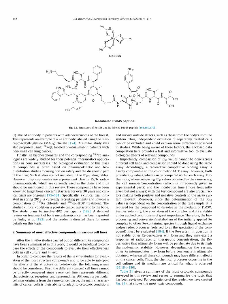
Fig. 33. Structures of Re-SSS and Re labeled P2045 peptide [163,169,170].
112 E.B. Bauer et al. / Coordination Chemistry Reviews 393 (2019) 79–117
(I) labeled antibody in patients with adenocarcinoma of the breast.This represents an example of a Re antibody labeled using the mer-captoacetyltriglycine (MAG3) chelate [174]. A similar study wasalso proposed using 186Re(I) labeled bivatuzumab in patients withnon-small cell lung cancer.
Finally, Re bisphosphonates and the corresponding 99mTc ana-logues are widely studied for their potential theranostics applica-tions in bone metastases. The biological evaluation of this classof compounds is often based on pharmacokinetic and bio-distribution studies focusing first on safety and the diagnostic partof the drug. Such studies are not included in the IC50-listing tables.However, bisphosphonates are a prominent class of Re/Tc radio-pharmaceuticals, which are currently used in the clinic and thusshould be mentioned in this review. These compounds have beenknown to target bone cancer/metastases for over 30 years and clin-ical trials are ongoing [175–181]. Specifically, a clinical trial initi-ated in spring 2018 is currently recruiting patients and involve acombination of 223Ra chloride and 188Re-HEDP treatment. Thestudied clinical condition is prostate cancer metastatic to the bone.The study plans to involve 402 participants [182]. A detailedreview on treatment of bone metastases/cancer has been reportedby Finlay et al. [183] and the reader is directed there for moredetails on this topic.
8. Summary of most effective compounds in various cell lines
After the in vitro studies carried out on different Re compoundshave been summarized in this work, it would be beneficial to com-pare the effects of the compounds to evaluate their effectivenessboth in cell culture and in vivo.
In order to compare the results of the in vitro studies for evalu-ation of the most effective compounds and to be able to interpretthe effects of the structure on cytotoxicity, the following issuesshould be considered. First, the different (cancer) cell lines cannotbe directly compared since every cell line represents differentcharacteristics, receptors, and surroundings. Although, a particularcell may originate from the same cancer tissue, the main character-istic of cancer cells is their ability to adapt to cytotoxic conditions
and survive outside attacks, such as those from the body’s immunesystem. Thus, independent evolution of separately treated cellscannot be excluded and could explain some differences observedin studies. While being aware of these factors, the enclosed datacompilation here provides a fast and informative tool to evaluatebiological effects of relevant compounds.
Importantly, comparison of IC50 values cannot be done acrossdifferent cell lines, and comparison should be done using the sameassay. Accordingly, a radioactive competitive binding assay ishardly comparable to the colorimetric MTT assay; however, bothprovide IC50 values, which can be compared within each assay. Fur-thermore, when comparing IC50 values obtained by the same assay,the cell number/concentration (which is infrequently given inexperimental parts) and the incubation time (more frequentlygiven but not always) with the test compound are also crucial fac-tors making both positive and negative controls in the assay sys-tem relevant. Moreover, since the determination of the IC50
values is dependent on the concentration of the test sample, it isrequired for the compound to dissolve in the medium or DMSO.Besides solubility, the speciation of the complex and its stabilityunder applied conditions is of great importance. Therefore, the bio-processing and conversion/metabolism of the initially applied Recomplex to other Re-containing species through ligand exchangeand/or redox processes (referred to as the speciation of the com-pound) must be evaluated [184]. If the Re-system in question isnot stable, other Re-derivatives will form and they may exert aresponse. At radiotracer or therapeutic concentrations, the Re-derivative that ultimately forms will be perrhenate due to its highthermodynamic stability. However, depending on the system,other Re intermediates may form before perrhenate is ultimatelyobtained, whereas all these compounds may have different effectson the cancer cells. Thus, the chemical processes occurring in thecell culture and its medium are critical for what is observed[37,184–186].
Table 31 gives a summary of the most cytotoxic compoundssurveyed in this review and serves to summarize the topic thathas been reviewed. For convenience of the reader, we have createdFig. 34 that shows the most toxic compounds.

Table 31Most cytotoxic Re compounds and its IC50 values for each cell line.
Cancer cell type Cell line Most active compound + IC50
value [mM]Assay + incubation time Log P
values
Cervical cancer HeLa 52 IC50 = 0.106 MTT; no time given 1.05125 IC50 = 0.03 MTT; 24 h after irradiation with
LED580 nm
Ovarian carcinoma cells A2780 167 IC50 = 0.150 MTT, 72 h 0.95Cisplatin-resistant ovarian carcinoma cells A2780R 166 IC50 = 0.042 MTT, 72 h 1.20Estrogen-positive breast cancer MCF-7 166 IC50 = 0.285 MTT, 72 h 1.20Estrogen-negative breast cancer MDA-MB-231 97 IC50 = 0.248 Resazurin, 72 hMammary epithelial cell line MCF-10A 98 IC50 = 0.023 Resazurin, 72 hEpidermoid carcinoma A431 130 IC50 = 2.0 MTT, 72 hMalignant melanoma A375 14 IC50 = 12.5 MTT, 72 hLung adenocarcinoma epithelial cells A549 167 IC50 = 0.15 MTT, 72 h 0.95Cisplatin-resistant lung carcinoma cells A549cisR 67e IC50 = 0.75 MTT, 48 h, darkMurine lung carcinoma LLC 57 IC50 = 4.8 WST-1, 24 h 0.98Colon carcinoma cells HT-29 166 IC50 = 0.085 MTT, 72 h 1.20Pancreatic adenocarcinoma PT-45 18 IC50 = 25.8 MTT, 72 hHepatocellular liver carcinoma HepG2 67b IC50 = 2.1 MTT, 48 h, dark 3.12Murine skin carcinoma B16 10 IC50 = 9.82 Resazurin, 24 hMyelogenous leukemia cell line K562 127 IC50 = 3.0 MTT, 48 hHuman acute T lymphoblastic leukemia MOLT-4
MOLT-3157 IC50 = 1.0115 IC50 = 3.01
MTT, 24 hTrypan blue; no time given
Leukemia monocytic cells THP-1 112 IC50 = 2.1 Trypan blue; no time givenHuman Osteosarcoma U2OS 167 IC50 = 0.209 MTT, 72 h 0.95Prostate cancer cells PC3 166 IC50 = 0.27 MTT, 72 h 1.20
Healthy cell typeHepatic cells LO2 67c IC50 = 3.1 MTT, 48 hEmbryonic kidney cells expressing mutant SV40 large
T antigenHEK293T 93 IC50 = 2.1 MTT, 48 h 3.63
Lung fibroblasts MRC-5 167 IC50 = 0.709 MTT, 72 h 0.95Fibroblasts (murine) Fibroblasts (like NIH/
3T3)93 IC50 = 1.8 MTT, 48 h 3.63
E.B. Bauer et al. / Coordination Chemistry Reviews 393 (2019) 79–117 113
Most of the compounds listed in Table 31 are organometallic Re(I) complexes, but there are also two Re(V)oxo complexes (166 and167). The first part of the table lists different cancer cell lines andthe corresponding most cytotoxic compounds. It has to be notedthat the cell lines listed are those used in the studies reportedand not necessarily the most suitable cell line to use to study aspecific form of cancer. Cell culture studies will provide informa-tion on cytotoxicity but results and interpretation will depend inpart on the cell line chosen. Cell culture studies provide some data,which allows for analysis of effects prior to embarking on moreexpensive and demanding in vivo studies. The second part ofTable 31 contains a few different non-cancerous cell lines andthe effects of Re compounds on these systems. A comparative eval-uation of the compounds in healthy cells provides insight into thecompounds’ toxicity for healthy cells and therefore the possibilityof side effects. Thus, the compounds having the lowest IC50 valuesin healthy cells, namely 67c, 93 and 167, are possibly too toxic forin vivo applications, although only the performance of in vivoexperiments will demonstrate how toxic the compounds are. How-ever, in the case of the Re(V)oxo complex 167 (IC50 = 0.709 in MRC-5 lung fibroblasts) no weight loss or other signs of toxicity wasobserved when administered to mice [116]. This important findingwas addressed in the in vivo studies of these compounds counter-ing the expectations that they would be toxic to healthy cells.
Phenyl-phenanthroline ligands, as used for Re(V)oxo coordina-tion complexes 166 and 167, are also used for Re(I) carbonylorganometallic complexes, like 67b, 67c, 67e, 75, 87, 88 and 97,which all have comparable IC50 values of 0.3 to 5 mM in HeLa,MCF7 or A549 cells, respectively (see Tables 24, 12, 13, and 16).Therefore, these phenanthroline based ligands were found to stabi-lize Re in different oxidation states, like Re(I), Re(V) and Re(VII)O3,however, the latter was not tested for its IC50 value in different celllines but in antibacterial assays, showing a certain amount ofantibacterial activity. Therefore, it would appear that complexes
with this ligand are very promising for the development of biolog-ically highly active Re complexes. However, the toxicity of thesecomplexes should not be considered without examining thephenanthroline ligands alone, as it is well-known that the phenan-throline ligand by itself is cytotoxic [187]. In this case, the stabilityand speciation of the compounds become very important in deter-mining if the phenanthroline dissociates [184–186,188]. If so, theobserved toxicity of these metal complexes are much less interest-ing and promising, because the likelihood that such systems wouldbe further developed is very low.
Most of the compounds shown in Fig. 34 contain the Re(I)(CO)3+
unit. This demonstrates that most of potent effects have beenobserved with organometallic Re compounds. Furthermore, inlarge parts of the cell lines listed in Table 31, the more active Recomplexes are coordinated to phenanthroline-based ligands.Moreover, 166 and 167 represent the most active compoundsreviewed in here, as they display the highest activity in 7 out of21 different cancer cell lines listed in Table 31. Investigations onthe mechanism of action of these compounds suggest, among othercharacterized effects, a necroptotic cell death with mitochondrialdamage. This is supported by a log P value between 0 and +5,which is consistent with mitochondrial accumulation. The deter-mined log P values are 1.2 and 0.95 for compounds 166 and 167,respectively (see Table 31). Mitochondria are essential for energyproduction in eukaryotic cells and therefore their survival. In addi-tion, they regulate intrinsic pathways leading to apoptosis, necrop-tosis and non-apoptotic cell death. Consequently, the mitochondriaregulate fundamental functions in healthy and cancerous cells.Therefore, it is not surprising that attributes of cancer cells likeexcessive proliferation are linked to mitochondrial dysfunctionand that anticancer research is developing mitochondria-targeting compounds [189].
It is often observed that the higher the lipophilicity (positive logP value) of the compounds, the lower the IC50 value will be (see

Fig. 34. Summary of the most cytotoxic compounds. Corresponding IC50 values are listed in Table 31.
114 E.B. Bauer et al. / Coordination Chemistry Reviews 393 (2019) 79–117
Table 31). Nevertheless, an increased lipophilicity may lead todecreased solubility, enhanced liver uptake and may cause lowerclearance from non-target tissues. However, the activity is alsohighly dependent on cellular uptake, molecular size, charge, andtherefore speciation [184–186,188]. Even small changes in the con-
stitution of the compounds can have a big influence, as it is shownfor the compounds in Tables 12, 20 and 22. Accordingly, the com-plexes listed in Table 22 show formation of dimers in solution afterloss of ancillary coordinated bromide, which results in increasedlipophilicity and cytotoxicity. These results are supported by previ-

E.B. Bauer et al. / Coordination Chemistry Reviews 393 (2019) 79–117 115
ous reports documenting that the speciation involved plays animportant role, because any bioprocessing of the administereddrug can affect uptake [185]. A possible explanation for increasedcytotoxicity by such small molecular changes might be theincrease in lipophilicity, and this is certainly the case for the com-pounds in Table 16. However, a good systematic study of the cyto-toxicity of different (CO)3Re(I) compounds in ASCP-1 pancreaticcells show that a greater influence is observed by the exchangeof the ancillary ligand. An increase in cytotoxicity was shown inthe order Br < NCS < Cl < I in cancer cells. The order changed toCl � Br < I < NCS in healthy cells (see Tables 19 and 20). Further-more, for Re(V)oxo compounds, similar observations were madeby evaluating different structurally related ligands indicating thatchelating ligands increase the stability, leading to a decrease incytotoxicity (see Table 25). Therefore, for the development ofpotent cytotoxic compounds, the right balance between stabilityand lability as well as lipophilicity and hydrophilicity must befound.
Although (CO)3Re(I) complexes have been extensively studiedin recent years, there are only a limited number of Re complexesthat are studied in vivo. The reviewed in vivo studies include twostudies on Re(I) tricarbonyl compounds, one study dealing withdirhenium(III) complexes encapsulated in liposomes, one studyperformed on Re(V)oxo complexes, two studies on hexarheniumcluster compounds and one on NIS addressing 188ReO4
�.Re compounds, which are currently in or have previously been
in clinical trials include Re-bisphosphonates, Re-labeled liposomes,Re-peptide or Re-protein complexes, Re-sulfide material and Re(V)oxo complexes. The analysis shown in this work points to the factthat the organometallic Re(I)(CO)3-containing compounds, particu-larly those with phenanthroline ligands, are particularly potent incell culture work. However, none of these compounds made it toclinical trials. Some studies indicate, that this might be due tothe high toxicity of the phenanthroline ligand, however, in othercases, in vivo studies have been done suggesting that the toxicityis not great for these systems.
9. Conclusions
The development of potent anticancer agents starts with theselection of compounds based on their in vitro activity. This is com-monly evaluated by the effect of the compounds on the growth ofappropriate cell lines and determination of the half-maximal inhi-bitory concentration (IC50) after treatment with a test compound.Making use of results obtained by this method, this review focuseson the cytotoxicity of Re compounds classified by their oxidationstate in order to identify the most cytotoxic Re complexes. In addi-tion to providing IC50 data, this review also highlighted correla-tions and basic information on the systems and (if available)modes of action. Furthermore, the few in vivo studies reportedfor non-radioactive Re complexes are reviewed. Finally, the mostcytotoxic Re compounds reported are compared to the classes ofcompounds that have made it to the 14 clinical trials, some ofwhich are currently active while others have been completed.The compounds that have or are in clinical trials include Re-bisphosphonates, Re labeled liposomes, Re-peptides or Re-proteincomplexes, Re-sulfide materials and Re(V)oxo complexes. Surpris-ingly, no (CO)3Re(I) compounds have made it to clinical trials. Con-sidering that this class of Re compounds is the one most frequentlystudied in cell culture, might be to expect that at least some one ofthese compounds would have been considered for human studies.As described in this review, the reasons for such discrepancy can beattributed to the toxicity on healthy cells, bioprocessing, com-pound stability, toxicity of the ligand itself or many other factors.However, this review demonstrates that the area of Re chemistry
is of growing interest in medicine and provides guidelines forfuture applications.
Acknowledgments
EBB thanks the TUM Graduate School of Chemistry for financialsupport and AAH thanks Colorado State University. DCC and FEKthank the Bavarian State Ministry of Education, Science and theArts for funding the Technische Universität München Visiting Pro-fessor Program given to DCC. DCC thanks the NSF (CHE-1709564)and The Arthur C. Cope Foundation, administered by the AmericanChemical Society for partial support.
Declaration of interest
None.
References
[1] R.L. Siegel, K.D. Miller, A. Jemal, CA-Cancer J. Clin. 68 (2018) 7–30.[2] V.T. DeVita, S.A. Rosenberg, New Engl. J. Med. 366 (2012) 2207–2214.[3] P.G. Corrie, Medicine 36 (2008) 24–28.[4] D.-W. Shen, L.M. Pouliot, M.D. Hall, M.M. Gottesman, Pharmacol. Rev. 64
(2012) 706–721.[5] R. Schibli, A.P. Schubiger, Eur. J. Nucl. Med. Mol. Imaging 29 (2002) 1529–
1542.[6] M. Frier, Mini-Rev. Med. Chem. 4 (2004) 61–68.[7] F. Tisato, C. Bolzati, M. Porchia, F. Refosco, Mass Spectrom. Rev. 23 (2004)
309–332.[8] U. Abram, R. Alberto, J. Brazil. Chem. Soc. 17 (2006) 1486–1500.[9] G. Ferro-Flores, C.A. de Murphy, Adv. Drug Deliver. Rev. 60 (2008) 1389–1401.[10] E.M. Hahn, A. Casini, F.E. Kühn, Coord. Chem. Rev. 276 (2014) 97–111.[11] S. Jürgens, W.A. Herrmann, F.E. Kühn, J. Organomet. Chem. 751 (2014) 83–89.[12] A. Boschi, P. Martini, L. Uccelli, Pharmaceuticals 10 (2017) 12.[13] K.K.W. Lo, Acc. Chem. Res. 48 (2015) 2985–2995.[14] S. Hostachy, C. Policar, N. Delsuc, Coord. Chem. Rev. 351 (2017) 172–188.[15] A.M.H. Yip, K.K.W. Lo, Coord. Chem. Rev. 361 (2018) 138–163.[16] A.G. Mayor, X. Gómez-Olivé, J.J. Aponte, S. Casimiro, S. Mabunda, M. Dgedge,
A. Barreto, P.L. Alonso, J. Infect. Dis. 183 (2001) 1413–1416.[17] A. Coleman, C. Brennan, J.G. Vos, M.T. Pryce, Coord. Chem. Rev. 252 (2008)
2585–2595.[18] D. Gupta, M. Sathiyendiran, ChemistrySelect 3 (2018) 7439–7458.[19] A. Leonidova, G. Gasser, A.C.S. Chem. Biol. 9 (2014) 2180–2193.[20] W. Noddack, I. Tacke, O. Berg, Naturwissenschaften 13 (1925) 567.[21] J.-C. Carlen, B.D. Bryskin, Mater. Manuf. Processes 9 (1994) 1087–1104.[22] M. Fleischer, Econ. Geol. 54 (1959) 1406–1413.[23] S. Liu, Chem. Soc. Rev. 33 (2004) 445–461.[24] D.E. Polyak, 2015 Minerals Yearbook: Rhenium, United States Geological
Survey, 2015.[25] C.D. Anderson, P. Taylor, C.G. Anderson, Miner. Metall. Process 30 (2013) 59–
73.[26] A.D. Dobrzanska-Danikiewicz, W. Wolany, Arch. Mater. Sci. Eng. 82 (2016)
70–78.[27] J.W. Kück, R.M. Reich, F.E. Kühn, Chem. Rec. 16 (2016) 349–364.[28] C.C. Romão, F.E. Kühn, W.A. Herrmann, Chem. Rev. 97 (1997) 3197–3246.[29] E. Deutsch, K. Libson, J.-L. Vanderheyden, A.R. Ketring, H.R. Maxon, Int. J. Rad.
Appl. Instrum. B 13 (1986) 465–477.[30] Y. He, Q. Zhu, M. Chen, Q. Huang, W. Wang, Q. Li, Y. Huang, W. Di, Oncotarget
7 (2016) 70803–70821.[31] W. Li, J. Zhou, Y. Xu, Biomed. Rep. 3 (2015) 617–620.[32] T.L. Riss, R.A. Moravec, A.L. Niles, S. Duellman, H.A. Benink, T.J. Worzella, L.
Minor, Assay Guidance Manual (2016).[33] J. O’Brien, I. Wilson, T. Orton, F. Pognan, Eur. J. Biochem. 267 (2000) 5421–
5426.[34] L. Sliwka, K. Wiktorska, P. Suchocki, M. Milczarek, S. Mielczarek, K. Lubelska,
T. Cierpiał, P. Ły _zwa, P. Kiełbasinski, A. Jaromin, PloS one 11 (2016) e0155772.[35] R. Roskoski, Assays of protein kinase, Methods Enzymol., Elsevier (1983) 3–6.[36] M.T. Makler, J.M. Ries, J.A. Williams, J.E. Bancroft, R.C. Piper, B.L. Gibbins, D.J.
Hinrichs, Am. J. Trop. Med. Hyg. 48 (1993) 739–741.[37] A.A. Haase, E.B. Bauer, F.E. Kühn, D.C. Crans, Coord. Chem. Rev. (2019), in
press.[38] D.A. Volpe, S.S. Hamed, L.K. Zhang, The AAPS Journal 16 (2014) 172–180.[39] J.L. Sebaugh, Pharm. Stat. 10 (2011) 128–134.[40] W. Hieber, H. Fuchs, Z. Anorg. Allg. Chem. 248 (1941) 269–275.[41] Y.K. Yan, S.E. Cho, K.A. Shaffer, J.E. Rowell, B.J. Barnes, I.H. Hall, Pharm. 55
(2000) 307–313.[42] R.G. Pearson, J. Am. Chem. Soc. 85 (1963) 3533–3539.[43] B. Safi, J. Mertens, F. De Proft, P. Geerlings, J. Phys. Chem. A 110 (2006) 9240–
9246.

116 E.B. Bauer et al. / Coordination Chemistry Reviews 393 (2019) 79–117
[44] J. Oyarzo, A. Acuna, H. Klahn, R. Arancibia, C.P. Silva, R. Bosque, C. Lopez, M.Font-Bardia, C. Calvis, R. Messeguer, Dalton Trans. 47 (2018) 1635–1649.
[45] K. Kowalski, Ł. Szczupak, S. Saloman, D. Steverding, A. Jabłonski, V. Vrcek, A.Hildebrandt, H. Lang, A. Rybarczyk-Pirek, ChemPlusChem 82 (2017) 303–314.
[46] R. Arancibia, F. Dubar, B. Pradines, I. Forfar, D. Dive, A.H. Klahn, C. Biot, Bioorg.Med. Chem. 18 (2010) 8085–8091.
[47] L. Glans, W.N. Hu, C. Jost, C. de Kock, P.J. Smith, M. Haukka, H. Bruhn, U.Schatzschneider, E. Nordlander, Dalton Trans. 41 (2012) 6443–6450.
[48] W. Hu, K. Splith, I. Neundorf, K. Merz, U. Schatzschneider, J. Biol. Inorg. Chem.17 (2012) 175–185.
[49] D. Can, H.W. Peindy N’Dongo, B. Spingler, P. Schmutz, P. Raposinho, I. Santos,R. Alberto, Chem. Biodivers. 9 (2012) 1849–1866.
[50] M. Munoz-Osses, F. Godoy, A. Fierro, A. Gomez, N. Metzler-Nolte, DaltonTrans. 47 (2018) 1233–1242.
[51] D. Rosita, M.A. Dewit, L.G. Luyt, J. Med. Chem. 52 (2009) 2196–2203.[52] C. Concha, C. Quintana, A.H. Klahn, V. Artigas, M. Fuentealba, C. Biot, I.
Halloum, L. Kremer, R. López, J. Romanos, Y. Huentupil, R. Arancibia,Polyhedron 131 (2017) 40–45.
[53] A. Kondratskyi, K. Kondratska, F. Vanden Abeele, D. Gordienko, C. Dubois, R.-A. Toillon, C. Slomianny, S. Lemière, P. Delcourt, E. Dewailly, R. Skryma, C.Biot, N. Prevarskaya, Sci. Rep. 7 (2017) 15896.
[54] K. Splith, I. Neundorf, W. Hu, H.W.P. N’Dongo, V. Vasylyeva, K. Merz, U.Schatzschneider, Dalton Trans. 39 (2010) 2536–2545.
[55] G. Jaouen, S. Top, A. Vessieres, G. Leclercq, M.J. McGlinchey, Curr. Med. Chem.11 (2004) 2505–2517.
[56] G. Jaouen, S. Top, A. Vessieres, P. Pigeon, G. Leclercq, I. Laios, Chem. Commun.(2001) 383–384.
[57] S. Top, A. Vessieres, P. Pigeon, M.N. Rager, M. Huche, E. Salomon, C.Cabestaing, J. Vaissermann, G. Jaouen, ChemBioChem 5 (2004) 1104–1113.
[58] I. Paterni, C. Granchi, J.A. Katzenellenbogen, F. Minutolo, Steroids 90 (2014)13–29.
[59] A. Vessieres, S. Top, W. Beck, E. Hillard, G. Jaouen, Dalton Trans. (2006) 529–541.
[60] M. Patra, M. Wenzel, P. Prochnow, V. Pierroz, G. Gasser, J.E. Bandow, N.Metzler-Nolte, Chem. Sci. 6 (2015) 214–224.
[61] L. Quental, P. Raposinho, F. Mendes, I. Santos, C. Navarro-Ranninger, A.Alvarez-Valdes, H.Y. Huang, H. Chao, R. Rubbiani, G. Gasser, A.G. Quiroga, A.Paulo, Dalton Trans. 46 (2017) 14523–14536.
[62] S. Imstepf, V. Pierroz, P. Raposinho, M. Bauwens, M. Felber, T. Fox, A.B.Shapiro, R. Freudenberg, C. Fernandes, S. Gama, G. Gasser, F. Motthagy, I.R.Santos, R. Alberto, Bioconj. Chem. 26 (2015) 2397–2407.
[63] S. Imstepf, V. Pierroz, P. Raposinho, M. Felber, T. Fox, C. Fernandes, G. Gasser, I.R. Santos, R. Alberto, Dalton Trans. 45 (2016) 13025–13033.
[64] S. Imstepf, V. Pierroz, R. Rubbiani, M. Felber, T. Fox, G. Gasser, R. Alberto,Angew. Chem. Int. Ed. 55 (2016) 2792–2795.
[65] A. Leonidova, V. Pierroz, R. Rubbiani, J. Heier, S. Ferrari, G. Gasser, DaltonTrans. 43 (2014) 4287–4294.
[66] A. Leonidova, V. Pierroz, R. Rubbiani, Y. Lan, A.G. Schmitz, A. Kaech, R.K.O.Sigel, S. Ferrari, G. Gasser, Chem. Sci. 5 (2014) 4044–4056.
[67] T. Joshi, G. Gasser, Synlett 26 (2015) 275–284.[68] G. Santoro, T. Zlateva, A. Ruggi, L. Quaroni, F. Zobi, Dalton Trans. 44 (2015)
6999–7008.[69] K.M. Knopf, B.L. Murphy, S.N. MacMillan, J.M. Baskin, M.P. Barr, E. Boros, J.J.
Wilson, J. Am. Chem. Soc. 139 (2017) 14302–14314.[70] A. Luengo, V. Fernández-Moreira, I. Marzo, M.C. Gimeno, Inorg. Chem. 56
(2017) 15159–15170.[71] I. Maisuls, E. Wolcan, O.E. Piro, E.E. Castellano, G. Petroselli, R. Erra-Balsells, F.
M. Cabrerizo, G.T. Ruiz, ChemistrySelect 2 (2017) 8666–8672.[72] R.-R. Ye, C.-P. Tan, Y.-N. Lin, L.-N. Ji, Z.-W. Mao, Chem. Commun. 51 (2015)
8353–8356.[73] G. Mion, T. Gianferrara, A. Bergamo, G. Gasser, V. Pierroz, R. Rubbiani, R. Vilar,
A. Leczkowska, E. Alessio, ChemMedChem 10 (2015) 1901–1914.[74] Y. Ren, K. Cheng, G. Liu, Y. Sun, Y. Shen, Acta Chim. Sincia 66 (2008) 459.[75] S. Clède, F. Lambert, R. Saint-Fort, M.A. Plamont, H. Bertrand, A. Vessières, C.
Policar, Chem. Eur. J. 20 (2014) 8714–8722.[76] A.W.-T. Choi, H.-W. Liu, K.K.-W. Lo, J. Inorg. Biochem. 148 (2015) 2–10.[77] M. Strack, A. Bedini, K.T. Yip, S. Lombardi, D. Siegmund, R. Stoll, S.M.
Spampinato, N. Metzler-Nolte, Chem. Eur. J. 22 (2016) 14605–14610.[78] J. Yang, J.-X. Zhao, Q. Cao, L. Hao, D. Zhou, Z. Gan, L.-N. Ji, Z.-W. Mao, A.C.S.
Appl. Mater. Interfaces 9 (2017) 13900–13912.[79] R.R. Ye, C.P. Tan, M.H. Chen, L. Hao, L.N. Ji, Z.W. Mao, Chem. Eur. J. 22 (2016)
7800–7809.[80] J. Yang, Q. Cao, H. Zhang, L. Hao, D. Zhou, Z. Gan, Z. Li, Y.-X. Tong, L.-N. Ji, Z.-W.
Mao, Biomaterials 176 (2018) 94–105.[81] I. Kitanovic, S.Z. Can, H. Alborzinia, A. Kitanovic, V. Pierroz, A. Leonidova, A.
Pinto, B. Spingler, S. Ferrari, R. Molteni, A. Steffen, N. Metzler-Nolte, S. Wolfl,G. Gasser, Chem. Eur. J. 20 (2014) 2496–2507.
[82] C. Redshaw, S. Watkins, S.M. Humphrey, P.C.B. Page, S. Ashby, Y.M. Chao, C.J.Herbert, A. Mueller, RSC Advances 3 (2013) 23963–23966.
[83] A. Leonidova, V. Pierroz, L.A. Adams, N. Barlow, S. Ferrari, B. Graham, G.Gasser, A.C.S. Med. Chem. Lett. 5 (2014) 809–814.
[84] M.W. Louie, H.W. Liu, M.H.C. Lam, Y.W. Lam, K.K.W. Lo, Chem. Eur. J. 17(2011) 8304–8308.
[85] M.-W. Louie, T.T.-H. Fong, K.K.-W. Lo, Inorg. Chem. 50 (2011) 9465–9471.[86] A.W.T. Choi, M.W. Louie, S.P.Y. Li, H.W. Liu, B.T.N. Chan, T.C.Y. Lam, A.C.C. Lin,
S.H. Cheng, K.K.W. Lo, Inorg. Chem. 51 (2012) 13289–13302.
[87] K. Yin Zhang, K. Ka-Shun Tso, M.-W. Louie, H.-W. Liu, K. Kam-Wing Lo,Organometallics 32 (2013) 5098–5102.
[88] L.C.-C. Lee, K.-K. Leung, K.K.-W. Lo, Dalton Trans. 46 (2017) 16357–16380.[89] F.F. Zhong, X.L. Yuan, J.Z. Zhao, Q. Wang, Sci. China Chem. 59 (2016) 70–77.[90] G. Balakrishnan, T. Rajendran, K. Senthil Murugan, M. Sathish Kumar, V.K.
Sivasubramanian, M. Ganesan, A. Mahesh, T. Thirunalasundari, S. Rajagopal,Inorg. Chim. Acta 434 (2015) 51–59.
[91] P.T. Wilder, D.J. Weber, A. Winstead, S. Parnell, T.V. Hinton, M. Stevenson, D.Giri, S. Azemati, P. Olczak, B.V. Powell, T. Odebode, S. Tadesse, Y.C. Zhang, S.K.Pramanik, J.M. Wachira, S. Ghimire, P. McCarthy, A. Barfield, H.N. Banerjee, C.Chen, J.A. Golen, A.L. Rheingold, J.A. Krause, D.M. Ho, P.Y. Zavalij, R. Shaw, S.K.Mandal, Mol. Cell. Biochem. 441 (2018) 151–163.
[92] J. Zhang, J.J. Vittal, W. Henderson, J.R. Wheaton, I.H. Hall, T.S.A. Hor, Y.K. Yan, J.Organomet. Chem. 650 (2002) 123–132.
[93] W.Wang, Y.K. Yan, T.S. Andy Hor, J.J. Vittal, J.R. Wheaton, I.H. Hall, Polyhedron21 (2002) 1991–1999.
[94] K. Wähler, A. Ludewig, P. Szabo, K. Harms, E. Meggers, Eur. J. Inorg. Chem.2014 (2014) 807–811.
[95] S. Wirth, A.U. Wallek, A. Zernickel, F. Feil, M. Sztiller-Sikorska, K. Lesiak-Mieczkowska, C. Brauchle, I.P. Lorenz, M. Czyz, J. Inorg. Biochem. 104 (2010)774–789.
[96] A. Bourkoula, M. Paravatou-Petsotas, A. Papadopoulos, I. Santos, H.-J. Pietzsch,E. Livaniou, M. Pelecanou, M. Papadopoulos, I. Pirmettis, Eur. J. Med. Chem. 44(2009) 4021–4027.
[97] K. Lyczko, M. Lyczko, S. Meczynska-Wielgosz, M. Kruszewski, J. Mieczkowski,J. Organomet. Chem. 866 (2018) 59–71.
[98] S.C. Marker, S.N. MacMillan, W.R. Zipfel, Z. Li, P.C. Ford, J.J. Wilson, Inorg.Chem. 57 (2018) 1311–1331.
[99] D.-L. Ma, C.-M. Che, F.-M. Siu, M. Yang, K.-Y. Wong, Inorg. Chem. 46 (2007)740–749.
[100] N. Margiotta, N. Denora, S. Piccinonna, V. Laquintana, F.M. Lasorsa, M. Franco,G. Natile, Dalton Trans. 43 (2014) 16252–16264.
[101] N. Denora, R.M. Iacobazzi, G. Natile, N. Margiotta, Coord. Chem. Rev. 341(2017) 1–18.
[102] R. Govindarajan, R. Nagarajaprakash, V. Veena, N. Sakthivel, B. Manimaran,Polyhedron 139 (2018) 229–236.
[103] C.A. Kumar, R. Nagarajaprakash, W. Victoria, V. Veena, N. Sakthivel, B.Manimaran, Inorg. Chem. Commun. 64 (2016) 39–44.
[104] B. Ramakrishna, R. Nagarajaprakash, V. Veena, N. Sakthivel, B. Manimaran,Dalton Trans. 44 (2015) 17629–17638.
[105] A. Kermagoret, G. Morgant, J. d’Angelo, A. Tomas, P. Roussel, G. Bastian, P.Collery, D. Desmaele, Polyhedron 30 (2011) 347–353.
[106] P. Collery, A. Mohsen, A. Kermagoret, S. Corre, G. Bastian, A. Tomas, M. Wei, F.Santoni, N. Guerra, D. Desmaële, J. d’Angelo, Invest. New Drugs 33 (2015)848–860.
[107] J.M. Ho, W.Y. Lee, K.J.T. Koh, P.P.F. Lee, Y.K. Yan, J. Inorg. Biochem. 119 (2013)10–20.
[108] M.L. Low, G. Paulus, P. Dorlet, R. Guillot, R. Rosli, N. Delsuc, K.A. Crouse, C.Policar, Biometals 28 (2015) 553–566.
[109] E. Rodríguez Arce, I. Machado, B. Rodríguez, M. Lapier, M.C. Zúñiga, J.D. Maya,C. Olea Azar, L. Otero, D. Gambino, J. Inorg. Biochem. 170 (2017)125–133.
[110] P.V. Simpson, I. Casari, S. Paternoster, B.W. Skelton, M. Falasca, M. Massi,Chem. Eur. J. 23 (2017) 6518–6521.
[111] S. Seifert, J.U. Künstler, E. Schiller, H.J. Pietzsch, B. Pawelke, R. Bergmann, H.Spies, Bioconjugate Chem. 15 (2004) 856–863.
[112] J. Martinez-Lillo, T.F. Mastropietro, R. Lappano, A. Madeo, M.E. Alberto, N.Russo, M. Maggiolini, G. De Munno, Chem. Commun. 47 (2011) 5283–5285.
[113] T.B.Y. Nguyen, C.T. Pham, T.N. Trieu, U. Abram, H.H. Nguyen, Polyhedron 96(2015) 66–70.
[114] A. Maalaoui, O.B. Said, S.T. Akriche, S.S. Al-Deyab, M. Rzaigui, Z. Naturforsch.B. 67 (2012) 1178–1184.
[115] A. Davison, C. Orvig, H.S. Trop, M. Sohn, B.V. DePamphilis, A.G. Jones, Inorg.Chem. 19 (1980) 1988–1992.
[116] K. Suntharalingam, S.G. Awuah, P.M. Bruno, T.C. Johnstone, F. Wang, W. Lin,Y.-R. Zheng, J.E. Page, M.T. Hemann, S.J. Lippard, J. Am. Chem. Soc. 137 (2015)2967–2974.
[117] P.I.d.S. Maia, H.H. Nguyen, A. Hagenbach, S. Bergemann, R. Gust, V.M. Deflon,U. Abram, Dalton Trans. 42 (2013) 5111–5121.
[118] B. Johannsen, M. Scheunemann, H. Spies, P. Brust, J. Wober, R. Syhre, H.J.Pietzsch, Nucl. Med. Biol. 23 (1996) 429–438.
[119] D.W. Demoin, A.N. Dame, W.D. Minard, F. Gallazzi, G.L. Seickman, T.L. Rold, N.Bernskoetter, M.E. Fassbender, T.J. Hoffman, C.A. Deakyne, S.S. Jurisson, Nucl.Med. Biol. 43 (2016) 802–811.
[120] C.J. Smith, G.L. Sieckman, N.K. Owen, D.L. Hayes, D.G. Mazuru, W.A. Volkert, T.J. Hoffman, Anticancer Res. 23 (2003) 63–70.
[121] Y. Barda, N. Cohen, V. Lev, N. Ben-Aroya, Y. Koch, E. Mishani, M. Fridkin, C.Gilon, Nucl. Med. Biol. 31 (2004) 921–933.
[122] A. Cappelli, A. Mancini, F. Sudati, S. Valenti, M. Anzini, S. Belloli, R.M. Moresco,M. Matarrese, M. Vaghi, A. Fabro, F. Fazio, S. Vomero, Bioconjugate Chem. 19(2008) 1143–1153.
[123] C. Fernandes, I.C. Santos, I. Santos, H.J. Pietzsch, J.U. Kunstler, W. Kraus, A. Rey,N. Margaritis, A. Bourkoula, A. Chiotellis, M. Paravatou-Pestsotas, I. Pirmettis,Dalton Trans. (2008) 3215–3225.
[124] Y. Li, L. Ma, V. Gaddam, F. Gallazzi, H.M. Hennkens, M. Harmata, M.R. Lewis, C.A. Deakyne, S.S. Jurisson, Inorg. Chem. 55 (2016) 1124–1133.

E.B. Bauer et al. / Coordination Chemistry Reviews 393 (2019) 79–117 117
[125] A.A. Krasilnikova, A.O. Solovieva, A.A. Ivanov, K.E. Trifonova, T.N. Pozmogova,A.R. Tsygankova, A.I. Smolentsev, E.I. Kretov, D.S. Sergeevichev, M.A.Shestopalov, Y.V. Mironov, A.M. Shestopalov, A.F. Poveshchenko, L.V.Shestopalova, Nanomedicine: NBM 13 (2017) 755–763.
[126] A.O. Solovieva, K. Kirakci, A.A. Ivanov, P. Kubat, T.N. Poznaogova, S.M.Miroshnichenko, E.V. Vorontsova, A.V. Chechushkov, K.E. Trifonova, M.S.Fufaeva, E.I. Kretov, Y.V. Mironov, A.F. Poveshchenko, K. Lang, M.A.Shestopalov, Inorg. Chem. 56 (2017) 13491–13499.
[127] M. Cokoja, M. Markiewicz, S. Stolte, F.E. Kühn, Toxicity Assessment ofMolecular Rhenium(VII) Epoxidation Catalysts, Encycl. Inorg. Bioinorg. Chem.(2016).
[128] H.B.T. Thu, M. Markiewicz, J. Thöming, R.M. Reich, V. Korinth, M. Cokoja, F.E.Kühn, S. Stolte, New J. Chem. 39 (2015) 5431–5436.
[129] R.I. Eggen, R. Behra, P. Burkhardt-Holm, B.I. Escher, N. Schweigert, Environ.Sci. Technol. 38 (2004) 58a–64a.
[130] I. Pastan, D. Fitzgerald, Science 254 (1991) 1173–1177.[131] A.A. Krasilnikova, M.A. Shestopalov, K.A. Brylev, I.A. Kirilova, O.P. Khripko, K.
E. Zubareva, Y.I. Khripko, V.T. Podorognaya, L.V. Shestopalova, V.E. Fedorov, Y.V. Mironov, J. Inorg. Biochem. 144 (2015) 13–17.
[132] M.A. Shestopalov, K.E. Zubareva, O.P. Khripko, Y.I. Khripko, A.O. Solovieva, N.V. Kuratieva, Y.V. Mironov, N. Kitamura, V.E. Fedorov, K.A. Brylev, Inorg.Chem. 53 (2014) 9006–9013.
[133] C. Echeverria, A. Becerra, F. Nunez-Villena, A. Munoz-Castro, J. Stehberg, Z.Zheng, R. Arratia-Perez, F. Simon, R. Ramirez-Tagle, New J. Chem. 36 (2012)927–932.
[134] S.J. Choi, K.A. Brylev, J.Z. Xu, Y.V. Mironov, V.E. Fedorov, Y.S. Sohn, S.J. Kim, J.H.Choy, J. Inorg. Biochem. 102 (2008) 1991–1996.
[135] S. Stolte, H.T.T. Bui, S. Steudte, V. Korinth, J. Arning, A. Bialk-Bielinska, U.Bottin-Weber, M. Cokoja, A. Hahlbrock, V. Fetz, R. Stauber, B. Jastorff, C.Hartmann, R.W. Fischer, F.E. Kühn, Green Chem. 17 (2015) 1136–1144.
[136] N. Shtemenko, P. Collery, A. Shtemenko, Anticancer Res. 27 (2007) 2487–2492.
[137] P.J. Blower, Int. J. Nucl. Med. Res. 4 (2017) 39–53.[138] E. Boros, A.B. Packard, Chem. Rev. (2018).[139] C.C. Konkankit, S.C. Marker, K.M. Knopf, J.J. Wilson, Dalton Trans. 47 (2018)
9934–9974.[140] R. Alberto, R. Schibli, U. Abram, A. Egli, F.F. Knapp, P.A. Schubiger, Radiochim.
Acta 79 (1997) 99–103.[141] J.D.G. Correia, I. Santos, R. Alberto, K. Ortner, H. Spies, A. Drews, J. Label.
Compd. Rad. 44 (2001) S507–S509.[142] S.R. Banerjee, C.A. Foss, M. Castanares, R.C. Mease, Y. Byun, J.J. Fox, J. Hilton, S.
E. Lupold, A.P. Kozikowski, M.G. Pomper, J. Med. Chem. 51 (2008) 4504–4517.[143] S.R. Banerjee, M.K. Levadala, N. Lazarova, L. Wei, J.F. Valliant, K.A. Stephenson,
J.W. Babich, K.P. Maresca, J. Zubieta, Inorg. Chem. 41 (2002) 6417–6425.[144] H. Struthers, B. Spingler, T.L. Mindt, R. Schibli, Chem. Eur. J. 14 (2008) 6173–
6183.[145] B.A. Rhodes, C.R. Lambert, M.J. Marek, F.F. Knapp, E.B. Harvey, Appl. Radiat.
Isot. 47 (1996) 7–14.[146] S.M. Quadri, B.W. Wessels, Int. J. Rad. Appl. Instrum. B 13 (1986) 447–451.[147] R. Schibli, R. Schwarzbach, R. Alberto, K. Ortner, H. Schmalle, C. Dumas, A.
Egli, P.A. Schubiger, Bioconjugate Chem. 13 (2002) 750–756.[148] A. Varvarigou, S. Archimandritis, M. Fani, P. Bouziotis, S. Vranjez, S.
Xanthopoulos, A. Leonti, T. Sourlingas, M. Sotiropoulou, IAEA-TECDOC-1359(2003) 85.
[149] P. Collery, G. Bastian, F. Santoni, A. Mohsen, M. Wei, T. Collery, A. Tomas, D.Desmaele, J. D’Angelo, Anticancer Res. 34 (2014) 1679–1689.
[150] P. Collery, F. Santoni, J. Ciccolini, T.N.N. Tran, A. Mohsen, D. Desmaele,Anticancer Res. 36 (2016) 6051–6057.
[151] A. Shtemenko, A. Golichenko, S. Tretyak, N. Shtemenko, M. Randarevich,Synthesis and antiradical properties of dirhenium cluster compounds, MetalIons in Biology and Medicine (2008) 229–234.
[152] A.V. Shtemenko, P. Collery, N.I. Shtemenko, K.V. Domasevitch, E.D.Zabitskaya, A.A. Golichenko, Dalton Trans. (2009) 5132–5136.
[153] A.V. Shtemenko, N.I. Shtemenko, S.A. Oliynik, M.A. Zelenuk, Lyposome formsof rhenium cluster compounds in models of haemolytic anemia, in: L.Khassanova, P. Collery, I. Maymard, Z. Khassanova, J.C. Etienne (Eds.), MetalIons in Biology and Medicine, 2002, pp. 558–561.
[154] N.I. Shtemenko, H.T. Chifotides, K.V. Domasevitch, A.A. Golichenko, S.A. Babiy,Z. Li, K.V. Paramonova, A.V. Shtemenko, K.R. Dunbar, J. Inorg. Biochem. 129(2013) 127–134.
[155] N.I. Shtemenko, E.D. Zabitskaya, O.V. Berzenina, D.E. Yegorova, A.V.Shtemenko, Chem. Biodivers. 5 (2008) 1660–1667.
[156] A.V. Shtemenko, N.I. Shtemenko, Ukr. Biochem. J. 89 (2017) 5–30.[157] M. Zhang, S. Shi, R. Guo, Y. Miao, B. Li, Oncol. Rep. 36 (2016) 2289–2297.[158] K. Brylev, M. Shestopalov, O. Khripko, V. Trunova, V. Zvereva, C. Wang, Y.V.
Mironov, V. Fedorov, Bull. Exp. Biol. Med. 155 (2013) 741–744.
[159] T.N. Pozmogova, A.A. Krasilnikova, A.A. Ivanov, M.A. Shestopalov, S.N.Gyrylova, L.V. Shestopalova, A.M. Shestopaloiv, V.A. Shkurupy, Bull. Exp.Biol. Med. 161 (2016) 64–68.
[160] P.L. Esquinas, C.F. Uribe, M. Gonzalez, C. Rodriguez-Rodriguez, U.O. Hafeli, A.Celler, Phys. Med. Biol. 62 (2017) 6379–6396.
[161] J.E. Cyr, D.A. Pearson, D.M. Wilson, C.A. Nelson, M. Guaraldi, M.T. Azure, J.Lister-James, L.M. Dinkelborg, R.T. Dean, J. Med. Chem. 50 (2007) 1354–1364.
[162] D.A. Pearson, J. Lister-James, W.J. McBride, D.M. Wilson, L.J. Martel, E.R.Civitello, J.E. Taylor, B.R. Moyer, R.T. Dean, J. Med. Chem. 39 (1996) 1361–1371.
[163] M.J. Edelman, G. Clamon, D. Kahn, M. Magram, J. Lister-James, B.R. Line, J.Thorac. Oncol. 4 (2009) 1550–1554.
[164] Y. Miao, N.K. Owen, D.R. Fisher, T.J. Hoffman, T.P. Quinn, J. Nucl. Med. 46(2005) 121–129.
[165] W.T. Phillips, B. Goins, A. Bao, D. Vargas, J.E. Guttierez, A. Trevino, J.R. Miller, J.Henry, R. Zuniga, G. Vecil, Neuro-oncology 14 (2012) 416–425.
[166] NIS, US Library of Medicine, ClinicalTrials.gov Identifier NCT01906385,Maximum Tolerated Dose, Safety, and Efficacy of Rhenium Nanoliposomesin Recurrent Glioblastoma, https://clinicaltrials.gov/ct2/show/NCT01906385?term=rhenium&rank=3 (last accessed 28 Nov 2018).
[167] NIS, US Library of Medicine, ClinicalTrials.gov Identifier NCT03255343,Treatment of Non-responding to Conventional Therapy Inoperable LiverCancers by in Situ Introduction of ImDendrim (ImDendrim),https://clinicaltrials.gov/ct2/show/NCT03255343?term=rhenium&draw=3&rank=8 (last accessed 28 Nov 2018).
[168] NIS, US Library of Medicine, ClinicalTrials.gov Identifier NCT01126463,188RE-SSS Lipiodol to Treat HepatoCellular Carcinomas (LIP-RE-I),https://clinicaltrials.gov/ct2/show/NCT01126463?term=rhenium&rank=7(last accessed 28 Nov 2018).
[169] E. Garin, B. Denizot, N. Noiret, N. Lepareur, J. Roux, M. Moreau, J.-Y. Herry, P.Bourguet, J.-P. Benoit, J.-J. Lejeune, Nuc. Med. Commun. 25 (2004) 1007–1013.
[170] N. Lepareur, V. Ardisson, N. Noiret, E. Garin, Int. J. Mol. Im. 2012 (2012).[171] NIS, US Library of Medicine, ClinicalTrials.gov Identifier NCT02030184, Phase
I/II Trial of Rhenium 188-P2045 in Small Cell Lung Cancer and OtherAdvanced Neuroendocrine Carcinomas, https://clinicaltrials.gov/ct2/show/NCT02030184?term=rhenium&rank=2 (last accessed 28 Nov 2018).
[172] NIS, US Library of Medicine, ClinicalTrials.gov Identifier NCT00100256,Rhenium Re 188 P2045 in Patients With Lung Cancer Who Have Receivedor Refused to Receive Prior Chemotherapy, https://clinicaltrials.gov/ct2/show/NCT00100256?term=rhenium&rank=4 (last accessed 28 Nov 2018).
[173] NIS, US Library of Medicine, ClinicalTrials.gov Identifier NCT01615991,Multicentre Canadian Study to Measure the Safety and Efficacy ofRadiosynoviorthesis (RSO), https://clinicaltrials.gov/ct2/show/NCT01615991?term=rhenium&draw=3&rank=5 (last accessed 28 Nov 2018).
[174] M. Koppe, F.v. Schaijk, J. Roos, P.v. Leeuwen, K.-H. Heider, H. Kuthan, R.Bleichrodt, Cancer Biother. Radiopharm. 19 (2004) 720–729.
[175] A.M. Denis-Bacelar, S.J. Chittenden, D.P. Dearnaley, A. Divoli, J.M. O’Sullivan,V.R. McCready, B. Johnson, Y. Du, G.D. Flux, Eur. J. Nucl. Med. Mol. Imaging 44(2017) 620–629.
[176] J.M. O’Sullivan, V.R. McCready, G. Flux, A.R. Norman, F.M. Buffa, S. Chittenden,M. Guy, K. Pomeroy, G. Cook, J. Gadd, J. Treleaven, A. Al-Deen, A. Horwich, R.A.Huddart, D.P. Dearnaley, Br. J. Cancer 86 (2002) 1715.
[177] H. Maxon, E. Deutsch, S. Thomas, K. Libson, S. Lukes, C. Williams, S. Ali,Radiology 166 (1988) 501–507.
[178] H.R. Maxon, S.R. Thomas, V.S. Hertzberg, L.E. Schroder, E.E. Englaro, R.Samaratunga, H.I. Scher, J.S. Moulton, E.A. Deutsch, K.F. Deutsch, Semin. Nucl.Med., Elsevier (1992) 33–40.
[179] M.C. Graham, H.I. Scher, G.-B. Liu, S.D. Yeh, T. Curley, F. Daghighian, S.J.Goldsmith, S.M. Larson, Clin. Cancer Res. 5 (1999) 1307–1318.
[180] M. Erfani, N. Rahmani, A. Doroudi, M. Shafiei, Nucl. Med. Biol. 49 (2017) 1–7.[181] C. Fernandes, S. Monteiro, A. Belchior, F. Marques, L. Gano, J.D.G. Correia, I.
Santos, Nuc. Med. Bio. 43 (2016) 150–157.[182] NIS, US Library of Medicine, ClinicalTrials.gov Identifier NCT03458559,
Rhenium-188-HEDP vs. Radium-223-chloride in Patients With AdvancedProstate Cancer Refractory to Hormonal Therapy (RaRe),https://clinicaltrials.gov/ct2/show/NCT03458559?term=rhenium&rank=1(last accessed 28 Nov 2018).
[183] I.G. Finlay, M.D. Mason, M. Shelley, Lancet Oncol. 6 (2005) 392–400.[184] A. Levina, D.C. Crans, P.A. Lay, Coord. Chem. Rev. 352 (2017) 473–498.[185] D.C. Crans, K.A. Woll, K. Prusinskas, M.D. Johnson, E. Norkus, Inorg. Chem. 52
(2013) 12262–12275.[186] K.A. Doucette, K.N. Hassell, D.C. Crans, J. Inorg. Biochem. 165 (2016) 56–70.[187] M. Le, O. Rathje, A. Levina, P.A. Lay, J. Biol. Inorg. Chem. 22 (2017) 663–672.[188] N. Samart, Z. Arhouma, S. Kumar, H.A. Murakami, D.C. Crick, D.C. Crans, Front.
Chem. 6 (2018).[189] S. Fulda, L. Galluzzi, G. Kroemer, Nat. Rev. Drug Discov. 9 (2010) 447.