Embed Size (px)
Citation preview

Cooperative Ethylene and Jasmonic Acid SignalingRegulates Selenite Resistance in Arabidopsis1[W][OA]
Masanori Tamaoki*, John L. Freeman, and Elizabeth A.H. Pilon-Smits
Biology Department, Colorado State University, Fort Collins, Colorado 80523 (M.T., J.L.F., E.A.H.P.-S.);Environmental Biology Division, National Institute for Environmental Studies, Tsukuba, Ibaraki 305–8506,Japan (M.T.); and Graduate School of Life and Environmental Sciences, University of Tsukuba, Tsukuba,Ibaraki 305–8577, Japan (M.T.)
Selenium (Se) is an essential element for many organisms, but excess Se is toxic. To better understand plant Se toxicity andresistance mechanisms, we compared the physiological and molecular responses of two Arabidopsis (Arabidopsis thaliana)accessions, Columbia (Col)-0 and Wassilewskija (Ws)-2, to selenite treatment. Measurement of root length Se tolerance indexdemonstrated a clear difference between selenite-resistant Col-0 and selenite-sensitive Ws-2. Macroarray analysis showedmore pronounced selenite-induced increases in mRNA levels of ethylene- or jasmonic acid (JA)-biosynthesis and -induciblegenes in Col-0 than in Ws-2. Indeed, Col-0 exhibited higher levels of ethylene and JA. The selenite-sensitive phenotype of Ws-2was attenuated by treatment with ethylene precursor or methyl jasmonate (MeJA). Conversely, the selenite resistance of Col-0was reduced in mutants impaired in ethylene or JA biosynthesis or signaling. Genes encoding sulfur (S) transporters and Sassimilation enzymes were up-regulated by selenite in Col-0 but not Ws-2. Accordingly, Col-0 contained higher levels of total Sand Se and of nonprotein thiols than Ws-2. Glutathione redox status was reduced by selenite in Ws-2 but not in Col-0.Furthermore, the generation of reactive oxygen species by selenite was higher in Col-0 than in Ws-2. Together, these resultsindicate that JA and ethylene play important roles in Se resistance in Arabidopsis. Reactive oxygen species may also have asignaling role, and the resistance mechanism appears to involve enhanced S uptake and reduction.
Selenium (Se) is a naturally occurring element com-monly found in sedimentary rocks formed during theCarboniferous to Quaternary periods (Wilber, 1980).The accumulation of Se in surface waters or soil canbecome a source of toxicity for livestock, wildlife, andhumans (Hamilton, 2004; Hira et al., 2004). At the sametime, Se is an essential element for many organisms,including mammals, many bacteria, and some greenalgae (Birringer et al., 2002; Fu et al., 2002; Obata andShiraiwa, 2005). These organisms contain selenopro-teins, which require seleno-Cys (SeCys) in their activesite (Stadtman, 1990, 1996). To date, there is no evi-dence that higher plants need Se for survival, althoughSe has been proposed to be a beneficial element, es-pecially for certain Se-tolerant hyperaccumulator spe-cies that grow on Se-rich soils; these include somespecies of Astragalus and Stanleya that are able to ac-cumulate Se up to 1% of their dry weight (Feist andParker, 2001; Pickering et al., 2003; Galeas et al., 2007).
High levels of Se are toxic for nonhyperaccumulatorplants, as for most organisms (White et al., 2004).
Se is chemically similar to sulfur (S) and can bemetabolized by S metabolic pathways (Lauchli, 1993).Due to the structural similarity between selenate(SeO4
22) and sulfate, plants take up selenate, the majorsoluble form of Se in soil, inadvertently via sulfatetransporters, and metabolize it through the sulfateassimilation pathway. This leads to the reductive as-similation of selenate to the analogs of Cys and Met,SeCys and seleno-Met, respectively (Lauchli, 1993;Terry et al., 2000; Sors et al., 2005). The incorporationof Se in Cys and Met by their selenoanalogs in proteinshas been shown to diminish protein synthesis, struc-ture, and function. For example, SeCys has been shownto inhibit the formation of S bridges (Gromer andGross, 2002), while seleno-Met is known to affect pro-tein synthesis (Eustice et al., 1981). Thus, when plantsare exposed to high level of Se, protein synthesis isadversely affected, causing symptoms including chlo-rosis and stunted growth that mimic S starvation, aswell as withering and drying of leaves and prematuredeath (Terry et al., 2000).
Knowing more about factors limiting plant Se accu-mulation and resistance may have applications forbreeding Se-fortified foods or for phytoremediation.For instance, overproduction of ATP sulfurylase, Se-Cys methyltransferase, or SeCys lyase in Brassicajuncea was shown to lead to enhanced Se accumulationwhen plants were grown on Se-polluted soil (Banueloset al., 2005, 2007). As an alternative to generating
1 This work was supported by the Ministry of Education, Science,Sports and Culture of Japan (grant no. 18780006 to M.T.).
* Corresponding author; e-mail [email protected] author responsible for distribution of materials integral to the
findings presented in this article in accordance with the policydescribed in the Instructions for Authors (www.plantphysiol.org) is:Masanori Tamaoki ([email protected]).
[W] The online version of this article contains Web-only data.[OA] Open Access articles can be viewed online without a sub-
scription.www.plantphysiol.org/cgi/doi/10.1104/pp.107.110742
Plant Physiology, March 2008, Vol. 146, pp. 1219–1230, www.plantphysiol.org � 2008 American Society of Plant Biologists 1219
laboratory-induced mutants or transgenic plants, anothersource of genetic variation can be found among natu-rally occurring populations of Arabidopsis (Koornneefet al., 2004). After comparison of Se resistance and ge-netic investigation of recombinant inbred lines betweenArabidopsis (Arabidopsis thaliana) accessions Columbia(Col)-4 and Landsberg erecta-0, it was reported that thedifference in selenate resistance between these acces-sions is controlled by multiple genes located on chro-mosomes 3 and 5 (Zhang et al., 2006b). In another studythat compared selenate or selenite resistance and Seaccumulation among 19 Arabidopsis accessions, no cor-relation was found between Se resistance and accumu-lation, either for selenate or selenite (SeO3
22; Zhanget al., 2007). Although there are a few studies for naturalvariation of Se resistance in Arabidopsis ecotypes, theunderlying mechanisms that cause the difference in Seresistance are largely unknown.
In this work, we investigated the natural differencein selenite resistance between Arabidopsis accessionsCol-0 and Wassilewskija (Ws)-2. In a previous study,Zhang et al. (2006a) showed Ws was sensitive toselenite while Col was resistant. Here, the molecularmechanisms behind this physiological difference werefurther investigated. The new results indicate thatjasmonic acid (JA) and ethylene play important rolesin Se resistance in Arabidopsis. Reactive oxygen spe-cies (ROS) may also have a signaling role, and theresistance mechanism appears to involve enhanced Suptake and reduction.
RESULTS AND DISCUSSION
Ws-2 Is More Sensitive to Selenite Than Col-0
To quantify differences in selenite resistance be-tween Col-0 and Ws-2, these accessions were grown onMurashige and Skoog medium containing differentconcentrations of sodium selenite for 7 d. The rootgrowth in the absence of added Se was not different inboth ecotypes (data not shown). With increasing sel-enite concentration in the medium, the selenite toler-ance index decreased in both accessions, but thetolerance index in Ws-2 was significantly more af-fected than in Col-0 for the five highest selenite con-centrations (Fig. 1). Thus, Ws-2 is more susceptible toselenite than Col-0. These results confirm the report byZhang et al. (2006a) that an Arabidopsis Col accessionwas more resistant to selenite than Ws. The differencein tolerance index was most pronounced (2-fold) whenplants were grown on medium containing 15 or 20 mM
selenite, and 15 mM was chosen for all subsequentexperiments.
Sulfur Transport and Assimilation Genes Are MoreInduced by Selenite in Col-0 Than Ws-2
Se is chemically similar to S and known to be takenup and assimilated by plants via the same transporters
and enzymes (Terry et al., 2000). Therefore, it can beexpected that high levels of Se treatment prevent Suptake and assimilation, resulting in S starvation. Themagnitude of S starvation induced by Se may dependon S transport and assimilation activity. To compareCol-0 and Ws-2 in this respect, macroarray analysiswas used to measure the selenite-related expression ofa large set of genes encoding sulfate transporters, Sassimilation proteins, iron (Fe)-S cluster-related pro-teins, homologs of selenoproteins and Se-binding pro-teins, and defense-related proteins. Of the 250 genesanalyzed (Supplemental Table S1), 55 genes werefound to be responsive only to selenite in Col-0 butnot Ws-2, using a P value of less than 0.05 and a mini-mal fold-change of greater than two (Table I; Supple-mental Fig. S1). Among these, three encode sulfatetransporters, 24 are related to S assimilation, five areFe-S cluster related, four encode homologs of seleno-protein or Se-binding proteins, and 12 are defense-related genes (Table I). Seven genes were up-regulatedonly by selenite in Ws-2 but not Col-0, and 27 geneswere induced in both ecotypes (Supplemental Table S1;Supplemental Fig. S1). Thus, the general trend was thatgenes involved in S assimilation or defense were moreup-regulated by selenite treatment in Col-0 than inWs-2.
Genes specifically up-regulated in the selenite-resistantCol-0 accession but not in selenite-sensitive Ws-2 in-cluded three sulfate transporters (Sultr2;2, Sultr3;1, andSultr3;5), three ATP sulfurylases (APS1, APS2, and APS4),three 5#-adenylylsulfate reductases (APR1, APR2, andAPR3), a sulfite reductase (SIR), four Ser O-acetyl-transferases (SAT1, SAT3, SAT52, and SAT106), threeCys synthases (CYSD1, CYSD2, and CYSC1), all in-volved in sulfate to Cys assimilation, and two gluta-thione biosynthesis genes encoding g-glutamyl-Cyssynthetase (GSH1) and glutathione synthetase (GSH2;Table I). The specific induction by selenite of several ofthese genes in Col-0 plants but not Ws-2 was further
Figure 1. Selenite tolerance index for Col-0 (black circles) and Ws-2(white circles). Plants were grown on control medium or on mediumwith various concentrations of sodium selenite for 7 d, then measuredfor root length. Shown are the means 6 SD (n 5 20). Lowercase lettersindicate significant differences between Col-0 and Ws-2 for a particularselenite concentration (P , 0.05).
Tamaoki et al.
1220 Plant Physiol. Vol. 146, 2008
Table I. Genes that were more induced in Col-0 than in Ws-2 by 15 mM selenite treatment
Macroarray analysis was carried out two times (experiments 1 and 2), and each macroarray membrane contained two duplicate spots per gene.Average and SD are calculated from fold-induction from the four replicate spots. Listed are genes where the fold-induction in Col-0 was .2, the fold-induction in Ws-2 was ,2, and the P value between the two accessions was ,0.05.
Gene Annotation MIPS Code
Col-0 (Fold-Induction) Ws-2 (Fold-Induction)
P ValueExperiment 1 Experiment 2Average SD
Experiment 1 Experiment 2Average SD
Spot 1 Spot 2 Spot 1 Spot 2 Spot 1 Spot 2 Spot 1 Spot 2
Fe-S cluster-related genes
APO1 Accumulation of PSI At1g64810 2.86 1.84 3.15 1.81 2.42 0.69 0.79 1.05 0.89 1.38 1.03 0.25 0.011
GRXS13 Chloroplastic glutaredoxin At1g03850 2.53 1.96 3.02 1.90 2.35 0.53 1.06 0.91 1.03 1.09 1.02 0.08 0.007
GRX Chloroplastic glutaredoxin At5g13810 2.66 2.34 2.52 1.95 2.37 0.31 1.63 1.18 1.79 1.64 1.56 0.27 0.004
GRX Chloroplastic glutaredoxin At5g58530 2.54 1.63 4.06 1.43 2.41 1.20 0.53 1.05 0.58 1.33 0.87 0.38 0.038
PSAD-2 PSI reaction center subunit II At1g03130 2.69 1.73 2.67 2.11 2.30 0.47 1.25 1.10 1.16 1.30 1.20 0.09 0.008
Selenoprotein
SFP Selenoprotein family protein At1g05720 3.25 1.98 3.50 2.23 2.74 1.32 0.94 1.21 1.17 1.49 1.20 0.22 0.008
HMT3 Homo-Cys S-methyltransferase 3 At3G22740 2.32 2.12 1.88 2.15 2.12 0.38 0.96 1.20 0.86 1.66 1.17 0.36 0.018
SELT Selenoprotein related At3g47300 1.99 2.21 2.51 2.13 2.21 0.22 1.28 1.23 1.58 1.72 1.45 0.24 0.002
Se-binding protein
SBP Putative Se-binding protein At5g40415 2.54 1.78 2.62 2.75 2.42 0.48 0.89 1.48 1.03 2.12 1.38 0.56 0.026
S assimilation-related genes
APS1 ATP sulfurylase 1 At3g22890 2.26 2.30 3.00 2.06 2.41 0.41 0.87 1.02 1.18 1.56 1.16 0.30 0.002
APS2 ATP sulfurylase 2 At1g19920 2.36 2.92 2.44 2.72 2.61 0.26 0.75 1.16 0.96 1.93 1.20 0.52 0.003
APS4 ATP sulfurylase 4 At5g43780 2.94 1.50 3.26 1.52 2.31 0.93 1.08 1.03 0.96 1.55 1.15 0.27 0.042
APR1 5#-Adenylylsulfate reductase 1 At4g04610 2.38 2.20 2.37 2.29 2.31 0.08 0.47 0.58 0.57 0.83 0.61 0.15 0.000
APR2 5#-Adenylylsulfate reductase 2 At1g62180 2.41 2.33 2.91 2.68 2.58 0.26 0.70 0.65 0.76 1.12 0.81 0.21 0.000
APR3 5#-Adenylylsulfate reductase 3 At4g21990 1.48 2.15 2.11 2.70 2.11 0.50 0.39 0.94 0.62 1.04 0.75 0.30 0.003
SIR Sulfite reductase At5g04590 2.18 2.84 2.95 1.89 2.47 0.51 0.74 0.97 0.94 1.43 1.02 0.29 0.003
SAT1 Ser O-acetyltransferase 1 At1g55920 2.12 1.90 2.50 2.14 2.16 0.25 1.31 1.28 1.49 1.57 1.41 0.14 0.002
SAT3 Ser O-acetyltransferase 3 At3g13110 2.69 2.42 3.96 2.14 2.80 0.80 0.89 0.90 0.98 1.45 1.06 0.26 0.009
SAT52 Ser O-acetyltransferase 52 At5g56760 4.10 2.15 4.61 2.46 3.33 1.21 1.26 1.08 1.46 1.91 1.43 0.36 0.023
SAT106 Ser O-acetyltransferase 106 At2g17640 1.57 2.30 1.96 2.69 2.13 0.48 1.15 1.06 1.01 1.65 1.22 0.30 0.011
CYSD1 Cys synthase At3g04940 1.99 2.50 2.87 2.67 2.51 0.38 1.36 1.22 1.28 1.53 1.35 0.13 0.003
CYSD2 Cys synthase At5g28020 2.41 1.40 3.16 1.82 2.20 0.76 1.02 1.25 1.01 1.35 1.16 0.17 0.035
CYSC1 Encodes a Cys synthase isomer At3g61440 1.95 3.07 3.10 2.41 2.63 0.56 1.25 1.31 1.39 1.55 1.37 0.13 0.009
OAS-TL O-acetyl-Ser (thiol) lyase At2g43750 2.13 1.97 2.95 2.02 2.27 0.46 1.00 1.38 1.09 1.82 1.32 0.37 0.010
GSH1 g-Glutamyl-Cys synthetase At4g23100 2.10 2.64 3.24 2.54 2.63 0.47 0.79 0.96 0.80 1.21 0.94 0.20 0.001
GSH2 Glutathione synthetase At5g27380 2.49 2.62 3.04 2.57 2.68 0.25 0.85 0.86 1.06 0.77 0.88 0.12 0.000
VTC4 3#(2#),5#-bisphosphatenucleotidase At5g09290 1.88 2.35 2.37 2.26 2.22 0.23 1.15 1.20 1.13 1.93 1.35 0.39 0.006
ISU1 Fe-S cluster assembly complex
protein
At4g22220 2.67 2.36 3.88 2.28 2.80 0.74 1.22 1.16 1.48 1.30 1.29 0.14 0.012
CPNIFS Similar to nitrogen fixation protein At1g08490 2.31 1.70 2.78 2.13 2.23 0.45 1.21 1.19 1.15 1.93 1.37 0.37 0.013
NFU1 Nitrogen fixation NifU-like family
protein
At4g01940 2.53 2.25 2.96 2.13 2.47 0.37 1.20 0.93 1.24 1.49 1.21 0.23 0.001
AtMtNlfS Cys desulfurase At5g65720 1.85 2.52 2.04 2.07 2.12 0.29 1.15 1.41 1.36 1.90 1.46 0.32 0.011
NIFSL Chloroplastic NifS-like protein At1g18490 1.63 1.94 2.39 2.09 2.01 0.32 1.04 1.10 1.23 1.88 1.31 0.39 0.016
ATMST1 Mercaptopyruvate
sulfurtransferase 1
At1g79230 2.82 1.47 2.99 1.96 2.31 0.72 1.17 1.67 1.02 1.74 1.40 0.36 0.040
Sulfur transporter
Sultr2;2 Sulfate transporter At5g10180 2.22 1.45 1.20 1.51 1.59 0.44 0.88 0.93 0.91 1.44 1.04 0.26 0.042
Sultr3;1 Sulfate transporter At3g51895 2.06 1.78 2.64 1.96 2.11 0.37 0.60 0.74 0.58 1.03 0.74 0.21 0.001
Sultr3;5 Sulfate transporter AT5g19600 2.23 1.39 2.55 2.00 2.04 0.49 1.11 1.60 1.31 1.14 1.29 0.23 0.023
Defense and hormone biosynthesis/signaling genes
ACS6 1-Aminocyclopropane-1-carboxylate
synthase 6
At4g11280 5.41 4.40 5.14 2.24 4.30 1.43 1.63 1.49 1.56 1.05 1.43 0.26 0.013
SAM1 S-adenosyl-Met synthetase 1 At1g02500 4.45 4.23 5.37 2.62 4.17 1.14 0.92 2.46 0.86 2.60 1.71 0.95 0.009
ERF1 Ethylene response factor 1 At3g23240 2.87 2.70 3.31 1.81 2.67 0.63 1.53 1.83 1.56 1.85 1.69 0.17 0.024
PR4 Pathogenesis-related protein 4 At3g04720 3.05 2.67 4.18 2.15 3.01 1.37 0.89 1.24 0.94 1.42 1.17 0.72 0.009
PDF1.2 Plant defensin 1.2 At5g44420 3.33 4.54 3.43 2.74 3.51 0.75 0.94 1.52 1.06 1.67 1.30 0.35 0.002
LOX2 Lipooxigenase 2 At3g45140 2.84 2.70 3.14 2.30 2.24 0.89 0.72 1.36 0.56 2.31 1.17 1.33 0.012
AOS Allene oxide synthase At5g42650 3.45 2.25 3.14 1.55 2.60 0.86 0.86 1.75 1.18 0.81 1.15 0.43 0.018
VSP1 Vegetative storage protein 1 At5g24780 3.94 2.29 2.96 1.26 2.61 0.63 1.04 1.28 1.08 1.27 1.17 0.12 0.041
PIN2 Proteinase inhibitor 2 At2g02100 2.32 2.35 2.16 1.96 2.20 0.18 1.16 1.32 1.20 1.35 1.25 0.09 0.000
JR JA-responsive gene At3g16470 2.79 2.00 3.31 1.40 2.37 0.85 0.71 1.86 0.80 1.73 1.28 0.61 0.042
BAH1 Benzoic acid hypersensitive 1 At1g02860 3.00 3.94 3.98 3.25 3.54 0.49 0.94 1.44 1.35 1.59 1.33 0.27 0.000
MPK6 MAP kinase 6 At2g43790 2.44 2.37 4.43 3.14 3.10 0.96 0.79 1.24 0.51 1.24 0.94 0.36 0.007
(Table continues on following page.)
Jasmonic Acid and Ethylene Regulate Selenite Resistance
Plant Physiol. Vol. 146, 2008 1221

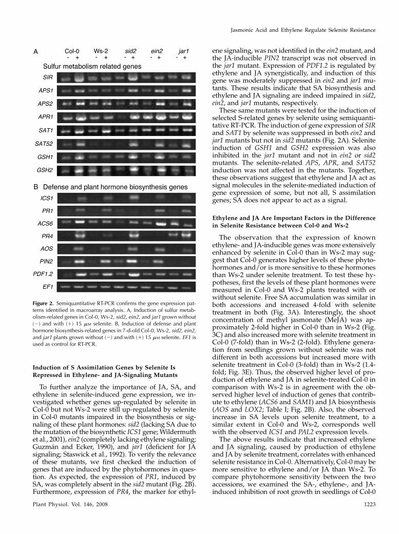
investigated by determining their expression profilesusing semiquantitative reverse transcription (RT)-PCR. The mRNA levels of SIR, APS1, APR1, SAT52,GSH1, and GSH2 again increased only with selenitetreatment in Col-0 but not in Ws-2 (Fig. 2A). Inductionof APR2 and SAT1 with selenite in Ws-2 was lowerthan in Col-0 (Fig. 2A). Therefore, the results from themacroarray and semiquantitative RT-PCR approachesboth indicate that the expression of several key genesinvolved in S uptake and assimilation are more en-hanced upon selenite treatment in Col-0 than in Ws-2.In this context, it is interesting to note that both APS1and a SAT gene (SAT1) are located in the chromosome3 quantitative trait loci region that was shown earlierto be associated with selenate tolerance in Col (Zhanget al., 2006b). Coordinated induction of S uptake andassimilation genes, including Sultr2;1, APR2, APR3,and GSH1, has also been observed in S-starved plants(for review, see Hirai and Saito, 2004), suggesting thatpart of the selenite responses observed in this exper-iment could be induced by S deficiency. However, theselenite-induced genes encoding homologs of seleno-proteins (At1g05720, At3G22740, and At3g47300) and aSe-binding protein (At5g40415) were not induced by Sdeficiency. Thus, the induction of some genes by selenitetreatment may occur via S starvation, while others maybe induced in a different, selenite-specific response.
Levels of Selenite-Induced Expression in Ethylene- andJA-Modulated Genes Are Higher in Col-0 Than in Ws-2
Earlier studies have shown that S deficiency inducesthe expression of 12-oxophytodienoate reductase, in-volved in JA biosynthesis (Hirai et al., 2003; Nikiforovaet al., 2003). JA is known to be a plant hormone whoseproduction is induced by various environmentalstresses (Pieterse and van Loon, 1999). In addition toJA, salicylic acid (SA) and ethylene are also known asstress-inducible phytohormones (Nurnberger andScheel, 2001). To study the involvement of JA, SA,and ethylene in selenite resistance, the selenite-relatedexpression of genes either involved in the biosynthesisof these phytohormones or known to be responsive to
these hormones was analyzed in the two accessionsusing macroarrays. The selenite-induced expressionof all SA-related genes tested, i.e. the biosyntheti-cally involved isochorismate synthase 1 (ICS1) andPhe ammonia-lyase 2 (PAL2), and the SA-responsivepathogenesis-related protein 1 (PR1), PR2, PR5, enhanceddisease susceptibility 1 (EDS1), and EDS5 were all sim-ilar in Col-0 and Ws-2 (Supplemental Table S1). In con-trast, the induction levels of genes known to be involvedin ethylene or JA biosynthesis, i.e. 1-aminocyclopropane-1-carboxylate synthase 6 (ACS6), S-adenosyl-Met syn-thetase 1 (SAM1), lipooxigenase 2 (LOX2), and alleneoxide synthase (AOS), were remarkably higher inCol-0 than in Ws-2 (Table I). Moreover, induction ofethylene response factor 1 (ERF1), PR4, plant defensin1.2 (PDF1.2), vegetative storage protein 1 (VSP1), pro-teinase inhibitor 2 (PIN2), and JA-responsive gene (JR),which are responsive to ethylene and/or JA, was alsosignificantly more pronounced in Col-0 (Table I). Toconfirm the selenite-induced gene expression observedfrom macroarray experiments with an independentexperimental approach and biological replicate, semi-quantitative RT-PCR was performed for selectedgenes, which were indeed significantly more inducedin Col-0 than in Ws-2 by the selenite treatment. Con-sistent with the macroarray data, the expression ofgenes ICS1 and PR1 was increased in both accessions,and the expression of genes ACS6, PR4, AOS, PIN2,and PDF1.2 was more induced by selenite in Col-0 thanin Ws-2 (Fig. 2B). The expression of PR4 is known to beinduced by ethylene (Lawton et al., 1994) and widelyused as a good marker for ethylene signaling. Induc-tion of the PIN2 gene is JA dependent (Farmer et al.,1992), and concomitant triggering of the ethyleneand JA pathways is required for PDF1.2 induction(Penninckx et al., 1998). ACS is the rate-limiting en-zyme and governs the major regulatory step in stress-induced ethylene production (Yang and Hoffman, 1984;Bleecker and Kende, 2000), and AOS is also a keyenzyme in JA synthesis (Stenzel et al., 2004). Together,these results show that ethylene and JA biosynthesisand/or signaling are induced by selenite in Col-0 butmuch less in Ws-2.
Table I. (Continued from previous page.)
Gene Annotation MIPS Code
Col-0 (Fold-Induction) Ws-2 (Fold-Induction)
P ValueExperiment 1 Experiment 2Average SD
Experiment 1 Experiment 2Average SD
Spot 1 Spot 2 Spot 1 Spot 2 Spot 1 Spot 2 Spot 1 Spot 2
Others
CA1 Carbonic anhydrase 1 At3g01500 2.40 1.22 3.07 1.37 2.01 0.88 0.80 0.84 0.81 1.08 0.88 0.13 0.040
PE3 Pectinesterase family protein At5g04960 2.34 1.71 3.50 2.31 2.46 0.75 1.02 1.45 1.13 1.59 1.30 0.27 0.023
SAUR Auxin-responsive family protein At2g46690 2.41 2.13 2.61 1.89 2.26 0.32 0.98 0.76 1.00 1.45 1.05 0.29 0.001
APX1 Cytosolic ascorbate peroxidase At1g07890 2.72 3.41 2.87 3.03 3.01 0.30 0.73 0.90 0.86 1.16 0.91 0.18 0.000
APX4 Ascorbate peroxidase,
thylakoid-bound (tAPX)
At1g77490 3.78 2.09 3.08 1.74 2.67 0.93 1.23 1.80 1.10 1.98 1.53 0.43 0.043
cytDHAR Dehydroascorbate reductase,
cytosol
At1g75270 2.10 2.68 2.99 1.52 2.32 0.65 1.04 1.46 0.92 1.38 1.20 0.26 0.017
MSD1 Mitochondrial, similar to MnSOD At3g10920 1.64 2.83 1.62 2.28 2.09 0.58 0.78 1.69 1.10 1.75 1.33 0.47 0.045
Tamaoki et al.
1222 Plant Physiol. Vol. 146, 2008
Induction of S Assimilation Genes by Selenite Is
Repressed in Ethylene- and JA-Signaling Mutants
To further analyze the importance of JA, SA, andethylene in selenite-induced gene expression, we in-vestigated whether genes up-regulated by selenite inCol-0 but not Ws-2 were still up-regulated by selenitein Col-0 mutants impaired in the biosynthesis or sig-naling of these plant hormones: sid2 (lacking SA due tothe mutation of the biosynthetic ICS1 gene; Wildermuthet al., 2001), ein2 (completely lacking ethylene signaling;Guzman and Ecker, 1990), and jar1 (deficient for JAsignaling; Staswick et al., 1992). To verify the relevanceof these mutants, we first checked the induction ofgenes that are induced by the phytohormones in ques-tion. As expected, the expression of PR1, induced bySA, was completely absent in the sid2 mutant (Fig. 2B).Furthermore, expression of PR4, the marker for ethyl-
ene signaling, was not identified in the ein2 mutant, andthe JA-inducible PIN2 transcript was not observed inthe jar1 mutant. Expression of PDF1.2 is regulated byethylene and JA synergistically, and induction of thisgene was moderately suppressed in ein2 and jar1 mu-tants. These results indicate that SA biosynthesis andethylene and JA signaling are indeed impaired in sid2,ein2, and jar1 mutants, respectively.
These same mutants were tested for the induction ofselected S-related genes by selenite using semiquanti-tative RT-PCR. The induction of gene expression of SIRand SAT1 by selenite was suppressed in both ein2 andjar1 mutants but not in sid2 mutants (Fig. 2A). Seleniteinduction of GSH1 and GSH2 expression was alsoinhibited in the jar1 mutant and not in ein2 or sid2mutants. The selenite-related APS, APR, and SAT52induction was not affected in the mutants. Together,these observations suggest that ethylene and JA act assignal molecules in the selenite-mediated induction ofgene expression of some, but not all, S assimilationgenes; SA does not appear to act as a signal.
Ethylene and JA Are Important Factors in the Differencein Selenite Resistance between Col-0 and Ws-2
The observation that the expression of knownethylene- and JA-inducible genes was more extensivelyenhanced by selenite in Col-0 than in Ws-2 may sug-gest that Col-0 generates higher levels of these phyto-hormones and/or is more sensitive to these hormonesthan Ws-2 under selenite treatment. To test these hy-potheses, first the levels of these plant hormones weremeasured in Col-0 and Ws-2 plants treated with orwithout selenite. Free SA accumulation was similar inboth accessions and increased 4-fold with selenitetreatment in both (Fig. 3A). Interestingly, the shootconcentration of methyl jasmonate (MeJA) was ap-proximately 2-fold higher in Col-0 than in Ws-2 (Fig.3C) and also increased more with selenite treatment inCol-0 (7-fold) than in Ws-2 (2-fold). Ethylene genera-tion from seedlings grown without selenite was notdifferent in both accessions but increased more withselenite treatment in Col-0 (3-fold) than in Ws-2 (1.4-fold; Fig. 3E). Thus, the observed higher level of pro-duction of ethylene and JA in selenite-treated Col-0 incomparison with Ws-2 is in agreement with the ob-served higher level of induction of genes that contrib-ute to ethylene (ACS6 and SAM1) and JA biosynthesis(AOS and LOX2; Table I; Fig. 2B). Also, the observedincrease in SA levels upon selenite treatment, to asimilar extent in Col-0 and Ws-2, corresponds wellwith the observed ICS1 and PAL2 expression levels.
The above results indicate that increased ethyleneand JA signaling, caused by production of ethyleneand JA by selenite treatment, correlates with enhancedselenite resistance in Col-0. Alternatively, Col-0 may bemore sensitive to ethylene and/or JA than Ws-2. Tocompare phytohormone sensitivity between the twoaccessions, we examined the SA-, ethylene-, and JA-induced inhibition of root growth in seedlings of Col-0
Figure 2. Semiquantitative RT-PCR confirms the gene expression pat-terns identified in macroarray analysis. A, Induction of sulfur metab-olism-related genes in Col-0, Ws-2, sid2, ein2, and jar1 grown without(2) and with (1) 15 mM selenite. B, Induction of defense and planthormone biosynthesis-related genes in 7-d-old Col-0, Ws-2, sid2, ein2,and jar1 plants grown without (2) and with (1) 15 mM selenite. EF1 isused as control for RT-PCR.
Jasmonic Acid and Ethylene Regulate Selenite Resistance
Plant Physiol. Vol. 146, 2008 1223

and Ws-2. Note that rather than ethylene gas, its pre-cursor, 1-aminocyclopropane-1-carboxylate (ACC), wassupplied. As shown in Supplemental Figure S2, thedegree of inhibition was not statistically different be-tween Col-0 and Ws-2 (approximately 20% and 45%inhibition at 1 and 10 mM SA; approximately 20% and32% inhibition at 1 and 0.1 mM MeJA; approximately15% and 42% inhibition at 0.1 and 1 mM ACC, respec-tively). This suggests that the enhanced ethylene and JAsignaling in Col-0 was not due to higher ethylene or JAsensitivity.
To assess whether the selenite resistance of thesensitive accession Ws-2 is limited by its lower ethyl-ene or JA concentration, we next tested whether sel-enite sensitivity could be mitigated via external supplywith MeJA or ACC. Indeed, the selenite toleranceindex of Ws-2 increased with increasing MeJA contentin the medium (Fig. 3D). As a result, no significantdifference in selenite tolerance index was observedany more between Col-0 and Ws-2 when grown on 0.5or 1 mM MeJA (Fig. 3D). Treatment with ACC resultedin a similar trend. Accession Ws-2 showed lowerresistance to selenite than Col-0 without ACC, but itsselenite tolerance index increased with increasingACC content in the media, and it became the sameas Col-0 when the plants were grown at or above 0.5mM ACC (Fig. 3F). In contrast, growing the plants on aseries of different concentrations of SA had no positiveeffect on the selenite tolerance index of Ws-2 (Fig. 3B);Col-0 was even significantly inhibited by external SA(Fig. 3B).
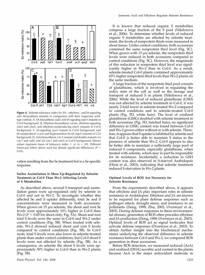
The importance of SA, ethylene, and JA for acqui-sition of selenite resistance in Arabidopsis was alsoinvestigated using mutants with defects in phytohor-mone biosynthesis or signaling. Mutants incapable ofSA production (sid2) or signaling (npr1; Cao et al.,1997) showed no statistical difference in selenite tol-erance index in comparison to their wild type, Col-0(Fig. 4A). In contrast, selenite resistance in mutantsdefective in ethylene production (acs6) or signaling(ein3 and ein2) was less than that in wild-type Col-0(Fig. 4B). Furthermore, an ethylene-overproducingmutant in the Col-0 background, eto1, showed higherresistance than wild-type Col-0 (Fig. 4B). Seleniteresistance in a mutant deficient for JA signaling, jar1,was significantly lower than in Col-0 (Fig. 4C). Con-versely, JA-hypersensitive mutant iop1 (Penninckxet al., 2003) and the constitutive JA-producing mutantcas1 (Kubigsteltig and Weiler, 2003) were more tolerantto selenite than their wild type, C24 (Fig. 4C). Takentogether, these results indicate that the selenite sus-ceptibility of Ws-2 is highly dependent on ethyleneand JA biosynthesis, while there is no evidence of SAinvolvement.
In view of the finding that both ethylene and JAappear to play important roles in selenite resistance inArabidopsis, it is interesting to note that in earlierstudies JA was implicated to be involved in the regu-lation of S metabolism because S starvation inducedgenes involved in JA synthesis, as well as JA-responsivegenes (Hirai et al., 2003; Maruyama-Nakashita et al.,2003; Nikiforova et al., 2003). The above-mentionedstudies showed no indication that ethylene plays a rolein S deficiency responses. However, a recent compre-hensive gene expression analysis showed that tran-scripts regulating ethylene synthesis (ACS6) andsignaling (ERF) were up-regulated by selenate treat-ment, and plants overexpressing ERF1 exhibited anincrease in selenate resistance (Van Hoewyk et al.,2008). These results indicate that Se resistance achievedthrough ethylene signaling is not mediated by S star-
Figure 3. Effect of selenite supply on endogenous SA (A), MeJA (C), andethylene (E) content in Col-0 and Ws-2, and effect on selenite toleranceindex of exogenous supply with SA (B), MeJA (D), and ACC (F). Theplants shown in A, C, and E were grown for 7 d without (white bars) orwith (black bars) treatment with 15 mM selenite. Values are means 6 SD
from three replicate samples, each consisting of several seedlings. Forthe results shown in B, D, and F, Col-0 (black circles) and Ws-2 (whitecircles) plants were grown on control medium or on medium with 15mM selenite that also contained various concentrations of SA (B), MeJA(D), or ACC (F). After 7 d of growth, root length was measured andselenite resistance index was calculated. Values are means 6 SD (n 5
20). Different lowercase letters above each bar denote significantdifferences (P , 0.05).
Tamaoki et al.
1224 Plant Physiol. Vol. 146, 2008
vation resulting from the Se treatment but is a Se-specificresponse.
Sulfur Assimilation Is More Up-Regulated by SeleniteTreatment in Col-0 Than Ws-2 Affecting Levelsof S Metabolites
As described above, several S transport and assim-ilation genes were up-regulated only by selenite inCol-0 and not in Ws-2. To investigate whether thisaffected Se and S uptake differently, total Se and Sconcentrations were measured in both accessions.When grown on 15 mM selenite, the shoot and root Selevels were approximately 10% higher in Col-0 thanWs-2 (P , 0.05 for shoot only; Fig. 5A). Shoot and roottotal S levels were the same in Col-0 and Ws-2 undercontrol conditions (Fig. 5B). When treated with sele-nite, Ws-2 showed reduced shoot and root S levelscompared to control conditions (Fig. 5B). In Col-0roots, total S levels were also lower in selenite-treatedplants than under control conditions, but Col-0 shoot Slevels were not affected by selenite (Fig. 5B). As aconsequence, on selenite the shoot S levels were ap-proximately 50% higher in Col-0 than in Ws-2 plants(Fig. 5B).
It is known that reduced organic S metabolitescompose a large fraction of the S pool (Nikiforovaet al., 2006). To determine whether levels of reducedorganic S metabolites are affected by selenite treat-ment, the levels of nonprotein thiols were measured inshoot tissue. Under control conditions, both accessionscontained the same nonprotein thiol level (Fig. 5C).When grown with 15 mM selenite, the nonprotein thiollevels were reduced in both accessions compared tocontrol conditions (Fig. 5C). However, the magnitudeof this reduction in nonprotein thiol level was signif-icantly higher in Ws-2 than in Col-0. As a result,selenite-treated Col-0 plants contained approximately65% higher nonprotein thiol levels than Ws-2 plants onthe same medium.
A large fraction of the nonprotein thiol pool consistsof glutathione, which is involved in regulating theredox state of the cell as well as the storage andtransport of reduced S in plants (Nikiforova et al.,2006). While the level of reduced glutathione (GSH)was not affected by selenite treatment in Col-0, it wasnearly 2-fold lower in selenite-treated Ws-2 comparedto control conditions and to selenite-treated Col-0plants (Fig. 5D, white bars). The level of oxidizedglutathione (GSSG) doubled with selenite treatment inboth accessions (Fig. 5D, hatched bars); no significantdifference in GSSG content was found between Col-0and Ws-2 grown either without or with selenite. There-fore, it appears that S uptake is inhibited by selenite andthat Col-0 is better able to maintain S status in thepresence of selenite than Ws-2. Col-0 appears to alsobe better able to maintain a sufficiently large pool ofreduced S compounds, especially glutathione, whentreated with selenite, which may in part be responsiblefor its resistance. Incidentally, a reduction in GSHcontent was also observed in S-starved Arabidopsis(Hirai et al., 2003), indicating that selenite treatmentinduced S starvation in Ws-2 plants.
Optimal Levels of ROS Are Necessary forSelenite Resistance
From the experiments described above, it appearsthat ethylene and JA play important roles in seleniteresistance in Arabidopsis. Ethylene and JA are knownto be required for plant defense responses such aspathogen attack, drought stress, and resistance to airpollutants (Dong, 1998; Zhu, 2002; Overmyer et al.,2003). During defense responses to these environmen-tal stresses, generation of ROS often precedes ethyleneand JA production (Dong, 1998; Overmyer et al., 2003).Optimal levels of ROS act as signal molecules thatactivate defense responses (Overmyer et al., 2003). Toobtain further insight into the biochemical mecha-nisms underlying the observed difference in seleniteresistance between Col-0 and Ws-2, we compared ROSgeneration in these accessions.
Before ROS detection, we measured reduced (AsA)and oxidized (DHA) ascorbic acid content in the plantsbecause AsA is the major antioxidant molecule in
Figure 4. Selenite tolerance index for SA-, ethylene-, and JA-signalingand -biosynthesis mutants in comparison with their respective wild-type controls. A, SA-biosynthesis (sid2) and SA-signaling (npr1) mutants inCol-0 background. B, Ethylene-biosynthesis (acs6), ethylene-signaling(ein3 and ein2), and ethylene-overproducing (eto1) mutants in Col-0background. C, JA-signaling (jar1) mutant in Col-0 background, andJA-overproducer (cas1) and hypersensitive for JA (iop1) mutants in C24background. D, AsA-biosynthesis (vtc1) mutant and double mutants v/s(vtc1 and sid2) and v/e (vtc1 and ein2) in Col-0 background. Shownvalues represent mean of tolerance index 6 SD (n 5 20). Differentlowercase letters above each bar denote significant differences (P ,
0.05).
Jasmonic Acid and Ethylene Regulate Selenite Resistance
Plant Physiol. Vol. 146, 2008 1225

plants (Smirnoff et al., 2001). The AsA content wassimilar in the two accessions and not affected byselenite treatment (Fig. 6A). The level of DHA wasalso similar in both accessions under control condi-tions but increased significantly in Col-0 when plantswere treated with selenite, in contrast to Ws-2 (Fig. 6A).The vtc1 mutant (Conklin et al., 1996) that is impairedin AsA production was also used in this experiment.As expected, the vtc1 mutant contained significantly
lower levels of AsA compared to the wild type (Col-0),which were not affected by selenite treatment; theDHA level was also reduced under control conditionsbut increased with selenite treatment, similar to theCol-0 wild type (Fig. 6A). The AsA redox state waslower (i.e. more oxidized) in the presence of selenite inall plant types (Fig. 6B). Under control conditions, theAsA redox state was similar in Col-0 and Ws-2, but dueto the higher DHA level in selenite-treated Col-0, theAsA redox state was significantly lower in Col-0 thanin Ws-2 on selenite medium (Fig. 6B). The AsA redoxstate in the vtc1 mutant was significantly lower than inCol-0 and Ws-2, both with and without selenite.
To assay cellular hydrogen peroxide and superoxideaccumulation, we performed in situ ROS detection asshown in Figure 6, C to E. The top row shows theaccumulation of hydrogen peroxide in Col-0 (C), Ws-2(D), and vtc1 (E). Hydrogen peroxide is visualized insitu as a reddish-brown precipitate, as 3,3#-diamino-benzidine (DAB) polymerizes on contact with hydro-gen peroxide in a reaction requiring peroxidase (Torreset al., 2002). Brown precipitates were observed inselenite-treated Col-0, Ws-2, and vtc1 leaves, but al-most no stain was detected in untreated plants. Thedensity of brown precipitates in selenite-treated Col-0appears higher than that in Ws-2 (Fig. 6, A and B). Theleaves of the vtc1 mutant were stained more than Col-0and Ws-2, likely due to its lack of AsA and low ROS-scavenging ability (Fig. 6B). The bottom row shows theaccumulation of superoxide in Col-0 (C), Ws-2 (D),and vtc1 (E), monitored in situ via the precipitation ofpurple formazan from the reaction of nitro blue tetra-zolium (NBT) with superoxide. Similar to the DABstaining, selenite-treated plants showed more super-oxide than control plants, and Col-0 accumulatedmore superoxide than Ws-2; as expected, a high levelof superoxide was also detected in the selenite-treatedvtc1 mutant. Thus, selenite treatment resulted in the
Figure 6. Effect of selenite treatment on the AsAcontent, redox state, and generation of ROS. A, ShootAsA content. White bars, Reduced form of AsA; blackbars, oxidized form of AsA. Values are means 6 SD
from three replications. B, AsA redox state, calculatedas reduced/oxidized AsA. Col-0 (C), Ws-2 (D), andvtc1 (E) plants were grown for 7 d on media with orwithout 15 mM selenite. In situ detection of the ROShydrogen peroxide and superoxide in leaves wascarried out with DAB and NBT staining, respectively.The presence of the brown precipitate and the purpleformazan precipitate indicates the location of hydro-gen peroxide and superoxide, respectively. F, Induc-tion of selected genes in 7-d-old Col-0 and vtc1plants grown without and with 15 mM selenite. Valuesare means 6 SD from three replicate samples, eachconsisting of several seedlings. Different lowercaseletters above each bar denote significant differences(P , 0.05).
Figure 5. Se, S, nonprotein thiols, and glutathione content in Arabi-dopsis. Col-0 and Ws-2 plants were grown on media without (whitebars) and with (black bars) 15 mM selenite. For results shown in A and B,seedlings were treated with Se for 1 month and Se (A) and S (B) wereanalyzed in shoot and root tissues. For results shown in C and D,seedlings were treated with Se for 7 d, and shoot tissues were analyzedfor nonprotein thiols (C) and reduced (white bars) and oxidized(hatched bars) glutathione (D). Values are means 6 SD from threereplicate samples, each consisting of several seedlings. Different low-ercase letters above each bar denote significant differences (P , 0.05).
Tamaoki et al.
1226 Plant Physiol. Vol. 146, 2008
formation of hydrogen peroxide and superoxide, andthis response was more pronounced in Col-0 than Ws-2.
The observation that the selenite-resistant accessionCol-0 generates more ROS than selenite-sensitive Ws-2when plants are treated with selenite may suggest thatthe generation of ROS is important for acquisition ofselenite resistance. If this is the case, the vtc1 mutantthat generates higher levels of ROS than Col-0 mayalso show enhanced selenite resistance. However, thevtc1 mutant actually showed lower selenite resistancethan Col-0 (Fig. 4D). It has been reported that extremelevels of ROS can trigger the production of SA that inturn leads to more ROS via a self-amplifying loop,activating the oxidative cell death cycle (Overmyeret al., 2003; Kangasjarvi et al., 2005). Indeed, a previousstudy showed that the vtc1 mutant produces highlevels of SA after inoculation with a pathogen (Barthet al., 2004). To assess whether a high level of SA wasalso present in the selenite-treated vtc1 mutant, wecarried out semiquantitative RT-PCR to evaluate theexpression of the PR1 gene, which encodes the SA-responsive PR1, which is frequently used as a markerfor SA signaling (Cao et al., 1997). Selenite treatmentenhanced PR1 expression in Col-0 and vtc1, but in-duction of PR1 in vtc1 was higher than in Col-0 (Fig.6F), suggesting that selenite induces SA productionand that this effect is more pronounced in the vtc1mutant, perhaps because of its higher ROS levels. Tofurther investigate whether the reduced selenite resis-tance of the vtc1 mutant is due to higher levels ofselenite-induced SA production, we tested the seleniteresistance of two double mutants: a vtc1/ein2 mutantthat lacks AsA biosynthesis and ethylene signaling,and a vtc1/sid2 mutant lacking AsA and SA biosyn-thesis. The selenite resistance in the vtc1/ein2 mutantwas not different from the vtc1 mutant, but the seleniteresistance in the vtc1/sid2 mutant was restored to thelevel of Col-0 (Fig. 4D). The finding that the vtc1mutant, which produces more SA when treated withselenite, is more selenite sensitive but if SA productionis knocked out in vtc1/sid2 selenite then resistance isrestored, suggests that SA inhibits the acquisition ofselenite resistance. Indeed, we show that treatment ofSA in Col-0 inhibited selenite resistance (see Fig. 3B).The next intriguing question is how the SA inhibitsselenite resistance in plants. Our results indicate SAmay inhibit JA and/or ethylene signaling because weobserved that selenite induction of PR4 (a marker forethylene signaling) and PIN2 (a marker for JA signal-ing) in Col-0 was completely repressed in the vtc1mutant (Fig. 6F). Inhibition of JA and/or ethylenesignaling by SA was also observed earlier in plantssuffering biotic and abiotic stresses (Pena-Cortes et al.,1993; Doares et al., 1995; Berrocal-Lobo et al., 2002).Therefore, it appears that in the selenite-treated vtc1mutant, an increase in SA production inhibited JA andethylene signaling, leading to impaired Se resistance.Indeed, the vtc1 mutant showed no selenite-relatedinduction of SIR and SAT1, whose expression is regu-lated by JA and/or ethylene (see Figs. 2A and 6F).
Taken together, our results suggest that an excesslevel of ROS production such as in the vtc1 mutantleads to a high level of SA, which inhibits JA andethylene signaling, thereby impeding S assimilationand selenite resistance. However, a low ROS responsesuch as in Ws-2 appears to be associated with a low Seresistance as well. Thus, an optimal level of ROS maybe needed to acquire selenite resistance. Recently, ROSinduction upon selenite treatment was also observedin a cell suspension of coffee (Gomes et al., 2007). Thecellular mechanisms regulating ROS production inresponse to Se in plants are not clear at this point andwill require further study. In this context, it is inter-esting to note that Zhang et al. (2006a) showed that amolecular marker on chromosome 4 (ciw7) appearedto be linked to selenite resistance in Col. Although noethylene- or JA-related gene is found around themarker, a potential defense-related gene, LSD1-like 2(LOL2; At4g21610), is located close to the marker. LOL2encodes a member of a small family of LSD1 proteinsthat contain three highly related zinc fingers, and mayfunction as either a transcriptional regulator or ascaffold protein (Dietrich et al., 1997). Eppel et al.(2003) showed LOL and LSD1 may function as antag-onistic transcriptional regulators or scaffolds that con-trol attenuation of cell death through regulation ofROS and/or SA production level. In future studies, itwill be interesting to test the involvement of LOL genesin selenite tolerance in Arabidopsis.
CONCLUSION
The results presented here indicate that the higherselenite resistance of accession Col-0 compared to Ws-2is dependent on its higher level of selenite-inducedJA and ethylene synthesis. Selenite-related ROS pro-duction was also higher in Col-0 than in Ws-2. Thismay indicate that JA and ethylene production requirean optimal level of ROS production to lead to Seresistance in plants. The resistance mechanism mayinvolve JA- and ethylene-enhanced S uptake andassimilation, as observed in Col-0. The higher levelsof organic S compounds observed in Col-0 mayenable it to more efficiently prevent Se analogs fromreplacing S in proteins and other S compounds.However, Se levels were also higher in Col-0 plantscompared to Ws-2. It is intriguing to speculate thatthe Se-binding protein homolog that was induced byselenite in Col-0 but not in Ws-2 may play an addi-tional role in alleviating Se toxicity. This would be inagreement with the study by Agalou et al. (2005),where overexpression of Se-binding protein resultedin enhanced Se resistance. Another possibility forincreasing Se resistance in Col-0 is caused by itshigher antioxidant levels, because higher Se causesthe extra oxidative stress. JA might also be involvedin this process because GSH and AsA biosyntheticpathways were enhanced after MeJA treatment (Sasaki-Sekimoto et al., 2005).
Jasmonic Acid and Ethylene Regulate Selenite Resistance
Plant Physiol. Vol. 146, 2008 1227
Se is an essential element for animals, includinghumans (Rayman, 2000). The recommended dietaryallowance is 40 to 70 mg/d. However, human diets inseveral countries lack sufficient Se, which leads toenhance susceptibility to cancer, viral infections, andheart problems (Rayman, 2000). On the other hand,soils containing .0.5 mg Se kg21 are considered sel-eniferous, and forage produced on such soils oftencontains more than the maximum permissible level foranimal consumption. Soils with elevated levels of Seare found in many countries, including Australia, China,India, and the United States (Dhillon and Dhillon,2003). Se accumulation by plants may help alleviateboth Se deficiency and toxicity. A better understandingof the mechanisms and rate-limiting factors controllingplant Se uptake and assimilation will be vital for theoptimal use of plants to alleviate dietary Se deficiencyor for cleanup of Se-polluted areas. Several transgenicplants with enhanced Se accumulation and resistancehave already been developed (Pilon-Smits et al., 1999;Agalou et al., 2005; Banuelos et al., 2005, 2007; VanHoewyk et al., 2005) and may be useful for Se phyto-remediation (for review, see Pilon-Smits and Freeman,2006). The new knowledge obtained here of the genesand processes that impact Se accumulation and resis-tance in Arabidopsis may lead to further developmentof plants with increased Se content.
MATERIALS AND METHODS
Plant Materials, Growth Conditions, and Selenite
Resistance Assays
Seeds of Arabidopsis (Arabidopsis thaliana) Col-0, Ws-2, npr1, acs6, ein3,
ein2, eto1, jar1, and vtc1 were obtained from the Arabidopsis Biological
Resource Center (ABRC; Columbus, OH). The sid2 mutant and iop1 mutant
were obtained from Christiane Nawrath (University of Fribourg) and Bart
P.H.J. Thomma (Wageningen University), respectively. C24 and cas1 mutant
were provided from Ines Kubigsteltig, Ruhr-Universitat Bochum. The dou-
ble mutants vtc1/sid2 and vtc1/ein2 were created by selecting F2 individuals
from the cross between vtc1 and sid2 or vtc1 and ein2. For selecting vtc1/sid2
double mutants, F2 plants that showed low levels of AsA were identified as
described by Conklin et al. (2000). F3 lines lacking ozone-inducible SA
accumulation were selected, then the point mutation of the ICS1 gene in the
sid2 mutant was identified with DNA sequencing. For selecting vtc1/ein2
double mutants, F2 plants lacking AsA biosynthesis were identified as de-
scribed above. Ethylene-insensitive F3 lines were selected on plates contain-
ing 20 mM ACC by screening for the lack of the triple response (Guzman and
Ecker, 1990).
Seeds were surface sterilized with 15% bleach and germinated on agar
plates containing 0.53 Murashige and Skoog medium and 1% Suc with or
without added sodium selenite at the indicated concentrations. Seedlings
were grown on plates in a growth chamber at 24�C under a photosynthetic
photon flux density of 150 mmol m22 s21 at a 16-h-light/8-h-dark cycle for 7 d.
For analysis of selenite resistance, plants were vertically grown for 7 d, and
seedling root length was measured from digital photographs of the seedlings
with the Image J program (http://rsb.info.nih.gov/ij/). The Se tolerance
index was calculated as root length in the presence of selenite divided by
mean of root length on control medium 3 100%.
Macroarray Analysis
The expression of 250 genes was studied by custom-made cDNA macro-
array using cDNA clones from the ABRC and RIKEN BioResource Center
(Ibaraki, Japan). These cDNA clones were resequenced for confirmation
before use. PCR-amplified sample was blotted onto Hybond N1 nylon
membranes with MultiPin Blotter 96 (Atto). Each gene was spotted on a
membrane in duplicate. lDNA was used as negative control. The constitu-
tively expressed gene EF1a (At5G12110) was included as internal standard.
For the macroarray studies, Col-0 and Ws-2 plants were grown on agar plates
with or without 15 mM sodium selenite for 7 d. Then shoots were separated
from roots and frozen with liquid nitrogen for total RNA extraction. Total
RNA was extracted using the RNeasy Plant Mini kit (Qiagen). Hybridization,
probe labeling, and signal detection were carried out according to Tamaoki
et al. (2003b). The signal intensity of each spot was obtained as described
previously (Tamaoki et al., 2003b). In brief, we subtracted the value of the
signal intensity of the negative control (lDNA) from the signal intensity of
each spot, then normalized the signal intensities against the intensity of EF1a,
the expression of which had been confirmed to be unchanged with or without
15 mM sodium selenite (see Fig. 2).
Expression Analysis via Semiquantitative RT-PCR
Plants were grown for 7 d on 0.53 Murashige and Skoog medium with or
without 15 mM sodium selenite. Total RNA was isolated from shoots as
described above. Five micrograms of DNase-treated total RNA was reverse
transcribed using the First Strand cDNA synthesis kit (Fermentas Interna-
tional) following the manufacturer’s instructions. PCR reactions were carried
out as described previously (Schiavon et al., 2007). A list of primers used in
these experiments is presented in Supplemental Table S2.
In Vitro Treatments and in Situ ROS Detection
The effect of exogenous SA, ACC, or MeJA on plant selenite resistance was
analyzed by sowing sterilized Arabidopsis seeds on plates containing 0.53
Murashige and Skoog with a range of concentrations of SA, ACC, or MeJA
with or without 15 mM sodium selenite. Seedlings were grown on vertical
plates for 7 d. Seminal root length was measured from digital photographs of
the seedlings with the ImageJ program (http://rsb.info.nih.gov/ij/). The Se
tolerance index was calculated as described above.
Se-induced in situ accumulation of superoxide was detected with NBT
(Boehringer Mannheim) as described by Jabs et al. (1996). To visualize in situ
accumulation of hydrogen peroxide, DAB staining was performed as de-
scribed by Torres et al. (2002).
Measurement of SA, MeJA, and JA
MeJA, salicylate, and jasmonate levels in shoot tissue were determined in
plants grown as described above with or without 15 mM sodium selenite for
7 d. For measurement of SA and MeJA, the extracts were prepared as described
by Wilbert et al. (1998). The extracts were analyzed by liquid chromatography-
mass spectrometry (LC-MS) using a Hewlett-Packard Agilent 1100 series HPLC
and a Finnigan LcQDuo thermoquest MS system equipped with Xcalibur
software. Through 30-mL injections, these extracts were separated at 40�C
using a Phenomenex Hypersil 5-mm C18 (ODS) column (250 3 2 mm, 5 mm)
at a flow rate of 0.32 mL/min using two eluents: A, water 1 0.1% formic
acid; and B, 100% methanol 1 0.1% formic acid. The following gradient
program was used during the 23-min run: 0 to 7 min, 50% A and 50% B; 7 to
9 min, 30% A and 70% B; 9 to 12 min, 100% B; 12 to 13 min, 50% A and 50% B,
with a 10-min postrun, column wash 50% A and 50% B. Standard curves
were established using chemicals purchased from Sigma Chemical; MeJA
(catalog no. 392707) had a retention time of 2.5 min, SA (catalog no. A–6262)
had a retention time of 4.45 min, and jasmonate (catalog no. J2500) had a
retention time of 6.85 min. Through MS, the different metabolites were
measured at their appropriate masses and retention times observed for each
of the standards. The MS detector settings were 1 to 3.5 min in positive ion
mode using parameters generated with the MeJA standard and the auto-
mated tune program, 3.5 to 5.5 min in negative ion mode using parameters
generated with the SA standard and the automated tune program, and 5.5 to
13 min in negative ion mode using parameters generated with the JA
standard and the automated tune program. Samples were kept at room
temperature (25�C) in the autosampler. The previously published (Wilbert
et al., 1998) and observed precursor and product ions for these standards
were exactly the same. These masses and those from the dimmers for MeJA
and JA were used to quantify these compounds.
Tamaoki et al.
1228 Plant Physiol. Vol. 146, 2008

Measurement of Ethylene, Nonprotein Thiols, Ascorbic
Acid, and Glutathione
For measurement of ethylene, 10 seedlings were enclosed in a 60-mL vial
and incubated for 12 h with illumination in a growth chamber. A 25-mL
gaseous phase of the vial was subjected into a Fisions 8000 gas chromatograph
equipped with a flame ionization detector. A 2-m Altec Hayesep N 80/100
column was used with isothermic oven temperature at 70�C and flame
ionization detector at 200�C. The program was 2 min in length with the
ethylene peak running from 1.180 to 1.633 min. Ethylene peak area was
determined by the PeakSimple program (ver. 3.39, 6 channel; SRI Instru-
ments). The amount of ethylene generated from the seedlings was estimated
from the peak area compared to that of ethylene standard.
Measurement of nonprotein thiol levels was performed using Ellman’s
reagent as described (Zhu et al., 1999). To measure total AsA, DHA, GSH, and
GSSG levels, 100 mg of fresh plants was homogenized in 2 mL of cold 5%
(w/v) metaphosphoric acid with sea sand. The AsA and DHA contents were
quantified as described previously (Tamaoki et al., 2003a). GSH and GSSG
contents were measured as described by Yoshida et al. (2006). All experiments
were carried out in three replicates, each consisting of 20 to 30 pooled plants.
Quantification of Se and S Accumulation
Col-0 and Ws-2 plants were grown on 0.53 Murashige and Skoog agar
medium with or without 15 mM selenite in a growth chamber for 3 weeks. Root
and shoot materials were harvested separately, rinsed with distilled water,
and dried at 37�C for a week. Three replicates consisting of 30 to 50 seedlings
were acid-digested and analyzed for Se and S by inductively coupled plasma
atomic emission spectrometry as described by Pilon-Smits et al. (1999).
Supplemental Data
The following materials are available in the online version of this article.
Supplemental Figure S1. Identified up-regulated selenite-responsive
genes (P , 0.05, fold change .2) that are categorized with biological
function.
Supplemental Figure 2. Sensitivity to SA, ACC, and MeJA in seedlings of
Col-0 and Ws-2.
Supplemental Table S1. Macroarray study showing induction of genes in
Col-0 and Ws-2 by 15 mM selenite treatment.
Supplemental Table S2. Primers used in semiquantitative RT-PCR.
ACKNOWLEDGMENTS
We are grateful to Dr. Christiane Nawrath and Dr. Silvia Heck (University
of Fribourg, Fribourg, Switzerland) for the gift of the sid2 mutant and to
Dr. Bart P.H.J. Thomma (Wageningen University, Wageningen, Netherlands)
for providing the iop1 mutant. We also thank Dr. Ines Kubigsteltig (Ruhr-
Universitat Bochum, Bochum, Germany) for providing C24 and the cas1
mutant.
Received October 10, 2007; accepted December 22, 2007; published January 4,
2008.
LITERATURE CITED
Agalou A, Roussis A, Spaink HP (2005) The Arabidopsis selenium-binding
protein confers resistance to toxic levels of selenium. Funct Plant Biol 31:
881–890
Banuelos G, LeDuc DL, Pilon-Smits EAH, Terry N (2007) Transgenic
Indian mustard overexpressing selenocysteine lyase or selenocysteine
methyltransferase exhibit enhanced potential for selenium phytoreme-
diation under field conditions. Environ Sci Technol 41: 599–605
Banuelos G, Terry N, LeDuc DL, Pilon-Smits EAH, Mackey B (2005) Field
trial of transgenic Indian mustard plants shows enhanced phytoreme-
diation of selenium-contaminated sediment. Environ Sci Technol 39:
1771–1777
Barth C, Moeder W, Klessig DF, Conklin PL (2004) The timing of senes-
cence and response to pathogens is altered in the ascorbate-deficient
Arabidopsis mutant vitamin c-1. Plant Physiol 134: 1784–1792
Berrocal-Lobo M, Molina A, Solano R (2002) Constitutive expression of
ETHYLENE-RESPONSE-FACTOR in Arabidopsis confers resistance to
several necrotrophic fungi. Plant J 29: 23–32
Birringer M, Pilawa S, Flohe L (2002) Trends in selenium biochemistry. Nat
Prod Rep 19: 693–718
Bleecker AB, Kende H (2000) Ethylene: a gaseous signal molecule in
plants. Annu Rev Cell Dev Biol 16: 1–18
Cao H, Glazebrook J, Clarke JD, Volko S, Dong X (1997) The Arabidopsis
NPR1 gene that controls systemic acquired resistance encodes a novel
protein containing ankyrin repeats. Cell 88: 57–63
Conklin PL, Saracco SA, Susan R, Norris SR, Last RL (2000) Identification
of ascorbic acid-deficient Arabidopsis thaliana mutants. Genetics 154:
847–856
Conklin PL, Williams EH, Last RL (1996) Environmental stress sensitivity
of an ascorbic acid deficient Arabidopsis mutant. Proc Natl Acad Sci USA
93: 9970–9974
Doares SH, Narvaez-Vasquez J, Conconi A, Ryan CA (1995) Salicylic acid
inhibits synthesis of proteinase inhibitors in tomato leaves induced by
systemin and jasmonic acid. Plant Physiol 108: 1741–1746
Dhillon KS, Dhillon SK (2003) Distribution and management of selenif-
erous soils. Adv Agron 79: 119–184
Dietrich RA, Richberg MH, Schmidt R, Dean C, Dangl JL (1997) A novel
zinc finger protein is encoded by the Arabidopsis LSD1 gene and
functions as a negative regulator of plant cell death. Cell 88: 685–694
Dong X (1998) SA, JA, ethylene, and disease resistance in plants. Curr Opin
Plant Biol 1: 316–323
Eppel P, Mack AA, Morris VRF, Dangl JL (2003) Antagonistic control of
oxidative stress-induced cell death in Arabidopsis by two related, plant-
specific zinc finger proteins. Proc Natl Acad Sci USA 100: 6831–6836
Eustice DC, Kull FJ, Shrift A (1981) Selenium toxicity: aminoacylation
and peptide bond formation with selenomethionine. Plant Physiol 67:
1054–1058
Farmer EE, Johnson RR, Ryan CA (1992) Regulation of expression of
proteinase inhibitor genes by methyl jasmonate and jasmonic acid. Plant
Physiol 98: 995–1002
Feist LJ, Parker DR (2001) Ecotypic variation in selenium accumulation
among populations of Stanleya pinnata. New Phytol 149: 61–69
Fu LH, Wang XF, Eyal Y, She YM, Donald LJ, Standing KG, Ben-Hayyim
G (2002) A selenoprotein in the plant kingdom: mass spectrometry
confirms that an opal codon (UGA) encodes selenocysteine in Chlamy-
domonas reinhardtii glutathione peroxidase. J Biol Inorg Chem 277:
25983–25991
Galeas ML, Zhang LH, Freeman JL, Wegner M, Pilon-Smits EAH (2007)
Seasonal fluctuations of selenium and sulfur accumulation in selenium
hyperaccumulators and related non-accumulators. New Phytol 173:
517–525
Gomes RA Jr, Gratao PL, Gaziola SA, Mazzafera PM, Lea PJ, Azevedo RA
(2007) Selenium-induced oxidative stress in coffee cell suspension
cultures. Funct Plant Biol 34: 449–456
Gromer S, Gross JH (2002) Methylseleninate is a substrate rather than an
inhibitor of mammalian thioredoxin reductase. Implications for the
antitumor effects of selenium. J Biol Chem 277: 9701–9706
Guzman P, Ecker JR (1990) Exploiting the triple response of Arabidopsis to
identify ethylene-related mutants. Plant Cell 2: 513–523
Hamilton SJ (2004) Review of selenium toxicity in the aquatic food chain.
Sci Total Environ 326: 1–36
Hira CK, Partal K, Dhillon K (2004) Dietary selenium intake by men and
women in high and low selenium areas of Punjab. Public Health Nutr 7:
39–43
Hirai MY, Fujiwara T, Awazuhara M, Kimura T, Noji M, Saito K (2003)
Global expression profiling of sulfur-starved Arabidopsis by DNA
macroarray reveals the role of O-acetyl-L-serine as a general regulator
of gene expression in response to sulfur nutrition. Plant J 33: 651–663
Hirai MY, Saito K (2004) Post-genomic approaches for the elucidation
of plant adaptive mechanism to sulfur deficiency. J Exp Bot 55:
1871–1879
Jabs T, Dietrich RA, Dangl JL (1996) Initiation of runaway cell death in an
Arabidopsis mutant by extracellular super oxide. Science 273: 1853–1856
Jasmonic Acid and Ethylene Regulate Selenite Resistance
Plant Physiol. Vol. 146, 2008 1229
Kangasjarvi J, Jaspers P, Kollist H (2005) Signalling and cell death in
ozone-exposed plants. Plant Cell Environ 28: 1021–1036
Koornneef M, Alonso-Blanco C, Vregdenhil D (2004) Naturally occurring
genetic variation in Arabidopsis thaliana. Annu Rev Plant Biol 55: 141–172
Kubigsteltig II, Weiler EW (2003) Arabidopsis mutants affected in the
transcriptional control of allene oxide synthase, the enzyme catalyzing
the entrance step in octadecanoid biosynthesis. Planta 217: 748–757
Lauchli A (1993) Selenium in plants: uptake, functions, and environmental
toxicity. Bot Acta 106: 455–468
Lawton KA, Potter SL, Uknes S, Ryals J (1994) Acquired resistance
signal transduction in Arabidopsis is ethylene independent. Plant Cell
6: 581–590
Maruyama-Nakashita A, Inoue E, Watanabe-Takahashi A, Yamaya T,
Takahashi H (2003) Transcriptome profiling of sulphur responsive
genes in Arabidopsis reveals global effects of sulphur nutrition on
multiple metabolic pathways. Plant Physiol 132: 597–605
Nikiforova V, Freitag J, Kempa S, Adamik M, Hesse H, Hoefgen R (2003)
Transcriptome analysis of sulphur depletion in Arabidopsis thaliana:
interlacing of biosynthetic pathways provides response specificity. Plant
J 33: 633–650
Nikiforova VJ, Bielecka M, Gakiere B, Krueger S, Rinder J, Kempa S,
Morcuende R, Scheible WR, Hesse H, Hoefgen R (2006) Effect of sulfur
availability on the integrity of amino acid biosynthesis in plants. Amino
Acids 30: 173–183
Nurnberger T, Scheel D (2001) Signal transmission in the plant immune
response. Trends Plant Sci 6: 372–379
Obata T, Shiraiwa Y (2005) A novel eukaryotic selenoprotein in the
Haptophyte alga Emiliania huxleyi. J Biol Chem 280: 18462–18468
Overmyer K, Brosch _e M, Kangasjarvi J (2003) Reactive oxygen species and
hormonal control of cell death. Trends Plant Sci 8: 335–342
Pena-Cortes H, Albrecht T, Prat S, Weiler EW, Willmitzer L (1993) Aspirin
prevents wound-induced gene expression in tomato leaves by blocking
jasmonic acid biosynthesis. Planta 191: 123–128
Penninckx IAMA, Eggermont K, Schenk PM, Ackerveken GVD, Cammue
BPA, Thomma BPHJ (2003) The Arabidopsis mutant iop1 exhibits in-
duced over-expression of the plant defensin gene PDF1.2 and enhanced
pathogen resistance. Mol Plant Pathol 4: 479–486
Penninckx IAMA, Thomma BPHJ, Buchla A, Metraux JP, Broekaert WF
(1998) Concomitant activation of jasmonate and ethylene response
pathway is required for induction of a plant defensin gene in Arabidop-
sis. Plant Cell 10: 2103–2114
Pickering IJ, Wright C, Bubner B, Ellis D, Persans MW, Yu EY, George
GN, Prince RC, Salt DE (2003) Chemical form and distribution of
selenium and sulfur in the selenium hyperaccumulator Astragalus
bisulcatus. Plant Physiol 131: 1460–1467
Pieterse CMJ, van Loon LC (1999) Salicylic acid-independent plant defence
pathway. Trends Plant Sci 4: 52–58
Pilon-Smits EAH, Freeman LJ (2006) Environmental cleanup using plants:
biotechnological advances and ecological considerations. Front Ecol
Environ 4: 203–210
Pilon-Smits EAH, Hwang S, Lytle CM, Zhu YL, Tai JC, Bravo RC, Chen Y,
Leustek T, Terry N (1999) Overexpression of ATP sulfurylase in Indian
mustard leads to increased selenate uptake, reduction, and tolerance.
Plant Physiol 119: 123–132
Rayman MP (2000) The importance of selenium to human health. Lancet
356: 233–241
Sasaki-Sekimoto Y, Taki N, Obayashi T, Aono M, Matsumoto F, Sakurai
N, Suzuki H, Yokota-Hirai M, Noji M, Saito K, et al (2005) Coordinated
activation of metabolic pathway for antioxidants and defence com-
pounds by jasmonates and their roles in stress tolerance in Arabidopsis.
Plant J 44: 653–668
Schiavon M, Zhang L-H, Abdel-Ghany SE, Pilon M, Malagoli M, Pilon-
Smits EAH (2007) Variation in copper tolerance in Arabidopsis thaliana
accessions Columbia, Landsberg erects and Wassilewskija. Physiol Plant
129: 342–350
Smirnoff N, Conklin PL, Loewus FA (2001) Biosynthesis of ascorbic acid in
plants: a renaissance. Annu Rev Plant Physiol Plant Mol Biol 52: 437–67
Sors TG, Ellis DR, Salt DE (2005) Selenium uptake, translocation, assim-
ilation and metabolic fate in plants. Photosynth Res 86: 373–389
Stadtman TC (1990) Selenium biochemistry. Annu Rev Biochem 59: 111–127
Stadtman TC (1996) Selenocysteine. Annu Rev Biochem 65: 83–100
Staswick PE, Su W, Howell SH (1992) Methyl jasmonate inhibition of root
growth and induction of a leaf protein are decreased in an Arabidopsis
thaliana mutant. Proc Natl Acad Sci USA 89: 6837–6840
Stenzel I, Hause B, Miersch O, Kurz T, Mauzher H, Weichert H, Ziegler J,
Feussner I, Wasternack C (2004) Jasmonate biosynthesis and the allene
oxide cyclase family of Arabidopsis thaliana. Plant Mol Biol 51: 895–911
Tamaoki M, Mukai F, Asai N, Nakajima N, Kubo A, Aono M, Saji H
(2003a) Light-controlled expression of a gene encoding L-galactono-
g-lactone dehydrogenase which affects ascorbate pool size in Arabidopsis
thaliana. Plant Sci 164: 1111–1117
Tamaoki M, Nakajima N, Kubo A, Aono M, Matsuyama T, Saji H (2003b)
Transcriptome analysis of O3-exposed Arabidopsis reveals that multiple
signal pathways act mutually antagonistically to induce gene expres-
sion. Plant Mol Biol 53: 443–456
Terry N, Zayed AM, de Souza MP, Tarun AS (2000) Selenium in higher
plants. Annu Rev Plant Physiol Plant Mol Biol 51: 401–432
Torres MA, Dangl JL, Jones JDG (2002) Arabidopsis gp91phox homologues
AtrbohD and AtrbohF are required for accumulation of reactive oxygen
intermediates in the plant defense response. Proc Natl Acad Sci USA 99:
517–522
Van Hoewyk D, Garifullina GF, Ackley AR, Abdel-Ghany SE, Marcus
MA, Fakra S, Ishiyama K, Inoue E, Pilon M, Takahashi H (2005)
Overexpression of AtCpNifS enhances selenium tolerance and accumu-
lation in Arabidopsis. Plant Physiol 139: 1518–1528
Van Hoewyk D, Takahashi H, Hess A, Tamaoki M, Pilon-Smits EAH
(2008) Transcriptome and biochemical analyses give insights into
selenium-stress responses and selenium tolerance mechanisms in Arabi-
dopsis. Physiol Plant 132: 236–253
White PJ, Bowen HC, Parmagure P, Fritz M, Spracklen WP, Spiby RE,
Meacham MC, Mead A, Harriman M, Trueman LJ, et al (2004) Inter-
action between selenium and sulfur nutrition in Arabidopsis thaliana.
J Exp Bot 55: 1927–1937
Wilber CG (1980) Toxicology of selenium: a review. Clin Toxicol 17:
171–230
Wilbert SM, Ericsson LH, Gordon MP (1998) Quantification of jasmonic
acid, methyl jasmonate, and salicylic acid in plants by capillary liquid
chromatography electrospray tandem mass spectrometry. Anal Bio-
chem 257: 186–194
Wildermuth MC, Dewdney J, Wu G, Ausubel FM (2001) Isochorismate
synthase is required to synthesize salicylic acid for plant defense.
Nature 414: 562–565
Yang SF, Hoffman NE (1984) Ethylene biosynthesis and its regulation in
higher plants. Annu Rev Plant Physiol 35: 155–189
Yoshida S, Tamaoki M, Shikano T, Nakajima N, Ogawa D, Ioki M, Aono
M, Kubo A, Kamada H, Inoue Y, et al (2006) Cytosolic dehydroascor-
bate reductase is important for ozone tolerance in Arabidopsis thaliana.
Plant Cell Physiol 47: 304–308
Zhang LH, Abdel-Ghany SE, Freeman JL, Ackley AR, Schiavon M, Pilon-
Smits EAH (2006a) Investigation of selenium tolerance mechanism in
Arabidopsis thaliana. Physiol Plant 128: 212–223
Zhang LH, Ackley AR, Pilon-Smits EAH (2007) Variation in selenium
tolerance and accumulation among 19 Arabidopsis thaliana accessions.
J Plant Physiol 164: 327–36
Zhang LH, Byrne PF, Pilon-Smits EAH (2006b) Mapping quantitative trait
loci associated with selenate tolerance in Arabidopsis thaliana. New
Phytol 170: 33–42
Zhu JK (2002) Salt and drought stress signal transduction in plants. Annu
Rev Plant Biol 53: 247–273
Zhu YL, Pilon-Smits EAH, Jouanin L, Terry N (1999) Overexpression of
glutathione synthetase in Brassica juncea enhances cadmium tolerance
and accumulation. Plant Physiol 119: 73–79
Tamaoki et al.
1230 Plant Physiol. Vol. 146, 2008