Embed Size (px)
Citation preview

Hemodynamic Effects after
Conversion of Arrhythmias
ROBERTJ. Comass, DAvID H. MCKENNA,CHARLESW. CRUMPTON,andGEORGEG. ROwE
Cardiovascular Research Laboratory, Department of Medicine, University ofWisconsin Medical School, Madison, Wisconsin 53706
A B S T R A C T Systemic and coronary hemody-namic parameters were determined during anarrhythmia and immediately after a direct currenttransthoracic shock given in an attempt to convertthe arrhythmia to a sinus mechanism. No anes-thesia or drugs were administered between the twostudies. 16 patients with atrial fibrillation con-verted to sinus rhythm and five did not. In twopatients with atrial flutter and one with supra-ventricular tachycardia, the arrhythmia was cor-rected. The arrhythmia persisted in a singlepatient with ventricular tachycardia. Utilizing eachpatient as his own control, we compared statisti-cally various hemodynamic parameters before andafter the shock. In addition, the group of patientswhose atrial fibrillation terminated was comparedto the group treated in the same manner but inwhich the atrial fibrillation persisted. Pressures inthe right side of the heart decreased in both groupsso that the changes appeared to be caused byfactors associated with the transthoracic directcurrent shock or the catheterization procedure.The differences between those with atrial fibrilla-tion who converted to sinus rhythm as comparedto those who did not were a decrease in heartrate, an increase in stroke volume, and an increasein cardiac efficiency. There was no immediateeffect on the cardiac output or coronary blood flow.
INTRODUCTION
The effects of arrhythmias on the coronary circu-lation and myocardial metabolism have not been
Received for publication 18 September 1967 and in re-svised form 15 January 1968.
adequately studied in human subjects. The sys-temic hemodynamic effects of arrhythmias havebeen investigated but the observed changes havenot been uniform (1-22). The cardiac output hasbeen reported to vary from no significant changeto more than doubling after conversion of atrialfibrillation to sinus rhythm (16, 9). The pressureresponses as well as other indices also have varied,both in direction and degree of change (6, 8, 15).
The various methods used to evaluate thesecirculatory changes have been criticized (14-16).The most frequent criticisms concern the time in-terval between the studies performed during thearrhythmia and after its conversion, and the ad-ministration of hemodynamically active drugs,such as quinidine or general anesthesia betweenthe two studies. Another unknown is the effect ofa transthoracic direct current shock when thismethod is used for conversion.
The present study was performed to evaluatethe effect of arrhythmias on the coronary circula-tion, myocardial metabolism, and cardiac efficiency.Additional information was also obtained regard-ing systemic hemodynamic effects of the arrhyth-mias and of the transthoracic direct current shockused for rhythm conversion. The protocol wasdevised to eliminate or control the variables men-tioned above.
METHODS
Studies were performed on 25 subjects ranging in agefrom 23 to 59 yr. Further information concerning age,sex, arrhythmia, and diagnosis is presented in Table I,as are the results of the attempt to change their rhythm.All patients were given a maintenance dose of quinidinesulfate for 24-48 hr before the study and, if they had
1774 The Journal of Clinical Investigation Volume 47 1968

Atrial Fibrillotion D CShock
FA
PA
LA
Sinus Rhythm (RBBB)
FIGURE 1 Simultaneous pressures in left atrium, left ventricle, pulmonary artery and femoral ar-
tery immediately before and after the electrical conversion of atrial fibrillation in a patient withmitral stenosis. Shaded area, mitral diastolic gradient.
been taking digitalis, it was withheld for at least 24 hr.Cardiac catheterization was performed in the postab-
sorptive state using 1%o lidocaine hydrochloride for localanesthesia. A Cournand needle was inserted into the fe-moral artery and cardiac catheters were placed fluoro-scopically in the pulmonary artery and coronary sinus
(CS). In seven subjects, transseptal catheterizations as
well as retrograde arterial (A) catheterizations were
performed so, in these subjects, there were catheters inthe pulmonary artery, coronary sinus, left ventricle,and left atrium throughout the study. Cardiac output was
determined by the Fick principle and coronary bloodflow (CBF) by the nitrous oxide saturation method.Analyses on the blood for oxygen and carbon dioxidewere done by the Van Slyke-Neill method. The methodof Orcutt and Waters was used for nitrous oxide (23).
The systemic arterial, pulmonary arterial, and coronary
sinus blood pressures were measured in all patients withStatham P23D or DB strain-gauge pressure transducersand recorded on a Waters photographic recorder. Leftatrial, left ventricular, and central aortic pressures were
recorded in patients with left-sided catheterizations.Slow-period galvanometers were used for determiningmean pressures. An index of cardiac efficiency was cal-culated in each group by relating external left ven-
tricular work to left ventricular oxygen consumption as
follows:
left ventricular work indexleft ventricular oxygen usage
When the procedure was underway and before anyhemodynamic measurements were made, meperidine was
Hemodynamic Effects after Conversion of Arrhythmias
- 150
- 100
C 5C
- 0E
. E
1775

TABLE IClinical Data and Systemic Hemodynamic Effects on
Patient Rhythm SVINo. Age Sex Diagnosis before shock %A
yr mlIIm2 b
Patients converted to sinus rhythm with DCshock55 M ASHDp
Infarct, 4 yr38 M ASHDp
Infarct, 3 wk41 F P. O. ASD
No shunt42 F MS3t
54 F P.O.MSRes TI2
48 M MSS
35 F MS3, MI2
34 F P. O. MS:
42 M IHSS3
38 F P.O. MI, MSGDV-MIi, MSo
38 F P. O. MI, MSGDV-MI:, MSi
47 F MS2
38 F P.O. MSRes MS2
45 F P. 0. MS1, AIl
43 F P.O.MSRes TIs. MS3
37 M P. O. MSi
60 M AIa
44 F Undiagnosed
48 M AI4, M14, MSI
shock-rhythm unchangedF MS1
M MS1, MI2
F P. O. MS1Res A12, MI:
F MS2
M Myocard.
Averages-AF
P value
Patients not converted after DC
19 L. S. 53
20 B. R. 49
21 A. B. 59
22 S. C. 41
23 G. A. 50
23 M Undiagnosed
Atrial flutter 2 :1
Suprevent. tach.
Atrial flutter
AF
AF
AF
AF
AF
AF
AF
AF
AF
AF
AF
AF
AF
AF
AF
AF
AF
AF
AF
AF
AF
Vent. tach.
34.2+ 19
13.9+112
35.46
17.3+ 59
23.4+ 29
31.9+100
27.9+44
21.5+ 39
16.3+120
21.8+ 57
18.7+ 44
46.0+ 36
19.5+ 31
36.3+ 37
29.51134.3
+ 2023.1
+ 5434.5
- 10
26.8+ 40< 0.001
35.91132.2
+ 1634.8
+533.6
+ 3040.9
+ 2035.5
+ 12< 0.2
20.3+ 18
HR CI%A %A
5eals/min liter/minper m2
126 4.31-25 -10169 2.35
-42 +23111 3.93
-14 -193.53
+24
129 2.23-37 0108 2.53
-31 -11111 3.54
-19 +62147 4.10
-39 -11104 2.24
-19 +12118 1.93
-47 +1795 2.07
-22 +23131 2.45
-31 - 185 3.91
-4 +2974 1.44
+12 +4787 3.16
-25 + 295 2.80
-8 -1868 2.33
+22 +48111 2.63
-50 -2469 2.38
+26 +14
102 2.65-22 +11< 0.01 < 0.2
73-4
95- 1
99+ 2
81-15106
- 291
-4< 0.4
135+ 1
2.62-14
3.06+15
3.45+ 8
2.72+ 9
4.34+18
3.24+ 8< 0.3
2.74+19
SVI, stroke volume index; HR, heart rate; CI, cardiac index; A-V 02, arterial venous oxygen difference; O2 cons, oxygen consumption; CO2prod,carbon dioxide production; PPA, mean pressures in pulmonary artery; Pcs. mean pressures in coronary sinus; PFA, mean pressures in femoralartery; LVWI, left ventricular work index; TPeR, total peripheral resistance; TPuR, total pulmonary resistance, AF, atrial fibrillation.Diagnosis: MS, mitral stenosis; MI. mitral insufficiency; AS. aortic stenosis; AI, aortic insufficiency; IHSS, idiopathic hypertrophic subaorticstenosis; TI, tricuspid insufficiency; P. O.. postoperative; Res, residual after surgery; GDV. Gott-Daggett prosthetic leaflet valve.
* Recording after conversion not technically acceptable.: Subscript in diagnosis column indicates severity: 1 = mild; 2 moderate; 3 = moderately severe; 4 = severe.
1776 R. J. Corliss, D. H. McKenna, C. W. Crumpton, and G. G. Rowe
1 E.F.
2 F.T.
3 E.H.
J. B.
4 M.E.
5 R.H.
6 A.K.
7 H.M.
8 P.K.
9 L.M.
10 C. H.
11 H. G.
12 L. C.
13 M. M.
14 E.W.
15 E. D.
16 R. Z.
17 J. B.
18 G. E.
Averages-AF%AP value
24 D. B.

Patients Undergoing DC Transthoracic Shock
A-V 02 02 cons. C02 prod. PPA P.Ca PFA LVWI TPeR TPuR
ml/100 ml ml/min mi/min mmHg mmHg mmHg kg M/ cgs cgs
30 11+ 7 0
29 9.8-28 -65
1 1 3.00 -33
40
-15
56
-20
47
6
33
-15
24
-17
50
-16
25
4
42
-14
43
9
25
+ 8
44
9
30
-10
18
0
20
+10
32
3
35
9
< 0.01
22
5
34
9
29
3
25
-20
35
-14
29
-10
< 0.05
26
8
17
-12
8.0
0
9.3
-32
10.0
0
6.5
-26
9.9
-34
5.3
-19
6.8
-44
7.5
-23
13.6
+10
12.2
-38
5.3
-43
5.8
-40
6.8
+54
10.0
0
9.0
-15
< 0.05
7.3
-11
10.0
-15
7.3
-18
6.7
0
9.4
-9
8.0
-10
< 0.05
10.8
-7
minim2
95 5.6- 6 -16
86 2.7~-3 +22
66 3.5- 6 -23
101
3
100
-10
99
9
69
+ 1
89
0
72
7
94
1
84
2
102
+ 3
77
+ 6
61
-10
95
+ 1
84
1
81
-16
81
+ 4
86
4
< 0.2
89
+ 2
102
+ 1
87
-13
77
-13
82
2
87
5
< 0.3
79
-11
3.1i3
3.4
-18
4.8
+46
3.8
8
2.7
+11
1.9
+11
2.6
+23
2.8
4
5.4
+33
1.5
+60
2.6
8
3.6
-17
2.6
+50
2.9
-34
2.6
+19
3.1
+10
< 0.3
3.2
-13
4.3
+14
4.1
7
2.8
4
4.9
+14
3.9
+ 3
> 0.8
2.9
+ 7
.Hemodynamic Effects after Conversion of Arrhythmias
blood
3.9+18
7.6-37
3.2+16
4.20
6.6-3
5.8+12
4.3-37
3.3+15
6.1-10
6.4-13
5.9-25
6.0-7
3.8-13
7.1-11
5.1-8
4.9+16
5.3-19
5.5+33
6.5-11
5.5-5
< 0.3
4.7+ 6
5.1I-4
4.0-5
4.603.5
-114.4
-2< 0.5
5.3-13
168+ 5179
-23126
- 6148
+24
147- 3147
0152+ 2135
+ 2137
+ 1124
+ 2122
- 8147
- 7149
+10102
+32161
- 6137
- 4123
+20144
+ 1155+ 1-139+ 2< 0.4
123- 9156
+10138
+ 2125
+10152+ 5139
+ 5< 0.5
145+ 3
155+ 3117-13
92+ 1115
+10
121- 9119
-11126
-10107
+ 2108
+1196-597
-13105
- 4115
+1377
+29128
-1297-789
+12101
- 2112+ 2107
- 2< 0.5
102-25126
+ 6104+ 6
92-3
III+ 9107
-1I> 0.8
112-1I
785+ 41741-21767
+16
2347- 31797
01388-44820
+151533-101594-21
2131-191814- 11468--212601-28
'1098-121324+231561-331521+101315- 91621-12< 0.05
1534+201287-121294-191459-20863-261287-10< 0.4
1130-25
248+18587
--41128
+20
930-151006-10645-41392- 4413
-251107-28567
-22907-13619-30844
-26792
-11418
+10334
-32376
+44519
-15658
-17< 0.01
379+12429
-21431-10474
-27326-26408-15< 0.2
372-22
1777

given subcutaneously in a dose of approximately 1 mg/kgof body weight. When the narcotic effect of meperidinewas clearly present, the first studies of cardiac outputand coronary blood flow were made. Conversion of thearrhythmia was then attempted using an Electrodynemodel E-100 M defibrillator (Electrodyne Co., Inc., Nor-wood, Mass.). No general or barbituate anesthesia wasemployed, nor were any drugs administered between thestudies. A single direct current shock of 200 w-sec wasdelivered through anterior and posterior paddles, re-spectively, 3- and 5 inches in diameter. In two patients, asecond direct current shock of 250 w-sec was appliedwhen the initial shock was unsuccessful. In one patient,15, this second shock was successful and in the otherpatient, 19, it was unsuccessful in converting atrial fibril-lation to a sinus rhythm. 20-25 min after the shock, thecardiac output and coronary blood flow was determinedand pressures recorded again as in the control study.Data from before and after the shock was analyzedstatistically by the t test only in the patients with atrialfibrillation. Data from patients with other arrhythmiasis presented for reader interest and is not used in statisti-cal analysis. Initially, comparisons were made with eachsubject as his own control. They also were divided intotwo groups and the data of those subjects whose atrialfibrillation converted was compared with the group inwhich it persisted. Thus, not only could the effects of
changing rhythm be evaluated but also those of theelectric shock.
RESULTS
16 of 21 patients with atrial fibrillation were con-verted to sinus rhythm. There was a trend forpatients whose atrial fibrillation persisted to havea higher cardiac output, lower pulmonary arterypressure, and a slower ventricular rate at rest be-fore the attempted conversion than the patients whoconverted, but the differences were not statisticallysignificant. However, the sensitivity of the statis-tical comparison of the two groups was decreasedby the small number of subjects whose atrial fibril-lation persisted. Two subjects with atrial flutterand one with an unidentified supraventriculartachycardia were converted to a sinus rhythm. Inone patient, ventricular tachycardia persisted aftera single shock. Two patients with hinged-flapmitral prosthetic valves were among those con-verted to sinus rhythm. The changes in the cardiacoutput after conversion of atrial fibrillation to nor-mal sinus rhythm ranged from a + 62 to -24%
TABLE I ILeft Heart Pressures after DC Transthoracic Shock
LAM LVEP MS
Patient before before beforeNo. Diagnosis after %A after %A after %A
Patients converted to sinus rhythm1 MS 36 -28* NR NR
266 MS, MI 40 -28 12.3 - 7 21.0 -16
31 11.4 17.510 G. D. mitral 13 -15 8.0 +15 5.9 -31
valve 11 9.2 4.112 P. 0. MS, MI 16 -13 11.0 -18 8.2 -10
14 9.0 7.413 P. O. MS, AI 15 + 3 15.8 - 8 3.0 +73
15.5 14.5 5.214 P. 0. MS, TI 28.5 -16 10.3 +16 23.0 -26
24 12.0 17.0
Patients not converted to sinus rhythm22 MS 19 -26 8.8 -11 10.1 -36
14 7.8 6.524 Myocard. 10.2 0 13.6 -10 No grad.
10.2 12.2
LAM, left atrial mean pressure; LVEP, left ventricular end-diastolicgradient; NR, not recorded.* Pulmonary artery wedge pressure.
pressure; MS, mitral stenosis, mean diastolic
1778 R. J. Corliss, D. H. McKenna, C. W. Crumpton, and G. G. Rowe

- 150
100--
50 -
01
-50
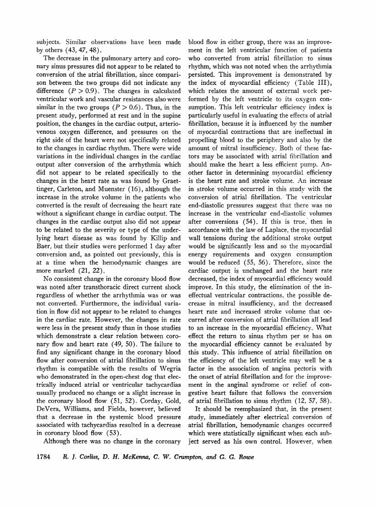
FIGURE 2 Patient J. B. Double dye dilution curve-injection of indocyaminegreen into the left ventricle with simultaneous sampling from the left atriumand left ventricle. Conclusion: no evidence of mitral insufficiency.
with a mean increase of 1% (P < 0.2). Therewas an increase of 8% (P < 0.3) in the cardiacoutput of patients whose atrial fibrillation per-sisted. The cardiac output of two patients withatrial flutter was at the upper limits of normal anddecreased, but remained within normal limits afterthe conversion to sinus rhythm. The mean oxygenconsumption and carbon dioxide production werenormal for this laboratory with no significantdifference between the two groups. There was anaverage decrease in the heart rate of 22% (P< 0.01) after conversion of the atrial fibrillation.Associated with this was an increase in strokevolume of 40%o (P < 0.001). Similar changeswere not observed in those whose atrial fibrillationpersisted.
There was a decrease in the pulmonary arterypressures of 9 (P < 0.01) and 15% (P < 0.05)in the coronary sinus pressure after conversion ofatrial fibrillation to sinus rhythm. There was a
similar decrease of 10% in the pulmonary arterypressure (P < 0.05) and of 10% in the coronarysinus pressure (P < 0.05) after similar directcurrent shocks in the group whose rhythm didnot change.
Table II summarizes the left-sided hemodynamicalterations after the shock. In the five patientswith atrial fibrillation who had left-sided catheteri-zations and whose rhythm converted, there was anaverage decrease of 16%o (P < 0.05) in the leftatrial mean pressure. There was also an averagedecrease in the mean diastolic gradient across themitral valve of 16% (P < 0.05) as demonstratedin Fig. 1. There was no significant change in theleft ventricular end-diastolic pressures after con-version of atrial fibrillation to sinus rhythm. Allthe patients with left-sided catheterizations hadmitral valvular disease except patient 24, who hadunderlying myocarditis with no demonstrable val-vular heart disease.
Hemodynamic Effects after Conversion of Arrhythmias 1779
150- Inject n
1.

There was a decrease in the calculated periph-eral and pulmonary resistances of 12 (P < 0.05)and 17% (P < 0.01), respectively, after conver-sion of atrial fibrillation to sinus rhythm withdirect current precordial shocks. Percentage-wise,there was also a similar decrease in the peripheralresistance (10%, P < 0.3) and in the pulmonaryresistance (15%o, P < 0.1) of patients with per-sistent atrial fibrillation after the shock. There wasno significant change in the right or left ventricu-lar work indices in either group of subjects.
The degree of mitral insufficiency was evaluatedbefore and after the conversion of atrial fibrillationto sinus rhythm in five subjects (Nos. 10, 12, 13,14, and J.B.). This evaluation was made by inject-ing indocyamine green into the left ventricle andby sampling simultaneously from the left atriumand femoral artery (24-26). In subject J.B.,1there was no mitral insufficiency detected duringatrial fibrillation (Fig. 2). In patients 10 and 14there was slight to moderate mitral insufficiencyduring atrial fibrillation, which was less after con-version. Two other patients, 12 and 13, with mod-erately severe and slight mitral insufficiency, re-
Table I, data not used in statistical analysis.
Before D C ShockAtriol Fibrillation
spectively, had no apparent change after theconversion from atrial fibrillation to sinus rhythm.Representative dye curves of patient 10 and 12 arepresented in Figs. 3 and 4.
The changes in the coronary hemodynamics arepresented in Table III. Changes in the coronaryblood- flow ranged from - 31 to + 24% after con-version of atrial fibrillation to sinus rhythm withnearly an identical range in the group that wasshocked but did not convert. There was no signifi-cant change in the mean coronary blood flow ineither group. The variation in coronary blood flowdid not correlate with changes in the cardiac rate.There was a slight decrease in the arterial oxygencontent and of the coronary sinus carbon dioxidecontent in all patients shocked. The changes in thecoronary sinus oxygen content, myocardial oxygenutilization, and carbon dioxide production were notof statistical significance. There was an increase of28% (P < 0.02) in the index of efficiency afterconversion of atrial fibrillation to sinus rhythm.There was no change in this -index of efficiencyafter direct current shock if atrial fibrillationpersisted.
In the two patients with atrial flutter and the
After DC ShookSinus Rhytnm
LA
FA
Injection
50
.1
FIGURE 3 Double dye dilution curve-injection of indocyamine green into the left ventriclewith simultaneous sampling from the left atrium and femoral artery. Conclusion: a decreasein the degree of mitral insufficiency after electrical conversion of atrial fibrillation.
1780 R. J. Corliss, D. H. McKenna, C. W. Crumpton, and G. G. Rowe
ll:: T. T. T IV: IT

Before DC SrockAtria! Fibrll~cilion
After D C ShoCKSinus Rhyh m
InjectionLV
u.wr~"
FIGURE 4 Patient 12. Double dye dilution curve-injection of indocyamine green into the leftventricle and simultaneous sampling from the left atrium and femoral artery. Conclusion: no
change in the degree of mitral insufficiency after electrical conversion of atrial fibrillation.
one with supraventricular tachycardia who were
converted to sinus rhythm, there was a decreasein the coronary blood flow, an increase in thecoronary sinus oxygen content, and a decrease in
-the coronary sinus carbon dioxide content (TableIII). Thus, there was a decrease in the myocardialoxygen consumption and carbon dioxide produc-tion and an increase in the left ventricular indexof efficiency.
DISCUSSION
An electrical discharge, which can be deliveredtransthoracically from a capacitor at a predeter-mined point in the cardiac cycle, provides an
efficient and, with proper precautions, a safemethod for the termination of many cardiac ar-
rhythmias (27-37). It also affords the opportunityto evaluate the hemodynamic state before andimmediately after the conversion of arrhythmiaswithout the administration of drugs or anesthesiabetween the studies which may, in themselves, pro-
duce hemodynamic changes and complicate inter-pretation of the results. The cardiovascular effectsresulting from the quinidine, digitalis, and meperi-dine given before this study were assumed to beconstant throughout both studies and not to con-
tribute to any of the observed changes (14, 38-42).Thus, the two variables in the present study were
the change in the rhythm and the effect of thedirect current transthoracic shock. The hemo-dynamic changes were analyzed statistically forevaluation of these two variables as describedunder Methods.
The average increase of 11 % in the cardiac out-put after conversion of atrial fibrillation to sinusrhythm while at rest was similar to that reportedby other investigators (12-15). There was alsoan 8% increase in the cardiac output after theshock in the group of subjects who failed to con-
vert from atrial fibrillation. Comparison of thesechanges between the two groups indicated no sig-nificant difference (P > 0.9). This suggests that
Hemodynamic Effects after Conversion of Arrhythmias
50-1 -.5,-
9...
.Iic.
0 -
i .I I-1ilm 'I li '101-0. I,- " o i jolr--T"r - - ..-- . -..r.-
-.1 J. I. IAll"J.- A I oil . li -J pi I -J i -J o" II A. -.U. ,"TTr 'r.-.9PPr T F - T., ,
IIIf IF I , -..
1781
Oman :..:

TABLE IIICoronary Blood Flow and Myocardial Metabolic Effects of DC Transthoracic Shock
CBF Index eff A 02 CS 02 A C02 CS C02Patient No. %A %,& %A % %A %A
ml/100 g per min
Patients converted-atrial fibrillation6 67 0.53
+22 + 407 88 0.38
-31 + 558 66 0.29
+14 - 39 45 0.30
+13 + 311 58 0.29
-10 + 2412 90 0.51
- 6 +4913 46 0.25
- 2 + 6814 85 0.29
-11 015 44 0.55
+ 2 -1316 79 0.24
+22 + 33Averages 67 0.36%A 0 + 28P value > 0.9 < 0.03
Patients converted-atrial flutter and vent. tach.1 54 0.80
-22 + 362 118 0.15
-37 +1473 90 0.40
-42 + 53
Patients not converted-atrial fibrillation19 52 0.48
+33 -3120 64 0.58
+11 + 321 54 0.73
-4 - 122 75 0.32
-16 + 2223 64 0.72
+ 2 + 14
Averages 62 0.57%A + 3 0P value < 0.8 > 0.8
24 64 0.40- 8 + 13
ml/100 ml blood
16.3-13
15.5-5
19.9+ 1
17.8-4
19.1+ 1
15.9+ 3
15.7-1
14.5-3
19.0-2
17.7-2
17.1-2< 0.03
20.2-4
17.4-1
14.7-3
16.0-3
15.7+1
16.0-5
15.1-1
15.0-1
15.6-2< 0.2
16.5- 1
ml/100 ml blood
2.9+52
4.1+12
6.2- 8
3.1+26
3.6+11
4.1- 5
3.0+10
3.503.9
+ 54.2
+ 53.9
+ 8< 0.2
7.2+22
3.6+39
4.5+22
3.5- 3
4.7- 6
5.4-11
3.2+19
3.9- 5
4.1- 2< 0.7
4.9+ 4
ml/100 ml blood
41.2+ 3
44.3-4
45.8-11
50.10
50.8+ 1
51.3-3
47.1-3
41.1-1
45.7-3
54.0-3
47.1-2< 0.1
48.8-27
44.7-2
46.1-26
48.6- 1
49.10
48.1-5
49.3-3
48.0-3
48.6-2< 0.03
49.2-2
ml/100 ml blood
52.9-353.1
-354.5
-760.1
-162;9
-361.7
-357.4
-448.3
+255.2
- 163.9
-457.0
-3<0.002
56.8-354.8
- 153.7
-2
58.7-357.9
055.2
-358.3
-455.3
-4
57.1-3<0.001
57.1-2
CBF, coronary blood flow; Index eff, index of efficiency, left ventricular work/left ventricular oxygen usage; A 02,arterial oxygen content; CS02, coronary sinus oxygen content; A C02, arterial carbon dioxide content; CS C02, coronarysinus carbon dioxide content.
1782 R. J. Corliss, D. H. McKenna, C. W. Crumpton, and G. G. Rowe

the increase in cardiac output after the conversionof atrial fibrillation may not be a result of thereturn of sinus rhythm but of other factors asso-ciated with the shock. The increase in the cardiacoutput in both groups and the observations byRodman, Pastor, and Figueroa would indicate thatthe precordial shock or other factors in this studywere not depressing the myocardial function andthus suppressing any hemodynamic changes thatotherwise may be evident (22).
Studies in which cardiac outputs are determinedhours or days after the conversion of atrial fibrilla-tion to sinus rhythm suggests that there is anincrease in the cardiac output after conversion,but it does not always appear to be immediate(15-22). Rodman et al. performed serial deter-minations of the cardiac output after the electricalconversion of atrial fibrillations and found that thehemodynamic improvements were often delayedfor hours or even days (22). The exact mechanismof this delayed benefit is not known, but in thisstudy the most significant change which followedconversion of atrial fibrillation to sinus rhythmwas the immediate increase in the left ventricularindex of efficiency. It is conceivable that this im-provement in myocardial function is cumulativeand involvtes the entire myocardium, both atrialand ventricular, and results in a progressively bet-ter mechanical systole. This temporal relationshipof hemodynamic improvements after conversion ofatrial fibrillation may account for some of the ob-served changes and at least in part explain someof the controversies arising from other studies onthe hemodvnamic effects of arrhythmias.
The failure for the cardiac output to increaseimmediately after the conversion of atrial fibrilla-tion to sinus rhythm with precordial shocks hasbeen noted by other investigators (16, 22). Thisis not unexpected since such factors as peripheralvascular resistance, the metabolic needs, myocar-dial function, and valvular lesions are the usualdetermining factors of the cardiac output and notthe cardiac rhythm per se, so long as the heartrate remains within the physiological range. How-ever, one factor that must be considered in therelationship of the conversion of atrial fibrillationto changes in the cardiac output is the contributionthat atrial contraction gives to diastolic filling.
In this study, an attempt was made to correlatethe height and contour of the left atrial "A" waves
after conversion of atrial fibrillation with thechanges in the cardiac output and in the left ven-tricular end-diastolic pressure. There was no con-sistent change in the left ventricular end-diastolicpressure, and no correlation was noted betweenthe character of the "A" wave and the cardiacoutput or left ventricular end-diastolic pressure.In general, the "A" waves were diminutive afterconversion. Similar results have been reported byothers (43).
The loss of left atrial contraction and its contri-bution to ventricular diastolic filling that occurswith the onset of atrial fibrillation has been re-ported to result in the elevation of the mean leftatrial pressure and a decrease in the left ventricu-lar end-diastolic pressure (44). Both the left atrialmean pressure and the mitral diastolic gradientdecreased in four of five subjects after conversionof atrial fibrillation to a sinus rhythm. The onesubject, 13, who increased his left atrial pressure,had a near normal pressure. Furthermore, theabsolute change was small after conversion andwas accompanied by an increase in oxygen con-sumption and cardiac output.
Experimental work suggests that the left atrialpressure changes noted in this study are causedby the correction of the arrhythmia (44), but itmust be pointed out that in the single patient inthis study with mitral valve disease and atrialfibrillation who did not convert, the changes in leftatrial pressure were similar to most of the patientswho were converted.
Experimental studies indicate that normal atrialcontractions are required for effective closure ofthe atrio-ventricular valves (45, 46). In this study,no evidence of significant mitral insufficiency wasfound in many subjects with atrial fibrillation whenevaluated by left ventricular angiography or byindicator dilution curves as shown in Fig. 2.Although there are many sources of error in thequantitation of mitral insufficiency by indicatordilution curves, evaluation by this method sug-gests that conversion of atrial fibrillation to sinusrhythm decreases the degree of mitral insufficiencyin some but not all patients (Fig. 3). Failure todemonstrate mitral insufficiency consistently in allpatients with atrial fibrillation also indicates thatatrial contraction may not be essential for adequateclosure of the atrio-ventricular valves in human
Hemodynamic Effects after Conversion of Arrhythmias 1783
subjects. Similar observations have been madeby others (43, 47, 48).
The decrease in the pulmonary artery and coro-nary sinus pressures did not appear to be related toconversion of the atrial fibrillation, since compari-son between the two groups did not indicate anydifference (P > 0.9). The changes in calculatedventricular work and vascular resistances also weresimilar in the two groups (P > 0.6). Thus, in thepresent study, performed at rest and in the supineposition, the changes in the cardiac output, arterio-venous oxygen difference, and pressures on theright side of the heart were not specifically relatedto the changes in cardiac rhythm. There were widevariations in the individual changes in the cardiacoutput after conversion of the arrhythmia whichdid not appear to be related specifically to thechanges in the heart rate as was found by Graet-tinger, Carleton, and Muenster (16), although theincrease in the stroke volume in the patients whoconverted is the result of decreasing the heart ratewithout a significant change in cardiac output. Thechanges in the cardiac output also did not appearto be related to the severity or type of the under-lying heart disease as was found by Killip andBaer, but their studies were performed 1 day afterconversion and, as pointed out previously, this isat a time when the hemodynamic changes aremore marked (21, 22).
No consistent change in the coronary blood flowwas noted after transthoracic direct current shockregardless of whether the arrhythmia was or wasnot converted. Furthermore, the individual varia-tion in flow did not appear to be related to changesin the cardiac rate. However, the changes in ratewere less in the present study than in those studieswhich demonstrate a clear relation between coro-nary flow and heart rate (49, 50). The failure tofind any significant change in the coronary bloodflow after conversion of atrial fibrillation to sinusrhythm is compatible with the results of Wegriawho demonstrated in the open-chest dog that elec-trically induced atrial or ventricular tachycardiasusually produced no change or a slight increase inthe coronary blood flow (51, 52). Corday, Gold,DeVera, Williams, and Fields, however, believedthat a decrease in the systemic blood pressureassociated with tachycardias resulted in a decreasein coronary blood flow (53).
Although there was no change in the coronary
blood flow in either group, there was an improve-ment in the left ventricular function of patientswho -converted from atrial fibrillation to sinusrhythm, which was not noted when the arrhythmiapersisted. This improvement is demonstrated bythe index of myocardial efficiency (Table III),which relates the amount of external work per-formed by the left ventricle to its oxygen con-sumption. This left ventricular efficiency index isparticularly useful in evaluating the effects of atrialfibrillation, because it is influenced by the numberof myocardial contractions that are ineffectual inpropelling blood to the periphery and also by theamount of mitral insufficiency. Both of these fac-tors may be associated with atrial fibrillation andshould make the heart a less efficient pump. An-other factor in determining myocardial efficiencyis the heart rate and stroke volume. An increasein stroke volume occurred in this study with theconversion of atrial fibrillation. The ventricularend-diastolic pressures suggest that there was noincrease in the ventricular end-diastolic volumesafter conversions (54). If this is true,' then inaccordance with the law of Laplace, the myocardialwall tensions during the additional stroke outputwould be significantly less and so the myocardialenergy requirements and oxygen consumptionwould be reduced (55, 56). Therefore, since thecardiac output is unchanged and the heart ratedecreased, the index of myocardial efficiency wouldimprove. In this study, the elimination of the in-effectual ventricular contractions, the possible de-crease in mitral insufficiency, and the decreasedheart rate and increased stroke volume that oc-curred after conversion of atrial fibrillation all leadto an increase in the myocardial efficiency. Whateffect the return to sinus rhythm per se has onthe myocardial efficiency cannot be evaluated bythis study. This influence of atrial fibrillation onthe efficiency of the left ventricle may well be afactor in the association of angina pectoris withthe onset of atrial fibrillation and for the improve-ment in the anginal syndrome or relief of con-gestive heart failure that follows the conversionof atrial fibrillation to sinus rhythm (12, 57, 58).
It should be reemphasized that, in the presentstudy, immediately after electrical conversion ofatrial fibrillation, hemodynamic changes occurredwhich were statistically significant when each sub-ject served as his own control. However, when
1784 R. I. Corliss, D. H. McKenna, C. W. Crumpton, and C. G. Rowe

the changes were compared between the group ofpatients who converted from atrial fibrillation andthose who did not, there was little difference. Thenotable differences between the two groups werea decrease in heart rate, an increase in strokevolume, and an increase in the left ventricularindex of efficiency in those patients who revertedto sinus rhythm. The conversion of atrial fibrilla-tion to sinus rhythm by a precordial shock had noimmediate effect on the cardiac output or coronaryblood flow.
ACKNOWLEDGMENTS
This work was supported in part by grants from the Na-tional Institutes of Health, HE-07754 of the U. S. PublicHealth Service, and the Wisconsin Heart Association.
REFERENCES
1. Meakins, J., L. Dautrebande, and W. J. Fetter. 1923.The influence of circulatory disturbances on thegaseous exchange of the blood. IV. The blood gasesand circulation rate in cases of mitral stenosis.Heart. 10: 153.
2. Smith, W. C., G. L. Walker, and H. L. Alt. 1930.The cardiac output in heart disease. I. Complete heartblock, auricular fibrillation before and after therestoration to normal rhythm, subacute rheumaticfever and chronic rheumatic valvular disease. Arch.Internal Med. 45: 706.
3. Kerkhof, A. C. 1936. Minute volume determinationsin mitral stenosis during auricular fibrillation and afterrestoration of normal rhythm. Am. Heart J. 11: 206.
4. Hecht, H. H., W. J. Osher, and A. J. Samuels. 1951.Cardiovascular adjustments in subjects with organicheart disease before and after conversion of atrialfibrillation to normal sinus rhythm. J. Clin. Invest.30: 647.
5. Kory, R. C., and G. R. Meneely. 1951. Cardiac out-put in auricular fibrillation with observations on theeffects of conversion to normal sinus rhythm. J. Clin.Invest. 30: 653.
6. Hansen, W. R., J. M. Kinsman, and R. L. McClendon.1952. Auricular fibrillation: hemodynamic studies be-fore and after conversion with quinidine. Am. HeartJ. 44: 499.
7. Wade, G., L. Werk6, H. Eliasch, A. Gidlund, and H.Lagerlbf. 1952. The hemodynamic basis of thesymptoms and signs in mitral valvular disease. Quart.J. Med. 21: 361.
8. Storstein, O., and H. Tveten. 1955. The hemodynamiceffect of restoring normal sinus rhythm in patientswith auricular fibrillation. Scand. J. Clin. Lab. Invest.7:167.
9. Harvey, R. M., M. I. Ferrer, D. W. Richards, andA. Cournand. 1955. Cardiocirculatory performancein atrial flutter. Circulation. 12: 507.
10. Broch, 0. J., and 0. Muller. 1957. Hemodynamicstudies during auricular fibrillation and after restora-tion of sinus rhythm. Brit. Heart J. 19: 222.
11. Seizer, A. 1960. Effects of atrial fibrillation upon thecirculation in patients with mitral stenosis. Am. HeartJ. 59: 518.
12. Gilbert, R., R. H. Eich, H. Smulyan, J. Keighley,and J. H. Auchincloss, Jr. 1963. Effect on circulationof conversion of atrial fibrillation to sinus rhythm.Circulation. 27: 1079.
13. Oram, S., I. Weinbren, J. P. H. Davies, P. Taggart,and L. D. Kitchen. 1963. Conversion of atrial fibril-lation to sinus rhythm by direct-current shock. Lancet.2: 159.
14. McIntosh, H. D., Y. Kong, and J. J. Morris, Jr.1964. Hemodynamic effects of supraventricular ar-rhythmias. Am. J. Med. 37: 712.
15. Morris, J. J., Jr., M. Entman, W. C. North, Y. Kong,and H. McIntosh. 1965. The changes in cardiac outputwith reversion of atrial fibrillation to sinus rhythm.Circulation. 31: 670.
16. Graettinger, J. S., R. A. Carleton, and J. J. Muenster.1963. Circulatory consequences of changes in cardiacrhythm produced in patients by transthoracic direct-current shock. J. Clin. Invest. 42: 938.
17. Halmos, P. B., and G. C. Patterson. 1965. Effect ofatrial fibrillation on cardiac output. Brit. Heart J.27: 719.
18. Polachek, A. A., A. E. Ruiz, and N. F. Nickerson.1965. Cardiac output by external radioisotope countingin patients reverted from atrial fibrillation. DiseasesChest. 47: 65.
19. Kahn, D. R., W. S. Wilson, W. Weber, and H.Sloan. 1964. Hemodynamic studies before and aftercardiover-sion. J. Thoracic Cardiovascular Surg. 48:898.
20. Ferrer, M. I., and R. M. Harvey. 1964. Some hemo-dynamic aspects of cardiac arrhythmias in man. Am.Heart J. 68: 153.
21. Killip, T., and R. A. Baer. 1966. Hemodynamic effectsafter reversion from atrial fibrillation to sinus rhythmby precordial shock. J. Clin. Invest. 45: 658.
22. Rodman, T., B. H. Pastor, and W. Figueroa. 1966.Effect on cardiac output of conversion from atrialfibrillation to normal sinus mechanism. Am. J. Med.41: 249.
23. Orcutt, F. S., and R. M. Waters. 1937. A method forthe determination of cyclopropane, ethylene andnitrous oxide in blood with the Van Slyke-Neillmanometric apparatus. J. Biol. Chem. 117: 509.
24. Woodward, E., Jr., H. J. C. Swan, and E. H. Wood.1957. Evaluation of a method for detection of mitralregurgitation from indicator dilution curves recordedfrom the left atrium. Proc. Mayo Clin. 32: 525.
25. Lacy, W. W., W. H. Goodson, W. G. Wheeler, andE. V. Newman. 1959. Theoretical and practical re-quirements for the valid measurement by indicator-dilution of regurgitant flow across incompetent valves.Circulation Res. 7: 454.
Hemodynamic Effects after Conversion of Arrhythmias 1785

26. Rowe, G. G. 1964. Investigation of heart disease bycatheterization methods. Wisconsin Med. J. 63: 507.
27. Lown, B. 1964. "Cardioversion" of arrhythmias. I.Mod. Conc. Cardiovascular Disease. 33: 863.
28. Lown, B., M. G. Pereroth, S. Kaidbey, T. Abe, andD. E. Harken. 1963. "Cardioversion" of atrial fibril-lation. A report of the treatment of 65 episodes in50 patients. New Engl. J. Med. 269: 325.
29. Lown, B., J. Neuman, R. Amarasingham, and B. V.Berkovits. 1962. Comparison of alternating currentwith direct current electroshock across the closedchest. Am. J. Cardiol. 10: 223.
30. Killip, T. 1963. Synchronized DC precordial shockfor arrhythmias; safe new technique to establish nor-mal rhythm may be utilized on an elective or anemergency basis. J. Am. Med. Assoc. 186: 1.
31. Rabbino, M. D., W. Likoff, and L. S. Dreifus. 1964.Complications and limitations of direct-current coun-tershock. J. Am. Med. Assoc. 190: 417.
32. Lemberg, L., A. Castellanos, Jr., J. Swenson, andA. Gosselin. 1964. Arrhythmias related to cardio-version. Circulation. 30: 163.
33. Miller, H. S., Jr. 1964. Synchronized precordial elec-troshock for control of cardiac arrhythmias; initialresults and success after three months. J. Am. Med.Assoc. 189: 549.
34. Morris, J. J., Jr., Y. Kong, W. C. North, and H. D.McIntosh. 1964. Experience with "cardioversion" ofatrial fibrillation and flutter. Am. J. Cardiol. 14: 94.
35. Hurst, J. W., E. A. Paulk, H. D. Proctor, and R. C.Schlant. 1964. Management of patients with atrialfibrillation. Am. J. Med. 37: 728.
36. Corliss, R. J., G. G. Rowe, D. H. McKenna, andC. W. Crumpton. 1966. Electrical conversion ofarrhythmias. Wisconsin Med. J. 65: 234.
37. Lown, B. 1967. Electrical reversion of cardiac ar-rhythmias. Brit. Heart J. 29: 469.
38. Evans, Frankis T., and T. C. Gray. 1965. Generalanaesthesia. Butterworth & Co., London. 2nd edition.1:150.
39. Johnson, S. R. 1951. The effect of some anaestheticagents on the circulation in man. Acta Chir. Scand.Suppl. 158: 1.
40. Ferrer, M. I., R. M. Harvey, L. Werko, D. T. Dres-dale, A Cournand, and D. W. Richards. 1948. Someeffects of quinidine sulfate on the heart and circula-tion in man. Am. Heart J. 36: 816.
41. Rowe, G. G., D. A. Emanuel, G. M. Maxwell, J. F.Brown, C. Castillo, B. Schuster, Q. R. Murphy, andC. W. Crumpton. 1957. Hemodynamic effects ofquinidine: including studies of cardiac work andcoronary blood flow. J. Clin. Invest. 36: 844.
42. Elliott, H. W., and M. A. A. Abdel-Rahman. 1965.Cardiovascular action of narcotic analgesics. Am.Heart J. 69: 567.
43. Braunwald, E. 1964. Symposium on cardiac arrhyth-mias, introduction. Am. J. Med. 37: 665.
44. Skinner, N. S., J. H. Mitchell, A. G. Wallace, andS. J. Sarnoff. 1964. Hemodynamic consequences ofatrial fibrillation at constant ventricular rates. Am.J. Med. 36: 342.
45. Little, R. C. 1951. Effect of atrial systole on ventricu-lar pressure and closure of the A-V valves. Am. J.Physiol. 166: 289.
46. Sarnoff, S. J., J. P. Gilmore, and J.- H. Mitchell.1962. Influence of atrial contraction and relaxation onclosure of the mitral valve. Circulation Res. 11: 26.
47. Burchell, H. B. 1964. A clinical appraisal of atriatransport function. Lancet. 1: 775.
48. Conn, H. L., Jr., D. F. Heiman, J. C. Wood, B.Jumbala, and W. S. Blakemore. 1957. Study of mitralregurgitant blood flow in subj ects with normal anddeformed mitral valves. Clin. Res. Proc. 5: 166.
49. Maxwell, G. M., C. A. Castillo, D. H. White, Jr.,C. W. Crumpton, and G. G. Rowe. 1958. Inducedtachycardia: its effects upon the coronary hemo-dynamics, myocardial metabolism, and cardiac effi-ciency of the intact dog. J. Clin. Invest. 37: 413.
50. Laurent, D., W. C. Bolene-Williams, F. L. Williams,and L. N. Katz. 1956. Effects of heart rate on coro-nary flow and cardiac oxygen consumption. Am. J.Physiol. 185: 355.
51. Wegria, R., C. W. Frank, H. H. Wang, and J. Lam-merant. 1958. The effect of atrial and ventriculartachycardia on cardiac output, coronary blood flow andmean arterial blood pressure. Circulation Res. 6: 624.
52. Wegria, R., C. W. Frank, G. A. Misrahy, R. S. Sious-sat, L. S. Sommer, and G. H. McCormack, Jr. 1950.Effect of auricular fibrillation on cardiac output, coro-nary blood flow and mean arterial blood pressure.Am. J. Physiol. 163: 135.
53. Corday, E., H. Gold, L. B. DeVera, J. H. Williams,and J. Fields. 1959. Effect of the cardiac arrhythmiason the coronary circulation. Ann. Internal Med. 50:535.
54. Braunwald, E., R. L. Frye, M. M. Aygen, and J. W.Gilbert, Jr. 1960. Studies on Starling's Law of theheart III. J. Clin. Invest. 39: 1874.
55. Burch, G. E., C. T. Ray, and J. A. Cronvich. 1952.Certain mechanical peculiarities of the human cardiacpump in normal and diseased states. Circulation. 5:504.
56. Levine, H. J, and R. J. Wagman. 1962. Energeticsof the human heart. Am. J. Cardiol. 9: 372.
57. Wolff, L. 1942. Clinical aspects of paroxysmal rapidheart action. New Engl. J. Med. 226: 640.
58. Phillips, E., and S. A. Levine. 1949. Auricular fibril-lation without other evidence of heart disease-a causeof reversible heart failure. Am. J. Med. 7: 478.
1786 R. J. Corliss, D. H. McKenna, C. W. Crumpton, and G. G. Rowe


![Nombre del informe id Norma - ISA...Nombre del informe id Norma [105000] Comentarios de la gerencia 105000 ps_mc [110000] Información general sobre estados financieros 110000 ias_1](https://img.dokumen.tips/doc/110x75/5eaf61a77f73c40a9e64aacd/nombre-del-informe-id-norma-nombre-del-informe-id-norma-105000-comentarios.jpg)