Embed Size (px)
Citation preview

Controversies in Primary care
Case Study ‘CVD risk reduction in the Elderly ‘
DR Jim Moore GPSI GLOS Heart Failure service

Declarations Received funding from BAYER, MSD, NOVARTIS, SERVIER (and possibly others) for various activities including attending and participating in educational events , advisory boards, etc Presently acting Chair of the GLOS CCG Cardiac Programme group Clinical Lead for the Don’t Wait to Anticoagulate Project -West of England Academic Health Science Network(WEAHSN)


Some of the highlights…..
• Much neglected areas of CVD risk reduction • Complexity of managing complex elderly
patients with CVD • An example of Multidisciplinary team working • Patient -Doctor relationship

Which of the following is likely to confer the greatest future Cardiovascular risk in
an elderly individual ?
• Managing risk factors for CVD • Left ventricular systolic dysfunction • Stroke risk in Atrial Fibrillation • Being a devout follower of Scottish
International Sport at any age

CVD risk reduction
• Primary prevention of CVD (NHC) 10% risk of CVD event over a ten year period reduce risk by 30%+ with statins (High risk) • AF -average risk of stroke 5(-10) % risk of debilitating CVA in one year reduce risk by 66%+ with anticoagulation • In Heart Failure (LVSD ) 40% risk of dying within a year of diagnosis 20% mortality at 2 years with modern triple therapy(PARADIGM)

Patient Profile “John”
81 year old Retired Taxi driver Retired and registered with my practice 35 years ago Married Playing golf till recently Healthy dose of “Northern attitude” Presents in Jan 2015 localised swelling in his face ? …. But what had gone before !!!

Case study
December 2004 Seen by GP • 71 year old • Persistent productive cough and SOBOE- x2 courses a/biotics • Initial URTI • Lifelong heavy smoker • No history of chest disease • No history of chest pain, fluid retention. • Gout

…presents for review with own GP 2 weeks later
• Cough was 60% better • 78/min regular 94/74 normal HS chest coarse basal crackles • No signs of fluid retention • FBC/TSH/LFTS/CRP all normal
Conclusions
• ? Chest infection/COPD/?HF/malignancy

What would you do next ?
1. Give further course of antibiotics 2. Chest x-ray 3. Spirometry 4. ECG 5. Natriuretic peptide testing
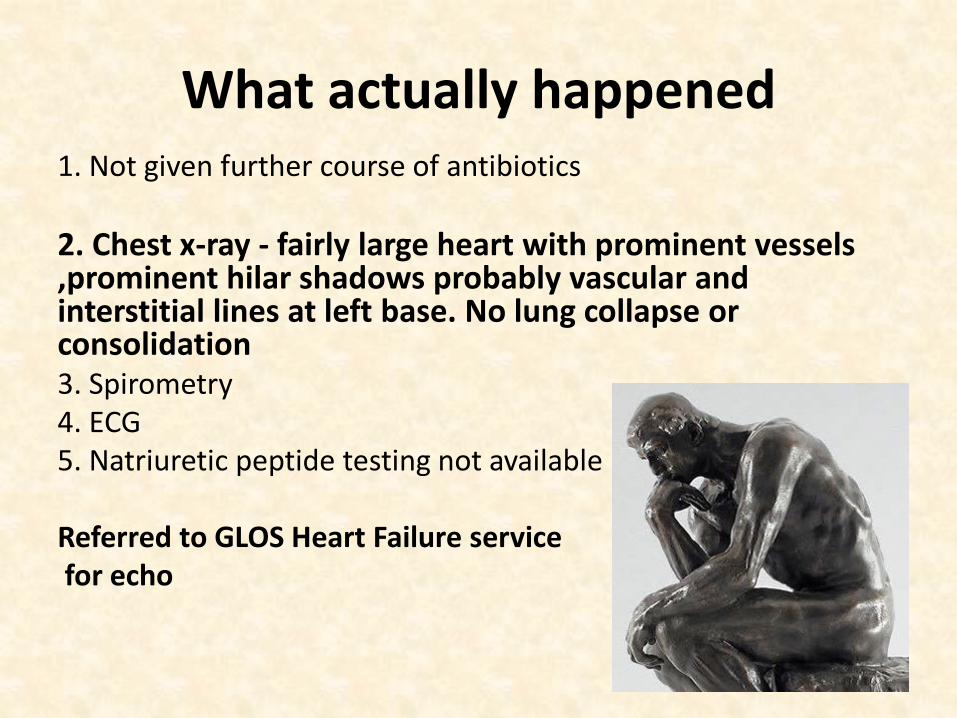
What actually happened 1. Not given further course of antibiotics
2. Chest x-ray - fairly large heart with prominent vessels ,prominent hilar shadows probably vascular and interstitial lines at left base. No lung collapse or consolidation 3. Spirometry 4. ECG 5. Natriuretic peptide testing not available
Referred to GLOS Heart Failure service for echo

Co-existence of COPD and Heart Failure
• Shared aetiological factors -age and smoking • Low incidence of COPD in HF trials relate to selection bias • Commonest reason for underuse of beta blocker in LVSD

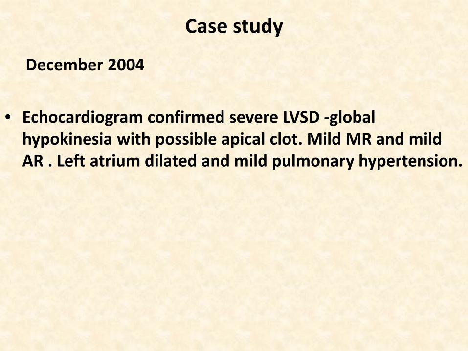
Case study
December 2004 • Echocardiogram confirmed severe LVSD -global
hypokinesia with possible apical clot. Mild MR and mild AR . Left atrium dilated and mild pulmonary hypertension.
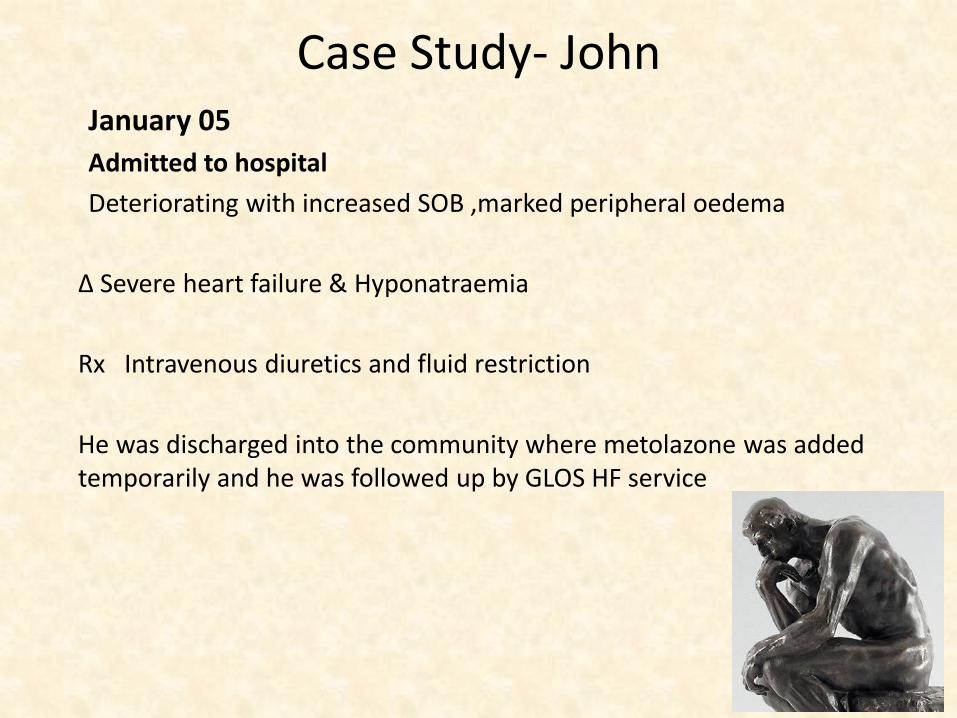
Case Study- John January 05 Admitted to hospital Deteriorating with increased SOB ,marked peripheral oedema
Δ Severe heart failure & Hyponatraemia Rx Intravenous diuretics and fluid restriction He was discharged into the community where metolazone was added temporarily and he was followed up by GLOS HF service

What followed
• Seen by Heart Failure service ….and titrated onto triple therapy.. - ramipril 2.5mg OD - carvedilol 25mg BD - spironolactone 25mg OD • Followed up by cardiology who undertook … 1. coronary angiogram which showed 30% stenosis in RCA and 60% stenosis in LAD 2.Mycocardial perfusion scan - inferior infarct with area of modest reversibility in basal septum /anteroapical area Not felt appropriate for coronary intervention


10Years

Patient Profile “John” 81 year old Retired Taxi driver Retired and registered with my practice 35 years ago Married and remains active though no longer playing golf PMH Hypertension? 2004 LVSD 2008 Primary Hyperparathyroidism (stable/no treatment) 2010 T2 DM 2013 PMR on low dose prednisolone Ramipril 2.5mg daily Carvedilol 25mg BD Spironolactone 25mg daily Aspirin 75mg OD Simvastatin 40mg OD Allopurinol 300mg daily Presents to dentist in Jan 2015 with facial swelling

Case study -John 6th Feb 15 ….....10 years later Undergoing assessment for surgery related to likely maxillary malignancy - faxed message from pre-admission clinic CT scan of his chest and abdomen shows 1.R basal atelectasis 2. possible small pleural effusions 3.gallstones and possible free fluid in abdomen … so could we sort out in Primary care !!! Extensive max-fax op scheduled for 10days time Therefore patient phoned practice to discuss with Duty Dr Asymptomatic but given course of antibiotics on the basis of CT findings

Case study - John 12th Feb …further message from pre-admission clinic concerned re his irregular ECG rhythm Faxed ECG not decipherable ??AF…. Clinical assessment(after hours) unremarkable -he felt his chest was back to normal and had no significant cardiac symptoms 62/min ?rhythm 104/68 Normal HS - soft apical systolic murmur (MR). JVPnormal. Chest clear and no oedema FBC -Platelets 64 ;eGFR 56 therefore ECG in the practice the following morning....


Temporal Relations of Atrial Fibrillation and Congestive Heart Failure and Their Joint Influence on Mortality
The Framingham Heart Study Wang et al Circulation 2003
• 1470 participants • AF or heart failure or both • 382 patients with both conditions, • 38% had AF first, 41% had CHF first, and 21% had both diagnosed at same
time. • Incidence of CHF in AF subjects was 33 per 1000 person-years • Incidence of AF in CHF subjects was 54 per 1000 person-years • In CHF subjects, later development of AF was associated with increased
mortality (men: HR 1.6; 95% CI, 1.2 to 2.1; women: HR 2.7, 95% CI, 2.0 to 3.6).
• In AF subjects, the subsequent development of CHF was associated with increased mortality (men: HR 2.7; 95% CI, 1.9 to 3.7; women: HR 3.1; 95% CI, 2.2 to 4.2)
• Conclusions— Individuals with AF or CHF who subsequently develop the other condition have a poor prognosis..
• Prevalence of AF in HF patients around 30-40% and increased in worsening LVSD


What would you do next ?
Given the ECG findings-in AF 60/min and LBBB in a patient with known AF would you 1.Make no alteration to his medication 2. Arrange a 24hour tape 3.Reduce the dose of carvedilol 4.Swap to an alternative beta blocker

What happened next
• Dose of carvedilol reduced

From: Is Heart Rate Important for Patients With Heart Failure in Atrial Fibrillation?
JCHF. 2014;2(3):213-220. doi:10.1016/j.jchf.2014.01.005
Multivariable Adjusted Survival Curves for Patients in Atrial Fibrillation at Baseline Divided by Heart Rate Quartiles CI = confidence interval; HR = hazard ratio.

Case study -Primary care 13th Feb 15
Full fax from pre-admission clinic Concerned re his ECG rhythm and echo Echo showed mild to moderate dilated LV with severe impaired systolic function. His RV is dilated with impaired systolic function. Bi-atrial dilatation. Moderate MR .Elevated RVSP ECG -AF 60/min LBBB Operation scheduled for 16th Feb


What happened next?
1. Urgent assessment re anticoagulation 2. Urgent assessment re anticoagulation and clinical review 3. Continue his present management which includes aspirin 4. Ask the pre admission clinic to make anaesthetist aware of clinical findings and need for further management (as they had initiated investigations)

What happened next
1. Urgent assessment re anticoagulation 2. Urgent assessment anticoagulation and haemodynamic review 3. Continue his present management which includes aspirin 4. GP saw patient and immediately co ntacted the pre-admission clinic asking them to inform the anaesthetist of the relevant findings…and reduced his dose of carvedilol . Patient and clinic told it was the anaesthetist decision about further management and whether he has op in 3 days time


What happened next
• Operation cancelled and rescheduled for 23rd March
• Anaesthetist requests cardiology opinion based on clinical detail
• There is a meeting of interested parties to discuss the way forward shortly (MDT meeting)

Cardiology opinion • Cardiology letter arrive 27th February… Salient points • AF and complete LBBB • Asymptomatic coronary artery disease but would he benefit from
revascularisation • Echo severe LVSD -EF 24%
• Needs anticoagulation • Needs 24hour tape • Needs myocardial perfusion scan • ?Biventricular pacemaker • Cardiology clinical assessment …...undertaken 25th March

What I did next • 27th Feb
• I phoned John unaware of developments over the past few weeks • He feels well-no symptoms cardiac decompensation • CHADS2VASC2 = 6…. HASBLED = 2- 3 • No overt bleeding • Arranged urgent bloods FBC showed platelets 64 eGFR 52 • Full and frank discussion re Stroke risk -“I like my alcohol” and has
significant misgivings re warfarin

Stroke risk assessment with CHADS2 and CHA2DS2-VASc

HAS-BLED should be used to identify modifiable risk factors for bleeding Score of ≥3 indicates need for regular clinical review Patients with a higher HAS-BLED score also have a higher CHA2DS2-VASc score There is net clinical benefit in anticoagulating CHA2DS2-VASc >0 whatever HAS-BLED score

What did I do next
1. Start Warfarin 2. Start LMWH and warfarin together 3. Start Dabigatran 4. Start Rivaroxaban 5. Start Apixaban 6. No change in medication 7. No change in medication other than stopping aspirin


What I did next
• Started Rivaroxaban 15mg as his estimated creatinine clearance was 46

Warfarin initiation and prothrombotic state
• UKCPRD database -70,000 patients • 1993-2003 • 71% increase risk of ischaemic stroke in first 30 days when
taking warfarin cf no anticoagulant • Highest risk in first week at around 3 days when around 2.3
fold increase • Warfarin blocks the activation of factors II, VII, IX and X but
also deactivates two other proteins, C and S, which are anticoagulants.
• Rapid depletion of protein C, in particular, might lead to a temporary hypercoagulable state.
• Consider bridging in the early use of warfarin
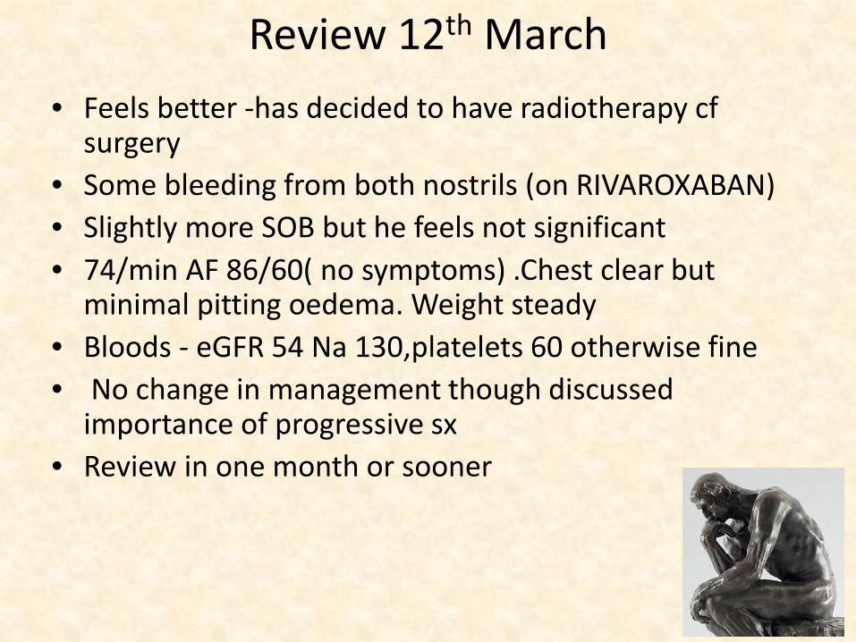
Review 12th March • Feels better -has decided to have radiotherapy cf
surgery • Some bleeding from both nostrils (on RIVAROXABAN) • Slightly more SOB but he feels not significant • 74/min AF 86/60( no symptoms) .Chest clear but
minimal pitting oedema. Weight steady • Bloods - eGFR 54 Na 130,platelets 60 otherwise fine • No change in management though discussed
importance of progressive sx • Review in one month or sooner

Review 24th March
• Increased SOB • Increased bleeding from nose • Felt awful on Rivaroxaban -dyspepsia, lethargy and SOB (he
blamed on NOAC) • Given PPI by GP partner a day previously • Pitting oedema to below knees but chest clear … 1. start furosemide 40mg daily and assess bloods 2. Stop RIVAROXABAN declines alternative anticoagulation (and dental extractions next week)
• ...results the next day Na119 and his platelets 50 ....his
furosemide was discontinued ( by another partner)
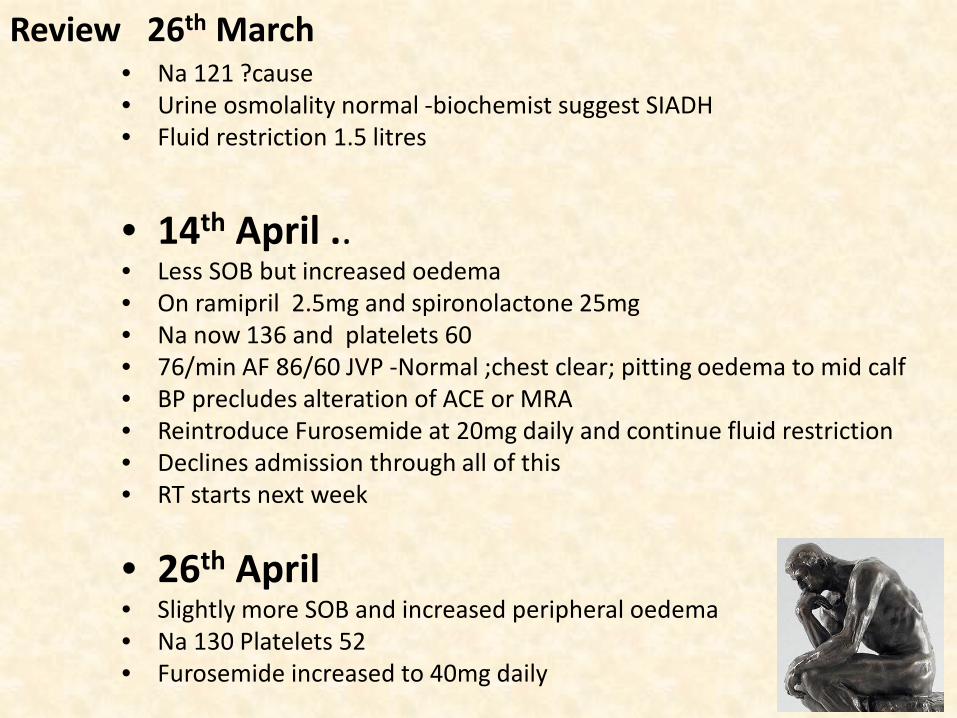
Review 26th March • Na 121 ?cause • Urine osmolality normal -biochemist suggest SIADH • Fluid restriction 1.5 litres
• 14th April .. • Less SOB but increased oedema • On ramipril 2.5mg and spironolactone 25mg • Na now 136 and platelets 60 • 76/min AF 86/60 JVP -Normal ;chest clear; pitting oedema to mid calf • BP precludes alteration of ACE or MRA • Reintroduce Furosemide at 20mg daily and continue fluid restriction • Declines admission through all of this • RT starts next week
• 26th April • Slightly more SOB and increased peripheral oedema • Na 130 Platelets 52 • Furosemide increased to 40mg daily

8th May ….. 6.50 pm Phoned patient Friday as ….... • Cardiology letter -fluid retention including ascites 1.increase diuretics 2.anticoagulation 3. refer HFS • Oncologist tells him four days earlier that he has a “A clot in his
Right chest on routine scan”. Cardiology opinion sought • Started RT and bleeding from both nostrils more so left this
evening • Recent bloods Platelets 48 and eGFR 46 • CT scan shows marked bi-atrial dilatation with substantial filling
defect in his Right atrial appendage consistent with thrombus. • Declines admission to hospital • What did I do next

What did I do next
1. Initiate anticoagulation with NOAC 2. Initiate anticoagulation with LMWH 3. Initiate anticoagulation with Warfarin 4. Discuss with …..ologist/specialist(s) 5. Wait till Monday to sort out
What I did next • Discussed with oncologists who advised discuss with
haematologists as not an oncology problem…........ • Discussed with haematologists …who said angrily to discuss with
oncology once more • Oncology refused to see him that night but agreed to review him at
his radiotherapy appointment the folltill the morning (12 hours later) ...
• The oncology team did not see him in the morning
...and when I phoned John on Monday morning to learn all of this what was my response


What happened next
• I discusssed the situation with the oncologist • I discussed the situation with the cardiologist • I discussed the situation with a haematologist • I initiated injections with LMW Heparin

…...John died less than 2 weeks after I last saw him ...he was admitted to hospital during his
radiotherapy.....“his breathing just got worse and worse ..and they made him comfortable”.
Death Certificate -Heart Failure secondary to mitral and
tricuspid valve disease

Learning points 1. Good communication central to effective health care -MDT/PCT/Patient -DR 2. Individual roles and responsibilities of MDT members need to be clearly understood and their possible limitations considered 3. Significant event discussion in primary care with agreed outcomes

Thank you ….any questions ?