Embed Size (px)
Citation preview

Controlling the morphology of copper-silica
nanocomposites from laser ablation in liquid
Mallory G. John and Katharine Moore TibbettsDepartment of Chemistry, Virginia Commonwealth University, Richmond, VA 23284, USA
Abstract
Synthesis of copper-silica nanocomposites with controllable morphology and
composition were produced with a one-step femtosecond reactive laser ablation
in liquid (fs-RLAL) technique. The composite nanomaterials were generated
by focusing femtosecond near-IR laser pulses onto a silicon wafer immersed in
an aqueous copper(II) nitrate solution, with the solution pH adjusted using ni-
tric acid or potassium hydroxide. Under acidic conditions (pH 3.0 and 5.4),
little copper was incorporated in the predominantly silica product (1.4 and 1.5
wt.%). These acidic conditions yielded large ⇠30�80 nm silica particles, with
some particles consisting of copper core/silica shell. In contrast, increasing the
solution pH to 10.4 resulted in extremely high Cu loading of 31.5 wt.% and a
composite product consisting of 1.5 nm copper clusters distributed throughout a
matrix of amorphous silica and copper phyllosilicate. The relationship between
the precursor solution pH and the product morphology and copper loading is
attributed to the point of zero charge (PZC) of silica, in which the high solution
pH allows for electrostatic adsorption to occur between the deprotonated silica
clusters from the ablated silicon wafer and the copper hydroxide dimer formed
in solution.
Keywords: reactive laser ablation in liquid, femtosecond laser, copper-silica
nanocomposite, copper phyllosilicate
Preprint submitted to Applied Surface Science December 9, 2019

1. Introduction
Copper nanoparticles (Cu NPs) are valued for their low cost, high conduc-
tivity, and thermal stability, making them a popular alternative to rare earth
metals for biological sensing and imaging [1], antimicrobial applications [2],
inkjet-printable electronics [3], and catalysis [4]. In particular, the ability of
copper to access many oxidation states makes supported Cu NPs active cat-
alysts towards reactions such as electrochemical reduction [5], thermochemical
hydrogenation [6] and photochemical reduction [7] of CO2, photocatalytic degra-
dation of organic dyes [8], and other organic transformations [9–11].
While Cu NPs possess high catalytic activity and high temperature sta-
bility, a major bottleneck to using copper-based nanomaterials for catalysis is
the propensity for small Cu NPs to agglomerate, and for Cu surfaces to oxidize.
Support materials such as graphene, oxides, polymers, and metal-organic frame-
works (MOFs) are added to prevent agglomeration and surface oxidation, while
preserving the high catalytic activity of Cu NPs [4, 12, 13]. In particular, silica
has been used as a support material for various metal NPs due to the silanol sur-
face groups that enhance binding with metal NPs [4, 14]. The majority of syn-
thetic approaches to fabricating copper-silica nanostructures involve wet chem-
ical methods such as incipient wetness impregnation, deposition-precipitation,
strong electrostatic adsorption, and ammonia evaporation [4, 5, 11, 13–15]. In
these methods, the silica is either prepared by the Stöber method or purchased
commercial amorphous/fumed silica, and the copper is added in the form of
a salt complex. Copper-silica bonding is achieved by heating up the slurry or
solution, followed by calcining the finished product. A drawback of many of
these methods is that uneven distribution of the copper complex throughout
the silica often results in poorly dispersed Cu NPs with large size distributions
and low copper loading.
Laser ablation in liquid (LAL) has recently emerged as a robust alternative
synthesis route to myriad (supported) NPs that can overcome many challenges
inherent in wet-chemical synthesis [16–19]. LAL involves focusing intense laser
2

pulses onto a solid target immersed in liquid, which produces a localized plasma
at the solid-liquid interface containing reactive electrons, radicals, and ions [20].
The plasma reaches transient temperatures exceeding 5000 K that cool on sub-
microsecond timescales due to the surrounding ambient liquid [21]. These highly
nonequilibrium conditions generate NPs comprised of the target material with
exotic metastable phases and bonding environments that are stable without
added capping agents, making them ideal for catalysis applications [16–18].
Because LAL generates nanomaterials under ambient conditions in water, it is
considered a ‘green’ and sustainable synthesis method [16, 22]. LAL of a Cu
target in various solvents have been widely used to synthesize Cu NPs with
solvent- and laser-dependent oxidation states and morphologies [23–26].
When LAL is carried out in a solution containing metal ions or other species
that interact with the ablated target atoms, the technique is referred to as
Reactive Laser Ablation in Liquid (RLAL) [16]. The first demonstration of
RLAL in 2008 produced Ag and Au NPs by ablating a silicon wafer immersed
in either Ag(NO3) or HAuCl4 aqueous solutions [27]. Since this initial work,
many metastable and unique bi- and multi-metallic nanostructures have been
reported. For instance, mixed-metal Pt-Co and Pt-Co-Cu oxide NPs for fuel
cell applications were synthesized by ablating a Co target immersed in Pt and
Cu metal salt solutions [28–30], and Ni-Fe layered hydroxides doped with Ti
and La for electrochemical water splitting were synthesized by ablation of Fe
powder in aqueous solutions of Ni, Ti, and La salts [31, 32]. Complex metal-
oxide mineral phases of copper and zinc were synthesized by RLAL of Zn or Cu
targets immersed in aqueous Zn or Cu salt solutions [33], and silica-supported
Au or Ag nanomaterials have been synthesized by ablating silicon wafers in
aqueous gold or silver salt solutions [27, 34–36]. Recently, we generated sub-3
nm Au NPs dispersed throughout a silica matrix by ablating a silicon wafer
immersed in a [AuCl4]– solution using femtosecond laser pulses [36].
In this work we report the synthesis and characterization of copper-silica
nanocomposites generated from a femtosecond-RLAL (fs-RLAL) technique, in
which fs laser pulses are focused onto a silicon wafer immersed in Cu(NO3)2
3
solutions under three pH conditions. The product morphology and Cu loading
on the silica were strongly dependent on the precursor solution pH, with the
highest copper loading achieved at pH 10.4. This pH-dependent copper loading
was reflected in the catalytic activity of the samples, determined using the
model reaction of catalytic para-nitrophenol reduction by sodium borohydride
[37]. We will discuss the role that the solution pH plays on the surface charge
of ablated silica species, and how surface interactions drive the formation of
different morphology and wt.% loading of Cu in the products.
2. Materials and Methods
2.1. Materials
Silicon wafers (n-doped, (111)-oriented, single side polished, 300 µm thick,
NOVA electronic materials), copper(II) nitrate, Cu(NO3)2 (Fisher), potassium
hydroxide, KOH (Fisher), nitric acid, HNO3 (Fisher), sodium borohydride,
NaBH4 (Acros Organics), and para-nitrophenol, PNP (Acros Organics) were
used as received. Stock and working solutions were prepared with purified wa-
ter from a Millipore Ultrapure water system (resistivity is 18.2 M⌦cm�1 at
25�C).
2.2. Sample Preparation
Working solutions of Cu(NO3)2 (2.0 mM) were prepared from a freshly pre-
pared aqueous stock solution (50 mM) and the pH was recorded as ⇠5.4. Either
HNO3 (1.0 mM) was added from a 10 mM stock solution or KOH (5.0 mM)
was added from a 200 mM stock, resulting in working solution pH values of 3.0
and 10.4, respectively. The working solution was transferred to a quartz cuvette
(3 mL) equipped with a stir bar, and a pre-cut silicon wafer was placed in the
cuvette and secured to one side.
fs-RLAL irradiation of the Si wafer immersed in the working solution was
conducted for 30 min while stirring (details of laser parameters in section 2.4),
followed by centrifugation for 15 min at 6,000 rpm (Thermo Fisher AccuSpin
4

Micro 17). The supernatant was replaced with water and centrifuged a second
time for 15 min at 6,000 rpm. The resulting pellet was collected for either
characterization or redispersed in water and tested for its catalytic activity.
The following naming convention was used for all samples: Cu-silica-[solution
pH]. For example, Cu-silica-3.0 corresponds to the sample containing 2.0 mM
Cu(NO3)2 and 1.0 mM HNO3, with a pH of 3.0. Table 1 displays the sample
names and solution compositions for clarity.
Sample Solution Compositiona Initial Solution pH
Cu-silica-3.0 2 mM Cu(NO3)2 + 1 mM HNO3 3.0±0.1
Cu-silica-5.4 2 mM Cu(NO3)2 5.4±0.1
Cu-silica-10.4 2 mM Cu(NO3)2 + 5 mM KOH 10.4±0.2
Table 1: Sample, solution composition, and initial solution pH values for experiments. aall
solution prepared in DI water.
2.3. Catalytic Reduction of Para-nitrophenol
The catalytic reduction of para-nitrophenol (PNP) by NaBH4 was carried
out in a home-built in situ UV-vis spectrometer (details in section 2.4). In a
typical catalytic run, the collected pellet (details in section 2.2) was dispersed
in water (3.0 mL) and 300 µL of this solution was added to a cuvette containing
PNP (0.1 mM) and NaBH4 (10 mM) while stirring. The PNP was added from a
stock solution (1.5 mM) and the NaBH4 was added from a freshly prepared stock
solution (100 mM). The absorbance at 400 nm (as the para-nitrophenolate ion)
was recorded, and the reaction was considered complete when it had disappeared
completely. Data processing for the PNP reaction was conducted based on the
methods of Ref. [38] and details are provided in the Supplemental Information
(Fig. S1).
2.4. Instrumentation
The experimental laser setup has been described in detail in Refs. [36,
39, 40]. Briefly, a Ti:Sapphire regenerative amplifier delivering 7 mJ, 30 fs
5

pulses with a bandwidth centered at 800 nm and a repetition rate of 1 kHz
was attenuated to 200 µJ for the ablation experiments. The 10⇥10⇥40 mm
quartz fluorescence cuvette containing the pre-cut silicon wafer was placed 10
mm before the focal point of a f = 50 mm lens. The converging beam propagates
through 9.7 mm of liquid to reach the 0.3 mm thick Si wafer. We note that no
white light was generated on the front face of the cuvette because the large initial
beam diameter of 11 mm and high numerical aperture of the focusing lens were
sufficient to eliminate any nonlinear optical effects until a few mm before the
Si-liquid interface. The spot size on the wafer was 85 µm diameter, measured
using an optical microscope of an ablated Si wafer. Under these conditions, the
laser fluence was 3.5 J cm�2 and peak intensity was 1.17⇥1014 W cm�2. The
cuvette was placed on a micro-stir plate (Thermo Scientific) mounted on x- and
y- motorized translation stages (Thorlabs), which moved in a zig-zag pattern at
a velocity of 0.5 mm/s.
PNP catalysis runs were carried out in a home-built in situ UV-vis spectrom-
eter, with a deuterium-tungsten light source (Ocean Optics, DH2000-DUV),
optical fibers, a sample holder for 10⇥10⇥40 mm cuvettes placed on a stir
plate (300 rpm stir rate), and a compact spectrometer (Ocean Optics HR4000).
Spectra were collected every 1.2 seconds using LabVIEW software (National
Instruments).
2.5. Characterization
Transmission Electron Microscopy (TEM) TEM images were collected
on a JEOL JEM-1230 TEM at 120 kV. High resolution TEM (HRTEM) images
and SAED patterns were collected on an FEI Titan 80�300 kV with a Gatan
794 Multi-Scan Camera. Samples were prepared by drop-casting the diluted
pellet onto a carbon-coated copper grid (100 mesh, Ted Pella, Inc.) and left to
dry for at least 24 hr at room temperature. Size distributions were determined
by measuring 300 individual particles from three separate parts of the grid
using ImageJ software. Gatan Microscopy Software Suite version 3.x was used
to determine the crystal lattices of the nanoparticles in the HRTEM images.
6
Details on this process are included in the Supplemental Information (Fig. S2).
Scanning Electron Microscopy-Energy Dispersive X-ray Spectroscopy
(SEM-EDX) SEM-EDX was carried out on a Hitachi FE SEM SU-70 (spa-
tial resolution 1.0 nm) equipped with an Energy Dispersive X-ray Spectroscopy
(EDX) detector. Images were obtained at 10 keV and elemental analysis was
conducted at 15 keV, with ZAF standardless quantification employed for EDS
measurements. Samples were prepared by drop casting the centrifuged pellets
onto conductive carbon tape stabilized on an aluminum stage, and drying under
vacuum at room temperature.
X-ray Photoelectron Spectroscopy (XPS) XPS was conducted on a
PHI VersaProbe III Scanning XPS Microprobe with a monochromatic Al k↵
X-ray source (1486.6 eV) run at 25 W and 15 KV, with a pass energy set to
112 eV for survey scans and 69 eV for high resolution spectra. A spot diameter
of 200 µm was irradiated using a take off angle of 90�, and a detector was
situated at an angle of 45�. Charge neutralization was achieved by employing
an ion gun and a flood gun during the analysis. Samples were prepared by drop
casting the centrifuged pellet onto conductive carbon tape. Sample analysis was
carried out using CasaXPS Software version 2.3.19PR1.0, employing Gaussian
and Lorentzian convolution to fit the spectral lines, and all high resolution
spectra were corrected by shifting the C1s peak at 284.8 eV.
X-ray Diffraction (XRD) XRD was conducted on a Panalytical Em-
pryrean Diffractometer with CuK↵ radiation (� = 0.15418 nm) at 40 kV and
45 mA, with scanning angle (2✓) of 10�90� and a gonio focusing geometry.
Samples were prepared for XRD analysis by drying the centrifuged pellet under
vacuum at room temperature.
Fourier Transform Infrared Spectroscopy (FTIR) FTIR analysis was
conducted on a Nicolet iS50 FTIR spectrometer equipped with a mid- and far-
IR-capable diamond ATR. Spectra were obtained using 32 scans in the range
of 4000 to 400 cm�1 with 5 cm�1 resolution. Samples were prepared for FTIR
analysis by drying the centrifuged pellet under vacuum at room temperature.
7

3. Results
3.1. Characterization
The three Cu-silica products are visualized in the TEM images displayed
in Fig. 1a-c with detailed insets, and additional TEM images are reported
in the Supplemental Information (Fig. S3). While all three products contain
large spherical particles around ⇠70�100 nm in diameter, these particles are
most abundant in the Cu-silica-3.0 sample, and rarely seen in the Cu-silica-10.4
sample. Many of the spherical particles in the Cu-silica-3.0 and Cu-silica-5.4
samples are smooth throughout the entire particle, while some have a darker
core and lighter shell, indicative of a Cu-core and silica-shell structure (insets in
Fig. 1a and b). The Cu-silica-3.0 sample had very few core-shell particles. Size
distribution analysis was possible on only 43 particles, and histograms of the
core and outer diameter are displayed in Fig. S3 in the Supplemental Material.
The Cu-silica-5.4 had substantially more core-shell particles than the Cu-silica-
3.0 sample, with size distribution analysis displayed in Figure 1d.The inner
core had a mean diameter of 22.4±14.4 nm with sizes ranging from 2�63 nm,
and the outer shell mean was 32.1±14.8 nm with sizes ranging 12�84 nm in
diameter. The Cu-silica-10.4 sample exhibits completely different morphology
from the samples produced at lower pH (Fig. 1c). This product predominantly
contains small, 1.52±0.75 nm Cu NPs dispersed throughout a matrix made up
of long nano-needles and amorphous structures, along with a few large spherical
particles decorated with small Cu NPs (inset). A histogram of the Cu NPs is
displayed in Fig. 1e fit to a Gaussian distribution.
HRTEM images of the Cu-silica-5.4 and Cu-silica-10.4 products are displayed
in Fig. 2a and c with SAED patterns (Fig. 2b and d). HRTEM analysis
was not performed on the Cu-silica-3.0 sample due to the low number of core-
shell particles. The inset in the Cu-silica-10.4 HRTEM image shows a small
crystalline nanoparticle with lattice spacings measuring 2.13 Å corresponding to
the (200) plane of Cu2O [41]. The bottom inset of Fig. 2a shows the amorphous
structure of the silica. The SAED pattern in Fig. 2b has two faint diffraction
8

Figure 1: TEM images of Cu-silica samples at pH 3.0 (a), pH 5.4 (b), and pH 10.4 with
histograms of Cu-silica-5.4 (e) and Cu-silica-10.4 (e).
9
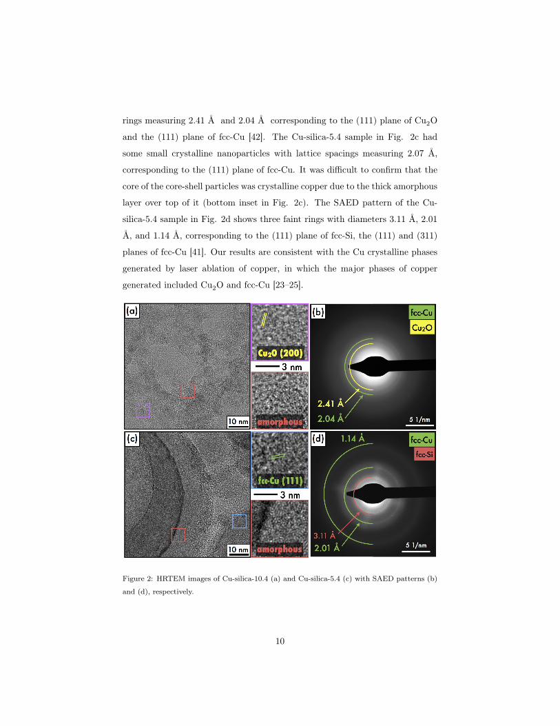
rings measuring 2.41 Å and 2.04 Å corresponding to the (111) plane of Cu2O
and the (111) plane of fcc-Cu [42]. The Cu-silica-5.4 sample in Fig. 2c had
some small crystalline nanoparticles with lattice spacings measuring 2.07 Å,
corresponding to the (111) plane of fcc-Cu. It was difficult to confirm that the
core of the core-shell particles was crystalline copper due to the thick amorphous
layer over top of it (bottom inset in Fig. 2c). The SAED pattern of the Cu-
silica-5.4 sample in Fig. 2d shows three faint rings with diameters 3.11 Å, 2.01
Å, and 1.14 Å, corresponding to the (111) plane of fcc-Si, the (111) and (311)
planes of fcc-Cu [41]. Our results are consistent with the Cu crystalline phases
generated by laser ablation of copper, in which the major phases of copper
generated included Cu2O and fcc-Cu [23–25].
Figure 2: HRTEM images of Cu-silica-10.4 (a) and Cu-silica-5.4 (c) with SAED patterns (b)
and (d), respectively.
10

SEM-EDX analysis was performed on the three Cu-silica samples, with a
representative EDX spectrum of the Cu-silica-10.4 sample displayed in Fig. 3.
The peaks located at 0.525 keV, 0.950 keV, and 1.74 keV correspond to the O
K↵, Cu L↵, and Si K↵ lines. The inset graph shows the wt.% Cu, Si, and O
quantified in the samples, and Table 2 displays the numerical values of wt.% Cu,
Si, and O from EDX, XPS, and ICP-OES analysis. The Cu-silica-10.4 sample
contains the highest amount of Cu, about ten to twenty times the amount as the
Cu-silica-5.4 and Cu-silica-3.0 samples. The significantly higher Cu loading in
the Cu-silica-10.4 sample is corroborated by the ICP-OES and XPS results, also
displayed in Table 2. XPS data was converted from atomic %, with calculations
provided in Table S1 in the Supplemental Information. XPS analysis shows
significant surface oxidation of the three samples, which is compensated for
by the decrease in Cu content in the Cu-silica-10.4 sample, and decrease in Si
content in the other two samples. The copper content did not decrease between
EDX and XPS analysis for the Cu-silica-5.4 sample, suggesting that the copper
present within the top 10 nm of this sample is protected from surface oxidation.
Figure 3: SEM-EDX spectrum of representative Cu-silica-10.4 sample with inset of wt.% of
SiK, OK, and CuL for different pH solutions.
11

ICP-OESa SEM-EDXa XPSbc
Sample Cu Cu Si O Cu Si O
Cu-silica-3.0 1.4±0.4 4.3±0.9 67.5±1.5 28.1±0.7 0.3±0.3 40.1±0.1 59.6±31
Cu-silica-5.4 1.5±0.1 2.6±0.4 86.5±2.1 11.0±1.7 2.6±2.0 45.9±0.9 51.5±6.8
Cu-silica-10.4 31.5±0.4 36.5±2.1 37.3±4.6 26.2±2.6 19.2±6.1 22.6±2.7 58.1±5.5
Table 2: weight % Cu from ICP-OES, weight % Cu, Si, and O from SEM-EDX, and XPS
analysis. aanalysis representative of entire sample material. banalysis representative of top
10 nm surface layer. cValues converted from atomic %, which is provided in Table S1 in the
Supplemental Information.
High resolution Cu2p3/2 and Si2p atomic orbital XP spectra are displayed
in Fig. 4a and b with the Cu-silica-10.4 spectra on the bottom panels and
the Cu-silica-5.4 and -3.0 samples on the top panels (a Si2p spectrum of silica
generated from ablating a silicon wafer in water is displayed in the Supplemental
Information). No Cu was detected in the Cu2p3/2 spectrum for the Cu-silica-3.0
sample. In the Cu2p3/2 spectra, a peak at 932.7 eV (purple) was deconvoluted
in both samples, which corresponds to either Cu0 (932.6 eV) or Cu+ as in
Cu2O (932.2 eV) [43–47]. The second peak around 934 eV in the Cu-silica-
5.4 sample (blue) corresponds to Cu2+ and matches that of a CuO species
[42, 48, 49]. In the Cu-silica-10.4 sample, this feature it is shifted to 935.3 eV
(black), corresponding to Cu2+ interacting with silica [11, 42]. In particular, this
feature matches the binding energy of copper phyllosilicate (Cu2Si2O5(OH)2,
Cu-PS) near 935�936 eV [11, 49, 50]. The peaks around 942�945 eV correspond
to shake up satellite features from the 2p!3d transition from the 3d9 ground
state electron configuration of Cu2+ [43, 44, 51]. This feature is strongly present
in the Cu-silica-10.4 sample, and only weakly visible in the Cu-silica-5.4 sample
due to its Cu low loading.
The Si2p atomic orbitals in Fig. 4b have several silicon species, with a large
peak centered around 103 eV corresponding to oxidized silica, and a small peak
near 99 eV (green) corresponding to Si0 [52, 53]. Within the large oxidized
silicon peak around 103 eV, two species are deconvoluted for all three samples-
12
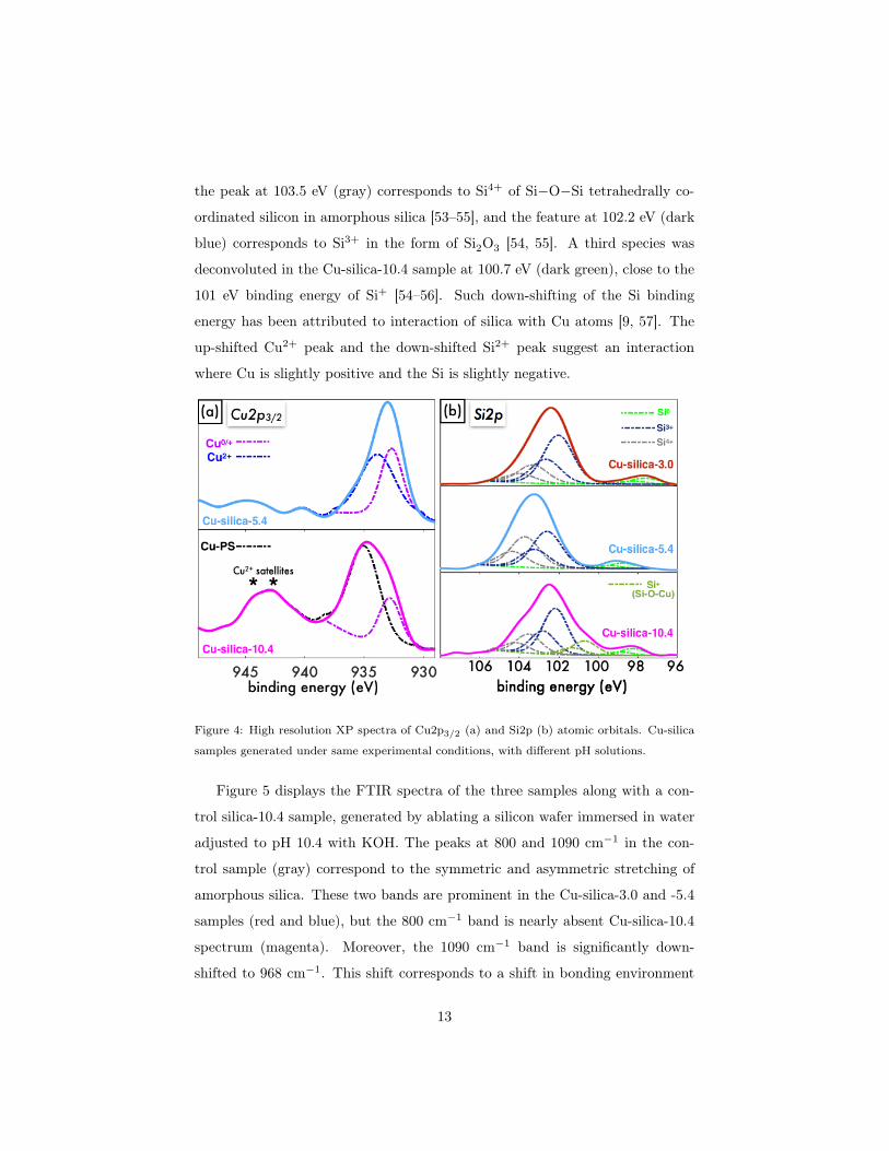
the peak at 103.5 eV (gray) corresponds to Si4+ of Si�O�Si tetrahedrally co-
ordinated silicon in amorphous silica [53–55], and the feature at 102.2 eV (dark
blue) corresponds to Si3+ in the form of Si2O3 [54, 55]. A third species was
deconvoluted in the Cu-silica-10.4 sample at 100.7 eV (dark green), close to the
101 eV binding energy of Si+ [54–56]. Such down-shifting of the Si binding
energy has been attributed to interaction of silica with Cu atoms [9, 57]. The
up-shifted Cu2+ peak and the down-shifted Si2+ peak suggest an interaction
where Cu is slightly positive and the Si is slightly negative.
Figure 4: High resolution XP spectra of Cu2p3/2 (a) and Si2p (b) atomic orbitals. Cu-silica
samples generated under same experimental conditions, with different pH solutions.
Figure 5 displays the FTIR spectra of the three samples along with a con-
trol silica-10.4 sample, generated by ablating a silicon wafer immersed in water
adjusted to pH 10.4 with KOH. The peaks at 800 and 1090 cm�1 in the con-
trol sample (gray) correspond to the symmetric and asymmetric stretching of
amorphous silica. These two bands are prominent in the Cu-silica-3.0 and -5.4
samples (red and blue), but the 800 cm�1 band is nearly absent Cu-silica-10.4
spectrum (magenta). Moreover, the 1090 cm�1 band is significantly down-
shifted to 968 cm�1. This shift corresponds to a shift in bonding environment
13
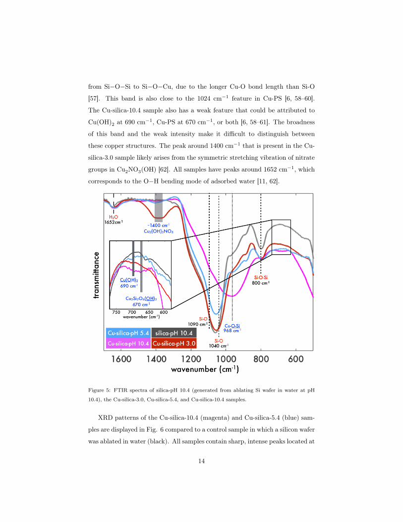
from Si�O�Si to Si�O�Cu, due to the longer Cu-O bond length than Si-O
[57]. This band is also close to the 1024 cm�1 feature in Cu-PS [6, 58–60].
The Cu-silica-10.4 sample also has a weak feature that could be attributed to
Cu(OH)2 at 690 cm�1, Cu-PS at 670 cm�1, or both [6, 58–61]. The broadness
of this band and the weak intensity make it difficult to distinguish between
these copper structures. The peak around 1400 cm�1 that is present in the Cu-
silica-3.0 sample likely arises from the symmetric stretching vibration of nitrate
groups in Cu2NO3(OH) [62]. All samples have peaks around 1652 cm�1, which
corresponds to the O�H bending mode of adsorbed water [11, 62].
Figure 5: FTIR spectra of silica-pH 10.4 (generated from ablating Si wafer in water at pH
10.4), the Cu-silica-3.0, Cu-silica-5.4, and Cu-silica-10.4 samples.
XRD patterns of the Cu-silica-10.4 (magenta) and Cu-silica-5.4 (blue) sam-
ples are displayed in Fig. 6 compared to a control sample in which a silicon wafer
was ablated in water (black). All samples contain sharp, intense peaks located at
14

28.4, 47.3, 56.1, 69.2, 76.4, and 88.1 2✓ corresponding to the (111), (222), (331),
(440), (533), and (640) planes of cubic silicon (ICDD: 04-012-7888). There is a
small, broad peak around 36 2✓ present only in the Cu-silica-10.4 sample that
corresponds to either the (111) CuO plane (35.6 2✓), the (111) plane of Cu2O
(36.5 2✓), or a Cu-PS structure [6, 58–60]. The inset shows the Gaussian peak
fit yielding a FWHM of 4.90 2✓ corresponding to a 1.78 nm diameter of the
crystalline nanoparticle, according to the Scherrer equation [63]. The FWHM
of the fcc-Si (111) peak at 28.4 2✓ was determined to be 0.25 2✓, yielding a
silicon crystalline diameter of 33.6 nm. The XRD pattern shows that there are
crystalline silicon particles present in these samples, consistent with the large
spherical particles visible in the TEM images in Fig. 1a. The absence of silica
in the XRD patterns supports the amorphous nature of the silica as evident in
the HRTEM images, Si2p XPS spectra, and FTIR spectra.
Figure 6: XRD patterns of Cu-silica-10.4, Cu-silica-5.4, and silica generated from laser ablation
of silicon wafer in water.
15

3.2. Catalytic Activity
The catalytic reduction of para-nitrophenol (PNP) by sodium borohydride
was employed as a model reaction to compare the catalytic activity of the silica-
Cu samples. Because all samples were irradiated under the same laser condi-
tions and underwent the same post-synthesis processing procedure, we assume
the amount of silica present in all samples is equivalent, and the rate constants
reported reflect the catalytically active copper particles in the samples. There-
fore, the same volume of re-dispersed pellet was added to all PNP reactions.
Experimental details and calculations for determining the catalytic rate con-
stants have been described in detail elsewhere [36, 38] and are provided in the
Supplemental Information.
Briefly, the catalytic reduction of PNP to para-aminophenol (PAP) by sodium
borohydride follows pseudo-first order reaction kinetics due to the excess of
NaBH4 added to the reaction
PNP +NaBH4(xs)Cu�silica�������! PAP. (1)
The para-phenolate ion absorbs strongly at 400 nm, allowing for the reaction
rate to be determined by monitoring its absorbance upon the addition of the
catalyst. The apparent rate constants, kapp (s�1) versus wt.% Cu from XPS
analysis are displayed in Fig. 7, with the different pH conditions labeled. There
is a linear relationship between the rate constant and the surface Cu content,
reflecting the high catalytic activity of the Cu-silica-10.4 sample which contains
the highest amount of surface Cu. While nearly no Cu was detected in the
Cu-silica-3.0 sample, it still possess catalytic activity, suggesting that the there
was Cu present in the sample, but in very small quantities.
16

Figure 7: Apparent rate constant (kapp) versus at.% Cu from XPS analysis.
4. Discussion
Wet chemical approaches to fabricating oxide supported metal nanoparticles
emphasize the importance of choosing the support, metal precursor, and solution
pH for maximizing the metal loading [64]. Determining the point of zero charge
(PZC) of the support material aids in identifying the pH conditions for optimal
interaction between the metal complex and the oxide support [64, 65]. The
PZC of a material is the pH at which the hydroxyl groups that populate the
surface of an oxide have a neutral charge. When the pH is below the PZC of
the support, the hydroxyl groups are protonated, and when the pH is above the
PZC, the hydroxyl groups are deprotonated [13, 15, 64]. Silica has a PZC of
pH 4, so when the solution pH is above this value, cationic metal complexes in
solution can adsorb onto the negatively charged surface of the silica [15].
The copper loadings reported in Table 2 demonstrate the need for basic
pH to achieve high copper loading under our synthesis conditions; even in the
weakly acidic solution at pH 5.4, little copper was found in the product. In
contrast, the Cu-silica-10.4 sample had a high copper loading of 31.5 wt%; much
17

higher than many previous reports using wet chemical techniques that typically
achieve around 10 wt.% [15, 65–69]. Previous investigations of pH-dependent
metal loading on silica that achieved copper loadings above 30 wt.% have formed
Cu-PS structures [10, 58, 70, 71], consistent with our results. However, the
wet-chemical synthesis methods used are considerably more time- and material-
consuming than our RLAL method. For instance, the method of Toupance et
al., in which a copper nitrate solution adjusted to pH 9 using ammonia mixed
with nonporous silica was stirred at room temperature for one week, required
100 times the amount of copper relative to our conditions to yield 36.6 wt.% Cu
with 4.5 nm Cu NPs [58].
In contrast to pre-synthesized fumed silica or nonporous silica spheres used in
wet chemical methods, our silica is produced in situ from laser ablation. As the
silicon atoms and clusters are ejected into solution, they may interact with other
nearby species including hydrated electrons and hydroxyl radicals, resulting in
oxidation to silica. Under basic conditions, nearby OH– ions may interact with
the oxidized silica clusters, deprotonating their surfaces [72]. The negatively
charged silica clusters attract nearby copper in the form of the bridged copper
hydroxide dimer, [Cu2(OH)2]2+, which is formed in the pH range 6.5�10.5
[15, 73]. The abundance of deprotonated silica clusters generated from laser
ablation provide numerous sites for these copper complexes to interact with,
driving the high copper loading under basic conditions.
Figure 8 displays a graphical representation of the formation mechanisms
of the copper-silica materials under the different pH conditions. We note that
the solution pH decreased from 10.4 to 8.5 and 5.4 to 4.2 during synthesis of
the Cu-silica-10.4 and -5.4 samples, respectively. The two samples generated at
final solution pH less than pH 4 are shown above the ‘silica PZC 4.0’ line [13, 15]
in Fig. 8. Under these conditions, silica clusters ablated off of the Si wafer into
solution become protonated and repel the surrounding Cu2+ ions, leading to
low amounts of Cu incorporated into the product. While it is possible that a
small amount of Cu in the product dissolves in the acidic solution following laser
synthesis (see details in the Supporting Information), this process is unlikely to
18

be the primary cause of low Cu incorporation because little Cu is incorporated
even at an initial pH of 5.4. For the particles in which Cu is incorporated into
the particles, segregation of the Cu-silica phases result in large Cu-core/silica-
shell particles with varying sizes and shell thicknesses. The formation of the
silica shell rather than silica core may be due to the higher surface energy of
silicon than copper in the liquid form. Synthesis of Cu-core/silica-shell parti-
cles by evaporating elemental Cu and Si using a high powered electron beam
resulted in phase segregation with the silicon shell forming around liquid Cu,
due to the higher surface energy of liquid silicon relative to liquid Cu [74, 75].
In contrast, the proposed formation mechanism of the Cu-silica-10.4 sample is
displayed below the line labeled ‘silica PZC 4.0’, where Cu-O-Si bonds form
due to the strong interaction between the deprotonated silica clusters and the
cationic copper hydroxide dimers. The morphology contains sheet- and needle-
like structures comprised of amorphous silica, copper phyllosilicate, or both,
decorated with sub-2 nm Cu clusters. The small size of the Cu particles likely
results from the strong interaction between the Cu nuclei and the silica clusters,
halting further Cu NP growth. The presence of sub-2 nm clusters dispersed
throughout the silica is similar to our recent report of fs-RLAL synthesis of sub-
3 nm Au NPs dispersed throughout a silica matrix under basic conditions [36].
Our observation of distinct copper-silica material structures using fs-RLAL at
different solution pH is consistent with previous RLAL studies showing a depen-
dence of Pt-Co NP properties on solution pH [29, 30] and suggests that solution
pH provides a generally applicable method to control nanomaterial properties
with RLAL.
5. Conclusion
We have synthesized copper-silica nanocomposites using a fs-RLAL approach,
with distinct copper-silica morphologies forming from different precursor solu-
tion pH conditions. The highest copper loading on silica of 31.5 wt.% achieved
with a precursor solution pH of 10.4 generated 1.52±0.75 nm Cu NPs well dis-
19
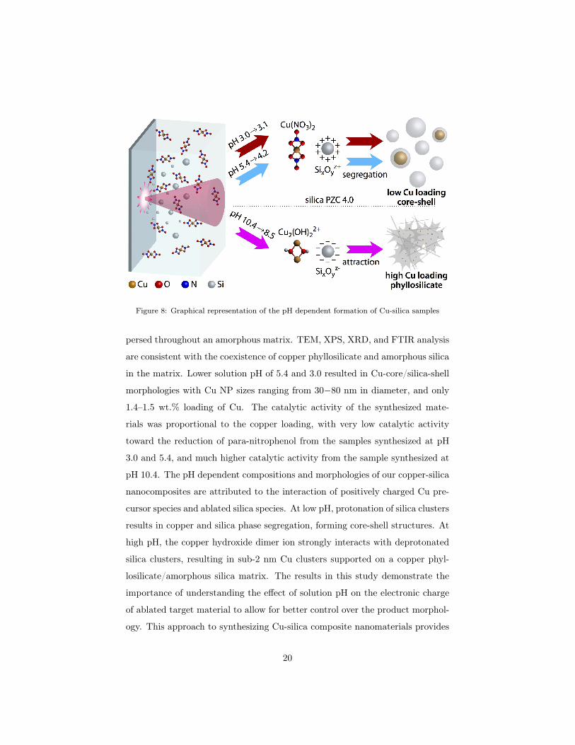
Figure 8: Graphical representation of the pH dependent formation of Cu-silica samples
persed throughout an amorphous matrix. TEM, XPS, XRD, and FTIR analysis
are consistent with the coexistence of copper phyllosilicate and amorphous silica
in the matrix. Lower solution pH of 5.4 and 3.0 resulted in Cu-core/silica-shell
morphologies with Cu NP sizes ranging from 30�80 nm in diameter, and only
1.4–1.5 wt.% loading of Cu. The catalytic activity of the synthesized mate-
rials was proportional to the copper loading, with very low catalytic activity
toward the reduction of para-nitrophenol from the samples synthesized at pH
3.0 and 5.4, and much higher catalytic activity from the sample synthesized at
pH 10.4. The pH dependent compositions and morphologies of our copper-silica
nanocomposites are attributed to the interaction of positively charged Cu pre-
cursor species and ablated silica species. At low pH, protonation of silica clusters
results in copper and silica phase segregation, forming core-shell structures. At
high pH, the copper hydroxide dimer ion strongly interacts with deprotonated
silica clusters, resulting in sub-2 nm Cu clusters supported on a copper phyl-
losilicate/amorphous silica matrix. The results in this study demonstrate the
importance of understanding the effect of solution pH on the electronic charge
of ablated target material to allow for better control over the product morphol-
ogy. This approach to synthesizing Cu-silica composite nanomaterials provides
20

valuable insight into designing RLAL reaction conditions for synthesizing addi-
tional metal-oxide nanocomposites with high metal loadings that may be used
for catalytic applications.
Acknowledgement
This work was supported by the American Chemical Society Petroleum Re-
search Fund through Grant 57799-DNI10. Microscopy was performed at the
VCU Department of Anatomy and Neurobiology Microscopy Facility, supported
by the Higher Education Equipment Trust Fund Grant No. 236160307. We
would like to acknowledge the VCU Nanomaterials Core Characterization Fa-
cility for additional characterization.
Supplemental Material
Details of PNP catalytic reactions; Details of HRTEM lattice fringe analysis;
Details of XPS composition analysis; additional TEM images; additional XPS
spectra of Si2p.
References
[1] X. Niu, Y. L, J. Tang, Y. Hu, H. Zhao, M. Lan, Electrochemical sensing
interfaces with tunable porosity for nonenzymatic glucose detection: A cu
foam case, Biosensors and Bioelectronics 51 (2014) 22 – 28. doi:https:
//doi.org/10.1016/j.bios.2013.07.032.
[2] A. M. Raspolli Galletti, C. Antonetti, M. Marracci, F. Piccinelli, B. Tellini,
Novel microwave-synthesis of cu nanoparticles in the absence of any stabi-
lizing agent and their antibacterial and antistatic applications, Appl. Surf.
Sci. 280 (2013) 610 – 618. doi:https://doi.org/10.1016/j.apsusc.
2013.05.035.
[3] Y. Lee, J. rak Choi, K. J. Lee, N. E. Stott, D. Kim, Large-scale synthesis
of copper nanoparticles by chemically controlled reduction for applications
of inkjet-printed electronics, Nanotechnology 19 (41) (2008) 415604. doi:
10.1088/0957-4484/19/41/415604.
URL https://doi.org/10.1088%2F0957-4484%2F19%2F41%2F415604
21

[4] M. B. Gawande, A. Goswami, F.-X. Felpin, T. Asefa, X. Huang, R. Silva,
X. Zou, R. Zboril, R. S. Varma, Cu and Cu-based nanoparticles: Synthesis
and applications in catalysis, Chemical Reviews 116 (6) (2016) 3722–3811.
doi:10.1021/acs.chemrev.5b00482.
[5] L. Cao, D. Raciti, C. Li, K. J. T. Livi, P. F. Rottmann, K. J. Hemker,
T. Mueller, C. Wang, Mechanistic insights for low-overpotential electrore-
duction of co2 to co on copper nanowires, ACS Catalysis 7 (12) (2017)
8578–8587. doi:10.1021/acscatal.7b03107.
[6] Y. Sheng, H. C. Zeng, Structured assemblages of single-walled 3d transition
metal silicate nanotubes as precursors for composition-tailorable catalysts,
Chemistry of Materials 27 (3) (2015) 658–667. doi:10.1021/cm502691s.
[7] G. Yin, M. Nishikawa, Y. Nosaka, N. Srinivasan, D. Atarashi, E. Sakai,
M. Miyauchi, Photocatalytic carbon dioxide reduction by copper oxide
nanocluster-grafted niobate nanosheets, ACS Nano 9 (2) (2015) 2111–2119.
doi:10.1021/nn507429e.
[8] L. Zhu, H. Li, Z. Liu, P. Xia, Y. Xie, D. Xiong, Synthesis of the 0d/3d
cuo/zno heterojunction with enhanced photocatalytic activity, The Journal
of Physical Chemistry C 122 (17) (2018) 9531–9539. doi:10.1021/acs.
jpcc.8b01933.
URL https://doi.org/10.1021/acs.jpcc.8b01933
[9] Y. Zhao, J. Zhao, Z. Su, X. Hao, Y. Li, N. Li, Y. Li, Sio2 capsulized cu
active nanoparticles: synthesis and activity study, J. Mater. Chem. A 1
(2013) 8029–8036. doi:10.1039/C3TA11281K.
[10] J. Gong, H. Yue, Y. Zhao, S. Zhao, L. Zhao, J. Lv, S. Wang, X. Ma,
Synthesis of ethanol via syngas on cu/sio2 catalysts with balanced cu0–cu+
sites, Journal of the American Chemical Society 134 (34) (2012) 13922–
13925. doi:10.1021/ja3034153.
22

[11] L.-F. Chen, P.-J. Guo, M.-H. Qiao, S.-R. Yan, H.-X. Li, W. Shen, H.-L.
Xu, K.-N. Fan, Cu/sio2 catalysts prepared by the ammonia-evaporation
method: Texture, structure, and catalytic performance in hydrogenation
of dimethyl oxalate to ethylene glycol, Journal of Catalysis 257 (1) (2008)
172 – 180. doi:https://doi.org/10.1016/j.jcat.2008.04.021.
[12] Z. Huang, F. Li, B. Chen, F. Xue, G. Chen, G. Yuan, Nitrogen-rich copoly-
meric microsheets supporting copper nanoparticles for catalyzing arylation
of n-heterocycles, Applied Catalysis A: General 403 (1) (2011) 104 – 111.
doi:https://doi.org/10.1016/j.apcata.2011.06.019.
[13] S. Eskandari, G. Tate, N. R. Leaphart, J. R. Regalbuto, Nanoparticle syn-
thesis via electrostatic adsorption using incipient wetness impregnation,
ACS Catal. 8 (11) (2018) 10383–10391. doi:10.1021/acscatal.8b03435.
[14] C. Xu, G. Chen, Y. Zhao, P. Liu, X. Duan, L. Gu, G. Fu, Y. Yuan,
N. Zheng, Interfacing with silica boosts the catalysis of copper, Nature
Commun. 9 (1) (2018) 3367. doi:10.1038/s41467-018-05757-6.
[15] L. Jiao, J. R. Regalbuto, The synthesis of highly dispersed noble and base
metals on silica via strong electrostatic adsorption: I. amorphous silica, J.
Catal. 260 (2) (2008) 329 – 341. doi:https://doi.org/10.1016/j.jcat.
2008.09.022.
[16] D. Zhang, B. Gökce, S. Barcikowski, Laser synthesis and processing of
colloids: Fundamentals and applications, Chemical Reviews 117 (5) (2017)
3990–4103. doi:10.1021/acs.chemrev.6b00468.
[17] D. Zhang, J. Liu, P. Li, Z. Tian, C. Liang, Recent advances in surfactant-
free, surface-charged, and defect-rich catalysts developed by laser
ablation and processing in liquids, ChemNanoMat 3 (8) (2017) 512–533.
doi:10.1002/cnma.201700079.
URL https://onlinelibrary.wiley.com/doi/abs/10.1002/cnma.
201700079
23

[18] S. Reichenberger, G. Marzun, M. Muhler, S. Barcikowski, Perspective of
surfactant-free colloidal nanoparticles in heterogeneous catalysis, Chem-
CatChem 11 (18) (2019) 4489–4518. doi:10.1002/cctc.201900666.
[19] D. Amans, W. Cai, S. Barcikowski, Status and demand of research to bring
laser generation of nanoparticles in liquids to maturity, Applied Surface
Science 488 (2019) 445 – 454. doi:https://doi.org/10.1016/j.apsusc.
2019.05.117.
[20] B. Rethfeld, D. S. Ivanov, M. E. Garcia, S. I. Anisimov, Modelling ultrafast
laser ablation, J. Phys. D 50 (19) (2017) 193001. doi:10.1088/1361-6463/
50/19/193001.
URL https://doi.org/10.1088%2F1361-6463%2F50%2F19%2F193001
[21] M. Dell’Aglio, R. Gaudiuso, O. De Pascale, A. De Giacomo, Mecha-
nisms and processes of pulsed laser ablation in liquids during nanopar-
ticle production, Applied Surface Science 348 (2015) 4 – 9, advanced Syn-
thesis of Functional Nanoparticles by Lasers in Liquids – From Funda-
mentals to Application in Catalysis, Energy Science, and Biomedicine.
doi:https://doi.org/10.1016/j.apsusc.2015.01.082.
[22] B. Gökce, V. Amendola, S. Barcikowski, Opportunities and challenges for
laser synthesis of colloids, ChemPhysChem 18 (9) (2017) 983–985. doi:
10.1002/cphc.201700310.
[23] J. M. J. Santillán, F. A. Videla, M. B. Fernández van Raap, D. C.
Schinca, L. B. Scaffardi, Size dependent cu dielectric function for plas-
mon spectroscopy: Characterization of colloidal suspension generated by
fs laser ablation, Journal of Applied Physics 112 (5) (2012) 054319. doi:
10.1063/1.4751328.
[24] J. M. J. Santillán, F. A. Videla, M. B. Fernández van Raap, D. C. Schinca,
L. B. Scaffardi, Analysis of the structure, configuration, and sizing of cu
and cu oxide nanoparticles generated by fs laser ablation of solid target in
24

liquids, Journal of Applied Physics 113 (13) (2013) 134305. doi:10.1063/
1.4798387.
[25] P. Liu, Z. Li, W. Cai, M. Fang, X. Luo, Fabrication of cuprous oxide
nanoparticles by laser ablation in pvp aqueous solution, RSC Adv. 1 (2011)
847–851. doi:10.1039/C1RA00261A.
[26] R. Tilaki, A. Iraji zad, S. Mahdavi, Size, composition and optical proper-
ties of copper nanoparticles prepared by laser ablation in liquids, Applied
Physics A 88 (2) (2007) 415–419. doi:10.1007/s00339-007-4000-2.
[27] E. Jiménez, K. Abderrafi, J. Martínez-Pastor, R. Abargues, J. L. Valdés,
R. Ibáñez, A novel method of nanocrystal fabrication based on laser abla-
tion in liquid environment, Superlattices and Microstructures 43 (5) (2008)
487 – 493, proceedings of the 7th International Conference on Physics of
Light-Matter Coupling in Nanostructures. doi:10.1016/j.spmi.2007.
06.025.
[28] S. Hu, G. Goenaga, C. Melton, T. A. Zawodzinski, D. Mukherjee,
Ptco/coox nanocomposites: Bifunctional electrocatalysts for oxygen reduc-
tion and evolution reactions synthesized via tandem laser ablation synthesis
in solution-galvanic replacement reactions, Applied Catalysis B: Environ-
mental 182 (2016) 286 – 296. doi:10.1016/j.apcatb.2015.09.035.
[29] S. Hu, M. Tian, E. L. Ribeiro, G. Duscher, D. Mukherjee, Tandem laser
ablation synthesis in solution-galvanic replacement reaction (lasis-grr) for
the production of ptco nanoalloys as oxygen reduction electrocatalysts, J.
Power Sources 306 (2016) 413 – 423. doi:10.1016/j.jpowsour.2015.11.
078.
[30] S. Hu, K. Cheng, E. L. Ribeiro, K. Park, B. Khomami, D. Mukherjee, A
facile and surfactant-free route for nanomanufacturing of tailored ternary
nanoalloys as superior oxygen reduction reaction electrocatalysts, Catal.
Sci. Technol. 7 (2017) 2074–2086. doi:10.1039/C7CY00073A.
25

[31] B. M. Hunter, J. D. Blakemore, M. Deimund, H. B. Gray, J. R. Winkler,
A. M. Müller, Highly active mixed-metal nanosheet water oxidation cata-
lysts made by pulsed-laser ablation in liquids, J. Am. Chem. Soc. 136 (38)
(2014) 13118–13121. doi:10.1021/ja506087h.
URL https://doi.org/10.1021/ja506087h
[32] B. M. Hunter, W. Hieringer, J. R. Winkler, H. B. Gray, A. M. Müller,
Effect of interlayer anions on [nife]-ldh nanosheet water oxidation activity,
Energy Environ. Sci. 9 (5) (2016) 1734–1743. doi:10.1039/C6EE00377J.
URL http://dx.doi.org/10.1039/C6EE00377J
[33] C. W. Roske, J. W. Lefler, A. M. Müller, Complex nanomineral formation
utilizing kinetic control by plal, Journal of Colloid and Interface Science
489 (2017) 68 – 75, laser Synthesis. doi:https://doi.org/10.1016/j.
jcis.2016.08.079.
[34] E. Jiménez, K. Abderrafi, R. Abargues, J. L. Valdés, J. P. Martínez-Pastor,
Laser-ablation-induced synthesis of sio2-capped noble metal nanoparti-
cles in a single step, Langmuir 26 (10) (2010) 7458–7463. doi:10.1021/
la904179x.
[35] V. A. Ermakov, E. Jimenez-Villar, J. M. C. d. Silva Filho, E. Yassitepe,
N. V. V. Mogili, F. Iikawa, G. F. de Sá, C. L. Cesar, F. C. Marques, Size
control of silver-core/silica-shell nanoparticles fabricated by laser-ablation-
assisted chemical reduction, Langmuir 33 (9) (2017) 2257–2262. doi:10.
1021/acs.langmuir.6b04308.
[36] M. G. John, K. M. Tibbetts, One-step femtosecond laser ablation synthesis
of sub-3âĂŕnm gold nanoparticles stabilized by silica, Applied Surface Sci-
ence 475 (2019) 1048 – 1057. doi:https://doi.org/10.1016/j.apsusc.
2019.01.042.
[37] P. Hervés, M. Pérez-Lorenzo, L. M. Liz-Marzán, J. Dzubiella, Y. Lu,
M. Ballauff, Catalysis by metallic nanoparticles in aqueous solution: model
reactions, Chem. Soc. Rev. 41 (2012) 5577. doi:10.1039/C2CS35029G.
26

[38] S. Wunder, F. Polzer, Y. Lu, Y. Mei, M. Ballauff, Kinetic analysis of cat-
alytic reduction of 4-nitrophenol by metallic nanoparticles immobilized in
spherical polyelectrolyte brushes, J. Phys. Chem. C 114 (19) (2010) 8814–
8820. doi:10.1021/jp101125j.
[39] V. K. Meader, M. G. John, C. J. Rodrigues, K. M. Tibbetts, Roles of free
electrons and h2o2 in the optical breakdown-induced photochemical reduc-
tion of aqueous [aucl4]âĹŠ, The Journal of Physical Chemistry A 121 (36)
(2017) 6742–6754. doi:10.1021/acs.jpca.7b05370.
[40] V. K. Meader, M. G. John, L. M. Frias Batista, S. Ahsan, K. M. Tib-
betts, Radical chemistry in a femtosecond laser plasma: Photochemical
reduction of ag+ in liquid ammonia solution, Molecules 23 (3). doi:
10.3390/molecules23030532.
URL https://www.mdpi.com/1420-3049/23/3/532
[41] T. A. Lastovina, A. P. Budnyk, G. A. Khaishbashev, E. A. Kudryavtsev,
A. V. Soldatov, Copper-based nanoparticles prepared from copper (ii) ac-
etate bipyridine complex, J. Serbian Chem. Soc. 81 (7) (2016) 751–762.
[42] X. Wang, K. Ma, L. Guo, Y. Tian, Q. Cheng, X. Bai, J. Huang, T. Ding,
X. Li, Cu/zno/sio2 catalyst synthesized by reduction of zno-modified cop-
per phyllosilicate for dimethyl ether steam reforming, Applied Catalysis A:
General 540 (2017) 37 – 46. doi:https://doi.org/10.1016/j.apcata.
2017.04.013.
[43] M. C. Biesinger, Advanced analysis of copper x-ray photoelectron spectra,
Surf. Interf. Anal. 49 (13) (2017) 1325–1334. doi:10.1002/sia.6239.
[44] M. C. Biesinger, L. W. Lau, A. R. Gerson, R. S. Smart, Resolving surface
chemical states in xps analysis of first row transition metals, oxides and
hydroxides: Sc, ti, v, cu and zn, Applied Surface Science 257 (3) (2010)
887 – 898. doi:https://doi.org/10.1016/j.apsusc.2010.07.086.
27
[45] M. Marques, A. Ferraria, J. Correia, A. Botelho do Rego, R. Vilar, Xrd, xps
and sem characterisation of cu–nbc nanocomposite produced by mechanical
alloying, Materials Chemistry and Physics 109 (1) (2008) 174 – 180. doi:
https://doi.org/10.1016/j.matchemphys.2007.10.032.
[46] Y. H. Kim, D. K. Lee, H. G. Cha, C. W. Kim, Y. C. Kang, Y. S.
Kang, Preparation and characterization of the antibacterial cu nanopar-
ticle formed on the surface of sio2 nanoparticles, The Journal of Physical
Chemistry B 110 (49) (2006) 24923–24928. doi:10.1021/jp0656779.
[47] J. F. Moulder, W. F. Stickle, P. E. Sobol, K. D. Bomben, Handbook of X-
ray Photoelectron Spectroscopy, Perkin-Elmer Corp., Physical Electronics
Division, 1979.
[48] D. Tahir, S. Tougaard, Electronic and optical properties of cu, CuO and
cu2o studied by electron spectroscopy, Journal of Physics: Condensed Mat-
ter 24 (17) (2012) 175002. doi:10.1088/0953-8984/24/17/175002.
[49] R.-P. Ye, L. Lin, J.-X. Yang, M.-L. Sun, F. Li, B. Li, Y.-G. Yao, A new
low-cost and effective method for enhancing the catalytic performance of
cu–sio 2 catalysts for the synthesis of ethylene glycol via the vapor-phase
hydrogenation of dimethyl oxalate by coating the catalysts with dextrin,
Journal of Catalysis 350 (2017) 122–132. doi:10.1016/j.jcat.2017.02.
018.
[50] X. Li, J. Zhang, M. Zhang, W. Zhang, M. Zhang, H. Xie, Y. Wu, Y. Tan,
The support effects on the direct conversion of syngas to higher alco-
hol synthesis over copper-based catalysts, Catalysts 9 (2). doi:10.3390/
catal9020199.
[51] X. Huang, M. Ma, S. Miao, Y. Zheng, M. Chen, W. Shen, Hydrogenation
of methyl acetate to ethanol over a highly stable cu/sio2 catalyst: Reac-
tion mechanism and structural evolution, Applied Catalysis A: General 531
(2017) 79 – 88. doi:https://doi.org/10.1016/j.apcata.2016.12.006.
28

[52] S. Dahle, L. Wegewitz, F. Qi, A. Weber, W. Maus-Friedrichs, Silicon diox-
ide coating of titanium dioxide nanoparticles from dielectric barrier dis-
charge in a gaseous mixture of silane and nitrogen, Plasma Chemistry and
Plasma Processing 33. doi:10.1007/s11090-013-9472-6.
[53] F. Lamastra, S. Mori, V. Cherubini, M. Scarselli, F. Nanni, A new green
methodology for surface modification of diatomite filler in elastomers,
Materials Chemistry and Physics 194 (2017) 253–260. doi:10.1016/j.
matchemphys.2017.03.050.
[54] J. Won Ma, W.-J. Lee, J. M. Bae, K. Jeong, S. Hoon Oh, J.-H. Kim,
S.-h. Kim, J.-H. Seo, J.-P. Ahn, H. Kim, M.-H. Cho, Carrier mobility
enhancement of tensile strained si and sige nanowires via surface defect
engineering, Nano letters 15. doi:10.1021/acs.nanolett.5b01634.
[55] A. Al-Kattan, Y. Ryabchikov, T. Baati, V. Chirvony, J. F. Sánchez Royo,
M. Sentis, D. Braguer, V. Timoshenko, M.-A. Estève, A. V. Kabashin,
Ultrapure laser-synthesized si nanoparticles with variable oxidation state
for biomedical applications, J. Mater. Chem. B 4 (2016) 7852–7858. doi:
10.1039/C6TB02623K.
[56] M. Bashouti, K. Sardashti, J. Ristein, S. Christiansen, Early stages of oxide
growth in h-terminated silicon nanowires: Determination of kinetic behav-
ior and activation energy, Physical chemistry chemical physics : PCCP 14
(2012) 11877–81. doi:10.1039/c2cp41709j.
[57] Y. Kong, H. Zhu, G. Yang, X. Guo, W. Hou, Q. Yan, M. Gu, C. Hu,
Investigation of the structure of mcm-41 samples with a high copper
content, Advanced Functional Materials 14 (8) (2004) 816–820. doi:
10.1002/adfm.200305111.
[58] T. Toupance, M. Kermarec, J.-F. Lambert, C. Louis, Conditions of forma-
tion of copper phyllosilicates in silica-supported copper catalysts prepared
by selective adsorption, The Journal of Physical Chemistry B 106 (9) (2002)
2277–2286. doi:10.1021/jp013153x.
29

[59] W. Di, J. Cheng, S. Tian, J. Li, J. Chen, Q. Sun, Synthesis and character-
ization of supported copper phyllosilicate catalysts for acetic ester hydro-
genation to ethanol, Applied Catalysis A: General 510 (2016) 244 – 259.
doi:https://doi.org/10.1016/j.apcata.2015.10.026.
[60] Z.-Q. Wang, Z.-N. Xu, S.-Y. Peng, M.-J. Zhang, G. Lu, Q.-S. Chen,
Y. Chen, G.-C. Guo, High-performance and long-lived cu/sio2 nanocat-
alyst for co2 hydrogenation, ACS Catalysis 5 (7) (2015) 4255–4259. doi:
10.1021/acscatal.5b00682.
[61] Z. Huang, F. Cui, H. Kang, J. Chen, X. Zhang, C. Xia, Highly
dispersed silica-supported copper nanoparticles prepared by precipita-
tionâĹŠgel method: A simple but efficient and stable catalyst for glyc-
erol hydrogenolysis, Chemistry of Materials 20 (15) (2008) 5090–5099.
doi:10.1021/cm8006233.
[62] H. Li, L. Ban, Z. Wang, P. Meng, Y. Zhang, R. Wu, Y. Zhao, Regulation of
cu species in cuo/sio2 and its structural evolution in ethynylation reaction,
Nanomaterials 9 (842). doi:10.3390/nano9060842.
[63] A. Monshi, M. R. Foroughi, M. Monshi, Modified scherrer equation to
estimate more accurately nano-crystallite size using xrd, World Journal of
Nano Science and Engineering 2 (2012) 154–160. doi:10.4236/wjnse.
2012.23020.
[64] P. Mäki-Arvela, D. Y. Murzin, Effect of catalyst synthesis parameters on
the metal particle size, Appl. Cat. A: General 451 (2013) 251 – 281. doi:
https://doi.org/10.1016/j.apcata.2012.10.012.
[65] A. Wong, Q. Liu, S. Griffin, A. Nicholls, J. R. Regalbuto, Synthesis of ultra-
small, homogeneously alloyed, bimetallic nanoparticles on silica supports,
Science 358 (6369) (2017) 1427–1430. doi:10.1126/science.aao6538.
[66] L. Trouillet, T. Toupance, F. Villain, C. Louis, In situ characterization of
the coordination sphere of cuii complexes supported on silica during the
30
preparation of cu/sio2 catalysts by cation exchange, Phys. Chem. Chem.
Phys. 2 (2000) 2005–2014. doi:10.1039/A909427J.
[67] E. Guerreiro, O. Gorriz, G. Larsen, L. Arrúa, Cu/sio2 catalysts for
methanol to methyl formate dehydrogenation: A comparative study
using different preparation techniques, Applied Catalysis A: General
204 (1) (2000) 33 – 48. doi:https://doi.org/10.1016/S0926-860X(00)
00507-X.
[68] C. Rudolf, F. Abi-Ghaida, B. Dragoi, A. Ungureanu, A. Mehdi, E. Du-
mitriu, An efficient route to prepare highly dispersed metallic copper
nanoparticles on ordered mesoporous silica with outstanding activity for
hydrogenation reactions, Catal. Sci. Technol. 5 (2015) 3735–3745. doi:
10.1039/C5CY00428D.
[69] N. H. Khdary, M. A. Ghanem, M. E. Abdesalam, M. M. Al-Garadah, Se-
questration of co2 using cu nanoparticles supported on spherical and rod-
shape mesoporous silica, Journal of Saudi Chemical Society 22 (3) (2018)
343 – 351. doi:https://doi.org/10.1016/j.jscs.2016.05.004.
[70] H. Yue, Y. Zhao, S. Zhao, B. Wang, X. Ma, J. Gong, A copper-phyllosilicate
core-sheath nanoreactor for carbon–oxygen hydrogenolysis reactions, Na-
ture Communications 4 (2013) 2339 EP –.
[71] C. van der Grift, P. Elberse, A. Mulder, J. Geus, Preparation of silica-
supported copper catalysts preparation of silica-supported copper catalysts
preparation of silica-supported copper catalysts by means of deposition-
precipitation, Appl. Cat. 59 (1990) 275–289.
[72] F. K. Crundwell, On the mechanism of the dissolution of quartz and silica
in aqueous solutions, ACS Omega 2 (3) (2017) 1116–1127. doi:10.1021/
acsomega.7b00019.
[73] M. Schreier, S. Teren, L. Belcher, J. Regalbuto, J. Miller, The nature of
31

’overexchanged’ copper and platinum on zeolites, Nanotechnology 16 (2005)
S582–91. doi:10.1088/0957-4484/16/7/036.
[74] A. V. Nomoev, S. P. Bardakhanov, M. Schreiber, D. G. Bazarova, N. A.
Romanov, B. B. Baldanov, B. R. Radnaev, V. V. Syzrantsev, Structure and
mechanism of the formation of core–shell nanoparticles obtained through
a one-step gas-phase synthesis by electron beam evaporation, Beilstein J.
Nanotechnol. 6 (2015) 874–880.
[75] N. Eustathopoulos, B. Drevet, Surface tension of liquid silicon: High or
low value?, Journal of Crystal Growth 371 (2013) 77 – 83. doi:https:
//doi.org/10.1016/j.jcrysgro.2013.02.010.
32