Embed Size (px)
Citation preview

CONTINUOUS, LOW DOSE IRRADIATION RESULTS IN NO DETRIMENT TO BONE
AFTER 4, 12, AND 28 DAYS
A Thesis
by
ALEXANDRA MARIE MARICH
Submitted to the Office of Graduate and Professional Studies of Texas A&M University
in partial fulfillment of the requirements for the degree of
MASTER OF SCIENCE
Chair of Committee, Susan Bloomfield Committee Members, Christopher Woodman
John Ford Head of Department, Melinda Sheffield-Moore
May 2018
Major Subject: Kinesiology
Copyright 2018 Alexandra M. Marich

ii
ABSTRACT
As the focus shifts to potential Lunar and Mars missions, there are many aspects of the
space environment that are still unknown, one of those being radiation exposure. There are many
radiation studies examining the effects of acute, high dose radiation on bone; however, it has been
discovered that the total exposure astronauts will experience will be much lower than the doses
these studies have utilized. The purpose of this study was to determine the effect of a continuous,
low-dose irradiation exposure on bone at various time points. Sixty-six male C57Bl/6 mice, aged
47-49 weeks, were placed in the continuous radiation field for either four, twelve, or twenty-eight
days. The radiation field was created by locating four activated 60Co wires emitting gamma
radiation around a mouse rack in a lead-shielded room. At a dose rate of 6.49 mGy/day, the total
cumulative dose was approximately 25.6 mGy for four days, 77.88 mGy for 12 days, and 181.72
mGy for 28 days. Fluorochrome calcein labels were injected to obtain bone formation rate post
termination. DXA scans were performed prior to radiation exposure and again just before
termination to determine bone mineral density and body composition. At termination, tibiae and
femurs were collected from all animals for histomorphometry assessments. Also, osteocyte
apoptosis was quantified in cancellous bone via staining for Annexin V. Continuous radiation
exposure at this low dose rate did not have any impact on total body bone mineral density, lean
mass, fat mass, and percent fat at any time point. There were also no differences in cortical or
cancellous bone formation rate at any time point in radiation-exposed mice vs. control animals.
Also, bone volume and trabecular microarchitecture were not affected by radiation at any time
point. Osteoid and osteoclast surfaces were not different after radiation at any time point, nor were
there any differences in osteocyte apoptosis at any time point examined. These results are quite
different from those seen after acute, high-dose radiation exposures, after which decreased bone

iii
volume, depressed bone formation rate, and increased osteoclast surface are often observed. This
data begins to elucidate a time course of continuous low dose radiation impacts on bone, similar
to radiation that might be experienced during transit to Mars and alludes to the potential that it may
not be deleterious to bone health, as previously assumed.

iv
ACKNOWLEDGEMENTS
I am extremely grateful for the opportunities given to me by Dr. Bloomfield during my
last year of my undergraduate degree and throughout my master’s program. Thank you for
allowing me the opportunity to create a project that I was passionate about for my thesis, and
your mentorship throughout my degree. I will forever be grateful for you giving me the
opportunity to fall in love with the research process.
Thank you to my lab mates for your support and friendship. To Rihana Bokhari for
teaching me how to do animal work and work in the radiation field. To Corinne Metzger for
teaching me how to do all the analyses that the Bone Biology lab does. To the both of you for
being a sounding board when I was trying to initially come up with this time course study.
Finally, a thank you to my family for their support throughout my education. To my
husband who has been my cheerleader throughout my degree, and who has constantly listened to
me talk about radiation and bone for the past two years. Thank you all for helping to reach the
finish line!

v
CONTRIBUTORS AND FUNDING SOURCES
This work was supervised by a thesis committee consisting of Dr. Susan Bloomfield,
Dr. Christopher Woodmen, and Dr. John Ford.
The animals used in the study were donated by Dr. Nancy Turner whose breeding colony
was supported by an award from NASA [ NNX14AC91G (awarded to Dr. Turner)]. The costs of
the animal protocol and all materials was supported by a NASA award (NNX13AL25G) to Dr.
Bloomfield. The radiation field was designed and assembled by Dr. Ford and his students. Dr. Ford
was also in charge of dosimetry for the radiation field. The Piximus DXA scanner used was
provided by Dr. Tim Lightfoot. All tissues used for cancellous histomorphometry were sectioned
by Rihana Bokhari.

vi
TABLE OF CONTENTS
Page
ABSTRACT ............................................................................................................................... ii
ACKNOWLEDGEMENTS ....................................................................................................... iv
CONTRIBUTORS AND FUNDING SOURCES ........................................................................ v
TABLE OF CONTENTS ........................................................................................................... vi
LIST OF FIGURES .................................................................................................................. vii
LIST OF TABLES................................................................................................................... viii
CHAPTER I INTRODUCTION AND LITERATURE REVIEW ............................................... 1
Bone Biology .................................................................................................................. 1 Spaceflight Environment ................................................................................................. 3 Whole Body Radiation Effects ........................................................................................ 4 Radiation Effect on Bone ................................................................................................ 5 Significance .................................................................................................................... 8
CHAPTER II MATERIALS AND METHODS .......................................................................... 9
CHAPTER III RESULTS ......................................................................................................... 13
CHAPTER IV CONCLUSIONS............................................................................................... 19
REFERENCES ......................................................................................................................... 25
APPENDIX A .......................................................................................................................... 30
APPENDIX B .......................................................................................................................... 31

vii
LIST OF FIGURES
Page
Figure 1.1 Depiction of osteocyte apoptosis and osteoclast response ........................................ 3
Figure 1.2 Depiction of the bone response to radiation exposure...................................................7
Figure 2.1 Continuous Radiation Field Layout ....................................................................... 10
Figure 3.1 Total body BMD and body composition measured via DXA .................................. 14
Figure 3.2 Cancellous bone volume and microarchitecture at the distal femur ........................ 15
Figure 3.3 Cortical BFR at the midshaft tibia...............................................................................16
Figure 3.4 Cancellous BFR at the distal femur............................................................................ 17
Figure 3.5 Indices of bone formation and resorption activity in the distal femur....................... 18
Figure 3.6 Immunohistochemistry staining results for Annexin V positive osteocytes in the distal femur cancellous bone.......................................................... 18

viii
LIST OF TABLES
Page
Table 3.1 Average body weights at the end of the study...............................................................13
Table 3.2 Cortical %sLS/BS in the midshaft tibia........................................................................15

1
CHAPTER I
INTRODUCTION AND LITERATURE REVIEW
Bone Biology
There are two different types of bone, cortical and cancellous. Cortical bone makes
approximately 80% of the human skeleton (1). It is the hard, outer shell of bone that provides the
supportive and protective function of the skeleton (1). Cancellous bone is the lattice-like structure
encased in cortical bone located at the ends of long bones and in the vertebral bodies. With more
surface area and a more rapid turnover rate than cortical bone, cancellous bone typically responds
to changes in mechanical loading and hormonal effects more quickly than cortical bone. Therefore,
it is important to examine both types of bone since they respond differently to stressors placed on
the body.
Bone is a dynamic tissue that has the ability to respond and adapt to many situations.
The variety of cells making up bone contribute to its ability to respond to environmental changes.
Osteoblasts are derived from mesenchymal precursor cells and are responsible for the bone
formation component of bone turnover. Bone formation takes place in two stages, matrix formation
followed by mineralization (1). Osteoblasts create osteoid, which is the organic component of bone
matrix, that is then mineralized by the deposition of calcium phosphate crystals and hydroxyapatite
which, in humans, starts about 15 days later (1, 2). Since there is a lag between matrix formation
and mineralization, osteoid remains visible on the bone surfaces for several weeks, and this can be
measured via bone histomorphometry to determine newly laid bone matrix (1). Osteoblasts can
also help to regulate the differentiation and activity of osteoclasts, by releasing macrophage-colony
stimulating factor (M-CSF) and RANKL (2). Osteoclasts are multinucleated cells that attach to the
bone surface and are responsible for the bone resorption component of the bone turnover equation.

2
They create a sealed space between the cell and bone matrix, transporting protons into the sealed
space to decrease the pH, and creating an acidic environment that solubilizes the bone mineral (3).
The activity of these bone cells is further coordinated by osteocytes, which help to
orchestrate bone formation and resorption in response to many physiological and environmental
changes. Osteocytes are osteoblasts that become embedded in the bone matrix and then change
their phenotype, developing dozens of dendritic cell extensions (4). They compose over 90% of
bone cells and could survive for decades within a bone that has a slow turnover rate (5,6).
Osteocytes are able to communicate with each other, cells on the bone surface and in the marrow
cavity through the vast network of dendritic processes (7). Through these dendrites they are able
to sense and respond to mechanical load placed on bone. Osteocytes are able to regulate mineral
metabolism through the release of a variety of molecules (8). Also, they express Dkk1 and
sclerostin, which are negative regulators of the Wnt/B-catenin pathway, inhibiting osteoblast
activity (8). A major recruiter of osteoclasts is osteocyte apoptosis due to the release of RANKL
(8). Osteocyte apoptosis can be induced by glucocorticoids, various cytokines, mechanical
overload, decrease in estrogen, hypoxia, immobilization, and nutrient limitation (5). Osteocyte
apoptosis is necessary to maintain healthy bone turnover; however, if there is excessive osteocyte
apoptosis, high bone resorption can result in bone loss (Figure 1.1).

3
Figure 1.1: Depiction of osteocyte apoptosis and the effect on osteoclasts (used with permission from 9)
There are dramatic changes in bone cell activity and bone structure as an animal or human
age. This specific study uses older male mice; therefore, it is important to take into account what
aging does to bone. As an animal grows bone mass increases rapidly, until approximately 16-20
weeks of age in which they reach skeletal maturity, after which bone mass can begin to slightly
decline with age. Bone loss due to aging differs between cortical and cancellous bone: typically,
trabecular number decreases rather than thickness in cancellous bone and cortical porosity is
increased (8,10). Also, with age the presence of reactive oxygen species (ROS) is increased in the
bone microenvironment, which can contribute osteocyte apoptosis and increase osteoclasts
resulting in an increase in resorption (11).
Spaceflight Environment
The spaceflight environment poses a number of health risks to astronauts including
microgravity and radiation exposure. We have limited information about the health effects of the
radiation sources and doses relevant to spaceflight. The major sources of radiation in deep space

4
are solar particle events (SPE) and galactic cosmic radiation (GCR). SPE’s result in large fluxes
of energetic particles, but, they are predictable and easy to shield against (12).
The GCR environment consists of ~98% protons and heavier ions, 88% of which are
hydrogen, 10% helium, and remaining 2% heavy ions, and ~2% electrons and positrons (12). GCR
is not easily shielded against and results in a continuous dose rate as low as 180 to 225 mGy/day
(13). The total mission radiation dose for a round trip Mars surface mission will result in
approximately 1.01 Gy of exposure, whereas a Lunar mission results in a GCR exposure of 380
mGy, during a solar minimum (13, 14). Therefore, GCR poses the greatest to human health.
Importantly, there is limited information available on the effects exposure to continuous, low-dose
radiation, particularly for bone.
Whole Body Radiation Effects
Earth-based radiation exposure is most known and commonly used in the treatment of
cancer. Radiation doses for radiotherapy are delivered in fractionated doses ranging from 2-20
Gy/fractionation, whereas acute studies can deliver as much as 60 Gy in a single dose, which
results in extremely different biological effects (15). There are a variety of effects that can be seen
throughout the body. After 16 Gy of radiation to the heart in 8-12-week-old male mice,
microvascular density was significantly decreased, which was accompanied by significant
endothelial damage (16). This damage was not restored after treatment with bone marrow-derived
endothelial progenitor cells (16). This is a great concern since this vasculature is not found to be
restored following irradiation, leading to decreased blood flow and damage to the heart (17). It has
also been found that there is both a rapid burst in free radicals and reactive oxygen species (ROS)
immediately following irradiation and prolonged increases in ROS and reactive nitrogen species
(RNS) for several days post-irradiation (18). This increase in oxidative species can result in DNA

5
damage and gene mutations in cells. DNA double strand breaks is the most common DNA damage
seen following ionizing radiation, and they pose the greatest risk to cell cycle survival (19). It has
been found that DNA damage rises linearly with dose, suggesting that there is a dose dependent
relationship with the effects of radiation (20).
Results of studies examining lower doses of radiation more relevant to the space
environment demonstrate a potentially different response then acute, high doses. When 10 cGy of
low-dose, low-LET gamma is delivered over 48 hours to normal AG1522 and GM1603 human
skin fibroblasts, the number of micronuclei, which result from un-rejoined DNA double strand
breaks, was significantly decreased (21). The researchers hypothesized that this decrease could be
due to an increase in antioxidant activity seen at this dose-rate (21). A similar effect has been
observed in immune function following low-dose, low-LET radiation exposure. Comparing the
immune response of an acute 1.6 Gy/min dose to a continuous low-dose-rate gamma irradiation at
1.2mGy/hour for three weeks in five week old female mice results in vastly different responses
(22). After acute irradiation the number of abnormal immune cells increased, while those that had
been exposed to continuous low-dose irradiation, no abnormal immune cells were detected (22).
In a study examining the impact of various doses of gamma radiation on splenocytes in 6-8 week
old female mice, they found that at a low dose (0.01 Gy) decreased apoptosis in splenocyte
subpopulations (specifically natural killer cells and dendritic cells), whereas 2 Gy of radiation
resulted in increased apoptosis in all splenocyte subpopulations (23). These data suggests that the
physiological response to low-dose radiation may be different than that of high doses.
Radiation Effect on Bone
Most radiation studies to date examining bone outcomes utilized higher, acute doses of
radiation. These studies demonstrate bone loss due to radiation, especially trabecular bone loss.

6
With doses as low as 2 Gy, there is a 29% loss in trabecular bone volume, due to a decrease in
trabecular number and an increase in trabecular spacing (24). After the delivery of an acute dose
of 2 Gy X-ray radiation to 20 week old male mice, the presence of early stage apoptotic osteocytes
increases in the trabecular bone compartment of the irradiated limb (25). This possibly contributed
to the observed increase in osteoclast number and subsequent decrease in trabecular bone volume
after irradiation (25). In another study using 2 Gy of gamma radiation on 17-week-old male mice,
there was an increase in osteoclast number (26). Also, with 2 Gy of X-ray radiation on 13-week-
old female mice, not only was there an increase in osteoclast number but also an increase of eroded
surface (27). This is concerning since osteoclasts are the bone resorbing cells, and if this increase
in osteoclasts is not matched by an increase in osteoblast activity, then net bone loss can occur
(Figure 1.2). Also, reactive oxygen species increase in the bone marrow cavity following gamma
radiation exposure, causing apoptosis of marrow cells which can be a contributing factor in the
increase of osteoclasts (26). The results of these studies, however, are much different than those
observed following even lower doses of radiation exposures.

7
Figure 1.2: Depiction of the bone response to radiation exposure. Following radiation exposure there is an increase in osteoclasts, and a decrease in osteoblasts and osteocytes (used with permission from 28).
A full year after an acute, non-targeted dose of 4.4 cGy delivered to 19-week-old male
mice, a 56% increase in trabecular bone volume, 16% increase in trabecular number, and a 17%
decrease in trabecular spacing in the distal femur is seen (29). These results are opposite than those
seen at higher, acute doses and suggest to a positive effect on bone at lower doses of radiation.
Exposing 14-week-old female mice to a dose rate of 8.5 cGy/day for 20 days, resulting in a total
dose of 1.7 Gy, produces no change in trabecular bone volume or trabecular microarchitecture
properties (30). Also, there was no increase in osteoclasts or osteoblasts following this continuous
radiation exposure (30). The results of these two studies paint a different picture of the effects of
continuous, low dose radiation on bone, suggesting it might not be as detrimental as higher doses
appear to be. Nonetheless, these studies still do not reach the prolonged low dose rate expected
during long-duration space missions that we aimed to mimic in our radiation field.

8
Significance
There are no published data describing a time course of the effects of continuous, low
dose irradiation (CIRR) on bone; therefore, my goal was to examine total body and site-specific
changes in bone at different durations of living in a CIRR field. The time points of 4, 12, and 28
days were chosen to establish a possible mechanism for how bone is being affected by the CIRR
field. Additionally, I aimed to study this effect in middle-aged mice to better represent the human
population that would likely embark on deep space exploration missions. An understanding of the
effects of low dose, continuous radiation is critical for discerning the risks astronauts could be
exposed to outside the protection of Earth’s orbit.
I hypothesized that continuous low-dose irradiation would not have an effect on cortical
bone formation rate; however, I hypothesized there will be a decrease in cancellous bone formation
rate (BFR) and therefore a decrease in cancellous bone volume. Finally, I expect there will also be
an increase in cancellous osteoclast surface and the prevalence of apoptotic osteocytes following
irradiation. Specifically, there would be the greatest increase of apoptotic osteocytes at the 4 day
time point. I hypothesized changes in BFR and osteoclast surface would be most different at day
12, while changes in bone volume would not be apparent until 28 days of radiation exposure.

9
CHAPTER II
MATERIALS AND METHODS
Animals: Sixty-six male wild type C57Bl/6 mice, aged 47-49 weeks, were obtained from
an in-house breeding colony and housed in a 12-hour light/dark cycle in an institutionally approved
animal facility. After a four-week acclimation to a purified rodent chow (AIN93G; Research Diets,
Inc, New Brunswick NJ), animals were randomly assigned to either the CIRR treatment (RAD) or
to a non-irradiated control (CON) group. Animals were terminated at one of three time points:
after four, twelve, or twenty-eight days of living in the radiation field. Each group had an n of 10-
12 animals. DXA whole body scans were performed on the same day as the start of radiation and
just prior to termination. Fluorochrome calcein labels (Sigma Aldrich, St Louis, MO) were injected
intraperitoneally prior to entering the radiation field for animals assigned to the four day time point,
and at 6 and 2 days prior to termination in mice assigned to the remaining time points. At the
completion of each duration of CIRR, animals were anesthetized via inhaled isoflurane, euthanized
via thoracotomy, and tissues were collected for analyses. All procedures were approved by the
Texas A&M Institutional Animal Care and Use Committee (IACUC).
Radiation Field: The CIRR field was set up in a dedicated animal facility room, formerly
used as the x-ray procedure room; lead-shielded walls prevented any leakage of gamma radiation
to adjacent rooms or hallways. The radiation for the field was created by activating 60Co wires, so
that they would emit gamma radiation. Four wires were placed in the corners to surround the rack
containing animal cages to create an equivalent exposure for all animals in the field (Figure 2.1).
The cart also contained standard dosimeters, 20 placed at various points on the cart, which
measured the dose rate of the radiation in the field. The animals were removed from the field for
10
approximately five minutes per day for health checks. The animals in the field experienced
approximately a dose of 6.49 mGy/day, which was a total of 25.6 mGy for four days, 77.88 mGy
for 12 days, and 181.72 mGy for 28 days.
Figure 2.1: Continuous Radiation Field Layout (with permission from R. Bokhari)
DXA: Dual-energy x-ray absorptiometry (DXA) (GE Lunar Prodigy; Madison, Wisconsin)
with small animal software was used to measure whole body bone mineral density, lean and fat
mass in animals prior to the start radiation and the day before termination. Animals were
anesthetized via inhaled isoflurane for the duration of the scan (~5 minutes) and positioned prone
on the scanner bed. The whole-body bone mineral density (BMD) value reported does not include
the skull and tail.
Dynamic histomorphometry assessment: Excised, undemineralized distal femurs saved for
histomorphometry were stored in 10% formalin solution for 24 hours and then switched to 70%
ethanol at 4°C. Samples were then serially dehydrated and embedded in methyl methacrylate (JT
Baker, Avantor Performance Materials, Center Valley, PA). Thin sections (4 and 8 microns) were
cut on a motorized Leica RM2255 microtome and mounted on slides. Image analysis was

11
performed using an epifluorescent microscope and interfaced with an OsteoMeasure Analysis
System, software V3.3 (OsteoMetrics, Inc., Atlanta, GA, USA), interfaced with an Olympus DP73
camera. For cancellous dynamic analysis, frontal 8 µm thick slices of the distal femur were
assessed at 200X magnification, 150 um from the growth plate and a 2.4 mm region of interest,
for single- (sLS) and double- labeled surfaces (dLS) and inter-label width (the mean distance
between two labels) at all doubly-labelled sites using standard methods (31). From these primary
data, % mineralized surface/total surface and mineral apposition rate (MAR, um/day) are
calculated. Percent mineralized surface is calculated as [(sLS/2) + dLS]/total surface. For the
four-day time point, only single-labeled surface (%sLS/BS) could be assessed, as the mice were
only given one injection of flurochrome label on day “zero” at the start of radiation. Bone
formation rate (BFR) was calculated as %MS/BS x MAR and expressed as um3/um2/d. For cortical
bone analysis, 100 µm cross-sectional slices of the midshaft tibia were cut on a Buehler saw and
then were assessed for %MS/BS and MAR, enabling calculation of BFR at the periosteal and
endocortical surfaces. For samples containing no double label, disallowing a direct measure of
MAR to use for calculating BFR, an imputed value of 0.02 (um/d) for cancellous and 0.02 (um/d)
for the endocortical surface and 0.06 (um/d) for the periosteal surface of cortical bone was used.
These imputed values were the lowest observed values for MAR on that bone surface across all
samples .
Static cancellous histomorphometry assessment: For cancellous bone analysis 4 µm frontal
sections of the distal femur were Von Kossa stained with a tetrachrome counterstain and assessed
for % osteoclast surface, osteoid surface and cancellous bone volume (%BV/TV) and trabecular
microarchitecture assessment (trabecular number, thickness and separation) at 400X using

12
standard methods in the same region of interest as specified above for the fluorochrome label
analyses (29).
Immunohistochemical staining for apoptosis marker Annexin V: Contralateral distal femurs
for immunohistochemistry fixed in 10% formalin solution for 24 hours and then switched to 70%
ethanol at 4°C. Whole femurs were decalcified in a formic acid/sodium citrate solution for
approximately 7 days, paraffin embedded, and sectioned to 4 µm thickness. Sections were
immunostained for Annexin V to quantify apoptotic osteocytes, using an avidin-biotion method as
previously described, with an antibody dilution of 1:200 (32). Cells were counterstained with
methyl green. Ten fields of view of the distal femur cancellous region, 150 um from the growth
plate, were analyzed. All osteocyte counts were performed using OsteoMeasure Analysis System
software V3.3 (OsteoMetrics, Inc., Atlanta, GA, USA) interfaced with an Olympus DP73 camera.
Statistical Analysis: All data sets were tested for normality using normality plots with tests.
Independent t-tests were used to compare CON and RAD animals’ values at each time point, for
normally distributed data; if data were not normally distributed, nonparametric Independent
Samples Kruskal-Wallis tests were used to compare group means. Although a 2X3 factorial could
have been used to test for changes over time as well, I chose to focus on the impact of radiation
within each time point with t-tests or the non-parametric equivalent, to maximize statistical power
of these comparisons. Statistical significance was accepted at p<0.05. All statistical analyses were
completed with SPSS (IBM, Armonk, NY).

13
CHAPTER III
RESULTS
Animals: After four, twelve, and twenty-eight days, there were no significant differences
in body weight across time points when pooling animals across groups (4 day p=0.672, 12 day
p=0.748, 28 day p=0.364; Table 3.1). The animals maintained normal eating and grooming
patterns and appeared to be in good health; there were no animal deaths during the protocol.
Table 3.1: Average body weights at the end of the study.
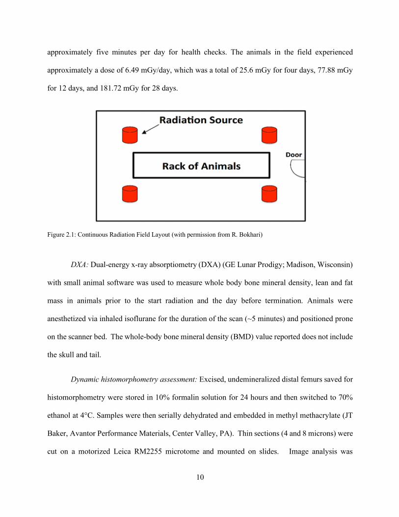
Radiation resulted in no differences in body composition at all time points compared to
age-matched controls: There were no differences between means for total body bone mineral
density (BMD) in CIRR-exposed mice when compared to age matched controls (4 day p=0.387,
12 day p=0.847, 28 day p=0.924; Figure 3.1A). There were also no differences in lean mass (4 day
p=0.387, 12 day p=0.328, 28 day p=0.416; Figure 3.1B), fat mass (4 day p=0.867, 12 day p=0.133,
28 day p=0.809; Figure 3.1C), and percent fat (4 day p=0.744, 12 day p=0.088, 28 day p=0.715;
Figure 3.1D) between radiation and CON mice.
14
Figure 3.1: Total body BMD and body composition measured via DXA (dual-energy X-ray absorptiometry). Effects of CIRR exposure on (A) total body BMD (B) total body lean mass (C) total body fat mass (D) % body fat. There were no significant differences between RAD and CON for any variable across all time points. DXA=
Cancellous microarchitecture remained unaltered at each time point of CIRR: There were
no significant differences in cancellous bone volume (%BV/TV) at any time point (4 day p=0.343,
12 day p=0.723, 28 day p=0.189; Figure 3.2A) in CIRR-exposed mice compared to CON. There
were also no significant differences in trabecular thickness (4 day p=0.981, 12 day p=0.063, 28
day p=0.497; Figure 3.2B), trabecular separation (4 day p=0.601, 12 day p=0.931, 28 day p=0.612;
Figure 3.2C), or trabecular number (4 day p=0.635, 12 day p=0.795, 28 day p=0.441; Figure 3.2D)
compared to CON.
A B
C D

15
Figure 3.2: Cancellous bone volume and microarchitecture at the distal femur. Effects of CIRR exposure on (A) BV/TV (B) trabecular thickness (C) trabecular separation (D) trabecular number. There were no significant differences between RAD and CON for any variable across all time points. BV/TV= bone volume/total volume
Cortical bone formation rate was not different from age-matched controls due to radiation
exposure: After four days of radiation there were no differences seen in % single-labeled surface
(%sLS/BS) at the periosteal (p=0.259; Table 3.2) or endocortical surfaces (p=0.724). There were
no differences seen in periosteal bone formation rate (BFR) (12 day p=0.279, 28 day p=0.666;
Figure 3.3A) and endocortical BFR (12 day p=0.798, 28 day p=0.796; Figure 3.3B) following
irradiation.
Table 3.2: Cortical %sLS/BS in the midshaft tibia. No significant differences between CON and RAD in the periosteal and endocortical sites at the 4 day time point.
C
A B
D

16
Figure 3.3: Cortical BFR at the midshaft tibia. Effects of CIRR exposure on (A) periosteal BFR (B) endocortical BFR. There were no significant differences between RAD and CON for any variable across all time points. BFR/BS= bone formation rate
CIRR-exposed mice had no alterations in cancellous bone formation rate compared to age-
matched controls: After four days of radiation there were no differences seen in %sLS/BS in
cancellous bone (CON= 2.34 +/- 1.21, RAD=3.28 +/- 2.19, p=0.244) compared to age-matched
controls. There were also no differences seen in %MS/BS (12 day p=0.384, 28 day p=0.189; Figure
3.4A), mineral apposition rate (MAR) (12 day p=0.451, 28 day p=0.867; Figure3.4B), and BFR
(12 day p=0.442, 28 day p=0.867; Figure3.4C) following 28 days of CIRR.
A B

17
Figure 3.4: Cancellous BFR at the distal femur. Effects of CIRR exposure on (A) % MS/BS (B) MAR (C) BFR. There were no significant differences between RAD and CON for any variable across all time points. MS/BS= mineralized bone surface; MAR= mineral apposition rate; BFR/BS= bone formation rate
Cancellous osteoclast and osteoid surfaces were not different between radiation and CON
mice at any time point: There were no significant differences detected in osteoclast surface
(%Oc.S/BS) (4 day p=0.109, 12 day p=0.796, 28 day p=0.299; Figure 3.5A) in CIRR-exposed
mice compared to age-matched controls. After irradiation, there were also no differences in
cancellous osteoid surface (%OS/BS) (4 day p=0.601, 12 day p=1.000, 28 day p=0.536; Figure
3.5B) as compared to age matched controls.
B A
C

18
Figure 3.5: Indices of bone formation and resorption activity in distal femur. Effects of CIRR exposure on (A) % osteoid surface (B) % osteoclast surface. There were no significant differences between RAD and CON for any variable across all time points. OS/BS= osteoid surface; Oc.S/BS= osteoclast surface
Osteocyte apoptosis, measured by % annexin V+ osteocytes, was not different between
radiation-exposed and CON mice: Following 28 days of CIRR there were no significant
differences in the % Annexin V positive osteocytes in the cancellous bone of the distal femur at
any time point (4 day p=0.950, 12 day p=0.330, 28 day p=0.279; Figure 3.6).
Figure 3.6: Immunohistochemistry staining results for Annexin V positive osteocytes in the distal femur cancellous bone. %Annexin V+ cells were not different between CON and RAD at any time point.
A B

19
CHAPTER IV
CONCLUSIONS
The primary findings of this study are that continuous, low dose gamma radiation (CIRR)
does not negatively impact bone outcomes in older (astronaut age-appropriate) male mice.
Specifically, at 3 time points during a 28-day exposure to CIRR, total bone mass, cortical and
cancellous bone formation rate, cancellous bone volume and microarchitecture, osteoid and
osteoclast surface, and prevalence of osteocyte apoptosis were not detrimentally impacted. This is
contrary to what has been previously observed in bone in studies examining higher, doses delivered
in one acute exposure
Although there are many factors that can influence astronaut’s health during spaceflight,
the focus of this study has been on simulating the continuous low dose radiation they will be
exposed to during a long duration mission to the moon or Mars. The majority of published
literature looking at the effect on radiation on bone has focused on the effect of acute, high dose
radiation, usually greater than 2 Gy. Although the energies and ions focused on in these studies
examining higher acute doses are relevant to the most damaging parts of the GCR spectrum, their
short-term delivery may render their results to not be biologically representative of the radiation
astronauts on long duration missions outside Earth’s orbit could experience. Only recently has
interest in the biological effects of spaceflight driven radiation researchers to examine low,
continuous doses of radiation and, to date, very few published studies exist in this arena.
Previous studies examining higher acute doses typically demonstrate that radiation leads
to an increase in osteoclast activity following radiation, triggering rapid resorption and bone loss.
Three days following a 2 Gy dose of X-ray radiation delivered acutely to 13-week-old mice, there

20
is a 44% increase in the number of osteoclasts present following radiation, along with a 213%
increase in osteoclast surface. (27). After 2 Gy of gamma radiation and hind limb unloading in
four-month-old male mice, tissues harvested three days post radiation, there was a 46% increase
in osteoclast surface in the tibiae due to radiation (26). This increase is similar to the increase
observed following two weeks of hind limb unloading (26). Also, after just three days a 16%
decrement in cancellous bone volume in the tibiae, determined by micro-CT, occurs. (26). Also,
in this particular study radiation had no effect on cancellous bone formation rate, suggesting that
the loss in bone volume was due to increased resorption by osteoclasts, rather than reduced BFR
(26). Results from my study indicate no changes in osteoclast surface in continuous low dose
radiation
The current knowledge base of radiation literature on bone points to a decrease in bone
formation rate due to radiation exposure. Following a single-limb exposure to an acute 2 Gy dose
of X-ray radiation in 20-week-old mice, with the other limb serving as a contralateral control, there
is a 22% decrease in bone volume one-week post irradiation, as determined by microcomputed
tomography (25). This radiation exposure also leads to a significant decrease in bone formation
rate compared to control animals, this is due primarily to reductions in mineralizing surface (25).
This means that there are fewer osteoblast teams being recruited to the bone surface resulting in a
decrease of formation activity and contributing to this decline of bone volume seen in this acute,
high dose radiation setting.
Osteocytes are key regulatory cells of bone tissue orchestrating changes in bone formation
and bone resorption. In the study mentioned previously, in which one limb was irradiated with 2
Gy of X-ray radiation, detection of fragmented DNA (TUNEL+) osteocytes was used to quantify
osteocytes in the early stage of apoptosis one week following irradiation (25). It was found that
21
TUNEL+ osteocytes were increased in the trabecular compartment of bone following irradiation
(25). They also quantified empty lacunae in trabecular bone, which is an indicator of osteocyte cell
death, and found that there were no differences between the irradiated limbs and control (25). As
osteocytes go through apoptosis they release RANKL, which is a primary stimulator of in
osteoclast formation (8). This study found a significant increase in osteoclast number, via TRAP
staining, following irradiation (25). This shows that osteocyte apoptosis is associated with a rise
in osteoclasts in the area of that osteocyte apoptosis and results in greater resorption, leading to
bone loss at that site. Therefore, osteocyte proteins and osteocyte apoptosis could be drivers of the
increase in bone resorption seen in high doses of radiation. In my study, osteocyte apoptosis, as
measured by staining for annexin V, was not affected by CIRR at short term or chronic time points
during a continuous period of radiation exposure. Additionally, there were no increases in
cancellous osteoclast surfaces, consistent with no differences in prevalence of osteocyte apoptosis.
Therefore, it can be concluded that the continuous low dose radiation to which the older male mice
were exposed did not lead to increases in osteocyte apoptosis as seen at higher, acute doses.
Fractionated doses have been studied to mimic the effects that could be seen after exposure
to a continuous exposure. However, when three fractionated doses of 0.17 Gy or an acute 0.5 Gy
dose of high-LET Si28 radiation were delivered to four-month-old female mice, detriments to bone
were seen with both dosing regimens (33). Bone volume was decreased in the fractionated and
acute dose group compared to sham irradiated, along with significant declines in trabecular number
(33). The periosteal and endocortical MS/BS were lower than sham after the fractionated and acute
dose (33). These results show that even at what is still considered low doses in the field, albeit
considerably higher than the doses in our study, either fractionated or acute, bone loss occurs. My
study examined the impact of delivering a low dose continuously and discovered this was not

22
detrimental for any of my outcome measures of bone mass or microarchitecture. However, it is
important to consider that my study used a low-LET radiation, which acts differently within the
body than a high-LET radiation. This highlights the importance of future studies examining the
impact of different LET radiation doses on bone, as there could be biological differences not only
due to the amount of radiation and the rate of delivery, but also the energy of the ionizing radiation.
One other published study examined the effect on low-dose continuous radiation and found
similar results to ours. Fourteen-week-old female mice were exposed to a daily dose of 8.5 cGy of
gamma for twenty days, resulting in a cumulative dose of 1.7 Gy (30). In these young female
mice, some in hindlimb unloading to induce disuse, radiation was found to have no independent
effect on cortical and cancellous bone volume and trabecular microarchitecture (30). Also, there
were no differences in osteoblast or osteoclast surfaces after twenty days of continuous radiation
(30). The authors concluded that the primary effect on bone was disuse-induced bone loss from
hindlimb unloading, with no detectable synergistic effects of radiation (30). The dose used in Yu
et. al was 10-fold higher than the dose delivered to the animals in our study over twenty-eight days
(at the longest time point) at approximately 181.72 mGy/day. Combined these two studies
demonstrate that continuous low dose radiation, even doses 10-fold different, appear to have no
measureable impact on bone structure or bone turnover, indicating this continuous, low dose-rate
radiation may not be as harmful as previously believed.
Another unpublished study performed in our CIRR field, examined the effects of 70 days
of this low-dose radiation on twenty male mice (36-41 weeks old) Ten were exposed to
approximately 7.14 mGy/day, resulting in a cumulative dose of 0.5 Gy after 70 days. In these mice
radiation resulted in an increase in whole body BMD and lean mass, with no effect on fat mass
(see Appendix B). These animals were on a modified AIN-76A Diet with 420 gm of sucrose, 60

23
gm of cellulose, and therefore could not be directly compared to the animals in this study.
However, it suggests that CIRR exposure does impact on bone and lean mass at higher,
accumulated dose and/or at a later time point that was not examined in this study.
Limitations of this study include examining only bone tissue and not the other factors that
could impact on bone cell activity and bone mass. For example, one area of research not addressed
in this study is the impact of continuous low dose radiation on immune functioning. Published
literature demonstrates an effect of low-dose radiation on immune markers (22). Alterations in
immune function can have a significant impact on bone and, even though this study is not seeing
a change in bone volume, changes in the immune system could be occurring and, eventually, these
changes could impact bone. The use of gamma radiation, as opposed to high energy particles such
as iron as a radiation source, can only approximate the effects that could be seen after GCR
exposure. This study examined the effects of this continuous low-dose irradiation in older, male
mice; younger mice could exhibit a different response due to the fact that they are still growing
and have a higher rate of bone turnover. However, using these “middle-aged” mice is also a major
strength of this study, since they are closer to typical astronaut age. Also, it is important to examine
the effects on female mice, to determine if there are sex-specific differences. It would also be
beneficial to look at later time points to ensure that prolonged exposure to this type of radiation
does not have a long-term negative impact.
In summary, these results demonstrate that CIRR is not having a detrimental effect on bone
microarchitecture nor on osteoblast/osteoclast activity in the first month of exposure. The findings
are similar to the one other published study that examined the effects of continuous, low-dose
radiation and/or hind limb unloading on bone, which demonstrated no detrimental effect of CIRR
on bone and that the major cause of reduced bone mass was the disuse resulting from hind limb

24
unloading. These results suggest that the radiation experienced from the GCR spectrum in space
may not be as detrimental to bone as previously thought.

25
REFERENCES
1. Cowin, S. C. (2001). Bone mechanics handbook(2nd ed.). New York: CRC Press.
2. Nam, N. H., & Kampa, N. (2013). Bone Cell Function: A Review. Thai J Vet Med,43(3),
329-336.
3. Buckwalter, J. A., Glimcher, M. J., Cooper, R. R., & Recker, R. (1996). Bone biology. I:
Structure, blood supply, cells, matrix, and mineralization. Instructional course lectures, 45,
371-386.
4. Bonewald LF. The amazing osteocyte. J Bone Miner Res. 2011;26(2):229-238.
5. Rochefort, G. Y., & Benhamou, C. (2013). Osteocytes are not only mechanoreceptive
cells. International Journal for Numerical Methods in Biomedical Engineering,29(10),
1082-1088. doi:10.1002/cnm.2561
6. Dallas, S. L., Prideaux, M., & Bonewald, L. F. (2013). The Osteocyte: An Endocrine Cell
… and More. Endocrine Reviews,34(5), 658-690. doi:10.1210/er.2012-1026
7. Bonewald, L. F., & Johnson, M. L. (2008). Osteocytes, mechanosensing and Wnt
signaling. Bone,42(4), 606-615. doi:10.1016/j.bone.2007.12.224
8. Buckwalter, J. A., Glimcher, M. J., Cooper, R. R., & Recker, R. (1996). Bone biology. II:
Formation, form, modeling, remodeling, and regulation of cell function. Instructional
course lectures, 45, 387-399.
9. Metzger, C.E., Depiction of osteocyte apoptosis and osteoclast response, January 2018
10. Boskey, A. L., & Coleman, R. (2010). Aging and Bone. J Dent Res,89(12), 1333-1348.
Retrieved February 24, 2018.

26
11. Callaway, D. A., & Jiang, J. X. (2015). Reactive oxygen species and oxidative stress in
osteoclastogenesis, skeletal aging and bone diseases. Journal of Bone and Mineral
Metabolism,33(4), 359-370.
12. Townsend, L. W. (2005). Implications of the space radiation environment for human
exploration in deep space. Radiation Protection Dosimetry,115(1-4), 44-50.
13. Hassler DM, Zeitlin C, Wimmer-Schweingruber RF et al. Mars’ surface radiation
environment measured with the Mars Science Laboratory’s Curiosity Rover. Science.
2014;343(6169):1244797.
14. Reitz, G., Berger, T., & Matthiae, D. (2012). Radiation exposure in the moon
environment. Planetary and Space Science,74(1), 78-83. doi:10.1016/j.pss.2012.07.014
15. Green, D. E., & Rubin, C. T. (2014). Consequences of irradiation on bone and marrow
phenotypes, and its relation to disruption of hematopoietic precursors. Bone,63, 87-94.
doi:10.1016/j.bone.2014.02.018
16. Seemann, I., Poele, J. A., Hoving, S., & Stewart, F. A. (2014). Mouse Bone Marrow-
Derived Endothelial Progenitor Cells Do Not Restore Radiation-Induced Microvascular
Damage. ISRN Cardiology,2014, 1-7. doi:10.1155/2014/506348
17. Seemann, I., Gabriels, K., Visser, N. L., Hoving, S., Poele, J. A., Pol, J. F., Stewart, F. A.
(2012). Irradiation induced modest changes in murine cardiac function despite progressive
structural damage to the myocardium and microvasculature. Radiotherapy and
Oncology,103(2), 143-150. doi:10.1016/j.radonc.2011.10.011
18. Robbins, M. E., & Zhao, W. (2004). Chronic oxidative stress and radiation-‐‑induced late
normal tissue injury: a review. International Journal of Radiation Biology,80(4), 251-259.
doi:10.1080/09553000410001692726

27
19. Li, L., Story, M., & Legerski, R. J. (2001). Cellular responses to ionizing radiation
damage. International Journal of Radiation Oncology*Biology*Physics,49(4), 1157-1162.
doi:10.1016/s0360-3016(00)01524-8
20. Feinendegen, L. E., Pollycove, M., & Neumann, R. D. (2007). Whole-body responses to
low-level radiation exposure: New concepts in mammalian radiobiology. Experimental
Hematology,35(4), 37-46. doi:10.1016/j.exphem.2007.01.011
21. De Toledo, S. M., Asaad, N., Venkatachalam, P., Li, L., Howell, R. W., Spitz, D. R., &
Azzam, E. I. (2006). Adaptive Responses to Low-Dose/Low-Dose-Rate γ Rays in Normal
Human Fibroblasts: The Role of Growth Architecture and Oxidative
Metabolism. Radiation Research,166(6), 849-857. doi:10.1667/rr0640.1
22. Ina, Y., & Sakai, K. (2005). Activation of immunological network by chronic low-dose-
rate irradiation in wild-type mouse strains: Analysis of immune cell populations and
surface molecules. International Journal of Radiation Biology,81(10), 721-729.
doi:10.1080/09553000500519808
23. Bogdándi, E. N., Balogh, A., Felgyinszki, N., Szatmári, T., Persa, E., Hildebrandt, G.,
Lumniczky, K. (2010). Effects of Low-Dose Radiation on the Immune System of Mice
after Total-Body Irradiation. Radiation Research,174(4), 480-489. doi:10.1667/rr2160.1
24. Hamilton, S. A., Pecaut, M. J., Gridley, D. S., Travis, N. D., Bandstra, E. R., Willey, J. S.,
Bateman, T. A. (2006). A murine model for bone loss from therapeutic and space-relevant
sources of radiation. Journal of Applied Physiology,101(3), 789-793.
doi:10.1152/japplphysiol.01078.2005
25. Wright LE, Buijs JT, Kim HS et al. Single-‐‑Limb Irradiation Induces Local and Systemic
Bone Loss in a Murine Model. Journal of Bone and Mineral Research. 2015.

28
26. Kondo H, Yumoto K, Alwood JS et al. Oxidative stress and gamma radiation-induced
cancellous bone loss with musculoskeletal disuse. Journal of Applied Physiology.
2010;108(1):152-61.
27. Willey JS, Lloyd SA, Robbins ME et al. Early increase in osteoclast number in mice after
whole-body irradiation with 2 Gy X rays. Radiation research. 2008;170(3):388-92.
28. Metzger C.E., Depiction of the bone response to radiation exposure, January 2018
29. Karim, L., & Judex, S. (2013). Low level irradiation in mice can lead to enhanced
trabecular bone morphology. Journal of Bone and Mineral Metabolism,32(5), 476-483.
doi:10.1007/s00774-013-0518-x
30. Yu, K., Doherty, A. H., Genik, P. C., Gookin, S. E., Roteliuk, D. M., Wojda, S. J., Donahue,
S. W. (2017). Mimicking the effects of spaceflight on bone: Combined effects of disuse
and chronic low-dose rate radiation exposure on bone mass in mice. Life Sciences in Space
Research,15, 62-68. doi:10.1016/j.lssr.2017.08.004
31. Dempster, D. W., Compston, J. E., Drezner, M. K., Glorieux, F. H., Kanis, J. A., Malluche,
H., Parfitt, A. M. (2012). Standardized nomenclature, symbols, and units for bone
histomorphometry: A 2012 update of the report of the ASBMR Histomorphometry
Nomenclature Committee. Journal of Bone and Mineral Research,28(1), 2-17.
doi:10.1002/jbmr.1805
32. Metzger, C. E., Narayanan, A., Zawieja, D. C., & Bloomfield, S. A. (2017). Inflammatory
Bowel Disease in a Rodent Model Alters Osteocyte Protein Levels Controlling Bone
Turnover. Journal of Bone and Mineral Research,32(4), 802-813. doi:10.1002/jbmr.3027
33. Macias, B. R., Lima, F., Swift, J. M., Shirazi-Fard, Y., Greene, E. S., Allen, M. R.,
Bloomfield, S. A. (2016). Simulating the Lunar Environment: Partial Weightbearing and

29
High-LET Radiation-Induce Bone Loss and Increase Sclerostin-Positive
Osteocytes. Radiation Research,186(3), 254-263. doi:10.1667/rr13579.1

30
APPENDIX A
Total body BMD values measured via DXA before the start of CIRR (pre) and after the completion of CIRR (post) at each time point in 47-49-week-old male mice. There was no significant differences between pre and post at each time point and between each treatment (either CON or RAD).

31
APPENDIX B
In-Vivo Total Body DXA after 70 days of CIRR exposure. Twenty male mice (36-41 weeks old) were used and a cumulative dose of 0.5 Gy was experienced by the RAD group. Seventy days of CIRR revealed that RAD lead to a 5.6% increase in total body BMD and a 7.5% increase in lean mass compared to SHAM
0
0.02
0.04
0.06
0.08
Sham Rad
BMD (g/cm
2 )
Total Body BMD
0
5
10
15
20
25
Sham Rad
Lean M
ass (g)
Lean Mass


![Guidelines for Certification of Irradiation Treatment Facilities to … · 2015-06-13 · dose (Dmin) The localized minimum absorbed dose within the process load [ISPM No. 18, 2003]](https://img.dokumen.tips/doc/110x75/5e511c448e85bc762110a755/guidelines-for-certification-of-irradiation-treatment-facilities-to-2015-06-13.jpg)