Embed Size (px)
Citation preview
15. 10. 1968 Specialia 1075
C o n t i n u o u s I s o l a t e d S p l e e n W e i g h t R e g i s t r a t i o n
I n th i s j o u r n a H a pe r fused i so la ted spleen m e t h o d was descr ibed in w h i c h a i r p l e t h y s m o g r a p h y is used for t h e c o n t i n u o u s r eg i s t r a t i on of spleen v o l u m e va r i a t ions .
T h e m e t h o d p rev ious ly desc r ibed h a s now been modi f ied in such a way t h a t sp leen v o l u m e v a r i a t i o n s a re p r e s e n t l y m o n i t o r e d b y m e a n s of a c o n t i n u o u s spleen we igh t reg is t ra - t ion . S imi la r m e t h o d s were used b y GREEN et al. * a n d GREENWAY et al. a in t h e course of in s i tu a n i m a l exper i - m e n t s . T h e s ame t h e r m o s t a t i c box as p rev ious ly desc r ibed is used, b u t a i r t i g h t n e s s is no longer necessary . G a s k e t s m a y be o m i t t e d e x c e p t for those in t he c a t h e t e r out le ts , w h i c h will p rov ide a f ixa t ion avo id ing u n w a n t e d move - m e n t s of t h e ca the te r s .
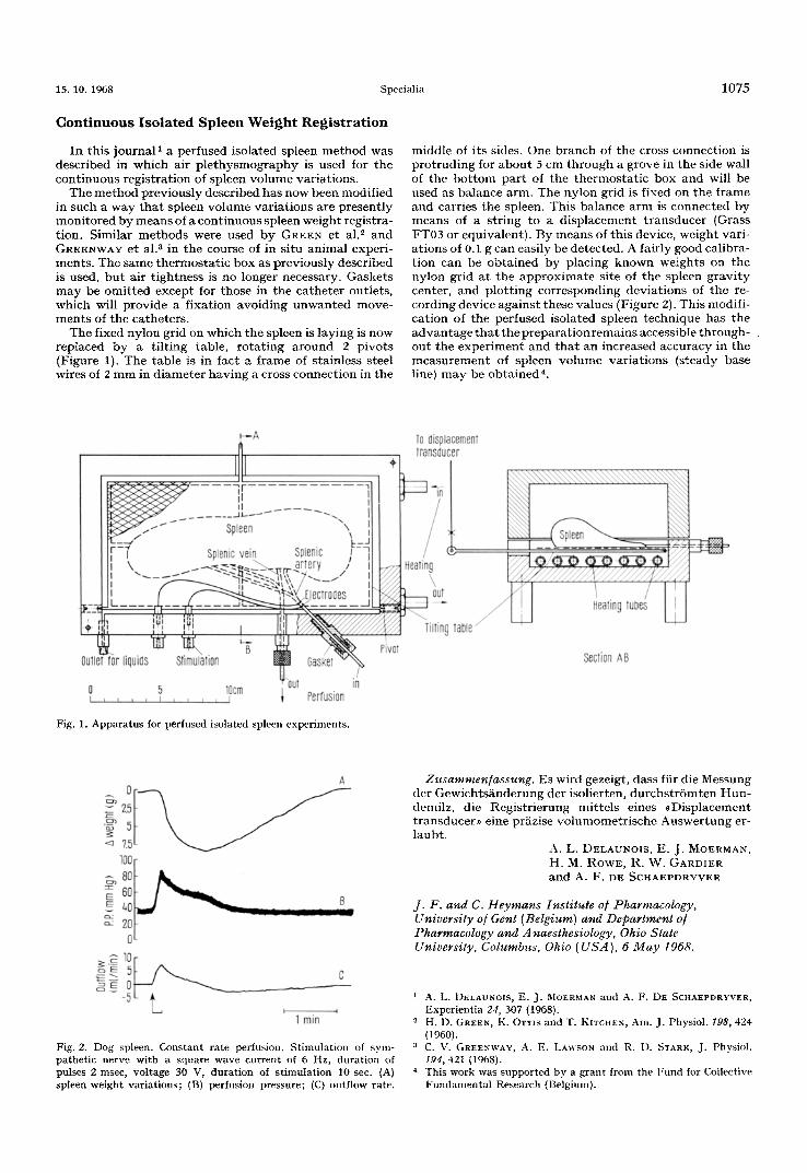
The f ixed n y l o n gr id on wh ich t h e spleen is l ay ing is now rep laced b y a t i l t i ng t ab le , r o t a t i n g a r o u n d 2 p i v o t s (F igure 1). T h e t a b l e is in f ac t a f r ame of s ta in less s teel wires of 2 m m in d i a m e t e r h a v i n g a cross c o n n e c t i o n in t h e
midd le of i t s sides. One b r a n c h of t h e cross c o n n e c t i o n is p r o t r u d i n g for a b o u t 5 cm t h r o u g h a g rove in t h e side wall of t h e b o t t o m p a r t of t h e t h e r m o s t a t i c b o x a n d will be used as b a l a n c e a rm. T h e n y l o n gr id is f ixed on t h e f r ame a n d carr ies t h e spleen. Th i s b a l a n c e a r m is c o n n e c t e d b y m e a n s of a s t r i ng to a d i s p l a c e m e n t t r a n s d u c e r (Grass FT03 or equ iva len t ) . By m e a n s of t h i s device, we igh t var i - a t i ons of 0.1 g c an easi ly be de tec ted . A fa i r ly good ca l ib ra - t i on can be o b t a i n e d b y p lac ing k n o w n we igh t s on t h e n y l o n gr id a t t h e a p p r o x i m a t e s i te of t h e spleen g r a v i t y center , a n d p l o t t i n g c o r r e s p o n d i n g d e v i a t i o n s of t h e re- co rd ing dev ice ag a i n s t these va lues (Figure 2). Th i s modif i - ca t ion of t h e pe r fused i so la ted spleen t e c h n i q u e has t h e a d v a n t a g e t h a t t h e p r e p a r a t i o n r e m a i n s accessible t h r o u g h - o u t t h e e x p e r i m e n t a n d t h a t a n inc reased accu racy in t h e m e a s u r e m e n t of sp leen v o l u m e v a r i a t i o n s ( s t eady base line) m a y be o b t a i n e d 4.
~---A To aisplacement , 11 Traosducer
Ill ! Splenic vei.n p u i II I I / I,-¢-¢-A ~ / , ~ - ~ ~ r - ~ a * - ~ ~ t / / / W II \ ". ....~...= ~"~.~., ~ artery / / I Hoatirl g ~ , , - = ~ = , ~ - - ~ , ,
I / ) ~C¢..~t ,~ " - I \ )
i ' - : : : ~ . - I L . . . . - ,J.r . . . .~_~-- . -.- - - - - - - J ~ . . . . . _ . I ~ ~ / I ) I H e a l i n l ] t u b e 8 I I I
• 7 ,, " - - . . j "
Outlet toe liquiEls Stimulation GasKet Section A8
o 5 lOcrn u . I i ~ , ~ I , i , ~ I ~ PCITUSIOf l
Fig. 1. Apparatus for perfused isolated spleen experiments.
A
~ 2
- ~ 7.5 100~ 80
6o ~ 4o ~ 2o
o ~ lO~- T rain
Fig. 2. Dog spleen. Constant rate perlusion. Stimulation of sym- pathetic nerve with a square wave current of 6 Hz, duration of pulses 2 msec, voltage 30 V, duration of stimulation 10 see. (A) spleen weight variations; (B) perfusion pressure; (C) outflow rate.
Zusammen/assung. Es wird gezeigt , dass fiir die Messung der G e w i c h t s i i n d e r u n g de r isol ier ten, d u r c h s t r G m t e n H u n - demilz, die R e g i s t r i e r u n g mi t t e l s e ines , D i s p l a c e m e n t t ransducer , , e ine prAzise v o l u m o m e t r i s c h e A u s w e r t u n g er- l aub t .
A. L. DELAUNOIS, E. J . MOERMAN, H, M. ROWE, R. W. GARDIER a n d A. F. DE SCHAEPDRYVER
j . F. and C. Heymans Institute o/Pharmacology, University of Gent (Belgium) and Department o/ Pharmacology and A naesthesiology, Ohio State University, Columbus, Ohio (USA), 6 May 1968.
i A. L. I)ELAUNOIS, E. J. MOERMAN and A. F. DE SCIiAEPDRYVER, Experientia 2if, 307 (1968).
2 H. D. GREEN, K. OTTlS and T. KITCHEN, Am. J. Physiol. 198, 424 (1960).
a C. V. GREENWAV, A. E. LAV~SON and R. 1). STARK, J. Physiol. t94, 421 (1968).
4 This work was supported by a grant from the Fund for Collective Fundamental Research (Belgium).