Embed Size (px)
Citation preview
CONTINUING EDUCATION
Preoperative Localization and RadioguidedParathyroid Surgery*Giuliano Mariani, MD1; Seza A. Gulec, MD2; Domenico Rubello, MD3; Giuseppe Boni, MD1; Marco Puccini, MD4;Maria Rosa Pelizzo, MD5; Gianpiero Manca, MD1; Dario Casara, MD3; Guido Sotti, MD3; Paola Erba, MD1;Duccio Volterrani, MD1; and Armando E. Giuliano, MD2
1Regional Center of Nuclear Medicine, University of Pisa Medical School, Pisa, Italy; 2Division of Surgery, John Wayne CancerInstitute, Santa Monica, California; 3Radiotherapy and Nuclear Medicine Department, Regional Hospital of Padua, Padua, Italy;4Institute of Endocrine Surgery, University of Pisa Medical School, Pisa, Italy; and 5Institute of General Surgery, University ofPadua Medical School, Padua, Italy
Clinical or subclinical hyperparathyroidism is one of the most com-mon endocrine disorders. Excessive secretion of parathyroid hor-mone is most frequently caused by an adenoma of �1 parathyroidgland. Unsuccessful surgery with persistent hyperparathyroidism,due to inadequate preoperative or intraoperative localization, maybe observed in about 10% of patients. The conventional surgicalapproach is bilateral neck exploration, whereas minimally invasiveparathyroidectomy (MIP) has been made possible by the introduc-tion of 99mTc-sestamibi scintigraphy for preoperative localization ofparathyroid adenomas. In MIP, the incision is small, dissection isminimal, postoperative pain is less, and hospital stay is shorter.Localization imaging techniques include ultrasonography, CT,MRI, and scintigraphy. Parathyroid scintigraphy with 99mTc-sesta-mibi is based on longer retention of the tracer in parathyroid thanin thyroid tissue. Because of the frequent association of parathy-roid adenomas with nodular goiter, the optimal imaging combina-tion is 99mTc-sestamibi scintigraphy and ultrasonography. Differentprotocols are used for 99mTc-sestamibi parathyroid scintigraphy,depending on the institutional logistics and experience (classicaldual-phase scintigraphy, various subtraction techniques in combi-nation with radioiodine or 99mTc-pertechnetate). MIP is greatlyaided by intraoperative guidance with a �-probe, based on in vivoradioactivity counting after injection of 99mTc-sestamibi. Differentprotocols used for �-probe–guided MIP are based on differenttiming and doses of tracer injected. �-Probe–guided MIP is a veryattractive surgical approach to treat patients with primary hyper-parathyroidism due to a solitary parathyroid adenoma. The proce-dure is technically easy, safe, with a low morbidity rate, and hasbetter cosmetic results and lower overall cost than conventionalbilateral neck exploration. Specific guidelines should be followedwhen selecting patients for �-probe–guided MIP.
Key Words: hyperparathyroidism; parathyroid adenomas, mini-mally invasive surgery; parathyroid adenomas, preoperative local-
ization; parathyroid adenomas, �-probe–guided surgery; 99mTc-sestamibi scintigraphy
J Nucl Med 2003; 44:1443–1458
Primary hyperparathyroidism (PHPT) is due to excessivesecretion of parathormone (parathyroid hormone [PTH]) by�1 enlarged parathyroid gland (1). Since the introduction ofroutine serum calcium measurement in the 1970s, this dis-order has become one of the most common endocrine dis-eases in the world (2). During the last 40 y, its estimatedprevalence in the United States jumped from about 0.08 per1,000 (18% asymptomatic) to about 0.5 per 1,000 (51%asymptomatic) (3,4). The prevalence of PHPT in Americanwomen �40 y old may be as high as 2 per 1,000 (0.5 per1,000 in men) (5). In Europe, the estimated prevalence ofPHPT is 3 per 1,000 overall, reaching 21 per 1,000 inwomen 55–75 y old (6). The prevalence in Asia seems to belower, especially in those countries where serum calcium isnot yet routinely measured, with some differences in thepattern of clinical presentations (7).
The classical presentation of PHPT is a clinically overtpattern of recurring nephrolithiasis associated with frankhypercalcemia and low serum phosphate, invalidating bonedisease, deep weakness, and reduced life expectancy. Dur-ing the last 10 y, the more widespread use of screening hasallowed clinicians to identify an earlier and more subtlepresentation of PHPT: Hypercalcemia is mild (combinedusually with normal or borderline-low serum phosphate)and symptoms are absent or subtle.
The earlier recognition of PHPT has increased the num-ber of parathyroidectomies performed for PHPT, consider-ing that about one tenth of all patients with PHPT undergosurgery in an early phase of disease.
ANATOMY, PHYSIOLOGY, AND PATHOPHYSIOLOGY
A detailed discussion of the pathogenesis, development,and clinical presentation of the various forms of hypercal-
Received Apr. 11, 2003; revision accepted May 29, 2003.For correspondence or reprints contact: Giuliano Mariani, MD, Regional
Center of Nuclear Medicine, University of Pisa Medical School, Via Roma 67,I-56126 Pisa, Italy.
E-mail: [email protected]*NOTE: FOR CE CREDIT, YOU CAN ACCESS THIS ACTIVITY THROUGH
THE SNM WEB SITE (http://www.snm.org/education/ce_online.html)THROUGH SEPTEMBER 2004.
RADIOGUIDED PARATHYROID SURGERY • Mariani et al. 1443
by on December 13, 2018. For personal use only. jnm.snmjournals.org Downloaded from
cemia and hyperparathyroidism is outside the scope of thepresent work. The interested reader is referred to excellentreviews on these topics (1,8,9). However, it is necessary toreview the basic surgical anatomy and physiology to under-stand the diagnostic and therapeutic challenges and surgicaltreatment strategies.
Anatomy and Embryology of Parathyroid GlandsNormally, there are 2 pairs of parathyroid glands in adult
humans. Each gland measures approximately 6 � 4 � 2 mmand weighs approximately 30–50 mg (total weight, approx-imately 130–140 mg). Although supernumerous parathy-roid glands are present in approximately 2%–5% of thepopulation, the presence of �4 glands is a rarity. Embryo-logically, the parathyroid glands originate from theendoderm of the third and fourth pharyngeal pouches. Theirrelative position, however, is inverted as they migrate dur-ing fetal life toward their final location (Fig. 1). The glandsthat are originally positioned in the third pharyngeal pouch(more cranially) initially follow the descent of the thymusand reach their final location at the posterolateral surface ofthe lower lobes of the thyroid. These glands occasionallyleave their connection to the thymus. The glands that areoriginally positioned in the fourth pharyngeal pouch (morecaudally) are closely associated with the embryologic struc-ture of the thyroid gland; they maintain a close associationwith the fetal thyroid until the descent of both structures totheir final location in the neck. These parathyroid glands arepositioned at the posterolateral surface of the upper poles ofthe thyroid lobes.
Migration of the parathyroid glands during fetal devel-opment from their original location to the final juxtathyroi-dal location explains why the surgical approach to theparathyroid glands can be intricate and variable. In mostcases, normal upper glands are located posterior to themiddle and upper third of the thyroid lobe and posterior tothe recurrent laryngeal nerves, cranially to the inferior thy-roid artery. The location of normal inferior parathyroidglands, however, is more variable, probably as a conse-quence of the more complicated migration process, whichbrings them from a cranial position to a caudal position relativeto the other pair of parathyroid glands. In approximately 50%of explorations, the lower parathyroid glands are found poste-riorly or laterally to the lower pole of the thyroid lobe, usuallywithin a 20-cm radius. With decreasing frequency, they arefound within the thyrothymic ligament, within the thymus inthe mediastinum, and intrathyroidal. When parathyroid glandsbecome adenomatous or hyperplastic and enlarge, their loca-tion may change somewhat.
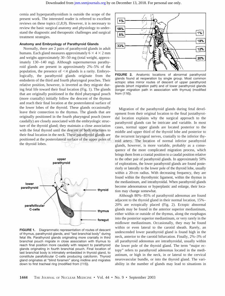
Although 80%–85% of parathyroid adenomas are foundadjacent to the thyroid gland in their normal location, 15%–20% are ectopically placed (Fig. 2). Ectopic abnormalglands may be found in the anterior superior mediastinum,either within or outside of the thymus, along the esophagusinto the posterior superior mediastinum, or very rarely in themidlower mediastinum. Occasionally, they may be foundwithin or even lateral to the carotid sheath. Rarely, anundescended lower parathyroid gland is found high in theneck, anterior to the carotid bifurcation. Finally, 2%–3% ofall parathyroid adenomas are intrathyroidal, usually withinthe lower pole of the thyroid gland. The term “major ec-topy” refers to parathyroid adenomas located in the medi-astinum, or high in the neck, in or lateral to the cervicalneurovascular bundle, or into the thyroid gland. The vari-ability in the number of glands may lead to situations in
FIGURE 1. Diagrammatic representation of routes of descentof thymus, parathyroid glands, and “last branchial body” duringfetal life. Parathyroid glands originating more cranially in thirdbranchial pouch migrate in close association with thymus toreach final position more caudally with respect to parathyroidglands originating in fourth branchial pouch. Final location oflast branchial body is intimately embedded in thyroid gland, toconstitute parafollicular C-cells producing calcitonin. Thyroidgland originates at “blind foramen” along midline and migratesdown to first tracheal ring (modified from (115)).
FIGURE 2. Anatomic locations of abnormal parathyroidglands found at reoperation by single group. Most commonectopic sites mirror routes of descent of upper parathyroidglands (short migration path) and of lower parathyroid glands(longer migration path in association with thymus) (modifiedfrom (116)).
1444 THE JOURNAL OF NUCLEAR MEDICINE • Vol. 44 • No. 9 • September 2003
by on December 13, 2018. For personal use only. jnm.snmjournals.org Downloaded from

which a patient has 4 normal glands in the neck and anabnormal fifth gland located in the mediastinum.
Calcium Homeostasis and PTHBy containing about 95% of all calcium in the body
in the form of calcium phosphate or hydroxyapatite(Ca5[PO4]3[OH]), bone and teeth serve as a balancing res-ervoir for the body fluids, ionized calcium in serum beingstrictly controlled in the approximate range of 1 mmol/L (or4 mg/dL).
The calcium ion stabilizes cellular and subcellular struc-tures (membranes and chromatin) and promotes cell adhe-sion. It is also involved in cell-to-cell signaling, through thevoltage-activating channels and interaction with the ryano-dine receptor and the inositol triphosphate receptor, as wellas with the myofibrillar apparatus and the N-methyl-D-aspartate receptor channel. Cells use the calcium ion as anintracellular messenger carrying information generated byhormones and neurotransmitters. Parathyroid cells express acalcium ion receptor (CaR). Calcium ion regulates both thesecretory balance and the cell proliferation in the parathy-roid glands through the CaR. The CaR is also expressed inthe parafollicular cells of the thyroid (regulating calcitoninsecretion), in the kidney (regulating calcium and waterhomeostasis), in osteoblasts and osteoclasts (regulatingmineral homeostasis), and in several cell types of the smallbowel and proximal colon (regulating intestinal absorption).Additionally, the CaR is also present in many tissues thatare not known to be directly involved in the regulation ofcalcium metabolism. The function of the CaR in suchcells—for example, neurones and keratinocytes—is possi-bly associated to a calcium-sensor–regulated hormone se-cretion, ion channel function, gene expression, cell prolif-eration, and cell apoptosis.
PTH, an 84-amino acid single-chain polypeptide with amolecular weight of 9,500 Da, controls the level of ionizedcalcium in blood and in the extracellular fluid. PTH issynthesized as a 115-amino acid large peptide called thepreproparathyroid hormone (prepro-PTH). The “pre” se-quence (a “signal” 25-amino acid peptide) is cleaved andrapidly degraded, thus leaving the pro-PTH peptide consti-tuted by 90 amino acids, during the endoplasmic reticulartransmembrane transportation. Further cleavage of the“pro” sequence (6 amino acids) produces mature PTH,which is concentrated in the secretory vesicles and granulesof parathyroid cells. Granules of a distinct subtype alsocontain cathepsin B and H proteases in addition to PTH. Theproteases’ effect in the granules results in formation of PTHfragments with carboxy-terminal fragments (a small portionof PTH secreted by the parathyroid cells) that are biologi-cally inactive on calcium balance. Intracellular fragmenta-tion of PTH probably represents a regulatory inactivatingpathway operating under as yet unidentified conditions.
Although catecholamines, magnesium, and other stimulican affect PTH secretion, the major determinant of PTHsecretion is the blood ionized calcium level. Small reduc-
tions in the extracellular calcium concentration result inincreased PTH secretion aimed at restoring the normocal-cemia. This regulation is mediated by different concomitanteffects of PTH, primarily on the kidney, by increasingtubular reabsorption of calcium, increasing excretion ofphosphorus, and increasing the transformation of precursorsinto the active form of vitamin D (which, in turn, stimulatesincreased absorption of calcium in the gastrointestinaltract). The resulting restoration of normal calcium levels inthe extracellular fluid terminates the feedback loop in theparathyroid glands.
There is a steep inverse relationship between extracellularcalcium concentration and PTH secretion, so that a smallincrease in calcium levels results in a large decrease in PTHsecretion (sigmoidal relationship). This effect, which is trig-gered by the CaR and is mediated via the G-protein andother modulators in the parathyroid cells, involves cellularevents ranging from PTH gene expression to transcription,translational and posttranslational processing, and intracel-lular trafficking regulating vesicular transport, vesicular fu-sion, and exocytosis.
The mechanism by which lowering of calcium in theextracellular fluid stimulates PTH secretion is less wellunderstood. Acute hypocalcemia induces the release of pre-formed PTH stored in the parathyroid cells within minutes.However, hypocalcemia lasting several hours stimulatessynthesis of PTH at translational level (increased PTH mes-senger RNA [mRNA]). Persistent or chronic hypocalcemiaincreases PTH secretion both by increasing the maximalsecretory rate per cell through an enhanced expression ofthe PTH gene and by stimulating proliferation of the para-thyroid cells.
Secreted PTH is rapidly metabolized by the liver (70%)and kidneys (20%), disappearing from the circulation with abiologic half-life of 2–3 min. Variations in the blood levelsof calcium or 1,25(OH)2 vitamin D, even in a wide range, donot affect this fast peripheral metabolism of PTH. There-fore, the PTH blood levels are determined mostly by the rateof PTH secretion.
Less than 1% of secreted hormone eventually finds itsway to PTH receptors in target organs. Cell-surface PTHreceptors are mainly located in bone and kidneys, wherethey trigger responses that increase calcium levels in blood.In the liver, only small amounts of hormone bind to PTHreceptors, while most of the intact PTH is cleaved or de-graded by cathepsins. In addition to representing one of themain target organs for PTH activity, the kidneys also rep-resent an important metabolic clearance site for PTH,through glomerular filtration and tubular degradation, amechanism that also involves the PTH carboxy-terminalfragments.
PHPTPHPT is a condition characterized by the inappropriate
secretion of PTH with respect to the extracellular calcium
RADIOGUIDED PARATHYROID SURGERY • Mariani et al. 1445
by on December 13, 2018. For personal use only. jnm.snmjournals.org Downloaded from

concentration. This condition can be caused by parathyroidadenoma(s), parathyroid hyperplasia, or carcinoma. Para-thyroid cells exhibit both increased proliferative activity(leading to enlarged glands) and decreased sensitivity to theinhibiting effect of increased calcium concentration on PTHsecretion (altered set point). Hyperparathyroidism due tohyperplasia may also be a component of a familial syn-drome, such as the multiple endocrine neoplasia 1 (MEN-1)syndrome (87%–97%), MEN-2 (5%–20%), familial hy-pocalciuric hypercalcemia, hyperparathyroidism-jaw-tumorsyndrome, and familial isolated hyperparathyroidism.
In the nonfamilial disease, molecular biology investiga-tions have shown that sporadic parathyroid tumors areclonal in origin, and there is evidence for both activation ofoncogenes and inactivation of tumor suppressor genes. In23%–40% of the parathyroid adenomas, the overexpressionof PRAD 1, a cyclin D1 oncogene product involved in cellcycle regulation, has been identified. Using comparativegenomic hybridization, candidate loci for dominant onco-genes involved in the genesis of sporadic parathyroid ade-nomas have been identified on chromosomes 1q, 9q, 16p,19p, and Xq. The loss of heterozygosity or suppressionfunction on chromosomes 1p, 6q, 9p, 11q, 13q, and 15q hasalso been detected. The loss of heterozygosity at the MEN-1locus has been identified in 25%–40% of patients withsporadic parathyroid adenoma, and, to date, this is the onlysuppressor gene implicated in the genesis of this disease.Evidence for the loss of the suppressor oncogenes retino-blastoma, p53, 1p, 4q, and 13q has been reported in para-thyroid carcinoma. Oncogene activation at 1q, 9q, 16p, 19p,and Xq is also seen more frequently in parathyroid carci-nomas than in benign parathyroid tumors.
The characteristic abnormality in hyperparathyroidism isthe downregulated response of parathyroid cells to reducePTH secretion in response to elevated calcium. In vitro andin vivo studies have focused on the CaR to explore the roleof the calcium sensor in the development of hyperparathy-roidism. Abnormalities in CaR expression or function as aconsequence of some as yet unidentified genetic mutation(s)may contribute to the failure of PTH secretory regulation.
Also, genetic variations of vitamin D metabolism have beenfound to be associated with some hyperparathyroidism states.In particular, certain vitamin D receptor (VDR) allelic variants(VDRb, VDRa, and VDRT) are overrepresented in patientswith PHPT and are associated with the decreased expression ofVDR mRNA; this combination makes the parathyroid cellsless susceptible to inhibition by active vitamin D and, there-fore, more likely to undergo hyperplastic or adenomatouschanges.
Finally, a well-known risk factor for the development ofhyperparathyroidism is irradiation of the neck and upperchest for benign diseases, including 131I treatment forGrave’s disease. Interestingly, such risk association has notbeen demonstrated in 131I treatment for thyroid cancer.Genetic analysis of radiation-associated parathyroid tumorshas disclosed multiple alterations, most commonly involv-
ing losses in 11q and 1p. Such changes in molecular profileare similar to those encountered in those adenomas showingMEN-1 gene abnormalities, thus suggesting some vulnera-bility of this gene to irradiation.
HistopathologyHistopathologic criteria used to distinguish adenoma
from hyperplasia are not well defined. Current evidence onthe histopathogenesis of parathyroid adenoma and hyper-plasia is also unclear. Although most adenomas are mono-clonal proliferations, suggesting a neoplastic origin, his-topathologic and cytogenetic evidence suggests evolution toa monoclonal adenoma from polyclonal hyperplasia andvice versa (10).
Parathyroid adenoma and hyperplasia could well be thesame disease entity, representing the opposite ends of thespectrum of phenotypic expression. To avoid confusion andspeculation, we prefer using the term “adenoma” inter-changeable with “single-gland disease” and “hyperplasia”interchangeable with “multigland disease.” MEN and thefamilial hyperparathyroidism syndromes are associated withmultigland disease as a rule. In sporadic PHPT, classically,single-gland involvement (adenoma) is observed in approx-imately 80%–85% of the cases, and hyperplasia is seen15%–20% of the cases.
The prevalence of hyperplasia varies considerably in dif-ferent series, and there appears to be a strong influence ofclinical (surgical) diagnosis on the pathologist’s diagnosis(11). When subtotal parathyroidectomy is performed, morehyperplasia is diagnosed. When a focused operation is per-formed with removal of a single gland, it is likely to becalled an adenoma. The importance of mild hyperplasia andthe best way of diagnosing this condition are yet to bedetermined. Parathyroid carcinoma is a rare cause of PHPT,accounting for �1% of all cases.
Secondary HyperparathyroidismThe most common cause of secondary hyperparathyroid-
ism is chronic renal failure. Even in its early stages, renalimpairment results in phosphorus retention; the strict in-verse balance existing between the serum levels of phos-phorus and calcium induces hypocalcemia, which in turnstimulates PTH secretion. Second, the impaired renal pa-renchyma cannot fully respond to the increased PTH stim-ulation by activating the tubular calcium-sparing mecha-nism and by increasing production of the active form ofvitamin D 1,25-(OH)2-D3. Lastly, there is a certain skeletalresistance to the calcemic action of PTH, due in part to adirect inhibiting effect of phosphorus retention, to down-regulation of the PTH receptors (possibly related to de-creased CaR expression), and to increased calcitonin secre-tion.
All of these mechanisms progressively augment and per-petuate the hyperparathyroid state. The altered cellularmechanisms in parathyroid glands in secondary hyperpara-thyroidism include the decreased inhibiting effect of the
1446 THE JOURNAL OF NUCLEAR MEDICINE • Vol. 44 • No. 9 • September 2003
by on December 13, 2018. For personal use only. jnm.snmjournals.org Downloaded from

extracellular calcium on PTH secretion consequent to thedecreased expression of CaR, despite increased PTHmRNA, and the decreased inhibiting effect of 1,25-(OH)2-D3 on PTH synthesis or secretion as well as on theproliferative activity of parathyroid cells.
Tertiary HyperparathyroidismSecondary hyperparathyroidism usually subsides after
successful renal transplantation, which restores a nearlynormal mass of functioning renal parenchyma. However, ina few instances, hyperparathyroidism persists after success-ful renal transplantation. This is due to the development offunctional autonomy in �1 enlarged parathyroid gland andis termed “tertiary hyperparathyroidism.” The incidence andtrue clinical significance of this condition are still a matterof debate. The term tertiary hyperparathyroidism is alsoused for patients in whom the PTH response due to second-ary hyperparathyroidism exceeds the hypocalcemic de-mands, resulting in hypercalcemia. The related term “re-fractory secondary hyperparathyroidism” denotes patientswho have nonsuppressible PTH secretion, but who are nor-mocalcemic. In both instances, the hyperparathyroid statehas reached an autonomous stage and is no longer sensitiveto medical treatment.
Persistent and Recurrent HyperparathyroidismPersistent or recurrent hyperparathyroidism occurs in
5%–10% of all patients who undergo surgery for PHPT.Persistent hyperparathyroidism is the most common situa-tion (75%) and is defined as a continuation of the preoper-ative abnormalities in calcium metabolism in the immediatepostoperative period. Causes for the immediate failure ofsurgical treatment include failure in localization of adeno-mas, inadequate resection of unrecognized multigland dis-ease, and presence of metastatic parathyroid carcinoma.Factors contributing to the failure involve inexperience onthe surgeon’s part and errors in intraoperative frozen sectionexamination on the pathologist’s part. Persistent hyperpara-thyroidism after surgery is particularly frequent in patientswith familial hyperparathyroidism, especially the MEN-1syndrome (usually in �25% of patients, but up to 40%–60% for less-experienced surgeons (12)).
Hyperparathyroidism presenting after a period of �6 moof normocalcemia following surgery is termed “recurrenthyperparathyroidism” and is usually linked to continuedgrowth of the remaining parathyroid tissue.
A rare case of recurrent or persistent hyperparathyroidismis termed “parathyromatosis,” which is described as multi-ple remnants of hyperfuctioning parathyroid tissue scatteredthroughout the neck or upper mediastinum. This conditionprobably is due either to inadvertent implantation of para-thyroid tissue at the time of parathyroidectomy or to growthof nests of parathyroid tissue left along the route of descentduring embryologic development of the parathyroid glands.
SURGICAL TREATMENT
Conventional Surgical ApproachThe first successful parathyroidectomy was performed in
1925 with bilateral neck exploration (13) and, ever since,has remained the standard treatment of PHPT (14). Thistime-honored approach, based on excision of any grosslyenlarged gland with or without biopsy of the remainingglands, yields a 95% success rate with minimal morbidity,even with 4-gland hyperplasia, in the hands of an experi-enced endocrine surgeon (15,16). When �1 gland is en-larged, the operative techniques include a 31⁄2-gland resec-tion (leaving approximately 50–100 mg of the most“normal-appearing” gland), excising only those glands thatare grossly enlarged at the exploration, and a less-common4-gland parathyroidectomy with subsequent autotransplan-tation. Success with this approach as measured by return tonormal calcium levels depends primarily on the experienceand the judgment of the surgeon in recognizing the differ-ence between enlarged or normal-sized glands, although thesize of the parathyroid gland does not always correlate wellwith the secretion of PTH.
When no preoperative imaging was available, bilateralexploration was mandatory, because discrimination be-tween single-gland and multigland disease was solely basedon macroscopic appearance of all glands. Introduction of theearlier imaging techniques (US and 201Tl/99mTc-pertechne-tate subtraction scintigraphy) did not really affect suchsurgical strategy, especially in the case of the first operation,because these techniques were unreliable in detecting mul-tigland disease. Difficulties associated with parathyroidec-tomy for PHPT relate to the variability in the number ofparathyroid glands, the different locations of normal andabnormal glands, and problems in distinguishing normalglands from those that are subtly diseased.
An especially challenging aspect of parathyroid surgeryis to distinguish an adenoma from hyperplasia, a distinctionthat can be difficult not only intraoperatively but also his-topathologically. In general, most experienced surgeons be-lieve that by evaluating the size, shape, and color of theparathyroid glands at surgery they can distinguish normalglands from abnormal ones. If 1 gland is enlarged and theothers are perfectly normal visually, the diagnosis is anadenoma. Some surgeons attempt to confirm this presump-tive diagnosis by obtaining a frozen section of a biopsy of anormal gland. On the other hand, hyperplasia (multiglanddisease) should result in enlargement of all 4 glands. How-ever, asymptomatic hyperplasia may appear with 1 or 2normal-sized glands and confuse the diagnosis, even withbiopsy. In the absence of a reliable pre- or intraoperativeimaging study to identify or localize abnormal parathyroidglands, conventional 4-gland exploration is mandatory.
Minimally Invasive ParathyroidectomyWith the introduction of 99mTc-sestamibi scintigraphy to
identify and locate the parathyroid adenoma preoperatively,
RADIOGUIDED PARATHYROID SURGERY • Mariani et al. 1447
by on December 13, 2018. For personal use only. jnm.snmjournals.org Downloaded from
the era of focused exploration or minimally invasive para-thyroidectomy (MIP) began (17–19). MIP is gradually re-placing the traditional 4-gland exploration as the procedureof choice in many institutions, with comparable cure rates(19,20). The different techniques associated with focusedexploration or MIP include �-probe–guided exploration andnovel endoscopic techniques.
The goal of surgery for PHPT is to return the patient’scalcium level to normal, although this cannot be the soleindex of successful parathyroid surgery; surgery should beaccomplished with minimal morbidity, no mortality, lowrecurrence rates, and a reasonable cost. The success of thesurgical treatment depends on the success in localizationand identification of abnormal glands. In this respect, theincreased sensitivity of parathyroid imaging allows the sur-geon to plan a localized exploration designed to remove thecommon single focus of disease. The incision is small,dissection is minimal, postoperative pain is less, and hos-pital stay is shorter. These procedures may be done asoutpatient surgery, even with local anesthesia.
Other relevant changes in the management of PHPT havebeen the introduction of intraoperative quick PTH (QPTH)assay and the fact that PHPT is caused in 85%–90% of thecases by a single adenoma. The first clinical application ofthese concepts resulted in the development of unilateral cervi-cal exploration (21–23), and the assay was refined and clini-cally popularized by Irvin (24) and Irvin and Carneiro (25).
This assay measures intact PTH levels in the patient’splasma using an immunochemiluminometric technique andcan be performed during the operation. Intact PTH has abiologic half-life of 2–3 min. Therefore, a significant dropin QPTH level is observed 5–10 min after removal of theabnormal gland. A fall in QPTH level of �50% of thepreoperative level is considered indicative of successfulremoval. Under these conditions, the QPTH assay has asensitivity and a specificity of 98% and 94%, respectively,and an overall accuracy of 97% in predicting success ofsurgery (24,25).
A recent survey of the members of the InternationalAssociation of Endocrine Surgeons indicated that, in theyear 2000, 99mTc-sestamibi-based MIP had been adopted by�50% of the surgeons worldwide (59% from America, 56%from Australia, and 49% from Europe and the Middle East).The most popular surgical technique (92%) was the focusedapproach with a small incision, followed by a video-assistedtechnique (22%), and true endoscopic technique with gasinsufflation (12%). Techniques used to ensure completenessof resection include the QPTH (68%) assay and �-probeguidance (14%) (20).
MIP has been used for only about 6–7 y; thus, no large-scale long-term follow-up data are available as yet. How-ever, the �95% success rate reported by most authors (asassessed by postoperative normalization of serum calciumand PTH levels) constitutes a sound basis for the favorableMIP results similar to those experienced with the conven-tional surgical approach also in the long run.
PREOPERATIVE PARATHYROID LOCALIZATION
UltrasonographyThe advantages of ultrasound imaging (US) as a nonin-
vasive, low-cost, and nonionizing procedure are somewhatcounterbalanced by the fact that this technique is highlydependent on the operator. The accuracy of the parathyroidtumor’s localization by US varies as a function of the sizeand location of the adenoma, whose location in the subster-nal, retrotracheal, and retroesophageal spaces entails poorsensitivity due to acoustic shadowing from overlying boneor air. The sensitivity of US identification of parathyroidadenomas ranges between 70% and 80% (26,27), whereasthe range is much wider (30%–90%) when the detection ofsimply enlarged parathyroid glands is considered (28).Moreover, the sensitivity of US evaluation falls to 40% inpatients who have had prior failed surgical exploration (29).
US is highly sensitive in detecting parathyroid adenomaslocated behind the thyroid gland (the upper parathyroidglands usually bulge out behind the thyroid lobe) or locatedbeyond the lower contour of the thyroid. However, it can bedifficult to visualize upper parathyroid glands located me-dially (close to the larynx and trachea), located deep in theneck in the para- and retropharyngeal space (28,30,31), orlocated close to the carotid bifurcation (32,33).
Moreover, parathyroid enlargement can be mimicked byother structures in the neck, such as muscles, vessels, en-larged lymph nodes, and esophagus (33). Therefore, the rateof false-positive US results is not negligible, with specific-ities reported between 40% and 100% (26–32,34).
On the other hand, US is often used in combination withother imaging procedures for the preoperative localizationof parathyroid adenomas before unilateral neck exploration.In particular, the combination of US with thyroid scintigra-phy is useful to differentiate enlarged parathyroid glandsfrom thyroid nodule(s) (28,30,35). Thus, US evaluationmight be more important in geographic areas where theprevalence of nodular goiter is high. Detection of thyroidnodular disease by US may identify individual patients whomay not be appropriate candidates for minimally invasivesurgery, even if presenting with a single parathyroid ade-noma (28,36). The diagnostic value of such US–thyroidscintigraphy combined imaging approach is limited in iden-tification of intrathyroidal parathyroid adenomas, whichusually appear as hypoechoic, low-uptake areas similar tomost of the thyroid nodules (33). US-guided fine-needleaspiration has been used to discriminate thyroid nodulesfrom enlarged parathyroid glands (33). The combination ofUS and thyroid scintigraphy appears to be of value indetecting parathyroid enlargement, particularly in patientswith secondary hyperparathyroidism (28,31), and these pa-tients should routinely be investigated with such combinedimaging procedure, possibly at a same session (27,28,35).
Despite the fact that it has a lower sensitivity and accu-racy than scintigraphy for detecting parathyroid tumors, insome circumstances US imaging plays an important clinical
1448 THE JOURNAL OF NUCLEAR MEDICINE • Vol. 44 • No. 9 • September 2003
by on December 13, 2018. For personal use only. jnm.snmjournals.org Downloaded from

role. In particular, US is useful to confirm the presence of ascan-positive solitary parathyroid adenoma (the cause of60%–70% of all cases of PHPT). In such patients, USimaging provides additional information on the depth of theparathyroid adenoma, a feature that is especially useful indistinguishing parathyroid adenomas located behind andclose to the thyroid gland from the 99mTc-sestamibi–avidthyroid nodules. Furthermore, morphologic imaging pro-vided by US can identify those rare adenomas that arenegative on parathyroid scintigraphy because of their prev-alent cystic component. Moreover, in patients with hyper-parathyroidism due to multigland disease, US can be helpfulfor detecting nondominant, scan-negative enlarged parathy-roid glands. Finally, by providing accurate morphologicevaluation of the thyroid gland, US allows identification ofsmall thyroid nodules (�1 cm), some of which should beconsidered for US-guided fine-needle aspiration cytology incase of equivocal features such as inhomogeneous echoicpattern, irregular margins, microcalcifications, and intra-nodular hypervascularization.
CT and MRICT and MRI are often used in the preoperative evaluation
of hyperparathyroid patients, especially when the probabil-ity of ectopic glands is high, as in patients with recurrent orpersistent hyperparathyroidism. When the ectopic adeno-mas are located in the mediastinum, the high anatomicdefinition of either CT or MRI provides topographic infor-mation of value for planning the most appropriate surgicalapproach.
Although CT localizes parathyroid adenomas located inthe retrotracheal, retroesophageal, and mediastinal spacesbetter than US, it performs poorly for ectopic lesions locatedin the lower neck at the level of the shoulders and lesionsclose to or within the thyroid gland (37–39) and even indiscriminating upper from lower parathyroid glands (39,40).The overall sensitivity of CT for preoperative identificationof hyperplastic parathyroid glands ranges between 46% and80%. The sensitivity consistently approaches 80% withintravenous contrast enhancement because adenomas andhyperplastic parathyroid glands are hypervascular. Priorneck surgery affects the sensitivity of CT imaging. Artifactscaused by metallic clips from previous surgery (“sparkler”effect) lower the sensitivity of CT to 46%–58% (39,41).
Imaging of normal parathyroid glands by MRI is poordue to their small size (�5 mm) (42). However, its ability tocharacterize nodular lesions on the basis of intensitychanges in the T1- and T2-weighted images as well as thepossibility of contrast enhancement and 3-dimensional re-construction make this technique particularly attractive(38). On MR examination, enlarged parathyroid glands dis-play a medium intensity on T1-weighted images (like thy-roid tissue or muscle) but have considerably increased in-tensity on T2-weighted and proton density images (43,44).These image patterns, however, do not distinguish parathy-roid adenomas from either simple hyperplasia or carcinoma.
Paired evaluations in the same patients have shown thatthe sensitivity of MRI is equivalent to that of parathyroidscintigraphy. However, the specificity has been found to beconsistently lower (44–47). Discrepancies reported by dif-ferent authors probably depend on the different patientpopulations studied (primary or secondary hyperparathy-roidism, before first or second operation, presence or ab-sence of thyroid nodules) and on the different imagingtechniques used.
Most authors believe that MRI is worth performing whenparathyroid scintigraphy is negative or equivocal or when itsuggests an ectopic gland (46–48). However, some authorssuggest the systematic use of combined MRI and parathy-roid scintigraphy (despite the associated high costs) becausethis approach should increase the accuracy and reliability ofpreoperative identification and localization of parathyroidlesions (43,47).
Because of the less-than-optimal accuracy of conven-tional imaging techniques, the National Institutes of Healthconsensus statement on the treatment of PHPT in 1990concluded that preoperative localization in patients withoutprior neck surgery was rarely indicated and not proven to becost-effective, recommending 4-gland exploration as theoperation of choice (49).
The whole scenario has dramatically changed more re-cently, after the introduction of more effective preoperativelocalization modalities, especially parathyroid scintigraphywith 99mTc-sestamibi.
Parathyroid ScintigraphyTo date, no specific radiopharmaceutical that is capable
of concentrating solely in the parathyroid glands has beendeveloped. Scintigraphic exploration of parathyroid tissue isfurther complicated by the intrinsic close proximity of para-thyroid glands to (sometimes true “embedding” within) ametabolically active thyroid gland. Early attempts to cir-cumvent this limitation included the concept of using 2tracers with different uptake patterns in thyroid and para-thyroid glands (50). The first such technique used 75Se-methionine/131I subtraction scanning. 75Se-Methionine is ac-tively concentrated in both thyroid and parathyroid tissuesbecause of their high metabolic activity. Simultaneously orsequentially administered 131I-iodide localizes in the thy-roid. After applying some “normalization” factor to the 2scintigraphic acquisitions, the thyroid-only image (131I) wassubtracted from the thyroid � parathyroid image (75Se-methionine), thus visualizing residual areas of radioactivityconcentration corresponding to abnormal parathyroidglands. The technique was rather disappointing due to thetechnical limitations linked to the imaging equipment avail-able at the time and to radiodosimetric considerations (51).
The subtraction imaging technique was later improved byFerlin et al. (52), replacing 75Se-methionine with 201Tl-chloride and 131I with 99mTcO4
�. Although 201Tl and 99mTcwere more suitable radionuclides than 75Se and 131I forgamma-camera imaging, the new technique still lacked op-
RADIOGUIDED PARATHYROID SURGERY • Mariani et al. 1449
by on December 13, 2018. For personal use only. jnm.snmjournals.org Downloaded from
timal imaging properties for parathyroid glands. Further-more, there was a nonnegligible radiation burden to patientsderiving from 201Tl, due to its low-energy x-ray emissionresulting from electron capture decay. The dual-isotope201Tl/99mTcO4
� procedure failed to demonstrate a definiteadvantage over other imaging modalities due, in part, to acertain degree of intrinsic variability and low reproducibil-ity in interpreting the results among different centers(53,54).
99mTc-SestamibiAfter initial experience with 99mTc-sestamibi for myocar-
dial perfusion studies, Coakley et al. incidentally observedthat this tracer exhibited significant uptake and retention inthe abnormal parathyroid glands of patients with PHPT(55). Successful utilization of 99mTc-sestamibi imaging forlocalizing abnormal parathyroid glands was subsequentlyconfirmed by numerous reports (56–60) and, with someprotocol modifications, has become the parathyroid imagingtechnique worldwide (61).
The localization of 99mTc-sestamibi in the parathyroidtissue is a function of metabolic activity, accumulationoccurring specifically in the mitochondria. The overall up-take in hyperplastic or adenomatous parathyroid glands islinked to the blood flow, gland size, and mitochondrialactivity (62). Like other radioactive imaging agents (such as201Tl), 99mTc-sestamibi accumulates both in the thyroid andin the parathyroid tissue within minutes after intravenousadministration. However, what makes this tracer especiallyuseful for parathyroid imaging is its different washout ratefrom the 2 tissues, in that 99mTc-sestamibi is released muchfaster from thyroid than from parathyroid tissue. This dif-ferential retention might be related to some downregulationof the P-glycoprotein system (normally acting as an outfluxcarrier molecule for various substrates, including 99mTc-sestamibi) in the parathyroid tissue (63–66). When feasible,concomitant suppression of thyroid uptake (induced by ex-ogenous thyroid hormone administration) obviously im-proves scintigraphic localization of parathyroid lesions with99mTc-sestamibi (67).
High-quality scintigraphy with 99mTc-sestamibi can accu-rately localize parathyroid adenomas in 85%–95% of pa-tients with PHPT. The addition of SPECT imaging consid-erably improved the localization of particular ectopic sitesotherwise difficult to explore, such as the retroesophagealspace or mediastinum (68–72). It is vital to include theentire chest in the imaging field in all 99mTc-sestamibiimaging protocols for evaluation of potential ectopic sites.
A great wealth of information currently supports theutility of 99mTc-sestamibi scintigraphy in preoperative local-ization and successful MIP with unilateral neck exploration(73–76). Different 99mTc-sestamibi imaging protocols havebeen adopted based on the institutional logistics and expe-rience.
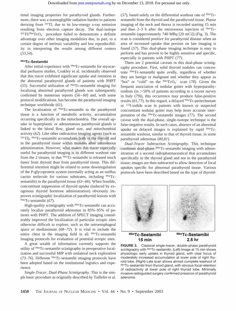
Single-Tracer, Dual-Phase Scintigraphy. This is the sim-ple basic procedure as originally described by Taillefer et al.
(57), based solely on the differential washout rate of 99mTc-sestamibi from the thyroid and the parathyroid tissue. Planarimaging of the neck and thorax is recorded starting 15 minand then 2–3 h after the intravenous injection of 99mTc-sestamibi (approximately 740 MBq [20 mCi]) (Fig. 3). Thescan is considered positive for parathyroid disease when anarea of increased uptake that persists on late imaging isfound (57). This dual-phase imaging technique is easy toperform and has proven to be highly sensitive and specific,especially in patients with PHPT (77).
There are 2 potential caveats in this dual-phase scintig-raphy procedure. First, solid thyroid nodules can concen-trate 99mTc-sestamibi quite avidly, regardless of whetherthey are benign or malignant and whether they appear as“hot” or “cold” on the 99mTcO4
� scan (78). Given thefrequent association of nodular goiter with hyperparathy-roidism (in �50% of patients according to a recent surveyin Italy (79)), this occurrence may produce false-positiveresults (61,77). In this regard, a delayed 99mTc-pertechnetateor 123I-iodide scan in patients with known or suspectedconcomitant nodular goiter may help more accurate inter-pretation of the 99mTc-sestamibi images (77). The secondcaveat with the dual-phase, single-isotope technique is thefalse-negative results. In such cases, absence of an abnormaluptake on delayed images is explained by rapid 99mTc-sestamibi washout, similar to that of thyroid tissue, in someparathyroid adenomas (80,81).
Dual-Tracer Subtraction Scintigraphy. This techniquecombines dual-phase 99mTc-sestamibi imaging with admin-istration of a second radiopharmaceutical that accumulatesspecifically in the thyroid gland and not in the parathyroidtissue; images are then subtracted to allow detection of focaluptakes specific for abnormal parathyroid tissue. Variousprotocols have been described based on the type of thyroid-
FIGURE 3. Classical single-tracer, double-phase parathyroidscintigraphy with 99mTc-sestamibi. (Left) Image at 15 min showsphysiologic early uptake in thyroid gland, with clear focus ofmoderately increased accumulation at lower pole of right thy-roid lobe. (Right) Late scan shows almost complete washout of99mTc-sestamibi from thyroid gland, with obvious focal retentionof radioactivity at lower pole of right thyroid lobe. Minimallyinvasive radioguided surgery confirmed presence of parathyroidadenoma.
1450 THE JOURNAL OF NUCLEAR MEDICINE • Vol. 44 • No. 9 • September 2003
by on December 13, 2018. For personal use only. jnm.snmjournals.org Downloaded from
imaging agent used and the sequence of the administrationof tracers.
The first such imaging protocol is the 123I/99mTc-sesta-mibi, dual-tracer subtraction technique. Patients are admin-istered 10 MBq 123I-NaI 2–4 h before 99mTc-sestamibi in-jection. Imaging is performed at different times orsimultaneously using 2 separate energy windows (140 keVfor 99mTc, 159 keV for 123I). The 123I thyroid image issubtracted from the 99mTc-sestamibi combined thyroid–parathyroid image (58). Wide routine application of thisprocedure is constrained by the high cost and limited avail-ability of 123I. The long imaging time required to obtainsatisfactory counting statistics at the doses used also is alimiting factor for 123I thyroid imaging.
Another practical parathyroid imaging technique is the99mTcO4
�/99mTc-sestamibi, dual-tracer subtraction tech-nique. Patients are injected with 185 MBq 99mTc-pertechne-tate and imaging is performed 20 min after injection. Im-mediately after completion of 99mTc-pertechnetate imaging,patients are injected with 300 MBq 99mTc-sestamibi withoutchanging the patient’s position, and a 20-min dynamic ac-quisition is performed. Although the technique has beenreported to have 89% sensitivity and 98% specificity (82), ithas its own shortcomings. The technique is hampered byhigh counting rates originating from 99mTc-pertechnetateuptake in the thyroid tissue overriding those originatingfrom 99mTc-sestamibi uptake. The identification of parathy-roid adenomas located behind the thyroid contour, espe-cially on planar images, may not be possible. Geatti et al.modified the technique by reducing the 99mTc-pertechnetatedose to 40 MBq and increasing the 99mTc-sestamibi dose to400–500 MBq. By adopting this modified protocol, a sen-sitivity of 95% was achieved in patients with PHPT, without
any false-positive result due to thyroid nodules being ob-served (27).
To achieve a rapid washout of 99mTc-pertechnetate fromthe thyroid tissue, potassium perchlorate (KClO4
�) has beenused in the 99mTcO4
�/99mTc-sestamibi protocol (83): 150MBq 99mTcO4
� are injected intravenously; 20 min later, justbefore positioning the patient under the gamma camera, 400mg KClO4
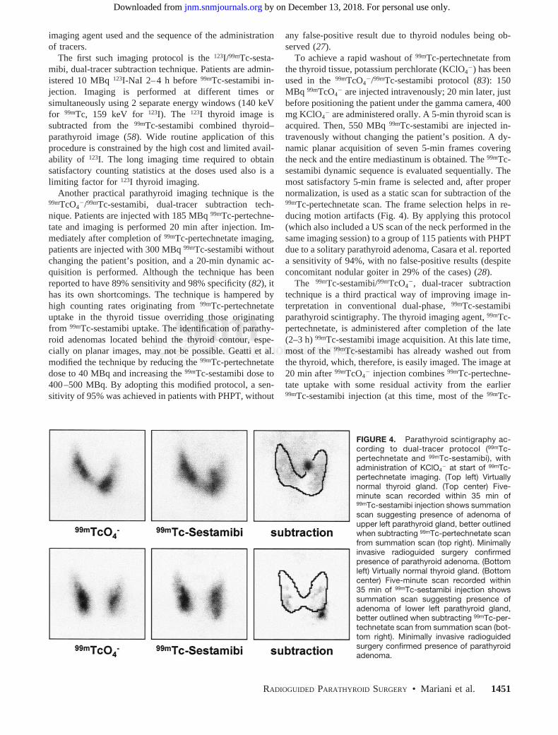
� are administered orally. A 5-min thyroid scan isacquired. Then, 550 MBq 99mTc-sestamibi are injected in-travenously without changing the patient’s position. A dy-namic planar acquisition of seven 5-min frames coveringthe neck and the entire mediastinum is obtained. The 99mTc-sestamibi dynamic sequence is evaluated sequentially. Themost satisfactory 5-min frame is selected and, after propernormalization, is used as a static scan for subtraction of the99mTc-pertechnetate scan. The frame selection helps in re-ducing motion artifacts (Fig. 4). By applying this protocol(which also included a US scan of the neck performed in thesame imaging session) to a group of 115 patients with PHPTdue to a solitary parathyroid adenoma, Casara et al. reporteda sensitivity of 94%, with no false-positive results (despiteconcomitant nodular goiter in 29% of the cases) (28).
The 99mTc-sestamibi/99mTcO4�, dual-tracer subtraction
technique is a third practical way of improving image in-terpretation in conventional dual-phase, 99mTc-sestamibiparathyroid scintigraphy. The thyroid imaging agent, 99mTc-pertechnetate, is administered after completion of the late(2–3 h) 99mTc-sestamibi image acquisition. At this late time,most of the 99mTc-sestamibi has already washed out fromthe thyroid, which, therefore, is easily imaged. The image at20 min after 99mTcO4
� injection combines 99mTc-pertechne-tate uptake with some residual activity from the earlier99mTc-sestamibi injection (at this time, most of the 99mTc-
FIGURE 4. Parathyroid scintigraphy ac-cording to dual-tracer protocol (99mTc-pertechnetate and 99mTc-sestamibi), withadministration of KClO4
� at start of 99mTc-pertechnetate imaging. (Top left) Virtuallynormal thyroid gland. (Top center) Five-minute scan recorded within 35 min of99mTc-sestamibi injection shows summationscan suggesting presence of adenoma ofupper left parathyroid gland, better outlinedwhen subtracting 99mTc-pertechnetate scanfrom summation scan (top right). Minimallyinvasive radioguided surgery confirmedpresence of parathyroid adenoma. (Bottomleft) Virtually normal thyroid gland. (Bottomcenter) Five-minute scan recorded within35 min of 99mTc-sestamibi injection showssummation scan suggesting presence ofadenoma of lower left parathyroid gland,better outlined when subtracting 99mTc-per-technetate scan from summation scan (bot-tom right). Minimally invasive radioguidedsurgery confirmed presence of parathyroidadenoma.
RADIOGUIDED PARATHYROID SURGERY • Mariani et al. 1451
by on December 13, 2018. For personal use only. jnm.snmjournals.org Downloaded from

sestamibi would be already washed out from the thyroid).The late 99mTc-sestamibi image is subtracted from the com-bined scan to obtain a “pure” 99mTc-pertechnetate image, theprofile of which will clarify the origin (thyroid vs. parathy-roid) of abnormal 99mTc-sestamibi uptake (Fig. 5).
Factors affecting the diagnostic accuracy of 99mTc-sesta-mibi imaging of parathyroid glands include regional perfu-sion, gland size and functional activity, cell cycle phase, andprevalence of mitochondria-rich oxyphil cells (84,85). Aslittle as 100 mg hyperfunctioning parathyroid glands can bedetected with application of an appropriate imaging proto-col and technique (28,86–89). The use of a pinhole colli-mator in the neck, with a trade-off in image acquisitiontime, increases imaging resolution. The chest is best eval-uated with a parallel-hole collimator either in the planar orin the SPECT mode with its added information on the depthof the lesion and topographic correlation with other ana-tomic structures. Although, in principle, SPECT offers the
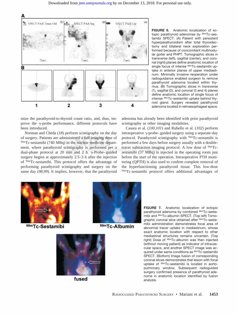
advantage of better discrimination of focal 99mTc-sestamibiretention in thyroid nodules and the parathyroid tissue, in abusy clinical practice this technique is rarely used for eval-uating the neck alone. SPECT is most helpful in evaluationof the mediastinum for possible sites of ectopic parathyroidglands, especially to better guide the surgeon in preopera-tive planning (Figs. 6 and 7). Because detection of parathy-roid tumors in ectopic locations is usually not hampered byproximity with the thyroid gland, SPECT can easily beperformed, with satisfactory counting statistics, relativelysoon after injection of 99mTc-sestamibi (e.g., within 30–40min, immediately after imaging of the neck and thorax withthe early planar views). Although only a marginal improve-ment in the overall detection rate of parathyroid adenomasis reported with SPECT (90,91) (so that the added cost of itssystematic routine use is not always justified), most authorsnow favor a wider application of this imaging modality,especially in patients with recurrent hyperparathyroidismafter prior surgery (30,32,68,72,85,91–93).
99mTc-TetrofosminAs an alternative to 99mTc-sestamibi, several authors have
proposed the use of 99mTc-tetrofosmin for parathyroid scin-tigraphy. Although their mechanism of intracellular accu-mulation and retention differs somewhat (99mTc-sestamibiaccumulates primarily in the mitochondria, whereas 99mTc-tetrofosmin is retained primarily in the cytosol fraction),these 2 tracers are used virtually interchangeably as myo-cardial perfusion agents and as nonspecific oncotropicagents. However, washout of 99mTc-tetrofosmin from thethyroid parenchyma is considerably slower than that of99mTc-sestamibi, so that the single-tracer, dual-phase proce-dure is not always reliable when using 99mTc-tetrofosmin(94–97). Therefore, the use of a second, thyroid-avid traceris mandatory for better discrimination of thyroid or para-thyroid areas of focal uptake in the neck. In conclusion,99mTc-tetrofosmin can be used for parathyroid scintigraphy,provided it is used within a dual-tracer subtraction protocol(96).
INTRAOPERATIVE PARATHYROID LOCALIZATION
Radioguided minimally invasive surgery for PHPT isfeasible when preoperative scintigraphy identifies a singlefocus of radiotracer uptake, indicating a parathyroid ade-noma.
Timing in Radioguided Surgery99mTc-Sestamibi is the only radiopharmaceutical cur-
rently used to identify the site of parathyroid adenomas bypre- or intraoperative radioguidance. The success of ra-dioguided surgery is dependent on the differential kineticsof 99mTc-sestamibi in thyroid and parathyroid glands. 99mTc-Sestamibi washes out more rapidly from the thyroid thanfrom the parathyroid glands. Intraoperative �-probe local-ization of an adenoma is most successful within a verynarrow time window, 2–3 h after tracer injection. To opti-
FIGURE 5. Double-phase parathyroid scintigraphy with99mTc-sestamibi in patient with multinodular goiter. (Top left)Image at 15 min shows physiologic early uptake in thyroidgland, with clear focus of increased accumulation at lower poleof left thyroid lobe. (Top right) Late 99mTc-sestamibi scan showssome residual activity in thyroid gland, with obvious focal 99mTc-sestamibi retention at lower pole of left thyroid lobe. Secondtracer (99mTc-pertechnetate) was administered after recordingdelayed 99mTc-sestamibi scan. (Bottom left) Image obtained bysubtracting delayed 99mTc-sestamibi scan from summation scanrecorded after 99mTc-pertechnetate administration was used todraw profile of thyroid gland, which was then superimposed ondelayed 99mTc-sestamibi scan (bottom right) for better anatomiclocalization of parathyroid adenoma (confirmed by minimallyinvasive radioguided surgery).
1452 THE JOURNAL OF NUCLEAR MEDICINE • Vol. 44 • No. 9 • September 2003
by on December 13, 2018. For personal use only. jnm.snmjournals.org Downloaded from
mize the parathyroid-to-thyroid count ratio, and, thus, im-prove the �-probe performance, different protocols havebeen introduced.
Norman and Cheda (18) perform scintigraphy on the dayof surgery. Patients are administered a full imaging dose of99mTc-sestamibi (740 MBq) in the nuclear medicine depart-ment, where parathyroid scintigraphy is performed per adual-phase protocol at 20 min and 2 h. �-Probe–guidedsurgery begins at approximately 2.5–3 h after the injectionof 99mTc-sestamibi. This protocol offers the advantage ofperforming parathyroid scintigraphy and surgery on thesame day (98,99). It implies, however, that the parathyroid
adenoma has already been identified with prior parathyroidscintigraphy or other imaging modalities.
Casara et al. (100,101) and Rubello et al. (102) performintraoperative �-probe–guided surgery using a separate-dayprotocol. Parathyroid scintigraphy with 99mTc-sestamibi isperformed a few days before surgery usually with a double-tracer subtraction imaging protocol. A low dose of 99mTc-sestamibi (37 MBq) is injected in the operating room justbefore the start of the operation. Intraoperative PTH moni-toring (QPTH) is also used to confirm complete removal ofthe hyperfunctioning parathyroid tissue. This low-dose99mTc-sestamibi protocol offers additional advantages of
FIGURE 6. Anatomic localization of ec-topic parathyroid adenomas by 99mTc-ses-tamibi SPECT. (A) Patient with persistenthyperparathyroidism after total thyroidec-tomy and bilateral neck exploration per-formed because of concomitant multinodu-lar goiter and PHPT. Tomographic slices intransverse (left), sagittal (center), and coro-nal (right) planes define anatomic location ofsingle focus of intense 99mTc-sestamibi up-take in anterior planes of upper mediasti-num. Minimally invasive reoperation underradioguidance enabled surgeon to removeparathyroid adenoma located within thy-mus. (B) Tomographic slices in transverse(1), sagittal (2), and coronal (3 and 4) planesdefine anatomic location of single focus ofintense 99mTc-sestamibi uptake behind thy-roid gland. Surgery revealed parathyroidadenoma located in retroesophageal space.
FIGURE 7. Anatomic localization of ectopicparathyroid adenoma by combined 99mTc-sesta-mibi and 99mTc-albumin SPECT. (Top left) Tomo-graphic coronal slice obtained after 99mTc-sesta-mibi administration demonstrates focal area ofabnormal tracer uptake in mediastinum, whoseexact anatomic location with respect to othermediastinal structures remains uncertain. (Topright) Dose of 99mTc-albumin was then injected(without moving patient) as indicator of intravas-cular space, and another SPECT image was ac-quired under same conditions as 99mTc-sestamibiSPECT. (Bottom) Image fusion of correspondingcoronal slices demonstrates that lesion with focaluptake of 99mTc-sestamibi is located in aorto-pulmonary window. Subsequent radioguidedsurgery confirmed presence of parathyroid ade-noma in anatomic location identified by fusionanalysis.
RADIOGUIDED PARATHYROID SURGERY • Mariani et al. 1453
by on December 13, 2018. For personal use only. jnm.snmjournals.org Downloaded from

minimized radiation exposure to the surgeon and to the teamand possibly improves the chance of identification of para-thyroid lesions with rapid 99mTc-sestamibi washout (80,81).This protocol was also found to be particularly useful inpatients with concomitant nodular goiter (27,28), althoughthese patients are not optimal candidates for minimallyinvasive radioguided parathyroid surgery, due to the highfalse-positive uptake sites in the thyroid mimicking para-thyroid adenomas.
Bozkurt et al. describe a patient-specific, optimal time-to-surgery protocol to improve operative success in�-probe–guided parathyroid localization (103). Using dou-ble-phase 99mTc-sestamibi parathyroid scintigraphy (500–700 MBq), the 99mTc-sestamibi time–activity curves forboth parathyroid and thyroid glands are generated. Theoptimal time to surgery was determined on the basis of thetime at which the target-to-background ratio was maximum.On the day of surgery, patients receive the same dose of99mTc-sestamibi and are taken to the operating room at thetime of optimal target-to-background ratio that was deter-mined by the preoperative imaging study.
In addition, several groups use a modified separate-dayprotocol, in which a higher dose of 99mTc-sestamibi is in-jected immediately before surgery (about 370 MBq) toreach higher target-to-background ratios, which are partic-ularly useful for identification of ectopic parathyroid ade-nomas.
Operative TechniqueOn the basis of the specific protocol adopted, minimally
invasive radioguided parathyroid surgery is started 30 minto 3 h after 99mTc-sestamibi administration. After inductionof the preferred anesthesia (local, regional, or general),external �-probe scanning is performed to locate the hotspot. Once the hot spot is identified (cutaneous marking bythe nuclear medicine physician could be very helpful), asmall incision is made and the space under the strap musclesis entered. The probe is then inserted into the skin incision(maximum, 1–2 cm), directly over the presumed location ofthe adenoma. The high-pitch signals produced by the�-probe lead the surgeon toward the location with highestradioactivity. A parathyroid-to-thyroid ratio higher than 1.5strongly suggests the presence of a parathyroid adenoma,whereas typical parathyroid-to-background ratios (exclud-ing the thyroid tissue) range between 2.5 and 4.5. After theparathyroid lesion is removed, the ex vivo counting rate ofan adenoma is at least 20% and usually is 50% higher thanthe thyroid background (104). The surgical bed is scannedagain to ensure the complete removal of the adenoma byestablishing a new level of background radioactivity. A ratiogreater than 1.2 between the ex vivo lesion counts and theresidual background counts is another criterion used todetermine successful excision of an abnormal gland (104).The errors due to equivocal or false-positive scans are thusdecreased by the use of an intraoperative �-probe. It shouldbe noted, however, that similar count ratios can occasionally
be observed also when removing simply hyperplastic para-thyroid tissue or even a thyroid nodule (89,101). A finalassessment of radioactivity in all 4 quadrants at the end ofthe surgical exploration increases the surgeon’s confidencein the completeness of the parathyroidectomy.
�-Probe guidance enables the surgeon to perform a rathersmall skin incision with improved cosmesis. The techniquecan also be performed under local anesthesia. The operatingtime is reduced, and the patient can be discharged from thehospital earlier (18,105,106). As with other radioguidedsurgical procedures (e.g., sentinel lymph node biopsy), asuccessful clinical outcome requires a smooth coordinationbetween the nuclear medicine physician, the surgeon, andthe pathologist).
GUIDELINES AND CONTROVERSIES IN RADIOGUIDEDMIP
The guidelines for a �-probe–guided MIP (GP-MIP) arebased on the clinical profile of patients and the preoperative99mTc-sestamibi imaging study findings. GP-MIP is an ap-propriate approach in patients who have a high probabilityof a solitary parathyroid adenoma established on the basis of99mTc-sestamibi scintigraphy (and US imaging), significant99mTc-sestamibi uptake in the parathyroid adenoma, no co-existing 99mTc-sestamibi–avid thyroid nodules, no history offamilial hyperparathyroidism or MEN, and no history ofprevious neck irradiation (18,28,100). Following these cri-teria, the fraction of patients who are candidates for GP-MIPis about 60%–70% of all cases of PHPT (39,100,107).Additional settings in which radioguided parathyroidectomyis indicated include reoperation for persistent or recurrenthyperparathyroidism and ectopic adenomas (especially, ma-jor ectopy).
In some clinical settings, the most frequent cause ofexclusion from GP-MIP was the diagnosis of a concomitantnodular goiter (101). In fact, thyroid nodules can givefalse-positive results both at preoperative scintigraphy(57,108,109) and at radioguided surgery (109,110) because�50%–60% of them can trap and retain 99mTc-sestamibisimilarly as parathyroid adenomas (61,110). This problem ispartly overcome by double-tracer subtraction protocols(123I/99mTc-sestamibi or 99mTc-pertechnetate/99mTc-sesta-mibi) that are therefore generally preferred for preoperativelocalization in areas with high prevalence of thyroid nodulargoiter, as in many European countries (61,109).
Considering also the possible association of a parathyroidadenoma with nodular goiter, the use of a double-tracertechnique or of 99mTc-sestamibi scintigraphy combined withUS examination might be useful in planning the type andextent of surgery (89,101): (a) bilateral neck exploration inthe case of any PHPT patient with concomitant multinodu-lar goiter; (b) unilateral neck exploration in the case of asolitary parathyroid adenoma with concomitant nodular goi-ter located in the ipsilateral thyroid lobe; (c) GP-MIP in thecase of a solitary 99mTc-sestamibi–avid parathyroid ade-noma with a normal thyroid gland; and (d) endoscopic
1454 THE JOURNAL OF NUCLEAR MEDICINE • Vol. 44 • No. 9 • September 2003
by on December 13, 2018. For personal use only. jnm.snmjournals.org Downloaded from

surgery in the infrequent case of a solitary 99mTc-sestamibi-negative (but US positive) parathyroid adenoma with anormal thyroid gland. Thus, intraoperative �-probe guid-ance can simply be considered as a completion of accuratepreoperative scintigraphic imaging.
The advantages of intraoperative �-probe guidance inparathyroid surgery can be summarized as follows: (a) The�-probe guides the surgeon to the site of the parathyroidadenoma, thus facilitating the approach and shortening thesurgical times, especially for parathyroid adenomas locatedin an ectopic site or deep in the neck (89,102). (b) �-Probeguidance is helpful in evaluating the completeness of para-thyroid tissue removal (18,89,102,104). (c) Use of the�-probe for measuring ex vivo radioactivity of the removedspecimen helps in verifying the effective removal of para-thyroid tissue and, thus, in evaluating the success of surgery(18,89,102,104). As mentioned above, the so-called “20%rule” proposed by Murphy and Norman (104) has beenreported to yield a 100% accuracy in distinguishing solitaryparathyroid adenomas from hyperplasia. An in vitro�-probe assessment that does not meet the above criteriashould direct the surgeon to continue the exploration, obtainintraoperative QPTH measurement, and perform frozen sec-tion analysis of the removed tissue.
Inconsistent results with the 20% rule, or even whenusing higher thresholds such as 30%–40% above back-ground, reported by other groups, might be related to thevariations in administered doses, timing of �-probe mea-surements, and background references (89,107,111,112).The different sensitivities of the various �-probes used andthe use of collimated or uncollimated probes also signifi-cantly contribute to the variability of reported results withGP-MIP (89,107).
Although �-probe guidance is highly effective for theintraoperative detection of solitary parathyroid adenomas,difficulties might arise in those rare patients with multiglanddisease with a “dominant” parathyroid gland. In these pa-tients, intraoperative �-probe counting in a nondominant,yet adenomatous, parathyroid gland can be significantlylower that the counting rate in the dominant gland, thusleading the surgeon to underestimate the presence of mul-tigland disease (89,107,113). In this clinical setting, intra-operative QPTH assay offers the advantage of identifyingpatients with multigland disease (114), including those withscan-negative enlarged parathyroid glands. On the basis ofthese considerations, confirmation of complete parathyroidremoval by intraoperative QPTH assay is still advisable,even when performing GP-MIP both at first surgery and atreoperation (89,100,102,107). Although intraoperativeQPTH assay does not seem to increase significantly thesuccess rate of surgery in carefully selected patients with asolitary parathyroid adenoma (114), there are no relative orabsolute contraindications to using this procedure, alone orin combination with intraoperative �-probe guidance.
�-Probe guidance has also been useful when performinga standard bilateral neck exploration, because it increases
the accuracy of preoperative 99mTc-sestamibi scintigraphy(89,99,110). A negative preoperative 99mTc-sestamibi scanand contemplation of bilateral neck exploration should notpreclude radioguided parathyroid surgery.
The cost-effectiveness of the image-guided minimallyinvasive approach—that is, the expense of imaging withequipment required—has been questioned. However, thepotential savings from decreased operating time and hos-pital stay were found to be comparable or in favor ofthe minimally invasive approach in many analyses(73,107,113). A major concern when using a 99mTc-sesta-mibi–guided focused unilateral surgical approach is a pos-sible failure to diagnose multigland disease. A recent reviewdemonstrated that the reported incidence of multigland dis-ease in a series in which bilateral explorations were done is20%, whereas the incidence with focused unilateral explo-rations is only 5% (11). The difference could be explainedby the lower sensitivity of 99mTc-sestamibi localization inmultigland disease, resulting in underdiagnosis of this oc-currence. However, the possibility of overdiagnosis of mul-tigland disease with the bilateral approach (biased pathol-ogy) cannot be discarded. The evidence-based support forMIP comes from the fact that the recurrence rates followingfocused unilateral approaches have not been higher thanthose of the bilateral approach.
CONCLUSION
GP-MIP is a very attractive surgical approach to treatpatients with PHPT secondary to solitary parathyroid ade-noma. GP-MIP has proven to be technically easy, safe, andwith a low morbidity rate in the hands of a skilled surgeon(18,28,89). The advantages of GP-MIP over bilateral neckexploration in patients with PHPT secondary to solitaryadenoma can be summarized as follows: (a) smaller inci-sion, less surgical trauma; (b) shorter length of surgery,anesthesia, and hospital stay; (c) less postsurgical pain; (d)better cosmetic results; and (e) lower overall cost. In con-trast to minimally invasive endoscopic surgery, GP-MIPcan be performed also for reintervention in patients withpersistent or recurrent hyperparathyroidism (89,99).
The guidelines for GP-MIP can be summarized as fol-lows: (a) Only patients with a high probability of a solitaryparathyroid adenoma and a normal thyroid gland should beconsidered. (b) The most appropriate preoperative scinti-graphic protocol should be selected on the basis of thyroidand parathyroid imaging information. (c) The radiation ex-posure dose to the surgeon and operating theater personnelshould be minimized by administering the lowest dose of99mTc-sestamibi proven to be effective for performing GP-MIP. (d) Both in vivo or ex vivo �-probe counting andintraoperative QPTH measurement should be used to eval-uate the success and completeness of surgery.
REFERENCES
1. Bilezikian JP, Silverberg SJ. Clinical spectrum of primary hyperparathyroidism.Rev Endocr Metab Disord. 2000;1:237–245.
RADIOGUIDED PARATHYROID SURGERY • Mariani et al. 1455
by on December 13, 2018. For personal use only. jnm.snmjournals.org Downloaded from
2. Sywak MS, Robinson BG, Clifton-Bligh P, et al. Increase in presentations andprocedure rates for hyperparathyroidism in Northern Sydney and New SouthWales. Med J Aust. 2002;177:246–249.
3. Heath H 3rd, Hodgson SF, Kennedy MA. Primary hyperparathyroidism: inci-dence, morbidity, and potential economic impact in a community. N Engl J Med.1980;302:189–193.
4. Clark OH, Siperstein AE. The hypercalcemic syndrome. In: Friesen SR, Thomp-son NW, eds. Surgical Endocrinology Clinical Syndromes. Philadelphia, PA:Lippincott; 1990:311–339.
5. Melton LJ 3rd. The epidemiology of primary hyperparathyroidism in NorthAmerica. J Bone Miner Res. 2002;17(suppl 2):N12–N17.
6. Adami S, Marcocci C, Gatti D. Epidemiology of primary hyperparathyroidismin Europe. J Bone Miner Res. 2002;17(suppl 2):N18–N23.
7. Bilezikian JP, Meng X, Shi Y, Silverberg SJ. Primary hyperparathyroidism inwomen: a tale of two cities—New York and Beijing. Int J Fertil Womens Med.2000;45:158–165.
8. Raisz LG, Kream BE, Lorenzo JA. Metabolic bone disease. In: Wilson JD,Foster DW, Kronenberg HM, Larsen PR, eds. Williams Textbook of Endocri-nology. 9th ed. Philadelphia, PA: Saunders; 1998:1211–1240.
9. Kinder BK, Stewart AF. Hypercalcemia. Curr Probl Surg. 2002;39:349–448.10. DeLellis RA. Tumors of the parathyroid gland. In: Atlas of Tumor Pathology.
Third Series, Fascicle 6. Washington, DC: Armed Forces Institute of Pathology;1993:25–51, 65–78.
11. Lee NC, Norton JA. Multiple gland disease in primary hyperparathyroidism: afunction of operative approach? Arch Surg. 2002;137:896–900.
12. Metz D, Jensen R, Allen B, et al. Multiple endocrine neoplasia type 1: clinicalfeatures and management. In: Bilezikian J, Levine M, Marcus R, eds. TheParathyroids. New York, NY: Raven Press; 1994:591–647.
13. Mandl F. Therapeutic attempt of osteitis fibrosa generalisata by excision of anepithelial-corpuscle tumors [in German]. Wien Klin Wochenschr. 1925;195:1343–1344.
14. Kaplan EL, Yashiro T, Salti G. Primary hyperparathyroidism in the 1990s:choice of surgical procedures for this disease. Ann Surg. 1992;215:301–317.
15. Rose MD, Wood TF, Van Herle AJ, et al. Long term management and outcomeof parathyroidectomy for sporadic primary multiple gland disease. Arch Surg.2001;136:621–626.
16. Udelsman R. Six hundred fifty-six consecutive explorations for primary hyper-parathyroidism. Ann Surg. 2002;235:665–672.
17. Sofferman RA, Nathan MH, Fairbank JT, Foster RS Jr, Krag DN. Preoperativetechnetium 99m sestamibi imaging: paving the way to minimal-access parathy-roid surgery. Arch Otolaryngol Head Neck Surg. 1996;122:369–374.
18. Norman J, Cheda H. Minimally invasive radioguided parathyroidectomy facil-itated by intraoperative nuclear mapping. Surgery. 1997;122:998–1004.
19. Rubello D, Pelizzo MR, Casara D. Nuclear medicine and minimally invasivesurgery of parathyroid adenomas: a fair marriage [editorial]. Eur J Nucl Med.2003;30:189–192.
20. Sackett WR, Barraclough B, Reeve TS, Delbridge LW. Worldwide trends in thesurgical treatment of primary hyperparathyroidism in the era of minimallyinvasive parathyroidectomy. Arch Surg. 2002;137:1055–1059.
21. Tibblin S, Bondeson AG, Ljungberg O. Unilateral parathyroidectomy in hyper-parathyroidism due to a single adenoma. Ann Surg. 1982;195:245–252.
22. Chapuis Y, Icard P, Fulla Y, et al. Parathyroid adenomectomy under localanesthesia with intra-operative monitoring of UcAMP and/or 1–84 PTH. WorldJ Surg. 1992;16:570–575.
23. Nussbaum SR, Thompson AR, Hutcheson KA, et al. Intraoperative measure-ment of parathyroid hormone in the surgical management of hyperthyroidism.Surgery. 1988;104:1121–1127.
24. Irvin GL 3rd. American Association of Endocrine Surgeons presidential ad-dress: chasin’ hormones. Surgery. 1999;126:993–997.
25. Irvin GL 3rd, Carneiro DM. Rapid parathyroid hormone assay guided explora-tion. Oper Tech Gen Surg. 1999;1:18–27.
26. Ammori BJ, Madan M, Gopichandran TD, et al. Ultrasound guided unilateralneck exploration for sporadic primary hyperparathyroidism: is it worthwhile?Ann R Coll Surg Engl. 1998;80:433–437.
27. Geatti O, Shapiro B, Orsolon P, et al. Localization of parathyroid enlargement:experience with technetium 99m methoxyisobutylisonitrile and thallium-201scintigraphy, ultrasound and computed tomography. Eur J Nucl Med. 1994;21:17–23.
28. Casara D, Rubello D, Pelizzo MR, Shapiro B. Clinical role of 99mTcO4/MIBIscan, ultrasound and intra-operative gamma probe in the performance of uni-lateral and minimally invasive surgery in primary hyperparathyroidism. EurJ Nucl Med. 2001;28:1351–1359.
29. Miller DL, Doppman MD, Shawker MD, et al. Localization of parathyroid
adenomas in patients who have undergone surgery. Radiology. 1987;162:133–137.
30. Uden P, Aspelin P, Berglund J. Preoperative localization in unilateral parathy-roid surgery: a cost-benefit study on ultrasound, computed tomography andscintigraphy. Acta Chir Scand. 1990;156:29–35.
31. Gofrit ON, Labensart PD, Pikarsky A, Lackstein D, Gross DJ, Shiloni E.High-resolution ultrasonography: highly sensitive, specific technique for preop-erative localization of parathyroid adenoma in the absence of multinodularthyroid disease. World J Surg. 1997;21:287–290.
32. Lloyd MN, Lees WR, Milroy EJG. Preoperative localisation in primary hyper-parathyroidism. Clin Radiol. 1990;41:239–243.
33. Beierwaltes WH. Endocrine imaging: parathyroid, adrenal cortex and medulla,and other endocrine tumors—Part 2. J Nucl Med. 1991;32:1627–1639.
34. Tomasella G. Diagnostic imaging in primary hyperparathyroidism—radiologi-cal techniques: US–CAT–MR [in Italian]. Minerva Endocrinol. 2001;26:3–12.
35. De Feo ML, Colagrande S, Biagini C, et al. Parathyroid glands: combination of99mTc-MIBI scintigraphy and US for demonstration of parathyroid glands andnodules. Radiology. 2000;214:393–402.
36. Udelsman R. Is unilateral neck exploration for parathyroid adenoma appropri-ate? Adv Surg. 2000;34:319–329.
37. Eisenberg H, Pallotta J, Sacks B, Brickman AS. Parathyroid localization,three-dimensional modeling, and percutaneous ablation techniques. EndocrinolMetab Clin North Am. 1989;18:659–700.
38. Mitchell BK, Merrel RC, Kinder BK. Localization studies in patients withhyperparathyroidism. Surg Clin North Am. 1995;75:483–498.
39. Koong HN, Choong LH, Soo KC. The role of preoperative localisation tech-niques in surgery for hyperparathyroidism. Ann Acad Med Singapore. 1998;27:192–195.
40. Levin KE, Clark OH. Localization of parathyroid glands. Annu Rev Med.1988;39:29–40.
41. Weber AL, Randolph G, Aksoy FG. The thyroid and parathyroid glands: CT andMR imaging and correlation with pathology and clinical findings. Radiol ClinNorth Am. 2000;38:1105–1129.
42. Kneeland JB, Krubsack AJ, Lawson TL, et al. Enlarged parathyroid glands:high-resolution local coil MR imaging. Radiology. 1987;162:143–146.
43. Lee VS, Spritzer CE, Coleman RE, Wilkinson RH Jr, Coogan AC, Leight GS Jr.The complementary roles of fast spin-echo MR imaging and double-phase99mTc-sestamibi scintigraphy for localization of hyperfunctioning parathyroidglands. AJR. 1996;167:1555–1562.
44. Lee VS, Spritzer CE. MR imaging of abnormal parathyroid glands. AJR.1998;170:1097–1103.
45. Hishibashi M, Nishida H, Hiromatsu Y, Kojima K, Tabuchi E, Hayabuchi N.Comparison of technetium-99m-MIBI, technetium-99m-tetrofosmin, ultra-sound, and MRI for localization of abnormal parathyroid glands. J Nucl Med.1988;39:320–324.
46. Fayet P, Hoeffel C, Fulla Y, et al. Techetium-99m-sestamibi, magnetic reso-nance imaging and venous blood sampling in persistent and recurrent hyper-parathyroidism. Br J Radiol. 1997;70:459–464.
47. Gotway MB, Reddy GP, Webb WR, Morita ET, Clark OH, Higgins CB.Comparison between MR imaging and 99mTc-MIBI scintigraphy in the evalua-tion of recurrent or persistent hyperparathyroidism. Radiology. 2001;218:783–790.
48. McDermott VG, Mendez Fernandez RJ, Meakem TJ 3rd, et al. Preoperative MRimaging in hyperparathyroidism: results and factors affecting parathyroid de-tection. AJR. 1996;166:705–710.
49. NIH Conference: diagnosis and management of asymptomatic primary hyper-parathyroidism—consensus development conference statement. Consensus De-velopment Conference, NIH. Ann Intern Med. 1991;114:593–597.
50. Di Giulio W, Morales J. An evaluation of parathyroid scanning using selenium-75-methionine. J Nucl Med. 1966;7:380–384.
51. Waldorf JC, van Heerden JA, Gorman CA, Grant CS, Wahner HW. [75Se]Sel-enomethionine scanning for parathyroid localization should be abandoned.Mayo Clin Proc. 1984;59:534–537.
52. Ferlin G, Borsato N, Camerani M, Conte N, Zotti D. New perspectives inlocalizing enlarged parathyroids by technetium-thallium subtraction scan.J Nucl Med. 1983;24:438–441.
53. Samanta A, Wilson B, Iqbal J, Burden AC, Walls J, Cosgriff P. A clinical auditof thallium-technetium subtraction parathyroid scans. Postgrad Med J. 1990;66:441–445.
54. Nicholson DA, Dawson P, Lavender JP. Imaging of the parathyroids. In: LynnJ, Bloom SR, eds. Surgical Endocrinology. Oxford, U.K.: Butterworth-Heine-mann; 1993:351–361.
55. Coakley AJ, Kettle AG, Wells CP, O’Doherty MJ, Collins REC. 99mTc-Sesta-
1456 THE JOURNAL OF NUCLEAR MEDICINE • Vol. 44 • No. 9 • September 2003
by on December 13, 2018. For personal use only. jnm.snmjournals.org Downloaded from

mibi: a new agent for parathyroid imaging. Nucl Med Commun. 1989;10:791–794.
56. O’Doherty MJ, Kettle AG, Wells P, Collins REC, Coakley AJ. Parathyroidimaging with technetium-99m-sestamibi: preoperative localization and tissueuptake studies. J Nucl Med. 1992;33:313–319.
57. Taillefer R, Boucher Y, Potvin C, et al. Detection and localization of parathyroidadenomas in patients with hyperparathyroidism using a single radionuclideimaging procedure with technetium-99m-sestamibi (double phase study). J NuclMed. 1992;33:1801–1807.
58. Weber CJ, Vansant J, Alazraki N, et al. Value of technetium 99m sestamibiiodine imaging in reoperative parathyroid surgery. Surgery. 1993;114:1011–1018.
59. Casas AT, Burke GJ, Mansberger AR, Wei JP. Impact of technetium-99msestamibi localization on operative time and success of operations for primaryhyperparathyroidism. Am Surg. 1994;60:12–17.
60. Feingold DL, Alexander HR, Chen CC, et al. Ultrasound and sestamibi scan asthe only preoperative imaging tests in reoperation for parathyroid adenomas.Surgery. 2000;128:1103–1110.
61. Giordano A, Rubello D, Casara D. New trends in parathyroid scintigraphy. EurJ Nucl Med. 2001;28:1409–1420.
62. Hetrakul N, Civelek AC, Stag CA, Udelsman R. In vitro accumulation oftechnetium-99m sestamibi in human parathyroid mitochondria. Surgery. 2001;130:1011–1018.
63. Piwnica-Worms D, Chiu ML, Budding M, Kronauge JF, Kramer RA, Croop JM.Functional imaging of multidrug resistant P-glycoprotein with organotechne-tium complex. Cancer Res. 1993;53:977–984.
64. Mitchell BK, Cornelius EA, Zoghbi S, et al. Mechanism of technetium 99msestamibi parathyroid imaging and possible role of p-glycoprotein. Surgery.1996;120:1039–1045.
65. Bhatnagar A, Vezza PR, Bryan JA, et al. Technetium-99m-sestamibi parathy-roid scintigraphy: effect of P-glycoprotein, histology and tumor size on detect-ability. J Nucl Med. 1998;39:1617–1620.
66. Yamaguchi S, Yachiku S, Hashimoto H, et al. Relation between technetium 99mmethoxyisobutylisonitrile accumulation and multidrug resistance protein in theparathyroid glands. World J Surg. 2002;26:29–34.
67. Royal RE, Delpassand ES, Shapiro SE, et al. Improving the yield of preoper-ative parathyroid localization: technetium-99m-sestamibi imaging after thyroidsuppression. Surgery. 2002;132:968–974.
68. Billotey C, Sarfati E, Aurengo A, et al. Advantages of SPECT in technetium-99m-sestamibi parathyroid scintigraphy. J Nucl Med. 1996;37:1773–1778.
69. Carty SE, Worsey MJ, Virji MA, Brown ML, Watson CG. Concise parathy-roidectomy: the impact of preoperative SPECT 99mTc sestamibi scanning andintraoperative quick parathormone assay. Surgery. 1997;122:1107–1116.
70. Gallowitsch JH, Mikosch P, Kresnik E, et al. Technetium-99m-tetrofosminparathyroid imaging: results with double-phase study and SPECT in primaryand secondary hyperparathyroidism. Invest Radiol. 1997;32:459–465.
71. Neumann DR, Esselstyn CB Jr, Go RT, Wong CO, Rice TW, Obuchowsky NA.Comparison of double-phase 99mTc-sestamibi with 123I-99mTc-sestamibi subtrac-tion SPECT in hyperparathyroidism. AJR. 1997;169:1671–1674.
72. Francis IS, Loney EL, Buscombe JR, Thakrar DS, Berger L, Hilson AJW.Technetium-99m-sestamibi dual-phase SPECT imaging: concordance with ul-trasound. Nucl Med Commun. 1999;20:487–488.
73. Denham DW, Norman J. Cost-effectiveness of preoperative sestamibi scan forprimary hyperparathyroidism is dependant solely upon the surgeons choice ofoperative procedure. J Am Coll Surg. 1998;186:293–304.
74. George EF, Komisar A, Scharf SC, Ferracci A, Blaugrund S. Diagnostic valueof the preoperative sestamibi scan in intraoperative localization of parathyroidadenomas: a case study. Laryngoscope. 1998;108:627–629.
75. Chen H, Sokoll LJ, Udelsman R. Outpatient minimally invasive parathy-roidectomy: a combination of sestamibi-SPECT localization, cervical blockanesthesia, and intraoperative parathyroid hormone assay. Surgery. 1999;126:1016 –1021.
76. Kumar A, Cozens NJA, Nash JR. Sestamibi scan-directed unilateral neckexploration for primary hyperparathyroidism due to a solitary adenoma. EurJ Surg Oncol. 2000;26:785–788.
77. Taillefer R. 99mTc sestamibi parathyroid scintigraphy. In: Freeman L, ed. Nu-clear Medicine Annual 1995. New York, NY: Raven Press; 1995:51–79.
78. Foldes I, Levay A, Stotz G. Comparative scanning of thyroid nodules withtechnetium-99m pertechnetate and technetium-99m methoxyisobutylisonitrile.Eur J Nucl Med. 1993;20:330–333.
79. dell’Erba L, Baldari S, Borsato N, et al. Retrospective analysis of the associationof nodular goiter with primary and secondary hyperparathyroidism. Eur JEndocrinol. 2001;145:429–434.
80. Benard F, Lefebvre B, Beuvon F, Langlois MF, Bisson G. Rapid wash-out of
technetium-99m-MIBI from a large parathyroid adenoma. J Nucl Med. 1995;36:241–243.
81. Leslie WD, Riese KT, Dupont JO, Teterdy AE. Parathyroid adenomas withoutsestamibi retention. Clin Nucl Med. 1995;20:699–702.
82. Casara D, Rubello D, Saladini G, Piotto A, Toniato A, Pelizzo MR. Imagingprocedures in the evaluation of hyperparathyroidism: the role of scintigraphywith 99mTc-MIBI [in Italian]. In: Rovelli E, Samori G, eds. Primary andSecondary Hyperparathyroidism [in Italian]. Milan, Italy: Wichtig Editore;1992:133–136.
83. Rubello D, Saladini G, Casara D, et al. Parathyroid imaging with pertechnetateplus perchlorate/MIBI subtraction scintigraphy: a fast and effective technique.Clin Nucl Med. 2000;25:527–531.
84. Carpentier A, Jeannotte S, Verreault J, et al. Preoperative localization ofparathyroid lesions in hyperparathyroidism: relationship between technetium-99m-MIBI uptake and oxyphil cell content. J Nucl Med. 1998;39:1441–1444.
85. Torregrosa JV, Fernandez-Cruz L, Canaleyo A, et al. 99mTc-Sestamibi scintig-raphy and cell cycle in parathyroid glands of secondary hyperparathyrodidism.World J Surg. 2000;24:1386–1390.
86. Sfakianakis GN, Irvin GL, Foss J, et al. Efficient parathyroidectomy guided bySPECT-MIBI and hormonal measurements. J Nucl Med. 1996;37:798–804.
87. Hindie E, Melliere D, Jeanguillame C, Perlemuter L, Chehade F, Galle P.Parathyroid imaging using simultaneous double window recording of techne-tium-99m-sestamibi and iodine-123. J Nucl Med. 1998;39:1100–1105.
88. Moka D, Eschner W, Voth E, Dietlein M, Larena-Avellaneda A, Schicha H.Iterative reconstruction: an improvement of technetium-99m-MIBI SPECT forthe detection of parathyroid adenoma? Eur J Nucl Med. 2000;27:485–489.
89. Rubello D, Casara D, Giannini S, et al. Importance of radio-guided minimallyinvasive parathyroidectomy using hand-held gamma probe and low 99mTc-MIBIdose: technical considerations and long-term clinical results. Q J Nucl Med.2003;47:224–232.
90. McHenry CR, Lee K, Saadey J, Neuman DR, Esselstyn CB Jr. Parathyroidlocalization with technetium-99m-MIBI scintigraphy to identify functional anat-omy in secondary hyperparathyroidism. J Nucl Med. 1996;37:565–569.
91. Pattou F, Huglo D, Proye C. Radionuclide scanning in parathyroid disease. Br JSurg. 1998;85:1605–1616.
92. Loney EL, Buscombe JR, Hilson AJW, Davenport A, Francis IS. Preoperativeimaging of parathyroid glands. Lancet. 1999;354:1819–1820.
93. Moka D, Voth E, Dietlein M, Larean-Avellanda A, Schicha H. Technetium99m-MIBI-SPECT: a highly sensitive diagnostic tool for localization of para-thyroid adenomas. Surgery. 2000;128:29–35.
94. Fjeld JG, Erichsen K, Pfeffer PF, Ole PF, Rootwelt C, Rootwelt K. Technetium-99m-tetrofosmin for parathyroid scintigraphy: a comparison with sestamibi.J Nucl Med. 1997;38:831–834.
95. O’Doherty MJ. Radionuclide parathyroid imaging. J Nucl Med. 1997;38:840–841.
96. Gallowitsch HJ, Mikosch P, Kresnik E, Unterweger O, Lind P. Comparisonbetween 99mTc-tetrofosmin/pertechnetate subtraction scintigraphy and 99mTc-tetrofosmin SPECT for preoperative localization of parathyroid adenoma in anendemic goiter area. Invest Radiol. 2000;35:453–459.
97. Froeberg AC, Valkema R, Bonjer HJ, Krenning EP. 99mTc-tetrofosmin or99mTc-sestamibi for double-phase parathyroid scintigraphy? Eur J Nucl Med.2003;30:193–196.
98. Norman JG. Minimally invasive radioguided parathyroidectomy: an endocrinesurgeon’s perspective. J Nucl Med. 1998;39:15N, 24N.
99. Norman J, Denham D. Minimally invasive radioguided parathyroidectomy inthe reoperative neck. Surgery. 1998;124:1088–1093.
100. Casara D, Rubello D, Piotto A, Pelizzo MR. 99mTc-MIBI radio-guided mini-mally invasive parathyroid surgery planned on the basis of a preoperativecombined 99mTc-pertechnetate/99mTc-MIBI and ultrasound imaging protocol.Eur J Nucl Med. 2000;27:1300–1304.
101. Casara D, Rubello D, Cauzzo C, Pelizzo MR. 99mTc-MIBI radio-guided mini-mally invasive parathyroidectomy: experience with patients with normal thy-roids and nodular goiters. Thyroid. 2002;12:53–61.
102. Rubello D, Casara D, Pelizzo MR. Symposium on parathyroid localization:optimization of peroperative procedures. Nucl Med Commun. 2003;24:133–140.
103. Bozkurt FM, Ugur O, Hamaloglu E, Sayek I, Gulec AS. Patient specificgamma-probe guided parathyroidectomy: preoperative determination of optimalsurgical timing. Am Surg. 2003;69:720–725.
104. Murphy C, Norman J. The 20% rule: a simple, instantaneous radioactivitymeasurement defines cure and allows elimination of frozen sections and hor-mone assays during parathyroidectomy. Surgery. 1999;126:1023–1029.
105. Flynn MB, Bumpous JM, Schill K, McMasters KM. Minimally invasive ra-dioguided parathyroidectomy. J Am Coll Surg. 2000;191:24–31.
106. Goldstein RE, Blevins L, Delbeke D, Martin WH. Effect of minimally invasive
RADIOGUIDED PARATHYROID SURGERY • Mariani et al. 1457
by on December 13, 2018. For personal use only. jnm.snmjournals.org Downloaded from
radioguided parathyroidectomy on efficacy, length of stay, and costs in themanagement of primary hyperparathyroidism. Ann Surg. 2000;231:732–742.
107. Perrier ND, Ituarte PHG, Morita E, et al. Parathyroid surgery: separatingpromise from reality. J Clin Endocrinol Metab. 2002;87:1024–1028.
108. Chapius Y, Fulla Y, Bonnichon P, et al. Values of ultrasonography, sesta-mibi scintigraphy and intraoperative measurement of 1– 84 PTH for unilat-eral neck exploration of primary hyperparathyroidism. World J Surg. 1996;20:835– 840.
109. Rubello D, Casara D, Shapiro B. Recent advances in preoperative and intraop-erative nuclear medicine procedures in patients with primary hyperparathyroid-ism. Panminerva Med. 2002;44:99–105.
110. Bonjer HJ, Bruining HA, Pols HAP, et al. Intraoperative nuclear guidance inbenign hyperparathyroidism and parathyroid cancer. Eur J Nucl Med. 1997;24:246–251.
111. McGreal G, Winter DC, Sookhai S, et al. Minimally invasive, radioguidedsurgery for primary hyperparathyroidism. Ann Surg Oncol. 2001;8:856 –860.
112. Rubello D, Casara D, Saladini G, Piotto A, Pagetta C, Pelizzo MR. 99mTc-MIBIradio-guided surgery in primary hyperparathyroidism: a prospective study of128 patients. Tumori. 2002;88:S63–S65.
113. Fahy BN, Bold RJ, Beckett L, Schneider PD. Modern parathyroid surgery: acost-benefit analysis of localizing strategies. Arch Surg. 2002;137:917–922.
114. Miura D, Wada N, Arici C, Morita E, Duh QY, Clark OH. Does intraoperativequick parathyroid hormone assay improve the results of parathyroidectomy?World J Surg. 2002;26:926–930.
115. Sadler TW. Langman’s Medical Embryology [Italian ed. of 8th U.S. ed.].Baltimore, MD: Lippincott Williams & Wilkins; 2000:370.6
116. Wang CA. Parathyroid re-exploration. Ann Surg. 1977;186:140–145.
1458 THE JOURNAL OF NUCLEAR MEDICINE • Vol. 44 • No. 9 • September 2003
by on December 13, 2018. For personal use only. jnm.snmjournals.org Downloaded from
2003;44:1443-1458.J Nucl Med. Manca, Dario Casara, Guido Sotti, Paola Erba, Duccio Volterrani and Armando E. GiulianoGiuliano Mariani, Seza A. Gulec, Domenico Rubello, Giuseppe Boni, Marco Puccini, Maria Rosa Pelizzo, Gianpiero Preoperative Localization and Radioguided Parathyroid Surgery
http://jnm.snmjournals.org/content/44/9/1443This article and updated information are available at:
http://jnm.snmjournals.org/site/subscriptions/online.xhtml
Information about subscriptions to JNM can be found at:
http://jnm.snmjournals.org/site/misc/permission.xhtmlInformation about reproducing figures, tables, or other portions of this article can be found online at:
(Print ISSN: 0161-5505, Online ISSN: 2159-662X)1850 Samuel Morse Drive, Reston, VA 20190.SNMMI | Society of Nuclear Medicine and Molecular Imaging
is published monthly.The Journal of Nuclear Medicine
© Copyright 2003 SNMMI; all rights reserved.
by on December 13, 2018. For personal use only. jnm.snmjournals.org Downloaded from