Embed Size (px)
Citation preview

CONTINUING EDUCATION
Brain SPECT in Clinical Practice.Part I: Perfusion*Ana M. Catafau
Nuclear Medicine Department, Autonomous University of Barcelona, Hospital de Sant Pau, Barcelona, Spain
Brain perfusion SPECT is a functional neuroimaging techniquethat allows noninvasive study of physiologic and physiopatho-logic events in the human brain. With the appropriate techniqueand careful interpretation of the information provided, brainperfusion SPECT has proven potential for patient management.SPECT has clinical value in the diagnosis, therapeutic manage-ment, and follow-up of patients. The diversity of central nervoussystem diseases and the still incomplete knowledge of themechanisms that underlie them have contributed to the successof brain perfusion SPECT as a research tool in neurosciences.This article provides fundamental knowledge on how and whento perform brain perfusion SPECT in clinical practice. A generaloverview of the clinical value of this technique is followed byrelevant information on cerebral physiology for proper under-standing of brain SPECT images. Practical considerations onquantification and interventional studies are also offered. Fi-nally, step-by-step recommendations for interpreting and re-porting brain perfusion SPECT images are provided to obtainthe maximum clinical benefit from this technique.
Key Words: brain SPECT; regional cerebral blood flow; neurol-ogy; psychiatry
J Nucl Med 2001; 42:259–271
Brain SPECT provides tridimensional information onthe perfusion and metabolic status of brain tissue. Thisinformation is often complementary to the anatomic detailprovided by structural neuroimaging techniques such as CTand MRI. However, brain perfusion SPECT has clinicalvalue by itself, because functional impairment in cerebraldiseases often precedes structural changes. SPECT imagesare often useful in the clinical management of patients,providing new and additional information that cannot beobtained from other techniques. Brain perfusion SPECT hasa role in the diagnosis, therapeutic management, and fol-low-up of patients. In addition, SPECT is a useful tool forresearch, because it is widely available and provides non-invasive in vivo assessment of human brain function.
CLINICAL VALUE OF BRAIN PERFUSION SPECT
Clinical Value in DiagnosisSPECT can be used to define a patient’s pathologic status
when neurologic or psychiatric symptoms cannot be ex-plained by structural neuroimaging findings. This dissocia-tion between clinical and radiologic findings is frequentlyseen. A good example is the usefulness of perfusion SPECTin the differential diagnosis of dementias (1–3). SPECT issensitive in detecting impairment of regional cerebral func-tion when CT or MRI shows only nonspecific findings suchas cerebral atrophy. Different perfusion patterns have beenassociated with different types of dementia. A decreasedperfusion in the temporoparietal regions of the brain isassociated with posterior dementia, such as Alzheimer’stype, whereas decreased perfusion in frontal or frontotem-poral regions suggests frontal lobe dementia, such as Pick’sdisease (Fig. 1). Accurate clasification of dementias is be-coming crucially important because of recent advances inmedical treatment (4,5). SPECT has an impact on therapeu-tic decisions by differentiating dementia of Alzheimer’stype from depressive pseudodementia, which can be effec-tively treated and presents with prefrontal perfusion impair-ment. The severity of the perfusion abnormality may evolvewith increasing clinical impairment, leading to an additionalrole of SPECT in disease staging (6–8).
The high sensitivity in detecting functional impairment iscounterbalanced by poor specificity; that is, the sameSPECT pattern may be encountered in different pathologies(Table 1). Detailed knowledge of the patient’s symptomsand the functional areas in the brain likely to be involved isimportant for interpretation of functional images. A closeworking relationship with the referring physician should beestablished, and the SPECT findings should be integratedwith those of CT or MRI. Interdisciplinary cooperationimproves the quality of the final report and optimizes theclinical yield of the SPECT technique.
Brain injuries of different causes, such as vascular, tu-moral, or traumatic, can impair neuronal impulses and in-terrupt connections with other cerebral regions, which maybe distant from the original lesion. This phenomenon isreferred to as deafferentation or diaschisis (9). Cerebralregions receiving poor afferent signals become hypofunc-tioning, decrease their metabolism, and appear as low-
Received May 10, 2000; revision accepted Sep. 12, 2000.For correspondence or reprints contact: Ana M. Catafau, MD, Medicina
Nuclear, Hospital Sant Pau, Pare Claret, 167, 08025-Barcelona, Spain.NOTE: FOR CE CREDIT, YOU CAN ACCESS THIS ACTIVITY THROUGH
THE SNM WEB SITE (http://www.snm.org) UNTIL AUGUST 2001.
BRAIN PERFUSION SPECT CLINICAL PRACTICE • Catafau 259
by on June 11, 2018. For personal use only. jnm.snmjournals.org Downloaded from
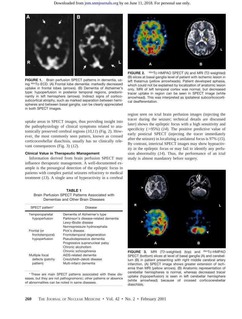
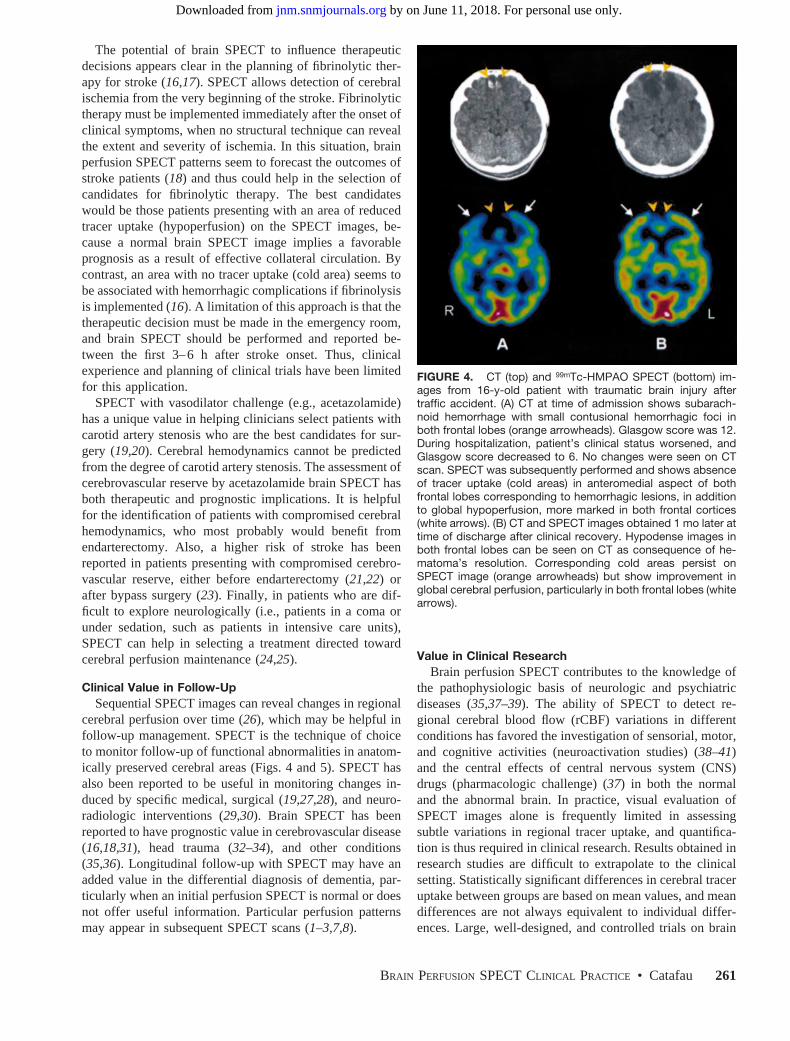
uptake areas in SPECT images, thus providing insight intothe pathophysiology of clinical symptoms related to ana-tomically preserved cerebral regions (10,11) (Fig. 2). How-ever, the most commonly seen pattern, known as crossedcorticocerebellar diaschisis, usually has no clinically rele-vant consequences (Fig. 3) (12).
Clinical Value in Therapeutic ManagementInformation derived from brain perfusion SPECT may
influence therapeutic management. A well-documented ex-ample is the presurgical detection of the epileptic focus inpatients with complex partial seizures refractory to medicaltreatment (13). A single area of hyperactivity in a cerebral
region seen on ictal brain perfusion images (injecting thetracer during the seizure; technical details are discussedlater) shows the epileptic focus with a high sensitivity andspecificity (.95%) (14). The positive predictive value ofearly postictal SPECT (injecting the tracer immediatelyafter the seizure) in localizing a unilateral focus is 97% (15).By contrast, interictal SPECT images may show hypoactiv-ity in the epileptic focus or may fail to identify any perfu-sion abnormality (14). Thus, the performance of an ictalstudy is almost mandatory before surgery.
FIGURE 1. Brain perfusion SPECT patterns in dementia, us-ing 99mTc-ECD. (A) Frontal lobe dementia: markedly decreaseduptake in frontal lobes (arrows). (B) Dementia of Alzheimer’stype: hypoperfusion in posterior temporal regions, predomi-nantly in left hemisphere (arrows). Indirect signs of cortico-subcortical atrophy, such as marked separation between hemi-spheres and between basal ganglia, can be clearly appreciatedin both SPECT images.
TABLE 1Brain Perfusion SPECT Patterns Associated with
Dementias and Other Brain Diseases
SPECT pattern* Disease
Temporoparietalhypoperfusion
Dementia of Alzheimer’s typeParkinson’s disease–related dementiaLewy–Bodie diseaseNormopressure hydrocephalia
Frontal (orfrontotemporal)hypoperfusion
Pick’s diseaseFrontotemporal degenerationPseudodepressive dementiaProgressive supranuclear palsyChronic alcoholismChronic schizophrenia
Multiple focaldefects (patchypattern)
AIDS-related dementiaCreutzfeldt–Jakob diseaseMulti-infarct dementia
* These are main SPECT patterns associated with these dis-eases, but they are not pathognomonic; other patterns or absenceof abnormalities can be noted in same diseases.
FIGURE 2. 99mTc-HMPAO SPECT (A) and MRI (T2-weighted)(B) slices at basal ganglia level of patient with ischemic lesion inleft thalamus (yellow arrowheads). Patient developed aphasia,which could not be explained by localization of anatomic lesiononly. MRI of left temporal cortex was normal, but decreasedtracer uptake in region can be seen in SPECT image (whitearrowhead). This was interpreted as ipsilateral subcorticocorti-cal deafferentation.
FIGURE 3. MRI (T2-weighted) (top) and 99mTc-HMPAOSPECT (bottom) slices at level of basal ganglia (A) and cerebel-lum (B) in patient presenting with right middle cerebral arteryinfarction. (A) SPECT image shows greater extension of isch-emia than MRI (yellow arrows). (B) Anatomic representation ofcerebellar hemispheres is normal, whereas decreased traceruptake (hypoperfusion) is seen in left cerebellar hemisphere(white arrowhead) because of crossed corticocerebellardiaschisis.
260 THE JOURNAL OF NUCLEAR MEDICINE • Vol. 42 • No. 2 • February 2001
by on June 11, 2018. For personal use only. jnm.snmjournals.org Downloaded from
The potential of brain SPECT to influence therapeuticdecisions appears clear in the planning of fibrinolytic ther-apy for stroke (16,17). SPECT allows detection of cerebralischemia from the very beginning of the stroke. Fibrinolytictherapy must be implemented immediately after the onset ofclinical symptoms, when no structural technique can revealthe extent and severity of ischemia. In this situation, brainperfusion SPECT patterns seem to forecast the outcomes ofstroke patients (18) and thus could help in the selection ofcandidates for fibrinolytic therapy. The best candidateswould be those patients presenting with an area of reducedtracer uptake (hypoperfusion) on the SPECT images, be-cause a normal brain SPECT image implies a favorableprognosis as a result of effective collateral circulation. Bycontrast, an area with no tracer uptake (cold area) seems tobe associated with hemorrhagic complications if fibrinolysisis implemented (16). A limitation of this approach is that thetherapeutic decision must be made in the emergency room,and brain SPECT should be performed and reported be-tween the first 3–6 h after stroke onset. Thus, clinicalexperience and planning of clinical trials have been limitedfor this application.
SPECT with vasodilator challenge (e.g., acetazolamide)has a unique value in helping clinicians select patients withcarotid artery stenosis who are the best candidates for sur-gery (19,20). Cerebral hemodynamics cannot be predictedfrom the degree of carotid artery stenosis. The assessment ofcerebrovascular reserve by acetazolamide brain SPECT hasboth therapeutic and prognostic implications. It is helpfulfor the identification of patients with compromised cerebralhemodynamics, who most probably would benefit fromendarterectomy. Also, a higher risk of stroke has beenreported in patients presenting with compromised cerebro-vascular reserve, either before endarterectomy (21,22) orafter bypass surgery (23). Finally, in patients who are dif-ficult to explore neurologically (i.e., patients in a coma orunder sedation, such as patients in intensive care units),SPECT can help in selecting a treatment directed towardcerebral perfusion maintenance (24,25).
Clinical Value in Follow-UpSequential SPECT images can reveal changes in regional
cerebral perfusion over time (26), which may be helpful infollow-up management. SPECT is the technique of choiceto monitor follow-up of functional abnormalities in anatom-ically preserved cerebral areas (Figs. 4 and 5). SPECT hasalso been reported to be useful in monitoring changes in-duced by specific medical, surgical (19,27,28), and neuro-radiologic interventions (29,30). Brain SPECT has beenreported to have prognostic value in cerebrovascular disease(16,18,31), head trauma (32–34), and other conditions(35,36). Longitudinal follow-up with SPECT may have anadded value in the differential diagnosis of dementia, par-ticularly when an initial perfusion SPECT is normal or doesnot offer useful information. Particular perfusion patternsmay appear in subsequent SPECT scans (1–3,7,8).
Value in Clinical ResearchBrain perfusion SPECT contributes to the knowledge of
the pathophysiologic basis of neurologic and psychiatricdiseases (35,37–39). The ability of SPECT to detect re-gional cerebral blood flow (rCBF) variations in differentconditions has favored the investigation of sensorial, motor,and cognitive activities (neuroactivation studies) (38–41)and the central effects of central nervous system (CNS)drugs (pharmacologic challenge) (37) in both the normaland the abnormal brain. In practice, visual evaluation ofSPECT images alone is frequently limited in assessingsubtle variations in regional tracer uptake, and quantifica-tion is thus required in clinical research. Results obtained inresearch studies are difficult to extrapolate to the clinicalsetting. Statistically significant differences in cerebral traceruptake between groups are based on mean values, and meandifferences are not always equivalent to individual differ-ences. Large, well-designed, and controlled trials on brain
FIGURE 4. CT (top) and 99mTc-HMPAO SPECT (bottom) im-ages from 16-y-old patient with traumatic brain injury aftertraffic accident. (A) CT at time of admission shows subarach-noid hemorrhage with small contusional hemorrhagic foci inboth frontal lobes (orange arrowheads). Glasgow score was 12.During hospitalization, patient’s clinical status worsened, andGlasgow score decreased to 6. No changes were seen on CTscan. SPECT was subsequently performed and shows absenceof tracer uptake (cold areas) in anteromedial aspect of bothfrontal lobes corresponding to hemorrhagic lesions, in additionto global hypoperfusion, more marked in both frontal cortices(white arrows). (B) CT and SPECT images obtained 1 mo later attime of discharge after clinical recovery. Hypodense images inboth frontal lobes can be seen on CT as consequence of he-matoma’s resolution. Corresponding cold areas persist onSPECT image (orange arrowheads) but show improvement inglobal cerebral perfusion, particularly in both frontal lobes (whitearrows).
BRAIN PERFUSION SPECT CLINICAL PRACTICE • Catafau 261
by on June 11, 2018. For personal use only. jnm.snmjournals.org Downloaded from

SPECT imaging are necessary before research results can beapplied in clinical practice.
PHYSIOLOGY OF THE BRAIN: A BASIS FORUNDERSTANDING BRAIN PERFUSION SPECT IMAGES
The Brain: A Unique OrganSpecial anatomic and functional features make the brain
a unique organ, substantially different from the other organsof the human body. Most of these differences are encoun-tered within the circulatory system, particularly in the reg-ulation of perfusion and metabolism (Table 2).
Intracranial arteries are different from arteries in the restof the body. Because the skull protects against externalpressures, vessel walls are thinner. No elastic fibers arefound in the media, but a well-developed inner elasticmembrane exists in the cerebral muscular arteries (42). Theblood–brain barrier, consisting of special endothelial cellswith tight junctions within brain capillaries, is a more strik-ing difference. It regulates the entrance of high-molecular-weight and hydrophilic substances to the CNS. These sub-stances are transported in the general blood pool and caneasily reach other organs of the body, but not the brain (43).
The brain is the only organ with almost no energeticstock. Neuronal activity depends on the continuity of thesupply of oxygen and glucose, which are provided by ce-rebral blood flow (CBF) (44). This dependence makes thebrain the most sensitive and vulnerable organ to CBF vari-ations. A lack of blood supply for only seconds leads tometabolic impairment, and, for more than 5 min, to irre-versible neuronal damage. The brain vascular system has anautoregulatory mechanism to maintain CBF.
Finally, the functional consequence of a lesion in thebrain depends not only on the severity or extension of the
lesion, but also on its location. A small lesion in otherorgans such as the kidneys, lungs, or liver does not com-promise their overall function. However, a very small lesionin specific brain structures can cause severe morbidity oreven death, whereas larger lesions in relatively silent brainareas may cause only mild symptoms.
Perfusion and Metabolism Coupling: Detection ofNeuronal Activity and Cerebral Function
Cerebral perfusion and metabolism are coupled in mostphysiologic and pathologic conditions, with a few excep-tions such as subacute stroke or some brain tumors. In mostconditions, adequate oxygen and glucose are provided toeach cerebral region according to its metabolic need, whichis determined by the intensity of neuronal activity. Hence,CBF is coupled to neuronal activity (44). Cerebral autoreg-ulation controls an adequate oxygen and glucose supply ineach cerebral region and involves a complex interplay ofmediators and active neurohumoral substances that exertspecific actions at a molecular level.
Classically, ions and metabolites such as potassium, hy-drogen ions (H1), CO2, and adenosine, which are excretedto the extracellular space as a result of neuronal oxidativemetabolism during increased neuronal activity, have beeninvolved in the accompanying vasodilator response andrelated hyperemia (45,46).
Recently, other mediators have been identified as intrinsicendothelial factors that regulate the physiologic and patho-physiologic responses of cerebral circulation (45,47,48). Suchmediators include:
● Nitric oxide (NO): The highest levels of NO in thebody are found in neurons. NO causes vasodilation bystimulating the activity of soluble guanylate cyclasewithin the vascular smooth muscle and elevating tissuelevels of cyclic guanosine monophosphate (GMP) (49).
● Calcium-activated potassium channels and potassiumchannels sensitive to adenosine 59-triphosphate, usingcyclic 39,59-adenosine monophosphate (cAMP) as asecond messenger: Agents that increase the intracellu-lar concentration of cAMP produce vasodilatation inpart by activation of these channels (45).
● Calcitonin gene-related peptide (CGRP), released from
TABLE 2Cerebral Requirements of Blood Flow and Metabolism
Parameter Requirement
Percentage of total body weight 2%Percentage of global cardiac output 20%Cerebral blood volume 120 mLBlood-flow needs 750 mL/minOxygen needs 50 mL/minGlucose needs 100 mg/minGlobal cerebral blood flow 55 mL/100 g/minGray-matter blood flow 80 mL/100 g/minWhite-matter blood flow 20 mL/100 g/min
FIGURE 5. Left temporal lobe infarction in 62-y-old patient.(A) MRI (T2-weighted) at admission shows hyperintensity at siteof infarction (arrowhead). (B) 99mTc-HMPAO SPECT image ob-tained 1 wk after stroke shows increased tracer uptake (hyper-perfusion) in left temporal lobe caused by luxury perfusion (ar-rowhead). Hypoperfusion is also seen in left frontal cortex(arrow), intrepreted as ischemia in anatomically preserved re-gion. (C) 99mTc-HMPAO SPECT image obtained 1 mo afterstroke shows left temporal lobe hypoperfusion (arrowhead) cor-responding to initial MR image of ischemia. Perfusion changesin left frontal lobe are also seen: improvement in anterior andmesial aspects caused by recovery of ischemia, as well asperfusion impairment in lateral aspect caused by extension ofthe infarction (arrow).
262 THE JOURNAL OF NUCLEAR MEDICINE • Vol. 42 • No. 2 • February 2001
by on June 11, 2018. For personal use only. jnm.snmjournals.org Downloaded from

trigeminal perivascular sensory nerves in the brain.CGRP may limit noradrenaline-induced constriction ofcerebral vessels and contribute to dilatation duringhypotension, reactive hyperemia, and seizures (48).
This parallelism between CBF, metabolism, and neuronalactivity (or cerebral function) is the basis for the use of brainperfusion SPECT in assessing cerebral function and, con-sequently, in detecting cerebral dysfunction. In the absenceof cerebrovascular disease, hypoperfusion detected inSPECT images can thus be related to impaired cerebralfunction, and a hyperperfused area can be related to in-creased neuronal activity. The feasibility of the noninvasivestudy of neuronal activity in different cerebral regions underdifferent conditions is the basis for interventional SPECTstudies such as neuroactivation and pharmacologic chal-lenges.
Autoregulation and Vasodilator Challenge: Insight intoCerebral Hemodynamics
Autoregulation is a mechanism that allows rCBF to re-main constant under a wide range of regional cerebralperfusion pressure (rCPP) variations (50). In physiologicconditions, systemic arterial pressure determines rCPP, be-cause cerebral venous back pressure is negligible. Condi-tions that affect either arterial inflow pressure or venousback pressure may alter rCPP globally (e.g., decreasedsystemic arterial pressure, increased intracranial pressure)or locally (e.g., local arterial occlusive disease, venousthrombosis). The main compensatory response depends onthe ability of precapillary resistance vessels to react to rCPPvariations. To maintain rCBF, pial arterioles vasoconstrictwhen rCPP increases and vasodilate when rCPP decreases.Intrinsic mechanisms involved in the ability of cerebralblood vessels to react to vasoactive stimuli were describedin the previous section. Cerebral vasodilation can also bepharmacologically induced, a factor in the use of brainSPECT examinations with vasodilator challenge. Amongthe most-used vasodilator stimuli for this purpose are in-haled CO2 and intravenous vasodilator drugs such as acet-azolamide (a carbonic anhydrase inhibitor) or adenosine.This modality of brain perfusion SPECT allows the assess-ment of the integrity of the cerebrovascular reserve andtherefore of the autoregulation mechanism and cerebralhemodynamics (51).
Under normal conditions, such pharmacologic stimuliproduce rCBF increases that can be detected by brain per-fusion SPECT. However, under pathologic conditions lead-ing to decreased rCPP, the ability of cerebral blood vesselsto react to other vasoactive stimuli is impaired or even lost.For these conditions, a maximal vasodilation could be re-quired to maintain CBF. In such cases, if rCPP continues tofall (between 60 and 30 mm Hg), the capacity for compen-satory vasodilation is exceeded and autoregulation fails,causing rCBF to decrease. Initially, an increase in the oxy-gen extraction fraction can maintain cerebral oxygen me-tabolism, but further rCBF declines will result in decreased
metabolism and function, and irreversible neuronal damagewill occur (52). For these conditions, pharmacologic vaso-dilator stimuli fail to induce further vasodilation in theaffected cerebral regions. Brain perfusion SPECT imageswould then reflect interhemispheric asymmetries caused byrCBF increases in preserved cerebral regions but minor orno rCBF increases in the affected regions. Brain perfusionSPECT with vasodilator challenge is therefore a feasiblemethod to explore the integrity of the cerebral autoregula-tion mechanism, providing insight into cerebral hemody-namics.
TECHNICAL ASPECTS OF BRAIN PERFUSION SPECT:RADIOPHARMACEUTICALS, PATIENT PREPARATION,AND IMAGE ACQUISITION AND RECONSTRUCTION
RadiopharmaceuticalsSeveral radiopharmaceuticals are commercially available
for brain perfusion SPECT (53). The selection of the radio-pharmaceutical is important, because both the pharmacoki-netic behavior of the compound and the physical character-istics of the isotopes influence the technical aspects. Thissituation is true from instrumentation to image interpreta-tion and relates to the kind of quantitative approach used orto the type of interventional studies to be performed.
To quantify rCBF (mL/min/100 g tissue) by means ofbrain perfusion SPECT, the diffusible gas133Xe is the bestradiopharmaceutical choice. By measuring cerebral wash-out of the inhaled133Xe, an absolute rCBF value is obtained(54). For this reason,133Xe SPECT has been considered astandard for rCBF quantification. However, this radiophar-maceutical has several limitations that have restricted its usein clinical practice. First, because of the rapid clearance of133Xe, the short acquisition time (around 5 min during theinhalation of the133Xe gas) requires dynamic SPECT in-strumentation, which provides high-sensitivity but low-res-olution SPECT images. Second, the lowg-ray energy of133Xe results in marked attenuation of deep structures,which makes this isotope less than optimal to obtain good-quality SPECT images. Finally, because133Xe inhalationrequires active cooperation, patients with respiratory orsevere cognitive impairment may not be studied adequately.
Other brain SPECT tracers, such as123I-labeled aminesand technetium-labeled compounds, offer higher-resolutionSPECT images after intravenous injection using conven-tional rotating gamma cameras. These tracers have foundmore acceptance, although they allow only quantitativeestimations through tracer uptake ratios. The common bio-logic properties of these radiopharmaceuticals are the fol-lowing: ability to cross the intact blood–brain barrier (smallmolecular size, lipophilic, and neutral); distribution in thebrain proportional to blood flow (this property is assessedby correspondence to the distribution of reference perfusiontracers such as iodoantipyrine or microspheres); retention inthe brain with a fixed regional distribution for a sufficienttime to permit image acquisition (20–30 min); sufficientuptake in the brain to allow imaging with standard SPECT
BRAIN PERFUSION SPECT CLINICAL PRACTICE • Catafau 263
by on June 11, 2018. For personal use only. jnm.snmjournals.org Downloaded from

instrumentation; rapid washout from background tissues(facial and glandular tissue, blood); and high gray-matter–to–white-matter uptake ratio to yield a good image defini-tion (55).
Among the initially used123I-labeled amines,123I-isopro-pyliodoamphetamine ([123I-IMP] IK-3; CIS Bio-Interna-tional, Gif-Sur-Yvette, France) has been the most frequentlyused (56). After crossing the intact blood–brain barrier,123I-IMP binds to amphetamine receptors on neurons. Thisradiopharmaceutical has good characteristics for brain per-fusion SPECT (57). However, its peak brain activity isreached as late as 20 min after injection, and it showsredistribution over time; that is, there is reuptake by thecerebral cortex that is not proportional to blood flow(57,58). These latter characteristics, in addition to the highcost and poor availability of123I-labeled compounds, havecontributed to the current predominant use of technetiumcompounds for brain perfusion SPECT examinations.
Two 99mTc-labeled radiopharmaceuticals are available:hexamethylpropyleneamine oxime ([99mTc-HMPAO] Ceretec;Nycomed-Amersham, Little Chalfont, U.K.) and ethylcystein-ate dimer ([99mTc-ECD] bicisate, Neurolite; Dupont-Pharma,Stevenage, U.K.). Their pharmacokinetic characteristics aresummarized in Table 3 (55,59). These compounds have severaladvantages over123I-IMP. The peak brain activity is reachedfaster (within 2 min after injection), and there is no redistribu-tion, so the initial tracer uptake and distribution, which areproportional to rCBF at the time of injection, remain un-changed up to at least 2 h, independent of rCBF variationsoccurring after the fixation time. This property of quick uptakeand prolonged stability allows for so-called frozen or shoot
images and makes interventional SPECT studies clearly feasi-ble. The tracer is injected while the subject is performing aspecific task or at the moment of maximum central effect of adrug, so that SPECT images reflect the rCBF distribution at thetime of injection, independent of the timing of SPECT acqui-sition. Thus, the tracer can be injected into the patient outsidethe nuclear medicine facility, and images can be acquired later.This procedure is generally not possible using PET or MRI,because patients must stay positioned in the device with thehead fixed while performing tasks to allow simultaneous imageacquisition.
The main differences between HMPAO and ECD relateto their in vitro stability, uptake mechanism, and dosimetry(55,59–61). HMPAO is highly unstable in vitro. Moreover,a high radiochemical purity must be assured before injec-tion, because only a small proportion of the injected dosewill reach the brain (Table 3). This purity is not difficult toachieve if the timing of the labeling process is done prop-erly, as follows (62): time since the last generator elution,24 h; time since the99mTc dose was eluted, 2 h; and timesince the cold vial was labeled with fresh99mTc , 20 min.Another recommendation is to avoid the mixture of99mTc-HMPAO with blood during intravenous injection, becausethe lipophylic compound enters into red blood cells. Stabi-lized forms of99mTc-HMPAO using either methylene blueor cobalt chloride have recently become available and alloweasier labeling and improvement of image quality by reduc-ing background activity (63). By contrast, ECD is stable upto at least 4 h in vitro, and freshly eluted99mTc is notrequired. However, the labeling procedure is longer, takingabout 30 min.
The rapid urinary excretion of ECD favors its dosimetry,so high doses can be administered. The dosimetry is similarto lower doses of HMPAO if patients are instructed to forcediuresis and void after the scan procedure. The use of higherdoses, together with the higher gray-matter–to–white-matterratio, contributes to the better image quality obtained withECD in comparison with HMPAO.
Although both HMPAO and ECD are distributed propor-tionally to rCBF, their retention is not completely linearwith rCBF because of an initial backdiffusion. High bloodflow may be underestimated and low blood flow may beoverestimated with both tracers (64,65).
In normal brain tissue, the kinetic properties are similarfor both perfusion agents. They enter the brain cells becauseof their lipophilic nature and remain there because of con-version into hydrophilic compounds. However, in patientswith brain disease, the distribution of these compounds maydiffer because of the biochemistry of lipophilic-to-hydro-philic conversion. Although a metabolic process of de-esterification accounts for ECD hydrophilic conversion, aninstability of the lipophilic form and glutation interactionhave been proposed for HMPAO. Thus, ECD would have apredominant cellular–metabolic uptake, and HMPAOwould reflect blood flow arrival to cerebral regions. Thissituation accounts for slight differences in the normal
TABLE 3Parmacokinetics of Technetium Radiopharmaceuticals for
Brain Perfusion SPECT
Parameter 99mTc-HMPAO 99mTc-ECD
Peak brainactivity
2 min 2 min
Brain uptake(% injecteddose)
2%–3% 4%–7%
Brain washout 12%–15% over15 min
12%–14% the firsthour; then 6%/h
Excretion (% at48 h pi)
50% liver–gut 15% liver–gut
40% kidneys 75% kidneysTarget organs Lachrymal
glandsUrinary bladder
Gallbladderwall
Gallbladder wall
Gray-matter–to–white-matter ratio
2–3:1 4:1
Imaging time Up to 4 h pi Up to 2 h pi
pi 5 postinjection.
264 THE JOURNAL OF NUCLEAR MEDICINE • Vol. 42 • No. 2 • February 2001
by on June 11, 2018. For personal use only. jnm.snmjournals.org Downloaded from

SPECT pattern (66), and it is one of the explanationssuggested for the different behavior of these tracers insubacute stroke. For cerebral perfusion–metabolic uncou-pling, increased HMPAO uptake may occur, thus reflectingthe luxury perfusion phenomenon (Fig. 5), whereas ECDuptake remains low, reflecting hypometabolism at the site ofischemia (67).
Patient PreparationThree things must be considered regarding patient prep-
aration for brain perfusion SPECT: environmental condi-tions, patient cooperation, and patient positioning.
Because of the sensitivity of brain perfusion SPECT indetecting rCBF changes coupled with neuronal activity,sensorial and cognitive stimuli must be kept at a minimumlevel during tracer injection and uptake. Injection in a quietroom and no interaction with patients at this time is desir-able. For each patient, the room conditions should be con-sistent during tracer injection and uptake. If the patient’seyes are open in a bright room during injection, a high traceruptake in the calcarine cortex is an expected SPECT pattern.On the other hand, if the patient’s eyes are closed in a dimlylit room during injection, a low tracer uptake in the calcarinecortex is found. These injecting conditions should be wellrecorded and considered in the evaluation of the SPECTimages.
Patient cooperation is of particular importance. To avoidhead movement during scanning (20–30 min), the patientshould be comfortable. Bladder voiding, a pad under theknees, and a blanket (the temperature is usually low inscanning rooms) are measures that would comfort and relaxpatients during the scan and help them to cooperate. Patientswith severe cognitive impairment or dementia may needsedation. If a technetium radiopharmaceutical (HMPAO orECD) is used, tracer injection must precede sedation toavoid sedation-induced metabolism/blood flow changes inthe SPECT images. In uncooperative children, sedation isnot recommended until other strategies to facilitate sleepduring the scan procedure have been exhausted. These in-clude instructing parents to keep the child very active duringthe previous hours and to feed the child after tracer injec-tion, just before image acquisition. If sedation remains theonly alternative, the same procedure described for adults hasto be followed.
Patient positioning largely contributes to the final qualityof SPECT images. As explained, keeping the patient com-fortable helps to avoid movement artifacts. Excludingshoulders from the field of view allows keeping the radiusof rotation to a minimum, thus maintaining the collimatorsas close as possible to the patient’s head. Belts at theforearm level help to hold the patient’s arms if the imagingbed is narrow. Keeping the head at flexion helps to reducethe radius of rotation. Lowering the chin to the chest helpsto include the entire cerebellum within the field of view andallows a better reorientation plane for oblique slices duringreconstruction.
For interventional SPECT studies, patients should beinformed about the procedure and possible secondary ef-fects. If pharmacologic intervention is required, writteninformed consent is advisable.
Practical Guides on Instrumentation and ImageAcquisition and Reconstruction
Brain perfusion SPECT is a technically demanding pro-cedure. The earlier sections described how selection of theradiopharmaceutical or patient preparation can influence thefinal result of the procedure. Instrumentation and selectionof adequate parameters for SPECT acquisition and recon-struction also play critical roles in the quality of the SPECTimages (68,69).
Because of the small size of important anatomically andfunctionally independent cerebral structures, spatial resolu-tion is the main concern in brain imaging. In principle,high-resolution equipment is recommended for brainSPECT. However, high-resolution dedicated cameras arenot widely available. A good compromise is to fit a general-purpose rotating camera with fanbeam collimators. SPECTinformation can also be obtained using standard rotatinggamma cameras fitted with parallel-hole, high-resolutioncollimators, if this is all that is available. Finally, even withthe most advanced equipment, the study cannot be optimalwithout appropriate quality control of the instruments (70).
Regarding image acquisition and reconstruction, the mainrule is to achieve a balance between detection sensitivity(i.e., number of total counts acquired) and spatial resolution.These parameters are influenced by dose, type of collimator,pixel size, acquisition time, number of projections, andfiltering (71).
Although high doses will increase detection sensitivity,dose is limited by safety rules. It is advisable to inject thehighest permitted dose to ensure enough photon flux.
A high-resolution collimator is recommended for brainSPECT, provided that enough total counts per study areguaranteed and at least a 1283 128 matrix is chosen. A lowcounting rate would require a smoother filtering duringreconstruction, which would result in loss of spatial resolu-tion. The same contradictory effect would occur if a 64364 matrix is used with a high-resolution collimator.
The matrix and pixel size should be assessed on eachspecific SPECT device. The pixel size can be calculated bydividing the useful field of view of the camera by the matrixsize. The matrix size chosen must provide a pixel size lessthan half the spatial resolution of the final image. Forexample, if the final resolution of brain SPECT slices is 10mm, the matrix size should be chosen to give a pixel size of5 mm or less.
Another factor to consider is the duration of the acquisi-tion process. Logically, a longer acquisition time will yielda higher number of total counts and better quality of thefinal images. However, long acquisition times increase therisk of patient motion, which will degrade the images.
BRAIN PERFUSION SPECT CLINICAL PRACTICE • Catafau 265
by on June 11, 2018. For personal use only. jnm.snmjournals.org Downloaded from

A particular rule that can be followed regarding thenumber of projections is to keep it close to the number ofpixels in the matrix. A higher number of projections willyield minimal reconstruction benefits, and a lower numberof projections will cause reconstruction artifacts. For exam-ple, the best choice for a 1283 128 matrix is one projectionfor each 3° in a 360° orbit for a total of 120 projections. Onthe other hand, for a 643 64 matrix, the best choice is oneprojection for each 6° in a 360° orbit for a total of 60projections.
During reconstruction, attenuation correction should beperformed except when special devices that intrinsicallycorrect attenuation, such as fanbeam collimators or trans-mission attenuation correction systems, are used. Filtering isanother step to carefully consider during SPECT reconstruc-tion, because the resulting image may vary substantially,depending on the filter applied. The diversity in type offilters and filter parameters that can be applied cannot becovered in depth in this article; concrete guidance can befound elsewhere (69,71). In simple terms, application of asmoothing filter will reduce the final resolution; thus, itwould not be sensible to use a high-resolution SPECTdevice and then lose resolution by applying a smoothingfilter to the acquired images. On the contrary, a very sharpfilter will result in noisy images. The number of final countsin the study can be a guide for the best filter to apply. Sharpfilters can be applied when a high number of total counts hasbeen achieved, whereas low total counts require a smootherfilter. It is advisable to select the appropriate filter for eachstudy. However, this process may not always be easy toimplement, depending on the software packages used. Anacceptable solution is to have three or four predefined filtersfor different ranges of total counts.
Another important process performed during SPECT im-age reconstruction is the orientation of the slicing planes.There is no consensus on the best reference to orientateslices in the transaxial plane. To be able to carefully com-pare SPECT images to structural images, a similar orienta-tion to that provided by CT, which is the canthomeatalplane, is the most popular. However, brain perfusionSPECT images lack bone references, and therefore it is notpossible to accurately find the cantomeatal line for orienta-tion. Hence, a reference plane that could be identified easilyin the perfusion SPECT images would be preferable to beconsistent with the orientation and to reproduce the sameorientation plane. A similar orientation to the canthomeatalline for oblique brain SPECT slices can be achieved by aline passing through the base of the frontal and occipitallobes (fronto-occipital plane). Nevertheless, other orienta-tions are also frequently used, such as the frontocerebellarplane. Apart from the classical oblique, coronal, and sagittalslices, slices parallel to the longitudinal edge of the temporallobe can be obtained. These temporal slices are particularlyuseful when clear differentiation between the mesial andlateral aspects of the temporal lobe is required. Some ex-
amples are temporal lobe epilepsy or the initial stages ofdementia of Alzheimer’s type.
For the display of final SPECT images, the choice ofblack-and-white or color display is arbitrary. However, dis-continuous multicolor scales may overestimate defects orinterhemispheric asymmetries. A method for normalizingthe images should be followed consistently. In our institu-tion, the maximum pixel count in the oblique slices is usedfor normalization (35). Regardless of the method used, itshould be consistent for representation of the final images,and subjective manipulation should be avoided.
SPECIFIC SITUATIONS: QUANTIFICATION ANDINTERVENTIONAL STUDIES
QuantificationThe absolute quantification of rCBF (mL/min/100 g tis-
sue) using SPECT is possible by measuring cerebral wash-out of the inhaled133Xe, as discussed earlier. An accuratemethod has been sought for measuring absolute rCBF val-ues using99mTc tracers. Such procedures require additionalinvasive or technically demanding steps (e.g., arterial bloodsampling or dynamic SPECT acquisition), thus increasingthe complexity of performance of the SPECT (72,73).Hence, absolute rCBF measurement using SPECT has notbeen fully implemented, and the region of interest (ROI)analysis of rCBF has become the preferred method of quan-tification. This SPECT quantification method, which hasbeen called semiquantification, is based on the calculationof tracer uptake ratios in different cerebral regions andallows an estimation of the relative rCBF distribution withinthe brain. Region/reference ratios can be obtained from theaverage counts per pixel of ROIs drawn and placed ondifferent cerebral areas. The left-to-right ratio of the samecerebral region or asymmetry indices are used to enhanceleft/right rCBF differences (74). Despite the simplicity ofthe concept, the laborious and time-consuming process ofgenerating a high number of ROIs and ratios, as well asproblems concerning ROI design and selection of the ref-erence region for uptake ratios, complicates brain SPECTquantification.
ROIs can be geometric (regular ROIs) and placed ongiven cerebral territories, or they can delineate anatomicstructures (irregular ROIs). They can also be specificallydesigned to fit cerebrovascular territories. Irregular ana-tomic ROIs or ROIs specifically designed for each studypurpose work better in detecting suspected abnormalities.However, drawing the same ROIs and placing them inexactly the same cerebral areas is a time-consuming processand requires special dedication. If this process is not fol-lowed carefully, reproducibility will be limited. Coregistra-tion of structural neuroimaging slices, or superpositioningto a stereotactic atlas, can definitely improve ROI position-ing, therefore reducing inter- and intraobserver variabilityand increasing reproducibility of these semiquantitative ap-proaches.
266 THE JOURNAL OF NUCLEAR MEDICINE • Vol. 42 • No. 2 • February 2001
by on June 11, 2018. For personal use only. jnm.snmjournals.org Downloaded from

The selection of the reference region is crucial in thecalculation of region-to-reference uptake ratios. The wholebrain and the cerebellum are the most common referencesused, but they do not always represent the best choicebecause the reference region must have anatomic and func-tional integrity (75). The choice of the whole brain as areference may be misleading. If a relative rCBF impairmentin a cerebral region does exist, it will influence the calcu-lation of the whole-brain average counts per pixel value, andthis reference value can be substantially different from onesubject to another. Similarly, when a cerebellar involvementhas been described or is indeed present, a region-to-cere-bellum ratio is not suitable. For these patients, anotherpreserved cerebral region should be used as a reference. Ingeneral, differences in ratios and the design of the ROIs willvary, depending on the purpose of each study.
In the search for more standardized and objective alter-natives for brain perfusion SPECT quantification, circum-ferential profiles and polar maps have been proposed withlimited success (76,77). Software programs allowing statis-tical comparison of different groups of subjects, such as thestatistical parametric mapping (SPM) package, are beingused mainly for research purposes. SPM offers clear advan-tages for PET examinations, but this approach appears lim-ited for SPECT because of the lower spatial resolution (78).
Brain perfusion SPECT quantification in the clinicalmanagement of patients should be encouraged when com-parison is required among SPECT studies performed on thesame subject at different times. This situation occurs whenpatients are followed up in search of rCBF modifications orwhen an interventional study is performed (e.g., neuroacti-vation or pharmacologic intervention). In these situations,quantitative data help to objectively report the degree ofrCBF changes and support visual impressions.
Interventional SPECT StudiesInterventional SPECT is the study of rCBF changes in-
duced by specific sensorial, motor, or cognitive stimuli(neuroactivation) or by specific drugs (pharmacologic inter-vention). Other SPECT studies that could be consideredinterventional are those in which it is desired to capture aspecific clinical feature with SPECT imaging. In these stud-ies, a tracer is injected when the specific clinical symptomappears. For example, the tracer is injected during theseizure in patients with epilepsy. The pathophysiology ofpositive symptoms in schizophrenic patients can be inves-tigated if the tracer is injected when the patient is experi-encing specific symptoms, for example, auditory hallucina-tions. The central effect induced by some therapeutic ordiagnostic procedures, such as electroconvulsive therapy ortranscranial magnetic stimulation, has also been investi-gated by SPECT using this approach.
Interventional SPECT studies require the performance ofat least two SPECT scans: one under baseline conditionsand the other during the task or under a pharmacologiceffect. For proper comparison of these two studies, quanti-
fication or subtraction techniques are used on slices recon-structed identically. Reference test–retest values in normalsubjects should be available for interpreting quantitativeresults (i.e., percentage of rCBF changes) to recognize thepercentage variation between two SPECT studies on thesame subject under the same baseline conditions (37). Thesetwo studies may be performed on separate days or on thesame day using the split-dose technique. When the twoSPECT studies are compared, obtaining the identical tomo-graphic slices may be a problem, which has been addressedby software for three-dimensional realignment (79). Thesplit-dose technique, which refers to baseline and interven-tional SPECTs in the same session, may help to overcomethis problem (80,81). The administered dose for the firstSPECT must be at least half of the second. Patients staypositioned in the camera until the second SPECT ends, andneither patient repositioning nor software realignment isrequired. However, other technical issues, such as correc-tions for decay and injected dose, need to be addressed.Moreover, the longer acquisition time increases the risk forpatient motion, that is, for image artifacts. The SPM soft-ware, mostly used in research, considers realignment andmakes statistical comparisons between two groups of sub-jects. The results are elegantly displayed on a reference MRIor brain surface atlas image (78).
Neuroactivation SPECT has been used successfully tostudy sensorial, motor, and cognitive functions (38–41).The tracer is injected while the subject is engaged in a task,which should be continued until tracer fixation in the brainis complete (at least 3–5 min). Tasks should be carefullyselected, because no other cerebral activity than the targetone is desirable. Cognitive tasks represent a major difficultyto overcome. The performance of a control task would bepreferable to a baseline “rest” condition. The task shouldinvolve the same parameters as the test task but without thecognitive function. For example, when using the WisconsinCard Sorting Test as a cognitive task, an acceptable controltask could be to match the cards with no criteria. Neuroac-tivation is a common approach in research SPECT studies.By contrast, neuroactivation SPECT is not widely used inthe clinical setting, most probably because of the technicaldemands, although it has been reported to be useful in thedifferential diagnosis of dementia.
Brain perfusion SPECT can also be performed duringpharmacologic interventions. For this purpose, a tracer isinjected at the expected time of maximum CNS drug effect.The most popular pharmacologic intervention that has clin-ical relevance is the acetazolamide SPECT for the study ofcerebrovascular reserve (32). Brain perfusion SPECT is alsouseful in showing the distribution and effect of intracarotidamobarbital during the Wada test used for presurgical eval-uation of epileptic patients (82–84). Pharmacologic inter-vention SPECT has been used to investigate the effect ofdifferent drugs on the CNS.
The methodology of ictal SPECT in patients withrefractory complex partial seizures requires special atten-
BRAIN PERFUSION SPECT CLINICAL PRACTICE • Catafau 267
by on June 11, 2018. For personal use only. jnm.snmjournals.org Downloaded from

tion. The clinical indication of this procedure is to iden-tify the lateralization and localization of an epilepticfocus before surgery. If patients are hospitalized in anepilepsy unit and monitored by video-electroencephalog-raphy (video-EEG), injecting the perfusion tracer duringa seizure is possible. The technologist in charge of thevideo-EEG monitoring should be trained in handlingradiopharmaceuticals. If the chosen tracer is HMPAO, acold vial and the corresponding freshly eluted99mTc dosein a separate syringe should be provided to the video-EEG technologist. The99mTc dose should be replacedevery 2 h if no seizures have appeared. At the verybeginning of seizure activity, as shown by the video-EEG, the technologist labels the cold vial with the fresh99mTc and immediately injects it into the patient throughan intravenous line. Using stabilized99mTc-HMPAO orECD, labeled vials can be provided and replaced every4 – 6 h. Patients can be scanned in the nuclear medicinedepartment after recovering from the seizure. The sameprocedure can be used when waiting for a patient’s par-ticular symptom or another event to be imaged (85).
INTERPRETING AND REPORTING IMAGES
The brain is not composed of uniform tissue, and onemust be trained to recognize the normal perfusion pattern.Perfusion patterns may differ from one subject to another,because the normal brain is not always completely symmet-ric, and small structural differences are frequent withinnormal subjects. Differences may be found between hemi-spheres, between subjects, and to a lesser extent within thesame healthy subject scanned twice under the same condi-tions (test–retest). This result is partly caused by variationsin functional activity and therefore by a varying vascularsupply to cerebral regions at the time of injection. Asmentioned previously, the radiopharmaceutical used and thepatient and environmental conditions during tracer injectionmust be considered.
In a normal brain perfusion SPECT image (74), regionswith higher perfusion, such as cortical and subcortical gray-matter structures, have the highest tracer uptake. Subcorticalwhite matter shows low tracer uptake, and no tracer uptakeis seen in areas containing cerebrospinal fluid (i.e., cerebralventricles, fissures, and sulcus) or bone (i.e., scalp or pe-trous part of temporal bones). The cerebral region showingthe maximum tracer uptake varies with the radiopharma-ceutical used (i.e., most probably the cerebellum withHMPAO, but the calcarine cortex with ECD), the patient’scondition at the time of injection (e.g., influence of visualactivity on calcarine cortex uptake), and image manipula-tion during reconstruction (i.e., influence of attenuationcorrection on basal ganglia activity).
The appropriate brain perfusion SPECT interpretationshould be made using a step-by-step approach, such as thefollowing:
1. Select a set of slices to use as a basis for identificationof the SPECT perfusion pattern. It is advisable to startwith the transaxial or oblique fronto-occipital slices.The other sets of slices will be of further help forconfirmation, localization, and extension of findings.
2. Anatomically identify the cerebral structures withinthe range of precision that spatial resolution of theimages allows. A good exercise to precisely recognizecerebral lobes on SPECT images is the identificationof cissures separating them, using an atlas as a guide.
3. Follow the same regional order of perfusion assess-ment, including all cerebral regions. For example, firstassess the cortical regions from bottom to top: cere-bellum, temporal lobes, frontal lobes (orbitofrontal,prefrontal, and superior frontal areas), occipital lobes,and parietal lobes. Mesial and lateral aspects of theseregions should be evaluated separately. Second, assessbrain stem and subcortical regions (striatum, thala-mus, and white matter).
4. Assess global and regional tracer uptake and distribu-tion (normal distribution vs. identification of perfusionabnormalities).4.1. Identify brain perfusion abnormalities. A good
practice is to check for marked asymmetries be-tween hemispheres (keep in mind that a normalbrain may display some asymmetry). For inade-quate reorientation of the SPECT study, asymme-tries caused by head tilts may be present, whichcan be easily identified because the pattern isreversed on consecutive slices.
4.2. Compare functional SPECT images to anatomicCT or MRI. For example, because of the slightdifference in uptake between periventricularwhite matter and lateral ventricles, it is difficult todifferentiate these two structures on SPECT im-ages. For marked hypoactivity of these regions,comparison with anatomic imaging will clarify ifthe pattern is caused by hydrocephalia or if itcould be attributable to white-matter ischemia (inthe latter case, anatomic images may be normal).
4.3. In patients with cerebral atrophy, look for theabsence of uptake in cerebrospinal fluid and nor-mal uptake in the cortex. These conditions maycoexist within the same pixel, resulting in a falseappearance of decreased uptake in the cerebralcortex. Although indirect signs of cerebral atro-phy can be identified in the SPECT images, suchas a marked separation between hemispherescaused by interhemispheric cissure enlargement,and between basal ganglia caused by ventricularenlargement (Fig. 1), anatomic imaging is re-quired to confirm this result. It is difficult to besure if hypofunction coexists in regions with at-rophy unless the image analyzing software pro-vides atrophy correction.
268 THE JOURNAL OF NUCLEAR MEDICINE • Vol. 42 • No. 2 • February 2001
by on June 11, 2018. For personal use only. jnm.snmjournals.org Downloaded from

5. Characterize perfusion abnormalities.5.1. Recognize abnormalities in brain perfusion
SPECT images such as decreased tracer uptake(hypoperfusion) (Figs. 2, 3, and 5), absence oftracer uptake (no perfusion) (Fig. 4), and in-creased tracer uptake (hyperperfusion) (Fig. 5).The most frequent causes of these uptake patternsare summarized in Table 4 . The etiology of hypo-and hyperperfusion can be purely vascular or re-lated to cerebral dysfunction, as described previ-ously. Knowledge of both the clinical context andanatomic images of patients is essential for inter-preting uptake abnormalities.
5.2. Recognize the distribution patterns of perfusionabnormalities such as vascular territories andeventual diaschisis patterns, dementia patterns,temporal lobe localization of epileptic foci, orherpetic encephalitis. This information often pro-vides valuable clues for the etiology.
The report of brain perfusion SPECT must be concise andreader friendly and may consist of three main sections:
● Heading: The report’s heading should include the ra-diopharmaceutical used and a summary of the clinicalproblem or indication of the study. When relevant,other factors known to influence perfusion SPECTimages, such as patient and environmental conditionsduring tracer injection or possible technical pitfalls,should be included.
● Description of findings: This part constitutes the bodyof the report and should include detailed informationon localization, type of abnormality (cold, hypo- orhyperperfusion), and extension and severity of the per-fusion abnormality. Comparison of SPECT with CTand MRI findings should also be added.
● Interpretation of SPECT findings and conclusion: If theSPECT indication fits within a well-recognized clinicalapplication (i.e., dementia, epilepsy, or stroke), an ac-curate interpretation statement leading to a diagnosiscan be provided. However, extreme caution must beapplied when the role of brain perfusion SPECT in thereferred clinical problem has not been studied in depthor is unknown, as occurs in behavioral disorders. Inthese cases, the nonspecificity of the technique and lackof consistent evidence of cause and effect need to beacknowledged in the report (86).
CONCLUSION
Brain perfusion SPECT has well-recognized clinical ap-plications mainly in dementia, cerebrovascular disease, andepilepsy. This technique generally adds valuable informa-tion to the clinical management of patients with brain dis-orders of a broad variety, helping in diagnosis, therapeuticmanagement, and follow-up. Accurate knowledge of thephysiologic and pathophysiologic basis of brain perfusionSPECT, together with the appropriate technique and carefulinterpretation of images and reporting, will enhance theclinical use of brain SPECT. Anatomic and functional cor-relations, as well as close interaction with the referringclinician, are additional practices of crucial importance infully exploiting brain perfusion SPECT capabilities in clin-ical practice.
ACKNOWLEDGMENTS
The author wishes to acknowledge Prof. Ignasi Carrio´,MD (Hospital Sant Pau, Barcelona, Spain) for his valuablecontribution, Juan Carlos Martin, MD (Hospital Sant Pau)for his help in preparing the figures, and Semih Dogan, MD,and Julia Buchanan (Johns Hopkins Medical Institutions,Baltimore, MD) for their help in editing the manuscript.
REFERENCES
1. Buttler CRE, Costa DC, Walker Z, Katona CLE. PET and SPECT imaging in thedementias. In: Murray IPC, Ell PJ, eds.Nuclear Medicine in Clinical Diagnosisand Treatment. 2nd ed. Edinburgh, Scotland: Churchill Livingstone; 1998:713–728.
2. Ryding E. SPECT measurements of brain function in dementia; a review.ActaNeurol Scand.1996;168(suppl):54–58.
3. Brooks DJ. Functional imaging techniques in the diagnosis of non-Alzheimerdementias. J Neural Transm.1996;47(suppl):155–167.
4. Nordberg A. Functional studies of new drugs for the treatment of Alzheimer’sdisease.Acta Neurol Scand.1996; 165(suppl):137–144.
5. Skoog I, Marcusson J, Blennow K. It’s getting better all the time.Lancet.1998;352(suppl IV):4.
6. Costa DC, Pilowsky LS, Ell PJ. Nuclear medicine in neurology and psychiatry.Lancet.1999;354:1107–1111.
7. Laatsch L, Jobe T, Sychra J, Lin Q, Blend M. Impact of cognitive rehabilitation
TABLE 4Frequent Causes of Different Tracer Uptake Patterns in
Brain Perfusion SPECT Images
Tracer uptake pattern Cause
Cold (no uptake) Cerebrospinal fluidEdemaNecrosisSpace-occupying lesions (e.g.,
hemorrhage, tumors,* cysts,arteriovenous malformation,postsurgery)
Hypoactive (decreaseduptake)
IschemiaHypometabolism (hypofunction):
degeneration, deafferentationAtrophy
Hyperactive (increaseduptake)
Luxury perfusionEncephalitisAcetazolamide-induced vasodilationHyperfunction (epilepsy [ictal],
neuroactivation, other causes ofincreased neuronal activity)
Tumors*
* Tumors can produce a variety of patterns in SPECT perfusionimages. The most frequent tumors with increased tracer uptake aremeningiomas.
BRAIN PERFUSION SPECT CLINICAL PRACTICE • Catafau 269
by on June 11, 2018. For personal use only. jnm.snmjournals.org Downloaded from

therapy on neuropsychological impairments as measured by brain perfusionSPECT: a longitudinal study.Brain Inj. 1997;11:851–863.
8. Greene JD, Miles K, Hodges JR. Neuropsychology of memory and SPECT in thediagnosis and staging of dementia of Alzheimer type.J Neurol.1996;243:175–190.
9. Reivich M. Crossed cerebellar diaschisis.AJNR.1992;13:62–64.10. Huguet M, Catafau A, Lomena F. Tc-99m HMPAO brain SPECT in frontal
cortex dysfunction: a case of subcortico-cortical diaschisis.Clin Nucl Med.1995;20:736–737.
11. Bakar M, Kirshner HS, Wertz RT. Crossed aphasia. Functional brain imagingwith PET or SPECT.Arch Neurol.1996;530:1026–1032.
12. Kim SE, Choi CW, Yoon BW, et al. Cerebellar vasoreactivity in stroke patientswith crossed cerebellar diaschisis assessed by acetazolamide and99mTc-HMPAOSPECT.J Nucl Med.2000;41:416–420.
13. Martınez M, Santamarıa J, Mercader JM, Catafau AM, Cardenal C, Lomen˜a F.Correlation of MRI hippocampal volume analysis, video/EEG monitoring andinter- and postictal single photon emission tomography in refractory focal epi-lepsy.Neuroradiology.1994;36:11–16.
14. Devous MD Sr, Thisted RA, Morgan GF, Leroy RF, Rowe CC. SPECT brainimaging in epilepsy: a meta-analysis.J Nucl Med.1998;39:285–293.
15. Rowe CC, Berkovic SF, Austin MC, et al. Patterns of postictal cerebral bloodflow in temporal lobe epilepsy: qualitative and quantitative analysis.Neurology.1991;41:1096–1103.
16. Alexandrov AV, Ehrlich LE, Bladin CF, Norris JW. Simple visual analysis ofbrain perfusion on HMPAO SPECT predicts early outcome in acute stroke.Stroke.1996;27:1537–1542.
17. Alexandrov AV, Masdeu JC, Devous MD Sr, Black SE, Grotta JC. Brainsingle-photon emission CT with HMPAO and safety of thrombolitic therapy inacute ischemic stroke. Proceedings of the meeting of the SPECT safe thromboli-sis study collaborators and the members of the brain imaging council of theSociety of Nuclear Medicine.Stroke.1997;28:1839–1834.
18. Ueda T, Sakaki S, Kumon Y, Ohta S. Multivariable analysis of predictive factorsrelated to outcome at 6 months after intra-arterial thrombolysis for acute ischemicstroke.Stroke.1999;30:2360–2365.
19. Cikrit DF, Dalsing MC, Harting PS, et al. Cerebral vascular reactivity assessedwith acetazolamide single photon emission computer tomography scans beforeand after carotid endarterectomy.Am J Surg.1997;174:193–197.
20. Cikrit DF, Dalsing MC, Lalka SG, Burt RW, Sawchuk AP, Solooki BA. Thevalue of acetazolamide single photon emission computed tomography scans inthe preoperative evaluation of asymptomatic critical carotid stenosis.J Vasc Surg.1999;30:599–605.
21. Knop J, Thie A, Fuchs C, Siepmann G, Zeumer H.99mTc-HMPAO-SPECT withacetazolamide challenge to detect hemodynamic compromise in occlusive cere-brovascular disease.Stroke.1992;23:1733–1742.
22. Kuroda S, Kamiyama H, Abe H, Houkin K, Isobe M, Mitsumori K. Acetazol-amide test in detecting reduced cerebral perfusion reserve and predicting long-term prognosis in patients with internal carotid artery occlusion.Neurosurgery.1993;32:912–918.
23. Ishikawa T, Houkin K, Abe H, Isobe M, Kamiyama H. Cerebral haemodynamicsand long-term prognosis after extracranial-intracranial bypass surgery.J NeurolNeurosurg Psychiatry.1995;59:625–628.
24. Carter LP, Weinand ME, Oommen KJ. Cerebral blood flow (CBF) monitoring inintensive care by thermal diffusion.Acta Neurochir Suppl (Wien).1993;59:43–46.
25. Matz PG, Pitts L. Monitoring in traumatic brain injury.Clin Neurosurg.1997;44:267–294.
26. Catafau AM, Sola M, Lomena F, Guelar A, Miro JM, Setoain J. Hyperperfusionand early HMPAO-SPECT appearance of CNS toxoplasmosis.J Nucl Med.1994;325:1041–1043.
27. Bryant CA, Jackson SH. Functional imaging of the brain in the evaluation of drugresponse and its application to the study of aging.Drugs Aging.1998;13:211–222.
28. Kachel R. Results of balloon angioplasty in the carotid arteries.J Endovasc Surg.1996;3:22–30.
29. Berger JD, Witte RJ, Holdeman KP, et al. Neuroradiologic applications of centralnervous system SPECT.Radiographics. 1996;16:777–785.
30. Moody EB, Dawson RC, Sandler MP.99mTc-HMPAO SPECT imaging in inter-ventional neuroradiology: validation of balloon test occlusion.AJNR.1991;12:1043–1044.
31. Mountz JM, Modell JG, Foster NL, et al. Prognostication of recovery followingstroke using the comparison of CT and technetium-99m HMPAO SPECT.J NuclMed.1990;31:61–66.
32. Masdeu JC, Abdel-Dayem H, Van Heertum RL .Head trauma: use of SPECT.J Neuroimaging. 1995;5(suppl 1):S53–S57.
33. Jacobs A, Put E, Ingels M, et al. One-year follow-up of technetium-99m-HMPAO
SPECT in mild and moderate traumatic brain injury.J Nucl Med. 1996;37:1605–1609.
34. Sakas DE, Bullock MR, Patterson J, et al. Focal cerebral hyperemia after focalhead injury in humans: a benign phenomenon?J Neurosurg.1995;83:227–284.
35. Nicolas J, Catafau A, Estruch R, et al. Regional cerebral blood flow-SPECT inchronic alcoholism: relation to neuropsychological testing.J Nucl Med. 1993;34:1452–1459.
36. Catafau AM, Kulisevsky J, Berna LL, et al. Cerebral perfusion in frontal-limbic-basal ganglia structures relates to neuropsychological impairment in patients withsubclinical hepatic encephalopathy.J Nucl Med.2000;41:405–410.
37. Catafau AM, Etcheberrigaray A, Perez de los Cobos J, et al. Regional cerebralblood flow changes in chronic alcoholic patients induced by naltrexone challengeduring detoxification.J Nucl Med.1999;40:19–24.
38. Catafau AM, Parellada E, Lomena F, et al. Prefrontal and temporal blood flow inschizophrenia: resting and activation technetium-99m-HMPAO SPECT patternsin young neuroleptic-naive patients with acute disease. J Nucl Med.1994;35:935–941.
39. Parellada E, Catafau A, Bernardo M, et al. The resting and activated issue ofhypofrontality: a SPECT study in young acute unmedicated schizophrenic femalepatients.Biol Psychiatry.1998;44:787–790.
40. Catafau AM, Parellada E, Lomena F, et al. Baseline, visual deprivation and visualstimulation99mTc-HMPAO related changes can be detected with a single-headSPECT system.Nucl Med Commun.1996;17:480–484.
41. Catafau AM, Parellada E, Lomena F, et al. Role of cingulate gyrus duringwisconsin card sorting test: a single-photon emission computed tomography(SPECT) study in normal volunteers.Psychiatry Res Neuroimaging.1998;83:67–74.
42. Stehbens WE. Cerebral atherosclerosis. Intimal proliferation and atherosclerosisin the cerebral arteries.Arch Pathol.1975;99:582–591.
43. Pardridge WM. CNS drug design based on principles of blood-brain barriertransport.J Neurochem.1998;70:1781–1792.
44. Miller JD, Bell BA. Cerebral blood flow variations with perfusion pressure andmetabolism. In: Wood JH, ed.Cerebral Blood Flow. Physiologic and ClinicalAspects.New York, NY: McGraw-Hill; 1987:19–130.
45. Dietrich HH, Dacey RG Jr. Molecular keys to the problems of cerebral vaso-spasm.Neurosurgery.2000;46:517–530.
46. Chen Y, McCarron RM, Bembry J, et al. Nitric oxide modulates endothelin1-induced Ca21mobilization and cyt filaments in human cerebromicrovascularendothelial cells.J Cereb Blood Flow Metab.1999;19:133–138.
47. Yang G, Chen G, Ebner TJ, Iadecola J. Nitric oxide is the predominant mediatorof cerebellar hyperemia during activation in rats.Am J Physiol.1999;277:R1760–1770.
48. Nishii T, Moriguchi A, Morishita R, et al. Angiotensinogen gene-activationgelements regulate blood pressure in the brain.Circ Res.1999;85:257–263.
49. Lee TJ. Nitric oxide and the cerebral vascular function.J Biomed Sci.2000;7:16–26.
50. Powers, WJ. Cerebral hemodynamics in ischemic cerebrovascular disease.AnnNeurol.1991;29:231–240.
51. Yudd AP, Van Heertum RL, Masdeu JC. Interventions and functional brainimaging.Semin Nucl Med.1991;21:153–158.
52. Sette G, Baron JC, Mazoyer B, et al. Local brain hemodynamics and oxygenmetabolism in cerebrovascular disease.Brain. 1989;112:931–951.
53. Bonte FJ, Devous MD Sr, Holman BL. Single photon emission computedtomographic imaging of the brain. In: Sandler MP, Coleman RE, Wackers FJTh,Patton JA, Gottschalk A, Hoffer PB, eds.Diagnostic Nuclear Medicine. 3rd ed.Baltimore, MD: Williams and Wilkins; 1996:1075–1093.
54. Lassen NA, Henriksen L, Paulson O. Regional cerebral blood flow in stroke by133Xe inhalation and emission tomography.Stroke.1981;12:284–287.
55. Neirinckx RD, Canning LR, Piper IM, et al. Technetium-99m d,l-HMPAO: a newradiofarmaceutical for SPECT imaging of regional cerebral blood perfusion.J Nucl Med.1987;28:191–202.
56. Cohen MB, Graham LS, Yamada LS. [123I]iodoamphetamine SPECT imaging.Int J Radiat Appl Instrum.1986;37:749–763.
57. Nishizawa S, Tanada S, Yonekura Y, et al. Regional dynamics ofN-isopropyl-(123I)p-iodo-amphetamine in the brain.J Nucl Med.1989;30:150–156.
58. Creutzig H, Schober O, Bielow P, et al. Cerebral dynamics ofN-isopropyl-(123I)p-iodoamphetamine.J Nucl Med.1986;27:178–183.
59. Vallabhajosula S, Zimmerman RE, Picard M, et al. Technetium-99m-ECD: a newbrain imaging agent: in vivo kinetics and biodistribution studies in normal humansubjects.J Nucl Med.1989;30:599–604.
60. Nakamura K, Tukatani Y, Kubo A, et al. The behavior of99mTc-hexamethylpro-pileneaminoxime (99mTc-HMPAO) in blood and brain.Eur J Nucl Med.1989;15:100–107.
61. Walovitch RC, Hill TC, Garrity ST, et al. Characterization of technetium-99m-
270 THE JOURNAL OF NUCLEAR MEDICINE • Vol. 42 • No. 2 • February 2001
by on June 11, 2018. For personal use only. jnm.snmjournals.org Downloaded from

l,l-ECD for brain perfusion imaging. Part 1: Pharmacology of technetium-99mECD in nonhuman primates.J Nucl Med.1989;30:1892–1901.
62. Piera C, Martinez A, Ramirez I. Radiochemical purity of technetium-99m-HMPAO depends on specific activity [letter].J Nucl Med.1995;36:706.
63. Barthel H, Kampfer I, Seese A, et al. Improvement of brain SPECT by stabili-zation of Tc-99m-HMPAO with methylene blue or cobalt chloride. Comparisonwith Tc-99m-ECD.Nuklearmedizin.1999;38:80–84.
64. Lassen NA,Andersen R, Friberg L, Paulson OB. The retention of [99mTc]-d,l-HM-PAO in the human brain after intracarotid bolus injection: a kinetic analysis.J Cereb Blood Flow Metab.1988;8(suppl. 1):S13–S22.
65. Friberg L, Andersen AR, Lassen NA, Holm S, Dam M. Retention of99mTc-bicisate in the human brain after intracarotid injection. J Cereb Blood FlowMetab.1994;14(suppl.1):S19–S27.
66. Koyama M, Kawashima R, Ito H, et al. SPECT imaging of normal subjects withtechnetium-99m-HMPAO and technetium-99m-ECD.J Nucl Med.1997;38:587–592.
67. Lassen NA, Sperling B.99mTc-bicisate reliably images CBF in chronic braindiseases but fails to show reflow hyperemia in subacute stroke: report of amulticenter trial of 105 cases comparing133Xe and99mTc-bicisate (ECD, Neuro-lite) measured by SPECT on the same day.J Cereb Blood Flow Metab.1994;14(suppl 1):S44–S48.
68. Devous MD. SPECT functional brain imaging. Technical considerations.J Neu-roimaging. 1995;5(suppl 1):S2–S13.
69. Keyes JW Jr. Single photon emission computed tomography (SPECT). Opera-tional Guidelines. In: Wagner HN Jr, Szabo Z, Buchanan JW, eds.Principles ofNuclear Medicine.Philadelphia, PA: Saunders; 1995:332–341.
70. Hines H, Kayayan R, Colsher J, et al. National electrical manufacturers associ-ation recommendations for implementing SPECT instrumentation quality control.J Nucl Med.2000;41:383–389.
71. English RJ, Brown SE.SPECT Single-Photon Emission Computed Tomography:A Primer.New York, NY: The Society of Nuclear Medicine; 1986.
72. Murase K, Tanada S, Fujita H, Sakaki S, Hamamoto K. Kinetic behavior oftechnetium-99m-HMPAO in the human brain and quantification of cerebral bloodflow using dynamic SPECT. J Nucl Med.1992;33:135–143.
73. Tsuchida T, Sadato N, Yonekura Y, et al. Quantification of regional cerebralblood flow with continuous infusion of technetium-99m-ethyl cysteinate dimer.J Nucl Med.1997;38:1699–1702.
74. Catafau AM, Lomena F, Pavia J, et al. Regional cerebral blood flow pattern innormal young and aged volunteers. A99mTc-HMPAO SPECT study.Eur J NuclMed.1996;23:1329–1337.
75. Syed GMS, Eagger S, Toone BK, Levy R, Barret JJ. Quantification of regionalcerebral blood flow (rCBF) using99mTc-HMPAO and SPECT: choice of thereference region.Nucl Med Commun.1992;13:811–816.
76. Spreafico G, Gadola G, Cammelli F, Caffarra P, Scaglioni A. Semiquantitativeassessment of regional cerebral perfusion using99mTc-HM-PAO and emissiontomography.Eur J Nucl Med.1988;14:565–568.
77. Ichise M, Crisp S, Ganguli N, Tsai S, Gray BG. A method of two-dimensionalmapping of cortical perfusion by cylindrical transformation of HMPAO SPECTdata.Nucl Med Commun. 1995;16:386–394.
78. Acton PD, Friston KJ. Statistical parametric mapping in functional neuroim-aging: beyond PET and fMRI activation studies. Eur J Nucl Med.1998.25:663– 667.
79. Pavıa J, Ros D, Catafau A, Lomena F, Huguet M, Setoain J. 3-D realignment ofactivation brain SPECT studies.Eur J Nucl Med. 1994;21:1298–1302.
80. Holm S, Madsen PL, Sperling B, Lassen NA. Use of99mTc-bicisate in activationstudies by split-dose technique.J Cereb Blood Flow Metab. 1994;14(suppl1):S115–S120.
81. Wong CO, MacIntyre WJ, Chen EQ, Saha GB, Chyatte D, Go RT. Is imagesubtraction necessary in the clinical interpretation of single-day split-dose stresscerebral perfusion single-photon emission tomography using technetium-99mcompounds?Eur J Nucl Med.1996;23:1309–1314.
82. Biersack HJ, Klemm E, Menzel C, Reichmann K, Shih WJ, Grunwald F.Interventional brain SPECT—a review.Ann Nucl Med.1996;10:277–280.
83. de Silva R, Duncan R, Patterson J, Gillham R, Hadley D. Regional cerebralperfusion and amytal distribution during the Wada test.J Nucl Med.1999;40:747–752.
84. Kim BG, Lee SK, Nam HW, Song HC, Lee DS. Evaluation of functional changesin the medial temporal region during intracarotid amobarbital procedure by use ofSPECT.Epilepsia.1999;40:424–429.
85. Arroyo S, Santamarıa J, Sanmartı F, et al. Ictal laughter associated with parox-ismal hypothalamopituitary dysfunction.Epilepsia.1997;38:114–117.
86. Society of Nuclear Medicine Brain Imaging Council. Ethical clinical practice offunctional brain imaging.J Nucl Med.1996;37:1256–1259.
BRAIN PERFUSION SPECT CLINICAL PRACTICE • Catafau 271
by on June 11, 2018. For personal use only. jnm.snmjournals.org Downloaded from

2001;42:259-271.J Nucl Med. Ana M. Catafau Brain SPECT in Clinical Practice. Part I: Perfusion*
http://jnm.snmjournals.org/content/42/2/259This article and updated information are available at:
http://jnm.snmjournals.org/site/subscriptions/online.xhtml
Information about subscriptions to JNM can be found at:
http://jnm.snmjournals.org/site/misc/permission.xhtmlInformation about reproducing figures, tables, or other portions of this article can be found online at:
(Print ISSN: 0161-5505, Online ISSN: 2159-662X)1850 Samuel Morse Drive, Reston, VA 20190.SNMMI | Society of Nuclear Medicine and Molecular Imaging
is published monthly.The Journal of Nuclear Medicine
© Copyright 2001 SNMMI; all rights reserved.
by on June 11, 2018. For personal use only. jnm.snmjournals.org Downloaded from