Embed Size (px)
Citation preview

Constructing a timetable of autumn senescence
in aspen
Johanna Keskitalo
Akademisk avhandling
som med vederbörligt tillstånd av rektorsämbetet vid Umeå universitet för avläggande av filosofie doktorsexamen i ämnet växtfysiologi med inriktning mot växtmolekylär biologi,
framläggs till offentligt försvar i stora hörsalen KB3B1, KBC-huset, Fredagen den 5 Maj 2006, kl. 10.00
Avhandlingen kommer att försvaras på engelska.
Fakultetsopponent: Prof. Howard Thomas, Institute of Grassland and Enviromental Research
(IGER), Plas Gogerddan, Aberystwyth, Ceredigion, UK
Umeå Plant Science Center Department of Plant Physiology
Umeå University, Sweden 2006

Constructing a timetable of autumn senescence in aspen Johanna Keskitalo Umeå Plant Science Center, Department of Plant Physiology, Umeå University, SE-901 87 Umeå, Sweden, May 2006 Doctoral dissertation ISBN 91-7264-075-8 Abstract: During the development and lifecycle of multicellular organisms, cells have to die, and this occurs by a process called programmed cell death or PCD, which can be separated from necrosis or accidental cell death (Pennell and Lamb, 1997). Senescence is the terminal phase in the development of an organism, organ, tissue or cell, where nutrients are remobilized from the senescing parts of the plant into other parts, and the cells of the senescing organ or tissue undergo PCD if the process is not reversed in time. Leaf senescence involves cessation of photosynthesis, loss of pigments and proteins, nutrient remobilization, and degradation of the plant cells (Smart, 1994). Initiation of leaf senescence is triggered by a wide range of endogenous and environmental factors, that through unknown pathways controls the process, and regulates the expression of senescence-associated genes (SAGs) (Buchanan-Wollaston, 1997). Autumn leaf senescence in deciduous trees is regulated by photoperiod and temperature, and is an attractive experimental system for studies on senescence in perennial plants. We have studied the process of autumn senescence in a free-growing aspen (Populus tremula) by following changes in pigment, metabolite and nutrient content, photosynthesis, and cell and organelle integrity. All data were combined in a cellular timetable of autumn senescence in aspen. The senescence process started on September 11 with degradation of pigments and other leaf constituents, and once initiated, progressed steadily without being affected by the environment. Chloroplasts were rapidly degraded, and mitochondria took over energy production after chlorophyll levels had dropped by 50%. At the end of remobilization, around 29th of September, some cells were still metabolically active and had chlorophyll-containing plastids. Over 80% of nitrogen and phosphorus was remobilized, and a sudden change in the δ15N of the cellular content on September 29, indicated that volatile compounds may have been released. We have also studied gene expression in autumn leaves by analysing EST sequences from two different cDNA libraries, one from autumn leaves of a field-grown aspen and the other from young, but fully expanded leaves of a green-house grown aspen. In the autumn leaf library, ESTs encoding metallothioneins, proteases, stress-related proteins and proteins involved in respiration and breakdown of macromolecules were abundant, while genes coding for photosynthetic proteins were massively downregulated. We have also identified homologues to many known senescence-associated genes in annual plants. By using Populus cDNA microarrays, we could follow changes in gene expression during the autumn over four years in the same free-growing aspen tree. We also followed changes in chlorophyll content to monitor the progression of leaf senescence. We observed a major shift in gene expression, occuring at different times the four years, that reflected a metabolic shift from photosynthetic competence to energy generation by mitochondrial respiration. Even though autumn senescence was initiated almost at the same date each year, the transcriptional timetables were different from year to year, especially for 2004, which indicates that there is no strict correlation between the transcriptional and the cellular timetables of leaf senescence. Key Words: Populus tremula, autumn senescence, senescence-associated genes, cDNA microarrays, EST sequencing, cellular timetable, transcriptional timetable.

Constructing a timetable of autumn senescence
in aspen
Johanna Keskitalo
Umeå Plant Science Center Department of Plant Physiology
Umeå University, Sweden 2006

Till pappa Copyright © Johanna Keskitalo, 2006 Umeå Plant Science Center Department of Plant Physiology Umeå University SE-90187 Umeå Sweden ISBN 91-7264-075-8 Printed by VMC, KBC, Umeå University, Umeå, 2006

Table of contents List of papers.............................................................................................................................. 2
Preface........................................................................................................................................ 3
My PhD time – a summary ........................................................................................................ 4
Background ................................................................................................................................ 5
Senescence, PCD and aging ................................................................................................... 5 Types of PCD......................................................................................................................... 5
Apoptosis............................................................................................................................ 5 Autophagy .......................................................................................................................... 6
The role of PCD in plants....................................................................................................... 7 Senescence ............................................................................................................................. 8 Leaf senescence...................................................................................................................... 9 Chloroplast senescence .......................................................................................................... 9 Chlorophyll degradation....................................................................................................... 10 Carotenoid degradation ........................................................................................................ 10 Protein degradation .............................................................................................................. 11 Export and oxidative metabolism......................................................................................... 11 Gene expression and regulation ........................................................................................... 12 Autumn senescence .............................................................................................................. 14
Aim........................................................................................................................................... 15
Results and discussion.............................................................................................................. 16
Creating a cellular timetable of autumn senescence in aspen .............................................. 16 Gene expression in autumn leaves ....................................................................................... 17 How does gene expression patterns change during autumn senescence? ............................ 18 Temperature and photoperiod effect on autumn senescence in Populus tremula erecta clones.................................................................................................................................... 20
Vad händer i ett löv när det blir höst? ...................................................................................... 22
Acknowledgements .................................................................................................................. 23
Literature .................................................................................................................................. 24

List of papers This thesis is based on the following papers, which will be referred to by their Roman numerals. I Bhalerao R, Keskitalo J, Sterky F, Erlandsson R, Björkbacka H, Birve SJ,
Karlsson J, Gardeström P, Gustafsson P, Lundeberg J, Jansson S (2003) Gene expression in autumn leaves. Plant Physiology 131: 430-442. ©ASPB
II Andersson A, Keskitalo J, Sjödin A, Bhalerao R, Sterky F, Wissel K, Tandre K,
Aspeborg H, Moyle R, Ohmiya Y, Brunner A, Gustafsson P, Karlsson J, Lundeberg J, Nilsson O, Sandberg G, Strauss S, Sundberg B, Uhlen M, Jansson S, Nilsson P (2004) A transcriptional timetable of autumn senescence. Genome Biology 5: R24. ©Authors
III Keskitalo J, Bergquist G, Gardestrom P, Jansson S (2005) A cellular timetable of
autumn senescence. Plant Physiology 139: 1635-1648. ©ASPB IV Keskitalo J, Sjödin A, Jansson S (2006) Autumn leaf senescence and gene
expression changes (manuscript)
2

Preface Senescence can be seen as both a positive and negative process. From a plant perspective nutrient recycling during senescence is a necessary mechanism for survival during adverse conditions, and to prepare for the coming winter. Even at minimal nutrient availability in the environment, plants can survive and even produce seeds by reusing internal nutrients from senescing parts. Senescence is also an effective and economic way to get rid of inefficient and aging organs. However, from a commercial viewpoint senescence causes large economical losses due to reduced plant productivity and post-harvest decay. For example, premature senescence caused by environmental stress may lead to yield losses and reduced nitrogen content of forage and cereal crops. Post-harvest senescence in fruits, vegetables and flowers during transportation and storage causes economic losses for producers and loss of quality to the consumer. In trees, the timing of autumn senescence affects the quantity and quality of wood, since early senescence negatively affects productivity and late senescence reduces the amount of remobilized nutrients. Research on senescence is therefore important not only to understand how plants function, but also to find ways to control the process and generate crops with improved yield, stress-resistance and post-harvest quality, as well as trees with higher quantity and quality of wood.
Johanna Keskitalo, 2006
3

My PhD time – a summary For the people out there that have managed to come this far in reading my thesis and for my opponent, who has to read everything, I want to introduce my work and explain why I have chosen to spend 5 years of my life with this particular subject. In the beginning the subject chose me, and I was dragged from my safe existence in the bacterial field into the unknown world of plant research! However, once I learned that plant research at the molecular level was not so different from studying bacteria or animals, I was fascinated with the versatile nature of plants and was quite happy with my choice. I soon realised that leaf senescence was a very huge, open field of research and that in examining genetic regulation of autumn leaf senescence in aspen we were practically alone. This reason alone would have been enough for me, since I am not a competitive person, but there were other points as well. Except for the obvious reasons of working with a good model system and easily being able to explain my field to outsiders, I had a previous interest in the field of programmed cell death (PCD) and here was a chance to investigate the similarity between apoptosis in animals and PCD in plants. When I started to work with the expressed sequence tag (EST) datasets, I was looking for homologues to animal apoptosis genes in the aspen senescence library as well as trying to find unique genes for our tree. As always in science, things are usually not clear-cut, and we could not find any obvious apoptosis homologues in aspen, but we did find genes that were unique for the senescence library and many genes that suggested a similar senescence process in annuals and perennials (Bhalerao et al., 2003). Now, the grand aim of my research was, of course, to find out how the autumn senescence process is regulated. We were hoping to find regulatory genes so that we could create transgenic hybrid aspens and work in a more controlled environment. As my PhD began at the time of the big microarray revolution, the plan was to work with the new cDNA arrays that were created in collaboration with Kungliga Tekniska Högskolan (KTH) in Stockholm. This was a long and very slow process and I will not go into details here, but the result was the Genome Biology article (Andersson et al., 2004), where we followed changes in gene expression during the autumn of 1999. At this point we realised that we had loads of expression data, but no physiological profile of the autumn senescence process in our tree. We needed to construct a timetable of biochemical and physiological events to be able to compare important events in cellular degradation to gene expression. The autumn of 2003 was the most eventful sampling period to that point, since we did sampling in 3 different leaf pools (for microarray, metabolomics and ATP measurements) as well as measured Fv/Fm and collected leaves for transmission electron microscopy (TEM). All this data ended up in one timetable of autumn senescence (Keskitalo et al., 2005) and everything looked quite nice. Since the array data was from 1999 and our timetable from 2003, we also needed more years of array data to combine the two timetables. I had already run experiments on 2001 leaves and started up experiments on 2003 and 2004 along with two undergraduate students. Our plan for the 2003 set was to run hybridizations every day for a period to cover the start of chlorophyll degradation (September 11) and then a few time-points after. The idea was to capture regulatory genes that were transiently up- or down-regulated before chlorophyll degradation. The results we got were quite surprising. We had four years where chlorophyll degradation started at approximately the same time, but were the transcriptional timetables differed up to one week! Even though we started our work believing in the importance of transcriptional regulation as a starting point for the senescence process, the evidence we have collected points toward other regulatory processes. From these results, and other unpublished data, which I will discuss in this thesis, we have learned a great deal about autumn senescence in aspen. My overall conclusions after 5 years is that senescence, and autumn senescence in particular, is a vast, unexplored field of science where there are many discoveries to be made before we can claim to understand the process.
4

Background Senescence, PCD and aging During the development and lifecycle of a multicellular organism, cells have to die. Organs and structures are formed by the disposing of unwanted cells or tissues, and cells damaged during pathogen attacks need to be removed. This occurs by a process called programmed cell death, or PCD, which is different than necrosis or accidental cell death (Pennell and Lamb, 1997; Laytragoon-Lewin, 1998; Dangl et al., 2000). PCD can be defined as an active process of cell death where developmental or environmental stimuli activate a specific series of events that culminate in cell death (Laytragoon-Lewin, 1998). Long before this term was invented, the word senescence was used to describe the same process of active cellular deterioration (van Doorn and Woltering, 2004). Being derived from the latin verb senescere, which means to grow old, senescence is often believed to be the same as aging. However, aging is a passive process where damage is accumulated over time and does not cause death, but increases the possibility of death due to decreased stress resistance (Leopold, 1975). Even today, the definition of senescence is not clear and has been discussed recently for plants (Thomas et al., 2003; van Doorn and Woltering, 2004). One view is that senescence is an active, endogenously-controlled process of cellular degradation, where a series of deteriorative changes leads to cell death (van Doorn and Woltering, 2004). This means that the definition of senescence is the same as for PCD. However, is a reversible process and does, in that aspect, not necessarily lead to cell death. Delorme et al. (2000) and Thomas et al. (2003) argues that the period where the process is reversible distinguishes senescence from PCD and claim that the two processes are mutually antagonistic. Whichever definition you prefer, senescence and PCD will probably continue to be used to define the same processes until we can clearly separate them. Types of PCD Apoptosis In 1972, John Kerr and colleagues coined the term apoptosis to describe an active process of cell death in animal cells (Kerr et al., 1972). This process can be defined by a series of morphological and biochemical events leading to cell death. These events include cell shrinkage, plasma membrane blebbing, nuclear condensation and cleavage of DNA into characteristic 180 bp fragments, formation of apoptotic bodies and engulfment of these apoptotic bodies by neighboring cells (Greenberg, 1996; Pennell and Lamb, 1997). The investigation of genetic mutants of the nematode C. elegans by Ellis and co-workers (Ellis and Horvitz, 1986) led to the identification of three CED genes essential for apoptosis. CED-3 was found to be homologous to caspase-1 in mammals, a cysteine aspartate-specific protease (Thornberry and Lazebnik, 1998). Although caspase-1 is not involved in cell death, many of its protein family members are, and caspases function as both effectors and initiators of apoptosis. Caspases are not the only components of the apoptotic machinery, nor are they essential for all kinds of apoptotic cell death (Jones, 2000; Orrenius et al., 2003). Bcl-2 family proteins, discovered in mammalian cells, are regulatory pro- or anti-apoptotic proteins that interact with each other and other complexes. (Hengartner, 2000). As seen in figure 1 (Orrenius, 2003), today we know of four different apoptotic pathways involving: death receptors outside the cell, mitochondria inside the cell, caspase-2 dependent and GranzymeA (a serine protease) -mediated cell death. Lysosomal proteases may also play an important role in initiation of apoptotic cell death (Guicciardi et al., 2004).
5

Figure 1. Signalling pathways that lead to apoptosis in mammalian cells (Orrenius et al., 2003). For a description on all factors involved, see Box 1 (Orrenius et al., 2003). Although the term apoptosis - derived from the greek words apo (away) and ptosis (to fall) - is referring to the falling of leaves in autumn (Kerr et al., 1972), the process do not seem to occur in plants (van Doorn and Woltering, 2005). The existence of cell walls in plants makes engulfment of cells undergoing this process by neighbouring cells difficult, which would mean that true apoptosis does not exist in plants. Interestingly, many instances of PCD in plants possess one or more characteristic apoptotic features (eg DNA laddering, cytochrome c release or formation of apoptotic bodies (Gunawardena et al., 2001; Rogers, 2005)) and in instances when animal regulators or effectors of apoptosis have been introduced into plant systems, the results were often positive (Lacomme and Cruz, 1999; Dickman et al., 2001; Woltering et al., 2002). However, very few homologues of animal apoptosis genes have been found in plants (Kang et al., 2006). For instance, Bcl2 or caspase encoding genes are missing, though a closely related group named meta-caspases exists in plants (Uren et al., 2000). The role of these meta-caspases during plant PCD is not clear. Autophagy In 1973, Schweichel and Merker presented ultrastructural data which suggested that apoptosis was not the only form of PCD (Schweichel and Merker, 1973). This data was not brought forward until 1990, when a publication by Clarke classified cell death into different morphological categories: apoptosis, autophagy, non-lysosomal degradation and cytoplasmic degeneration. Apoptosis and autophagy have been studied extensively and represent the majority of PCD cases, while the other two classes are rare and may easily be confused with other types of cell death (Clarke, 1990). Autophagy (reviewed in Gozuacik and Kimchi, 2004; Baehrecke, 2005; van Doorn and Woltering, 2005) is characterized by the formation of
6

vesicles that encapsulate cytoplasm and organelles and then fuse with lytic vacuoles/lysosomes so that the vacuolar contents can be degraded by hydrolytic enzymes. In yeast, autophagy occurs by microautophagy and macroautophagy. Microautophagy involves the sequestration of small portions of the cytoplasm at the vacuolar surface into autophagic bodies, while macroautophagy entails formation of double-membrane vesicles called autophagosomes, which can contain both cytoplasm and organelles. The autophagic bodies degrade both their contents and themselves, whereas autophagosomes merge with a lysosomal vacuole. Autophagy is a mechanism whereby large amount of cellular constituent can be degraded and recycled, which makes it important for survival of cells during starvation. However, it may also lead to cell death. Caspase-independent cell death in mouse L929 fibroblastic cells was shown to be dependent on expression of two key autophagy genes, ATG7 and Beclin-1, and to be regulated by caspase-8 (Yu et al., 2004). This, along with other evidence (see Gozuacik and Kimchi, 2004; Baehrecke, 2005; van Doorn and Woltering, 2005), shows that autophagy and apoptosis are not mutually exclusive mechanisms and may even be regulated in the same way. Autophagy has been documented in plant cells during developmental PCD, for example during formation of embryonic suspensor cells and during senescence (for references see Rogers, 2005). If we include another form of autophagy, called megaautophagy - defined as autophagy by permeabilization or rupture of the tonoplast - we can find even more examples of autophagic plant cell death, like tracheary element (TE) PCD and aerenchyma formation (van Doorn and Woltering, 2005). Homologues to the autophagy ATG (or APG) genes in yeast have been found in Arabidopsis, and the large number of homologues indicate a complex regulation of autophagy in plants (Thompson et al., 2005). Little is known about the functions of these genes, although some groups working with Arabidopsis atg mutants have detected and interfered with autophagy during the hypersensitive response (HR) and senescence (Liu et al., 2005; Thompson et al., 2005). However, in the first of these articles, autophagy was involved in nutrient recycling and in the other it prevented spreading of local PCD. This evidence points toward a role for autophagy in cell survival, and not cell death in these plants. The role of PCD in plants Cell death plays an important role in the lifecycle of a plant. Various physiological processes from germination to senescence are dependent on PCD. These include megaspore formation, embryo development, formation of tracheids and aerenchyma, trichome rootcap and aleurone development, fruit ripening, organ senescence and the HR (Pennell and Lamb, 1997; Dangl et al., 2000). If you divide cells that undergo death into different categories (Krishnamurthy et al., 2000), you will end up with four basic reasons for PCD in plant cells. The first category comprises cells that have already served their functions and therefore have no role in the organism or may even be in the way of other cells. Some examples of this in plants are: root cap cells, which die off fast as the roots grow, cells involved in abscission, cells after senescence is finished, the transmitting cells of the stigma, style and ovary wall, and the suspensor cells of a developing embryo (for references see Rogers, 2005). The second category includes cells that are unwanted from the beginning, like for instance the stamen primordial cells in female flowers and carpel cells in males (Dellaporta and Calderon-Urrea, 1994). A third category involves cells that redifferentiate into specialized cell types that require partial or complete cell death for their functionality. The most common example is the formation of xylem tracheary elements (TEs), which are the water-conducting tubes in plants. TE PCD has been extensively studied (Fukuda, 2000) and occurs through vacuolar collapse
7

which releases hydrolytic enzymes that degrades the cell and leaves only the thick cell walls intact. Other examples in category three include the formation of thorns and spines, scelerenchyma and cork cells as well as phloem sieve cells, which are only partially dead at maturity (Noodén, 1988; Krishnamurthy et al., 2000). The last category consists of cells that are subjected to enviromental stresses and the hypersensitive response, a highly localized cell death program which functions to prevent pathogen spread. During the HR, defense responses are induced and cells in and around the infection area are rapidly killed to avoid spreading of the disease, and to kill the pathogen (Lam et al., 2001). Aerenchyma formation is a well-studied example of abiotic stress-induced PCD, where programmed cell death of cortical cells in the roots causes the formation of hollow spaces to allow for better gas exchange during water stress in some species (Gunawardena et al., 2001). Senescence At this point, I would like explain how I define senescence to avoid confusion for the reader. Senescence is the terminal phase in the development of an organism, organ, tissue or cell, where nutrients are remobilized from the senescing parts of the plant to other parts, and the cells of the senescing organ or tissue undergo PCD if the process is not reversed in time. Natural (ie non-stressed) senescence is an endogenously controlled process induced by unknown age factors (Guo and Gan, 2005), and it seems to be affected by photosynthetic rate (Woo et al., 2002). Other abiotic and biotic factors influencing senescence include light, temperature, mineral and water relations, and pathogens (Smart, 1994). Senescence can occur locally in individual cells, or be co-ordinately regulated in tissues, organs or organisms. Senescence does not occur synchronously in all the cells and tissues of one individual organ, and it is likely the main tissue that signals the progress of senescence (Thomas and Donnison, 2001). As an example, senescence of the yellowing leaf mesophyll tissue in leaves precedes the senescence of other cell types, like the stomatal guard cells - which remain green even after abscission (Noodén, 1988; Thomas and Donnison, 2001). Leaves can senesce in different patterns depending on the timing of senescence in a plant. Older leaves senesce progressively as new leaves are formed at the top, and single leaves may senesce after being subjected to different kinds of stress (Noodén, 1988). Synchronous senescence of leaves occurs i) during the autumn in deciduous trees, ii) during monocarpic senescence and iii) in perennials plants during top senescence, where the aboveground parts senesce and only the underground parts survive until next season (Leopold, 1961). In the case of whole-plant or monocarpic senescence, occurring in herbaceous plants after reproduction, signals transported from the reproductive organs may induce senescence in the rest of the plant. Soybean is a good example of a plant where reproductive growth controls vegetative growth. At the start of pod-filling, leaf growth stops in these plants, and if the pods are removed, senescence and death of the plant is delayed. (Noodén, 1988; Noodén et al., 1997). By way of correlative control, the various parts of the plant influence and control each other to coordinate their development. However, reproductive control is not always this clear-cut. In some species, removal of reproductive structures accelerates senescence or does not affect leaf age at all, as in Arabidopsis (Nooden and Penney, 2001). Although the main research on cell death and senescence have focused on leaves, flower senescence and fruit ripening have also been studied in similar ways in a variety of species. In flowers, senescence in many species occurs directly after pollination, induced by a burst of ethylene, and petals can be abscised from the flower either before or after wilting, allowing the plant to fully mobilize nutrients or not (Rubinstein, 2000). Petal senescence has been
8

studied extensively in different species, and is very similar to leaf senescence, involving expression of many genes, nutrient recycling and autophagy of cell contents. (Breeze, 2004; Rogers, 2005). Fruit ripening is the maturation process of the fruit and involves a burst of respiration and ethylene production, which induces ripening-related activities that result in changes in colour, texture and flavour in the fruit (Dangl et al., 2000; Adams-Phillips et al., 2004). During ripening of fruits, chloroplasts differentiate into chromoplasts, a process very similar to gerontoplast formation during leaf senescence (Bonora et al., 2000). Fruit ripening is also a genetically controlled process, and many ripening-related genes have been identified (Hadfield and Bennett, 1997; Adams-Phillips et al., 2004). Leaf senescence Leaves are the carbohydrate-producing, photosynthetically active organs of the plant and a lot of energy is invested in them. As a mature leaf shifts from being an autotrophic source to a senescing organ, metabolism changes markedly. Catabolic metabolism leads to breakdown of pigments, proteins, lipids and nucleic acids, causing dismantling of the photosynthetic machinery and loss of assimilatory functions (Matile, 1992). Amino acids, carbohydrates and inorganic nutrients are exported from the senescing organ and translocated to other parts of the plant before the leaf is abscised (Feller and Fischer, 1994). This recycling process is active and requires energy in the form of ATP. Respiration using lipids and carbohydrates in the mitochondria supplies the cells with ATP for transport and degradation (Solomos, 1988). Cellular membranes also lose integrity during senescence, but compartments are kept intact until a very late stage to keep the cells viable and able to perform their physiological task (Smart, 1994). Leaf senescence is controlled by the nucleus, and the organisation of this complex process requires expression of many genes and proteins, as well as massive post-transcriptional and post-translational regulation (Thomas et al., 2003; Guo and Gan, 2005). Although many senescence-associated genes (SAGs) have been identified, and many mutants characterized, we still don’t know how this process is regulated, and only bits and pieces of the complete picture can be presented here. Chloroplast senescence In the beginning of leaf senescence, the chloroplasts start deteriorating. The thylakoids are broken down, plastoglobuli (osmiophilic spherical bodies) are formed, and ribosomes are lost. (Matile, 1992). Chloroplast dismantling is followed by a decrease in photosynthetic enzymes, and subsequent decline in photosynthesis (Gepstein, 1988; Dangl et al., 2000). Although thylakoid membranes are degraded inside the chloroplast, the chloroplast outer membrane stays intact until the late stages of senescence (Matile, 1992). These structurally and functionally altered chloroplasts are called gerontoplasts to clearly distinguish them from chromoplasts, which show a similar pattern of degradation during fruit ripening (Matile, 1992; Thomas et al., 2001). One large distinction between the two plastid types is that gerontoplasts develop from mature chloroplasts and are unable to divide, while chromoplasts, derived from immature plastids, retain the ability to divide and synthesize carotenoids until a late stage of ripening (Matile, 1992; Bonora et al., 2000; Thomas et al., 2001). Although unable to divide, gerontoplasts can re-differentiate into green chloroplasts during re-greening of leaves (Zavaleta-Mancera et al., 1999). Chloroplasts are thought to generally be degraded from within, instead of being engulfed by the vacuole, but this may vary between species. Some species seem to retain their chloroplasts, while others show a decreasing number of chloroplasts during senescence (Thomas et al., 2001).
9

Chlorophyll degradation The most visible change in senescent leaves is the loss of chlorophyll, which is apparent through the yellowing of the leaves. Breakdown of chloroplastic proteins provides a major source of nitrogen, and chlorophylls need to be removed from the chlorophyll-binding proteins to make them available for degradation (Feller and Fischer, 1994; Thomas et al., 2001). Unbound chlorophyll is also toxic for the cell and needs to be degraded properly to avoid free radical production (Matile, 1992). Chlorophyll degradation begins in the chloroplast and involves a complex enzymatic pathway spanning several subcellular compartments (Thomas et al., 2001; Hörtensteiner and Feller, 2002, see figure 2). Before chlorophyll can be degraded it has to be removed from the thylakoids by an unknown factor, and transported to the inner envelope membrane, where it can come in contact with the enzyme chlorophyllase. Both Chlorophyll a and b are degraded by the same pathway, but
chlorophyll b are first converted into chlorophyll a. Chlorophyll a is converted into pheophorbide a by chlorophyllase and Mg-dechelatase, which are two enzymes that sequentially remove the phytol side chain and the Mg atom respectively. Phytol accumulates in the plastoglobuli in its esterified form. Another inner envelope enzyme, pheophorbide a oxygenase (PaO), catalyzes the next step in degradation, where the tetrapyrrole ring is opened and a bright-red intermediate, red chlorophyll catabolite (RCC), is produced. RCCs are immediately metabolized by the stromal enzyme RCC reductase to produce almost colorless but strongly fluorescent tetrapyrroles called fluorescent chlorophyll catabolites, or FCCs. FCCs are probably modified and transported to the vacuole via an ABC transporter The acidic enviroment in the vacuole further metabolizes FCCs into various non-fluorescent catabolites (NCCs). Carotenoid degradation Chlorophylls are not the only pigments associated with the photosynthetic machinery. Carotenoids function as accessory light-harvesting pigments and
Figure 2. The chlorophyll breakdown pathway of higher plants (Hörtensteiner, 2006). Putative enzymatic reaction are indicated with a question mark.
10

protect against photo-oxidative damage by dissipating excess light. This class of pigments includes the carotenes (a and b) and their oxygenated derivatives, the xanthophylls (violaxanthin, antheraxanthin, zeaxanthin, neoxanthin and lutein). (Biswal, 1995). Carotenoids are also degraded during senescence. In fact, the yellow and orange colours of senescing leaves are due to the slower degradation of carotenoids versus chlorophylls (Goodwin, 1958; Ougham et al., 2005). The pathway for carotenoid breakdown is not known, but it has been found that xanthophyll acyl esters are formed during leaf senescence, and other conversions of both xanthophylls and b-carotene may occur (Biswal, 1995). Plastoglobuli are formed along with thylakoid breakdown and have been shown to contain both carotenoid esters and free carotenoids (Tevini and Steinmuller, 1985). Plastoglobuli increase both in number and size during senescence, fill the whole plastid and have been shown to eventually escape from the gerontoplast (Guiamet et al., 1999). Accumulation of red anthocyanin pigments occurs in some plant species and will be discussed in the context of autumn coloration. Protein degradation Massive degradation of proteins occurs during senescence and is one of the typical parameters measured when studying the process. The largest source of mobilizable proteins within the cell is the chloroplast, where Rubisco and the chlorophyll-binding light-harvesting proteins of PSII (LHCIIs) are the major proteins (Hörtensteiner and Feller, 2002). Soluble stromal proteins like Rubisco can be directly degraded, while membrane-bound proteins like LHCII are more stable and need to be modified first (see chlorophyll degradation). Many different classes of proteases, including cysteine and aspartic proteases, have been shown to increase in activity, amount or gene expression during senescence, but most of them are targeted to the ER or vacuole and not the chloroplast (Feller and Fischer, 1994; Thomas and Donnison, 2001; Buchanan-Wollaston et al., 2003). There are many known chloroplastic proteases that regulate turnover of proteins in the chloroplast, but their role during senescence is unknown (Hörtensteiner and Feller, 2002; Sakamoto, 2006). The metalloprotease FtsH6, a member of the FtsH VAR1/VAR2 group, has been shown to degrade Lhcb3 during dark-induced senescence and high-light acclimation in barley and Arabidopsis thaliana (Zelisko et al., 2005). This, together with other evidence, shows that at least the initial degradation of chloroplast proteins occurs inside the plastid (see Gou and Gan, 2005 for a review). Transport of proteases in and/or proteins out of the chloroplasts could be an alternative pathway for protein degradation, and the existence of protease- or chloroplast protein-containing vesicles in senescing leaves supports this theory (Schmid et al., 2001; Chiba et al., 2003; Otegui et al., 2005). In the cytoplasm, proteins are often targeted and degraded by the ubiquitin/proteasome system. The increased expression of genes encoding proteins involved in ubiquitin-dependent proteolysis (see (Buchanan-Wollaston et al., 2003) and identification of delayed senescence mutants in this category (Woo et al., 2001; Yoshida et al., 2002), point toward an important role for this system during senescence. Export and oxidative metabolism Amino acids from degraded proteins do not accumulate in the leaves; instead they are converted into transportable compounds, most likely the N:rich amides glutamine and asparagine (Fischer and Feller, 1994). Ammonia is released during the metabolism of organic compounds. Ammonia sources include the photorespiratory pathway and the catabolic pathways for amino acids and nucleic acids. (Lam et al., 1996). The formation of glutamine from glutamate and ammonia is catalyzed by glutamine synthetase (GS). GS is an enzyme
11

that exists in two isoforms, one plastidial (GS2) and one cytosolic (GS1). (Lam et al., 1996). During senescence, both GS2 activity and gene expression has been shown to decrease, while GS1 activity increases and protein accumulates in the cytosol of mesophyll cells (Fischer and Feller, 1994; Brugiere et al., 2000). GS functions in a nitrogen assimilation cycle with glutamate synthase (GOGAT), which also exists in two forms, ferredoxin (FD) -dependent GOGAT and NADH-dependent GOGAT (Lam et al., 1996). FD-GOGAT is chloroplastic and decreases in activity and expression during senescence (Masclaux et al., 2000; Guo and Gan, 2005), while NADH-GOGAT has been shown to increase in activity (See Guo and Gan, 2005 for references). Another source of glutamate is the mitochondrial enzyme NAD(H)-dependent glutamate dehydrogenase (GDH), which increases in activity, protein and transcript levels during senescence (Masclaux et al., 2000). Major amino acids, sugars and ammonia have been shown to regulate GS1 and GDH expression and activity in old leaves, indicating that the nutritional status (carbon:nitrogen ratio) of the plant is related to nutrient remobilization and senescence (Masclaux-Daubresse et al., 2005). Asparagine is synthesized from aspartate by the enzyme asparagine synthase. Aspartate can be formed from glutamine by aspartate aminotransferase. (Lam et al., 1996) Transcripts for both enzymes have been shown to increase during dark-induced senescence and suspension culture senescence, but not during developmental senescence (Buchanan-Wollaston et al., 2005), which implies a main role for glutamine export during developmental senescence and asparagine export during dark-induced senescence (Lin and Wu, 2004; Buchanan-Wollaston et al., 2005). In addition to amino acids, sugars and other phloem-mobile nutrients are exported from senescing leaves to other parts of the plant, including reproductive structures, branches, stems and roots. (Himelblau and Amasino, 2001) measured nutrient remobilisation in Arabidopsis thaliana during leaf senescence. They showed that C, N, P, S, K and various metals were efficiently remobilised from senescing leaves. However, the senescence process is very energy demanding and requires active respiration of sugars. Carbon skeletons from amino acid metabolism and lipids can be converted to sugar via the tricarboxylic acid (TCA) cycle, gluconeogenesis and the glyoxylate cycle (Smart, 1994; Dangl et al., 2000). Peroxisomes are reversibly differentiated into glyoxysomes during senescence in some species to help salvage carbon from lipid sources (Thomas and Donnison, 2001). However, genes encoding key enzymes in the glyoxylate cycle are not upregulated in all species, so it is unknown if this is a common phenomenon or not. Acetyl CoA released from β-oxidation in species lacking these enzymes may be respired directly instead of being converted into glucose by the gluconeogenesis pathway. (Buchanan-Wollaston et al., 2005). Mitochondria appear to remain intact until late senescence, and there is an increase in respiration during early senescence (Solomos, 1988). Gene expression and regulation Senescence is dependent on the active nucleus, as shown by enucelation experiments and the requirement of protein synthesis and gene expression for senescence to proceed (Jones, 2001). Early evidence for nuclear control of leaf senescence came from a senescence mutant in the grass Festuca pratensis, where a single recessive gene, Sid (senescence-induced deficiency), gave rise to a “stay-green” phenotype during senescence. Chlorophyll degradation is inhibited in this mutant due to a deficiency in the chlorophyll-degrading enzyme PaO, but protein degradation and photosynthesis decline occur at a normal rates, indicating an otherwise functional senescence process. (Vicentini et al., 1995). Other mutants with both delayed and early onset of senescence have been found and characterized (Lim and Nam, 2005), giving us important clues on the functional role of specific genes during senescence.
12

Recently, there has been extensive research on the genetic regulation of the senescence process, with special emphasis on the identification of senescence-specific and senescence-associated genes (Buchanan-Wollaston et al., 2003; Lim and Nam, 2005). By using high-throughput techniques like microarrays, thousands of genes showing induced expression during senescence have been found (Gepstein et al., 2003; Guo et al., 2004; Lin and Wu, 2004; Buchanan-Wollaston et al., 2005). Particular interest lies in the identification of regulatory genes encoding transcription factors and signalling components that could be responsible for the onset of senescence (Guo and Gan, 2005; Lim and Nam, 2005). Since leaf senescence is affected by both endogenous factors like phytohormones, age and reproductive development, as well as by environmental abiotic and biotic stresses, a lot of work is devoted to finding out which genes are important in which processes. For instance, natural leaf senescence and dark-induced leaf senescence have been considered very similar, but a recent transcriptome comparison in Arabidopsis shows big differences in gene expression patterns between developmental and dark/starvation-induced senescence (Buchanan-Wollaston et al., 2005). The two senescence marker genes, GS1 and GDH (see export and oxidative metabolism), have been shown to be induced by pathogen infection, stress hormones, reactive oxygen species, as well as during developmental senescence in tobacco leaves (Masclaux et al., 2000; Pageau et al., 2006). Many factors are important for regulating senescence: hormones, light, sugars, nutrient availability, rate of metabolism, etc., and a normal senescence process requires a complex balance between these regulators. An example of this is presented by Wingler and co-workers (2005), where hypersensitive cell death was initiated instead of senescence in nutrient deficient SAG12-IPT mutants of Tobacco. The SAG12 gene encodes a cysteine protease with senescence-specific expression isolated from Arabidopsis thaliana, and the promoter of this gene has been isolated and used to modify senescence in dicots (Gan and Amasino, 1995; Noh and Amasino, 1999) and monocots (Robson et al., 2004). If the SAG12 promoter controls expression of the gene IPT, encoding the cytokinin biosynthesis enzyme isopentenyl transferase, cytokinin levels increase in senescing tissues which lead to delayed leaf senescence. Cytokinins are known to delay senescence and even cause re-greening of yellow leaves in some species by stimulating re-differentiation of senescing plastids (Smart, 1994; Zavaleta-Mancera et al., 1999). However, cytokinins are powerful growth regulators and can affect many processes in the leaves if not controlled properly (Van Staden et al., 1988; Smart, 1994). The SAG12-IPT construct allows for an efficient regulation of hormone levels due to its autoregulatory capacity (Gan and Amasino, 1995). Wingler et al. (2005) discovered that under nitrogen-limiting conditions, SAG12-IPT Tobacco leaves developed lesions that did not occur in well-fertilized leaves. They hypothesize that cytokinins alter the sugar balance in the plants, which under nitrogen starvation could lead to a decrease in Calvin cycle enzymes and cause an imbalance between energy capture and consumption. This imbalance leads to the formation of reactive oxygen species (ROS), which will efficiently kill the cell from within. Induction of defence genes in these leaves is also interesting in light of the discussion of crosstalk between senescence, defence pathways and oxidative stress (Quirino et al., 2000; Buchanan-Wollaston et al., 2003; Guo and Gan, 2005). Other plant hormones like ethylene, jasmonic acid (JA) and salicylic acid (SA) are known to accelerate senescence (Buchanan-Wollaston et al., 2003; Lim et al., 2003; Buchanan-Wollaston et al., 2005; Lim and Nam, 2005). Mutants that are insensitive to ethylene show a delayed senescence phenotype, but ethylene induction is not sufficient or necessary for senescence, since even these mutants enter senescence eventually (Grbic and Bleecker, 1995).
13

Ethylene induction of senescence is dependent on age and can only occur after the leaves have reached a certain developmental stage (Lim et al., 2003), and potential regulators of this process are the OLD (onset of leaf death) genes found in Arabidopsis (Jing et al., 2002). Characterization of old mutants in Arabidopsis shows that at least eight genes are involved in modifying ethylene induction of leaf senescence (Jing et al., 2005). The signalling molecules JA and SA are known to regulate gene expression during plant pathogen and stress responses, as well as senescence (Buchanan-Wollaston et al., 2003). In a recent comparison between gene expression during stress-induced and developmentally-induced senescence, it was shown that signalling pathways involving SA, JA and ethylene were required for the expression of many genes during developmental senescence (Buchanan-Wollaston et al., 2005). The carbon/nitrogen balance in leaves is important for regulation of senescence, and sugar accumulation combined with low nitrogen represses photosynthesis and induces the remobilization process (Paul and Pellny, 2003). Sugar signalling may be important for integration of environmental signals that affect senescence, like different light conditions, nitrogen supply, CO2 concentrations, etc. (Wingler et al., 2006). Extracellular invertase (Lara et al., 2004) and hexokinase (Moore et al., 2003) are both implicated as important components during sugar signalling (Wingler et al., 2006). Autumn senescence When we look at trees in the autumn, the first word that jumps into our minds would most likely be: colour. A dazzling show of green, yellow, orange and red awaits us every year at the same time, and informs us that winter is approaching. This is a fact that has not escaped scientists, and today we know much about the pigments involved in these colourful changes (Ougham et al., 2005). Loss of chlorophyll leads to the unmasking of the yellow and orange carotenoids (see carotenoid degradation), and accumulation of the red anthocyanin pigments. Anthocyanins are water-soluble compounds located in the vacuoles of mesophyll cells, which through structural changes can give rise to a range of different colours, from red to pink, blue and purple. (Ougham et al., 2005). The brightest colours occur during bright and cold, but not freezing autumn conditions, which has led to the assumption that anthocyanins are important for protection against damaging light (Feild et al., 2001; Hoch et al., 2003). In connection to insect herbivory, a role for autumn colours as a signal about the nutrient status of the tree has also been discussed recently (Archetti, 2000; Hagen et al., 2003; Archetti and Brown, 2004; Ougham et al., 2005). Autumn senescence in deciduous trees is a developmental process that, together with other stages of the annual growth cycle in trees (eg bud burst, growth cessation, bud set), occurs at a specific time of the year in response to seasonal variations in the local climate (Pauley and Perry, 1954). The timing of these processes is crucial for the survival of the tree, and determines the length of the growth season, the amount of retained nutrients and the ability to resist freezing temperatures in the winter (cold hardiness). Bud burst is mainly controlled by temperature (Heide, 1993), while growth cessation, bud set and senescence in most species are induced by the shorter days of autumn (Downs and Borthwick, 1956) - a photoperiodic response regulated by phytochromes (Olsen et al., 1997). Although the induction of autumn senescence in trees and leaf senescence in annuals may be different, the purpose of leaf senescence is the same: to recycle nitrogen and other important minerals. Literature on senescence is still very scattered, and we believe that senescence
14

studies in another system could help us understand the process better. In contrast to the increasing number of papers on molecular genetic research during annual leaf senescence, this approach is still in its infancy in the field of autumn leaf senescence. Studies on autumn senescence have mainly focused on photosynthesis and respiratory changes, pigments and nutrient remobilization from a physiological and ecological perspective (Merzlyak et al., 1993; Collier and Thibodeau, 1995; Lee et al., 2003). One aim of our research has been to try and bridge this gap between leaf senescence in annual plants and trees by using a genomics approach. In describing the genetic basis of autumn senescence in aspen, we can find the differences and similarities in the two processes. Aim Once upon a time, there was a beautiful aspen tree that captured the attention of nearby researchers because of its synchronized change of colors every autumn. The story began in 1999, and every year since, this tree, and later others as well, has been subjected to sampling of leaves and various measurements in the name of science. The rationale for this yearly harassment and the main aim of our research can be summarized in one simple question: How is autumn senescence regulated in aspen? To address this question, we have followed the process of autumn senescence in aspen leaves both on a molecular and cellular level. Our aim was to find similarities and differences between leaf senescence in the perennial aspen tree and annual plants like Arabidopsis and soybean, and to find interesting genes that could be important for regulating autumn senescence. Since we were interested in the synchronous process of autumn leaf senescence, which is difficult to induce in a controlled environment, we chose to work with a free-growing aspen tree on campus. Aspen (Populus tremula) was chosen for this study because it grows naturally in Umeå and the Populus species is rising as a model system for tree research. Poplar trees are fast-growing and easy to propagate. Additionally, they have a small, recently-sequenced genome and the hybrid aspen (Populus tremula x tremuloides) is transformable. A large collection of Poplar ESTs from 19 different tissue libraries has been used to create cDNA arrays with more than 20,000 genes, making this model system very interesting for gene expression studies (Sterky et al., 2004).
15
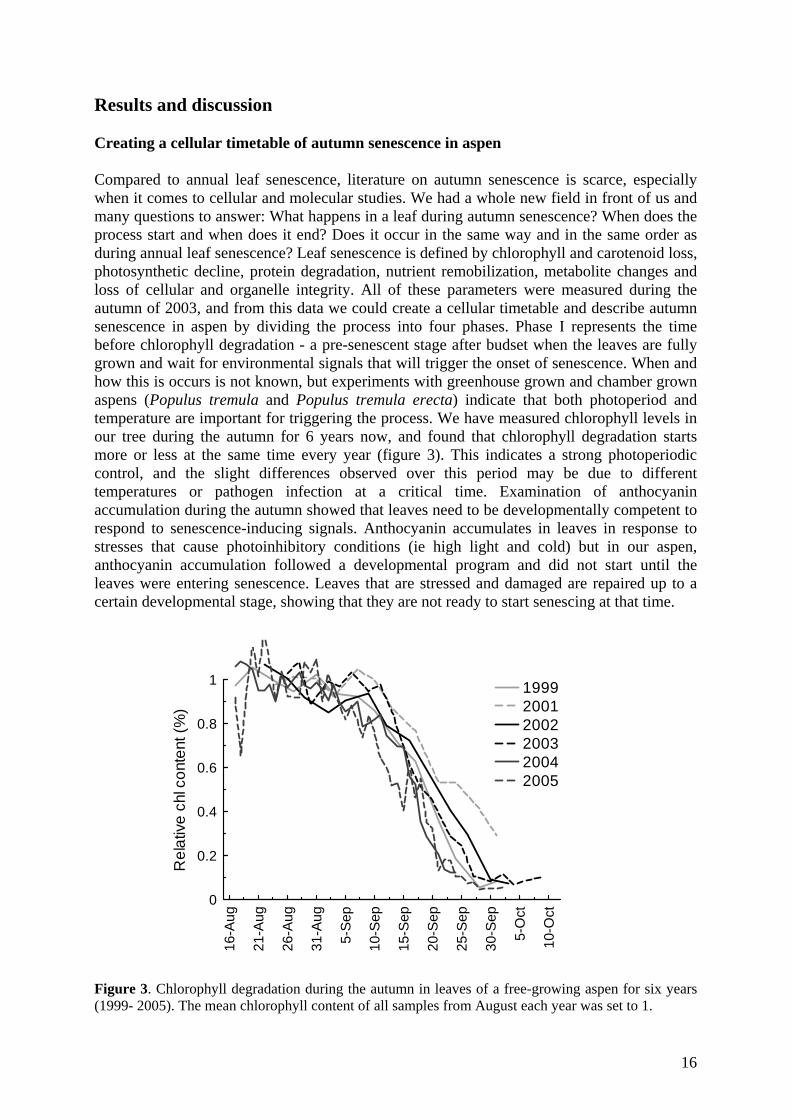
Results and discussion Creating a cellular timetable of autumn senescence in aspen Compared to annual leaf senescence, literature on autumn senescence is scarce, especially when it comes to cellular and molecular studies. We had a whole new field in front of us and many questions to answer: What happens in a leaf during autumn senescence? When does the process start and when does it end? Does it occur in the same way and in the same order as during annual leaf senescence? Leaf senescence is defined by chlorophyll and carotenoid loss, photosynthetic decline, protein degradation, nutrient remobilization, metabolite changes and loss of cellular and organelle integrity. All of these parameters were measured during the autumn of 2003, and from this data we could create a cellular timetable and describe autumn senescence in aspen by dividing the process into four phases. Phase I represents the time before chlorophyll degradation - a pre-senescent stage after budset when the leaves are fully grown and wait for environmental signals that will trigger the onset of senescence. When and how this is occurs is not known, but experiments with greenhouse grown and chamber grown aspens (Populus tremula and Populus tremula erecta) indicate that both photoperiod and temperature are important for triggering the process. We have measured chlorophyll levels in our tree during the autumn for 6 years now, and found that chlorophyll degradation starts more or less at the same time every year (figure 3). This indicates a strong photoperiodic control, and the slight differences observed over this period may be due to different temperatures or pathogen infection at a critical time. Examination of anthocyanin accumulation during the autumn showed that leaves need to be developmentally competent to respond to senescence-inducing signals. Anthocyanin accumulates in leaves in response to stresses that cause photoinhibitory conditions (ie high light and cold) but in our aspen, anthocyanin accumulation followed a developmental program and did not start until the leaves were entering senescence. Leaves that are stressed and damaged are repaired up to a certain developmental stage, showing that they are not ready to start senescing at that time.
16-A
ug
21-A
ug
26-A
ug
31-A
ug
5-S
ep
10-S
ep
15-S
ep
20-S
ep
25-S
ep
30-S
ep
5-O
ct
10-O
ct
0
0.2
0.4
0.6
0.8
1
Rel
ativ
e ch
l con
tent
(%)
199920012002200320042005
Figure 3. Chlorophyll degradation during the autumn in leaves of a free-growing aspen for six years (1999- 2005). The mean chlorophyll content of all samples from August each year was set to 1.
16

Phase II begins when chlorophyll degradation starts and is a period of massive degradation and remobilization of nutrients. Once phase II has begun the senescence process in autumn leaves is very similar to annual leaf senescence. Cellular contents are degraded at a constant rate in a highly organized way and most organelles keep their integrity and function until the cells die. Chloroplast degradation occurs early, beginning with loss of chloroplast pigments, followed by the breakdown of photosynthetic proteins, destabilization of the thylakoid membranes and accumulation of plastoglobuli containing the highly-accumulating carotenoid catabolites. We believe that chloroplast-to-gerontoplast differentiation occurs very quickly, since we could not find cells which contained both chloroplasts and gerontoplasts and Fv/Fm values are kept high until late senescence, ruling out the possibility of a progressive loss of PSII activity and of active chloroplasts. Autophagy has been proposed as a mechanism for degradation of cytoplasmic and organellar contents, and vesicles containing Rubisco or proteases have been found in annuals like Arabidopsis. Small vesicle-like structures located in the vacuolar space appeared in TEM micrographs during phase II as the vacuole increased in size, indicating that autophagy may be important during autumn senescence as well. The shift in metabolism that occurs during the transition from photosynthetically active leaves to senescing leaves can be seen by measuring metabolites like sugars and ATP. In the middle of the second phase the adenylate content decreases and the ATP/ADP ratio increases sharply, indicating a change in ATP production from the chloroplast to the mitochondria. The energy requirement of the senescing leaves is met by respiration using the lipids, sugars and amino acids that are not remobilized. The timing of this metabolic shift is confirmed by changes in leaf sugar composition and amounts. Accumulation of starch and sucrose ends at approximately the same time as we detect metabolic indications of a transfer to reliance on the mitochondria for energy production. This halt in starch and sucrose accumulation indicates a loss of photosynthetic activity and results in subsequent depletion of sugars in the leaves by respiration or remobilization of sucrose. Nutrient remobilization in aspen during the autumn is of the same magnitude as in Arabidopsis, with an 80% loss of nitrogen and phosphorous and a lesser degree of S, Fe and K retained. At the very end of phase II, we measure a sharp increase in d15N/d14N ratio, which we believe occurs because of the formation of the abscission layer, which blocks transport out of the leaves through the phloem and causes a build up of nitrogen compounds to toxic levels. Volatile amines like ammonia are released into the air and isotope discrimination during the formation of these compounds results in the sudden change in isotope ratio. As phase II ends, possibly with the formation of the abscission layer, the senescence process also finishes. However, many cells are still metabolically active in phase III, and these leaves retain chlorophyll-containing plastids with active PSII centers. It could be possible that some leaves retain phloem transport capability until late in the autumn, allowing the tree to continue to draw nutrients from these leaves until they die from cold temperatures. If not, and all leaves are cut off from the tree at the same time, the cells may continue catabolism until they die but this will not benefit the tree. Gene expression in autumn leaves What is the genetic basis of autumn senescence? Senescence is an active process, requiring an active nucleus and the expression of many genes and proteins. Gene expression during
17

senescence has been studied extensively in annual plants and thousands of SAGs have been identified to date. However, to our knowledge, autumn senescence has not been studied on the level of gene expression, leaving the question open as to whether there is a difference at the transcriptional level between annuals and trees. Differences in induction of the senescence process between perennials and annual plants could be represented by differences in gene expression patterns. Our approach to this was to study gene expression changes during autumn senescence in aspen by taking advantage of the recently developed genomics resources at our centre, ie EST sequencing and cDNA microarray projects in poplar (Sterky et al., 1998; Hertzberg et al., 2001). In the initial study, a digital northern approach was used to compare EST frequencies between young greenhouse grown hybrid aspen leaves and senescing free-growing aspen leaves. This comparison allowed us to find differences in gene expression caused by both environmental stresses and senescence, and gave us a snapshot of the senescence process at the beginning of chlorophyll breakdown. By looking at the most abundant clones in each library and the percentage of clones found in different functional classes (according to UPSC-MIPS), we found a striking difference between the two libraries, mainly due to downregulation of photosynthesis genes and upregulation of stress-related genes in the autumn leaves. Genes encoding proteins involved in respiration, proteolysis, and pigment and lipid degradation were highly represented in the autumn leaves compared to the young leaves. This, together with the fact that we identified homologues to many SAGs in Arabidopsis, led us to the conclusion that autumn senescence in aspen is very similar to leaf senescence in Arabidopsis. How does gene expression patterns change during autumn senescence? To continue our investigation, we wanted to follow gene expression patterns in aspen leaves during the autumn, and create a transcriptional timetable of autumn senescence. The recently developed cDNA microarray approach lends itself well to the screening of large numbers of transcripts, so this technique was perfect for our needs. Since this technique was in its infancy at the time of our experiments, we wanted to quantify transcript changes in the same material (1999 leaves) used in the EST study to confirm our results from this experiment, and compare expression patterns from northern blots and microarrays to determine if the results were reliable. The populus microarrays were constructed from a unigene set consisting of 13.490 cDNA clones, representing around 9,000 unique genes, where 3,792 clones were expressed in all studied samples after quality filtering. Seven samples, harvested between the middle of August and the end of September 1999, were chosen to cover the autumn senescence process, and cDNA synthesized from the total RNA of these leaves were hybridized against a common reference (a collection of different aspens from Sept 4, 2000). Although the study of single genes may not be reliable, a large microarray dataset is advantageous for following genes with coordinated expression patterns and to study different functional categories of genes. In this way we could verify and follow up our earlier EST data with gene expression profiles. Clustering of the data showed a major shift in gene expression between 7 and 14th of September, at the beginning of chlorophyll degradation. At this time, gene expression followed three basic patterns: increasing, decreasing and constant. Transcripts coding for proteins involved in photosynthesis started decreasing before visible senescence and continued throughout the study period, whereas many genes encoding proteases, metallothioneins and other proteins increased in expression. Transcript levels for the genes encoding enzymes involved in energy-generating metabolism in the cell (eg mitochondrial respiration, glycolysis, gluconeogenesis and TCA cycle) did not change significantly, indicating that these processes continued during senescence. This expression data is consistent with our earlier EST data, and the timing for the massive gene expression changes
18

occured at the same time as, or right before, chlorophyll degradation started in both 1999 and 2003, making it possible to combine our transcriptional timetable with the cellular one. However, although the changes in the 1999 transcriptional timetable started at the same time as chlorophyll degradation, this was not the case for the other years we studied. When we started working with a free-growing tree, we knew that it would be difficult to separate developmentally-regulated genes from those that were regulated by environmental stresses, but believed that this could be accomplished by studying gene expression in different trees during many years. Even though the timing and rate of senescence could differ between years, the underlying genetically controlled program would be the same, and this would be represented by developmentally regulated transcript patterns that varied in timing, but otherwise stayed the same every year. The benefit of field studies would be that we could capture the natural autumn senescence process and avoid losing important information about unknown environmental contributions to senescence while examining the process in a controlled environment. After initially running microarray experiments on two years, 1999 and 2001, we decided to continue during 2003 and 2004. At this time, we had finished our combined timetable and decided to focus on the period right before chlorophyll degradation started to screen for regulatory genes involved in controlling the process. In 2003, we studied gene expression every day for a brief period to capture transient changes. When we compared the different timetables for all years we were surprised to find that there was a 7-10 day difference in the timetables between 2004 and the other years, even though chlorophyll degradation started at more or less the same time. These results indicate that start of chlorophyll degradation, and possibly the whole cellular timetable, is not tightly coupled to the start of gene expression changes during autumn senescence in aspen. However, a more thorough investigation of our gene expression data is needed before we can draw any definite conclusions from our data. Recently, we have also done a metabolomics study on leaves (for M&M see Külheim et al., 2006) collected during the autumn of 2003 (not analysed) and 2004. Results from 2004 show that the rate of change in metabolism in the leaves was fairly constant, and started long before chlorophyll degradation and continued until the end of senescence (Figure 4 and 5).
Figure 4. Metabolomics analysis of aspen leaves 2004. PCA column plot (t1) showing the median scores for each class (sample date). The rate of change in metabolism in the leaves is fairly constant from end of August to17-18th of September.
19

Figure 5. Metabolomics analysis of aspen leaves 2004. PCA score plot (t1/t3) showing the median scores for each class (sample date).
Temperature and photoperiod effect on autumn senescence in Populus tremula erecta clones What are the responsible environmental signals regulating autumn senescence in aspen? Populus is known to grow continuously in long days and the induction of growth cessation and bud set is regulated by shortening of the photoperiod (Paul and Pellny). Although our experiments with free-growing aspen imply an important role for photoperiod in the induction of autumn senescence, there may be other signals involved in timing of the process. The most conspicuous of them would be decreasing temperature during the autumn. To separate the effects of photoperiod and temperature on aspen trees, we needed to perform controlled environment experiments, and decided to work with clonal tree material to avoid large genotypic variations. 50 young stem cuttings of Populus erecta were grown in a greeenhouse on a 20h photoperiod at 20 C. After reaching full growth 3 clones were moved to the cold room (8h, 5 C), 3 clones to a chamber with decreasing photoperiod (1 h / week, 24C) and 9 clones were moved to a chamber were both photoperiod and temperature were decreased (12h, 20C for 3 weeks, then 11h, 6C). Unfortunately, almost directly after moving the trees into a chamber they became infected, and many leaves shriveled up and died. However, we were still able to determine the critical photoperiod for budset (14h), and we also saw that autumn senescence was not induced by SD alone during the study period, which lasted until 3 months after budset using a 12h photoperiod and 24C air temperature. Similar experiments with our SweAsp collection (aspen trees collected from different latitudes in Sweden) after changing photoperiod abruptly from 23h to 12h in the greenhouse showed, however, that lower night temperatures induced autumn senescence 1-2 months after budset, depending on ecotype. When is “the point of no return”? As another part of this experiment, we were interested in determining what would happen if we moved clones at different stages of senescence in the field into long day conditions back in the greenhouse. 20 potted Populus erecta clones were placed outside in the beginning of
20

July, and following the completion of growth cessation and budset in September, one tree was moved inside every second day until the end of October. By scoring chlorophyll changes and bud break, we could determine at which time the trees did no longer break dormancy to resume growth, and when the remaining leaves did not re-green, but continued senescence and dropped off. Figure 6 shows the natural photoperiod in Umeå from the time when we started to move trees from the field back into the greenhouse, and until chlorophyll measurements were stopped. Three time-points representing different stages of vegetative dormancy are shown. Until October 2 (stage 1) buds break after a couple of days, and growth continues. At October 8 (stage 2), leaves retain chlorophyll but buds do not break until much later. Finally, after October 10 (stage 3), all leaves fall off and buds do not break during the study period (although all buds opened eventually). “The point of no return” in these trees occured somewhere between stage 2 and 3.
Figure 6. Natural photoperiod in Umeå (Sävar) during the study period. Populus tremula erecta clones were moved from the field into the greenhouse between September 15 and October 20, and the experiment continued until the beginning of December. Three time-points representing different stages of vegetative dormancy are shown. 1. Buds break and leaves continue to grow. 2. Buds stay dormant but leaves stay green. 3. Buds stay dormant and leaves drop off.
21

Vad händer i ett löv när det blir höst? När man tänker på hösten är det oftast ett ord som dyker upp i huvudet - färg! Dessa färger uppstår hos bladen när det gröna pigmentet klorofyll bryts ner snabbare än de gula karotenoid pigmenten och andra sekundära pigment, tex de röda antocyaninerna, bildas. Träden kan via ljuskänsliga proteiner i bladen, sk fytokromer, känna av att dagarna blir kortare och nätterna längre och detta kombinerat med att temperaturen sjunker signalerar att det är dags att sluta växa och göra sig redo för vintern. Detta startar en process som kallas för höst-senescence och är en genetiskt styrd sk programmerad celldöd där cellerna induceras att brytas ner på ett organiserat sätt. Höst-senescence är en viktig fas i bladens utveckling när näringsämnen (framförallt kväve) som använts till att bygga upp bladet och fotosyntesmaskineriet transporteras till stammen och rötterna och lagras inför nästa års tillväxt. Trots att man länge vetat en del om den fysiologiska och ekologiska processen bakom höst-senescence, så saknas molekylär kunskap om processen och hur den styrs. Vi har studerat processen i asplöv genom att använda oss av olika molekylärbiologiska metoder och använt dessa data för att konstruera en cellulär och genetisk tidtabell för höst-senescence hos friväxande asp (Populus tremula). För att ta reda på vad som egentligen händer i ett asplöv under hösten så har vi studerat cellerna under elektronmikroskop och mätt fotosyntes samt nivåer av pigment, näringsämnen och metaboliter, som tex socker. Genom att studera dessa data kan vi säga när processen börjar och slutar, och vi kan även se hur olika delar av cellerna bryts ner, tex kloroplasterna och det mesta av cellmaterialet. När kloroplasterna bryts ner förlorar cellerna förmågan att fotosyntetisera, alltså producera energi, och eftersom nedbrytning och transport av näringsämnen är en väldigt energikrävande process, så måste energi produceras på annat sätt, dvs genom respiration i mitokondrierna. Genom att titta på hur nivåer av socker och energirika metaboliter (sk adenosin di- och tri-fosfater, ATP och ADP) förändras under hösten kan vi säga ungefär när mitokondrierna tar över energiproduktionen. Nedbrytningsprocessen börjar i mitten av September och fortsätter tills i början på Oktober, då löven lossnar från trädet och faller av. Ca 80% kväve och fosfor, men även andra näringsämnen, har vid det laget transporterats ut från löven till andra delar av trädet. Höst-senescence processen styrs, som alla andra processer i en levande organism, av gener. Vi har studerat vilka gener som uttrycks i asplöv under hösten genom sk EST sekvensering av uttryckta gener i höstlöv. Genom att analysera dessa gensekvenser och jämföra med gensekvenser från andra delar av trädet, framförallt gröna löv, kunde vi se vilka gener som är påslagna under hösten och även hitta gener som bara uttrycks i höstlöv och inte andra delar av trädet. De gener som var högst uttryckta i höstlöv kodade för proteiner involverade i nedbrytningen av proteiner, pigment osv, samt proteiner som uttrycks när trädet blir utsatt för stress, som tex insektsangrepp. Antalet gener som kodar för proteiner i fotosyntesmaskineriet var betydligt lägre i höstlöv än i gröna blad. Många av generna som uttrycks i höstlöv hos asp är samma som de gener som uttrycks i gulnande blad hos olika ettåriga växter, exempelvis backtrav (Arabidopsis thaliana). Vi har även följt genuttryck i asplöv under hösten under flera år genom att använda oss av sk DNA-mikromatriser (microarrays), som är glasskivor med över 20.000 aspgener på. På detta sätt har vi konstruerat genetiska tidtabeller under fyra säsonger, där vi kan se att uttrycket av de flesta gener ändras markant vid en viss tidpunkt på hösten. Denna tidpunkt varierar dock från år till år, även om klorofyllmätningar visar att höst-senescence processen startar ungefär samtidigt varje år, vilket tyder på att det inte finns någon tydlig koppling mellan de genetiska och den cellulära tidtabellen.
22

Acknowledgements First and foremost, I would like to thank the person who helped me to get me this far, my supervisor Stefan Jansson. Without your support, inexhaustible patience and motivational speeches, I would not be writing this thesis now. A warm thanks also goes to the past and present Jansson group, a group of people who I always felt very comfortable around - except when I occasionally forgot fika! I would like to thank all collaborators and all the people who had time to help or advise me even though they probably had a very busy schedule. Special thanks goes to: • The people at KTH, who did all the sequencing and produced the arrays. • All the Genomics people, working with clone preparations and PopulusDB. • The electron microscopy people, Lenore Johansson and Tatsuya Awano for introducing
me into the fascinating world of toxic fixation fluids, and performing all the sectioning. • Ewa Mellerowitz for producing beautiful confocal microscopy pictures. • Per Gardeström and Gunilla Malmberg for help with ATP and sugar mesurements. • Thomas Moritz and his group, for the metabolomics work. Thanks also to all hard-working students doing their exam work or project work and helping me with a lot of boring and time-consuming work, especially Gustaf Bergquist, who also is a co-author on the cellular timetable article. Special thanks to: Catherine - for help with the language and inviting me to her death-by-chocolate evenings! Maria and Magnus - for friendship and Depeche Mode evenings! Carsten, Jenny and Linda - for starting their PhD at the same time as me, and just being very easy persons to be around. Ulrika and Jenny - for helping me get started, and giving me advice on how long time it actually takes to run experiments. Frank - for knowing everything and for being an extremely annoying and wonderful person at the same time! Andreas – for being a fun person to annoy, and for having patience with me and my computer skills! Charlene – for being a real party person, and fun to have as a travel companion! Mattias – for our lunch-time and general discussions! Ola – for making life at Fysbot a little more interesting! Jan Eklöf – for helping me with grafic stuff! A special thanks also to all people at UPSC for providing a nice working environment during my years there! To all the people I have forgot: Thank you and forgive me for leaving you out! My love goes to my family and friends, the people who matters most to me in the world! And to Lars, my love, who has been there for me all the way, I can only say: - nu är det min tur att diska!
23

Literature Adams-Phillips L, Barry C, Giovannoni J (2004) Signal transduction systems regulating
fruit ripening. Trends in Plant Science 9: 331-338 Andersson A, Keskitalo J, Sjödin A, Bhalerao R, Sterky F, Wissel K, Tandre K,
Aspeborg H, Moyle R, Ohmiya Y, Brunner A, Gustafsson P, Karlsson J, Lundeberg J, Nilsson O, Sandberg G, Strauss S, Sundberg B, Uhlen M, Jansson S, Nilsson P (2004) A transcriptional timetable of autumn senescence. Genome Biology 5: R24
Archetti M (2000) The origin of autumn colours by coevolution. Journal of Theoretical Biology 205: 625-630
Archetti M, Brown SP (2004) The coevolution theory of autumn colours. Proceedings of the Royal Society of London Series B-Biological Sciences 271: 1219-1223
Baehrecke EH (2005) Autophagy: Dual roles in life and death? Nature Reviews Molecular Cell Biology 6: 505-510
Bhalerao R, Keskitalo J, Sterky F, Erlandsson R, Björkbacka H, Birve SJ, Karlsson J, Gardeström P, Gustafsson P, Lundeberg J, Jansson S (2003) Gene expression in autumn leaves. Plant Physiology 131: 430-442
Biswal B (1995) Carotenoid Catabolism During Leaf Senescence And Its Control By Light. Journal of Photochemistry and Photobiology B: Biology 30: 3-13
Bonora A, Pancaldi S, Gualandri R, Fasulo MP (2000) Carotenoid and ultrastructure variations in plastids of Arum italicum Miller fruit during maturation and ripening. Journal of Experimental Botany 51: 873-884
Brugiere N, Dubois F, Masclaux C, Sangwan RS, Hirel B (2000) Immunolocalization of glutamine synthetase in senescing tobacco (Nicotiana tabacum L.) leaves suggests that ammonia assimilation is progressively shifted to the mesophyll cytosol. Planta 211: 519-527
Buchanan-Wollaston V, Earl S, Harrison E, Mathas E, Navabpour S, Page T, Pink D (2003) The molecular analysis of leaf senescence - a genomics approach. Plant Biotechnology Journal 1: 3-22
Buchanan-Wollaston V, Page T, Harrison E, Breeze E, Lim PO, Nam HG, Lin JF, Wu SH, Swidzinski J, Ishizaki K, Leaver CJ (2005) Comparative transcriptome analysis reveals significant differences in gene expression and signalling pathways between developmental and dark/starvation-induced senescence in Arabidopsis. Plant Journal 42: 567-585
Chiba A, Ishida H, Nishizawa NK, Makino A, Mae T (2003) Exclusion of ribulose-1,5-bisphosphate carboxylase/oxygenase from chloroplasts by specific bodies in naturally senescing leaves of wheat. Plant and Cell Physiology 44: 914-921
Clarke PGH (1990) Developmental Cell-Death - Morphological Diversity and Multiple Mechanisms. Anatomy and Embryology 181: 195-213
Collier DE, Thibodeau BA (1995) Changes in Respiration and Chemical Content During Autumnal Senescence of Populus Tremuloides and Quercus Rubra Leaves. Tree Physiology 15: 759-764
Dangl JL, Dietrich RA, Thomas H (2000) Senescence and programmed cell death. In Biochemistry & molecular biology of plants. B Buchanan, W Gruissem, R Jones, eds, American society of plant physiologists
Dellaporta SL, Calderon-Urrea A (1994) The Sex Determination Process in Maize. Science 266: 1501-1505
24

Delorme VGR, McCabe PF, Kim DJ, Leaver CJ (2000) A matrix metalloproteinase gene is expressed at the boundary of senescence and programmed cell death in cucumber. Plant Physiology 123: 917-927
Dickman MB, Park YK, Oltersdorf T, Li W, Clemente T, French R (2001) Abrogation of disease development in plants expressing animal antiapoptotic genes. Proceedings of the National Academy of Sciences of the United States of America 98: 6957-6962
Downs RJ, Borthwick HA (1956) Effects of photoperiod on growth of trees. Botanical Gazette 117: 310-326
Ellis HM, Horvitz HR (1986) Genetic-Control of Programmed Cell-Death in the Nematode C. elegans. Cell 44: 817-829
Feild TS, Lee DW, Holbrook NM (2001) Why leaves turn red in autumn. The role of anthocyanins in senescing leaves of red-osier dogwood. Plant Physiology 127: 566-574
Feller U, Fischer A (1994) Nitrogen-Metabolism in Senescing Leaves. Critical Reviews in Plant Sciences 13: 241-273
Fischer A, Feller U (1994) Senescence and Protein-Degradation in Leaf Segments of Young Winter-Wheat - Influence of Lead Age. Journal of Experimental Botany 45: 103-109
Fukuda H (2000) Programmed cell death of tracheary elements as a paradigm in plants. Plant Molecular Biology 44: 245-253
Gan SS, Amasino RM (1995) Inhibition of Leaf Senescence by Autoregulated Production of Cytokinin. Science 270: 1986-1988
Gepstein S (1988) Photosynthesis. In Senescence and aging in plants. LD Noodén, AC Leopold, eds. Academic press, San Diego, pp 85-109
Gepstein S, Sabehi G, Carp MJ, Hajouj T, Nesher MFO, Yariv I, Dor C, Bassani M (2003) Large-scale identification of leaf senescence-associated genes. Plant Journal 36: 629-642
Goodwin TW (1958) Studies in carotenogenesis: XXIII the changes in carotenoid and chlorophyll pigments in the leaves of deciduous trees during autumn necrosis. Biochemistry Journal 68: 503-511
Gozuacik D, Kimchi A (2004) Autophagy as a cell death and tumor suppressor mechanism. Oncogene 23: 2891-2906
Grbic V, Bleecker AB (1995) Ethylene Regulates the Timing of Leaf Senescence in Arabidopsis. Plant Journal 8: 595-602
Greenberg JT (1996) Programmed cell death: A way of life for plants. Proceedings of the National Academy of Sciences of the United States of America 93: 12094-12097
Guiamet JJ, Pichersky E, Nooden LD (1999) Mass exodus from senescing soybean chloroplasts. Plant and Cell Physiology 40: 986-992
Guicciardi ME, Leist M, Gores GJ (2004) Lysosomes in cell death. Oncogene 23: 2881-2890
Gunawardena A, Pearce DM, Jackson MB, Hawes CR, Evans DE (2001) Characterisation of programmed cell death during aerenchyma formation induced by ethylene or hypoxia in roots of maize (Zea mays L.). Planta 212: 205-214
Guo Y, Cai Z, Gan S (2004) Transcriptome of Arabidopsis leaf senescence. Plant Cell and Environment 27: 521-549
Guo YF, Gan SS (2005) Leaf senescence: Signals, execution, and regulation. Current Topics in Developmental Biology, Vol 71, Vol 71, pp 83-112
Hadfield KA, Bennett AB (1997) Programmed senescence of plant organs. Cell Death and Differentiation 4: 662-670
Hagen SB, Folstad I, Jakobsen SW (2003) Autumn colouration and herbivore resistance in mountain birch (Betula pubescens). Ecology Letters 6: 807-811
25

Heide OM (1993) Daylength and Thermal Time Responses of Budburst During Dormancy Release in Some Northern Deciduous Trees. Physiologia Plantarum 88: 531-540
Hengartner MO (2000) The biochemistry of apoptosis. Nature 407: 770-776 Hertzberg M, Aspeborg H, Schrader J, Andersson A, Erlandsson R, Blomqvist K,
Bhalerao R, Uhlen M, Teeri TT, Lundeberg J, Sundberg B, Nilsson P, Sandberg G (2001) A transcriptional roadmap to wood formation. Proceedings of the National Academy of Sciences of the United States of America 98: 14732-14737
Himelblau E, Amasino RM (2001) Nutrients mobilized from leaves of Arabidopsis thaliana during leaf senescence. Journal of Plant Physiology 158: 1317-1323
Hoch WA, Singsaas EL, McCown BH (2003) Resorption protection. Anthocyanins facilitate nutrient recovery in autumn by shielding leaves from potentially damaging light levels. Plant Physiology 133: 1296-1305
Hörtensteiner S (2006) Chlorophyll degradation during Senescence. Annual Review of Plant Biology 57 (http://arjournals.annualreviews.org)
Hörtensteiner S, Feller U (2002) Nitrogen metabolism and remobilization during senescence. Journal of Experimental Botany 53: 927-937
Jing HC, Schippers JHM, Hille J, Dijkwei PP (2005) Ethylene-induced leaf senescence depends on age-related changes and OLD genes in Arabidopsis. Journal of Experimental Botany 56: 2915-2923
Jing HC, Sturre MJG, Hille J, Dijkwel PP (2002) Arabidopsis onset of leaf death mutants identify a regulatory pathway controlling leaf senescence. Plant Journal 32: 51-63
Jones A (2000) Does the plant mitochondrion integrate cellular stress and regulate programmed cell death? Trends in Plant Science 5: 225-230
Jones AM (2001) Programmed cell death in development and defense. Plant Physiology 125: 94-97
Kang CH, Jung WY, Kang YH, Kim JY, Kim DG, Jeong JC, Baek DW, Jin JB, Lee JY, Kim MO, Chung WS, Mengiste T, Koiwa H, Kwak SS, Bahk JD, Lee SY, Nam JS, Yun DJ, Cho MJ (2006) AtBAG6, a novel calmodulin-binding protein, induces programmed cell death in yeast and plants. Cell Death and Differentiation 13: 84-95
Kerr JFR, Wyllie AH, Currie AR (1972) Apoptosis: a basic biological phenomenon with wide-ranging implications in tissue kinetics. British Journal of Cancer 26: 239-257
Keskitalo J, Bergquist G, Gardestrom P, Jansson S (2005) A cellular timetable of autumn senescence. Plant Physiology 139: 1635-1648
Krishnamurthy KV, Krishnaraj R, Chozhavendan R, Christopher FS (2000) The programme of cell death in plants and animals - A comparison. Current Science 79: 1169-1181
Külheim C, Skogström O, Frenkel M, Moen J, Moritz T, Jansson S (2006) Metabolomics and transcriptomics in the field - PsbS levels strongly influence plant metabolism (manuscript).
Lacomme C, Cruz SS (1999) Bax-induced cell death in tobacco is similar to the hypersensitive response. Proceedings of the National Academy of Sciences of the United States of America 96: 7956-7961
Lam E, Kato N, Lawton M (2001) Programmed cell death, mitochondria and the plant hypersensitive response. Nature 411: 848-853
Lam HM, Coschigano KT, Oliveira IC, MeloOliveira R, Coruzzi GM (1996) The molecular-genetics of nitrogen assimilation into amino acids in higher plants. Annual Review of Plant Physiology and Plant Molecular Biology 47: 569-593
Lara MEB, Garcia MCG, Fatima T, Ehness R, Lee TK, Proels R, Tanner W, Roitsch T (2004) Extracellular invertase is an essential component of cytokinin-mediated delay of senescence. Plant Cell 16: 1276-1287
26

Laytragoon-Lewin N (1998) Programmed cell death: the influence of CD40, CD95 (Fas or Apo-I) and their ligands. Medical Oncology 15: 15-19
Lee DW, O'Keefe J, Holbrook NM, Feild TS (2003) Pigment dynamics and autumn leaf senescence in a New England deciduous forest, eastern USA. Ecological Research 18: 677-694
Leopold AC (1961) Senescence in plant development. Science 134: 1727-1732 Leopold AC (1975) Aging, senescence, and turnover in plants. Bioscience 25: 659-662 Lim PO, Nam HG (2005) The molecular and genetic control of leaf senescence and
longevity in Arabidopsis. Current Topics in Developmental Biology, Vol 67, Vol 67, pp 49-83
Lim PO, Woo HR, Nam HG (2003) Molecular genetics of leaf senescence in Arabidopsis. Trends in Plant Science 8: 272-278
Lin JF, Wu SH (2004) Molecular events in senescing Arabidopsis leaves. Plant Journal 39: 612-628
Liu Y, Schiff M, Czymmek K, Talloczy Z, Levine B, Dinesh-Kumar SP (2005) Autophagy regulates programmed cell death during the plant innate immune response. Cell 121: 567-577
Masclaux C, Valadier MH, Brugiere N, Morot-Gaudry JF, Hirel B (2000) Characterization of the sink/source transition in tobacco (Nicotiana tabacum L.) shoots in relation to nitrogen management and leaf senescence. Planta 211: 510-518
Masclaux-Daubresse C, Carrayol E, Valadier MH (2005) The two nitrogen mobilisation- and senescence-associated GS1 and GDH genes are controlled by C and N metabolites. Planta 221: 580-588
Matile P (1992) Chloroplast senescence. In Crop photosynthesis:spatial and temporal determinants. NR Baker, H Thomas, eds. Elsevier, Amsterdam, pp 413-440
Merzlyak MN, Hendry GAF, Atherton NM, Zhigalova TV, Pavlov VK, Zhiteneva OV (1993) Pigment Degradation, Lipid-Peroxidation, and Free-Radicals in Leaves During Autumn Senescence. Biochemistry-Moscow 58: 129-135
Moore B, Zhou L, Rolland F, Hall Q, Cheng WH, Liu YX, Hwang I, Jones T, Sheen J (2003) Role of the Arabidopsis glucose sensor HXK1 in nutrient, light, and hormonal signaling. Science 300: 332-336
Noh YS, Amasino RM (1999) Identification of a promoter region responsible for the senescence-specific expression of SAG12. Plant Molecular Biology 41: 181-194
Noodén LD (1988) The phenomena of senescence and aging. In Senescence and aging in plants. LD Noodén, AC Leopold, eds. Academic press, San Diego, pp 1-50
Noodén LD, Guiamet JJ, John I (1997) Senescence mechanisms. Physiologia Plantarum 101: 746-753
Nooden LD, Penney JP (2001) Correlative controls of senescence and plant death in Arabidopsis thaliana (Brassicaceae). Journal of Experimental Botany 52: 2151-2159
Olsen JE, Junttila O, Nilsen J, Eriksson ME, Martinussen I, Olsson O, Sandberg G, Moritz T (1997) Ectopic expression of oat phytochrome A in hybrid aspen changes critical daylength for growth and prevents cold acclimatization. Plant Journal 12: 1339-1350
Orrenius S, Zhivotovsky B, Nicotera P (2003) Regulation of cell death: The calcium-apoptosis link. Nature Reviews Molecular Cell Biology 4: 552-565
Otegui MS, Noh YS, Martinez DE, Vila Petroff MG, Andrew Staehelin L, Amasino RM, Guiamet JJ (2005) Senescence-associated vacuoles with intense proteolytic activity develop in leaves of Arabidopsis and soybean. Plant Journal 41: 831-844
27

Ougham HJ, Morris P, Thomas H (2005) The colors of autumn leaves as symptoms of cellular recycling and defenses against environmental stresses. Current Topics in Developmental Biology 66: 135-160
Pageau K, Reisdorf-Cren M, Morot-Gaudry JF, Masclaux-Daubresse C (2006) The two senescence-related markers, GS1 (cytosolic glutamine synthetase) and GDH (glutamate dehydrogenase), involved in nitrogen mobilization, are differentially regulated during pathogen attack and by stress hormones and reactive oxygen species in Nicotiana tabacum L. leaves. Journal of Experimental Botany 57: 547-557
Paul MJ, Pellny TK (2003) Carbon metabolite feedback regulation of leaf photosynthesis and development. Journal of Experimental Botany 54: 539-547
Pauley SS, Perry TO (1954) Ecotypic variation of the photoperiodic response in Populus. Journal of the Arnold Arboretum 35: 167-188
Pennell RI, Lamb C (1997) Programmed cell death in plants. Plant Cell 9: 1157-1168 Quirino BF, Noh YS, Himelblau E, Amasino RM (2000) Molecular aspects of leaf
senescence. Trends in Plant Science 5: 278-282 Robson PRH, Donnison IS, Wang K, Frame B, Pegg SE, Thomas A, Thomas H (2004)
Leaf senescence is delayed in maize expressing the Agrobacterium IPT gene under the control of a novel maize senescence-enhanced promoter. Plant Biotechnology Journal 2: 101-112
Rogers HJ (2005) Cell death and organ development in plants. Current Topics in Developmental Biology, Vol 71, Vol 71, pp 225-+
Rubinstein B (2000) Regulation of cell death in flower petals. Plant Molecular Biology 44: 303-318
Sakamoto W (2006) Protein Degradation Machineries in Plastids. Annual Review of Plant Biology 57 (http://arjournals.annualreviews.org)
Schmid M, Simpson DJ, Sarioglu H, Lottspeich F, Gietl C (2001) The ricinosomes of senescing plant tissue bud from the endoplasmic reticulum. Proceedings of the National Academy of Sciences of the United States of America 98: 5353-5358
Schweichel JU, Merker HJ (1973) The morphology of various types of cell death in prenatal tissues. Teratology 7: 253-266
Smart CM (1994) Gene-Expression During Leaf Senescence. New Phytologist 126: 419-448 Solomos T (1988) Respiration in senescing plant organs: its nature, regulation, and
physiological significance. In Senescence and aging in plants. LD Noodén, AC Leopold, eds. Academic press, San Diego, pp 111-145
Sterky F, Bhalerao RR, Unneberg P, Segerman B, Nilsson P, Brunner AM, Charbonnel-Campaa L, Lindvall JJ, Tandre K, Strauss SH, Sundberg B, Gustafsson P, Uhlen M, Bhalerao RP, Nilsson O, Sandberg G, Karlsson J, Lundeberg J, Jansson S (2004) A Populus EST resource for plant functional genomics. Proceedings of the National Academy of Sciences of the United States of America 101: 13951-13956
Sterky F, Regan S, Karlsson J, Hertzberg M, Rohde A, Holmberg A, Amini B, Bhalerao R, Larsson M, Villarroel R, Van Montagu M, Sandberg G, Olsson O, Teeri TT, Boerjan W, Gustafsson P, Uhlen M, Sundberg B, Lundeberg J (1998) Gene discovery in the wood-forming tissues of poplar: Analysis of 5,692 expressed sequence tags. Proceedings of the National Academy of Sciences of the United States of America 95: 13330-13335
Tevini M, Steinmuller D (1985) Composition and function of plastoglobuli II. Lipid composition of leaves and plastoglobuli during beech leaf senescence. Planta 163: 91-96
28

Thomas H, Donnison I (2001) Back from the brink: plant senescence and its reversibility. In Programmed cell death in animals and plants. J Bryant, SG Hughes, JM Garland, eds. Oxford: Bios, pp 149-162
Thomas H, Ougham H, Hortensteiner S (2001) Recent advances in the cell biology of chlorophyll catabolism. Advances in Botanical Research, Vol 35, Vol 35, pp 1-52
Thomas H, Ougham HJ, Wagstaff C, Stead AD (2003) Defining senescence and death. Journal of Experimental Botany 54: 1127-1132
Thompson AR, Doelling JH, Suttangkakul A, Vierstra RD (2005) Autophagic nutrient recycling in Arabidopsis directed by the ATG8 and ATG12 conjugation pathways. Plant Physiology 138: 2097-2110
Thornberry NA, Lazebnik Y (1998) Caspases: Enemies within. Science 281: 1312-1316 Uren AG, O'Rourke K, Aravind L, Pisabarro MT, Seshagiri S, Koonin EV, Dixit VM
(2000) Identification of paracaspases and metacaspases: Two ancient families of caspase-like proteins, one of which plays a key role in MALT lymphoma. Molecular Cell 6: 961-967
van Doorn WG, Woltering EJ (2004) Senescence and programmed cell death: substance or semantics? Journal of Experimental Botany 55: 2147-2153
van Doorn WG, Woltering EJ (2005) Many ways to exit? Cell death categories in plants. Trends in Plant Science 10: 117-122
Van Staden J, Cook EL, Nooden LD (1988) Cytokinins and senescence. In Senescence and aging in plants. LD Nooden, AC Leopold, eds. Academic Press, San Diego, pp 281-328
Vicentini F, Hortensteiner S, Schellenberg M, Thomas H, Matile P (1995) Chlorophyll Breakdown in Senescent Leaves - Identification of the Biochemical Lesion in a Stay-Green Genotype of Festuca-Pratensis Huds. New Phytologist 129: 247-252
Wingler A, Brownhill E, Pourtau N (2005) Mechanisms of the light-dependent induction of cell death in tobacco plants with delayed senescence. Journal of Experimental Botany 56: 2897-2905
Wingler A, Purdy S, MacLean JA, Pourtau N (2006) The role of sugars in integrating environmental signals during the regulation of leaf senescence. Journal of Experimental Botany 57: 391-399
Woltering EJ, van der Bent A, Hoeberichts FA (2002) Do plant Caspases exist? Plant Physiology 130: 1764-1769
Woo HR, Chung KM, Park JH, Oh SA, Ahn T, Hong SH, Jang SK, Nam HG (2001) ORE9, an F-box protein that regulates leaf senescence in Arabidopsis. Plant Cell 13: 1779-1790
Woo HR, Goh CH, Park JH, de la Serve BT, Kim JH, Park YI, Nam HG (2002) Extended leaf longevity in the ore4-1 mutant of Arabidopsis with a reduced expression of a plastid ribosomal protein gene. Plant Journal 31: 331-340
Yoshida S, Ito M, Callis J, Nishida I, Watanabe A (2002) A delayed leaf senescence mutant is defective in arginyl-tRNA : protein arginyltransferase, a component of the N-end rule pathway in Arabidopsis. Plant Journal 32: 129-137
Yu L, Alva A, Su H, Dutt P, Freundt E, Welsh S, Baehrecke EH, Lenardo MJ (2004) Regulation of an ATG7-beclin 1 program of autophagic cell death by caspase-8. Science 304: 1500-1502
Zavaleta-Mancera HA, Franklin KA, Ougham HJ, Thomas H, Scott IM (1999) Regreening of senescent Nicotiana leaves I. Reappearance of NADPH-protochlorophyllide oxidoreductase and light-harvesting chlorophyll a/b-binding protein. Journal of Experimental Botany 50: 1677-1682
29

Zavaleta-Mancera HA, Thomas BJ, Thomas H, Scott IM (1999) Regreening of senescent Nicotiana leaves II. Redifferentiation of plastids. Journal of Experimental Botany 50: 1683-1689
Zelisko A, Garcia-Lorenzo M, Jackowski G, Jansson S, Funk C (2005) AtFtsH6 is involved in the degradation of the light-harvesting complex II during high-light acclimation and senescence. Proceedings of the National Academy of Sciences of the United States of America 102: 13699-13704
30