Embed Size (px)
Citation preview

Histology 2nd Lec.
Connective tissue
Connective tissue is diverse in structure and function. Despite their differences, all
types of connective tissue have three similar components: specialized cells, ground
substance, and protein fibers (1). The ground substance is a non-cellular material that
separates the cells. It varies in consistency from solid (bone) to semifluid (cartilage)
to fluid (blood). The fibers are of three types. White collagen fibers contain collagen,
a protein that gives them flexibility and strength. Reticular fibers are very thin
collagen fibers, highly branched proteins that form delicate supporting networks.
Yellow elastic fibers contain elastin, a protein that is not as strong as collagen but is
more elastic. Elastic fibers return to their original shape and may stretch to over 100
times their relaxed size without damage.
Figure 1 Components of connective tissues. All connective tissues have three components: specialized cells, ground substance, and protein fibers. Loose fibrous connective tissue is shown here.
Fibrous Connective Tissue
Fibrous tissue exists in two forms: loose fibrous tissue and dense fibrous tissue. Both
loose fibrous and dense fibrous connective tissues have cells called fibroblasts

Histology 2nd Lec.
located some distance from one another and separated by a jellylike ground
substance containing white collagen fibers and yellow elastic fibers (Fig. 2). Matrix
includes ground substance and fibers.
A. Loose fibrous connective tissue, which includes areolar and reticular
connective tissue, supports epithelium and many internal organs. Its presence
in lungs, arteries, and the urinary bladder allows these organs to expand. It
forms a protective covering enclosing many internal organs, such as muscles,
blood vessels, and nerves.
Adipose tissue is a special type of loose connective tissue in which the cells
enlarge and store fat. Adipose tissue has little extracellular matrix. Its cells,
which are called adipocytes, are crowded, and each is filled with liquid fat. The
body uses this stored fat for energy, insulation, and organ protection. Adipose
tissue also releases a hormone called leptin, which regulates appetite-control
centers in the brain. Adipose tissue is primarily found beneath the skin, around
the kidneys, and on the surface of the heart.
B. Dense fibrous connective tissue contains many collagen fibers packed
together. This type of tissue has more specific functions than does loose
connective tissue. For example, dense fibrous connective tissue is found in
tendons, which connect muscles to bones, and in ligaments, which connect
bones to other bones at joints.

Histology 2nd Lec.
Figure 2 Connective tissues in the knee. Most types of connective tissue may be found in the knee.
Does dieting get rid of fat cells?
Unfortunately, dieting can shrink the size of fat cells (adipocytes), but their number
stays the same. This means that it is easier to regain the weight if diet and exercise
are not maintained. Liposuction is the only way to remove fat cells from the body.
Supportive Connective Tissue
Cartilage and bone are the two main supportive connective tissues. Each provides
structure, shape, protection, and leverage for movement. Generally cartilage is more
flexible than bone, because it lacks mineralization of the matrix. The supportive
connective tissues are covered in more detail in Section 12.1.
A. Cartilage
In cartilage, the cells lie in small chambers called lacunae (sing., lacuna),
separated by a solid, yet flexible, matrix. This matrix is formed by cells called
chondroblasts and chondrocytes. Because this tissue lacks a direct blood

Histology 2nd Lec.
supply, it often heals slowly. The three types of cartilage are distinguished by
the type of fiber found in the matrix.
1. Hyaline cartilage (Fig. 4.2), the most common type of cartilage, contains only
fine collagen fibers. The matrix has a glassy, translucent appearance. Hyaline
cartilage is found in the nose and at the ends of the long bones and the ribs,
and it forms rings in the walls of respiratory passages. The fetal skeleton also is
made of this type of cartilage. Later, the cartilaginous fetal skeleton is replaced
by bone.
2. Elastic cartilage has more elastic fibers than hyaline cartilage does. For this
reason, it is more flexible and is found, for example, in the framework of the
outer ear.
3. Fibrocartilage has a matrix containing strong collagen fibers. Fibrocartilage is
found in structures that withstand tension and pressure, such as the disks
between the vertebrae in the backbone and the cushions in the knee joint.
B. Bone
Bone is the most rigid connective tissue. It consists of an extremely hard matrix
of inorganic salts, notably calcium salts. These salts are deposited around
protein fibers, especially collagen fibers. The inorganic salts give bone rigidity.
The protein fibers provide elasticity and strength, much as steel rods do in
reinforced concrete. Cells called osteoblasts and osteoclasts are responsible for
forming the matrix in bone tissue.
1. Compact bone makes up the shaft of a long bone (Fig. 4.2). It consists of
cylindrical structural units called osteons (see Section 12.1). The central canal
of each osteon is surrounded by rings of hard matrix. Bone cells are located in
lacunae between the rings of matrix. In the central canal, nerve fibers carry
nerve impulses, and blood vessels carry nutrients that allow bone to renew

Histology 2nd Lec.
itself. Thin extensions of bone cells within canaliculi (minute canals) connect
the cells to each other and to the central canal. The ends of the long bones are
composed of spongy bone covered by compact bone. Spongy bone also
surrounds the bone marrow cavity. This, in turn, is covered by compact bone,
forming a “sandwich” structure.
2. Spongy bone appears as an open, bony latticework with numerous bony bars
and plates, separated by irregular spaces. Although lighter than compact bone,
spongy bone is still designed for strength. Just as braces are used for support in
buildings, the solid portions of spongy bone follow lines of stress.
Fluid Connective Tissue
A. Blood
Blood is a fluid connective tissue. Blood, which consists of formed elements
(Fig. 3) and plasma, is located in blood vessels. Blood transports nutrients and
oxygen to interstitial fluid, also called extracellular fluid. Interstitial fluid
bathes the body’s cells and removes carbon dioxide and other wastes. Blood
helps distribute heat and plays a role in fluid, ion, and pH balance. The systems
of the body help keep blood composition and chemistry within normal limits.
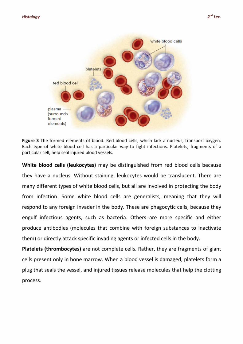
Each formed element of blood has a specific function. The red blood cells
(erythrocytes) are small, biconcave, disk-shaped cells without nuclei. The
presence of the red pigment haemoglobin makes the cells red, which in turn
makes the blood red. Hemoglobin is composed of four units. Each unit is
composed of the protein globin and a complex, iron-containing structure called
heme. The iron forms a loose association with oxygen; therefore, red blood
cells transport oxygen.
Histology 2nd Lec.
Figure 3 The formed elements of blood. Red blood cells, which lack a nucleus, transport oxygen. Each type of white blood cell has a particular way to fight infections. Platelets, fragments of a particular cell, help seal injured blood vessels.
White blood cells (leukocytes) may be distinguished from red blood cells because
they have a nucleus. Without staining, leukocytes would be translucent. There are
many different types of white blood cells, but all are involved in protecting the body
from infection. Some white blood cells are generalists, meaning that they will
respond to any foreign invader in the body. These are phagocytic cells, because they
engulf infectious agents, such as bacteria. Others are more specific and either
produce antibodies (molecules that combine with foreign substances to inactivate
them) or directly attack specific invading agents or infected cells in the body.
Platelets (thrombocytes) are not complete cells. Rather, they are fragments of giant
cells present only in bone marrow. When a blood vessel is damaged, platelets form a
plug that seals the vessel, and injured tissues release molecules that help the clotting
process.

Histology 2nd Lec.
B. Lymph
Lymph is also a fluid connective tissue. It is a clear (sometimes faintly yellow)
fluid derived from the fluids surrounding the tissues. It contains white blood
cells. Lymphatic vessels absorb excess interstitial fluid and various dissolved
solutes in the tissues. They transport lymph to particular vessels of the
cardiovascular system. Lymphatic vessels absorb fat molecules from the small
intestine. Lymph nodes, composed of fibrous connective tissue, occur along
the length of lymphatic vessels. Lymph is cleansed as it passes through lymph
nodes, in particular, because white blood cells congregate there. Lymph nodes
enlarge when you have an infection.
Figure 4.4 summarizes the classification of each of the major types of
connective tissue.

Histology 2nd Lec.
Figure 4.4 Types of connective tissue. Connective tissue is divided into three general categories—fibrous, supportive, and fluid.

Histology 2nd Lec.
OVERVIEW OF CONNECTIVE TISSUE
Connective tissue forms a continuous compartment throughout the body that
connects and supports other tissue. It is bounded by the basal laminae of various
epithelia and by the external laminae of muscle cells and nerve-supporting cells.
Connective tissue comprises a diverse group of cells within a tissue-specific
extracellular matrix (ECM). ECM contains protein fibers and ground substance.
Classification of connective tissue is primarily based on the composition and
organization of its extracellular components and on its functions: embryonic,
connective tissue proper, and specialized connective tissue.
EMBRYONIC CONNECTIVE TISSUES
A. Mesenchyme derives from embryonic mesoderm and gives rise to various
connective tissues of the body. It contains a loose network of spindle-shaped
cells that are suspended in a viscous ground substance containing fi ne
collagen and reticular fibers.
B. Mucous connective tissue is present in the umbilical cord. It contains widely
separated spindle-shaped cells embedded in a gelatin-like, hyaluronan-rich
ECM; its ground substance is called Wharton’s jelly.
CONNECTIVE TISSUE PROPER
Connective tissue proper is divided into loose and dense connective tissue. Dense
connective tissue is further subdivided into dense irregular and dense regular
connective tissue.
A. Loose connective tissue is characterized by large number of cells of various
types embedded in an abundant gel-like ground substance with loosely
arranged fibers. It typically surrounds glands, various tubular organs, blood
vessels, and is found beneath the epithelia that cover internal and external
body surfaces.
B. Dense connective tissue
1. Dense irregular connective tissue contains few cells (primary fibroblasts),
randomly distributed bundles of collagen fibers, and relatively little ground
substance. It provides significant strength and allows organs to resist
excessive stretching and distension.
2. Dense regular connective tissue is characterized by densely packed, parallel
arrays of collagen fibers with cells (tendinocytes) aligned between the fiber

Histology 2nd Lec.
bundles. It is the main functional component of tendons, ligaments, and
aponeuroses.
CONNECTIVE TISSUE FIBERS
There are three principal types of connective tissue fibers: collagen, reticular, and
elastic fibers.
1. Collagen fibers are the most abundant structural components of the
connective tissue. They are flexible, have a high tensile strength, and are
formed from collagen fibrils that exhibit a characteristic 68-nm banding
pattern.
2. Collagen fiber formation involves events that occur both within the fibroblasts
(production of procollagen molecules) and outside the fibroblasts in the ECM
(polymerization of collagen molecules into fibril, which are assembled into
larger collagen fibers).
3. Reticular fibers are composed of type III collagen and provide a supporting
framework for cells in various tissues and organs (abundant in lymphatic
tissues).
In the lymphatic and hemopoietic tissues, reticular fibers are produced by specialized
reticular cells. In most other tissues, reticular fibers are produced by fibroblasts.
Elastic fibers are produced by fibroblasts and smooth muscle cells. They allow tissues
to respond to stretch and distension. Elastic fibers are composed of a central core of
elastin associated with a network of fibrillin microfibrils, which are made of fibrillin
and emilin.
EXTRACELLULAR MATRIX
The ECM provides mechanical and structural support for connective tissue, influences
extracellular communication, and provides pathways for cell migration. In addition to
protein fibers, the ECM contains ground substance that is rich in proteoglycans,
hydrated glycosaminoglycans (GAGs), and multi adhesive glycoproteins. The GAGs
are the most abundant heteropolysaccharide components of ground substance.
These molecules are composed of long-chain unbranched polysaccharides containing
many sulfate and carboxyl groups. They covalently bind to core proteins to form
proteoglycans that are responsible for the physical properties of ground substance.

Histology 2nd Lec.
The largest and longest GAG molecule is hyaluronan. By means of special link
proteins, proteoglycans indirectly bind to hyaluronan, forming giant macromolecules
called proteoglycan aggregates. The binding of water and other molecules (e.g.,
growth factors) to proteoglycan aggregates regulates movement and migration of
macromolecules, microorganisms, or metastatic cancer cells in the ECM.
Multiadhesive glycoproteins (e.g., fibronectin, laminin, and tenascin) are
multifunctional molecules that possess binding sites for a variety of ECM proteins
(e.g., collagens, proteoglycans, and GAGs). They also interact with cell-surface
receptors such as integrin and laminin receptors.
CONNECTIVE TISSUE CELLS
Connective tissue cells are classified as part of the resident cell population (relatively
stable, nonmigratory) or the wandering (or transient) cell population (primarily cells
that have migrated from blood vessels).
1. Resident cells include fibroblasts (and myofibroblasts), macrophages,
adipocytes, mast cells, and adult stem cells.
2. Wandering (transient) cells include lymphocytes, plasma cells, neutrophils,
eosinophils, basophils, and monocytes.
3. Fibroblasts are the principal cells of connective tissue. They are responsible for
the synthesis of collagen and other components of the ECM. Fibroblasts that
express actin fi laments and associated actin motor proteins such as
nonmuscle myosin are called myofibroblasts.
4. Macrophages are phagocytic cells derived from monocytes that contain an
abundant number of lysosomes and play an important role in immune
response reactions.
5. Adipocytes are specialized connective tissue cells that store neutral fat and
produce a variety of hormones.
6. Mast cells develop in bone marrow and differentiate in connective tissue. They
contain basophilic granules that store mediators of inflammation. Upon
activation, mast cells synthesize leukotrienes, interleukins, and other
inflammation-promoting cytokines.
7. Adult stem cells reside in specific locations (called niches) in various tissues
and organs. They are difficult to distinguish from other cells of connective
tissue.

Histology 2nd Lec.
Loose and dense irregular connective tissue, mammary gland, human. H&E
This micrograph shows at low magnification both loose connective tissue (LCT) and dense irregular connective tissue (DICT) for comparative purposes. The loose connective tissue surrounds the glandular epithelium (GE). The dense irregular connective tissue consists mainly of thick bundles of collagen fibers with few cells present, whereas the loose connective tissue has a relative paucity of fibers and a considerable number of cells. The upper inset is a higher magnification of the dense connective tissue. Note that only a few cell nuclei are present relative to the larger expanse of collagen fibers. The lower inset, revealing the glandular epithelium and surrounding loose connective tissue, shows very few fibers but large numbers of cells. Typically, the cellular component of loose connective tissue contains a relatively small proportion of fibroblasts but large numbers of lymphocytes, plasma cells, and other connective tissue cell types.

Histology 2nd Lec.
Dense regular connective tissue, tendon, longitudinal section, human, H&E This specimen includes the surrounding dense irregular connective tissue of the tendon, the epitendineum (Ept). The tendon fascicles (TF) that make up the tendon are surrounded by a less dense connective tissue than that associated with the epitendineum. In longitudinal sections such as this, the connective tissue that surrounds the individual fascicles, the endotendineum (Ent), seems to disappear at certain points, with the result that one fascicle appears to merge with a neighbouring fascicle. This is due to obliqueness in the plane of section rather than an actual merging of fascicles. The collagen that makes up the bulk of the tendon fascicle has a homogeneous appearance as a result of the orderly packing of the individual collagen fibrils. The nuclei of the tendinocytes appear as elongate profi les arranged in linear rows. The cytoplasm of these cells blends in with the collagen, leaving only the nuclei as the representative feature of the cell.

Histology 2nd Lec.
Loose connective tissue
Dense irregular connective tissue

Histology 2nd Lec.
Dense regular connective tissue
Brown and white adipose tissue


![EMBRYONIC CONNECTIVE TISSUE CONNECTIVE TISSUE · PDF fileConnective tissue is composed of two elements: ØCells ØExtracelullar matrix [ECM] üFibers -collagen fibers, elastic fibers,](https://img.dokumen.tips/doc/110x75/5aa81cc77f8b9aa7258b710d/embryonic-connective-tissue-connective-tissue-tissue-is-composed-of-two-elements.jpg)