Embed Size (px)
Citation preview

Congenital Toxoplasmosis in SoutheasternBrazil: Results of Early OphthalmologicExamination of a Large Cohort of Neonates
Daniel Vitor Vasconcelos-Santos, MD, PhD,1,2 Danuza O. Machado Azevedo, MD, PhD,1
Wesley R. Campos, MD, PhD,1 Fernando Oréfice, MD, PhD,1 Gláucia M. Queiroz-Andrade, MD, PhD,2,3
Éricka V. Machado Carellos, MD, MSc,2,3 Roberta M. Castro Romanelli, MD, PhD,2,3
José Nelio Januário, MD, MSc,2,4 Luciana Macedo Resende, MSc,5 Olindo Assis Martins-Filho, MSc, PhD,6
Ana Carolina de Aguiar Vasconcelos Carneiro, MSc,7 Ricardo W. Almeida Vitor, MSc, PhD,7
Waleska Teixeira Caiaffa, MPH, PhD,8 for the UFMG Congenital Toxoplasmosis Brazilian Group
Objective: To report results of early ophthalmologic examinations in a large cohort of newborns withcongenital toxoplasmosis (CT) after neonatal screening.
Design: Cross-sectional analysis of a cohort.Participants: A total of 178 newborns with confirmed CT from 146,307 screened babies (95% of live births)
from Minas Gerais state, southeastern Brazil.Methods: From November 2006 to May 2007, newborns underwent neonatal screening by immunoglobulin
(Ig)M capture of dried blood samples. On all positive or suspected cases, confirmative serology was performedon babies and their mothers. Congenital toxoplasmosis was confirmed in newborns who had IgM and/or IgA andIgG, or IgG associated with suggestive ocular lesions (with IgM and IgG in the mother). Ophthalmologicevaluation consisted of indirect ophthalmoscopy with a lid speculum. Pediatric examination and radiologicstudies of the central nervous system were also performed. In selected cases, biomicroscopy of the anteriorsegment, fundus photographs, or ultrasonography (B-scan) was performed.
Main Outcome Measures: Prevalence of retinochoroidal lesions, either cicatricial or active, and theirlocation and associated findings, such as vascular sheathing, hemorrhage, vitreous opacities, and retinaldetachment, were evaluated. The occurrence of cataract, microphthalmia, microcephaly, intracranial calcifica-tion, and hydrocephalus was also recorded.
Results: Of 146,307 neonates screened, 190 had CT, yielding a prevalence of 1 in 770 live births, of whom178 (93.7%) underwent standardized ophthalmologic examination at an average age of 55.6�16.6 days. Of these178 infants, 142 (79.8%) had retinochoroidal lesions consistent with CT in at least 1 eye. Bilateral involvementwas noted in 113 patients (63.5%). Macular involvement was seen in 165 eyes (46.3%) of 111 patients (62.4%).Active lesions were observed in 142 eyes (39.9%) of 85 patients (47.8%). These lesions were located in themacula of 75 eyes (21.1%) and were associated with retinal vascular sheathing in 44 eyes (12.4%).
Conclusions: A high prevalence of CT was encountered (1/770) with high rates of early retinochoroidalinvolvement (�80%) and many active lesions (in �50%), indicating a possibly more severe ocular involvementby CT in Brazil than in other parts of the world. The hypotheses of higher parasite virulence and increasedindividual susceptibility are being currently investigated.
Financial Disclosure(s): The author(s) have no proprietary or commercial interest in any materials discussedin this article. Ophthalmology 2009;116:2199–2205 © 2009 by the American Academy of Ophthalmology.
Toxoplasma gondii is an extremely ubiquitous parasite,infecting up to one third of the human population.1,2 Al-though disease caused by T. gondii is usually subclinical, itmay be severe, especially in immunosuppressed subjectsand in fetuses after vertical transmission.2–4
Congenital toxoplasmosis (CT) presents asymptomati-cally in most newborns. However, retinochoroidal involve-ment is of concern, being seen in up to 85% of infected
subjects before adulthood.4–6 Bilateral and macular dis-© 2009 by the American Academy of OphthalmologyPublished by Elsevier Inc.
ease may be responsible for significant morbidity in thesepatients,7–10 with CT being a leading cause of childhoodlow vision and blindness in many countries, includingBrazil.11,12
Despite some evidence of higher prevalence and severityof CT in Brazil,12 large population-based studies have notbeen done. Reported prevalences vary from 3 to 20 casesper 10,000 live births, with highly variable rates of retino-
choroidal involvement; as a rule, however, these figures2199ISSN 0161-6420/09/$–see front matterdoi:10.1016/j.ophtha.2009.04.042

Ophthalmology Volume 116, Number 11, November 2009
have been based on a relatively small number of infectedsubjects.13–17
The aim of the study reported was to investigate ocularinvolvement of newborns with CT who were identified by alarge comprehensive neonatal screening program in thestate of Minas Gerais (MG), in southeastern Brazil.
Materials and Methods
This study is part of a prospective investigation on neonatalscreening for CT in the Brazilian state of MG. It is being carriedout by a multidisciplinary research group (UFMG-Congenital Tox-oplasmosis Brazilian Group) under the coordination of one of theauthors (GMQ-A). The study protocol followed the precepts of theDeclaration of Helsinki and was approved by the Ethics Commit-tee at our institution (Ethics Commitee at Universidade Federal deMinas Gerais).
From November 1, 2006, to May 31, 2007, 146,307 newbornsunderwent screening for CT using a capture enzyme-linked im-munosorbent assay for anti-T. gondii immunoglobulin (Ig)M de-tection (TOXO IgM Q-Preven, Symbiosis, Leme, Brazil). Bloodsamples were collected from 1560 public health care centerslocated in 853 cities within the state; these were then sent foranalysis to the laboratory of Núcleo de Ações e Pesquisa em ApoioDiagnóstico in the capital of the state, Belo Horizonte.
Confirmative plasma/serum tests were run on cases with posi-tive or undetermined screening results. These mothers and babieswere tested for IgA (enzyme-linked immunosorbent assay) andIgG and IgM anti-T. gondii (Enzyme-Linked Fluorometric Assay,ELFA-VIDAS, BioMérrieux SA, Lyon, France). Babies also un-derwent a thorough pediatric examination, neuroimaging studies(cranial radiographs or transfontanel ultrasound; computer-assistedtomography in selected cases), hearing assessment, and ophthal-mologic evaluation.
Cases of confirmed CT were defined as those infants withpositive testing of the serum/plasma for IgM and/or IgA and IgG,and those with IgG associated with suggestive retinochoroidallesions and positive IgM and IgG test results on their mothers.
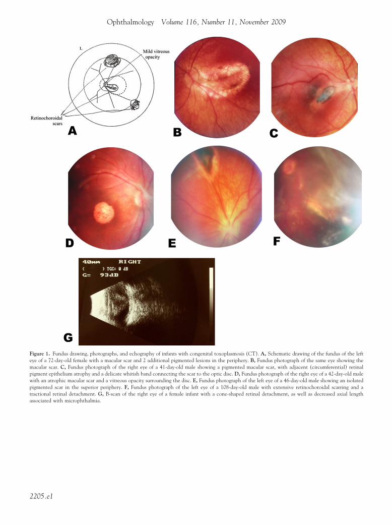
Each ophthalmologic evaluation of the babies was performedwithout sedation by 2 retina/uveitis specialists (DVV-S andDOMA) with the help of a trained nursing professional, accordingto a standardized protocol. After external inspection, pupils weredilated with 2 drops of tropicamide 0.5% and 1 drop of phenyl-ephrine 2.5%. All newborns, after being placed on an appropriatebed, underwent indirect ophthalmoscopy with a 20-diopter lens,with careful immobilization of the body with a cloth sheet, of thehead with the hands of the nurse, and after topical anesthesia andplacement of a tailored lid speculum. Most examinations wererecorded with a video charged-coupled device camera coupled tothe ophthalmoscope. When possible, fundus photographs werealso recorded; lesions were carefully drawn on an appropriate form(Fig 1A; available at http://aaojournal.org). In cases in which thefundus examination was limited because of media opacity,echographic evaluation of the posterior segment of the eye wasperformed. Biomicroscopy of the anterior segment with a slit-lamp was also performed in these cases, as well as in those withactive retinochoroiditis, looking for signs of anterior segmentinflammation.
All cases with confirmed or strongly suspected T. gondii infec-tion were started on classic treatment with sulfadiazine (100 mg/kg/d [in 2 divided doses]), pyrimethamine (1 mg/kg/d for the first6 months followed by the same dose 3 times per week thereafter),and folinic acid (7.5 mg 3 times per week) from the time of
diagnosis until completing 1 year of treatment, with careful bi-2200
monthly monitoring of the complete blood count. Cases of activeretinochoroiditis in the macular area and of neurologic involve-ment with increased protein content in the cerebrospinal fluid alsoreceived oral prednisolone (1 mg/kg/d) in a tapering fashion.
After the first examination, follow-up visits were scheduled at6 and 12 months of life and yearly thereafter, unless active reti-nochoroiditis lesions were detected. In this case, more frequentexaminations were performed, with the intervals between exami-nations individualized for each case.
Information from pregnancy cards on which the assistant ob-stetrician had registered data from the mother and the fetus, alongwith results of serologic tests during pregnancy, were alsocollected.
Data were stored and analyzed by a statistical package (SPSSfor Windows, Version 12, Chicago, IL), with frequency and de-scriptive statistics. Age at the first ophthalmologic examinationwas compared between infants with active and those with cicatri-cial retinochoroidal lesions using the Mann–Whitney U test. A Pvalue �0.05 was regarded as statistically significant.
Results
A total of 146,307 newborns were screened during the 6-monthperiod (November 1, 2006 to May 31, 2007), corresponding toapproximately 95% of the live births in the state of MG duringthose months. Of these screened babies, 235 had suspected CTand, along with their mothers, underwent confirmative serology.Infection was confirmed in 190 cases, corresponding to a preva-lence of 1 in 770 liveborn infants.
Among the 45 cases that were not confirmed, 35 (77.8%) hadthe infection ruled out because anti-T. gondii IgG antibodies de-creased during the follow-up, ultimately reaching undetectablelevels (passively transferred antibodies from their mothers). Of theremaining 10 cases, 9 could not be evaluated because of neonataldeath (4 newborns), loss to follow-up (3 newborns), or refusal toparticipate (2 newborns whose parents opted for private care). Theother had decreasing specific IgG antibodies, but these had notreached undetectable levels.
Of the 189 women who delivered infected newborns (onedelivered dizygotic twins, both of whom were infected), only 11(5.8%) had been treated during pregnancy, 10 of whom receivedspiramycin and 1 of whom was treated with sulfadiazine, py-rimethamine, and folinic acid.
Of the 190 patients with confirmed CT, 103 were male (54.2%)and 87 were female (45.8%). Pediatric examination results werenormal (absence of hepatomegaly, splenomegaly, microphthalmia,microcephaly, and convulsions) in 89 patients (46.8%) at a meanage of 57.3 days. In 9 patients (4.7%), there had been clinicalsuspicion of a congenital infection soon after birth. Neuroimagingrevealed intracranial calcifications in 39 patients (20.5%). Micro-cephaly was observed in 5.3% (10 patients) and hydrocephalus in6.3% (12 patients).
Standardized ophthalmologic examination was performed at amean age of 55.6�16.6 days (median: 56 days) in 178 of the 190patients (93.7%). The remaining 12 infected newborns (6.3%)were examined by local ophthalmologists in their home towns andare not included in this report. Retinochoroidal lesions consistentwith CT were found in 142 infants (79.8%), being bilateral in63.5% (113 cases). The macula was affected in 165 eyes (46.3%)of 111 patients (62.4%) (Fig 1A–D; available at http://aaojournal.org). Involvement of the foveal center (which posed an ominousvisual prognosis) was observed in 120 eyes (33.7%) of 88 infants(49.4%). Isolated peripheral lesions were seen in 51 eyes (14.3%)(Fig 1E; available at http://aaojournal.org) and were bilateral in 9
patients (5.1%). Fundus examination was not possible in 7 eyes
Vasconcelos-Santos et al � Congenital Toxoplasmosis in Southeastern Brazil
(2.0%) of 5 infants (2.8%) because of a cataract (2 eyes of 2patients), pupillary seclusion (4 eyes of 3 patients), a dense vitre-ous opacity, either inflammatory (7 eyes, 4 patients) or hemor-rhagic (3 eyes, 2 patients), or a combination of these factors.Microphthalmia was seen in 14 eyes (3.8%) of 8 patients (4.5%).In 11 eyes (3.1%) of 7 patients (3.9%), retinal detachment wasobserved by indirect ophthalmoscopy (Fig 1F; available at http://aaojournal.org) or echographic examination (Fig 1G; available athttp://aaojournal.org; Table 1).
Active retinochoroidal lesions were seen in 142 eyes (39.9%)of 85 patients (47.8%) and were associated with retinal scars in 66eyes (18.5%) of 53 patients (29.8%). These exudative retinocho-roidal lesions involved the macula in 75 eyes (21.1%) (Fig 2A, B),with associated features being retinal vascular sheathing in 44 eyes(12.4%) (Fig 2C) and vitreous or retinal hemorrhage in 11 eyes(3.1%) (Fig 2D). Vitreous opacities were relatively rare (26 eyes,7.3%) and mild (1–2�/4�), despite the high prevalence of activeretinochoroidal lesions. Moreover, none of these cases showedsigns of anterior segment inflammation, such as keratic precipitatesor cells in the anterior chamber. Active retinochoroidal lesionswere not associated with an earlier ophthalmologic examination,because babies with the active lesions were examined at an agecomparable to that of babies with only retinochoroidal scars (me-dian of 58 and 57 days, respectively; P � 0.393).
Ophthalmologic evaluation was decisive in 28 infants (15.7%)who had suspected CT at the neonatal screening (positive or unde-termined IgM) but who did not have a confirmative serology at thattime (positive IgG but negative IgA and IgM, along with maternalseroconversion). In these infants, indirect ophthalmoscopy disclosedretinochoroidal lesion(s) suggestive of toxoplasmosis, allowing con-firmation that they had, in fact, been congenitally infected.
Discussion
The prevalence of CT (1/770 live births) is higher in thepresent study than in most other reported series in the rest ofthe world19–23 and in Brazil.13–17 These few published
Table 1. Early* Ophthalmologic Findings in 190 Newboof Immun
Ophthalmologic Findings No. Eyes (%)
Retinochoroiditis 255 (71.6%)Unilateral lesionsBilateral lesionsActive lesions 142 (39.9%)Inactive lesions 173 (48.6%)Macular lesions 165 (46.4%)Foveal lesions† 120 (33.7%)Isolated peripheral lesions‡ 51 (14.3%)Cataract 2 (0.6%)Microphthalmia 14 (3.9%)Vascular sheathing (vasculitis) 44 (12.4%)Vitreous/retinal hemorrhage 11 (3.1%)Vitreous opacities (mild [1–2�]) 26 (7.3%)Retinal detachment 11 (3.1%)
CI � confidence interval.*Examination performed at a mean of 55.6�16.6 days after birth (media†Involving the foveal center.‡In zone 2 or 3 of Holland’s scheme,18 with no lesion in zone 1.
reports on the prevalence of CT in Brazil13–17 have involved
limited geographic areas and had selection and referral biasthat may limit interpretation and extrapolation of the result-ant data. In contrast, our study had coverage of approxi-mately 95% of liveborn infants in a large geographic area:a Brazilian state with approximately 20 million peopledistributed over 853 cities in an area of 586,528 square km.Furthermore, the use of a well-established public networkfor neonatal screening allowed for prompt screening, refer-ral, treatment, and follow-up of both children and theirmothers, with relatively few losses (3.8%, representing9/235 suspected of being infected).
In agreement with other reports on neonatal14,19,23 andprenatal24,25 screening, but not on referred patients (who aretheoretically more severely affected), only 4.7% of new-borns in our study had a suspected congenital infection, anda large proportion (46.8%) was completely asymptomatic,even after careful pediatric examination.
Retinochoroidal lesions consistent with toxoplasmosiswere present in approximately 80% of our cases; theseinfants underwent ophthalmologic examination at an aver-age of 55.6�16.6 days (median: 56 days) after birth. Theprevalence rates of retinochoroidal lesions at early evalua-tion in the literature are significantly lower, ranging between4% and 24%.8,19,20,23,26 As the children grow up, however,months or even years later, the reported prevalence ratesincrease to 30% to 90%5,6,8,9,27–29 because of either occur-rence of new lesions or better visualization of the retina.Ophthalmoscopic examination was decisive for the earlydiagnosis of CT in 15.7% of newborns in our study, whohad negative confirmatory serology for IgM and IgA butwho were positive for IgG anti-T. gondii. These newbornshad retinochoroidal lesions that were consistent with CT,allowing confirmation of the diagnosis by the end of the firstyear of life, before completion of the IgG kinetics study.This suggests that a careful ophthalmologic examination
ith Congenital Toxoplasmosis in Brazil with Detectionbulin-M
95% CI No. Patients (%) 95% CI
.9%–76.3% 142 (79.8%) 73.9%–85.7%29 (16.3%) 10.9%–21.7%
113 (63.5%) 56.4%–70.6%.8%–45.0% 85 (47.8%) 40.4%–55.1%.4%–53.8% 108 (60.7%) 53.5%–67.9%).2%–51.5% 111 (62.4%) 55.2%–69.5%.8%–38.6% 88 (49.4%) 42.1%–56.8%.7%–18.0% 42 (23.6%) 17.4%–29.8%.0%–1.3% 2 (1.1%) 0.0%–2.7%.9%–6.0% 8 (4.5%) 1.5%–7.5%.9%–15.8% 32 (18.0%) 12.3%–23.6%.3%–4.9% 7 (3.9%) 1.1%–6.8%.6%–10.0% 16 (9.0%) 4.8%–13.2%.3%–4.9% 7 (3.9%) 1.1%–6.8%
days).
rns woglo
66
3443412810
018141
n: 56
may in fact help, not only in evaluating the extent of ocular
2201

Ophthalmology Volume 116, Number 11, November 2009
involvement in CT but also in making an early diagnosis inuncertain cases.
Active lesions also were frequently observed in 39.9% ofeyes (47.8% of patients). This has been described onlyrarely in other series, in approximately 4% of newborns whowere examined early.8,23,30 Notably, the vitreous body wasusually clear, even in the presence of relatively large exu-dative retinochoroidal lesions (Fig 2A, B). Involvement ofthe macula in our series (46.3% of eyes, and 62.4% ofpatients) is similar to that of other reports,8,23,31 and thismacular tropism (considering the relatively small area of themacula) is thought to be due to anatomic, developmental,
Figure 2. Active retinochoroiditis in newborns with congenital toxoplasman active macular lesion and adjacent retinal edema and pigment abnormactive macular lesion with circumjacent retinal edema. C, Fundus photogrsuperiorly in an area with multiple punctate exudative lesions. D, Fundusan exudative lesion inferonasally.
and immunologic factors.7,32 We also reported involvement
2202
of the foveal center, which is more apt to be associated withsignificant visual impairment, especially if the involvementis bilateral.33 Unilateral and bilateral foveal involvementwere seen in 33.7% (120) and 18.0% (32) of newborns,respectively, in our study, but further follow-up shouldprovide more information as to its true functional impact,because relatively large central lesions in CT may be asso-ciated with reasonably good visual acuity.9
The high prevalence of retinochoroidal lesions that wefound, many of which were active, may have been influ-enced by the neonatal screening strategy, as well as by thelack of prenatal treatment. Only 10 of the women (5.7%)
(CT). A, Fundus photograph of the right eye of a 42-day-old female with. B, Fundus photograph of the right eye of a 30-day-old male showing anf the left eye of a 30-day-old male showing vascular sheathing (phlebitis)graph of the left eye of a 48-day-old female with vitreous hemorrhage and
osisalitiesaph ophoto
had received spiramycin during their pregnancy, which did
Vasconcelos-Santos et al � Congenital Toxoplasmosis in Southeastern Brazil
not prevent vertical transmission, and only 1 woman hadreceived sulfadiazine, pyrimethamine, and folinic acid. Al-though an earlier ophthalmologic examination would theo-retically be more likely to allow detection of active retino-choroidal lesions, this association was not confirmed in ourstudy (P � 0.393).
It is also possible that the high rate of ocular diseasedescribed may be associated at least in part with a greatervirulence and retinotropism of T. gondii in Brazil, wherehighly virulent parasites with atypical and recombinant ge-notypes have been observed infecting intermediate hosts, aswell as humans with ocular disease.34–36 This is an issuethat is currently being addressed by the UFMG CongenitalToxoplasmosis Brazilian Group, because all babies hadblood collected at the initial evaluation, which was inocu-lated into mice for isolation, phenotyping, and genotypingof T. gondii. Preliminary results show that 27 infants had apositive mouse inoculation (Carneiro ACAV, AndradeGMQ, Januário JN, et al. Isolation and characterization of T.gondii from newborns with CT identified by the neonatalscreening program in MG, Brazil. Poster presented at: Tox-oplasma Centennial Congress, September 23, 2008; Bú-zios), but further results from our parasitology team shouldbetter elucidate these findings.
Individual susceptibility also likely influenced our re-sults. Correlation between disease outcome in CT and HLA-DQ3 has been reported.37 An association between oculartoxoplasmosis and a polymorphism of interleukin-10 geneat position �1082 was also recently demonstrated in adultsattending a university uveitis referral center in Brazil.38 Astudy of the immune response in the children in our cohortand their mothers is currently under way, and it is hoped thatthis study will answer some questions.
Ocular toxoplasmosis is a recurrent disease, especiallythe congenital form, and detection of new retinochoroidallesions, in either normal-appearing retina (which has beendemonstrated to be “seeded” with T. gondii cysts39–42) orthe margins of preexisting scars (satellite lesions), may beobserved in up to 66% of patients.1,10,43,44 Phan et al28
recently demonstrated that new lesions occurred in only31% of infected children who were treated during their firstyear of life, after a mean follow-up of 11 years. In most oftheir patients with new lesions (79.4%), lesions were pe-ripherally located, in contrast with 44.1% of their patientswith central lesions; 38.2% had new lesions in both eyes.28
The new retinochoroidal lesions were detected at 2 peaks:one around entry to school (ages 5–7.5 years) and another inadolescence (15–20 years of age); 41% of the patients were10 years old or older when they developed new lesions.28
Long-term longitudinal follow-up of our cohort will allowmonitoring of recurrences and new lesions.
All children in our cohort received triple therapy withsulfadiazine, pyrimethamine, and folinic acid during thefirst year of life (and those with active macular lesions alsoreceived a tapering course of oral corticosteroids). Despitethe concerns regarding their negligible activity against T.gondii tissue cysts, and on their erratic pharmacokinetics innewborns, antiparasitic drugs may be beneficial in this set-ting of an immature or more tolerant immune system. The
finding of a significant number of newborns with activeretinochoroidal lesions, some of which were in the macula,as well as the preliminary data on parasitemia, reinforcesthis possibility.
Study Limitations
A neonatal screening strategy using detection of IgM mayunderestimate the prevalence of CT by missing fetal lossesand perinatal/early neonatal deaths (which are rare), as wellas by false-negative cases, which may be reflected by thesmaller number of newborns who were infected in the firstgestational trimester45,46 (and who may ultimately havemore severe disease). However, neonatal screening offersthe advantages of lower cost and relative simplicity, andthereby allows the study of large samples3,46 such as re-ported in this article.
Examination under sedation would have been desirable,because it would have allowed more thorough evaluation ofthe retina and prevented overlooking of peripheral lesionsthat may be detected later in life, at which time they may beconsidered “new.”23,28,29 We managed to attenuate this po-tential problem by standardizing the ophthalmologic exam-ination, which was performed by specialists experienced inocular toxoplasmosis, which accounts for approximately75% of cases of posterior uveitis in our service.47 In addi-tion, the use of indirect ophthalmoscopy with a lid speculumallowed careful inspection of the retina. Lesions located inthe far periphery of the retina might have been missed, butthis would have underestimated the rate of ocular involve-ment, which was already found to be high. Furthermore,these are early results on the first ophthalmologic examina-tion, and further data will be available because the cohort isto be followed in the long-term.
In conclusion, we found a high prevalence of CT (1/770)in a large population-based cohort of newborns who hadundergone neonatal screening in Brazil, with high rates ofearly retinochoroidal involvement (�80%), often with ac-tive lesions (�50%). Whether this may be associated withmore virulent parasites or with individual susceptibility isnow being investigated.
References
1. Oréfice F, Bahia-Oliveira LM. Toxoplasmose. In: Oréfice F,ed. Uveíte: Clínica e Cirúrgica: Texto e Atlas. 2nd ed. Rio deJaneiro, Brazil: Cultura Médica; 2005:699–804.
2. Montoya JG, Liesenfeld O. Toxoplasmosis. Lancet 2004;363:1965–76.
3. Petersen E. Toxoplasmosis. Semin Fetal Neonatal Med 2007;12:214–23.
4. Remington JS, McLeod R, Thulliez P, Desmonts G. Toxo-plasmosis. In: Remington JS, Klein JO, eds. Infectious Dis-eases of the Fetus and Newborn Infant. 5th ed. Philadelphia,PA: Saunders; 2001:205–346.
5. Wilson CB, Remington JS, Stagno S, Reynolds DW. Devel-opment of adverse sequelae in children born with subclinicalcongenital Toxoplasma infection. Pediatrics 1980;66:767–74.
6. Koppe JG, Loewer-Sieger DH, de Roever-Bonnet H. Resultsof 20-year follow-up of congenital toxoplasmosis. Lancet
1986;1:254–6.2203
Ophthalmology Volume 116, Number 11, November 2009
7. Roberts F, Mets MB, Ferguson DJ, et al. Histopathologicalfeatures of ocular toxoplasmosis in the fetus and infant. ArchOphthalmol 2001;119:51–8.
8. Kodjikian L, Wallon M, Fleury J, et al. Ocular manifestationsin congenital toxoplasmosis. Graefes Arch Clin Exp Ophthal-mol 2006;244:14–21.
9. Mets MB, Holfels E, Boyer KM, et al. Eye manifestations ofcongenital toxoplasmosis. Am J Ophthalmol 1996;122:309–24.
10. Hogan MJ, Kimura SJ, Lewis A, Zweigart PA. Early and delayedocular manifestations of congenital toxoplasmosis. Trans AmOphthalmol Soc 1957–1958;55:275–93; discussion 293–6.
11. de Carvalho KM, Minguini N, Moreira Filho DC, Kara-JoséN. Characteristics of a pediatric low-vision population. J Pe-diatr Ophthalmol Strabismus 1998;35:162–5.
12. Gilbert RE, Freeman K, Lago EG, et al; European MulticentreStudy on Congenital Toxoplasmosis (EMSCOT). Ocular se-quelae of congenital toxoplasmosis in Brazil compared withEurope [report online]. PLoS Negl Trop Dis 2008;2:e277. Avail-able at: http://www.plosntds.org/article/info%3Adoi%2F10.1371%2Fjournal.pntd.0000277. Accessed April 8, 2009.
13. Petersen E, Pollak A, Reiter-Owona I. Recent trends in re-search on congenital toxoplasmosis. Int J Parasitol 2001;31:115–44.
14. Neto EC, Anele E, Rubim R, et al. High prevalence of con-genital toxoplasmosis in Brazil estimated in a 3-year prospec-tive neonatal screening study. Int J Epidemiol 2000;29:941–7.
15. Carvalheiro CG, Mussi-Pinhata MM, Yamamoto AY, et al.Incidence of congenital toxoplasmosis estimated by neonatalscreening: relevance of diagnostic confirmation in asymptom-atic newborn infants. Epidemiol Infect 2005;133:485–91.
16. Mozzatto L, Procianoy RS. Incidence of congenital toxoplas-mosis in southern Brazil: a prospective study. Rev Inst MedTrop Sao Paulo 2003;45:147–51.
17. Neto EC, Rubin R, Schulte J, Giugliani R. Newborn screeningfor congenital infectious diseases. Emerg Infect Dis 2004;10:1068–73.
18. Holland GN, Buhles WC Jr, Mastre B, Kaplan HJ, UCLACMV Retinopathy Study Group. A controlled retrospective studyof ganciclovir treatment for cytomegalovirus retinopathy: use ofa standardized system for the assessment of disease outcome.Arch Ophthalmol 1989;107:1759–66.
19. Paul M, Petersen E, Pawlowski ZS, Szczapa J. Neonatalscreening for congenital toxoplasmosis in the Poznan regionof Poland by analysis of Toxoplasma gondii-specific IgMantibodies eluted from filter paper blood spots. Pediatr InfectDis J 2000;19:30–6.
20. Schmidt DR, Hogh B, Andersen O, et al. The national neo-natal screening programme for congenital toxoplasmosis inDenmark: results from the initial four years, 1999–2002. ArchDis Child 2006;91:661–5.
21. Tornqvist K, Källén B. Risk factors in term children for visualimpairment without a known prenatal or postnatal cause.Paediatr Perinat Epidemiol 2004;18:425–30.
22. Evengård B, Petersson K, Engman ML, et al. Low incidenceof toxoplasma infection during pregnancy and in newborns inSweden. Epidemiol Infect 2001;127:121–7.
23. Guerina NG, Hsu HW, Meissner HC, et al; New EnglandRegional Toxoplasma Working Group. Neonatal serologicscreening and early treatment for congenital Toxoplasma gon-dii infection. N Engl J Med 1994;330:1858–63.
24. Foulon W, Villena I, Stray-Pedersen B, et al. Treatment oftoxoplasmosis during pregnancy: a multicenter study of im-pact on fetal transmission and children’s sequelae at age 1year. Am J Obstet Gynecol 1999;180:410–5.
25. Gras L, Wallon M, Pollak A, et al. Association between
prenatal treatment and clinical manifestations of congenital2204
toxoplasmosis in infancy: a cohort study in 13 Europeancentres. Acta Paediatr 2005;94:1721–31.
26. Couvreur J, Desmonts G, Tournier G, Szusterkac M. A ho-mogeneous series of 210 cases of congenital toxoplasmosis in0 to 11-month-old infants detected prospectively [in French].Ann Pediatr (Paris) 1984;31:815–9.
27. Vutova K, Peicheva Z, Popova A, et al. Congenitaltoxoplasmosis: eye manifestations in infants and children.Ann Trop Paediatr 2002;22:213–8.
28. Phan L, Kasza K, Jalbrzikowski J, et al; Toxoplasmosis StudyGroup. Longitudinal study of new eye lesions in treated con-genital toxoplasmosis. Ophthalmology 2008;115:553–9.
29. Brézin AP, Thulliez P, Couvreur J, et al. Ophthalmic out-comes after prenatal and postnatal treatment of congenitaltoxoplasmosis. Am J Ophthalmol 2003;135:779–84.
30. McAuley J, Boyer KM, Patel D, et al. Early and longitu-dinal evaluations of treated infants and children and un-treated historical patients with congenital toxoplasmosis:the Chicago Collaborative Treatment Trial. Clin Infect Dis1994;18:38 –72.
31. McLeod R, Boyer K, Karrison T, et al; ToxoplasmosisStudy Group. Outcome of treatment for congenital toxo-plasmosis, 1981-2004: the National Collaborative Chicago-Based, Congenital Toxoplasmosis Study. Clin Infect Dis2006;42:1383–94.
32. Yang P, Das PK, Kijlstra A. Localization and characterizationof immunocompetent cells in the human retina. Ocul ImmunolInflamm 2000;8:149–57.
33. Tan HK, Schmidt D, Stanford M, et al; European MulticentreStudy On Congenital Toxoplasmosis (EMSCOT). Risk ofvisual impairment in children with congenital toxoplasmicretinochoroiditis. Am J Ophthalmol 2007;144:648–53.
34. Vallochi AL, Muccioli C, Martins MC, et al. The genotype ofToxoplasma gondii strains causing ocular toxoplasmosis inhumans in Brazil. Am J Ophthalmol 2005;139:350–1.
35. de Melo Ferreira A, Vitor RW, Gazzinelli RT, Melo MN.Genetic analysis of natural recombinant Brazilian Toxoplasmagondii strains by multilocus PCR-RFLP. Infect Genet Evol2006;6:22–31.
36. Khan A, Jordan C, Muccioli C, et al. Genetic divergence ofToxoplasma gondii strains associated with ocular toxoplasmo-sis, Brazil. Emerg Infect Dis 2006;12:942–9.
37. Mack DG, Johnson JJ, Roberts F, et al. HLA-class II genesmodify outcome of Toxoplasma gondii infection. Int J Para-sitol 1999;29:1351–8.
38. Cordeiro CA, Moreira PR, Andrade MS, et al. Interleukin-10gene polymorphism (-1082G/A) is associated with toxoplasmicretinochoroiditis. Invest Ophthalmol Vis Sci 2008;49:1979–82.
39. McMenamin PG, Dutton GN, Hay J, Cameron S. The ultra-structural pathology of congenital murine toxoplasmic retino-choroiditis. Part I: The localization and morphology of Toxo-plasma cysts in the retina. Exp Eye Res 1986;43:529–43.
40. Gazzinelli RT, Brézin A, Li Q, et al. Toxoplasma gondii: ac-quired ocular toxoplasmosis in the murine model, protective roleof TNF-alpha and IFN-gamma. Exp Parasitol 1994;78:217–29.
41. Pavésio CE, Chiappino ML, Gormley P, et al. Acquired reti-nochoroiditis in hamsters inoculated with ME 49 strain Tox-oplasma. Invest Ophthalmol Vis Sci 1995;36:2166–75.
42. Hogan MJ. Ocular toxoplasmosis. Am J Ophthalmol 1958;46:467–94.
43. Bosch-Driessen EH, Rothova A. Recurrent ocular disease inpostnatally acquired toxoplasmosis. Am J Ophthalmol 1999;
128:421–5.
Vasconcelos-Santos et al � Congenital Toxoplasmosis in Southeastern Brazil
44. Bosch-Driessen LE, Berendschot TT, Ongkosuwito JV, RothovaA. Ocular toxoplasmosis: clinical features and prognosis of 154patients. Ophthalmology 2002;109:869–78.
45. Dunn D, Wallon M, Peyron F, et al. Mother-to-child transmissionof toxoplasmosis: risk estimates for clinical counselling. Lancet
1999;353:1829–33.6 Centro de Pesquisas René Rachou/FIOCRUZ, Belo Horizonte, Brazil.
46. Wallon M, Dunn D, Slimani D, et al. Diagnosis of congenitaltoxoplasmosis at birth: what is the value of testing for IgM andIgA? Eur J Pediatr 1999;158:645–9.
47. Fernandes LC, Oréfice F. Clinical and epidemiological aspects ofuveitis in reference services in Belo Horizonte between 1970 and
1993: part II [in Portuguese]. Rev Bras Oftalmol 1996;55:579–92.Footnotes and Financial Disclosures
Originally received: December 12, 2008.Final revision: April 23, 2009.Accepted: April 23, 2009.Available online: September 10, 2009. Manuscript no. 2008-1484.
1 Uveitis Unit, Hospital São Geraldo/HC–Universidade Federal de MinasGerais, Belo Horizonte, Brazil.
2 Núcleo de Ações e Pesquisa em Apoio Diagnóstico–NUPAD–FM/Uni-versidade Federal de Minas Gerais, Belo Horizonte, Brazil.
3 Department of Paediatrics–FM/Universidade Federal de Minas Gerais,Belo Horizonte, Brazil.
4 Department of Internal Medicine–FM/Universidade Federal de MinasGerais, Belo Horizonte, Brazil.
5 Department of Phonoaudiology–FM/Universidade Federal de MinasGerais, Belo Horizonte, Brazil.
7 Department of Parasitology–ICB/Universidade Federal de Minas Gerais,Belo Horizonte, Brazil.8 Department of Preventive Medicine–FM/Universidade Federal de MinasGerais, Belo Horizonte, Brazil.
Presented in part at the III International Congress on Congenital Toxoplas-mosis, May 13–16, 2007, Montenegro, Colombia, and at the 9th Congressof the International Ocular Inflammation Society, September 17–20, 2007,Paris, France.
Financial Disclosure(s):The author(s) have no proprietary or commercial interest in any materialsdiscussed in this article.
Financial support provided by Secretaria do Estado de Saúde de MinasGerais, Brazil.
Correspondence:Daniel V. Vasconcelos-Santos, MD, PhD, Rua Martim de Carvalho, 66/1402. Santo Agostinho, Belo Horizonte–MG. Brazil. E-mail: dvitor@
ufmg.br.2205
Ophthalmology Volume 116, Number 11, November 2009
Figure 1. Fundus drawing, photographs, and echography of infants with ceye of a 72-day-old female with a macular scar and 2 additional pigmentemacular scar. C, Fundus photograph of the right eye of a 41-day-old mapigment epithelium atrophy and a delicate whitish band connecting the scwith an atrophic macular scar and a vitreous opacity surrounding the disc.pigmented scar in the superior periphery. F, Fundus photograph of thetractional retinal detachment. G, B-scan of the right eye of a female infassociated with microphthalmia.
ongenital toxoplasmosis (CT). A, Schematic drawing of the fundus of the leftd lesions in the periphery. B, Fundus photograph of the same eye showing thele showing a pigmented macular scar, with adjacent (circumferential) retinalar to the optic disc. D, Fundus photograph of the right eye of a 42-day-old maleE, Fundus photograph of the left eye of a 46-day-old male showing an isolated
left eye of a 108-day-old male with extensive retinochoroidal scarring and aant with a cone-shaped retinal detachment, as well as decreased axial length
2205.e1


![Countermeasures against Congenital Toxoplasmosis and ... South/Monday...When the Japanese government (or specifically, the Ministry of Health, Labor and Welfare [MHLW]) tries to change](https://img.dokumen.tips/doc/110x75/5ea4f3f2fec58d28874feb7d/countermeasures-against-congenital-toxoplasmosis-and-southmondaywhen-the.jpg)