Embed Size (px)
Citation preview

J. clini. Path. (1948), 1, 302.
CONGENITAL DEFECTS FOLLOWING RUBELLAREPORTS OF TWO CASES, ONE OF WHICH SHOWS A HITHERTO UNDESCRIBED LESION
BY
IAN MACKENZIE, A. P. PRIOR, AND A. HOLZELFrom the Department of Pathology, Warwickshire County Council, and the
Department of Child Health, University of Manchester
(RECEIVED FOR PUBLICATION, juNE 29, 1948)
The association of maternal rubella in the thirdmonth of pregnancy with ocular defects in theinfant was first noticed in New South Wales byGregg (1941). Subsequently (1946) the matter wasinvestigated by Swan and his collaborators inSouth Australia. Further investigations have beenmade in the United States and in this country. Bynow there have also been reports of maternalrubella preceding the following defects in theinfant: congenital abnormalities of the heart,abnormal size of the head, deafness, feeding diffi-culties, subnormal weight.
Apart from records by Swan (1944) very fewpost-mortem observations have been made. His" Final Report " (1946) mentions " heart disease "in a number of the cases, but there are no furtherpost-mortem records. We have been unable totrace any such records in this country. Thealready voluminous literature on the subject dealsin the main with case reports, probabilities, andtheories of mechanism.The purpose of the present paper is to record
two further instances in which post-mortem exami-nations were made and to discuss relevant findings.
Case ReportsCase 1.-An infant was born to a primipara of
26 years. It was known that the mother had hadrubella when ten weeks pregnant. The delivery wasnormal, but the child lived for only fifteen minutes.Post-mortem findings.- Full-term female child
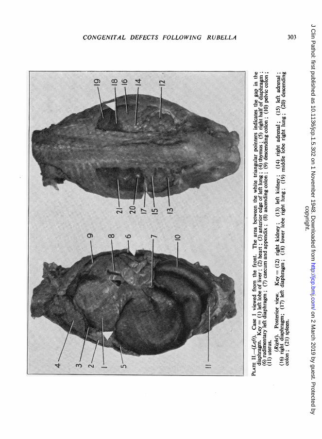
weighing 6 lb. There were no external abnor-malities. The body was radiographed, but no bonyabnormalities found. The eyes were bisected andfound normal. Cranium and brain were also normal.The thorax and abdomen were anatomically grossly
abnormal (Plate II). The small bowel and the leftlobe of the liver (1) were seen to be in the thoraciccavity. The heart (2) and lungs were deviated to theright, and both lungs were much compressed. A thinedge of both lobes of the left lung ran diagonally
across the thorax (3) from the apex of the cavity tothe lower right-hand corner, where it met the dis-placed heart and pericardium. A large thymus gland(4) covered the upper portion of the pericardium andfilled the whole of the superior mediastinum.The right muscular portion of the diaphragm was
well formed (5, 16). The posterior muscular portionof the left half of the diaphragm was present (17),but there was no anterior two-thirds of the left halfof the diaphragm (6). Through the gap thus madehad herniated the left lobe of the liver, the stomachand nearly all the small intestines, the spleen, thepancreas, and in an extraordinary fashion the bulkof the colon.The caecum and appendix were to the left of the
mid-line in the epigastric region (7). The ascend-ing colon (8) traversed the left side of the liver andstomach to reach the top of the left thoracic cavity.It descended (9) antero-superiorly to the spleen (21),which itself lay on the left diaphragm, to come intomore normal lateral relationship with the left kidney(13).The pelvic colon (10) was unnaturally mobile and
ran from the left iliac fossa upwards and then acrossin a wide sweep to the right iliac fossa, where itentered the pelvis minor on the right side. In its sweepit embraced the left ovary and fallopian tube whichlay on its mesentery to the right side of the mid-line.The uterus (11) was rotated through 30 degrees, so asto face to the left.The heart (2) was of normal size and shape,
although it lay in the right thoracic cavity.There was a large patent inter-auricular septum, but
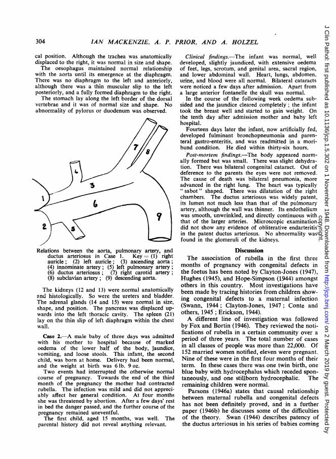
no interventricular communication. The pulmonaryartery was of average size, both in length anddiameter. The ductus arteriosus was more prominentand of wider lumen than the aorta. It was 2 cm. inlength from the origin of the left pulmonary arteryto its junction with the aorta beyond the left sub-clavian artery. It formed a complete right angle withthe descending aorta, and ran almost parallel to theascending aorta in the manner shown (see diagram).
In its descending and abdominal portions the aortaappeared to be normal in length, lumen, and anatomi-
copyright. on 2 M
arch 2019 by guest. Protected by
http://jcp.bmj.com
/J C
lin Pathol: first published as 10.1136/jcp.1.5.302 on 1 N
ovember 1948. D
ownloaded from
CONGENITAL DEFECTS FOLLOWING RUBELLA
-~
0
~u
t o >
0 <_o #_
2i.=r-
00
'00
, em
-E_
3t.: e- ,
c0'0
Ur QC1
4)>£-, sr@ 8
303
copyright. on 2 M
arch 2019 by guest. Protected by
http://jcp.bmj.com
/J C
lin Pathol: first published as 10.1136/jcp.1.5.302 on 1 N
ovember 1948. D
ownloaded from
IAN MACKENZIE, A. P. PRIOR, AND A. HOLZEL
cal position. Although the trachea was anatomicallydisplaced to the right, it was normal in size and shape.The oesophagus maintained normal relationship
with the aorta until its emergence at the diaphragm.There was no diaphragm to the left and anteriorly,although there was a thin muscular slip to the leftposteriorly, and a fully formed diaphragm to the right.The stomach lay along the left border of the dorsal
vertebrae and it was of normal size and shape. Noabnormality of pylorus or duodenum was observed.
Relations between the aorta, pulmonary artery, andductus arteriosus in Case 1. Key== (1) rightauricle; (2) left auricle; (3) ascending aorta;(4) innominate artery; (5) left pulmonary artery;(6) ductus arteriosus; (7) right carotid artery;(8) subclavian artery; (9) descending aorta.
The kidneys (12 and 13) were normal anatomicallyand histologically. So were the ureters and bladder.The adrenal glands (14 and 15) were normal in size,shape, and position. The pancreas was displaced up-wards into the left thoracic cavity. The spleen (21)lay on the thin slip of left diaphragm within the chestwall.
Case 2.-A male baby of three days was admittedwith his mother to hospital because of markedoedema of the lower half of the body, jaundice,vomiting, and loose stools. This infant, the secondchild, was born at home. Delivery had been normal,and the weight at birth was 6 lb. 9 oz.Two events had interrupted the otherwise normal
course of pregnancy. Towards the end of the thirdmonth of the pregnancy the mother had contractedrubella. The infection was mild and did not appreci-ably affect her general condition. At four monthsshe was threatened by abortion. After a few days' restin bed the danger passed, and the further course of thepregnancy remained uneventful.The first child, aged 15 months, was well. The
parental history did not reveal anything relevant.
Clinical findings.-The infant was normal, welldeveloped, slightly jaundiced, with extensive oedemaof feet, legs, scrotum, and genital area, sacral region,and lower abdominal wall. Heart, lungs, abdomen,urine, and blood were all normal. Bilateral cataractswere noticed a few days after admission. Apart froma large anterior fontanelle the skull was normal.
In the course of the following week oedema sub-sided and the jaundice cleared completely; the infanttook the breast well and started to gain weight. Onthe tenth day after admission mother and baby lefthospital.
Fourteen days later the infant, now artificially fed,developed fulminant bronchopneumonia and paren-teral gastro-enteritis, and was readmitted in a mori-bund condition. He died within thirty-six hours.
Post-mortem findings.-The body appeared norm-ally formed but was small. There was slight dehydra-tion. There was bilateral congenital cataract. Out ofdeference to the parents the eyes were not removed.The cause of death was bilateral pneumonia, moreadvanced in the right lung. The heart was typically" sabot" shaped. There was dilatation of the rightchambers. The ductus arteriosus was widely patent,its lumen not much less than that of the pulmonaryartery, although the wall was thinner. Its endotheliumwas smooth, unwrinkled, and directly continuous withthat of the larger arteries. Microscopic examinationdid not show any evidence of obliterative endarteritisin the patent ductus arteriosus. No abnormality wasfound in the glomeruli of the kidneys.
DiscussionThe association of rubella in the first three
months of pregnancy with congenital defects inthe foetus has been noted by Clayton-Jones (1947),Hughes (1945), and Hope-Simpson (1944) amongstothers in this country. Most investigations havebeen made by tracing histories from children show-ing congenital defects to a maternal infection(Swann, 1944; Clayton-Jones, 1947; Conte andothers, 1945 ; Erickson, 1944).A different line of investigation was followed
by Fox and Bortin (1946). They reviewed the noti-fications of rubella in a certain community over aperiod of three years. The total number of casesin all classes of people was more than 22,000. Of152 married women notified, eleven were pregnant.Nine of these were in the first four months of theirterm. In these cases there was one twin birth, oneblue baby with hydrocephalus which receded spon-taneously, and one stillborn hydrocephalic. Theremaining children were normal.
Parsons (1946a) states that causal relationshipbetween maternal rubella and congenital defectshas not been definitely proved, and in a furtherpaper (1946b) he discusses some of the difficultiesof the theory. Swan (1944) describes patency ofthe ductus arteriosus in his series of babies coming
304
copyright. on 2 M
arch 2019 by guest. Protected by
http://jcp.bmj.com
/J C
lin Pathol: first published as 10.1136/jcp.1.5.302 on 1 N
ovember 1948. D
ownloaded from

CONGENITAL DEFECTS FOLLOWING RUBELLA
to necropsy. Gregg is quoted (Parsons, 1946b) asfinding forty-four cases of congenital heart diseaseout of seventy-eight affected infants.
Other abnormalities described have been mentalretardation, microcephaly, mongolism, hypo-spadias, talipes equino-varus, and synostosis ofradius and ulna or tibia and fibula.
Congenital diaphragmatic hernia has not hithertobeen described in the literature as one of thedevelopmental defects occurring in associationwith maternal rubella.The diaphragmatic hernia in Case 1 differs from
the common form, in which all diaphragmatic seg-ments are present but a patency exists between thepleural and peritoneal cavities. This pleuroperi-toneal hiatus, or foramen of Bachdalek, may varyin size, but it is surrounded by diaphragmaticelements. Abassy has quoted Dickson as sayingthat diaphragmatic hernia is seen in one case outof each hundred routine gastric radiographs.
In this case there was a total absence of theanterior two-thirds of the left half of the dia-phragm. There was, however, a small muscu-lar slip to represent the posterior third of thediaphragm.
Developmentally the diaphragm arises in fourmain parts: a ventral, a dorsal, and a right anda left lateral. The ventral part is formed from a
septum transversum which is gradually differen-tiated into a caudal, an intermediate, and a cephalicpart. It is from the intermediate portion that theventral part of the diaphragm is formed. Thedorsal portion of the diaphragm arises from themesoderm of the dorsal mesentery of the foregut.The two lateral portions grow towards the medianplane until they fuse with the dorsal portion.The present appearance of the diaphragm would
suggest that there had been a failure of growth ofthe left lateral portion and of the left half of theventral portion. It resulted in' a very rudimentaryleft half of the diaphragm, and this gross dia-phragmatic hernia.The time of formation of the diaphragm from
its various elements is from the eighth weekonwards, after the pericardial cavity has alreadybeen shaped. The inter-atrial and interventricularsepta begin to develop between the fourth and sixthweeks. The former is the result of the coalescenceof the primary and secondary septa. Their fusionis partial and leaves the foramen ovale. The valveof the foramen is formed in the fourth month.The interventricular septum arises in the lowerpart of the primitive ventricle, and grows upwardsto meet the endocardial cushions and the septumdeveloping in the bulbus arteriosus. The lower
z
part is complete about the eighth week, and theseptum of the bulbus arteriosus a few days earlier.
In Case 1 the mother was stated to have sufferedfrom rubella when ten weeks pregnant. The timeof formation of the diaphragm is about the eighthweek. The possibility that the maternal diseaseand the time of formation of the affected partcoincided is not to be lightly dismissed.
In Case 2 the mother suffered from rubella atabout the third month. The ductus arteriosusforms about the sixth week; the valve of the intra-atrial septum about the sixteenth week; the inter-atrial septum about the eighth week. Thus theinfection would have had ample opportunity tointerfere with the development of the cardiac septa.v Parsons (1946b) has canvassed the possibility ofstrain differences, as well as emphasizing the diffi-culties of diagnosis. This is a point which mostauthors in this country stress. Some support forthe theory is given by Conte and others (1945),who found that the incidence of rubella in theappropriate months of pregnancy in mothers ofchildren with congenital abnormalities was tentimes the expected rate for women of the child-bearing age of the population at large.Swan and others (1946) do not deal only with
rubella as an antecedent of congenital defect.They list cases in which influenza, scarlet fever,herpes zoster, varicella, and mumps, together withone of "pustular rash," were antecedents.
SummaryPost-mortem records of two infants with con-
genital malformations following maternal rubellaare described. Case 1 presented an unusual formof congenital diaphragmatic hernia and a widelypatent ductus arteriosus. Case 2 had bilateral con-genital cataract and a wide patency of the ductusarteriosus.
We wish to thank Mr. G. G. Alderson, under whosecare Case 1 was born and who has encouraged publi-cation of the case; Dr. J. Wearing for taking x-rayphotographs; and Mr. T. L. Skuse and Mr. G. Wrightfor technical assistance.
REFERENCESAbassy, A. S. A. (1945). Arch. Pediat., 62, 285.Clayton-Jones, E. (1947). Lancet, 1, 56.Conte, W. R., McCammont C. S., and Christie, A. (1945). Amer. J.
Dis. Child., 70, 301.Erickson, C. A. (1944). J. Pediat., 25, 281.Fox, M. J., and Bortin, M. M. (1946). J. Amer. med. Ass., 130, 568.Gregg, H. McA. (1941). Trans. Ophthalmological Soc. Aust. Brit.
Med. Ass., 111, 35.Hope-Simpson, R. E. (1944). Lancet, 1, 483.Hughes, I. (1945). Proc. R. Soc. Med., 39, 17.
5Parsons, L. G. (1946a). J. Obstet. Gynaec. Brit. Emp., 53, 1.Parsons, L. G. (1946b). Brit. med. Bull., 4, 193.Swan, C. (1944). J. Path. Bact., 56, 289.Swan, C., Tostevin, A. L., and Barham Black, G. H. (1946). Med. J.
Aust., 11, 889.
305
copyright. on 2 M
arch 2019 by guest. Protected by
http://jcp.bmj.com
/J C
lin Pathol: first published as 10.1136/jcp.1.5.302 on 1 N
ovember 1948. D
ownloaded from