Embed Size (px)
Citation preview

Confocal Laser Endomicroscopy
Policy Number: 2.01.87 Last Review: 9/2018
Origination: 3/2013 Next Review: 3/2019
Policy Blue Cross and Blue Shield of Kansas City (Blue KC) will not provide coverage for
confocal laser endomicroscopy. This is considered investigational.
When Policy Topic is covered Not Applicable
When Policy Topic is not covered Use of confocal laser endomicroscopy is considered investigational.
Description of Procedure or Service Populations Interventions Comparators Outcomes
Individuals:
With suspected or known colorectal lesions
Interventions of interest are:
Confocal laser endomicroscopy as an adjunct to colonoscopy
Comparators of interest are:
White-light colonoscopy alone
Colonoscopy used with alternative adjunctive diagnostic aids
Relevant outcomes include:
Overall survival
Disease-specific survival
Test accuracy
Test validity
Resource utilization
Individuals:
With Barrett esophagus who are undergoing surveillance
Interventions of interest are:
Confocal laser endomicroscopy with targeted biopsy
Comparators of interest are:
Standard endoscopy with random biopsy
Relevant outcomes include:
Overall survival
Disease-specific survival
Test accuracy
Test validity
Resource utilization
Individuals:
With gastrointestinal lesions and have had endoscopic treatment
Interventions of interest are:
Confocal laser endomicroscopy to assess adequacy of endoscopic treatment
Comparators of interest are:
Standard endoscopy (ie, white-light endoscopy)
Relevant outcomes include:
Overall survival
Disease-specific survival
Test accuracy
Test validity
Resource utilization
Individuals:
With suspicion of a condition diagnosed by identification and biopsy of lesions (eg, lung, bladder, or gastric cancer)
Interventions of interest are:
Confocal laser endomicroscopy
Comparators of interest are:
Standard diagnostic procedures
Relevant outcomes include:
Overall survival
Disease-specific survival
Test accuracy
Test validity
Resource utilization
Confocal laser endomicroscopy (CLE), also known as confocal fluorescent
endomicroscopy and optical endomicroscopy, allows in vivo microscopic imaging of cells during endoscopy. CLE is proposed for a variety of purposes, especially as a
Confocal Laser Endomicroscopy 2.01.87
real-time alternative to histology during colonoscopy and for targeting areas to
undergo biopsy in patients with inflammatory bowel disease and Barrett’s
esophagus.
For individuals who have suspected or known colorectal lesions who receive CLE as
an adjunct to colonoscopy, the evidence includes multiple diagnostic accuracy
studies. Relevant outcomes are overall survival, disease-specific survival, test accuracy and validity, and resource utilization. While the reported sensitivity and
specificity in these studies are high, it is uncertain whether the accuracy is
sufficiently high to replace biopsy/polypectomy and histopathologic analysis.
Moreover, issues remain about the use of this technology in practice (eg, the learning curve, interpretation of lesions). The evidence is insufficient to determine
the effects of the technology on health outcomes.
For individuals who have Barrett esophagus who are undergoing surveillance who receive CLE with targeted biopsy, the evidence includes several randomized
controlled trials (RCTs) and a meta-analysis. Relevant outcomes are overall
survival, disease-specific survival, test accuracy and validity, and resource
utilization. Evidence from RCTs has suggested CLE is more sensitive than standard
endoscopy for identifying areas of dysplasia. However, a 2014 meta-analysis found that the pooled sensitivity, specificity, and negative predictive value of available
studies were not sufficiently high to replace the standard surveillance protocol.
National guidelines continue to recommend 4-quadrant random biopsies for
patients with Barrett esophagus undergoing surveillance. The evidence is insufficient to determine the effects of the technology on health outcomes.
For individuals who have gastrointestinal lesions and have had endoscopic
treatment who receive CLE, the evidence includes 1 RCT and a systematic review. Relevant outcomes are overall survival, disease-specific survival, test accuracy and
validity, and resource utilization. The single RCT, which compared high definition
(HD) white-light endoscopy with HD white-light endoscopy plus CLE, was stopped
early because an interim analysis did not find a between-group difference in
outcomes. The evidence is insufficient to determine the effects of the technology on health outcomes.
For individuals who have a suspicion of a condition diagnosed by identification and
biopsy of lesions (eg, lung, bladder, or gastric cancer) who receive CLE, the evidence includes a small number of diagnostic accuracy studies. Relevant
outcomes are overall survival, disease-specific survival, test accuracy and validity,
and resource utilization. There is limited evidence on the diagnostic accuracy of
these other indications. The evidence is insufficient to determine the effects of the technology on health outcomes.
Background
CLE, also known as confocal fluorescent endomicroscopy and optical endomicroscopy, allows in vivo microscopic imaging of the mucosal epithelium
during endoscopy. The process involves using light from a low-power laser to
illuminate tissue and, subsequently, the same lens detects light reflected from the
Confocal Laser Endomicroscopy 2.01.87

tissue through a pinhole. The term confocal refers to having both illumination and
collection systems in the same focal plane. Light reflected and scattered at other
geometric angles that is not reflected through the pinhole is excluded from detection, which dramatically increases the special resolution of CLE images.
To date, 2 types of CLE systems have been cleared by the U.S. Food and Drug
Administration (FDA). One is an endoscope-based system in which a confocal probe is incorporated onto the tip of a conventional endoscope. The other is a
probe-based system; the probe is placed through the biopsy channel of a
conventional endoscope. The depth of view is up to 250 um with the endoscopic
system and about 120 um with the probe-based system. A limited area can be examined; no more than 700 um in the endoscopic-based system and less with
the probe-based system. As pointed out in review articles, the limited viewing area
emphasizes the need for careful conventional endoscopy to target the areas for
evaluation. Both CLE systems are optimized using a contrast agent. The most widely used agent is intravenous fluorescein, which is FDA-approved for
ophthalmologic imaging of blood vessels when used with a laser scanning
opthalmoscope.
Unlike techniques such as chromoendoscopy, which are primarily intended to improve the sensitivity of colonoscopy, CLE is unique in that it is designed to
immediately characterize the cellular structure of lesions. CLE can thus potentially
be used to make a diagnosis of polyp histology, particularly in association with
screening or surveillance colonoscopy, which could allow for small hyperplastic lesions to be left in place rather than removed and sent for histological evaluation.
This would reduce risks associated with biopsy and reduce the number of biopsies
and histological evaluations.
Another key potential application of CLE technology is targeting areas for biopsy in
patients with BE undergoing surveillance endoscopy. This is an alternative to the
current standard approach recommended by the American Gastroenterological
Association (AGA) which is that patients with BE who do not have dysplasia
undergo endoscopic surveillance every 3 to 5 years.(1) AGA further recommends that random 4-quadrant biopsies every 2 cm be taken with white-light endoscopy
in patients without known dysplasia.
Other potential uses of CLE under investigation include better diagnosis and differentiation of conditions such as gastric metaplasia, lung cancer, and bladder
cancer.
As noted previously, limitations of CLE systems include a limited viewing area and depth of view. Another issue is standardization of systems for classifying lesions
viewed with CLE devices. Although there is not currently an internationally
accepted classification system for colorectal lesions, 2 systems have been
developed that have been used in a number of studies conducted in different countries. These are the Mainz criteria for endoscopy-based CLE devices and the
Miami classification system for probe-based CLE devices. (2) Lesion classification
systems are less developed for non-gastrointestinal lesions viewed by CLE devices
Confocal Laser Endomicroscopy 2.01.87
e.g., those in the lung or bladder. Another potential issue is the learning curve for
obtaining high-quality images and classifying lesions. Several recent studies,
however, have found that the ability to acquire high-quality images and interpret them accurately can be learned relatively quickly; these studies were limited to
colorectal applications of CLE. (3, 4)
Regulatory Status Two confocal laser endomicroscopy devices have been cleared for marketing by
the FDA. These include:
Cellvizio® (Mauna Kea Technologies, Paris, France) is a confocal microscopy with a fiber optic probe (ie, a probe-based CLE system). The device consists of a laser
scanning unit, proprietary software, a flat-panel display, and miniaturized fiber
optic probes. The F-600 system, cleared by FDA in 2006, can be used with any
standard endoscope with a working channel of at least 2.8 mm. According to FDA, the device is intended for confocal laser imaging the internal microstructure of
tissues in the anatomic tract (gastrointestinal or respiratory) that are accessed by
an endoscope. The 100 series version of the system was cleared by FDA in 2015
for imaging the internal microstructure of tissues and for visualization of body
cavities organs and canals during endoscopic and laparoscopic surgery. FDA product code: GCJ.
Confocal Video Colonoscope (Pentax Medical, Montvale, NJ) is an endoscopy-based
CLE system. The EC-3S7OCILK system, cleared by FDA in 2004, is used with a Pentax Video Processor and with a Pentax Confocal Laser System. According to
FDA, the device is intended to provide optical and microscopic visualization of and
therapeutic access to the lower gastrointestinal tract. FDA product code: GCJ/KOG
(endoscope and accessories).
Rationale This evidence review was created in January 2013 and has been updated regularly
with searches of the MEDLINE database. The most recently literature update was
performed through September 11, 2017.
Assessment of a diagnostic technology typically focuses on 3 categories of
evidence: (1) technical reliability (test-retest reliability or interrater reliability); (2)
clinical validity (sensitivity, specificity, and positive and negative predictive value) in relevant populations of patients; and (3) clinical utility (ie, demonstration that
the diagnostic information can be used to improve patient outcomes). The
following is a summary of the key literature to date.
Confocal Laser Endomicroscopy
Clinical Context and Test Purpose
The purposes of confocal laser endomicroscopy (CLE) scanning in patients with
suspected or known colorectal lesions; Barrett esophagus (BE) who are undergoing surveillance; gastrointestinal lesions following endoscopic treatment;
Confocal Laser Endomicroscopy 2.01.87

or suspected other conditions diagnosed by identification and biopsy of lesions (eg,
lung, bladder, head and neck, esophageal, or gastric cancers) are to provide a
real-time alternative to histology and assist in targeting areas for biopsy.
The question addressed in this evidence review is: Does the use of CLE improve
the net health outcome in individuals with suspected or known colorectal lesions,
BE who are undergoing surveillance, gastrointestinal lesions who have had endoscopic treatment, or suspected other conditions (eg, lung, bladder, head and
neck, esophageal, or gastric cancers)?
The following PICOTS were used to select literature to inform this review.
Patients
The populations of interest include patients with suspected or known colorectal
lesions; BE undergoing surveillance; gastrointestinal lesions who have had endoscopic treatment; and suspected other conditions diagnosed by identification
and biopsy of lesions.
Interventions
The intervention of interest is CLE.
Comparators
The comparators of interest for each indication include:
For suspected colorectal lesions: white-light colonoscopy alone or alternative
adjunctive diagnostic aids
For BE undergoing surveillance: standard endoscopy with random biopsy
For gastrointestinal lesions following endoscopic treatment: standard endoscopy (white-light endoscopy)
For other condition diagnosed by identification and biopsy of lesions: standard
diagnostic procedures
Outcomes For patients with suspected colorectal lesions, the outcome of interest is confirmed
diagnosis. With a confirmed diagnosis, appropriate treatment options can be
pursued.
For patients with BE undergoing surveillance, the outcome of interest is a
reduction in the number of biopsies without compromising diagnostic accuracy.
For patients with gastrointestinal lesions following endoscopic treatment, the outcome of interest is determining whether residual disease is present following
the endoscopic treatment.
For patients with other conditions diagnosed by identification and biopsy of lesions, the outcome of interest is a diagnosis confirmation to inform clinical management
decisions.
Confocal Laser Endomicroscopy 2.01.87

Timing
For patients with suspected colorectal lesions, BE, and other conditions, the timing
of CLE would be during the disease confirmation process. For patients with gastrointestinal lesions following endoscopic treatment, the timing would be
following the endoscopic treatment.
Setting The setting is a facility equipped with CLE.
Colorectal Lesions
Clinical Validity
Systematic Reviews
Several systematic reviews of studies have compared the diagnostic accuracy of CLE with a reference standard. In 2013, Su et al reviewed studies on the efficacy
of CLE for discriminating colorectal neoplasms from non-neoplasms.5 To be
included in the review, studies had to use histologic biopsy as the reference
standard, and the pathologist and endoscopist had to be blinded to each other’s
findings. Selected studies also used a standardized CLE classification system. Patients had to be at increased risk of colorectal cancer (CRC) due to personal or
family history, have previously identified polyps, and/or have inflammatory bowel
disease (IBD). Two reviewers independently assessed the quality of individual
studies using the modified Quality Assessment of Diagnostic Accuracy Studies tool, and studies considered at high risk of bias were excluded from further
consideration.
Fifteen studies (total N=719 adults) were eligible for the systematic review. All were single-center trials, and two were available only as abstracts. In all studies,
suspicious lesions were first identified by conventional white-light endoscopy with
or without chromoendoscopy and then further examined by CLE. Meta-analysis of
the 15 studies found an overall sensitivity for CLE of 94% (95% confidence
interval [CI], 88% to 97%) and a specificity of 95% (95% CI, 89% to 97%) compared with histology. Six studies included patients at increased risk of CRC
who were undergoing surveillance endoscopy; 5 studies included patients with
colorectal polyps and 4 studies included patients with IBD. In a predefined
subgroup analysis by indication for screening, the pooled sensitivity and specificity for surveillance studies were 94% (95% CI, 90% to 97%) and 98% (95% CI, 97%
to 99%), respectively. For patients presenting with colorectal polyps, the pooled
sensitivity of CLE was 91% (95% CI, 87% to 94%) and the specificity was 85%
(95% CI, 78% to 90%). For patients with IBD, the pooled sensitivity was 83% (95% CI, 70% to 92%) and the specificity was 90% (95% CI, 87% to 93%). In
other predefined subgroup analyses, the summary sensitivity and specificity were
significantly higher (p<0.001) in studies of endoscopy-based CLE (97% and 99%,
respectively) than in studies of probe-based CLE (87% and 82%, respectively). In addition, the summary sensitivity and specificity were significantly higher (p<0.01)
with real-time CLE in which the macroscopic endoscopy findings were known (96%
and 97%, respectively) than in blinded CLE in which recorded confocal images
Confocal Laser Endomicroscopy 2.01.87

were subsequently analyzed without knowledge of macroscopic endoscopy findings
(85% and 82%, respectively).
Another 2013 systematic review by Dong et al included studies that compared the
diagnostic accuracy of CLE with conventional endoscopy.6 Reviewers did not
explicitly state that the reference standard was histologic biopsy, but this was the
implied reference standard. Six studies were included in a meta-analysis. All were prospective, and at least five included blinded interpretation of CLE findings (in 1
study, it was unclear whether interpretation was blinded). In a pooled analysis of
data from all 6 studies, the sensitivity was 81% (95% CI, 77% to 85%) and the
specificity was 88% (95% CI, 85% to 90%). Reviewers also conducted a subgroup analysis by type of CLE used. When findings from the 2 studies on endoscopy-
based CLE were pooled, the sensitivity was 82% (95% CI, 69% to 91%) and the
specificity was 94% (95% CI, 91% to 96%). Two studies may not have been
sufficient to obtain a reliable estimate of diagnostic accuracy. When findings from the 4 studies on probe-based endoscopy were pooled, the sensitivity was 81%
(95% CI, 76% to 85%) and the specificity was 75% (95% CI, 69% to 81%).
A 2013 meta-analysis by Wanders et al searched for studies that reported the
diagnostic accuracy of several new technologies used to differentiate between colorectal neoplasms and non-neoplasms.7 To be selected, studies had to use the
technology to differentiate between non-neoplastic and neoplastic lesions and to
use histopathology as the reference standard. Blinding was not an inclusion
criterion. Eleven eligible studies identified included an analysis of CLE. Meta-analysis yielded an estimated sensitivity of 93.3% (95% CI, 88.4% to 96.2%) and
a specificity of 89.9% (95% CI, 81.8% to 94.6%). Meta-analysis limited to the 5
studies that used endoscopy-based CLE found a sensitivity of 94.8% (95% CI,
90.6% to 98.92%) and a specificity of 94.4% (95% CI, 90.7% to 99.2%). When findings of the 6 probe-based CLE studies were pooled, sensitivity was 91.5%
(95% CI, 86.0% to 97.0%) and specificity was 80.9 (95% CI, 69.4% to 92.4%).
Cohort Studies
A 2011 study by Xie et al in China included 116 consecutive patients who had polyps found during CLE (1 patient was excluded from the analysis).8 All patients
had an indication for colonoscopy (19 were undergoing surveillance after
polypectomy, 2 had a family history of CRC, 3 had IBD, 91 were seeking a
diagnosis). All patients first underwent white-light colonoscopy. Endoscopy-based CLE was used on the first polyp identified during withdrawal of the endoscope (ie,
1 polyp per patient was analyzed). Intravenous fluorescein sodium was used.
Real-time diagnosis of the polyp was performed based on criteria used at the
study center (adapted from the Mainz classification system). The polyps were biopsied or removed, and a histopathologic diagnosis was determined. Real-time
CLE diagnosis correctly identified 109 (95%) of 115 adenomas or hyperplastic
polyps. Four adenomas were misdiagnosed by CLE as hyperplastic polyps (two
were tubulous adenomas, two were tubulovillous adenomas) and 2 hyperplastic polyps were misdiagnosed as adenomas. The overall sensitivity, specificity,
positive predictive value (PPV), and negative predictive value (NPV) of CLE
diagnosis were 93.9% (95% CI, 85.4% to 97.6%), 95.9% (95% CI, 86.2% to
Confocal Laser Endomicroscopy 2.01.87

98.9%), 96.9% (95% CI, 89% to 99%), and 94.8% (95% CI, 89.1% to 97.6%),
respectively. For polyps less than 10 mm in size, CLE diagnosis had a sensitivity of
90.3% and a specificity of 95.7%; for polyps 10 mm or larger, sensitivity was 97.1% and specificity was 100%.
In 2010, Buchner et al published findings on 75 patients who had a total of 119
polyps.9 Patients were eligible for participation if they were undergoing surveillance or screening colonoscopy or undergoing evaluation of known or
suspected polyps identified by other imaging modalities or endoscopic resection of
larger flat colorectal neoplasia. White-light colonoscopy was used as the primary
screening method. When a suspicious lesion was identified, it was evaluated by virtual chromoendoscopy and a probe-based CLE system. Intravenous fluorescein
sodium was administered after the first polyp was identified. After the imaging
techniques, the appropriate intervention (ie, polypectomy, biopsy, endoscopic
mucosal resection) was performed, and all resected specimens underwent histopathologic analysis by a pathologist blinded to CLE information. Confocal
images of the 199 polyps were evaluated after all procedures were completed; the
evaluator was blinded to the histology diagnosis and the endoscopic appearance of
the lesion. Diagnosis of confocal images used modified Mainz criteria; polyps were
classified as benign or neoplastic. According to histopathologic analysis, there were 38 hyperplastic polyps and 81 neoplastic lesions. CLE correctly identified 74 of 81
neoplastic polyps (sensitivity, 91%; 95% CI, 83% to 96%). In addition, CLE
correctly identified 29 of 38 hyperplastic polyps (specificity, 76%; 95% CI, 60% to
89%). In contrast, virtual chromoendoscopy correctly identified 62 neoplastic polyps (sensitivity, 77%; 95% CI, 66% to 85%) and 27 hyperplastic polyps
(specificity, 71%; 95% CI, 54% to 85%).
Another study from the same academic medical center as Buchner was published in 2012 by Shadid et al.10 The study compared 2 methods of analyzing CLE
images: real-time diagnosis and blinded review of video images after endoscopy
(known as “offline” diagnosis). The study included 74 patients with 154 colorectal
lesions. Eligibility criteria were similar to the Buchner study (previously
discussed)selected patients were undergoing surveillance or screening
colonoscopy. Patients had white-light colonoscopy, and identified polyps were also
evaluated with virtual chromoendoscopy and probe-based CLE. Intravenous fluorescein sodium was administered after the first polyp was identified. At
examination, an endoscopist made a real-time diagnosis based on CLE images.
Based on that diagnosis, the patient underwent polypectomy, biopsy, or
endoscopic mucosal resection, and histopathologic analysis was done on the specimens. CLE images were deidentified and reviewed offline by the same
endoscopist at least 1 month later. At the second review, the endoscopist was
blinded to the endoscopic and histopathologic diagnosis. Of the 154 polyps, 74
were found by histopathologic analysis to be non-neoplastic, and 80 were neoplastic (63 tubular adenomas, 12 tubulovillous adenomas, 3 mixed
hyperplastic-adenoma polyps, 2 adenocarcinomas). Overall, there was no
statistically significant difference in the diagnostic accuracy between real-time CLE
diagnosis and blinded offline CLE diagnosis (ie, confidence intervals overlapped).
The sensitivity, specificity, PPV, and NPV for real-time CLE diagnosis were 81%,
Confocal Laser Endomicroscopy 2.01.87
76%, 87%, and 79%, respectively. For offline diagnosis, these values were 88%,
77%, 81%, and 85%, respectively. For larger polyps, there was a nonsignificant
trend in favor of better diagnostic accuracy with real-time compared with offline CLE. However, in the subgroup of 107 smaller polyps (<10 mm in size), the
accuracy of real-time CLE was significantly less than offline CLE. For smaller
polyps, sensitivity, specificity, PPV and NPV of real-time CLE were 71%, 83%,
78%, and 78%, respectively; for offline CLE, they were 86%, 78%, 76%, and 87%, respectively.
A 2011 study by Hlavaty et al included patients with ulcerative colitis or Crohn
disease.11 Thirty patients were examined with standard white-light colonoscopy, chromoendoscopy, and an endoscopy-based CLE system. Another 15 patients
were examined only with standard colonoscopy. All lesions identified by white-light
colonoscopy or chromoendoscopy were examined using CLE to identify neoplasia
using the Mainz classification system. Suspicious lesions were biopsied, and random biopsies were taken from 4 quadrants every 10 cm per the standard
surveillance colonoscopy protocol. All specimens underwent histologic analysis by
a gastrointestinal pathologist blinded to CLE diagnosis. Diagnostic accuracy of CLE
was calculated for examinable lesions only. Compared with histologic diagnosis,
the sensitivity of CLE for diagnosing low-grade and high-grade intraepithelial neoplasia was 100%, specificity was 98.4%, PPV was 66.7%, and NPV was 100%.
However, whereas CLE was able to examine 28 (93%) of 30 flat lesions, it could
examine only 40 (57%) of 70 protruding polyps. Moreover, 6 (60%) of 10
dysplastic lesions, including 3 of 5 low-grade and high-grade intraepithelial neoplasms were not evaluable by CLE. It is also worth noting that the diagnostic
accuracy of chromoendoscopy (see evidence review 2.01.84) was similar to that of
CLE. The sensitivity, specificity, PPV, and NPV of chromoendoscopy were 100%,
97.9%, 75%, and 100%, respectively.
Clinical Utility
In patients at average risk of CRC, no randomized controlled trials (RCTs) or
nonrandomized comparative studies were identified that evaluated the impact of
CLE on subsequent development of CRC or on CRC mortality. In addition, it is not clear that the diagnostic performance of this technology is sufficient to obviate the
need for biopsy of identified polyp lesions. Thus, there is insufficient evidence to
support a chain of evidence to demonstrate an improvement in net health
outcome.
Section Summary: Colorectal Lesions
Multiple studies have compared the accuracy of CLE with the histopathology for
diagnosing colorectal lesions. In 3 published systematic reviews, pooled estimates of overall sensitivity of CLE ranged from 81% to 94%, and pooled estimates of the
specificity ranged from 88% to 95%. Although the reported diagnostic accuracy
tended to be relatively high, it is unclear whether the accuracy is high enough to
replace biopsy/polypectomy and histologic analysis. Moreover, there are no controlled studies on the impact of using CLE on CRC incidence or mortality, and
the available evidence is insufficient to support a chain of evidence.
Confocal Laser Endomicroscopy 2.01.87

Barrett Esophagus
This section addresses whether CLE can distinguish BE without dysplasia from BE
with low- (LGD) and high-grade dysplasia (HGD) and/or lead to fewer biopsies of benign tissue compared with surveillance with random biopsies. The ideal study to
answer this question would include an unselected clinical population of patients
with BE presenting for surveillance and would randomized patients to CLE with
targeted biopsy or a standard biopsy protocol without CLE. Relevant outcomes include the diagnostic accuracy for detecting dysplasia, the detection rate for
dysplasia, and the number of biopsies.
Systematic Reviews In 2014, Gupta et al published a systematic review and meta-analysis of
prospective studies comparing the accuracy of CLE with targeted biopsy to
standard 4-quadrant biopsy in patients with BE.12 Reviewers noted that, according
to the Preservation and Incorporation of Valuable Endoscopic Innovation Initiative of the American Society of Gastrointestinal Endoscopy, in order to replace the
standard Seattle protocol, an alternative approach would need to have a per-
patient sensitivity of at least 90%, specificity of at least 80%, and NPV of at least
98% for detecting HGD or esophageal adenocarcinoma compared with the current
protocol.
Eight studies published through May 2014 met inclusion criteria; one was a
parallel-group RCT, and one was a randomized crossover study. The other six
were single- or double-blind nonrandomized comparative studies. Seven studies had data suitable for pooling on a per-lesion basis; together they included 345
patients and 3080 lesions. In a meta-analysis of the diagnosis of HGD or
esophageal adenocarcinom, pooled sensitivity was 68% (95% CI, 64% to 73%)
and pooled specificity was 88% (95% CI, 87% to 89%). Four studies were included in the per-patient meta-analysis. The pooled sensitivity and specificity
were 86% (95% CI, 74% to 96%) and 83% (95% CI, 77% to 88%), respectively.
NPV (calculated using the sensitivity, specificity, and overall prevalence) was 96%.
Thus, according to the criteria in the Preservation and Incorporation of Valuable
Endoscopic Innovation Initiative, the diagnostic accuracy of CLE in the studies published to date is not sufficiently high for this technique to replace the standard
Seattle protocol. HGD and esophageal adenocarcinoma were much higher in the
studies included in the meta-analysis than is generally seen in clinical practice and
therefore diagnostic accuracy results should be interpreted cautiously.
In 2016, Xiong et al published a meta-analysis of prospective studies evaluating
the diagnostic accuracy of CLE in patients with BE, using histopathologic analysis
as the criterion standard.13 Studies were not required to compare CLE with standard 4-quadrant biopsy. Fourteen studies were included. In a pooled analysis
7 studies (n=473 patients) reporting a per-patient analysis, the sensitivity of CLE
for detecting neoplasia was 89% (95% CI, 82% to 94%) and the specificity was
83% (95% CI, 78% to 86%). The pooled positive and negative likelihood ratios were 6.53 (95% CI, 3.12 to 13.4) and 0.17 (95% CI, 0.11 to 0.29), respectively.
Reviewers did not report PPV or NPV. Moreover, they provided estimates of pretest
probability to aid in the interpretation of the likelihood ratios (ie, to evaluate a
Confocal Laser Endomicroscopy 2.01.87

person’s risk level before and after getting the test). Sensitivity and specificity
were similar to those calculated in the Gupta systematic review (discussed earlier).
Randomized Controlled Trials
In 2011, Sharma et al published an international, multicenter RCT that included
122 consecutive patients presenting for surveillance of BE or endoscopic treatment
of HGD or early carcinoma.14 Patients were randomized to both standard white-light endoscopy and narrow-band imaging. Following these 2 examinations, done
in a blinded fashion, the location of lesions was unblinded and, subsequently, all
patients underwent probe-based CLE. All examinations involved a presumptive
diagnosis of suspicious lesions. Also, in both groups, after all evaluations were performed, all suspicious lesions were biopsied, as well as random locations (4
quadrants every 2 cm). The histopathologic analysis was the reference standard.
Twenty-one patients were excluded from the analysis. Of the remaining 101
patients, 66 (65%) were found on histopathologic analysis to have no dysplasia, 4 (4%) had LGD, 6 (6%) had HGD, and 25 (25%) had early carcinoma. Sensitivity
of CLE plus white-light endoscopy for detecting HGD or early carcinoma was
68.3% (95% CI, 60.0% to 76.7%), which was significantly higher than white-light
endoscopy alone (34.2%; 95% CI, 25.7% to 42.7%; p=0.002). However,
specificity of CLE plus white-light endoscopy was significantly lower (87.8%; 95% CI, 85.5% to 90.1%) than white-light endoscopy alone (92.7%; 95% CI, 90.8% to
94.6%; p<0.001). For white-light endoscopy alone, PPV was 42.7% (95% CI,
32.8% to 52.6%), and NPV was 89.8% (95% CI, 87.7% to 92.0%). For white-
light endoscopy with probe-based CLE, PPV was 47.1% (95% CI, 39.7% to 54.5%), and NPV was 94.6% (95% CI, 92.9% to 96.2%). White-light endoscopy
alone missed 79 (66%) of 120 areas with HGD or early carcinoma, and white-light
endoscopy plus CLE missed 38 (32%) areas. On a per-patient basis, 31 patients
were diagnosed with HGD or early carcinoma. White-light endoscopy alone failed to identify 4 of these patients (sensitivity, 87%), whereas white-light endoscopy
plus CLE failed to identify 2 patients (sensitivity, 93.5%).
A single-center crossover RCT was published in 2009 by Dunbar et al.15 Forty-six
patients with BE were enrolled, and 39 (95%) completed the study protocol. Of these, 23 were undergoing BE surveillance, and 16 had BE with suspected
neoplasia. All patients received endoscopy-based CLE and standard endoscopy, in
random order. One endoscopist performed all CLE procedures, and another
endoscopist performed all standard endoscopy procedures; endoscopists were blinded to the finding of the other procedure. During the standard endoscopy
procedure, biopsies were taken of any discrete lesions followed by 4-quadrant
random biopsy (every 1 cm for suspected neoplasia, every 2 cm for BE
surveillance). During the CLE procedure, only lesions suspicious of neoplasia were biopsied. Endoscopists interpreted CLE images using the Confocal Barrett’s
Classification system, developed in a previous research study. Histopathologic
analysis was the reference standard. Among the 16 study completers with
suspected high-risk dysplasia, there were significantly fewer biopsies per patient with CLE (mean, 9.8 biopsies per patient) than with standard endoscopy (mean,
23.9 biopsies per patient; p=0.002). Although there were fewer biopsies, the
mean number of biopsy specimens showing HGD or cancer was similar in the 2
Confocal Laser Endomicroscopy 2.01.87

groups (3.1 during CLE vs 3.7 during standard endoscopy). The diagnostic yield
for neoplasia was 33.7% with CLE and 17.2% with standard endoscopy. None of
the 23 patients undergoing BE for surveillance had HGD or cancer. The mean number of mucosal specimens obtained for patients in this group was 12.6 with
white-light endoscopy and 1.7 with CLE (p<0.001).
Another RCT was published in 2014 by Canto et al.16 This single-blind, multicenter trial was conducted at academic centers with experienced endoscopists. It included
consecutive patients undergoing endoscopy for routine BE surveillance or for
suspected or known neoplasia. Patients were randomized to high-definition white-
light endoscopy with random biopsy (n=98) or white-light endoscopy with endoscopy-based CLE and targeted biopsy (n=94). In the white-light endoscopy-
only group, 4-quadrant random biopsies were taken every 1 to 2 cm over the
entire length of the BE for patients undergoing surveillance and every 1 cm for
patients with suspected neoplasia. In the CLE group, biopsy specimens were obtained only when there was CLE evidence of neoplasia. Final pathologic
diagnosis was the reference standard. A per-patient analysis of diagnostic
accuracy for diagnosing BE-related neoplasia found a sensitivity of 40% with
white-light endoscopy only and 95% with white-light endoscopy plus CLE.
Specificity was 98% with white-light endoscopy only and 92% with white-light endoscopy plus CLE. When the analysis was done on a per-biopsy specimen basis
and when CLE was added, sensitivity was substantially higher, and specificity was
slightly lower. The median number of biopsies per patient was significantly higher
in the white-light endoscopy group (n=4) compared with the CLE group (n=2; p<0.001).
The investigators analyzed the number of cases in which CLE resulted in a
different diagnosis. Thirty-two (34%) of 94 patients in the white-light plus CLE group had a correct change in dysplasia grade after CLE compared with initial
endoscopic findings. Six (19%) of the 32 patients had lesions, and the remaining
26 did not. In 21 of the 26 patients without lesions, CLE changed the plan from
biopsy to no biopsy. The remaining 62 (65%) of 94 patients in the white-light
endoscopy plus CLE group had concordant diagnoses with both techniques. Because the trial was conducted at academic centers and used endoscopy-based
CLE, findings may not be generalizable to other clinical settings or to probe-based
CLE.
Section Summary: Barrett Esophagus
Several RCTs and nonrandomized comparative studies evaluating CLE for detecting
dysplasia and neoplasia in patients with BE have been published. A 2014 meta-
analysis found that the pooled sensitivity, specificity, and NPV of available studies were not sufficiently high to replace the standard Seattle protocol, according to
criteria adopted by the Initiative of the American Society of Gastrointestinal
Endoscopy. There are limited data comparing standard protocols using random
biopsies with protocols using CLE and targeted biopsies; therefore, data are inconclusive on the potential for CLE to reduce the number of biopsies in patients
with BE undergoing surveillance without compromising diagnostic accuracy.
Confocal Laser Endomicroscopy 2.01.87

Moreover, studies do not appear to have used a consistent approach to classifying
lesions as dysplastic using CLE.
Adequacy of Endoscopic Treatment of Gastrointestinal Lesions
This section addresses whether the use of CLE improves the detection of residual
disease compared with conventional techniques (ie, white-light endoscopy). In
2015, Ypsilantis et al published a systematic review that included retrospective and prospective studies reporting the diagnostic accuracy of CLE for the detection
of residual disease after endoscopic mucosal resection of gastrointestinal lesions.17
After examining full-text articles, 3 studies (1 RCT, 2 prospective, nonrandomized
comparative studies) met the eligibility criteria. Studies included patients with BE, gastric neoplasia, and colorectal neoplasia. There was significant heterogeneity
among studies. In a per-lesion meta-analysis, pooled sensitivity of CLE for
detecting neoplasia was 91% (95% CI, 83% to 96%) and pooled specificity was
69% (95% CI, 61% to 76%). Based on the small number of studies and heterogeneity among studies, reviewers concluded that the evidence on the
usefulness of CLE in assessing the adequacy of endoscopic mucosal resection is
weak.
The single RCT in the Ypsilantis review was published in 2012 by Wallace et al.18 This multicenter trial included patients with BE who were undergoing ablation.
After an initial attempt at ablation, patients were randomized to follow-up with
high-definition white-light endoscopy or high-definition white-light endoscopy plus
CLE. The primary outcome was the proportion of optimally treated patients, defined as those with no evidence of disease at follow-up, and those with residual
disease who were identified and treated. Trial enrollment was halted after an
interim analysis showed no difference between groups and higher than expected
residual BE in both arms. Among the 119 patients enrolled at the interim analysis, 15 (26%) of 57 in the high-definition white-light endoscopy group and 17 (27%)
of 62 in the high-definition white-light endoscopy plus CLE group were optimally
treated; the difference was not statistically significant. Moreover, other outcomes
were similar in the 2 groups.
Section Summary: Adequacy of Endoscopic Treatment of Gastrointestinal
Lesions
There is insufficient evidence to demonstrate that CLE improves on standard
practice for assessing the adequacy of endoscopic treatment of gastrointestinal lesions. The single RCT on this topic was stopped early because an interim analysis
found that CLE did not improve on high-definition white-light endoscopy.
Other Potential Applications of CLE Studies have evaluated CLE for diagnosing a variety of conditions, including lung
cancer,19-21 bladder cancer,22,23 head and neck cancer,24,25 esophageal cancer,26,27
atrophic gastritis,28 gastric cancer,29,30 pancreatic cysts,31,32 breast surgery,33 and
biliary strictures.34 These studies, mostly pilot feasibility studies and diagnostic accuracy studies, are insufficient to determine the accuracy of CLE and its
potential role in clinical care for patients with these conditions.
Confocal Laser Endomicroscopy 2.01.87

Summary of Evidence
For individuals who have suspected or known colorectal lesions who receive CLE as
an adjunct to colonoscopy, the evidence includes multiple diagnostic accuracy studies. Relevant outcomes are overall survival, disease-specific survival, test
accuracy and validity, and resource utilization. While the reported sensitivity and
specificity in these studies are high, it is uncertain whether the accuracy is
sufficiently high to replace biopsy/polypectomy and histopathologic analysis. Moreover, issues remain concerning the use of this technology in clinical practice
(eg, the learning curve, interpretation of lesions). The evidence is insufficient to
determine the effects of the technology on health outcomes.
For individuals who have Barrett esophagus who are undergoing surveillance who
receive CLE with targeted biopsy, the evidence includes several RCTs and a meta-
analysis. Relevant outcomes are overall survival, disease-specific survival, test
accuracy and validity, and resource utilization. Evidence from RCTs has suggested CLE is more sensitive than standard endoscopy for identifying areas of dysplasia.
However, a 2014 meta-analysis found that the pooled sensitivity, specificity, and
negative predictive value of available studies were not sufficiently high to replace
the standard surveillance protocol. National guidelines continue to recommend 4-
quadrant random biopsies for patients with Barrett esophagus undergoing surveillance. The evidence is insufficient to determine the effects of the technology
on health outcomes.
For individuals who have gastrointestinal lesions and have had endoscopic treatment who receive CLE to assess adequacy of endoscopic treatment, the
evidence includes an RCT and a systematic review. Relevant outcomes are overall
survival, disease-specific survival, test accuracy and validity, and resource
utilization. The single RCT, which compared high-definition white-light endoscopy with high-definition white-light endoscopy plus CLE, was stopped early because an
interim analysis did not find a between-group difference in outcomes. The
evidence is insufficient to determine the effects of the technology on health
outcomes.
For individuals who have a suspicion of a condition diagnosed by identification and
biopsy of lesions (eg, lung, bladder, or gastric cancer) who receive CLE, the
evidence includes a small number of diagnostic accuracy studies. Relevant
outcomes are overall survival, disease-specific survival, test accuracy and validity, and resource utilization. There is limited evidence on the diagnostic accuracy of
CLE for these other indications. The evidence is insufficient to determine the
effects of the technology on health outcomes.
Supplemental Information
Practice Guidelines and Position Statements
American Society for Gastrointestinal Endoscopy
In 2006 (reaffirmed in 2011), the American Society for Gastrointestinal Endoscopy
published guidelines on the role of endoscopy in the surveillance of premalignant
Confocal Laser Endomicroscopy 2.01.87

conditions of the upper gastrointestinal (GI) tract.35 The guidelines included the
following statements on surveillance of patients with Barrett esophagus:
2. “The cost effectiveness of surveillance in patients without dysplasia is
controversial. Surveillance endoscopy is appropriate for patients fit to
undergo therapy, should endoscopic/histologic findings dictate. For patients
with established Barrett's esophagus of any length and with no dysplasia, after 2 consecutive examinations within 1 year, an acceptable interval for
additional surveillance is every 3 years.”
3. “Patients with high-grade dysplasia are at significant risk for prevalent or
incident cancer. Patients who are surgical candidates may elect to have definitive therapy. Patients who elect surveillance endoscopy should undergo
follow-up every 3 months for at least 1 year, with multiple large capacity
biopsy specimens obtained at 1 cm intervals. After 1 year of no cancer
detection, the interval of surveillance may be lengthened if there are no dysplastic changes on 2 subsequent endoscopies performed at 3-month
intervals. High-grade dysplasia should be confirmed by an expert GI
pathologist.”
4. “Surveillance in patients with low-grade dysplasia is recommended. The
significance of low-grade dysplasia as a risk factor for cancer remains poorly defined; therefore, the optimal interval and biopsy protocol has not been
established. A follow-up EGD [screening esophagogastroduodenoscopy] (i.e.,
at 6 months) should be performed with concentrated biopsies in the area of
dysplasia. If low-grade dysplasia is confirmed, then one possible management scheme would be surveillance at 12 months and yearly
thereafter as long as dysplasia persists.”
The Society published a technology status evaluation on confocal laser endomicroscopy (CLE) in 2014.36 It concluded that CLE is an emerging technology
with the potential to improve patient care. However, before it can be widely
accepted, further studies are needed in the following areas:
1. “[T]he applicability and practicality of CLE, especially in community settings [because the research has been done] primarily in academic centers.”
2. The “learning curve of CLE image interpretation … and additional time
needed to perform the procedure….”
3. The clinical efficacy of the technology … compared to other available advanced imaging technologies….”
4. Improvements in CLE imaging and image interpretation….”
American Gastroenterological Association In 2011, the American Gastroenterological Association published a position
statement on the management of Barrett esophagus.1 The statement included the
following recommendations on endoscopic surveillance of Barrett esophagus (see
Table 1).
Confocal Laser Endomicroscopy 2.01.87

Table 1. Recommendations on Endoscopic Surveillance of Barrett
Esophagus Recommendation LOR QOE
“The guideline developers suggest that endoscopic surveillance be performed in patients with Barrett’s esophagus.”
Weak Moderate
“The guideline developers suggest the following surveillance intervals: No dysplasia: 3-5 years Low-grade dysplasia: 6-12 months High-grade dysplasia in the absence of eradication therapy: 3 months”
Weak Low
“For patients with Barrett’s esophagus who are undergoing surveillance, the guideline developers recommend: Endoscopic evaluation be performed using white-light endoscopy. 4-quadrant biopsy specimens be taken every 2 cm. Specific biopsy specimens of any mucosal irregularities be submitted
separately to the pathologist. 4-quadrant biopsy specimens be obtained every 1 cm in patients with
known or suspected dysplasia.”
Strong Strong Strong Strong
Moderate Moderate Moderate Moderate
The guideline developers suggest against requiring chromoendoscopy or advanced imaging techniques for the routine surveillance of patients with
Barrett’s esophagus at this time.”
Weak Low
LOR: level of recommendation; QOE: quality of evidence.
U.S. Preventive Services Task Force Recommendations
The U.S. Preventive Services Task Force recommendations on colorectal cancer screening do not mention CLE.
Medicare National Coverage
There is no national coverage determination. In the absence of a national coverage
determination, coverage decisions are left to the discretion of local Medicare carriers.
Ongoing and Unpublished Clinical Trials
Currently ongoing and unpublished trials that might influence this review are listed in Table 2.
Table 2. Summary of Key Trials
NCT No. Trial Name Planned Enrollment
Completion Date
Ongoing NCT01887509 Evaluation of Rectal Tumor Margin Using Confocal
Endomicroscopy and Comparison to Histopathology 21 Oct 2016
(ongoing) NCT02552004 Assessment of Intraoperative Probe-based Confocal
Laser Endomicroscopy in Digestive and Endocrine Surgery: a Pilot Study (Pilot pCLE)
30 Oct 2016 (ongoing)
NCT02930616 A Comparison of pCLE Based Targeted Biopsy and WLE Based Standard Biopsy in Staging the Operative Link on Gastric Intestinal Metaplasia (OLGIM): A Randomized Cross-over Study
40 Jun 2017 (ongoing)
NCT02672774 Study of Minimally Invasive Endoscopic Imaging Methods for the Evaluation of Neoangiogenesis in
Gastrointestinal Cancers
50 Sep 2017 (ongoing)
NCT03013894 Feasibility of Confocal Laser Microendoscopy in Bladder Cancer Diagnosis
60 Dec 2017
Confocal Laser Endomicroscopy 2.01.87
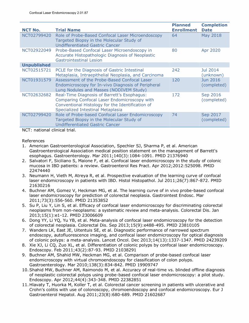
NCT No. Trial Name Planned Enrollment
Completion Date
NCT02799420 Role of Probe-Based Confocal Laser Microendoscopy Targeted Biopsy in the Molecular Study of Undifferentiated Gastric Cancer
64 May 2018
NCT02922049 Probe-Based Confocal Laser Microendoscopy in Accurate Histopathologic Diagnosis of Neoplastic Gastrointestinal Lesion
80 Apr 2020
Unpublished NCT02515721 PCLE for the Diagnosis of Gastric Intestinal
Metaplasia, Intraepithelial Neoplasia, and Carcinoma 242 Jul 2014
(unknown) NCT01931579 Assessment of the Probe-Based Confocal Laser
Endomicroscopy for In-vivo Diagnosis of Peripheral Lung Nodules and Masses (NODIVEM Study)
120 Jun 2016
(completed)
NCT02632682 Real-Time Diagnosis of Barrett’s Esophagus: Comparing Confocal Laser Endomicroscopy with Conventional Histology for the Identification of Specialized Intestinal Metaplasia
172 Sep 2016 (completed)
NCT02799420 Role of Probe-based Confocal Laser Endomicroscopy Targeted Biopsy in the Molecular Study of Undifferentiated Gastric Cancer
74 Sep 2017 (completed)
NCT: national clinical trial.
References 1. American Gastroenterological Association, Spechler SJ, Sharma P, et al. American
Gastroenterological Association medical position statement on the management of Barrett's esophagus. Gastroenterology. Mar 2011;140(3):1084-1091. PMID 21376940
2. Salvatori F, Siciliano S, Maione F, et al. Confocal laser endomicroscopy in the study of colonic mucosa in IBD patients: a review. Gastroenterol Res Pract. Apr 2012;2012:525098. PMID 22474440
3. Neumann H, Vieth M, Atreya R, et al. Prospective evaluation of the learning curve of confocal laser endomicroscopy in patients with IBD. Histol Histopathol. Jul 2011;26(7):867-872. PMID 21630216
4. Buchner AM, Gomez V, Heckman MG, et al. The learning curve of in vivo probe-based confocal laser endomicroscopy for prediction of colorectal neoplasia. Gastrointest Endosc. Mar 2011;73(3):556-560. PMID 21353852
5. Su P, Liu Y, Lin S, et al. Efficacy of confocal laser endomicroscopy for discriminating colorectal neoplasms from non-neoplasms: a systematic review and meta-analysis. Colorectal Dis. Jan 2013;15(1):e1-12. PMID 23006609
6. Dong YY, Li YQ, Yu YB, et al. Meta-analysis of confocal laser endomicroscopy for the detection of colorectal neoplasia. Colorectal Dis. Sep 2013;15(9):e488-495. PMID 23810105
7. Wanders LK, East JE, Uitentuis SE, et al. Diagnostic performance of narrowed spectrum endoscopy, autofluorescence imaging, and confocal laser endomicroscopy for optical diagnosis of colonic polyps: a meta-analysis. Lancet Oncol. Dec 2013;14(13):1337-1347. PMID 24239209
8. Xie XJ, Li CQ, Zuo XL, et al. Differentiation of colonic polyps by confocal laser endomicroscopy. Endoscopy. Feb 2011;43(2):87-93. PMID 21038291
9. Buchner AM, Shahid MW, Heckman MG, et al. Comparison of probe-based confocal laser
endomicroscopy with virtual chromoendoscopy for classification of colon polyps. Gastroenterology. Mar 2010;138(3):834-842. PMID 19909747
10. Shahid MW, Buchner AM, Raimondo M, et al. Accuracy of real-time vs. blinded offline diagnosis of neoplastic colorectal polyps using probe-based confocal laser endomicroscopy: a pilot study. Endoscopy. Apr 2012;44(4):343-348. PMID 22382851
11. Hlavaty T, Huorka M, Koller T, et al. Colorectal cancer screening in patients with ulcerative and Crohn's colitis with use of colonoscopy, chromoendoscopy and confocal endomicroscopy. Eur J
Gastroenterol Hepatol. Aug 2011;23(8):680-689. PMID 21602687
Confocal Laser Endomicroscopy 2.01.87

12. Gupta A, Attar BM, Koduru P, et al. Utility of confocal laser endomicroscopy in identifying high-grade dysplasia and adenocarcinoma in Barrett's esophagus: a systematic review and meta-analysis. Eur J Gastroenterol Hepatol. Apr 2014;26(4):369-377. PMID 24535597
13. Xiong YQ, Ma SJ, Zhou JH, et al. A meta-analysis of confocal laser endomicroscopy for the detection of neoplasia in patients with Barrett's esophagus. J Gastroenterol Hepatol. Jun 2016;31(6):1102-1110. PMID 26676646
14. Sharma P, Meining AR, Coron E, et al. Real-time increased detection of neoplastic tissue in Barrett's esophagus with probe-based confocal laser endomicroscopy: final results of an international multicenter, prospective, randomized, controlled trial. Gastrointest Endosc. Sep 2011;74(3):465-472. PMID 21741642
15. Dunbar KB, Okolo P, 3rd, Montgomery E, et al. Confocal laser endomicroscopy in Barrett's esophagus and endoscopically inapparent Barrett's neoplasia: a prospective, randomized,
double-blind, controlled, crossover trial. Gastrointest Endosc. Oct 2009;70(4):645-654. PMID 19559419
16. Canto MI, Anandasabapathy S, Brugge W, et al. In vivo endomicroscopy improves detection of Barrett's esophagus-related neoplasia: a multicenter international randomized controlled trial (with video). Gastrointest Endosc. Feb 2014;79(2):211-221. PMID 24219822
17. Ypsilantis E, Pissas D, Papagrigoriadis S, et al. Use of confocal laser endomicroscopy to assess the adequacy of endoscopic treatment of gastrointestinal neoplasia: a systematic review and
meta-analysis. Surg Laparosc Endosc Percutan Tech. Feb 2015;25(1):1-5. PMID 24910941 18. Wallace MB, Crook JE, Saunders M, et al. Multicenter, randomized, controlled trial of confocal
laser endomicroscopy assessment of residual metaplasia after mucosal ablation or resection of GI neoplasia in Barrett's esophagus. Gastrointest Endosc. Sep 2012;76(3):539-547 e531. PMID 22749368
19. Sorokina A, Danilevskaya O, Averyanov A, et al. Comparative study of ex vivo probe-based confocal laser endomicroscopy and light microscopy in lung cancer diagnostics. Respirology.
Aug 2014;19(6):907-913. PMID 24909555 20. Wellikoff AS, Holladay RC, Downie GH, et al. Comparison of in vivo probe-based confocal laser
endomicroscopy with histopathology in lung cancer: A move toward optical biopsy. Respirology. Aug 2015;20(6):967-974. PMID 26094505
21. Fuchs FS, Zirlik S, Hildner K, et al. Confocal laser endomicroscopy for diagnosing lung cancer in vivo. Eur Respir J. Sep 20 2012. PMID 22997220
22. Sonn GA, Jones SN, Tarin TV, et al. Optical biopsy of human bladder neoplasia with in vivo
confocal laser endomicroscopy. J Urol. Oct 2009;182(4):1299-1305. PMID 19683270 23. Liu JJ, Droller MJ, Liao JC. New optical imaging technologies for bladder cancer: considerations
and perspectives. J Urol. Aug 2012;188(2):361-368. PMID 22698620 24. Nathan CA, Kaskas NM, Ma X, et al. Confocal laser endomicroscopy in the detection of head and
neck precancerous lesions. Otolaryngol Head Neck Surg. Apr 3 2014;151(1):73-80. PMID 24699456
25. Moore C, Mehta V, Ma X, et al. Interobserver agreement of confocal laser endomicroscopy for
detection of head and neck neoplasia. Laryngoscope. Mar 2016;126(3):632-637. PMID 26372409
26. Liu J, Li M, Li Z, et al. Learning curve and interobserver agreement of confocal laser endomicroscopy for detecting precancerous or early-stage esophageal squamous cancer. PLoS One. Jun 2014;9(6):e99089. PMID 24897112
27. Guo J, Li CQ, Li M, et al. Diagnostic value of probe-based confocal laser endomicroscopy and high-definition virtual chromoendoscopy in early esophageal squamous neoplasia. Gastrointest
Endosc. Jun 2015;81(6):1346-1354. PMID 25680899 28. Liu T, Zheng H, Gong W, et al. The accuracy of confocal laser endomicroscopy, narrow band
imaging, and chromoendoscopy for the detection of atrophic gastritis. J Clin Gastroenterol. May-Jun 2015;49(5):379-386. PMID 25485568
29. He XK, Liu D, Sun LM. Diagnostic performance of confocal laser endomicroscopy for optical diagnosis of gastric intestinal metaplasia: a meta-analysis. BMC Gastroenterol. Sep 05 2016;16:109. PMID 27596838
30. Qian W, Bai T, Wang H, et al. Meta-analysis of confocal laser endomicroscopy for the diagnosis of gastric neoplasia and adenocarcinoma. J Dig Dis. Jun 2016;17(6):366-376. PMID 27129127
Confocal Laser Endomicroscopy 2.01.87

31. Karia K, Waxman I, Konda VJ, et al. Needle-based confocal endomicroscopy for pancreatic cysts: the current agreement in interpretation. Gastrointest Endosc. May 2016;83(5):924-927. PMID 26382051
32. Napoleon B, Lemaistre AI, Pujol B, et al. In vivo characterization of pancreatic cystic lesions by needle-based confocal laser endomicroscopy (nCLE): proposition of a comprehensive nCLE classification confirmed by an external retrospective evaluation. Surg Endosc. Jun
2016;30(6):2603-2612. PMID 26428198 33. De Palma GD, Esposito D, Luglio G, et al. Confocal laser endomicroscopy in breast surgery: a
pilot study. BMC Cancer. Apr 10 2015;15:252. PMID 25885686 34. Slivka A, Gan I, Jamidar P, et al. Validation of the diagnostic accuracy of probe-based confocal
laser endomicroscopy for the characterization of indeterminate biliary strictures: results of a prospective multicenter international study. Gastrointest Endosc. Feb 2015;81(2):282-290.
PMID 25616752 35. Hirota WK, Zuckerman MJ, Adler DG, et al. ASGE guideline: the role of endoscopy in the
surveillance of premalignant conditions of the upper GI tract. Gastrointest Endosc. Apr 2006;63(4):570-580. PMID 16564854
36. ASGE Technology Committee. Confocal laser endomicroscopy. Gastrointest Endosc. Dec 2014;80(6):928-938. PMID 25442092
Billing Coding/Physician Documentation Information 0397T Endoscopic retrograde cholangiopancreatography (ERCP), with optical
endomicroscopy (List separately in addition to code for primary
procedure) (new code 1/1/2016) 43206 Esophagoscopy, flexible, transoral; with optical endomicroscopy
43252 Esophagogastroduodenoscopy, flexible, transoral; with optical
endomicroscopy
88375 Optical endomicroscopic image(s), interpretation and report, real-time
or referred, each endoscopic session
ICD10 Codes
K22.70-
K22.719
Barrett's esophagus code range
Z13.810 Encounter for screening for upper gastrointestinal disorder
Z13.811 Encounter for screening for lower gastrointestinal disorder
Z13.83 Encounter for screening for respiratory disorder NEC
The interpretation and report of optical endomicroscopic image(s) would be
reported with the following code:
88375: Optical endomicroscopic image(s), interpretation and report, real-time or
referred, each endoscopic session.
Code 88375 cannot be reported in conjunction codes 43206 and 43252.
Additional Policy Key Words N/A
Policy Implementation/Update Information 3/1/13 New policy; considered investigational.
9/1/13 No policy statement changes.
3/1/14 No policy statement changes.
Confocal Laser Endomicroscopy 2.01.87

9/1/14 No policy statement changes. Added Billing and Coding Information
section. Added CPT: 43206, 43252, 88375.
3/1/15 No policy statement changes. 9/1/15 No policy statement changes.
3/1/16 Added Cat III CPT code. No policy statement changes.
9/1/16 No policy statement changes.
3/1/17 No policy statement changes. 9/1/17 No policy statement changes.
3/1/18 No policy statement changes.
9/1/18 No policy statement changes.
State and Federal mandates and health plan contract language, including specific provisions/exclusions, take precedence over Medical Policy and must be considered first in determining eligibility for coverage. The medical policies contained herein are for informational purposes. The medical policies do not constitute medical advice or medical care. Treating health
care providers are independent contractors and are neither employees nor agents Blue KC and are solely responsible for diagnosis, treatment and medical advice. No part of this publication may be reproduced, stored in a retrieval system or transmitted, in any form or by any means, electronic, photocopying, or otherwise, without permission from Blue KC.
Confocal Laser Endomicroscopy 2.01.87