Embed Size (px)
Citation preview

NEUROSYSTEMS
Cone arrestin confers cone vision of hightemporal resolution in zebrafish larvae
Sabine L. Renninger, Matthias Gesemann and Stephan C. F. NeuhaussInstitute of Molecular Life Sciences, Neuroscience Center Zurich and Center for Integrative Human Physiology, University of Zurich,Winterthurerstrasse 190, CH-8057 Zurich, Switzerland
Keywords: arrestin, cone photoreceptor, phototransduction, retina, zebrafish
Abstract
Vision of high temporal resolution depends on careful regulation of photoresponse kinetics, beginning with the lifetime of activatedphotopigment. The activity of rhodopsin is quenched by high-affinity binding of arrestin to photoexcited phosphorylated photopigment,which effectively terminates the visual transduction cascade. This regulation mechanism is well established for rod photoreceptors,yet its role for cone vision is still controversial. In this study we therefore analyzed arrestin function in the cone-dominated vision oflarval zebrafish. For both rod (arrS ) and cone (arr3 ) arrestin we isolated two paralogs, each expressed in the respective subset ofphotoreceptors. Labeling with paralog-specific antibodies revealed subfunctionalized expression of Arr3a in M- and L-cones, andArr3b in S- and UV-cones. The inactivation of arr3a by morpholino knockdown technology resulted in a severe delay inphotoresponse recovery which, under bright light conditions, was rate-limiting. Comparison to opsin phosphorylation-deficientanimals confirmed the role of cone arrestin in late cone response recovery. Arr3a activity partially overlapped with the function of thecone-specific kinase Grk7a involved in initial response recovery. Behavioral measurements further revealed Arr3a deficiency to besufficient to reduce temporal contrast sensitivity, providing evidence for the importance of arrestin in cone vision of high temporalresolution.
Introduction
The visual system of vertebrates is effective over a wide range of lightintensities as it exploits different types of photoreceptor cells. Rodphotoreceptors are exceptionally light sensitive, with their transduc-tion cascade tuned for maximal quantum yield. Cone photoreceptorsare less sensitive but allow vision of high temporal and spatialresolution, which necessitates fast response kinetics (reviewed inRodieck, 1998 and Kawamura & Tachibanaki, 2008).Signal amplification and temporal aspects of the photoresponse are
precisely regulated by controlling the lifetime of activated photopig-ment. In the rod phototransduction cascade, phosphorylation ofphotoexcited rhodopsin reduces the efficacy of transducin activation.Subsequent high-affinity binding of the arrestin protein (Arr) toactivated and phosphorylated rhodopsin completely quenches signaltransduction by competing with the G-protein transducin for rhodop-sin binding (Krupnick et al., 1997). Deficiency in this quenchingmechanism in rods causes congenital stationary night blindness inhumans but leaves photopic vision unaffected (Fuchs et al., 1995;Yamamoto et al., 1997).The identification of a cone-specific G-protein-coupled receptor
kinase, GRK7 (Hisatomi et al., 1998), and a cone arrestin protein(Craft et al., 1994) strongly supports the hypothesis that, similarly to
rods, normal cone response kinetics depend on regulated pigmentinactivation.Impaired cone photopigment phosphorylation has been shown to
cause a prolonged photoresponse (Lyubarsky et al., 2000; Nikonovet al., 2005, 2008; Shi et al., 2007) and a delay in photoresponserecovery (Rinner et al., 2005). Furthermore, studies in carp retina haverevealed that fast cone response kinetics depend on distinct molecularcharacteristics of GRK7. Compared to GRK1 found in rods and (insome species) cones, higher expression levels and phosphorylationactivity of GRK7 account for fast inactivation of photoexcited opsinand low transducin activation in cones (Tachibanaki et al., 2005,2007).The function of cone arrestin in the final shutoff of cone pigment
and its impact on cone response kinetics or visual function is stillelusive. In functional studies of transgenic mice expressing S-opsin inrhodopsin-deficient rods the inactivation of S-pigment was slowed inthe absence of rod arrestin (Arr1 in the mouse). The prolonged photo-response of these transgenic rods could not be rescued by expressionof cone arrestin (Arr4 in the mouse), indicating that cone arrestin isnot sufficient to regulate photoexcited S-opsin (Shi et al., 2007).Recently, co-expression of mouse Arr1 and Arr4 in the outer
segments of cones with higher protein levels of rod arrestin has beendemonstrated (Nikonov et al., 2008). Flash responses of cones lackingeither cone or rod arrestin did not show any severe impairment ofpigment recovery. However, cone response inactivation in arrestindouble knockouts was slowed down, which indicates that arrestin isinvolved in quenching photoexcited cone pigment.
Correspondence: Prof Dr Stephan C. F. Neuhauss, as above.E-mail: [email protected]
Received 7 September 2010, revised 5 November 2010, accepted 29 November 2010
European Journal of Neuroscience, Vol. 33, pp. 658–667, 2011 doi:10.1111/j.1460-9568.2010.07574.x
ª 2011 The Authors. European Journal of Neuroscience ª 2011 Federation of European Neuroscience Societies and Blackwell Publishing Ltd
European Journal of Neuroscience

To directly address the function of cone arrestin in photopigmentinactivation and the impact of arrestin on photopic vision we utilizedthe cone-dominated visual system of zebrafish (Danio rerio) larvae(Bilotta & Saszik, 2001; Fadool & Dowling, 2008). We found thatfunctional loss of cone arrestin causes a prolonged photoresponserecovery and reduced temporal contrast sensitivity of zebrafish larvalvision.
Materials and methods
Fish husbandry
Zebrafish of the Tu strain were bred and maintained under standardconditions at 28 !C and a 14 h light and 10 h dark cycle (Westerfield,1994). Larvae were raised in E3 medium containing (in mm): NaCl, 5;KCl, 0.17; CaCl2, 0.33; MgSO4, 0.33; and methylene blue, and werestaged in days post-fertilization (dpf) according to Kimmel et al.(1995).
Annotation of arrestin cDNAs
As many genes predicted within GenBank are produced by automatedprocesses and have been shown to contain numerous errors, arrestincDNA sequences used in this study were manually annotated.Sequences were identified and annotated using combined informationfrom expressed sequence tags and genome databases (GeneBank,http://www.ncbi.nlm.nih.gov; Ensembl, http://www.ensembl.org/index.html; version 50 ⁄ 51, 2008). Human and mouse sequences wereused as initial query (for more details on sequence annotation seeGesemann et al., 2010).
Phylogenetic tree analysis
Coding sequences of arrestin genes were translated into proteins usingthe EditSeq software (Lasergene; DNASTAR, Madison, WI, USA)and obtained protein sequences were used to generate a combinedsequence file in FASTA. Sequence alignment and phylogeneticanalysis was performed on the Phylogeny.fr platform (Dereeper et al.,2008; http://www.phylogeny.fr/version2_cgi/phylogeny.cgi). Sequenceswere aligned using muscle (v3.7; Edgar, 2004) configured for highestaccuracy (muscle with default settings). After alignment, ambiguousregions (i.e. containing gaps and ⁄ or being poorly aligned) wereremoved with Gblocks (v0.91b; Castresana, 2000). The phylogenetictree was reconstructed by the maximum likelihood method (Guindon &Gascuel, 2003) using the WAG amino acid replacement matrix (Whelan& Goldman, 2001) implemented in the PhyML program (v3.0). Theapproximate likelihood ratio test (aLRT; Anisimova & Gascuel,2006) was used to judge branch reliability. Graphical representationand editing of the phylogenetic tree was done using TreeDyn (v198.3)and the obtained svg files were colored using the CorelDrawprogram.
Cloning of arrestin genes
Total RNA was isolated from larvae 5 dpf using the RNAeasy kit(Qiagen, Hombrechtikon, Switzerland) and cDNA was synthesizedusing the SuperScript II Reverse Transcriptase kit (Invitrogen, Basel,Switzerland) according to manufacture’s instructions.
For identified arrestin genes, the coding sequences were amplifiedfrom cDNA using the following primer pairs: arr3a sense ATGGC-TGACAAAGTTTACAAG, antisense GCCCTGTGGAATCTGATATG;
arr3b sense CATGACAAAGGTTTACAAGAAG, antisenseTGCTCCTCACTGGCTGTAG; arrSa sense CAATGAGTCC-AAAAAATGTCG, antisense TAACCGAGAAGTGCTCTTTC; arrSbsense ATGAGTCCCAAGCACATCATC, antisense CAGCCAG-CTCAAAACACG. Products were cloned into the pCRII-TOPOvector (Invitrogen) and sequenced using T7 and SP6 primers.
In situ hybridization (ISH)
Arrestin sense and antisense RNA probes were transcribed in thepresence of digoxigenin-labeled nucleotides (DIG RNA Labeling Mix;Roche, Rotkreuz, Switzerland) using SP6 and T7 polymerases(Roche), respectively. For the rhodopsin antisense RNA probe T7polymerases (Roche) were used. Transcripts were hydrolyzed to yieldfragments of approximately 300–500 nt in length. For ISH, zebrafishlarvae were fixed in 4% paraformaldehyde (PFA), dehydrated andstored in methanol until usage. To reduce pigmentation, larvae werebleached with 3% H2O2 and 1% KOH directly before ISH. For mRNAdetetction in adult eyes, retinas from albino zebrafish were cryosec-tioned to 14 lm thickness. ISH was carried out in an automated ISHapparatus (Holle&Huttner, Tubingen, Germany) according to Thisse& Thisse (2008). RNA probes were hybridized overnight at 58 !C anddetected using an alkaline phosphatase-based color reaction.
Generation of antibodies
A highly immunogenic hydrophilic peptide specific for either Arr3a orArr3b but not conserved in ArrS (Supporting Information Fig. S1) hasbeen chosen for immunization. Rabbits were immunized with theArr3a peptide AKSADDPDEKVDKKDTC (147–163) whereas chick-en were immunized with the Arr3b peptide ANE-EDNIDEKVEKKDTC (146–162). All antibodies were affinity-purified against the respective peptide by Eurogentec (Seraing,Belgium).
Immunohistochemistry
Immunohistological staining was performed as previously described(Fleisch et al., 2008). For detection of Arr3b, tissue was fixed in 2%trichloroacetic acid (Sigma-Aldrich, Switzerland) for 30 min at roomtemperature, equilibrated in 30% sucrose and embedded in Tissue Tek(Sakura Finetek Europe, Netherlands). Cryosections of 18 lm thick-ness were cut. Rabbit anti-Arr3a antibody was used in a 1 : 400dilution and chicken anti-Arr3b antibody in a 1 : 250 dilution.The following antibodies were used for co-labeling: Zpr1 antibody
(1 : 400; Zebrafish International Resource Center, Eugene, OR, USA),rabbit anti-blue opsin and rabbit anti-ultraviolet (UV) opsin (1 : 500;kindly provided by David R. Hyde, University of Notre Dame, NotreDame, IN, USA). For visualization, the secondary antibodies AlexaFluor 488 goat anti-chicken or goat anti-mouse (1 : 1000; Invitrogen)and Alexa Fluor 568 goat anti-mouse or goat anti-rabbit (1 : 500;Invitrogen) were applied.
Histology
Larvae were fixed at room temperature for 50 min in 4% PFA in 0.2 mphosphate buffer (pH 7.4). Fixed larvae were dehydrated in a standardethanol series, infiltrated and embedded in Technovit 7100 (KulzerHistotechnik, Wehrheim, Germany). Sections (3 lm) were cut andmounted on slides. For contrast, tissue was stained with Richardson
Arrestin function in cone vision 659
ª 2011 The Authors. European Journal of Neuroscience ª 2011 Federation of European Neuroscience Societies and Blackwell Publishing LtdEuropean Journal of Neuroscience, 33, 658–667

(Bromeis) solution (1% methylene blue, 1% Borax and 1% azur II,1 : 1 : 2).
Microscopy
Histological sections were imaged under a BX61 microscope (Olym-pus) equipped with a ColorView IIIu digital camera and edited usingAdobe Photoshop CS2 (Adobe Systems). Z-stacks of immunostain-ings were recorded using a CLSM SP2 confocal laser scanningmicroscope (Leica) and processed in Imaris 7.0 (Bitplane).
Western blot
Eyes of 35 zebrafish larvae, 5 dpf, were isolated and sonicated in40 lL Ringer solution (in mm: NaCl, 116; KCl, 2.9; CaCl2, 1.8; andHEPES, 5; pH 7.4) containing serine and cysteine protease inhibitors(complete, Mini, EDTA-free; Roche). Insoluble material wasremoved by ultracentrifugation. The total protein concentrationswere measured using the DC Protein Assay (Bio-Rad, Reinach,Switzerland) according to manufacture’s instruction before sampleswere mixed with 3· Urea buffer [Tris, 65 mm, pH 6.75; urea, 8 m;glycine, 20%; b-mercaptoethanol, 5%; SDS, 5%; and bromphenolblue]. As loading control, acetylated tubulin of approximately50 kDa was detected simultaneously with Arr3a or Arr3b by mouseanti-acetylated tubulin antibody (Sigma-Aldrich). Immunoblots weredeveloped using the ECL system (Supra Signal West; ThermoScientific, Applied Biosystems, Switzerland) and chemiluminescencewas detected with the Fujifilm LAS-3000 imaging system. For thedetection of Arr3 and acetylated tubulin different exposure timeswere applied. The arrestin knockdown was semi-quantitativelyevaluated in ImageJ (NIH).
Targeted gene knockdown
Antisense morpholino oligonucleotides (MOs) covering the transla-tional start site were obtained from Gene Tools (Philomath, Oregon,USA): arr3a-MO (5¢-ATAATCCGCAGCCCGTCTGGTGTAG-3¢),arr3b-MO (5¢-CAAGTTCTGAGCTTTTCAAGTTCTG-3¢) and grk7a-MO (5¢-ATCGAGTCCCCCCATGTCACACATT-3¢). For control, astandard control MO (5¢-CCTCTTACCTCAGTTACAATTTATA-3¢)was used.All MOs were dissolved in ddH2O to a stock concentration of
2 mm. Prior to injection, MOs were diluted in 1· Danieau’s solution[in mm: NaCl, 58; KCl, 0.7; MgSO4, 0.4; Ca(NO3), 0.6; HEPES, 5;and phenol red, 0.2%] to the desired concentration. If not indicateddifferently, embryos were injected in the one- and two-cell stage with5 ng control-MO, 4.2 ng arr3a-MO, 27 ng arr3b-MO or 10.8 nggrk7a-MO. For double-knockdown experiments we injected 2.8 ngarr3a-MO and 13 ng arr3b-MO.
Electroretinography
Electroretinograms (ERGs) were recorded from unanesthetized zebra-fish larvae as described previously (Makhankov et al., 2004). Larvaewere first dark-adapted for at least 40 min. The following pre-recording steps were carried out under dim red illumination.For paired flash recordings, a 500 ms conditioning flash (7000 or
70 lux) followed by a probing flash of the same light intensity andduration was presented. The interstimulus interval between condition-ing and probing flash was varied (1, 2, 3, 5, 10 or 20 s). Cone response
recovery was calculated as the ratio of b-wave peak amplitudes toconditioning and probe flash (Rinner et al., 2005). The b-waveamplitudes were measured as the range between the minimum andmaximum potential within 200 ms of light onset.
Psychophysics
The optokinetic response of zebrafish larvae was measured using theexperimental setup described in Mueller & Neuhauss (2010). Forimmobilization, larvae were placed dorsal up in a 35 mm Petri dishcontaining 3% methylcellulose.An LCD Projector (VPL-CX1; Sony Corporation) was used to
present a vertical sinusoidal grating pattern on a white paper drum(r = 4.5 cm) with the grating rotating around the larvae. Eye angle andvelocity were recorded by means of an infrared-sensitive CCD camera(Guppy F-038B NIR; Allied Vision Technologies). Custom-developedsoftware based on LabView 7.1 and NI-IAQ 3.7 (National Instru-ments) was used to control stimulation and to record eye movements(Mueller & Neuhauss, 2010).To determine temporal sensitivity at different illumination levels a
sine-wave grating of varying pattern velocity was presented. Stimulusintensity was adjusted via the color values defining the stimulus. Priorto recording, larvae were adapted for 60 s to background light levelsof 20 or 120 cd ⁄m2 and then pre-stimulated for 9 s with a lowtemporal frequency pattern (0.033 cycles ⁄ deg) at a angular velocity of3.5 deg ⁄ s. Eye velocity was measured from larvae stimulated with agrating pattern of 0.083 cycles ⁄ deg and varying angular velocities of3.5, 5.25, 7, 14, 21 and 28 deg ⁄ s.
Statistical analysis
For electrophysiological experiments, statistical significance wascalculated in GraphPad Prism 4 (GraphPad Software). The b-wavepeak amplitude recovery of morphants was compared using two-wayanova with Bonferroni’s post-test. For behavioral measurements theeye velocity of the two eyes, the left and the right, were averaged anddata were analyzed in pasw Statistics 17.0 (SPSS Inc.) using repeated-measures anova with angular velocity as within-subject effect andgenotype as between-subjects effect. Graphs were generated inGraphPad Prism 4.
Results
The zebrafish genome habors four visual arrestin genes
To investigate the role of arrestin function in cone vision, we first useda combined bioinformatic and cDNA cloning approach and identifiedparalogs of the mammalian cone arrestin (arrestin 3, arrestin 4 orX-arrestin). We denoted the two identified zebrafish paralogs as arr3aand arr3b. Similarly, we isolated two rod arrestin paralogs ofthe mammalian ortholog (arrestin 1, S-arrestin or retinal S-antigen),denoted by arrSa and arrSb. For the two broadly expressedmammalian b-arrestin genes we isolated three orthologs, namely theparalogs b-arr2a, b-arr2b and b-arr1a. The additional arrestin genesfound in the zebrafish genome probably originated from the teleost-specific whole-genome duplication event (Postlethwait et al., 2000),as radiation hybrid mapping determined all four visual arrestins tolocate on different chromosomes (Supporting Information Table S1).Pairwise amino acid sequence alignment of the visual arrestinsrevealed that protein identity between two zebrafish paralogs did notexceed 70% (Supporting Information Table S2).
660 S. L. Renninger et al.
ª 2011 The Authors. European Journal of Neuroscience ª 2011 Federation of European Neuroscience Societies and Blackwell Publishing LtdEuropean Journal of Neuroscience, 33, 658–667

To confirm that the identified zebrafish paralogs are true orthologs ofthe respectivemammalian genes, we performed a phylogenetic analysis,which supported our assumption (Supporting Information Fig. S2).
Visual arrestins are expressed in a complementary manner inzebrafish photoreceptor subtypes
In order to determine the cellular distribution of visual arrestintranscripts in the zebrafish retina, we performed whole-mount in situhybridization experiments with sequence-specific riboprobes at dif-ferent developmental stages.
At 3 dpf the two cone arrestin paralogs showed distinct expressionpatterns: arr3a was evenly expressed throughout the photoreceptorcell layer (PRCL) whereas arr3b expression was confined to theventral half of the retina (Fig. 1A and B). Both arrS transcripts were,in contrast, detected in a ventrotemporal patch (Fig. 1C and D),consistent with expression of rod photoreceptor markers (Raymondet al., 1995; Hamaoka et al., 2002). At 5 dpf arrS transcripts appearedsporadically over the PRCL with prominence at the ventral sidewhereas mRNAs for both arr3 paralogs were detected evenlythroughout the PRCL of the retina (Fig. 1E–H). These expressionpatterns are consistent with the differentiation of cone (arr3) and rod(arrS) photoreceptors in the developing zebrafish retina (Raymondet al., 1995).
No mRNA transcripts of b-arr genes were found in photoreceptorcells of the zebrafish retina at 3 and 5 dpf (data not shown).
To elucidate if this cell type-specific visual arrestin expression ispreserved throughout development we next analyzed adult retinasections. arrS mRNAs could be found in the inner most part of theouter nuclear layer where the nuclei of rods are located (Fig. 1K andL). Comparison of arrS expression with rhodopsin expressionconfirmed the specificity of both arrS paralogs for rod photoreceptors(Supporting Information Fig. S3).
The expression pattern of cone arrestin transcripts was comple-mentary to the expression of rod arrestins in the adult retina. Whilearr3a mRNA was detected in the inner segment of most conephotoreceptors, arr3b expression was restricted to a small subset ofcone cells (Fig. 1I and J).
To further investigate this distinct expression of arr3a and arr3b,we raised peptide antibodies against the two arr3 paralogs. Immuno-histochemical analysis revealed a complementary expression of Arr3aand Arr3b in different photoreceptor cell types (Fig. 2A and D).Interestingly, both arrestin proteins were distributed throughout thephotoreceptor cells with high protein levels in the inner segment andthe cone pedicles as observed for mouse cone arrestin (Zhu et al.,2002).
In co-immunostainings we ascertained the cell type specificity ofArr3a and Arr3b. Double staining with the Arr3a antibody and thegreen ⁄ red double cone specific Zpr1 antibody (Larison & Bremiller,1990) revealed zebrafish Arr3a to be specifically expressed in mid-and long-wavelength-sensitive cone (M-cone and L-cone, respec-tively) photoreceptors (Fig. 2B and E). In contrast, we found Arr3b tobe expressed in short- and UV-wavelength-sensitive cones (S-conesand UV-cones, respectively) as revealed by co-labeling of Arr3b andblue- and UV-opsin (Fig. 2C and F).
Generation of Arr3-deficient zebrafish larvae
Expression analysis has shown that arr3 is exclusively expressed inzebrafish cone photoreceptors. In order to explore the functional role
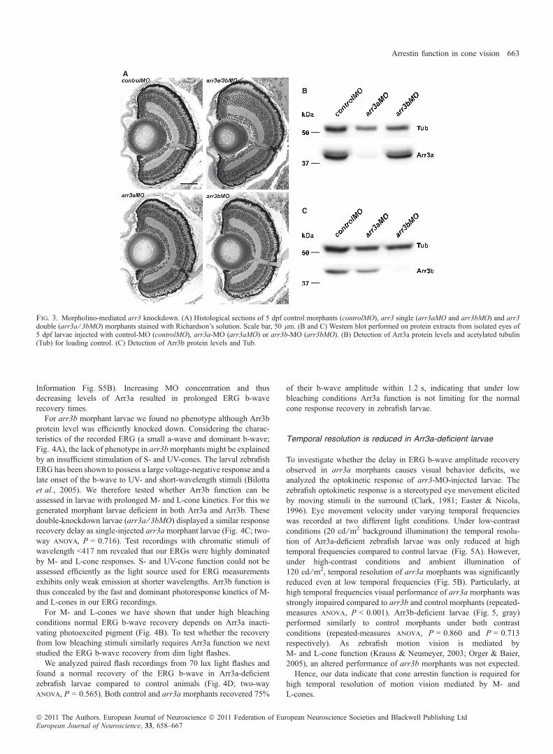
of arrestin in cone vision, we thus generated Arr3-deficient zebrafishlarvae. Protein expression was knocked down using MOs targetedagainst the translational start sites of arr3a or arr3b. Injection of theseMOs did not cause any apparent malformations and the developmentof injected larvae was indistinguishable from that of control-injectedlarvae. Similarly, we found no changes to retinal integrity on standardhistological sections. Neither arr3 single morphants (arr3aMO andarr3bMO) nor arr3 double morphants (arr3a ⁄ 3bMO) displayed anymorphological abnormality of the retina (Fig. 3A).The specificity of the knockdown was evaluated in Western blot and
immunohistological analysis. At 5 dpf, the Arr3a protein level wasspecifically reduced in arr3a-MO-treated larvae but not in larvaeinjected with arr3b-MO or control-MO (Fig. 3B and SupportingInformation Fig. S4). Vice versa, the Arr3b protein level was onlyreduced in arr3b morphants but not in arr3a or control morphants(Fig. 3C and Supporting Information Fig. S4). Thus, using MOstargeting the translational start site we could specifically knock downeither arr3a or arr3b.To quantify the knockdown efficiency in larvae 5 dpf, acetylated
tubulin was additionally detected in Western blot analysis forstandardization. Semi-quantitative evaluation determined an estimatedknockdown efficiency of 95% for Arr3a and 88% for Arr3b. Hence,MO-mediated knockdown is highly efficient at 5 dpf.
Cone response recovery is delayed in Arr3a-deficientzebrafish larvae
The larval zebrafish retina is strongly dominated by cone function. Rodphotoreceptors are sparse and exhibit short outer segments (Branchek &Bremiller, 1984). Their function can not be assessed in electroretino-graphic recordings before 15 dpf (Bilotta et al., 2001).Taking advantage of this cone dominance we studied the functional
impact of cone arrestin on normal photopigment inactivation and coneresponse recovery using ERG recordings. The ERG is a sum fieldresponse of the retina to light (Dowling, 1987). In zebrafish larvae,the ERG is dominated by a prominent b-wave which mainly reflectsthe response of ON-bipolar cells. As the b-wave largely masks theresponse of the photoreceptors, represented by the a-wave, we usedthe b-wave as an indirect measure of photoreceptor function.Arr3 function was assessed in ERG paired-flash recordings. Two
white light flashes, a conditioning and a probing flash (Fig. 4A, blackand gray lines respectively) were separated by varying interstimulusintervals. The recovery of the b-wave amplitude between conditioningand probing flash was calculated. In 4 and 5 dpf zebrafish larvae, lossof Arr3b did not cause any alteration in the time course of b-waverecovery when compared to control-injected larvae (Fig. 4B, two wayanova, P = 0.862; and Supoprting Information Fig. S5A, P = 0.069).However, knockdown of arr3a caused a significant delay of b-waveamplitude recovery (Fig. 4B; two way anova, P < 0.0001). Five-day-old control morphants recovered 75% of their b-wave amplitudewithin 1.8 s. arr3a morphants, in contrast, required approximately 4 sto recover the b-wave to the same extent. This delay in recovery wasenhanced in 4-day-old Arr3a-deficient larvae (Supporting InformationFig. S5A; P < 0.0001). As MO efficacy decreases during larvaldevelopment the reversibility of this delay in response recovery arguesagainst nonspecific effects induced by arrestin knockdown.To further confirm the specificity of the arr3a morphant phenotype,
we analyzed an Arr3a dosage-dependent recovery of the ERG b-wave.Zebrafish larvae treated with increasing concentrations of arr3a-MOrevealed a dose–response relationship with MO concentrationdependent lengthening of the b-wave recovery time (Supporting
Arrestin function in cone vision 661
ª 2011 The Authors. European Journal of Neuroscience ª 2011 Federation of European Neuroscience Societies and Blackwell Publishing LtdEuropean Journal of Neuroscience, 33, 658–667

A B C
D E F
Fig. 2. Photoreceptor cell type-specific expression of the arr3 paralog genes in zebrafish larvae. Z-projections of confocal image stacks from immunostainings ontransverse sections of 5-day-old albino larvae. (A and D) Double immunostaining of Arr3a (red) and Arr3b (green). (B and E) Co-staining with antibody againstArr3a (red) and double cone specific Zpr1 (green). (C and F) Co-labeling of Arr3b (green) and short-wavelength-sensitive blue- and UV-opsin (S-opn, red). Scalebars, 50 lm (A–C), 10 lm (D–F).
A B C D
E F G H
I J K L
Fig. 1. Spatiotemporal expression analysis of visual arrestin genes in zebrafish. (A–H) Lateral view of zebrafish larvae with dorsal side up and anterior to the left.(I–L) Sections of adult albino retina. (A, E and I) mRNA expression analysis revealed arr3a to be evenly expressed throughout the PRCL at 3 and 5 dpf (A and E)and restrictedly in the outer nuclear layer of adult retina (I). (B, F and J) arr3b expression was detected only in the ventral half of the retina in larvae 3 dpf (B) butthroughout the PRCL at 5 dpf (F). In the adult retina arr3b expression was restricted to a subset of photoreceptor cells (J). (C and D) arrSa and arrSb were detectedin a ventral patch of the retina at 3 dpf. (G and H) At 5 dpf arrSa and arrSb expression had expanded at the ventral site with sparse expression dorsally. (K and L) Onadult retina sections, both arrS paralogs were detected in a restricted part of the outer nuclear layer. OS, outer segments; ONL, outer nuclear layer; OPL, outerplexiform layer; INL, inner nuclear layer; IPL, inner plexiform layer; GCL, ganglion cell layer. Scale bars, 150 lm (in A for A–H), 50 lm (in I for I–L).
662 S. L. Renninger et al.
ª 2011 The Authors. European Journal of Neuroscience ª 2011 Federation of European Neuroscience Societies and Blackwell Publishing LtdEuropean Journal of Neuroscience, 33, 658–667
Information Fig. S5B). Increasing MO concentration and thusdecreasing levels of Arr3a resulted in prolonged ERG b-waverecovery times.
For arr3b morphant larvae we found no phenotype although Arr3bprotein level was efficiently knocked down. Considering the charac-teristics of the recorded ERG (a small a-wave and dominant b-wave;Fig. 4A), the lack of phenotype in arr3bmorphants might be explainedby an insufficient stimulation of S- and UV-cones. The larval zebrafishERG has been shown to possess a large voltage-negative response and alate onset of the b-wave to UV- and short-wavelength stimuli (Bilottaet al., 2005). We therefore tested whether Arr3b function can beassessed in larvae with prolonged M- and L-cone kinetics. For this wegenerated morphant larvae deficient in both Arr3a and Arr3b. Thesedouble-knockdown larvae (arr3a ⁄ 3bMO) displayed a similar responserecovery delay as single-injected arr3amorphant larvae (Fig. 4C; two-way anova, P = 0.716). Test recordings with chromatic stimuli ofwavelength <417 nm revealed that our ERGs were highly dominatedby M- and L-cone responses. S- and UV-cone function could not beassessed efficiently as the light source used for ERG measurementsexhibits only weak emission at shorter wavelengths. Arr3b function isthus concealed by the fast and dominant photoresponse kinetics of M-and L-cones in our ERG recordings.
For M- and L-cones we have shown that under high bleachingconditions normal ERG b-wave recovery depends on Arr3a inacti-vating photoexcited pigment (Fig. 4B). To test whether the recoveryfrom low bleaching stimuli similarly requires Arr3a function we nextstudied the ERG b-wave recovery from dim light flashes.
We analyzed paired flash recordings from 70 lux light flashes andfound a normal recovery of the ERG b-wave in Arr3a-deficientzebrafish larvae compared to control animals (Fig. 4D; two-wayanova, P = 0.565). Both control and arr3a morphants recovered 75%
of their b-wave amplitude within 1.2 s, indicating that under lowbleaching conditions Arr3a function is not limiting for the normalcone response recovery in zebrafish larvae.
Temporal resolution is reduced in Arr3a-deficient larvae
To investigate whether the delay in ERG b-wave amplitude recoveryobserved in arr3a morphants causes visual behavior deficits, weanalyzed the optokinetic response of arr3-MO-injected larvae. Thezebrafish optokinetic response is a stereotyped eye movement elicitedby moving stimuli in the surround (Clark, 1981; Easter & Nicola,1996). Eye movement velocity under varying temporal frequencieswas recorded at two different light conditions. Under low-contrastconditions (20 cd ⁄m2 background illumination) the temporal resolu-tion of Arr3a-deficient zebrafish larvae was only reduced at hightemporal frequencies compared to control larvae (Fig. 5A). However,under high-contrast conditions and ambient illumination of120 cd ⁄m2, temporal resolution of arr3a morphants was significantlyreduced even at low temporal frequencies (Fig. 5B). Particularly, athigh temporal frequencies visual performance of arr3a morphants wasstrongly impaired compared to arr3b and control morphants (repeated-measures anova, P < 0.001). Arr3b-deficient larvae (Fig. 5, gray)performed similarly to control morphants under both contrastconditions (repeated-measures anova, P = 0.860 and P = 0.713respectively). As zebrafish motion vision is mediated byM- and L-cone function (Krauss & Neumeyer, 2003; Orger & Baier,2005), an altered performance of arr3b morphants was not expected.Hence, our data indicate that cone arrestin function is required for
high temporal resolution of motion vision mediated by M- andL-cones.
A
B
C
Fig. 3. Morpholino-mediated arr3 knockdown. (A) Histological sections of 5 dpf control morphants (controlMO), arr3 single (arr3aMO and arr3bMO) and arr3double (arr3a ⁄ 3bMO) morphants stained with Richardson’s solution. Scale bar, 50 lm. (B and C) Western blot performed on protein extracts from isolated eyes of5 dpf larvae injected with control-MO (controlMO), arr3a-MO (arr3aMO) or arr3b-MO (arr3bMO). (B) Detection of Arr3a protein levels and acetylated tubulin(Tub) for loading control. (C) Detection of Arr3b protein levels and Tub.
Arrestin function in cone vision 663
ª 2011 The Authors. European Journal of Neuroscience ª 2011 Federation of European Neuroscience Societies and Blackwell Publishing LtdEuropean Journal of Neuroscience, 33, 658–667

Cone arrestin affects later aspects of photoresponse recoverythan cone opsin kinase
Photoexcited visual pigment of rod photoreceptors is inactivated byphosphorylation followed by arrestin binding (Palczewski & Saari,1997). To establish whether this holds true for the inactivation of thevisual pigment in cone photoreceptors, we compared response recoveryin phosphorylation-deficient and Arr3a-deficient zebrafish larvae.GRK7a has recently been identified as the cone-specific rhodopsinkinase important for cone pigment phosphorylation and normal coneresponse recovery in zebrafish (Rinner et al., 2005). Confirming theseresults, we found a severe delay in initial response recovery in GRK7a-deficient larvae (Fig. 6; two-way anova, P < 0.0001). Controlmorphants showed a half-maximal recovery of their ERG b-waveamplitude within <1 s. The grk7a morphants, in contrast, exhibited ahalf-maximal recovery time of 1.7 s. Similarly, half-maximal recoveryin Arr3a-deficient larvae was not reached before 1.8 s.In the late phase of response recovery, arr3a morphants showed a
pronounced delay compared to grk7a morphants. GRK7a-deficientlarvae recovered 75% of their b-wave amplitude within 3.1 s but arr3amorphants required 4.3 s to recover to the same extent.
A B
C D
Fig. 4. Cone response recovery is prolonged inArr3-deficient zebrafish larvae at 5 dpf. (A) ERGrecordings from a control (controlMO) and anarr3a (arr3aMO) morphant larvae stimulated with500 ms conditioning (black line) and probing (grayline) flashes separated by an interstimulus intervalof 3 s. (B) Time course of the ERG b-waverecovery in control (black, n ‡ 30), arr3a (red,n ‡ 19) and arr3b (gray, n ‡ 10) morphant larvae.For stimulation, 500 ms flashes of 7000 lux wereseparated by 1, 2, 3, 5, 10 or 20 s interstimulusinterval (ISI). (C) Time course of the b-waverecovery in arr3 double morphants (arr3a ⁄ 3bMO,red, n = 14) compared to arr3a (black, n ‡ 10)and arr3b (gray, n ‡ 10) single morphants. Larvaewere injected with 2.8 ng arr3a-MO and ⁄ or 13 ngarr3b-MO. (D) ERG b-wave recovery of coneresponses from paired dim light flashes (500 ms,70 lux) in control-MO-treated (black, n ‡ 10) andarr3a-MO-treated (red, n ‡ 10) zebrafish larvae.All data points represent the means ± SEM.
A B
Fig. 5. Temporal contrast sensitivity was reducedin Arr3a-deficient zebrafish larvae. The optokineticresponse was recorded from 5 dpf larvae injectedwith control-MO (black), arr3a-MO (red) orarr3b-MO (gray). Animals were adapted to back-ground illumination for 60 s and eye velocity wasmeasured at the temporal frequencies of 3.5, 5.25,7, 14, 21 and 28 deg ⁄ s. Background illuminationsof (A) 20 cd ⁄m2 and (B) 120 cd ⁄m2. Plotted aremean values ± SEM (n ‡ 14).
Fig. 6. Cone response recovery differed between GRK7a- and Arr3a-deficientlarvae. Time course of the ERGb-wave recovery of arr3amorphants (red, n ‡ 19)compared to grk7amorphants (black, n ‡ 10) or control morphants (gray, n ‡ 30)in response to paired light flashes (500 ms, 7000 lux) separated byan interstimulusinterval (ISI) of 1, 2, 3, 5 or 10 s. Data points represent the means ± SEM.
664 S. L. Renninger et al.
ª 2011 The Authors. European Journal of Neuroscience ª 2011 Federation of European Neuroscience Societies and Blackwell Publishing LtdEuropean Journal of Neuroscience, 33, 658–667

Thus, we found that loss of photopigment phosphorylation in grk7amorphants had a severe effect on the initial phase of ERG b-waverecovery. Loss of Arr3a function, in contrast, strongly prolonged lateb-wave recovery, indicating sequential actions of GRK7a and Arr3a.
Discussion
Low light sensitivity and fast response kinetics make cone photore-ceptors well suited for vision of high spatial and temporal resolutionunder bright daylight conditions. Efficient mechanisms to ensureprecise signal amplification are indispensable for high resolution conevision. Regulation of the lifetime of activated photopigment has beenelucidated in considerable detail as the first stage of control for signalamplification in the rod visual transduction cascade. Phosphorylationof photoexcited rhodopsin and subsequent quenching by arrestinbinding are required for normal recovery of the rod photoresponse (Xuet al., 1997; Chen et al., 1999).
A similar mechanism for the inactivation of cone visual transduc-tion has remained controversial as thermal decay of activated visualpigment is more rapid in cones than in rods (Shichida et al., 1994;Imai et al., 1997). However, pigment phosphorylation is also faster incone photoreceptors in the presence of the cone-specific kinase Grk7(Tachibanaki et al., 2001, 2005). Recently, the importance of pigmentphosphorylation for cone vision has been demonstrated, suggesting asimilar quenching mechanism for cone responses (Rinner et al., 2005).
In line with this we now showed that cone arrestin is essential forphotopic vision, particularly under high bleaching conditions. Incontrast to mice, in which cones co-express rod and cone arrestin(Lyubarsky et al., 1999; Nikonov et al., 2005), zebrafish conephotoreceptors express cone arrestin (arr3) exclusively, while rodarrestin (arrS) is confined to rod photoreceptors (Figs 1 and 2). Thiscell-type specificity of zebrafish arr3 and arrS resembles the cone-and rod-specific expression of ortholog genes in the human andsalamander retina (McKechnie et al., 1986; Sakuma et al., 1996;Smith et al., 2000). Interestingly, the two arr3 paralogs of zebrafishexhibited complementary expression in cone photoreceptor subtypes.As these paralogs are located on different chromosomes in thezebrafish genome, they probably arose in a teleost-specific additionalwhole-genome duplication event (Postlethwait et al., 2000; Tayloret al., 2003). The retention of initially redundant genes in the genomecan be explained by subfunctionalization, where complementarydegenerative mutations in regulatory elements lead to the preservationof both gene copies (Force et al., 1999). For cone arrestin, we foundarr3a being exclusively expressed in M- and L-cones whereas arr3bwas complementary expressed in S- and UV-cone photoreceptors(Fig. 2).
The absence of arrS expression in cones and the lack of efficientrod function in larval zebrafish allowed us to specifically probe thecontribution of cone arrestin to visual function. Reduced levels ofzebrafish Arr3a protein caused a severe delay of b-wave amplituderecovery in the two-flash electroretinogram paradigm (Fig. 4B andSupporting Information Fig. S5A). As the ERG b-wave, generated byON-bipolar cell responses, depends on cone signaling this delayreflects a prolonged cone response recovery and is consistent with anincomplete inactivation of M- and L-opsin in Arr3a-deficient larvae.This recovery delay is analogous to the prolonged rod photoresponsesafter rod arrestin depletion in mice (Xu et al., 1997) as well as coneresponses in double cone and rod arrestin knockouts (Nikonov et al.,2008).
Hence, our results support a mechanism for photopigment inacti-vation in zebrafish double cones in which photoactivated conepigment is shut off through specific phosphorylation and subsequent
arrestin binding, similar to the inactivation of rhodopsin in rodphotoreceptors (Gross & Burns, 2010). The temporal difference inERG response recovery under Arr3a-deficient and GRK7a-deficientconditions further affirmed this quenching mechanism (Fig. 6).Comparison of grk7a and arr3a knockdown revealed a pronounceddelay of late response recovery in Arr3a-deficient larvae. This delayexceeded the effect of GRK7a deficiency, thus substantiating thenotion that Arr3 binds to light-activated and phosphorylated photo-pigment to regulate efficient pigment inactivation. Consistent with ourdata, inactivation of mouse M-cone pigment has been shown todepend on phosphorylation (Nikonov et al., 2005) and arrestinfunction (Nikonov et al., 2008).Interestingly, we found normal ERG response recovery to dim light
flashes in arr3a knockdown larvae (Fig. 4D). Mice lacking both rodand cone arrestin similarly showed a light intensity dependentprolongation of the photoresponse of S-dominant cones (Nikonovet al., 2008). We suggest that the large-scale recovery of bleachedcone photopigment strongly relies on arrestin function to assure fastcone response kinetics and normal visual function. For the recovery oflow amounts of bleached photopigment cone arrestin appears,however, to be not rate-limiting in zebrafish. We presume that, underlow bleaching conditions, the recovery of 11-cis retinal or cGMP(Takemoto et al., 2009) or the lifetime of downstream signalingcomponents dominates response kinetics in cones. In line with this,analysis of rod arrestin function in mouse rods necessitated theacceleration of downstream deactivation kinetics (Gross & Burns,2010). Alternatively, a different mechanism of pigment inactivationmight compensate for arrestin absence. One such mechanism is thethermal decay of light-activated photopigment (Kennedy et al., 2004).Overall, multiple factors may converge to regulate cone response
kinetics. Our data indicate that, at a certain threshold of bleachedphotopigment, arrestin function becomes limiting for ERG responserecovery in larval zebrafish. Differences between the normal dim flashresponse recovery observed in zebrafish arr3a morphant ERGs andthe slowed tail-phase recovery from dim light flashes found for mousecones lacking rod and cone arrestin might on the one hand result fromdifferent sensitivities of the measurements. On the other hand, mousecones express GRK1, not the highly effective GRK7 (Weiss et al.,2001), that is likely to result in different inactivation kinetics.Moreover, bioinformatical analysis revealed a shorter C-terminus ofzebrafish cone arrestin compared to its mouse ortholog. C-terminaltruncations of arrestin molecules may vary activation kinetics andreceptor specificity (Gurevich & Gurevich, 2004).In physiological analysis Arr3a deficiency was limiting to normal
photoresponses depending on the level of bleached photopigment. Itsfunctional relevance for cone-mediated visual behavior was assessedby exploiting the zebrafish optokinetic response, a stereotypic ocularmovement probably mediated by modulation of M- and L-cone input(Schaerer & Neumeyer, 1996; Orger & Baier, 2005). Consistent withour electrophysiological data, we found an impaired visual perfor-mance of Arr3a-deficient zebrafish larvae (Fig. 5). The optokineticbehavior of the arr3a morphant larvae under low-contrast (dark-adapted) conditions was only affected at high temporal frequencypatterns, whereas the general temporal sensitivity was highly reducedunder high-contrast (light-adapted) conditions. arr3a morphants thusshowed a deceleration of the temporal transfer function under light-adapted conditions, similarly to grk7a-morphant larvae (Rinner et al.,2005). This deceleration contrasts with an improved temporalsensitivity in control larvae under light-adapted conditions (Fig. 5and Kelly, 1961) and presumably results from differences in thelifetime of photoactivated cone pigment. Hence, Arr3a function inpigment inactivation is essential for high temporal resolution of
Arrestin function in cone vision 665
ª 2011 The Authors. European Journal of Neuroscience ª 2011 Federation of European Neuroscience Societies and Blackwell Publishing LtdEuropean Journal of Neuroscience, 33, 658–667

zebrafish cone vision, particularly when exceeding a critical level ofbleached photopigment. In accordance with our electrophysiologcalresults, we assume that under conditions of low pigment bleachingcone response kinetics are dominated by other factors than the lifetimeof cone pigment.Our analysis did not show electrophysiological or behavioral
alterations in Arr3b-deficient larvae. Although it was suggested thatS-opsin has additional access to an efficient phosphorylation-independent pathway (Nikonov et al., 2005), there is ample evidencethat S-opsin inactivation in human, mouse and salamander can beregulated by phosphorylation and arrestin binding (Lyubarsky et al.,2000; Kefalov et al., 2003). Recent studies in mouse cones haveshown that the S-cone response is prolonged in the absence of arrestin(Nikonov et al., 2008), and transgenic expression of S-opsin in mouserods revealed that Arr1 is effective in S-pigment inactivation (Shiet al., 2007).Consistently, arrestin is found to localize in the outer segments of
mammalian short-wavelength-sensitive photoreceptors, where photo-pigment inactivation takes place (Sakuma et al., 1996; Nikonov et al.,2008). Similarly, zebrafish Arr3b protein localized in the outersegments of S- and UV-light sensitive photoreceptors (Fig. 2),implying that Arr3b is functional in pigment inactivation. The lackof sensitive physiological and behavioral measurements testing S- andUV-cone function impeded the direct proof of Arr3b function in thisstudy. However, zebrafish S-opsin has been shown to be phosphor-ylated in a light-dependent manner (Kennedy et al., 2004), indicatingthat S-pigment may be inactivated by phosphorylation and subsequentarrestin binding.In conclusion, zebrafish cones complementarily express two arrestin
paralogs. Arr3a is expressed in M- and L-cones, and Arr3b is found inS- and UV-cones. In loss-of-function studies we found Arr3a to act ina sequential order to GRK7a in cone response recovery. Behavioraltesting of arr3a morphants demonstrated for the first time the crucialrole of arrestin in cone vision of high temporal contrast sensitivity.
Supporting Information
Additional supporting information may be found in the online versionof this article:Fig. S1. Sequence alignment of zebrafish arrestin3 and arrestinSparalogs.Fig. S2. Visual arrestins are duplicated in teleost fish.Fig. S3. Specificity of arrS transcript expression in zebrafish rodphotoreceptors.Fig. S4. Knockdown of arr3 in cone photoreceptors of the zebrafishretina.Fig. S5. Arr3 dosage-dependent recovery of the photoresponse.Table S1. Genome localization of visual arrestin genesTable S2. Conservation of visual arrestins between zebrafish andhumanPlease note: As a service to our authors and readers, this journalprovides supporting information supplied by the authors. Suchmaterials are peer-reviewed and may be re-organized for onlinedelivery, but are not copy-edited or typeset by Wiley-Blackwell.Technical support issues arising from supporting information (otherthan missing files) should be addressed to the authors.
AcknowledgementsThe authors would like to thank Dr Robert Geisler for assistance with themapping of arrestin 3b and Peter Frosch for assistance with standardhistological analysis. This work was supported by the Swiss National ScienceFoundation (3100A0-117782).
AbbreviationsArr, arrestin; Arr1, rod arrestin in mice; Arr3, cone arrestin in zebrafish; Arr4,cone arrestin in mice; arrS, rod arrestin in zebrafish; dpf, days post-fertilization;ERG, electroretinogram; GRK, G-protein-coupled receptor kinase; L-cone,long-wavelength-sensitive cone; M-cone, mid-wavelength-sensitive cone; MO,morpholino oligonucleotide; PRCL, photoreceptor cell layer; S-cone, short-wavelength-sensitive cone; UV, ultraviolet; UV-cone, UV-wavelength-sensitivecone.
ReferencesAnisimova, M. & Gascuel, O. (2006) Approximate likelihood-ratio test for
branches: a fast, accurate, and powerful alternative. Syst. Biol., 55, 539–552.Bilotta, J. & Saszik, S. (2001) The zebrafish as a model visual system. Int. J.
Dev. Neurosci., 19, 621–629.Bilotta, J., Saszik, S. & Sutherland, S.E. (2001) Rod contributions to the
electroretinogram of the dark-adapted developing zebrafish. Dev. Dyn., 222,564–570.
Bilotta, J., Trace, S.E., Vukmanic, E.V. & Risner, M.L. (2005) Ultraviolet- andshort-wavelength cone contributions alter the early components of the ERGof young zebrafish. Int. J. Dev. Neurosci., 23, 15–25.
Branchek, T. & Bremiller, R. (1984) The development of photoreceptors in thezebrafish, Brachydanio rerio. I. Structure. J. Comp. Neurol., 224, 107–115.
Castresana, J. (2000) Selection of conserved blocks from multiple alignmentsfor their use in phylogenetic analysis. Mol. Biol. Evol., 17, 540–552.
Chen, C.K., Burns, M.E., Spencer, M., Niemi, G.A., Chen, J., Hurley, J.B.,Baylor, D.A. & Simon, M.I. (1999) Abnormal photoresponses and light-induced apoptosis in rods lacking rhodopsin kinase. Proc. Natl. Acad. Sci.U S A, 96, 3718–3722.
Clark, D.T. (1981) Visual Responses in the Developing Zebrafish (Brachydaniorerio). Universtiy of Oregon Press, Eugene.
Craft, C.M., Whitmore, D.H. & Wiechmann, A.F. (1994) Cone arrestinidentified by targeting expression of a functional family. J. Biol. Chem., 269,4613–4619.
Dereeper, A., Guignon, V., Blanc, G., Audic, S., Buffet, S., Chevenet, F.,Dufayard, J.F., Guindon, S., Lefort, V., Lescot, M., Claverie, J.M. &Gascuel, O. (2008) Phylogeny.fr: robust phylogenetic analysis for the non-specialist. Nucleic Acids Res., 36, W465–W469.
Dowling, J.E. (1987) The Retina: An Approachable Part of the Brain. HarvardUniversity Press, Cambridge, MA.
Easter, S.S. Jr & Nicola, G.N. (1996) The development of vision in thezebrafish (Danio rerio). Dev. Biol., 180, 646–663.
Edgar, R.C. (2004) MUSCLE: multiple sequence alignment with high accuracyand high throughput. Nucleic Acids Res., 32, 1792–1797.
Fadool, J.M. & Dowling, J.E. (2008) Zebrafish: a model system for the study ofeye genetics. Prog. Retin. Eye Res., 27, 89–110.
Fleisch, V.C., Schonthaler, H.B., von Lintig, J. & Neuhauss, S.C. (2008)Subfunctionalization of a retinoid-binding protein provides evidence for twoparallel visual cycles in the cone-dominant zebrafish retina. J. Neurosci., 28,8208–8216.
Force, A., Lynch, M., Pickett, F.B., Amores, A., Yan, Y.L. & Postlethwait, J.(1999) Preservation of duplicate genes by complementary, degenerativemutations. Genetics, 151, 1531–1545.
Fuchs, S., Nakazawa, M., Maw, M., Tamai, M., Oguchi, Y. & Gal, A. (1995) Ahomozygous 1-base pair deletion in the arrestin gene is a frequent cause ofOguchi disease in Japanese. Nat. Genet., 10, 360–362.
Gesemann, M., Lesslauer, A., Maurer, C.M., Schonthaler, H.B. & Neuhauss,S.C. (2010) Phylogenetic analysis of the vertebrate excitatory ⁄ neutral aminoacid transporter (SLC1 ⁄ EAAT) family reveals lineage specific subfamilies.BMC Evol. Biol., 10, 117.
Gross, O.P. & Burns, M.E. (2010) Control of rhodopsin’s active lifetime byarrestin-1 expression in mammalian rods. J. Neurosci., 30, 3450–3457.
Guindon, S. & Gascuel, O. (2003) A simple, fast, and accurate algorithm toestimate large phylogenies by maximum likelihood. Syst. Biol., 52, 696–704.
Gurevich, V.V. & Gurevich, E.V. (2004) The molecular acrobatics of arrestinactivation. Trends Pharmacol. Sci., 25, 105–111.
Hamaoka, T., Takechi, M., Chinen, A., Nishiwaki, Y. & Kawamura, S. (2002)Visualization of rod photoreceptor development using GFP-transgeniczebrafish. Genesis, 34, 215–220.
Hisatomi, O., Matsuda, S., Satoh, T., Kotaka, S., Imanishi, Y. & Tokunaga, F.(1998) A novel subtype of G-protein-coupled receptor kinase, GRK7, inteleost cone photoreceptors. FEBS Lett., 424, 159–164.
Imai, H., Terakita, A., Tachibanaki, S., Imamoto, Y., Yoshizawa, T. & Shichida,Y. (1997) Photochemical and biochemical properties of chicken blue-sensitive cone visual pigment. Biochemistry, 36, 12773–12779.
666 S. L. Renninger et al.
ª 2011 The Authors. European Journal of Neuroscience ª 2011 Federation of European Neuroscience Societies and Blackwell Publishing LtdEuropean Journal of Neuroscience, 33, 658–667

Kawamura, S. & Tachibanaki, S. (2008) Rod and cone photoreceptors:molecular basis of the difference in their physiology. Comp. Biochem.Physiol., Part A Mol. Integr. Physiol., 150, 369–377.
Kefalov, V., Fu, Y., Marsh-Armstrong, N. & Yau, K.W. (2003) Role of visualpigment properties in rod and cone phototransduction. Nature, 425, 526–531.
Kelly, D.H. (1961) Visual response to time-dependent stimuli. I. Amplitudesensitivity measurements. Rinsho Eiyo, 51, 422–429.
Kennedy, M.J., Dunn, F.A. & Hurley, J.B. (2004) Visual pigment phosphor-ylation but not transducin translocation can contribute to light adaptation inzebrafish cones. Neuron, 41, 915–928.
Kimmel, C.B., Ballard, W.W., Kimmel, S.R., Ullmann, B. & Schilling, T.F.(1995) Stages of embryonic development of the zebrafish. Dev. Dyn., 203,253–310.
Krauss, A. & Neumeyer, C. (2003) Wavelength dependence of the optomotorresponse in zebrafish (Danio rerio). Vision Res., 43, 1273–1282.
Krupnick, J.G., Gurevich, V.V. & Benovic, J.L. (1997) Mechanism ofquenching of phototransduction. Binding competition between arrestin andtransducin for phosphorhodopsin. J. Biol. Chem., 272, 18125–18131.
Larison, K.D. & Bremiller, R. (1990) Early onset of phenotype and cellpatterning in the embryonic zebrafish retina. Development, 109, 567–576.
Lyubarsky, A.L., Falsini, B., Pennesi, M.E., Valentini, P. & Pugh, E.N. Jr(1999) UV- and midwave-sensitive cone-driven retinal responses of themouse: a possible phenotype for coexpression of cone photopigments.J. Neurosci., 19, 442–455.
Lyubarsky, A.L., Chen, C., Simon, M.I. & Pugh, E.N. Jr (2000) Mice lackingG-protein receptor kinase 1 have profoundly slowed recovery of cone-drivenretinal responses. J. Neurosci., 20, 2209–2217.
Makhankov, Y.V., Rinner, O. & Neuhauss, S.C. (2004) An inexpensive devicefor non-invasive electroretinography in small aquatic vertebrates. J. Neuro-sci. Methods, 135, 205–210.
McKechnie, N.M., Al-Mahdawi, S., Dutton, G. & Forrester, J.V. (1986)Ultrastructural localization of retinal S antigen in the human retina. Exp. EyeRes., 42, 479–487.
Mueller, K.P. & Neuhauss, S.C. (2010) Quantitative measurements of theoptokinetic response in adult fish. J. Neurosci. Methods, 186, 29–34.
Nikonov, S.S., Daniele, L.L., Zhu, X., Craft, C.M., Swaroop, A. & Pugh, E.N.Jr (2005) Photoreceptors of Nrl - ⁄ - mice coexpress functional S- and M-coneopsins having distinct inactivation mechanisms. J. Gen. Physiol., 125, 287–304.
Nikonov, S.S., Brown, B.M., Davis, J.A., Zuniga, F.I., Bragin, A., Pugh, E.N.Jr & Craft, C.M. (2008) Mouse cones require an arrestin for normalinactivation of phototransduction. Neuron, 59, 462–474.
Orger, M.B. & Baier, H. (2005) Channeling of red and green cone inputs to thezebrafish optomotor response. Vis. Neurosci., 22, 275–281.
Palczewski, K. & Saari, J.C. (1997) Activation and inactivation steps in thevisual transduction pathway. Curr. Opin. Neurobiol., 7, 500–504.
Postlethwait, J.H., Woods, I.G., Ngo-Hazelett, P., Yan, Y.L., Kelly, P.D., Chu,F., Huang, H., Hill-Force, A. & Talbot, W.S. (2000) Zebrafish comparativegenomics and the origins of vertebrate chromosomes. Genome Res., 10,1890–1902.
Raymond, P.A., Barthel, L.K. & Curran, G.A. (1995) Developmental patterningof rod and cone photoreceptors in embryonic zebrafish. J. Comp. Neurol.,359, 537–550.
Rinner, O., Makhankov, Y.V., Biehlmaier, O. & Neuhauss, S.C. (2005)Knockdown of cone-specific kinase GRK7 in larval zebrafish leads to
impaired cone response recovery and delayed dark adaptation. Neuron, 47,231–242.
Rodieck, R.W. (1998) The First Steps in Seeing. Sinauer Associates Inc.,Sunderland.
Sakuma, H., Inana, G., Murakami, A., Higashide, T. & McLaren, M.J. (1996)Immunolocalization of X-arrestin in human cone photoreceptors. FEBS Lett.,382, 105–110.
Schaerer, S. & Neumeyer, C. (1996) Motion detection in goldfish investigatedwith the optomotor response is ‘‘color blind’’. Vision Res., 36, 4025–4034.
Shi, G., Yau, K.W., Chen, J. & Kefalov, V.J. (2007) Signaling properties of ashort-wave cone visual pigment and its role in phototransduction.J. Neurosci., 27, 10084–10093.
Shichida, Y., Imai, H., Imamoto, Y., Fukada, Y. & Yoshizawa, T. (1994) Ischicken green-sensitive cone visual pigment a rhodopsin-like pigment? Acomparative study of the molecular properties between chicken green andrhodopsin. Biochemistry, 33, 9040–9044.
Smith, W.C., Gurevich, E.V., Dugger, D.R., Vishnivetskiy, S.A., Shelamer,C.L., McDowell, J.H. & Gurevich, V.V. (2000) Cloning and functionalcharacterization of salamander rod and cone arrestins. Invest. Ophthalmol.Vis. Sci., 41, 2445–2455.
Tachibanaki, S., Tsushima, S. & Kawamura, S. (2001) Low amplificationand fast visual pigment phosphorylation as mechanisms characterizingcone photoresponses. Proc. Natl. Acad. Sci. U S A, 98, 14044–14049.
Tachibanaki, S., Arinobu, D., Shimauchi-Matsukawa, Y., Tsushima, S. &Kawamura, S. (2005) Highly effective phosphorylation by G protein-coupledreceptor kinase 7 of light-activated visual pigment in cones. Proc. Natl.Acad. Sci. U S A, 102, 9329–9334.
Tachibanaki, S., Shimauchi-Matsukawa, Y., Arinobu, D. & Kawamura, S.(2007) Molecular mechanisms characterizing cone photoresponses. Photo-chem. Photobiol. , 83, 19–26.
Takemoto, N., Tachibanaki, S. & Kawamura, S. (2009) High cGMP syntheticactivity in carp cones. Proc. Natl. Acad. Sci. U S A, 106, 11788–11793.
Taylor, J.S., Braasch, I., Frickey, T., Meyer, A. & Van de Peer, Y. (2003)Genome duplication, a trait shared by 22000 species of ray-finned fish.Genome Res., 13, 382–390.
Thisse, C. & Thisse, B. (2008) High-resolution in situ hybridization to whole-mount zebrafish embryos. Nat. Protoc., 3, 59–69.
Weiss, E.R., Ducceschi, M.H., Horner, T.J., Li, A., Craft, C.M. & Osawa, S.(2001) Species-specific differences in expression of G-protein-coupledreceptor kinase (GRK) 7 and GRK1 in mammalian cone photoreceptor cells:implications for cone cell phototransduction. J. Neurosci., 21, 9175–9184.
Westerfield, M. (1994) The Zebrafish Book: A Guide for the Laboratory Use ofZebrafish Danio (Brachydanio) rerio. University of Oregon, Eugene, OR.
Whelan, S. & Goldman, N. (2001) A general empirical model of proteinevolution derived from multiple protein families using a maximum-likelihood approach. Mol. Biol. Evol., 18, 691–699.
Xu, J., Dodd, R.L., Makino, C.L., Simon, M.I., Baylor, D.A. & Chen, J. (1997)Prolonged photoresponses in transgenic mouse rods lacking arrestin. Nature,389, 505–509.
Yamamoto, S., Sippel, K.C., Berson, E.L. & Dryja, T.P. (1997) Defects in therhodopsin kinase gene in the Oguchi form of stationary night blindness. Nat.Genet., 15, 175–178.
Zhu, X., Li, A., Brown, B., Weiss, E.R., Osawa, S. & Craft, C.M. (2002)Mouse cone arrestin expression pattern: light induced translocation in conephotoreceptors. Mol. Vis., 8, 462–471.
Arrestin function in cone vision 667
ª 2011 The Authors. European Journal of Neuroscience ª 2011 Federation of European Neuroscience Societies and Blackwell Publishing LtdEuropean Journal of Neuroscience, 33, 658–667