Embed Size (px)
Citation preview
U. S. DEPARTMENT OF AGRICULTURE.BUREAU OF ANIMAL INDUSTRY.
BULLETIN NO. 3.
MISCELLANEOUS INVESTIGATIONS
CONCERNING
INFECTIOUS AND PARASITIC DISEASES
DOMESTICATED ANIMALS.
CONDUCTED UNDER THE DIRECTION OF DR. D. E. SALMON,CHIEF OF THE BUREAU OF ANIMAL INDUSTRY,
BY
F. L. KILBORNE, VERANUS A. MOORE, E C. SCHROEDER,THEOBALD SMITH, AND C. W. STILES.
PUBLISHED BY AUTHORITY OF THE SECRETARY OF AGRICULTURE.
WASHINGTON:GOVERNMENT PRINTING OFFICE.
18 9 3.

Historic, archived document
Do not assume content reflects current scientific knowledge, policies, or practices.

U. S. DEPARTMENT OFlllSRICULTURE^BLBEAU
BULLETIN NO
MISCELLANEOUS INVESTIGATIONS
CONCERNING
INFECTIOUS AND PARASITIC DISEASESOF
DOMESTICATED ANIMALS.
CONDUCTED UNDER THE DIRECTION OF DR. D. E. SALMON,CHIEF OF THE BUREAU OF ANIMAL INDUSTRY,
BY
F. L. KILBORNE, VERANUS A. MOORE, E C. 'SCHROEDER,THEOBALD SMITH, AND C. W. STILES.
BY AUTHORITY OF THE SECRETARY OF AGRICULTURE.
WASHINGTON:GOVERNMENT PRINTING OFFICE.
18 9 3.


TABLE OF CONTENTS.Observations on the morphology, biology, and pathogenic properties of twenty-
eight streptococci found in the investigation of animal diseases. By VE-RANUS A. MOORE 9
Anon-motile pathogenic bacillus, closely resembling the bacillus of hog cholera,found in the lung and spleen of a pig. By VERANUS A. MOORE 31
Pathogenic and toxicogenic bacteria in the upper air passages of domesticatedanimals. By VERANUS A. MOORE 38
An outbreak of abortion in mares. By F. L. KILBORNE 49On a pathogenic bacillus from the vagina of a mare after abortion. By THEO-
BALD SMITH 53
Some experimental observations on the presence of tubercle bacilli in the milkof tuberculous cows when the udder is not visibly diseased. By THEOBALDSMITH and E. C. SCHROEDER 60
Additional observations on Texas cattle fever. By THEOBALD SMITH, F. L.KILBORNE, and E. C. SCHROEDER 67
Preliminary notes on a sporozoon in the intestinal villi of cattle. By THEO-BALDSMITH 73
Notes on parasites.—18: On the presence of Sarcosporidia in birds. By C. W.STILES 79
ILLUSTRATIONS.Page.
Plate I. Sporozoon in the intestinal villi of cattle 78II. Balbiania Bileyi sp. n 86
III. Balbiania falcatula sp. n. and saroocyslis falcatula sp. n 883


LETTER OF TRANSMITTAL
TJ. 8. DEPARTMENT OF AGRICULTURE,BUREAU OF ANIMAL INDUSTRY,
Washington, D. C., Augmt 29,1893.SIR : I have the honor to herewith transmit the results of some im-
portant investigations concerning infectious and parasitic diseases, asrevealed by researches made by my assistants in the laboratory andat the Experimental Station of the Bureau while engaged in efforts todetermine the causes of some of the more destructive communicable andcontagious diseases of domesticated animals.
Very respectfully,D. E. SALMON,
Chief of Bureau.Hon. J. STERLING MORTON, ,
Secretary of Agriculture.


LETTER OF SUBMITTAL
U. S. DEPARTMENT OF AGRICULTURE, -BUREAU OF ANIMAL INDUSTRY,
Washington, D. 0., April 25, 1893.SIR : I have the honor to submit herewith some investigations con-
cerning infectious and parasitic diseases of domesticated animals, whichare either the outgrowth of more important researches already pub-lished or else fragments of such work under way. The slow progressmade, largely due to the want of suitable material for investigation,makes it very desirable that integral portions of our work be not with-held too long from publication, provided premature conclusions are notdrawn therefrom. The minor investigations herewith presented are allof them important in aiding our understating of infectious animaldiseases, and in paving the way for their control or suppression.
Very respectfully,THEOBALD SMITH,
Chief of the Division of Animal Pathology.Dr. D. E. SALMON,
Chief of the Bureau of Animal Industry.7


MISCELLANEOUS INVESTIGATIONS CONCERNING IN-FECTIOUS AND PARASITIC DISEASES OF
DOMESTICATED ANIMALS.
OBSERVATIONS ON THE MORPHOLOGY, BIOLOGY, AND PATHOGENICPROPERTIES OF TWENTY-EIGHT STREPTOCOCCI FOUND IN
THE INVESTIGATION OF ANIMAL DISEASES.
By VERANUS A. MOORE.
In the bacteriological investigation of diseased animal tissues whichhas been carried on in this laboratory, streptococci have frequentlybeen found, either alone or associated with other bacteria. Their oc-casional appearance in greater or less numbers in the variously affectedorgans, more especially when associated with those morbid conditionswhich could not be attributed to any specific cause, suggested the de-sirability of determining as far as possible the relations existing be-tween the streptococci and the lesions in which they were found. Theresults that had been obtained, and the observations that had beenmade by a few investigators with reference to the etiological value ofcertain streptococci, gave hope that with a better knowledge of thisgroup of bacteria the cause of a greater or less number of morbid con-ditions might be explained. Since the summer of 1888, therefore, thestreptococci that have appeared in the regular examinations in thislaboratory have for the greater part been isolated and subjected to asomewhat thorough investigation with reference to their morphology,biology, and especially their pathogenic properties. It should be statedthat several of the streptococci that had appeared prior to this timehad been quite carefully studied, but no attempt had been made toisolate and compare all of these forms. My work upon this group ofbacteria has been done under the direction of Dr. Theobald Smith,Director of the Laboratory, to whom I am indebted for valuable sug-gestions and assistance in the isolation of many of these streptococci.For the inoculation of pigs and the careful watching of the same sub-sequently, I am indebted to Dr. F. L. Kilborne. Other and more im-portant work has necessarily confined this study within quite narrowlimits, and prevented the carrying out of extended experiments in anyof the many directions suggested by these preliminary observations.
9

1 0 MISCELLANEOUS INVESTIGATIONS.
Since Billroth called attention to this group of bacteria, much hasbeen written concerning the streptococci that have been found to beassociated with lesions in a large number of diseases in both man andthe lower animals. A few of these streptococci have been found bycareful study and investigation to be of specific etiological value, but aconsiderable number of them, on account of their frequent appearanceand their pathogenic effect on certain of the experimental animals, havebeen considered as bearing a more or less causal relation to the lesionswith which they were associated. While it is my purpose to presentsimply the results of the observations made on the streptococci ex-amined, a brief mention of the more important work that has beendone on this subject, and from which many suggestions have been ob-tained, should not be omitted.
Koch found in putrid fluids a streptococcus which, when inoculatedinto the ear of a mouse, produced necrosis of the surrounding tissue,destruction of the cartilage cells, and death of the animal in aboutthree days.
In the earlier investigations of swine diseases carried on by the TT.S. Department of Agriculture, Dr. Salmon isolated micrococci from thediseased organs of pigs. The micrococci grew in shorter or longerchains, and in stained cover-glass preparations they exhibited a lightcenter. These forms were so constantly present that they were sup-posed to have a more or less causal relation to the diseases with whichthey were associated.
Klein, in his investigation of a cattle disease known as "cow scar-latina," or the "Hendon cow disease," found a streptococcus in the soreson the udders of the diseased cows. He isolated and carefully studiedthis streptococcus. When it was inoculated into calves and mice itproduced a definite disease, which he considered to be identical withthe disease that was produced by the scarlatina streptococcus. Inanother outbreak, which manifested itself by a contagious, ulcerativeprocess on the teats and udders of certain cows which furnished milkto the Edinburgh dairy, other streptococci were obtained, one of whichKlein considered as standing in some "necessary relation to thedisease."
The same author, in his investigations of u foot-and-mouth disease"in sheep, concludes that the cause of the disease is a micrococcus, whichgrew in chains. He obtained the germ from cultures made from thelymph of a sheep which was affected with the disease. Inoculationexperiments with this germ were negative, but by feeding sheep withcultures of the twentieth generation the typical disease was produced.From t ie vesicles of the artificially-produced disease pure cultures ofthe micrococcus were obtained.
Sand and Jensen in 1887 investigated the cause of strangles inhorses. In 29 cases they invariably found in the pus from the suppu-rating glands and in the nasal discharge a characteristic streptococcus.

PROPERTIES OF STREPTOCOCCI. 11
It grew on all of the ordinary culture media. It destroyed mice, andtwo of the three rabbits that were inoculated with it in the veins died.Guinea-pigs were immune. Horses.were not affected, but colts devel-oped typical strangles after spraying the nasal mucous membrane witha bouillon culture of the streptococcus. One of these died. Twocolts that were inoculated intravenously suffered from phlebitis andabscesses, after which they were immune.
Schiitz found a streptococcus in the pus from the glands of horsessuffering from strangles. It was fatal to mice, but rabbits, guinea-pigs,and pigeons were immune. The nasal cavities of a colt, 6 to 8 weeksold, were sprayed several times with a pure bouillon culture of thisgerm. It died of strangles on the 23d day. Schiitz examined the pus fromthe suppurating glands of a considerable number of horses suffering fromstrangles, in all of which he found the streptococcus. Zschokke, in1886, found a streptococcus in horses suffering from strangles, but sup-posed it, prior to Schiitz's investigations, to be identical with Strepto-coccus pyogenes.
In addition to these observations it is a fact worthy of note thatstreptococci are frequently found associated with a considerable num-ber of morbid conditions which thus far have not been satisfactorilyattributed to any specific germ.
In. human pathology the presence of streptococci is not unusual.The etiological value of Streptococcus pyogenes Ogston, and Strepto-coccus erysipelatos Fehleisen, is very generally conceded. Such affec-tions as erysipelas, pyjaemia, empyema, septicaemia, diphtheria, progres-sive gangrene, scarlatina, Impetigo contagiosa, pericarditis, ostemyeli-tis, and influenza have been attributed with more or less positiveevidence to the invasion of certain streptococci. The investigations ofthe etiology of la grippe has revealed the fact that streptococci arevery frequently associated with the inflammatory condition of the airpassages in that disease. Prudden has made the observation thatstreptococci are found associated, apparently as the etiological factor,with those inflammations, particularly of the serous membranes, inwhich the formation of fibrin is a most marked feature.
Investigators are not unanimous in the belief that streptococci whichhave been found to be associated with the diseases mentioned belong todifferent species. It is a well known fact that their common morphologi-cal and biological properties are much more numerous than theirdifferences, and that it is exceedingly difficult to point out a distin-guishing trait of any one of these streptococci that is sufficientlycharacteristic to differentiate it from the others.
The description by Fliigge of Streptococcus pyogenes applies equallyas well to Streptococcus erysipelatos, and also to a large number of otherstreptococci which have no pathogenic properties by which they can bedifferentiated. Hajek could not find any morphological or culturaldifference between Streptococcus erysipelatos and Streptococcus pyogenes.

1 2 MISCELLANEOUS INVESTIGATIONS.
but by inoculation experiments he found that they possessed markeddifferences.
Prudden compared very carefully Streptococcus pyogenes, Streptococ-cus erysipelatos, and Streptococcus diphtherice, but found that there wasnot a single constant feature of difference between them that couldbe detected by cultural characters (parallel cultures), or by inoculationsinto the smaller experimental animals.
Fraenkel isolated from a case of purulent peritonitis a streptococcuswhich had apparently played the part of a pyogenic agent, and suc-ceeded in producing erysipelas on the ear of a rabbit with a pureculture of this streptococcus. When it was inoculated beneath theskin on the back of mice and rabbits it produced suppuration.
Prof. Welch was unable to determine any decisive differences be-tween Streptococcus erysipelatos and Streptococcus pyogenes. He addsthat " the efforts to differentiate into distinct species the pathogenicstreptococci have thus far met with little success, so that the weightof opinion favors the view that the streptococci of erysipelas, phleg-monous inflammations, septicaemia, puerperal fever, and of the variousforms of angina belong to one and the same species."
Klein, however, in his report on " The Morphology and Biology ofthe Streptococcus," in which he discusses the properties of nine selectedstreptococci, concludes that they are separate and belong to distinctspecies or varieties. He also believes that a large number of strepto-cocci that have the appearance, on a superficial examination, of belong-ing to the same species, may, by careful study, be differentiated intoseparate species or varieties.
Yon Lingelsheim has recently divided the streptococci into twogroups; one non-pathogenic, called Streptococcus brevis, and the otherpathogenic, called Streptococcus longus. The streptococci which hestudied were from various sources, principally, however, from thenormal human saliva and diphtheritic membranes. He made someinteresting investigations upon the effect of disinfectants on thevarious streptococci which he stud ied. He lays much stress upon theimportance of blood serum from v arious animals as a culture mediumfor these germs. His streptococci grew very vigorously on the serumfrom rabbit's blood, while they failed, with one exception, to grow onthat from pig's blood.
Kurth, in his extensive work upon the differentiation of streptococciand their appearance, especially Streptococcus conglomeratus in scarla-tina, makes the following provisional classification. Streptococci rigidlywhich grow in short chains in bouillon, the sediment loose, the chainsnot hanging together. Streptococci fiexuosi, which grow in long chainsin bouillon, the sediment hanging together.
Pasquale, in an exhaustive discussion of the greater number of thestreptococci that have heretofore been described, has tabulated thevarious properties of 33 different species, making note of their effectson milk, acid formations, pigment-producing properties, growths upon
PROPERTIES OF STREPTOCOCCI. 13
various kinds of blood serum, and their pathogenic effects. Theirmorphology and cultural characters on the ordinary media are alsocarefully considered. The large variety of species with which he hasworked has enabled him to take a more comprehensive view of theentire group of streptococci than had heretofore been done. The re-sult of his study of the pathogenic powers of the different species ofstreptococci differs in many respects from those obtained by otherauthors. He has, as a result of his observations, formulated a newclassification of this group of bacteria based upon their morphologicaland pathogenic properties. He does not, however, draw sharp linesbetween the different streptococci, but on the contrary groups themwithin the natural boundaries which he finds to exist between them.
Pasquale's classification of the streptococci is as follows:
I. SHORT SAPROPHYTIC STREPTOCOCCI.
(1) Develop in alow temperature (Feces and external surroundings).(2) Develop in a higher temperature (Mouth and bronchial mucosa).
II. LONG, MOT VIRULENT, STREPTOCOCCI.
(1) Feces. (Example, Streptococcus coli gracilis.)(2) Mucous membrane of the mouth (Strepococcus of Kruse u. Pansini),
III. LONG PATHOGENIC STREPTOCOCCI.
(1) Streptococci of erysipelas, suppuration, pneumonia, diphtheria, scarlatina,etc.
(2) Sputum of pneumonia (Streptococcus of Kruse u. Pansini),IV. SHORT, HIGHLY INFECTIOUS, STREPTOCOCCI.
(1) From tuberculosis, etc. (Diplococous pyogenes),(2) From pneumonia (Diplococcus pneumonia).
It is believed that the investigations cited are sufficient to show thegeneral scope of the work that has been done and the more importantopinions entertained concerning this very interesting class of the shiz-omycetes, although a very long list of minor observations could beadded. Without entering into detail, it may be stated that subse-quent investigations have shown that in some cases, especially in hu-man pathology, the streptococcus which was first supposed to be theetiological factor in the disease gave way to other bacteria* and in afew other cases the question is still an open one. With this brief re-view I will pass to the consideration of the streptococci that have comeunder my observation. It should be stated at the outset that thestreptococci which are here considered were obtained exclusively fromthe organs and secretions of domesticated animals, and that no at-tempt is made to identify them with the streptococci that have hereto-fore been described. This is omitted on account of the great similarity,
* Streptococcus diphtherice is a marked example of this fact. Prudden found thisstreptococcus almost invariably in the false membrane and occasionally in thespleen, lfver, and kidneys in a series of twenty-four cases in children who perishedwith the disease. In none of these did he find the Klebs-Loeffler baciUus. In alater series of investigations, however, he found the Klebs-Loeffler bacillus inevery case.

1 4 MISCELLANEOUS INVESTIGATIOIfS.
generally speaking, that exists in the morphological and biologicalcharacters between the streptococci isolated from different sources andthe variability of their cultural characters when grown under differentconditions.
The first streptococcus to which my attention was given was discov-ered by Dr. Theobald Smith, in the spring of 1887, while he was inves-tigating an outbreak of swine plague.* Since that time several otheroutbreaks of swine disease have been investigated, in each ofwhich one or more streptococci have been isolated from the diseasedorgans of one or more of the pigs that were examined. In addition tothese, streptococci have been isolated from diseased organs taken fromanimals that died from varied and widely separated diseases. Theyhave also been found to to be quite numerous in the secretions cov-ering the different mucuous membranes of the animail body as wellas in various extraneous material. This varied, and we may almostsay, universal distribution of these organisms, renders it of muchimportance in view of th,e great similarity that is known to existbetween them, that differential biological or morphological charac-ters should be determined, if they exist, between the purely sapro-phytic streptococci and those that have a greater or less etiologicalvalue.
In all we have isolated during the past four years more than fiftystreptococci, many of which perished before they were sufficientlystudied to be considered here. In a few of the earlier cases strepto-cocci, which appeared upon a somewhat superficial examination to beidentical, were found in several organs of the same animal, and con-sequently only one culture was retained for further study and com-parison. This was unfortunate, as subsequently it was found in afew cases that the streptococci that were present in different parts ofthe body could be differentiated by one or more distinct characteristics.A few of the streptococci were obtained in pure culture from the dis-eased organs of the animals that were examined, while others wereisolated from impure original cultures by means of agar plates. Thegerms thus obtained have been carefully studied and compared micro-scopically and in their growth upon certain culture media. Yariousexperimental animals have been inoculated with pure cultures of thesegerms, and subsequently very carefully watched in order to detect anyeffect that might have been produced by the inoculation. The greaternumber of the streptococci considered here were isolated from morbidtissues, but for the purpose of comparison a few that were obtainedfrom the mucous membranes of certain animals are inserted.
The cultivation of these streptococci has shown them to be verysensitive to any change in the conditions under which they were grown,especially to any variation in the composition or chemical reaction ofthe culture media. On account of this extreme delicacy in th6 greatmajority of their differential biological characters, it seems best at
#This streptococcus was described by Dr. Smith in the Bureau Report for 1887.

PROPERTIES OF STREPTOCOCCI. 15
this time to limit the descriptions of these characters to those morehardy properties which repeated cultures have shown to be constant,and consequently recognizable under slightly varying conditions ashaving a more or less differential value. It will be observed subse-quently that the greater number of constant differences that existbetween these streptococci were manifested in their physiological effect,and it is believed by the writer that when our methods for the deter-mination of these properties are sufficiently elaborated the facts neces-sary for a specific differentiation of the various classes of the schizomy-cetes will be forthcoming.
Although several classifications of the streptococci have been made,there appears to be no system of subdivision which is applicable to allof the series of streptococci which have been described. My observa-tions have shown that there are streptococci which possess biologicalcharacters that give them an intermediate position between the formswhich have been designated as belonging to subgroups. On this ac-count I have not adopted any of the classifications which have beenformulated. The streptococci which I have studied, as the subsequentdescriptions will show, exhibit a more or less marked variation in theirvarious properties, but5 with one exception, they were as sensitive tochanging conditions of growth as Streptococcus erysipelatous and otherrecognized pathogenic forms. It would seem, therefore, that many otthese forms have become parasitic in their habits. A classificationbased on the various morphological, biological, or pathogenic properties'of a limited number of streptococci must continue to suffer modifica-tions as more extended observations are made, until the saprophytic,parasitic, and pathogenic forms are more clearly defined, and the rangeof their biological variations more accurately determined.
Before considering the special qualities of the streptococci about tobe described, a few of the more important properties which have beenfound to be common to them all may be mentioned:
1. They are readily stained with the ordinary aniline dyes.2. They grow in gelatine, but do not liquefy it.3. The growth on the different media is not viscid.4. The growth at the ordinary temperature is less vigorous than at
the temperature of the body (excepting streptococcus O).5. In simple beef broth the growth is more feeble than in peptonized
bouillon.6. The growth is most vigorous in peptonized bouillon containing
glucose.7. With one exception (streptococcus O) the time during which cul-
tures will remain alive, is very variable and uncertain. (It is due tothis fact that several fbrms were lost before their characteristics weredetermined.)
In order to present, in a form most convenient for comparison, a de-scription of the more important properties of the streptococci studiedthe following table has been constructed:

16 MISCELLANEOUS INVESTIGATIONS.
Description of the
Source and date. Accompanyingdisease.
Length ofchains.
Diameterof cocci.
Growth on solid media.
Agar. Gelatine. Potato,
DE
F
G
H
I
J
K
L
M
N0
PQ
E
S
T
IT
VW
XTZ
Lung, pic, 1887...Spleen, pig, 1888 .
Lung, pig, 1889...
. . . .do
Spleen, pig, 1889 .
. . . .do
. . . . do
Lung, pig, 1889...
. . . . do
Blood,pig.l889...Peritoneal e x u -
date, pig, 1890.
Liver, pig, 1890...
Spleen, pig, 1890 .
Kidney, cow, 1890.Feceb, cow, 1890...
Trachea, pig, 1890.Lung, cow, 1891...
Abscess, k n e e ,pig, 1891.
Abscess, elbow,pig, 1891.
Lung, cow, 1891...
Trachea, h o r s e ,1891.
Lung, pig, 1891Liver, pig, 1892....
Blood,pig, 1892 . . .Spleen, pig, 1892 . .Liver, pig, 1892....Blood,pig, 1892 . . .
N a s a l m u c u s,horfee, 1892.
Swine plague.Disease (?) . . .
Hog cholera . .
. . .do
. . .do
Modified hogcholera.
Disease (?) . . .
. . .do
Swine plague.
Modified hogcholera.
Swine plague.
. . .do
Disease (?) . . .
Texas fever...Healthy
. . .doPneumonia. . .
Chronic h o gcholera.
. . .do
P]euro - pneu-monia.
Healthy (? ) . . .
Swine plague.Disease (?) . . .
. . .do. d o .
. . .do
. . .do
Glanders
Very long.Long
Short andlong.
...doLong andvery long.
...do
Very long.
Short
Long . . . . . .
...do
...do
...do
...do
Short....do . .
. . . d o . .Long .
Short .
. . . d o . .
. . d o . .
. . d o . .
Long ...do . .
Short.Long .Short.Long .
Short.
0.8 /i oval0.6-0.9/*..
0.7-0.9 fjL . .
0.7-0.9/*..0.5-0.7/* . .
0.7-0.8/* . .
.0.8-1.0/* . .
0.7-0.9 /A . .
0.6-0.7/x..
0.6-0.7/* . .
0.6-0.8 n...
0.8-1.8/*..
1.0-1.5 /* . .
0.8-1.0/*..0.8-1.8/1 . .
0.8-1.0/1 . .1.2-2.0 /* . .
0.7-0.8/x..
0.7-0.8 /* . .
1.0-2.0/*..
0.9-1.0/*.-
0.7-0.8/1..0.8-0.9/1 . .
0.7-0.9/*..0.7-0.8 u . .0.8-1.3/1..0.7-0.9 /* . .
0.9-1.0/*..
Nucleart..
. . .do
. . .do
. . .do
. . . do j
. . .do
. . .do
. . .do
. . . d o . . . . . .
. . .do§
. . .do
. . . d o t
...doVigorous .Nuclear ..Moderate.
...do
..do
..do
Translu-cent.
Moderate .Nuclear...
..do
Moderate '...do
Nuclear ..
Feeble.....do
...do
...do
...do
...do
...do
...do
...do
...do
...do
...do
...do
...doVigorousModerateFeeble...
...do
...do
...do
..do
...do
...do
...do
...do
...do
...do
...do
No growth.Grayish ...
No growth .
Thin, gray-ish.
Feeble
No growth.
Feeble
No growth
No growth.
Feeb le . . . .
No growth.. . .do
. . .dodo
.. .do
. . .do
. . .do
*The streptococci from Q to /3 inclusive were cultivated in peptonized boullin containing 2 per centglucose in the fomentation tube. Streptococci Q, R, and S, did not develop in the closed portion ofthe tube. The growth in the open bulb was exceedingly vigorous. The others developed in both sidesof the tube. The growth was more vigorous but otherwise similar to that in simple bouillon. Thealkaline reaction or the bouillon was changed in every case to an acid one during their multiplication.
tThere was no formation of pigment in the growth on the surface of agar.tAn unusually vigorous growth.J An unusually feeble growth.(I Several wee**.

PROPERTIES OF STREPTOCOCCI.
twenty-eighth streptococci.
17
Growth inbouillon.* Reaction.
Bouil-lon
clearedEffect on milk.
Stain after—
Gjram'smethod.
Weigert-Gram method
Pathogenesis.
FlakesF'ntlycl'd'd,
flakes.Uniformly
clouded,do
Flakes, faint-ly olouded.
Uniformlyclouded.
F lakes insuspension.
Uniformlyclouded.
....do
..do.
Flaky sedi-ment.
....do
Heavilv cl'd-ed, flakes insuspension.
Turbid.Ao.
F'tlycloud'dFlocculent
sediment.Flakes
Acid*....
. do
....do
Uniformlyclouded.
....do
....doFt'ly cloud-
ed, flakes.....do....do....do....do
Very faintlyolouded.
...do
Alkaline.
Acid
Alkaline.Acid..do..do..do..do
Alkaline..
Days.
%
8
8
4
75
2422
6-8
Not changed..Thickened in
30 days.No change
...doCasein coag-
ulated.No change
....do
....do
Casein coag-ulated.
Thickened
No change
Casein coag-ulated.
No change
....doCasein coag-
ulated.....doNo change
Casein coag-ulated
No change
Casein coasc-uated.
....do
....do
....do
....doNo change
do....do
....do
Nicely
Not stained.
Nicely
,.do
Feebly
Nicely
Feebly
..do
Nicely
..do
Not stained...doNicely.
..doNot stained.Feebly......
..do
..do
Nicely
..do
Feebly
NicelyFeeblyNicely..do..do..do
...do 1
Not stained
Feebly
...do
...do
Nicely
...do
...do
...do
Feebly
Very feebly.
Feebly
Nicely
FeeblyNot stained.NicelyFeeblyNicely
Not stained.
Nicely
Not stained .
NicelyNot stained.Nicely...doNot stained.Nicely
...do
Destroys mice.Local swelling in rab-
bits.No effect.
Do.Destroys mice and
rabbits.No effect.
Do.
Do.
Do.
Do.
In rabbit elevation oftemperature. L o-cal swelling.
No effect.
Do.
Do.Do.
Do.Slight local reaction
in rabbits.No effect.
Do.
Destroys mice andrabbits.Do.
No effect.Do.
Do.Do.
Destroys mice.Destroys mice, rab-
bits, and pigs.No effect.
•According to observations recently made by Dr. Theobald Smith, the initial acidity of the culturesof many bacteria is dcmonstrably due to the presence of muscle-glucose in the bouilion. As thisvaries in quantity and is occasionally absent, the inconstant and conflicting; results on tlie acid andalkaline reaction*of cultures is readily explained.
6311—s p, 3 3

1 8 MISCELLANEOUS INVESTIGATIONS.
A few explanations seem necessary, however, for an understandingof the terms employed in tabulating these facts:
1. In discussing these streptococci it was necessary to assign to eachsome name by which it could be designated. For this purpose the let-ters of the alphabet were chosen.
2. In the beginning of our work several of the tests were not em-ployed that have been used in studying the forms more recently iso-lated.
3. The streptococci are given in the order of their discovery.4. The words short, long, etc., used in designating the length of the
chains, have been assigned the following arbitrary definite meaning:Very short chains are composed of from 3 to 4 cocci or segments; shortchains of from 4 to 10 cocci; long chains of from 10 to 40 cocci; verylong chains of more than 40 cocci. In the table these terms have refer-ence to the growth in bouillon only. In cultures containing chainsranging from 3 to 50 or more cocci, the term has been employed thatapplies to the greatest number of individuals.
5. The media used were faintly alkaline in reaction. The agar, gel-atine, and bouillon contained one-quarter per cent peptone and one-half per cent sodium chloride. Bouillon used in the fermentationtubes contained, in addition, 2 per cent glucose. The word " nuclear"is used to designate colonies from 1 to 2 millimeters in diameter (onthe inclined surface of agar) which have a convex, grayish, opaquecenter, with a spreading translucent border. The word "moderate"represents a corresponding homogenous non-nuclear growth. In thegrowth in gelatine "feeble" indicates the development of coloniesvarying from one-eighth to three-fourths millimetre in diameter.
The fermentation tube* enables us to determine approximately theaerobic and anaerobic properties of the bacteria; as the liquid in theclosed bulb is deprived of its air during the sterilization, all bacteriathat grow in it can be considered as more or less anaerobic. The glu-cose was added in order to detect the power of the various streptococcito cause the fermentation of sugar with or without the production ofgas.
BIOLOGY.
In addition to the media mentioned, glycerine agar, blood serum,agar-gelatine, glycerinated bouillon, acid bouillon, and potato brothhave been employed in the cultivation of these streptococci. Althoughvon Lingelsheim found blood serum very advantageous, my experiencewith the streptococci in question showed this medium to be quite un-satisfactory. (I have used the serum from cattle only.) The growthwas variable on the various other solid media and in the bouillon con-
*Dr. Theobald Smith called attention to the value of the fermentation tube inbacteriological work in 1890. Centralblatt f. Bacteriologie, Bd. vn, p. 502. It is fig-ured and briefly described by the same author in his Report on the Cause and Pre-vention of Swine Plague, U. S. Department of Agriculture, 1891, p. 81.

PROPERTIES OF STREPTOCOCCI. 19
taming glycerine. Like the swine-plague germ, the great majority ofthese streptococci would not grow in. acid bouillon, although many ofthem would chauge the reaction of the alkaline medium to an acid oneduring their multiplication.
The character of the growth in bouillon is interesting. With fewexceptions it will be observed that the long-chained streptococci grewin flakes, while those composed of short chains imparted a uniformcloudiness to the liquid. This property was constant. StreptococcusQ was carried through a series of 20 subcultures made at short inter-vals with no alteration in the character of the growth. Shorter serieswere made with several of the other streptococci with the same result.In a few cases, there was a faintly clouded appearance of the liquid inwhich the streptococci grew very largely in flaky masses. The flakesare due to the interlacing of the chains, presumably during the processof multiplication. The time required for the liquid to become clearwas very constant (as tested by several cultures), although it variedvery much with the different species (?). The settling of the strepto-cocci did not indicate their death, but the limit of their growth in theliquid. The sediment was not viscid.
Although the growth on agar and gelatine varied slightly, the con-stant characters were very similar, and, with few exceptions, thesecultures could not be considered of any differential value. Theirgrowth on potato, their aerobic and anaerobic powers, and their effecton the reaction of bouillon and the casein of milk show more markeddifferences. Although with every germ tested the reaction of thealkaline bouillon containing glucose was changed to a very acid one,no gas was produced. These facts are significant in showing thedifferential importance of the physiological properties of these bacteria.
In considering the minor differences that exist between these strep*tococci, especially those that were isolated from animal tissues, itshould be remembered that they were obtained presumably at differentperiods of time after their invasion, and that there is no positive infor-mation by which we can determine the changes produced in theirvarious properties by the influence of their new environments and thevariable time in which they were subjected to them,
MORPHOLOGY.
When examined in a fresh condition, (hanging drop) with a magnifi-cation of 500 diameters, the chains appear, as a rule, to be made up ofmicrococci 0.7 to 1.8 // in diameter. When stained with methyleneblue, or fuchsin, they appear as deeply stained oval or spherical bodies.With a magnification of 1,000 diameters, the cocci, in the majority ofthe forms, appear to be more or less elongated. Many of these exhibitthe polar stain or belted appearance quite characteristic of the swineplague group of bacteria, while others show simply a light unstainedcenter. The spherical forms usually appear as deeply stained bodies.

2 0 MISCELLANEOUS INVESTIGATIONS.
In many species (?) there seems to be a tendency of the segments to beclosely united in pairs. This is believed to be due to the incompletedivision of the cocci. The Gram and Weigert-Grani staining methodscan not be considered of much importance as means of differentiation,on account of the variable degree to which the stain is retained by thedifferent species. The individual deviations from the typical strepto-coccus (micrococci united in chains) are as follows:
In streptococci A, F, G, O, Q, and W, the individual segments wereoval, elongated, and resembled somewhat, in a stained specimen, theappearance of the swine plague organism. In G, the long diameterappeared to be transverse to the long axis of the chain. An examina-tion of a properly stained specimen, magnified 1,000 diameters, revealedthe fact that a single segment was composed of two of the supposedcocci separated by an unstained band. The ends were nearly square,giving them the appearance of minute rectangular parallelograms. Instreptococci B, E, M, and U, the terminal cocci, and frequently one or twowithin the chains, were much larger than the other segments. Whenonly two were present they were invariably at .the extremities. Theywere never observed in the other streptococci, and their significance isstill somewhat speculative. Streptococcus L presented a great varia-tion in the size of the cocci and length of the chains. So varied wasthe size that it was thought for a considerable time that it was an impureculture. Repeated plate cultures, however, developed only one form ofcolonies which were composed of streptococci of a similar character.The shorter chains were composed of the larger cocci and the longerones of the smaller cocci. This variation * was also present, but to aless degree, in streptococcus Z.
Streptococcus O varied very considerably when grown in differentmedia. In bouillon the segments were spherical, while on agar and gela-tine they were oval. The morphological as well as biological charactersof this streptococcus indicated its close relationship to the ordinarysaprophytic micrococci. The length of the chains in nearly all of thespecies varied with the media; usually they were longer in bouillonthan on solid media, and longest in bouillion containing glucose.
Although considerable variation was found in the length of thechains, and in a few cases in the size of the individual cocci, there wasno appreciable change produced by artificial cultivation in the mor-phology of any of these forms. Streptococcus Q was carried througha long series of cultures in a liquid containing very little nutriment(0.5 cc. of bouillon and 9.5 cc. of sterile distilled water). The numberof chains was greatly diminished, but no appreciable change was pro-duced in the morphology of the individual chains or cocci. Culturesin bouillon containing 1 per cent peptone gave similar results. It is
* Koplik and Van Arsdale hare recently noted the variation in the diameter ofthe cocci; in the different chains of the streptococcus found by them in diseasedjoints (osteomyelitis).

PROPERTIES OF STREPTOCOCCI. 21
interesting to note in this connection that in the pathogenic forms thechains were very short, and often single cocci appeared in cover-glasspreparations from the organs of the dead animals. The few forms thatwere fatal to experimental animals possessed no morphological or bio-logical peculiarities that would suggest their pathogenic properties,or separate them as a class or group from the others. The parasitictendency in the cultural characters of all the streptococci (exceptingO) was very marked.
It is observable that, while there are many minor differences, thereis still a strong similarity existing between the different streptococcimentioned (excluding streptococcus O), and that the difference in thespecies of the host animal or the disease with which they were associ-ated was not marked by any distinct characteristic of the invadingstreptococci. It will be observed from the table that in the differen-tial tests no two of the streptococci that were studied gave preciselythe same reaction throughout, but each streptococcus differed in somerespect from all of the others. This fact appears to support Klein'sstatement that there is a specific or at least variatal difference exist-ing between the streptococci (isolated from different sources) whichappear in a superficial examination to be identical. In many cases,however, the variation was so slight that it seems possible to find itscause in external conditions rather than in the inherent properties ofthe germ.
The statements at hand concerning the variations in the morphology,biology, and pathogenic properties of certain bacteria as found innature, and the power to modify to a greater or less degree theseproperties in certain species by cultivating them under different condi-tions, would warrant the supposition that many of the minor differ-ences in these streptococci could have been brought about by thedifferent influences to which they had been subjected prior to theirisolation. The close resemblance of the streptococcus from the tra-cheal mucus of a pig (streptococcus P), to those found within theorgans of diseased swine is at least suggestive in tracing tl^e sourceof these organisms. The fact that intestinal and tracheal bacteriainvade the inner organs and cavities of the body is so well establishedthat it is not difficult to believe that streptococci, known to be moreor less numerous in the flora of the mucous membranes of the healthyanimal, should occasionally, under favorable conditions, make theirway into the various organs of their host.
A few very interesting differences in the reaction of streptococci iso-lated from different organs of the same animal have been observed.Streptococci G and H were isolated from the liver and lung respec-tively pf a pig that presented the lesions on post-mortem examinationof a somewhat modified case of swine plague. In sections of the lungstreptococcus H was seen winding in and out among the cells in thealveoli. As there was considerable lung disease but no swine plague

22 MISCELLANEOUS INVESTIGATIONS.
or hog cholera bacteria discovered, the pathogenesis of this germ wasthoroughly tested on mice and rabbits, but with negative results.Streptococcus G grew without exception in the longest chains of anyform studied, but like streptococcus H it produced no effect on animals,the difference between them existing in their morphology and biology.
Streptococci R and S were isolated from the pas in the joint absces-ses (knee and elbow) of a pig tha-t died of chronic hog cholera. (Thehog-cholera bacilli were obtained from the same abscesses). As theywere isolated from the abscesses, produced presumably by the samecause in the same animal, it is evident that they were subjected to prac-tically the same conditions after their invasion. They were not dis-tinguishable excepting in their reaction to Gram's and Weigert-Grain'sstaining processes and in their effect upon milk, where repeated ex-periments proved conclusively that streptococcus R would invariablycoagulate the casein, while streptococcus S produced no appreciablechange. The constancy in their reaction to the staining reagents waslikewise determined. A third and similar illustration of this peculiarvariation of streptococci inhabiting the" same host and apparentlyidentical in the majority of their characteristics is found in streptococciZ and a. These were obtained from the same animal and differed prin-cipally in their virulence, the one being fatal to mice only, the otherdestroying mice, rabbits and swine.
Pasquale makes the remark that streptococci differing in theircharacters were isolated from different organs of the same animal.
OTHER STREPTOCOCCI.
The streptococci that perished before they were studied sufficientlyto be considered here did not, as far as they were observed, presentany characteristics that were markedly different from those here de-scribed. A large number of them were inoculated into mice or rab-bits, or both, but always with negative results. There are, however,several streptococci that have been isolated and studied more or lessthoroughly, which have not been included in the preceding series, butwhich appear to be of sufficient importance in throwing light upon thedistribution of the delicate and more saprophytic forms of streptococcito be briefly considered. A glance at the table will show that strep-tococcus O (isolated from the feces of a cow) differs very decidedlyfrom the other forms in its biological characters, and that in every wayit wa$ more saprophytic in its tendencies. Several apparently similarstreptococci have been isolated, which, upon subsequent cultivation,lost their power to grow in chains and appeared as ordinary sapro-phytic micrococci or diplococci. Some of these only softened gelatine,while others liquefied it. I will describe briefly two of these supposedstreptococci:
(X) April 4,1890.—I examined a rabbit which had. a large and apparently closedabscess m one of its nasal cavities. From the pns in this abscess I obtained a strep-

PROPERTIES OP STREPTOCOCCI. 23
tococcas. It grew in short chains, and was composed of inicrococci 1.2 to 1.5 /* indiameter. It stained nicely after Gram's method, and grew very vigorously on theordinary media. After ahont four generations, however, it ceased to grow inchains, and appeared subsequently as simply micrococci.
(2) From the tracheal mucus of a sheep a streptococcus was isolated by means ofgelatine plates. It consisted of micrococci 1.2 to 1.8 jn in diameter, united in shortchains. It took the Gram stain and grew very vigorously on all of the ordinarymedia. After a few generations very few chains were observed, but diplococci andsingle germs appeared in large numbers. It liquefied gelatine. The variations inthe size of the cocci is especially worthy of note.
During an investigation of an outbreak of abortion in mares, cul-tures were made from the vaginal secretion of five healthy animals,from four of which a streptococcus which grew in long chains wasobtained, which, in the minuteness of its structure and delicacy of itscultural characters, surpassed any of those described in this article.The streptococci from the different animals appeared to be identicalin their microscopic appearance and gr#wth on certain media. Theywere cultivated only with difficulty. In the examination of Potomacriver water and a series of soil examinations various streptococci werefound, some of which, upon superficial examination, presented charac-teristics of strongly saprophytic bacteria, while others were parasiticin their tendencies. Dr. Smith gave me a culture of a streptococcusisolated by him from drinking water that was biologically as parasiticin its tendencies as any of those obtained from the organs of diseasedanimals.
Although these observations on streptococci from extraneous mate-rial could be multiplied, the facts stated are sufficient to show the widevariations found to exist in this group of bacteria, and that thoseforms which we have isolated from the organs of diseased animals donot possess, biologically, parasitic tendencies that are not equally aswell marked in some of the streptococci found apparently in their nor-mal habitat in nature. These facts are of much importance in consid-ering the economic value of this group of bacteria.
PATHOGENIC PROPERTIES.
It will be observed from the table that fifteen of the streptococciwere obtained from the organs of animals that had perished from aknown disease, while nine of them were isolated from animals thatwere either healthy or that had died from a disease the specific causeof which was not positively determined. These facts are important intheir bearing upon the economic value of these forms. Of the twenty-eight streptococci described, six were fatal to some one or more of theexperimental animals. The source of these destructive forms showsthat two of them were associated with specific disease germs; twowere found in a pig in which no disease germ was discovered, althoughthe pig was from a herd in which several of the animals had succumbedto hog cholera: one from a pleuro-pneumonia lung, and the other was

2 4 MISCELLANEOUS INVESTIGATIONS.
from the traoheal mucus of a lame, but otherwise healthy horse. Inaddition to these, three of the forms produced more or less local reac-tion in rabbits, but apparently had no effect on mice. Of these, twowere associated with specific pathogenic bacteria, and the third wasfound in the organs of an animal that perished from an undetermineddisease. The other nineteen forms exhibited no pathogenic propertieswhatever. In addition to the usual subcutaneous and intravenous in-oculations several of them were injected in large quantities into thepleura! and abdominal cavities of rabbits with no appreciable effect.It should be observed that six of the nine more or less pathogenicforms grew in long chains, while in the other three the chains wereshort.
Besides the fact that 79 per cent of these streptococci, and all of aconsiderable number of other forms isolated from similar sources,failed to destroy experimental animals, we should note a few othergeneral facts which are important in considering the pro and con oftheir etiological value.
1. The streptococci were isolated from a very large variety of lesions,from many of which the specific germ was obtained.
2. Comparatively speaking, a small minority of apparently similarlyaffected organs contained streptococci.
3. The streptococci isolated, even those from undoubtedly the samekind of lesions, were not identical as indicated by the various differ-ential tests applied.
In these respects my observations have differed from those of Fehl-eisen, Klein, Schiitz, and others who have attributed specific etiologicalvalue to certain of the streptococci which they have studied. It willbe remembered that in their investigations the same streptococci werefound to be nearly or quite universally present in a greater or lessnumber of similarly diseased conditions.
As my observations and results do not conform with the necessaryrequirements to establish a specific pathogenesis for a micro-organism,it would seem that the streptococci here described (that were obtainedfrom animal tissues) must be considered as having accidentally* invadedthe organs from which they were isolated. Their etiological effect istherefore reduced to a secondary place, which assures them of nogreater economic value than that possessed by a large number of otherbacteria that are frequently found in diseased animal tissues. A moredetailed account of the inoculation experiments with the nine path-ogenic forms is appended:
Streptococcus A.—For the pathogenic effect of this streptococcus I quote from theReport of the Bureau of Animal Industry for 1887: "Two rabbits and two mice wereinoculatedsubcutaneously with one-twelfth cc. each of a pure bouiUon culture. Both
*By the term " accidentally" is meant a passive invasion, brought about by con-ditions produced by other causes in contradistinction to the invasion of virulentpathogenic bacteria.

PE0PEETIE8 OP STREPTOCOCCI. 25
mice were found dead on the morning of the second day. One, on account of decom-position, was not examined; the other presented at the point of inoculation a slightreddish serous infiltration containing numerous streptococci. There was a moderatenumber in the spleen and blood from the heart. The two rabbits remained well.Cultures of this microbe were injected beneath the skin into the thorax and tracheaof pigs without causing any disturbance."
Two series of inoculations in mice were made with this streptococcusfor the purpose of testing the effect of repeated inoculations upon thevirulence of the germ. The first mouse was inoculated with a pureculture, the second from the blood of the first and so on. The appendedtable is self-explanatory:
Inoculation experiments.
MouseNo.
1
2345
Dateof inocu-lation.
1887.Aug. 15
Aug. 17Aug. 20Aug. 25Aug. 29
Inoculated subcu-taneonsly with—
2 to 3 drops bouillonculture.
Loop blood mouse 1..Loop blood mouse 2..Loop blood mouse 3..Loup blood mouse 4..
Date ofdeath.
1887.Aug. 17
Aug. 20Aug. 25Aug. 29
Time re-quired to de-stroy life.
2 days . . . . .
3 days5 days4 days
Remarks.
Streptococci in blood and spleen notfound in cover-glass preparationsfrom liver.
Do.Do.Do.
Remained alive and well.
A second series gave the same result, showing that the streptococ-cus lost its virulence very rapidly.
Streptococcus B.—This streptococcus was obtained in pure cultures (bouiUon andagar) from the spleen of a pig that died at the Experiment Station apparently from adisease of the liver. Mice inoculated subcutaneously with a pure culture of thismicrobe remained apparently perfectly well. A rabbit inoculated in a similar man-ner with 0.25 cc. of a turbid suspension in bouillon of the growth from an agarcult are exhibited on the following day considerable reddening and slight swellingat the point of inoculation. No elevation of temperature. These symptoms soondisappeared.
Streptococcus E.—This streptococcus was obtained from the spleen of a pig. Theanimal was one among others that perished in an outbreak of modified hog choleranear Washington, D. C, in 1889. Streptococci C and D were obtained from pigs inthe same outbreak.
Mice inoculated subcutaneously with from 1 to 3 drops of a bouillon culture diedin from 2 to 14 days. The livers and spleens hyperaemic. Coagulation necrosis inthe liver when the animals lived several days after inoculation. Stained cover-glasspreparations from the organs showed the cocci single, in small clumps, and shortchains, never in long chains. Pure cultures of the streptococcus were obtained fromthe organs in every case. A rabbit inoculated with one-eighth cc. of a bouillon cul-ture in the ear vein died within 24 hours. Slight enlargement of spleen, liver hyper-semic, blood-stained serum in pericardial sac. In stained cover-glass preparations ofthe blood a few micrococci, usually in pairs, were discovered. No bacteria were ob-served in similar preparations from the spleen. Pure cultures of the streptococcuswere obtained from both spleen and blood. A rabbit inoculated subcutaneouslywith one-fourth cc. of a bouillon culture was found dead on the seventh day. Sur-rounding the point of inoculation a slight injection of the blood vessels. Over thecoils of the intestines, liver, and spleen a thin, semi-membranous exudate. Spleensomewhat enlarged, liver hypersBmic. The exudate contained a large number ofsingle micrococci, also short chains. No bacteria were observed in stained cover-

2 6 MISCELLANEOUS INVESTIGATIONS.
glass preparations from the spleen, liver, and blood. A pure culture was obtainedfrom the spleen. This streptococcus lost its virulence after having been cultivatedfor about one year, so that it would not destroy either mice or rabbits even wheninoculated with very large quantities of a pure culture.
Streptococcus K.—This germ was associated with swine plague bacteria in a bouil-lon culture which was made from the peritoneal exudate of a pig that died of swineplague in an outbreak of that disease in New Jersey in 1890. (Streptococcus L wasobtained from the liver of another pig in the same outbreak.) It produced no effecton mice. A rabbit was inoculated subcutaneously with 0.25 cc. of a turbid suspen-sion of agar growth in bouillon. On the following day there was considerable red-dening and slight swelling at the point of inoculation, temperature 104.4° F. (Nor-mal temperature 103 °F.) On the second day the rabbit appeared unwell; the furwas ruffled and it ate sparingly. Temperature 103.6° F. The next day it appearedwell. Temperature normal and the local reaction had subsided.
Streptococcus Q.—This germ was found in large numbers in the lung of a cow af-fected with pneumonia (bronchial?). It produced no appreciable effect on mice.A rabbit inoculated in the ear vein with 0.25 cc. of a bouillon culture appeared un-well on the following day. Temperature 102.8° F. A rabbit inoculated subcutane-ously in the ear with a loop of the surface growth from an agar-culture developed asmall abscess at the point of inoculation, with slight reddening of the surroundingtissue. No other symptoms.
Streptococcus T.—This was obtained from a pleuro-pneumonia lung in 1891. Miceinoculated subcutaneously with a few drops of a bouillon culture died in from 3 to4 days. No local reaction. Abdominal organs more or less hypersemic. In cover-glass preparations from the spleen and liver were a large number of oval bacteria,surrounded apparently by a capsule. Pure cultures of the streptococcus were ob-tained from the blood. A rabbit inoculated in the ear vein with 0.50 cc. of a bouillonculture was found dead on the fifth day. The heart, muscles, and abdominal organspale. About the right knee joint were several small abcesses in the inter-musculartissue containing grayish pus. No other abcesses were found. No bacteria wereobserved in stained cover-glass preparations from the blood, liver, and spleen. Ina similar preparation from the abcesses about the joint a large number of micrococcisingle, in pairs, and in short chains, were found. A pure culture was obtained fromthe blood.
Streptococcus U.—On June 24, 1891, two rabbits were inoculated with the mucusfrom the trachea of a horse which was killed on account of lameness. One of therabbits died on June 28, from the organs of which this streptococcus was obtained.No other bacteria were discovered. Mice inoculated subcutaneously with a pureculture died on the fifth day. At the place of inoculation there was a purulentthickening of the skin and subcutis over an area one-half inch in diameter. Liver,spleen, and kidney swollen and discolored. No bacteria found in stained prepara-tions made from the local lesions. Pure cultures were obtained from the spleen andblood.
A rabbit, which was injected subcutaneously with 0.10 cc. of a bouillon culturedied on the second day. At the point of inoculation there was a slight injection ofbloodvessels. Liver pale, spleen enlarged, dark, and friable. Kidneys hypereemic.Punctifonn hemorrhages in lower colon. Lung emphysematous. Blood-stainedserum in pericardial sac. Many cocci, usually in short chains, in cover-glass prep-arations from the organs. A pure culture from the blood. After four months thisgerm had lost its virulence to such a degree that a rabbit inoculated subcutaneouslywith 0.2 cc. of a bouillon culture remained well. It was reinoculated with 0.5 cc.five days later, and died on the second day following the inoculation. At point ofinoculation a slight purulent infiltration into the subcutis, liver reddened, swollen,and sprinkled with minute round grayish areas (coagulation necrosis?). Spleenmuch enlarged, dark-colored, and friable. Considerable blood-stained liquid in

PEOPEHTIES OF STREPTOCOCCI. 2 7
pericardial eao. Cover-glass preparations showed many microcoeci, but no chainsor capsules were observed. After five months (nine months in all) 0.75 cc. of a buil-lon culture destroyed a rabbit in five days. A slight local reaction; exudative peri-tonitis ; exudate contained cocci. Spleen dark. Liver enlarged, hyjaersemic, kid-neys reddened. Many bacteria appeared in preparations made from the liver, butfew in those made from the blood. Pure cultures were obtained from the organs.
Streptococcus Z.— This germ was found in the liver of a pig that died in an out-break of swine disease in Illinois in 1892. No hog-cholera or swine-plague bacteriawere found in this animal, although the hog-cholera germ was found in several ofthe animals that perished in the same outbreak. Two mice inoculated subouta-oeously with this streptococcus died, one on the fourth and the other on the fifthday. At point of inoculation a purulent thickening of the skin and subcutis ex-tended over an area one-half inch in diameter. Livers pale (fatty), spleens and kid-neys congested. Stained cover-glass preparations from the internal organs showedno bacteria, while those from local lesions exhibited a very large number of micro-cocci. Agar tubes inoculated with the blood of the first mouse that died, and withthe spleen of the second one, developed pure cultures of the streptococcus. Arabbit inoculated subcutaneously with 0.75 cc. of a bouillon culture remained per-fectly well.
Streptococcus a.—This organism was obtained from the blood of the same pig fromwhich streptococcus Z was isolated. Mice inoculated with this germ died on thethird day, and presented lesions similar to those produced by streptococcus Z. Thelocal lesion was more extended in this case.
A rabbit that received 0.25 cc. of a bouillon culture in the ear vein was founddead on the following morning. Blood-stained liquid was found in the peritonealcavity and pericardial sac. Heart muscle (right ventricle) hemorrhagic. Enor-mous numbers of oval bacteria, many of which exhibited a light central band, werefound in cover-glass preparations from the various organs. They were usuallyunited in short chains. A rabbit inoculated subcutaneously with 0.75 cc. of abouillon culture remained well.
March 1, 1892, two pigs (Nos. 91 and 92) were each inoculated intravenously with5 cc. of a bouillon culture. On the following day both animals were apparentlyunwell, ate sparingly, and moved about very little. March 4, improvement in appe-tite and general appearance, but both animals used the right fore leg very little.March 6, appetite fairly good, but pigs have no use of their right fore limbs. March7, no apparent change in their condition when seen in the morning. At 1 p. m. pig91 was found dead; examination made at once.
Pig No. 91 (female), Essex grade, aged 3 months, weight 65 pounds, conditiongood. The capsular ligament of the right shoulder joint thickened, deeply reddenedareas on the inner surface, otherwise the capsule pale and apparently necrosed.Articular cartilage normal in appearance. An infiltration of a thick yellowishpurulent substance in the intermuscular tissue surrounding the joint. Other jointsnormal.
In the abdominal cavity about 300 cc. of light amber-colored liquid, which coagu-lated on exposure to the atmosphere. A few elastic shreds adhered to the abdom-inal parietes. Spleen very much enlarged, discolored, and on the lesser surfaceseveral blood tumors. Liver swollen, hypersemic. Kidneys swollen, surface mot-tled. A microscopical examination showed parenchymatous degeneration of bothliver and kidneys. Stomach contained a small quantity of undigested food; mucosaof fundus slightly congested and pigmented. Upper portion of the duodenalmucosa sprinkled with stellate areas of blood injection: mucosa and contents deeplybile-stained. This was true of the small intestine throughout. Large intestinenormal. Lungs hyperaemic and slightly emphysematous; a considerable area of theTight lung agglutinated to costal surface by a fibro-gelatinous substance. An area 4by 2 cm., of right lung, was deeply congested and partially hepatizod. Heart well

28 MISCELLANEOUS INVESTIGATIONS.
filled with post-mortem clots; several punctiform hemorrhages on right auricle andbase of right ventricle.
Stained cover-glass preparations from the various organs showed many micrococci,often in shqrt chains. In the liver, the chains were from 4 to 8 cocci long, surroundedby an unstained border which appeared to be a capsule. From the peritoneal andpleural exudatos, and from the organs pure cultures of the streptococcus wereobtained.
Pig No. 95 remained apparently quite well, excepting the loss of the use of theright fore limb. Killed for examination March 23. Female, Essex, weight 90pounds, in good condition. Right elbow joint considerably enlarged, articular sur-faces normal, capsule very much thickened, cartilaginous and sprinkled with centersof ossification. Spleen considerably enlarged, engorged and on section shows a fewhemorrhagic infarcts. Otherwise the tissues appeared to be normal. No bacteriafound in cover-glass preparations made from the different organs, and media inocu-lated from the organs remained sterile.
The appended table contains a summary of the inoculation experiments with thepathogenic forms:
Summary of inoculation expeiiments with the pathogenic streptococci*
Strepto-coccus. Fatal to—
-Time requiredin destroy-ing life.
Kemarks.
A.B.
Mice
E
Mice.
2day8
2 to 14 days.
Rabbits (ear vein)Eabbits (subcutane-
ously).
24 hours7 days
!=!
MiceRabbits (ear vein)Babbits (subcutane-
ously).Mice :MiceRabbit (ear vein)
Pig* (in vein)
3 to 4 days..5 days2 days
2 to 4 days..3 days24 hours
7 days
Serous infiltration at point of inoculation; strep-tococci in spleen and blood.
Slight local reaction in rabbits.Spleen and liver hyperaeraic; coagulation necrosis
in liver when life was prolonged for severaldays.
Spleen slightly enlarged.Local reaction; exudative peritonitis; many strep-
tococci in exudate.Local reactions and elevation of temperature in
fabbits.Slight effect on rabbits; local abscess; no fever.Spleen and liver hyperaemic.Small abscesses in intermnscular tissue.Spleen and liver hyperamio; punctiform hemor-
rhages in colon.Local reaction; spleen and liver hyperaBmic.Local reaction; spleen and liver hyperaemic.Spleen and liver hyperaBmic; innumerable strep-
tocooci in organs.Inflammation about right shoulder joint; spleen,
liver, and kidneys hypercemic; streptococci inorgans.
•Streptococci A a and are the only ones that were inoculated into pigs.
Unfortunately streptococci Z and a perished before a further studycould be made of their pathogenic properties.
Although the effect produced on animals by the nine pathogenicstreptococci varied to a greater or less degree, in general the lesionaproduced were more characteristic of septicaemia and pyaemia than ofany other general or local disease. The localization of the lesions inrabbits appeared tfnly when the animals lived for several days—a con-dition comparable to that produced by attenuated rabbit-septicaemiabacteria. The comparatively rapid attenuation of the more virulentstreptococci (excepting a, which perished before this point was deter-mined) is also observable. This is important, as it points to a temporaryvirulence rather than to distinctive specific pathogenic species. Thetemporary virulence may, in some cases at least, be explained on the

PROPERTIES OF STREPTOCOCCI. 29
ground suggested by Lesage and Macargne,* who discovered thatBacillus coli comunis acquired more or less virulence when exposed indiseased animal tissues.t Although we are not able to show thatstreptococci were in any case primarily the cause of the lesions in whichthey were found, or that they are capable of producing any disease inthe experimental animals manifested by characteristic lesions,f theirmultiplication within the organs of the body undoubtedly had a moreor less secondary influence in the production of the lesions from whichthey were obtained. The similarity that exists between the descriptionof the streptococcus, supposed to be the cause of strangles and strep-tococcus U of this series, should be noted. The toxic effect of filteredor sterilized cultures of these streptococci was not tested.
The organs from which these streptococci were originally obtainedpresented such a variety of appearances that it was impossible topredict with any degree of certainty the presence of streptococci in anyof the lesions examined. This is further evidence of their accidentalinvasion and non-specific etiological value. The results obtained andrecorded in the preceding pages—limited and variable as they are—appear to be sufficiently clear to warrant the following conclusions:
CONCLUSIONS.
1. Streptococci are very generally distributed in nature, and espe-cially on the mucous membranes of healthy animals. They fall verynaturally into two classes: (1) Those that are strongly saprophyticinallof their properties; and (2) those that have decidedly parasitic ten-dencies in their morphological and cultural characters. The secondclass only has been found (by the writer) in diseased animal tissues.
2. Under certain conditions, presumably a weakened condition ofthe body with lesions of the mucosa in the air passages or intestinaltract, the streptococci of the mucous membranes invade the inner cav-ities and organs of the body. The large number of streptococci fre-quently found in the tissues indicate their invasion and subsequentmultiplication prior to the death of the animal.
3. The streptococci that have been isolated in my investigations arecapable of being differentiated by one or more distinct and apparentlyconstant characteristics. In several instances, however, the differencesare very slight, and may be due to previous conditions of life or unob-served irregularities in their cultivation.
4. Streptococci, quite as delicate and parasitic in their cultural char-acters as those obtained from diseased organs, are found in the floraof water and the mucosa of healthy animals, although no two of these
"Archiv. de Med., Exper. No.*2, 1892.tA series of experiments is now in progress which it is hoped win throw some light
upon this question with reference to certain streptococci. N
X Excepting more or less purulent infiltration of the subcutaneous tissue at thepoint of inoculation and general changes indicative of septicaemia or pyaemia.

3 0 MISCELLANEOUS INVESTIGATIONS.
contrasted species (I) have been found that possess precisely the sameproperties. ~
5. The grouping of the pathogenic and non-pathogenic streptococciby von Lingelsheim into Streptococcus longus and Streptococcus brevisdoes not hold true wjth all the forms that I have studied. The patho-genic effect of at least the majority of the destructive forms is septicin its character. Their virulence is soon lost.
6. Like other bacteria, streptococci are frequently found in theorgans of animals that have perished from different and widely sepa-rated diseases. A few of these streptococci, as well as a small percent of those from other sources, have been found to be fatal to certainof the experimental animals, while a large majority of them possess(when isolated) no pathogenic powers whatever as indicated by animalinoculations.
LIST OF AUTHORS REFERRED TO IN THE TEXT.
BILLROTH.—Untersuchungen iiber die Vegetationsformen von Coccobacteria septicau. s. w. Berlin, 1874.
KOCH.—Untersuchungen iiber die Aetiologie der Wund-infecti onskrankheiten. Leip-zig, 1878, p. 47.
SALMON.—Reports of the Department of Agriculture, 1880, 1881-1882.KLEIN.—Sixteenth Annual Report of the Local Government Board. Supplement
containing Report of the Medical Officer. London, 1886-'87.KLEIN.—The Veterinarian, 1886, p. 92.SAND AND JENSEN.—Reviewed in Baumgarten's Jahresbericht, 1888, p. 86.ZSCHOKKE.—Schweiz. Archiv f. Thierheilk., Bd. xxx, p. 209. Reviewed in Baum-
garten's Jahresbericht, 1888.SCHttTZ.—Archiv f. wiss. u. prakt. Thierheilkunde, Bd. xiv, 1888, p. 172.OGSTON.—Archiv f. klinisch. Chirurgie, Bd. xxv, 1880; British Med. Journal, 1881,
p. 369.FEHLEISEN.—Aetiologie des Erysipels. Berlin, 1883.ROSENBACH.—Mikro-Organismenbei den Wund-Infectionskrankheiten des Menschen.
Wiesbaden, 1884.BARBIER.—Archives de me'decine expe"rimentale et d'anatomie pathologique, 1891,
No. 3, p. 361.BARBIER.—Ibid., 1892, No. 6, p. 827.PRUDDEN.—The American Journal of the Medical Sciences, xcvn, 1889, p. 229.FLttGGE.—Die Mikroorganismen, 1886.HAJEK.—Reviewed in Baumgarten's Jahresbericht, 1888.FRAENKEL.—Centralblatt f. Bakteriologie u. Parasitenkunde, Bd. vi, 1889, p. 691.WELCH.— The American Journal of the Medical Sciences, en, 1891, p. 439.KLEIN.—Seventeenth Annual Report of the Local Government Board. Supplement
containing Report of th&Afedical Officer, 1887, p. 256.VON LiNGELftHEiM.—Zeitschrift f. Hygiene, Bd. x, Heffe 2, 1891, p. 331.SMITH.—Report of the Bureau of Animal Industry, 1887-1888.KOPLIK AND VAN ARSDALE.—The American Journal of the Medical Sciences, cm, 1892,
p. 422.KURTH.—Arbei ten a. d. kaiserlichen Gesundheitsamte, Bd. VII, 1891, p. 389.KURTH.—Ibid., Bd. VIII, 1892, Heffc 2, p. 294.PFUHL.—Zeitschrift f. Hygiene, Bd. xn, 1892, Heft 4, p. 517.PASQUALE.—Beit rage zur path. Anat. u. zur allgeineinen Pathologie, Bd. xn, Heft 3,
1893, p. 433.

A NON-MOTILE PATHOGENIC BACILLUS CLOSELY RESEMBLING THEBACILLUS OF HOG CHOLERA FOUND IN THE LUNG AND THE SPLEENOF A PIG.
By VERANUS A. MOORE.
In the bacteriological examination of the spleen and a portion of thelung of a pig early in 1891, a non-motile bacillus was discovered which,on account of its pathogenic properties, has been somewhat carefullystudied. Its appearance in a bouillon culture from the spleen and instained cover-glass preparations from the bronchial secretions of thelung suggested a slightly modified form of the swine-plague germ.Further investigation, however, showed that biologically and in itseffect upon animals it resembled very closely the hog-cholera bacillus.In the lung it was associated with attenuated swine-plague bacteria,but it appeared in a pure culture from the spleen. Unfortunately theother organs of this pig were not bacteriologically examined.
The pig from which this bacillus was isolated was of Essex grade,about 10 months old, and weighed 50 pounds. For several weeksprior to its death it had been kept in a pen at the Experiment Stationwith three other pigs. It had not been exposed to any swine disease.The other animals remained well. From the autopsy notes I take thefollowing:
There was extensive broncho-pneumonia of the cephalic and median lobes, andsmall areas of hepatization in the principal lobes of both lungs. No pleurisy. Ab-dominal organs apparently normal. A comminuted fracture of the right femur and.suppuration, the pus burrowing in all directions, but more especially downward.Both hock joints much enlarged and deformed. The injury to the femur was prob-ably due to fighting.
The spleen and a small portion of the right lung* were removed withantiseptic precautions, and taken to the laboratory in a sterilizedbottle and jar. These tissues were examined at once. No bacteriawere found in cover-glass preparations from the spleen. A tube ofbouillon inoculated with the spleen pulp developed a pure culture of a
*The piece of lung received at the laboratory was quite firm, pale, the lobulesseparated by opaque grayish lines, due to thickening of the interlobular tissue.The surface mottled with grayish, more or less elevated nodules, varying from one-half to two millimeters in diameter. On section the nodules appeared as grayishnecrosed tissue. Upon pressure, muco-pus exuded from the larger bronchioles.Near the border there was ecchymosis beneath the pleura.
31

32 MISCELLANEOUS INVESTIGATIONS.
non-motile bacillus. Cover-glass preparations from the muco-pus fromthe bronchioles contained a very large number of oval, elongated bac-teria slightly larger than the swine-plague germ. A smaller numberof apparently the same bacteria were found in similar preparationsfrom the consolidated lobules. A rabbit inoculated subcutaneouslywith the hepatized tissue died of swine plague on the fourth day. Thecharacteristic polar-stained bacteria were found in cover-glass prepara-tions from the organs. Plate cultures in agar developed colonies ofswine-plague bacteria only. A second rabbit, inoculated with themuco-pus from the bronchioles, died on the eleventh day with lesionsresembling those produced by hog-cholera bacteria. Pure cultureswere obtained fro m the spleen, liver, and blood of a non-motile bacillusslightly larger than that of swine plague, but apparently identicalwith those obtained from the spleen of the pig.
The marked difference in the character of the lesions and the numberof bacteria present in the organs of the two rabbits led to further in-vestigations of the bacteria obtained (1) from the hepatized lungtissue, (2) from the muco-pus of the bronchioles, and (3) from thespleen. The methods employed and the results obtained were as fol-lows:
INOCULATION OP RABBITS.
The necessary information concerning the inoculations for differen-tial purposes is summarized in the appended table:
BabbitNo.
1
2
3
4
6
Date ofinocula-
tion.
1891.Jan. 9
Jan. 24
Jan. 9
Jan. 22
Jan. 17
Tissue and cultures inocu-lated subcutaneously.
Hepatized tissue pig's lung.
0.01 cc. bouillon culturerabbit No. 1.
Muco-pus bronchioles, pig'slung.
0.01 cc. bonillon culturerabbit No. 3.
0.01 cc. bouillon culturepig's spleen.
Date ofdeath.
1891.Jan. 14
Jan. 28
Jan. 20
Jan. 26
Jan. 20
Timerequired to
destroyrabbits.
5 days
4 days
11 days...
4 days. . . .
3 days. . . .
Remarks.
Severe local reaction, perito-nitis, swine-plague bacteria.
Do.
Local reaction; no peritonitisjnecrosis in liver; very fewbacteria in organs.
Slight local reaction; no peri-tonitis ; few bacteria.
Slight local reaction; necrosisin liver; few bacteria.
The examination of rabbits Kos. 1 and 2 showed typical lesions ofattenuated swine plague. There was severe local reaction and perito-nitis with the polar-stained bacteria in cover-glass preparations. Thelesions in rabbits ISTos. 3, 4, and 5 were characterized by local reaction,which was slight in Nos. 4 and 5^enlarged, discolored spleen ;*necrosisin the liver 5 heart muscle pale. In stained cover-glass preparationsfrom the spleen and liver were very few elongated oval bacteriaslightly larger than the swine-plague germ and usually united in pairs.They exhibited a light unstained center and deeply stained periphery. Insimilar preparations from the blood a much smaller number of bacteriawas found.

NON-MOTILE PATHOGENIC BACILLUS. 33
The fact that the rabbit which was inoculated with the virus fromthe bronchioles lived a day longer than the one inoculated with theculture from the spleen, is worthy of note, as it points to the supposi-tion that the bacteria after penetrating the spleen had become slightlyincreased in thek virulence.
It should be observed that in the cover-glass preparations from thebronchial secretions and the hepatized lung tissue of the pig there wasnot the visible difference in the size of these bacteria that was appar-ent in the preparations made from the organs of rabbits that perishedfrom their inoculation, or from subsequent cultures.*
CULTURE TEST.
Eepeated cultures were made on all of the ordinary media of thebacteria obtained from the organs of the pig. Those made from thehepatized lung tissue and from the germ obtained from* the rabbit in-oculated with the same could not be distinguished from cultures ofswine-plague bacteria. The cultures made from the bacteria obtainedfrom the spleen and from the muco-pus of the bronchioles, by rabbitinoculation, could not be differentiated. They were identical in everyrespect, aud resembled similar cultures of hog-cholera bacteria as thesubsequent description will show.
DESCRIPTION OF THE BACILLUS.
Morphology.—A non-motile, rod-shaped germ, from 1.0 to 2.0/* inlength and from 0.7 to 1.2/* in thickness. In stained cover-glass prepar-ations from an agar culture they are about 1.0/* long and from 0.7 to 0.8/twide; the ends are rounded, giving them the appearance of elongatedovals. In bouillon they are appreciably larger, usually single, but oc-casionally united in pairs. In stained preparations from the tissues ofrabbits they are much larger (1.5 to 2.0/* long and from 0.8 to 1.2/*wide). The periphery stains more deeply than the central portion; thisis also frequently observed in preparations from cultures. It does notretain its coloring matter when treated after the .Gram or Weigert-Gram methods.
Biology.—Agar at 36° C. On the surface of agar, after 24 hours,
*Tho non-appearance of the swine-plague germ in the organs of the rabbit inocu-lated with the muco-pus of the bronchioles is explained by the fact that they werevery largely localized in the hepatized tissue as shown from the plate cultures.
. In preparing the material for inoculation the surface of the lung was scorchedand an incision made with a sterile knife. The section was then scorched andthe muco-pus obtained by pressure. The few swine-plague bacteria that werepresumably present in the secretions were, on account ojf their attenuation, over-come by the groat excess of other bacteria* It is probable that the swine-plaguegerms in this case belonged to the tracheal bacteria not distinguishable from thoseof swine plague that are normally present in the air passages of many healthy swine.(Sep report on Swine Plague, U. S. Department of Agriculture, 1891, p* 107.)
6311—No. 3-—-3

34 MISCELLANEOUS INVESTIGATIONS.
round, convex, grayish, non-viscid colonies appear, varying from 0.5 to1.5 mm. in diameter, according to the number present. The edge of thecolonies is sharply defined, the surface glistening. Within the agarsmall grayish colonies develop. When grown on agar plates (" doubledishes") a peculiar, somewhat musty, penetrating odor is given off.
Gelatine.—At the ordinary temperature the growth is quite slow.After several days round, convex, glistening colonies are developed.Within the gelatine the colonies are smaller, 0.7mm. in diameter, whichhave, under a hand lens, a slightly yellowish color and homogeneousappearance. The gelatine is not liquefied.
Potato.—At 36° 0. a thin, brownish growth is formed over the sur-face. The growth continues slowly for several days. In old culturesit is of a yellowish brown color.
Alkaline p&ptonized bouillon.—After twenty-four hours, at 36° O.,theliquid is uniformly heavily clouded with a very slight quantity of agrayish sediment. "No appreciable odor. Acid reaction, a few bubblesat surface. In two weeks the liquid is nearly cleared, with the forma-tion of a grayish, friable sediment. At this time the reaction is alka-line.
Acid peptonized bouillon.—In this medium the growth is quite feeble,imparting a faintly uniform cloudiness to the liquid. A grayish bandcomposed of bacteria is formed on the sides of the tube at the surfaceof the liquid. The reaction becomes alkaline in about two weeks.
Alkaline peptonized bouillon, plus 2 per cent glucose.—In fermenta-tion tubes containing this liquid the growth is quite vigorous through-out. The liquid becomes clouded and acid after twenty-four hours.Gas is formed, which replaces about one-third of the liquid in theclosed bjilb. The fermentation is very slow; only a few bubbles of gasappear in the first twenty-four hours. The fermentation is completedin about six days. When treated with a solution of caustic potashabout one-third of the gas is absorbed. When the remaining gas isbrought in contact with a flame there is a feeble explosion.
'JfiZft.-—In this medium, at 36° 0., the growth is vigorous. In stainedcover-glass preparations, the bacteria are often observed in clumps.The milk remains unchanged in appearance for about two weeks, whenit presents a pearly, semi-translucent appearance. A microscopicalexamination shows that the fat globules are destroyed.* It isstrongly alkaline in reaction. The casein is precipitated by the ad-dition of a few drops of acetic acid. There is a doubtful peptone re-'action.t A few experiments were made to test its power of resisting -drying, and to determine its thermal death point.
*TMs peculiar appearance of the milk is also produced by the hog-cholera bac-teria, but usually a much longer time is required.
t As a few drops of caustic potash or soda solution added to a tube of milk willproduce the same appearance, it is probable that the change is largely due to thealkali which is produced duribg the multiplication of the bacteria.

NON-MOTII.E PATHOGENIC BACILLUS. 3 5
Drying.—Cover-glasses were placed on a glass stand and coveredwith a bell jar, all of which were previously sterilized. A drop of abouillon culture, one day old, was placed, by means of a flamedpipette, on each cover-glass. A tube of bouillon was inoculated withone of these cover-glasses on each day for seven consecutive days. Thetubes inoculated on the fifth, sixth, and seventh days remained sterile.The others developed pure cultures of the bacillus.
Thermal death point—Five tubes of bouillon were inoculated withabout six drops each of a fresh bouillon culture and heated in a waterbath for five, ten, fifteen, twenty, and twenty-five minutes at 58° C.The tube that was heated for twenty-five minutes remained clear. Allof the others, including check, developed pure cultures of this bacillus.
PATHOGENIC PROPERTIES.
In the detailed account of the isolation and differentiation of this bac-.illus, its effect upon rabbits was partially described. The germ (theone from the spleen) was preserved by means of subcultures until Sep-tember (eight months), when a series of inoculations were made whichdemonstrated niore fully the range of its pathogenic effect. Babbitsinoculated subcutaneously with 0.01 cc. of a bouillon culture died infrom four to six days. The lesions were marked necrosis in the liver,enlarged and discolored spleen, heart muscle pale. In some cases Pey-.er's patches were pigmented and the kidneys more or less hyperaemic.Occasionally a few shreds of exudate over the intestines, but no bac-teria present as indicated by cover-glass preparations. The organs-contained very few elongated oval bacteria which exhibited a light un-stained center and deeply stained periphery in cover-glass prepar-ations. This was true in similar preparations from other animals.An intravenous inoculation of the same quantity proved fatal in aboutforty-eight hours. Guinea pigs were destroyed in eight days by a sub-cutaneous injection of 0.03 cc. of a bouillon culture. The lesions weresimilar to those produced by hog cholera bacteria. Mice perished insix days after a subcutaneous injection of from one to two drops of abouillon culture.
A pig weighing 50 pounds, inoculated intravenously with 6 cc. of afour-day bouillon culture, died on the afternoon of the second day. Thetesions were not distinguishable from those produced by a similar inoc-ulation of strong hog cholera virus. Pure jcultures were obtaiued fromthe different organs.
A rabbit that had been made immune to swine plague was inocu-lated subcutaneously, together with a check rabbit, with 0.01 cc. of abouillon culture. The check died in four days. The treated rabbitperished on the sixth day, two days later than the check. This isinteresting, as rabbits that have been made immune to swine plagueoffer no resistance whatever to hog-cholera bacteria.
In June, 1892, this bacillus was as fatal to rabbits as in September,

3 6 MISCELLANEOUS INVESTIGATIONS.
1891. This is important in differentiating it from certain so-called tox-icogenic bacteria that have been found in water and in diseased ani-mal tissues which were fatal to rabbits when first isolated, but whichsoon lost their virulence.
From the description of the various properties of this bacillus it willbe observed that it resembles the hog-cholera bacillus in (1) its biologi-cal characters; (2) the light center and deeply stained periphery incover-glass preparations f (3) the appearance of the bacteria in pairs inpreparation from animal tissues, and (4) the character of the lesionsproduced in experimental animals. It differs from it in (1) morphology,(2) its feeble resistance to drying, (3) the small number of bacteriapresent in the tissues of animals dead from its inoculation, and (4) itis more rapidly fatal to rabbits. Its non-motility, however, is the onlyspecific difference that can be considered between it and the bacillusof hog cholera. Cultures on the various media have been very care-folly examined in the very early and advanced stages for the purposeof determiniitg whether or not this germ was motile under any of theordinary conditions of cultivation, or at any age of the culture, butonly negative results were obtained. No flagella could be found, althougha very careful examination was made in which the same methods wereemployed that were satisfactory in demonstrating the flagella on thehog-cholera, coli, and typhoid bacilli. As flagella can be demonstratedon motile but not on non-motile bacteria, it seems fair to presume thatafter these tests this bacillus can be safely considered a non-motileorganism.
Although morphologically it resembles the swine-plague group ofbacteria its cultural characters and pathogenic effect on animals dif-ferentiate it very distinctly from that group of organisms. The dif-ferences between it and the swine-plague and hog-cholera bacteriaappear to be sufficiently great to consider it a species rather than amodified form of either of the specific disease germs. Further inves-tigations, however, may reveal intermediate forms which will connectit unquestionably with one or the other of these specific bacteria. Theclose resemblance that exists between these organisms suggests theimportance of a thorough examination of all of the various propertiesof a germ before it is placed unreservedly in any species, and especiallyso in cases of pathogenic organisms where a description based on in-complete observations may lead to unexplainable differences in thecorrelation of our disease-producing bacteria.
#In the earlier investigations of hog cholera (Second Annual Report Bureau ofAnimal Industry, U. S. Department of Agriculture, 1885, p. 212) this reaction of theoacteria to the staining fluids was very constant. It is not, however, so universallyobserved in the bacteria that have been isolated from more recent outbreaks,although it is frequently noticed. The difference may be due to slight variations inthe germs themselves, or to some inappreciable difference in the staining fluids ormode of their application.

NON-MOTILE PATHOGENIC BACILLUS. 3 7
From a bacteriological standpoint the natural habitat of this bacillus is of much interest. The fact that it has not heretofore been ob-served would indicate that it does not belong to the normal flora of thelungs. As it was the predominating form in the bronchial secretions,it is presumable that it first gained entrance to the air passages, fromwhich it invaded the inner organs of the body. Although it was uni-versally fatal to experimental animals, there is very little if any evi-dence that it was to any degree responsible for the death of the pigfrom which it was obtained. This fact renders it of little economicimportance when considered with reference to#this case alone, but itseems reasonable to suppose that a germ which possesses such markedpathogenic properties might, under certain conditions, be the cause oia more or less serious outbreak of infectious disease.

PATHOGENIC AND TOXTCOGENTC BACTERTA IN THE UPPER AIR PASSAGESOF DOMESTICATED ANIMALS.
By VKRANUS A. MOORE.
In 1888 Dr. Theobald Smith* demonstrated the fa<5t that in the se-cretions covering the posterior nares, pharynx and larynx of certainnumbers of apparently healthy pigs, were bacteria, which could notbe distinguished either by their cultural characters or in their patho-genic properties from the swine-plague germs. In an appendix to thesame report are the results of a few preliminary examinations of thesecretions from the upper air passages of certain other domesticatedanimals which I made for the purpose of determining the extent of thedistribution of these bacteria. In these investigations it was foundthat similar bacteria were present in the secretions covering the mu-cosa of the upper air passages in certain numbers of such animals ascattle, dogs, and cats. Since the publication of that report, farther ex-aminations have been made, from time to time, from both the same ai}dother species of animals. As a final result of these observations theswine-plague group of bacteria was found to be present, apparently inits natural habitat, in the upper air passages of a certain per cent ofhealthy pigs, cattle, cats, dogs, and horses. In sheep very attenuatedforms were occasionally found. Pathogenic bacteria were not discov-ered in the flora of the mucosa of healthy rabbits, fowls, and guineapigs. A few examinations were also made from frogs and turtles, butwith negative results.
The method that was employed in the latter examinations is thesame as that used in the earlier investigations, viz, the subcutaneousinoculation of rabbits with the secretions taken from the mucosa of theupper air passages of the living or freshly killed animals. It is givenin detail in the report on swine plague (I. e., p. 110), and consequentlyneed not be repeated here.
The pathogenic bacteria previously reported from the healthy mucosaof the different species of animals could not be distinguished from theswine-plague group, and only slight variations were found to exist be-tween the germs isolated from the various animals, either of the same
•Report on the Cause and Prevention of Swine Plague. U. S. Department of Agri-culture, 1891, p. 109. .
88

BACTERIA IN AIR PASSAGES OF ANIMALS. 39
or different species. The more recent investigations, however, haverevealed the existence of bacteria which vary in a marked degreefrom the more typical swine-plague germ, and also the presence of tox-icogenic bacteria which are widely separated from the swine-plaguegroup of organisms.
The total results, as reported in 1891, are, for the sake of compari-son, tabulated here. As previously stated, all the pathogenic bacte-ria which were obtained could not be distinguished from the swine-plague germs. They varied from the rapidly fatal septicaemia formswhich destroyed rabbits in sixteen hours when inoculated subcuta-neously, to those that possessed so little virulence that thirteen dayswere required for the rabbits to die.
Table showing the species of animals from which rabbits had been inoculated and theresults reported up to 1891.
Species of animals.
Number ofanimals
from w.rabbits
were inoc-ulated.
hich plagrnIts teria
Swine->lagne bac-
pres-ent In.
Numberpathogenicbacteria not
found inv
Per centnormallyinfected.
Virulence of the germs.
P i g s . .Cattle.Cats . .Dogs . .Horse.Sheep.Fowls.Babbit
Attenuated.Do.
Very virulent and attenuated.Virulent.No pathogenic bacteria.
' Do.Do.Do.
In addition to the inoculations from pigs which were reported, an-other series was made which gave uniformly positive results. Thebacteria, however, as in the other cases were attenuated* producing inthe rabbit more or less cellular infiltration with the localization of thebacteria on one or more of the serous membranes. The facts necessaryfor an understanding of these inoculations are given in the appendedtable:
Inoculation of rabbits with the mucus from the upper air passages of pigs.
PigNo*
181
267
803
805
808
219
202
7
Mucusfrom—
Larynx...
Trachea ..
Pharynx..
Larynx...
Pharynx..
. . . .do .
Larynx.. .
. . . .do
Rsft>-bit
inoculated.
Ao..128456789
101112181415
• Date ofinoculation.
Sept. 23,1889do
Jan. 2,1890. . . .doJan. 28,1890. . . .do. . . .do . . . . . . .. . . .doFeb. 11,1890. . . . d o . . . . . . .Feb. 14,1890. . . . d o .. . . .do. . . .doApr. 2,1890
Rabbitdiedi n -
Pay*.22563445453
. . . . . . . .
44
Remarks.
Local reaction; peritonitis.Do.Do.
Local reaction; hemorrhages in caecum.Local reaction; peritonitis.
Do.Do.
Local reaction; hemorrhagio areas in caecum.Local reaction; peritonitis; pleuritis.
Do.Do.
Remained apparently well.Local reaction; peritonitis; hemorrhages in caecum.Local reaction; pleuritia.Local reaction; peritonitis; hemorrhages in caecum.

40 MISCELLANEOUS INVESTIGATIONS.
• The pigs from which these inoculations were made came from differentherds, excepting Nos. 181 and 305. These were secured from the samefarm with an interval of nearly nine months between the times of pur-chase. The inoculations were made in a few cases as soon as the pigswerepurchased, but in other instances they were kept at the ExperimentStation for a greater or less period of time prior to the examination.The uniform positive results obtained in this series would indicate amore general distribution of the swine-plague group of bacteria in theflora of the upper air passages of healthy pigs than would be supposedfrom the previously reported results.
In the consideration of the more recent rabbit inoculations from thevarious animals, a table has been constructed for each of the speciesexamined. Very few additional examinations have been made fromthose species from which a considerable number of examinations wereheretofore reported. It seemed, for obvious reasons, more desirableto obtain the secretions from freshly killed animals, and on this ac-.eount the work has extended over a considerable length of time inorder to obtain the secretions from suitable animals that were killedfor other and more important purposes. In a few instances thecattle and sheep were slaughtered at the abattoir for beef and mutton.I am indebted to Dr.F.L. Kilborne,Director of the Experiment Station,for assistance in making many of the inoculations.
INOCULATIONS FROM CATTLE.
Babbits have been inoculated with the secretions from the upper airpassages of three animals. Two of these (Nos. 8 and 9), were from theExperiment Station, and the other (No. 10) was a steer killed1 at theabattoir for beef.. No. 8 died of Texas fever. The mucus was takenfrom the amygdaloid cavities very soon after death. This fact does notdetract from the justice in using this animal, as the disease from whichshe suffered was not bacterial in its nature. The subjoined table is self-explanatory:
Inoculation of rabbits with the mucus from the upper air passages of cattle.
Ani-TnalNo.
8
0
10
Mucus from—
Amygdaloid cavities..
dod o . . . .
Larynx
Rabbitinocu-lated.
No.27
2829
30
Date ofinoculation.
Aug. 27,1891
. . .doMay 13,1892
June 2,1892
Rabbitdied in—
Days.2
215
5
. Remarks.
Local reaction, spleen, liver, andkidneys hyperffimic.
Local reaction, peritonitis.Severe local reaction, spleen and
liver hyperomic.Severe local reaction, thoracic and
abdominal organs normal, culturesremained clear.
The bacteria* obtained from rabbits inoculated from cow No. 8 could
* The various morphological, biological, and pathogenic properties of these bac-teria will not be mentioned excepting where they differ from the descriptions pub-lished in the previous report (I. c.) or where additional facts concerning any oftheae properties have been obtained*

BACTERIA IN AIR PASSAGES OF ANIMALS. 4 1
irot be distinguished from the swine-plague germ, in their morphology,biological characters, or pathogenic properties. They gave the phenolreaction, but no trace of indol could be found. They perished afterdrying on cover-glasses for twenty-four hours, and were destroyed byheating at a temperature of 60° 0. in a water bath for ten minutes.
In order to compare more fully the pathogenic effect of these bacteriawith that of the swine plague germ a pig was inoculated with a cul-ture from rabbit No. 27. The facts concerning the inoculation and itsresults are as follows:
October 16,1891.—Pig No. 39, weighing abont 25 pounds, was inoculated with 5cc.of a fresh bouillon culture in a subcutaneous vein in the right thigh.
October 17.—Pig barely able to stand.October 19.—Pig down, unable to rise; ate very little.October 20.—Pig lies on one side, can not be roused up; refuses food.October 26.—The pig still refuses food. Metatarsal joints considerably swollen.
It breathes with apparent difficulty.October 27. —No appreciable difference in its condition. It was killed by a blow
on the head.Post-mortem examination.—The skin over the ears and above the eyes and nose was
of a purplish color; a small amount of subcutaneous fat. Both metatarsal jointsand right elbow joint swollen, fluctuation detected. Subcutis about these jointscedematous. The sections showed the joint capsules to contain a considerablequantity of thick, greenish-yellow, semi-solid substance. About the edges of thejoint cartilages were small pockets of pus. The suppuration extended from the cap-sule between the muscles for from 1 to 2 inches. Spleen slightly enlarged,pulp firm. Liver slightly discolored, fatty, contained several cysts of Tcenia eehi-nocooci. * The gall bladder contained a small quantity of very thick, yellowish-brownbile which could not be forced through the duct. Kidneys, cortex pale (paren-chjmatous degeneration), pyramids of a glairy, rfeddish appearance. Capsule easilyremoved. Stomach contained no food; a small quantity of bile-stained mucus inpyloric end. Miicosa pale. Small intestines contained bile-stained mucus; mncosa.slightly reddened from injected blood vessels. Lymphatics pale and oedematousthroughout. Lungs partially collapsed, deeply congested, but no hepatization orhemorrhages. Heart muscle very pale.
Stained cover-glass preparations from the blood, spleen,.liver, and pus from elbowjoint showed no bacteria. Similar preparations from the kidneys exhibited a largenumber of oval bacteria, many of which took the polar stain. Tubes of agar andbouillon inoculated from the blood and spleen remained clear. Those inoculatedfrom the liver, kidneys, and pus from the affected joints developed pure cultures ofbacteria which were not distinguishable from the swine-plague germ.
A rabbit was inoculated subcutaneously with a bit of the pus from one of themetatarsal joints. It perished on the third day with lesions characteristic of at-tenuated swine plague. .
The suppurative condition of the joints which formed the most markedlesion is not unlike that found by Dr. Smith in pigs inoculated in-traveneously with swine-plague bacteria (I. c. p. 75). The resemblanceof this germ to that of bovine pneumonia is worthy of careful attention.
In the rabbit inoculated from cow No. 9 there was a marked differ-ence. It lived fop an unusually long period after the inoculation. The
•This appears to be the nrst time that this parasite has been found in pigs fromthe District of Columbia.

42 MISCELLANEOUS INVESTIGATIONS.
subcutaneous and intermuscular tissue was infiltrated with a purulentsubstance over the entire abdomen and.thorax. The abdominal organswere hyperaemic but no exudate. Cover-glass preparations revealedthe presence of no bacteria, but culture media inoculated from thespleen and blood developed feeble growths of an elongated non-motilebacillus, which will be briefly described under the provisional name ofbacillus No. 1.
BACHJ.U8 NO. 1.—Source: From the spleen and blood of a rabbit inoculated withthe mucus from the amygdaloid cavities of cow No. 9. Morphology: A non-motileoval bacillus 1.5 to 2.0/* in length. Stains deeply or with a light center. Polar ar-rangement of the cellular protoplasm well marked when examined at the edge of ahanging drop preparation. Biology : On the surface of agar it develops a somewhatvigorous grayish, non-viscid growth. Growth very feeble in gelatine. It does notdevelop on potato. Bouillon becomes very faintly clouded with an acid reaction. Inpepionized bouillon containing 2 per cent glucose in a fermentation tube it impartsa uniform cloudiness to the liquid throughout the tube. It ferments the sugar withthe production of a large quantity of gas (five-ninths of the contents of the tube)one-third of which was absorbed by potassium hydrate. Milk was thickened intwenty-four hours, not viscid. Pathogenesxs: Rabbits inoculated with 0.5 co. of afresh bouiUon culture exhibited an elevation of temperature which lasted for aboutfive days. Otherwise they appeared to be unaffected.
Babbit No. 30, inoculated from cow No. 10, exhibited an extensivepurulent infiltration and sanguinolent effusion in the subcutaneous tis-sue over the entire ventral aspect of the body. No bacteria werefound in the organs, but cover-glass preparations from the local lesioncontained a very large number of oval elongated bacteria, resemblingthose isolated from rabbit No. 29. Unfortunately no cultures weremade from the local lesion aild those made from the spleen and bloodremained clear.
INOCULATIONS FBOM SHEEP.
The sheep from which secretions were obtained were killed in partfor mutton at the abattoir and in part for various purposes at the Ex-periment Station. In the examinations that have thus far been madeno bacteria have been found which were identical in their variousproperties with the swine-plague group of organisms. The factsnecessary for an understanding of these inoculations are tabulated below.
Inoculation of rabbits with the mucus from the upper air passages of sheep.
SheepKo.
S
8
4
J5
Muousfrom—
Larynx* a n dpharynx.do
Mouth
. . . * d o . . . .
. . . d o , . . .Pharynx and
larynx., . . . d o . . . .
Rabbit in-ooulated.
No.33
3436
36
27&8
88
Date of in-oculation.
NOT. 6,1891
May 26,1892
. . . d o . . . . . . .
. . .do . . . . . . .May 28,1892
. . . . d o . . . . . . .
Babbitdied i n -
Bays.
• 18
19
22*
Remarks.
Severe local reaction; bacteria from locallesions only.
Killed after thirty-two days; no bacteria.Local abscess; oral bacteria from local
lesion only.Severe local reaction: no pathogenic
baoteria.Babbit remained well.Slight local reaction; many coccidia in
liver, no bacteria.Babbit remained well.

BACTERIA IN AIR PASSAGES OF ANIMALS,. 4 3
Babbit No. 33 exhibited an extensive purulent infiltration in thesubcutaneous tissue over the ventral aspect of the body, starting fromthe point of inoculation. The thoracic and abdominal organs were ap-parently normal. No bacteria were found in cover-glass preparationsmade from the spleen and blood. A large number of oval, elongatedbacteria were present in similar preparations from the local abscess.Cultures from the spleen and blood remained clear. A tube of bouilloninoculated from the local abscess developed an impure culture of abacillus resembling morphologically the swine-plague germ. The other"rabbit was killed after thirty-two days. No lesions.
A. rabbit was inoculated subcutaneously with 0.50 cc. of a bouillonculture of the germ made directly from the local abscess of rabbit No.33. It perished on th& second day. It exhibited slight local reaction;liver large, hypersemic. Spleen slightly.enlarged. No localization ofbacteria on serous membranes. Bacteria appeared in •cover-glass prepara-tions from the liver as deeply stained bodies. No polar stain observed.Similar preparations from the spleen and blood exhibited no bacteria.Tubes of bouillon inoculated with bits of the liver developed pure cul-tures of a non-motile bacillus 1.5 to 2.5 ji in length; ends rounded. Atthe edge of a hanging drop preparation the cell protoplasm wasobserved to be more dense at the poles or extremities. Many of themexhibited the polar stain. The periphery stained more deeply thanthe center. The cultures from the spleen and blood remained sterile.This bacillus differed very slightly from the swine-plague germ in itsbiological properties.
The rabbit inoculated from sheep No. 4 presented on post-mortemexamination an extensive purulent infiltration into the subcutis. Noinflammation of.the serous membranes. From the local lesion culturesof an elongated, oval germ were obtained which subsequently destroyeda rabbit in three days when inoculated subcutaneously with 0.75 cc. ofa pure bouillon culture. It exhibited a local reaction, but no localiza-tion of the bacteria on the serous membranes. This germ differedfrom the swiue-plague organism in (1) not exhibiting a polar stain incover-glass preparations from the organs of the rabbit; (2) it wasslightly larger; (3) the growth on the agar was not viscid, and (4)-'its localization at the place of inoculation only.
Babbit No. 36 perished evidently from the effect of the local reac-tion, which was extremely severe. A inicrococcus which grew inclumps was isolated from the infiltrated substance, but it possessed nopathogenic properties, as indicated by an intravenous inoculation inrabbits. Babbit No. 38 perished presumably from the effect of thecoccidia in the liver.
Although the bacteria isolated from the air passages of sheep dif-fered in a marked degree from each other, and from the swine-plaguegerms in their virulence and pathogenic manifestations, their generalmorphological and biological characters would indicate that they

44 . MISCELLANEOUS INVESTIGATIONS.
belong to that group of organisms. The severe purulent infiltrationinto the subcutaneoue tissue, extending from the point of inoculation,was undoubtedly partially due to other germs that were introducedwith the mucus. The non-localization of the bacteria on the serousmembranes is the most important variation that was found to existbetween these and the attenuated swine-plague bacteria.
INOCULATIONS. FROM HOESES.
Babbits were inoculated with the secretions from the upper air pas-sages of five horses. Two of these were the work horses at the Exper-iment Station. The other three were from Washington, D. C, two ofwhich were killed for lameness. The following table is self-explanatory:
Inoculation oX rabbits with tht mucus from the upper air passages of horses.
HorseNo.
2
34
56
Mucus from—
TracheaLarynx
Root of tongue.-Nares
PharynxHoot of tongue..
Rab-bit
inocu-lated.
No.4243
44454647484951
Date of in-oculation.
June 24,1891....do
May 26,1892Apr. 23,1892
do . . . . . . .Mar. 8,1892Apr. 7,1892.-..do. . . d o
Rab-bit-
diedin—
Dayt.59
26
Remarks.
Spleen enlarged; streptococci from organs.Slight local reaction; peritonitis; polar-stained
bacteria.Rabbit remained well.
Do.Do.Do.
Rabbit emaciated; no bacteria.Rabbit remained well.
Do.
The inoculations with the mucus from horses showed the flora oftheir mouth, nose, and throat to be comparatively free from pathogenicbacteria. No. 2, however, was unusually fertile in th&ie forms. Bab-bit No. 42 died evidently from the effect of a streptococcus which wasisolated from its organs. This germ was rapidly fatal to rabbits andmice; 0.10 cc. of a boullion culture destroyed a rabbit, when inoculatedsubcutaneously, in less than two days. In cover-glass preparations fromthe organs of the rabbit it appeared very largely in single forms,frequently^however, in very short chains. The individual segmentswere elongated and frequently exhibited a polar stain. The resem-blance of the individual elements to the swine-plague germ was verymarked. This was true of its biological properties. It became muchattenuated in about nine months. This germ is described more fullyunder the name "Streptococcus U," in the article on Streptococci.
Babbit No. 28 presented lesions typical of attenuated swine plague.Its organs contained bacteria that could not be differentiated from theswine-plague germ.
A rabbit inoculated subcutaneously with 0.10 cc. of abouillon culturedied on the third day with severe local reaction, but no marked locali-zation of the bacteria on the serous membranes. No phenol or indolreaction could be detected in bouillon cultures of this germ. Its power

BACTERIA IN AIR PASSAGES OF ANIMALS. 4 5
to resist drying and its thermal death point were not appreciably dif-ferent from those of the swine-plague germ.
Babbit No. 33 perished presumably from the effects of the inocula-tion, but no bacteria could be found in any of the organs. The otherrabbits remained well.
INOCULATION OF GUINEA-PIGS FROM THE Ni?SAL DISCHARGE OF AMULE.
September 10, 1892, Dr. F. L. Kilborne inoculated a guinea-pigwith mucus .from the nose of a mule which was suspected of havingglanders. The guinea-pig was found dead September 12. From theorgans cultures of bacteria were obtained which were fatal to rabbits infrom two to four days. They were not distinguishable from the swine-plague germ in their biological and pathogenic properties. In cover-glass preparations from tissues, however, they did not exhibit the polarstain.
September 12 a second guinea-pig was inoculated from the samemule. It died September 14. Tubes of bouillon inoculated from theorgans developed pure cultures of the Bacillus fluorescem liquefacims.A rabbit inoculated subcutaneously with 0.75 cc. of a bouillon cultureof this bacillus perished in forty hours. At the point of inoculationthere was an infiltration of serum into the subcutaneous tissue. Ab-dominal organs hyperaemic. Cover-glass preparations from the spleenand liver contained many bacilli. Pure cultures were obtained fromthe spleen and liver. A ifcbbit inoculated in the ear vein with 0.10 cc.of a fresh bouillon culture perished in seven days. After three weekscultivation this germ lost its virulence to such an extent that 0.75 cc.produced no effect on rabbits. This is the first time, so far as I am aware,that this bacillus has been reported as possessing pathogenic or septicproperties.* It did not produce gas. When grown in the fermentationtube it developed in the open bulb only.
INOCULATIONS FROM CATS.
Babbits have been inoculated with the mucus from the upper airpassages of three healthy cats, all of which contained bacteria whichbelong to the swine-plague group of organisms. The facts concerningthese inoculations are tabulated below.
*In the summer of 1892, while making a series of inoculations in pigeons with an-other germ, the cultures became contaminated with the Bacillus fiuoresoens liquefydens. It was isolated by means of agar plate cultures and a pigeon inoculated inthe pectoral muscle with 0.50 cc. of a pure bouillon culture. The pigeon died inforty hours with extensive necrosis of the pectoral muscle. Heart muscle pale.Many bacilli were found in cover-glass preparations from the organs. A secondpigeon, inoculated with 0.50 cc. of an emulsion of the pectoral muscle from the firstpigeon, remained welL

4 6 MI8CELLANBOtJS INVESTIGATIONS.
Inoculation of rabbits with mucus from the upper air passages of cats.
CatNo.
8
9
10
Mucus from—
Larynx. . . . . . .
Trachea andlarynx.
Pharynx andlaruyx.
Rabbitinoculated.
No.636469
60
Date ofinoculation.
Aug. 31,1890Aug. 31,1890Sept. 4,1892
May 31,1892
Babbitdied in—
Hour*.212120
48
Remarks.
Septicaemia.Do.Do.
Local reaction; peritonitis.
The bacteria obtained from cat No. 8 by rabbit inoculation was com-pared very carefully with the germs of acute swiue plague. They gave
,in bouillon (containing 1 per cent peptone) cultures 22 days old a de-cided phenol reaction, but no trace of indol could be found. Theirpower to resist drying and their thermal death point were identicalwith those of the swine-plague germ.
The pathogenic power of these bacteria was tested by a pig, guinea-pig, and rabbit inoculations.
October -16,1891.—-A. pig weighing about 30 pounds was inoculated in the vein(inside of right thigh) with 5 oc. of a fresh bouillon culture. It was found dead onthe morning of October 17, twenty-four hours after its inoculation. The abdominalorgans were slightly hypersBmic, lymphatic glands reddened. About 100 cc. of aclear straw-colored serum in the right pleural cavity, a smaller quantity on the leftside. Beneath the pleura over the lungs were several irregular areas containingpeteohiffi. Lungs not collapsed. Slight emphysema. Cover-glass preparationsfrom the organs contained very many polar-stained, oval bacteria.
A pig inoculated at the same time, and wJ4h a similar quantity of alike culture of virulent swine-plague bacteria, perished at the same timeand with similar lesions.
A guinea-pig inoculated subcutaneously with 0.001 ce. of a bouillonculture perished on the tenth day. It exhibited considerable cellularinfiltration into the subcutis, involving the superficial layer of the sub-jacent muscle. The abdominal organs were covered with a cellular,pasty exudate. There was beginning pleuritis as exhibited by a largenumber of bacteria in cover-glass preparations made by drawing a cover-glass over the surface of the visceral pleura. A guinea-pig inoculatedwith a similar quantity of a like culture of a virulent swine-plaguegerm recovered after some weeks. This fact would indicate a slightincrease in the virulence of the tracheal germs over that of the swine-plague organism used.
They destroyed rabbits in about sixteen hours. In cover-glass pre-parations from their organs the bacteria were appreciably larger thauthe swine-plague germ. They appeared frequently in clumps, and when
; stained were separated by unstained septa, which suggested the pos-sibility of a capsule. There were many involution forms, in some ofwhich the cellujar protoplasm was condensed near the center of thecells rather than at the ends as indicated by their reaction to the stain-ing fluids.
'BACTERIA IN AIR PASSAGES OF ANIMALS. 4 7
The bacteria from cat Kb. 9 differed from those from cat No. 8, in thatthey were larger, did not exhibit the polar stain (excepting involutionforms), and were localized more especially in the liver of the rabbit.
Babbit No. 66 exhibited lesions characteristic of attenuated swineplague.
The almost uniform presence of the swine plague group of bacteriain the normal saliva of cats is of considerable interest, inasmuch asthese animals are so largely the living pets of children and the effect ofthese germs on the human species is not yet demonstrated. Their ef-fect on the smaller experimental animals, however, does not differ, toany marked degree, from those of the germs of pneumonia in man.
Fiocca* has recently described a pathogenic bacillus which he foundin the saliva of cats and dogs, and which resembles the germ of rabbitsepticaemia, but only one-half its size. The variation which I havefound in the size of the bacteria from the saliva of cats would indicatethat Fiocca's germ belongs to the same group of bacteria as thosewhich I have found in these animals. It will be observed that* thereis a considerable variation in both the morphology and virulence of thepathogenic bacteria found in the upper air passages of apparentlyhealthy cats.
OTHER INOCULATIONS.
Babbits were inoculated with the secretions from the pharynx, larynx,or trachea of one healthy dog, six rabbits, five guinea-pigs, and fotfrfowls, with negative results.
It is of farther interest to note that pathogenic bacteria were notdiscovered in the flora of the upper air passages of rabbits, guinea-pigs, and fowls, although it frequently happens that these animalsperish from sporadic diseases, the lesions of which resemble those pro-duced by the inoculation of the swine-plague germ, and that bacteriawhich belong to the swine-plague group can be isolated from the af-fected organs. On the other hand it should be mentioned that catsare immune to large doses of the virluent swine-plague germ.
From the results recorded in the precediug pages it is found that 48per cent of pigs,t 80 per cent of cattle, 50 per cent of sheep, 16 percent of horses, 90 per cent of cats, and nearly 30 per cent of dogs con:-,tain among the flora of their upper air passages bacteria which possessmore or less pathogenic or septic properties. Of these the greaternumber belong to the swine-plague group, and can be designated aspathogenic bacteria, while the other forms which possess only tempo-rarily destructive power appear to more properly belong to the toxico-genic bacteria. The extremely attenuated forms from the sheep andthe great variation found in the pathogenic properties of those from
*Centralblatt f. Bakteriologie u. Parasitenkunde, Bfl. xi, 1892, p. 406.tThis is based upon both lny own results as recorded in the table and those here-
tofore published by Dr. Smith.

4 8 MISCELLANEOUS INVESTIGATIONS.
tbe cats are interesting in showing the graduation of the swine-plaguegroup of bacteria from the very virulent to the extremely attenuatedforms. .
The close resemblance of the pathogenic bacteria to the swine-plaguegerms has been determined by a large number of inoculations and par-allel cultures, and by a careful study of their morphology in both freshand stained preparations. Their feeble power to resist drying and theirthermal death point are identical with those of the virulent swine-plague bacteria.
Bacillus No. 1 from cow No. 9 is interesting, from the fact that whileit resembles in its general morphological characters the swine-plaguegerm, it possessed the property of fermenting sugar with the formationof gas. These variations are important in the classification of disease-producing bacteria. The streptococcus isolated from the nares of ahealthy horse differed very slightly in its more hardy biological char-acters, as well as in its effect upon certain animals from the descriptionof the streptococcus which has been described as the supposed causeof strangles.
The very general distribution, in the* secretions covering the mucosaof the upper air passages of domesticated animals, of bacteria whichpossess such marked pathogenic and toxicogenic properties, is of muchimportance, as it explains the cause of sporadic cases of swine-plague,*and, to some extent at least, the possible source and significance ofvarious toxicogenic bacteria which are frequently isolated from variouslydiseased animal tissues.
*For a discussion of the relation of these bacteria to isolated cases of pneumonia inswine, see Dr. Smith's Report on Swine Plague, I. o.; pp. 109-117.

AN OUTBREAK OF ABORTION IN MARES.
By F. L. KILBORNH.
During the winter of 1890-'91 an outbreak of abortion occurred amongthe mares of a large stud near Franklin, Pa. On visiting the farmFebruary 5, 1891, I found the stud to comprise about one hundredbrood mares and several stallions.
The first mare to abort, due to foal March 21, dropped her foal De-cember 2, or 109 days before full term. Between December 2 and Feb-ruary 5, ten abortions occurred, at periods ranging from 23 days to 195days, or an average of 100 days, before the completion of the period ofgestation/ No history of any introduction of the infection from with-out could be obtained beyond the fact that mares were daily being re-ceived at the stock farm from various parts of the State, to be bred tothe stallions of the stud. But as far as could be ascertained no marehad been received from any stable in which abortions had previouslyoccurred.
The brood mares were all kept in box stalls, bedded with peat moss,on a dirt floor. The box stalls were arranged in one row around theoutside of the barn, and another within. Between the two rows there ,was a tan-bark track for exercising the animals. The barn was ratherdamp, but otherwise apparently well suited to the purpose. Eachpregnant mare received, on an average, about 6 quarts of oats and alittle bran, with a moderate allowance of timothy hay, daily. Mares inpoor condition received more, while those in high condition were fedless. The mares were exercised daily, by being ridden on the tan-barktrack.
The veterinarian in charge of the stables had taken the usual pre-caution of removing the aborting mares as soon as discovered, and dis-infecting the stall. Such mares were disinfected per vaginum by re-peated injections of a diluted solution of corrosive sublimate, 1 part to10,000 of water, and the external organs, tail and hind limbs spongedover with a stronger solution, 1 part to 1,500 of water. The stable hadalso been repeatedly fumigated with burning sulphur. Fluid extractof black haw (Viburnum prunifolium) in half ounce doses once daily r
had been given the pregnant mares during the month preceding Feb-ruary 5.
At the time of the above visit there had been no indications ofanother abortion during the past week, the last abortion having occur-
§311—No, 3 * 49

50 MISCELLANEOUS INVESTIGATIONS.
red January 29. In order to guard against a continuation of the out-break, a more thorough system of disinfection of the whole stable wasadvised and carried out. For this purpose a 2 per cent solution (byvolume) of commercial sulphuric acid was used. All stalls were to bedisinfected once a week, and any stall occupied by a mare aborting wasto be immediately vacated, thoroughly cleaned and disinfected.
The practice of exercising the mares by riding was discontinued, andthey were exercised by beiiig led at the halter. On account of the ex-treme cold weather the drinking water was to be given in quantity notto exceed one-half horse-bucketful at one time, and with the cliill takenoff. All pregnant mares were to have the tail and external genitalorgans sponged over daily with a solution of the mercuric chloride 1part to 1,000 or 1,500 of water. The fluid extract of black haw wascontinued throughout February, but apparently without result. Themares aborting February 15 to March 4, were among the number receiv-ing the extract.
From January 29 until February 15, no abortions occurred $ but fromthis date until March 4 six mares aborted. A second visit was madeto the farm at this time. The additional precautions taken during thepast month having failed to check the outbreak, it was deemed advis-able to remove all the pregnant mares from the large barn to otherstables. Accordingly eighteen of the remaining mares were removedto a stable, formerly used only for colts, with high plank floor androomy box stalls. The other mares were to be removed as soon as ac-commodations, could be provided for them. Owing to some delay ofthe carpenters.in completing another stable, these mares were notremoved from the large barn as intended.
In the barn to which the eighteen mares were removed, all stallswere to be entirely emptied of bedding, and swept and sprinkled with2 per cent solution of sulphuric acid, twice a week. The disinfectionof the large barn was continued as before. All pregnant mares con-tinued to have the root of tail and vulva washed, once daily, with themercuric chloride solution. In washing, the lips of the vulva werepressed apart with the sponge in order that the solution might beworked in as far as possible. In attempting to inject a diluted solution,even a short distance into the vagina, it was found to produce more orless straining, and was therefore discontinued.
Beginning with March 4, one-half of the mares were to receive thefluid extract of black haw, in doses of one-half ounce once daily; the re-maining half of the mares to receive one heaping tablespoonful of thefollowing powder in the feed twice daily, omitting the powders for fouror five days after each two weeks: Chlorate of potash, phosphate of soda,of each 1 pound ; sulphate of quinine 3 ounces, mix. This treatment wascontinued to the end of the season. No further abortions occurred.
From the last case to abort, in the evening of March 3, cultures weremade on agar-agar early the following morning. The cultures weremade by separating the lips of the vulva as far as possible, and lightly

ABORTION IN MARES. 51scraping the mucosa of the vulva with a flamed platinum loop and rub-bing this upon the inclined agar surface. In these tubes a bacillusappeared, which is described in an article by Dr. Smith, following thisaccount.
Table showing date of abortion and stage of gestation of each mare aborting.
Kumber of mare. Due to foal/ Aborted. close ofnormalperiod.
were infoal.
1.2.3.4.5.6.7,8.9
10.11.12.13.14.15.16.
Mar. 21,1891Apr. 14,1891Feb. 13,1891Apr. 5,1891May 6,1891May 1, Itf91Apr. 26,1891June 12,1891Aug. 11,1891Apr. 24,1891Apr. 24,1891June 2,1891May 14,1891Apr. 15,1891Aug. 25,1891July 4,1891
Dec.Jan.Jan.Jan.Jan.Jan.Jan.Jan.Jan.Jan.Fob.Feb.Feb.Feb.Feb.Mar.
2,18905,189121,189123,189123,189126,189127,189127,189128,189129.189115,189117,189120,189124,189128,18913,1891
109992372
lO'.J958913619585901058350178123
No.l.No.lNo.l.•No. 2.No. 3.No.l.No.l.No. 4.No. 2.No.l.No.l.No.l.No. 5.No. 6.No.l.No. 2.
•Eleven months allowed as period of gestation.
Of the sixteen mares nine were in foal by stallion No. 1; three bystallion No. 2, and one each by four other stallions.
Mare No. 5 died after the abortion. The others all made a good re-covery. No. 7, however, was a chronic aborter, having lost two orthree foals previously.
There still remained, March 4, thirty-six pregnant mares, due to foalMarch 19 to September 14. Two mares due to foal February 18 and 26,making bag. Live foals at term were dropped February 2 and 11, andMarch 4. One dead foal dropped at term February 19.
In the fall of 1891, when the mares were first brought in from thepasture, three abortions occurred in a small barn where several mareswere kept. The aborting mares were at once isolated and the stallsdisinfected with the solution of sulphuric acid. The pregnant mareswere again given the fluid extract of the black haw in half-ounce doses,once daily, and the vulva and root of tail washed with mercuric chlo-ride solution, as before. No further abortions occurred during theseason of 1891->92.
In order to test the pathogenic properties of the bacillus obtainedfrom mare No. 16, and described by Dr. Smith, vaginal injections weremade with pure cultures on one pregnant mare and two pregnant cows.Inoculations were also made on two pigs.
Vaginal injection of pregnant mares.—On April 22 a large bay mare,age 7 years, supposed to be in the ninth month of gestation, receivedby injection into the vagina 10 cc. of a peptone bouillon cul-ture, two days old, of the bacillus. Twenty-four hours after the in-jection an abundant, purulent discharge of a grayish-white color was

52 MISCELLANEOUS INVESTIGATIONS.
coming from the vagina. This discharge continued for two days, afterwhich it ceased. The mare developed well-marked symptoms of influ-enza on the day following the injection, and from April 23 to 30 suf-fered a severe attack, with a temperature varying from 102.4° F. to106.2° F. There were several cases of influenza in the stable fromwhich the mare came, so that it is evident the mare contracted thedisease before coming to the station. On May 2 the mare dropped alive foal, probably owing to the attack of influenza. The birth wasevidently premature, as shown by the lack of development and weak-ness of the colt, which lived but two days.
Vaginal injection of pregnant cows.—On April 11 a two-year-oldheifer, No. 160, and a cow, No. 152, five years old, were chosen for theinjection. No. 160 was supposed to be in the seventh or eighth monthof pregnancy, while No. 152 was in the fifth month, having taken thebull November 29,1890. Each cow received by injection into the vagina10 cc. of a peptone bouillon culture of the bacillus twenty-four hoursold. Twenty-four hours after the injection there was quite an abun-dant, purulent discharge of a dirty grayish-white color from the vaginaof both cases. After two days this discharge gradually diminished andhad entirely ceased April 15. On April 22 both cows were given asecond vaginal injection of a similar culture of two days' growth. No.152 received 10 cc. as before, and No. 160 received 25 cc. Twenty-four hours after the injection No. 152 showed a slight vaginal discharge,and No. 160 a much more abundant discharge of the same characteras before. The discharge had entirely ceased on the third day.
Below is given the temperature of the two cows taken morning andevenin g durin g the peri od of inj ection:
Date.
April 12April 13April 14April 15.April 16April 17.April 20April 22.April 23April 24April 25
Cow No. 160.*
9 a.m.
OF.102.8101.2104103103.2103102102.2102.6102.7102.5
5 p.m.
mmm104.4104.2102.8103103.7103.5104103.8103103.5
Cow No. 152.
9 a .m.
OF.101.2101.2101101101.4101.4101.4100.8101.6101.6101.4
5 p .m.
Ojp.
" 101.6101.4102102101.6100.8102.2102100.8102
*The continued elevated temperature of No. 160 is due to t h e advanced stage of pregnancy, per*haps . No. 160 drops a healthy calf May 9. No. 152 calves September 5.
Intravenous inoculation of pigs.—April 11, two pigs, five to sixmonths old, numbers 472 and 473, received each by inoculation intothe vein on the inside of the thigh, 5 cc. of a peptone bouillon culturetwenty-four hours old. On April 12 and 13 both pigs were quite dulland eating but very little. On April 14 to 16 the pigs were active andappetite returning. On April 17 they had apparently entirely recov-ered.

ON A PATHOGENIC BACILLUS FROM THE VAGINA OF A MARE AFTERABORTION.
By THEOBALD SMITH.
The two tubes of inclined agarinoculated by Dr. F. L. Kilborne, asdescribed in his report, were handed to me the day following the inocu-lation. 1 may repeat here that these tubes were inoculated directlywith a platinum loop rubbed over the mucosa of the vulva. Theywere inoculated in the morning, the abortion having occurred theevening before. In each tube there had appeared, twenty-four hoursafter inoculation, about a dozen roundish, grayish-white coloniesseveral millimeters in diameter. Some of the growth from each tubeseems to have run down the inclined surface of the agar as if semi-liquid. Microscopic examination revealed small rods which did notshow any motion when stirred up in sterile water and examined in ahanging drop. Both tubes evidently contained but this one species.From a» colony in each peptone bouillon, tubes were inoculated andplaced in the thermostat. Both were clouded on the following dayand contained short motile rods closely resembling in size and motilityhog-cholera bacilli. From both cultures gelatine plates were prepared.On the third day colonies appeared on both sets of plates which wereall alike. The bacteria composing them were motile and identicalwith those in the original tubes. From a colony on a gelatine plate apeptone bouillon culture was prepared, and from this two rabbits wereinoculated. The source of the culture may be tabulated thus:
March 5,1891, agar inoculated from vagina of mare.
March 6, peptone bouillon.
March 9, gelatine plates.
March 12, peptone bouillon(from colony).
March" 14, two rabbits inoculated.
Rabbit No. 1 received into an ear vein 0.3 cc. of the culture liquid.,. Rabbit No. 2 received under the skin of the abdomen the samequantity.

5 4 MISCELLANEOUS INVESTIGATIONS.
The first rabbit appeared ill on the second day; the inoculated earslightly swollen and turned back on the head. The same conditionprevailed on the third day. Seven days after inoculation it was founddead. As the writer was confined to his bed at this time the animalwas examined and the cultures prepared by Dr. Y. A. Moore. Thespleen very much enlarged, dark, friable. The liver sprinkled with min-ute areas of necrosis as in hog cholera. In pericardial sac blood-stainedserum. The left lung cedematous and some serum in left pleural sac.In cover-glass preparations of both spleen and liver stained withalkaline methylene blue are observed groups of short bacilli. In theblood they are not detected. An inclined agar tube inoculated witha loop of blood showed on the following day about ten nearly round,isolated colonies. They are nearly flat, slightly raised disks from 1Jto 2 millimeters in diameter, of a grayish-white color. An agar tubeinoculated with a trace of spleen pulp showed on the following day alarge number of isolated and confluent, quite small colonies. The con-densation water was covered with a very thin, papery membrane. Apeptone bouillon tube inoculated from the spleen at the same timeshows, as did the examination of the colonies from the agar tubes, thesame motile bacillus which had been injected into the rabbit. Thesecond rabbit remained well until the sixteenth day, when it was unableto move its hind limbs. It*was chloroformed at once. No internallesions were detected. At the place of inoculation slight purulent in-filtration of the skin, with enlargement of the neighboring kneefoldglands. The spinal cord was not examined.
A second pair of rabbits were subsequently inoculated, the sourceof the culture used being as follows:
Blood of inoculated rabbit.
March 21, agar tube.
April 1, peptone bouillon(from colony).
April 2, two rabbits inoculated.
Rabbit No. 3 received 0.3 cc. into abdominal cavity.Eabbit Eb. 4 received 0.3 cc into an ear vein.No. 3 did not become ill. Killed on the twelfth day. It showed no
trace of local or general infection.No. 4 died suddenly March 6, four days after inoculation.The spleen very large, dark and quite firm. A portion of dorsal surface converted
into a yellowish necrotic layer. Liver pale. Besides the general parenchymatonschanges the tissue was permeated with a network of yeUowish necrotic lines.Parenchymatous degeneration of kidneys. Urine turbid; contains very short frag-ments of granular casts.
Cloudy swelling of myocardium associated with fatty degeneration. Scattered

BACILLUS FROM VAGINA OF MARE. 55
over ventricular surfaces are yellowish lines parallel to the course of the fibers andabout 1 or 2 mm. long. These are made up of necrosed fibers.
Lungs Congested and cedematous. Scattered petechi® over surfaces. One-halfof the two small right (cephalic and ventral) lobes hepatized. Hepatized foci inleft cephalic lobe.
Blackish, hemorrhagic condition of edge of pyloric valve in duodenum. Forseveral inches below this petcchiaB in mucosa. Very many bacteria in preparationsfrom spleen, kidneys, and liver. Few in blood.
From the spleen two gelatine roll cultures prepared. From theblood (right ventricle) agar plates. In all these cultures colonies ap-peared, to be described more fully below, which were made up of theinjected bacteria and these only. At the same time a tube of inclinedagar was inoculated with a trace of spleen pulp. On the followingday about fifteen colonies of the same bacteria appeared. These inoc-ulations proved the pathogenic character of these bacilli and made itdesirable to examine them more carefully.
On gelatine plates or in rolls colonies of these bacilli become dis-tinctly visible to the naked eye about thirty-six to forty-eight hoursafter sowing. When magnified the deep colonies appear as round,finely granular, brownish disks, which may show later on a more orless distinct peripheral, paler zone. Their size depends on the num-ber of colonies. When about 1 cm. apart they reach a diameter of0.5 mm. The surface colonies are roundish, slightly convex, whitish,glistening disks with margin slightly wavy or delicately notched.When magnified they show a faintly stippled surface. They becomenot more than 1 to 1-6 mm. in diameter.
On agar plates the colonies are quite large twenty-four hours aftersowing when kept in the thermostat. The deep ones are round or len-ticular, opaque with finely knobbed periphery, and 1 mm. in diam-eter. The surface colonies are flattish, barely translucent, grayish-white disks. When magnified, finely granular in appearance. Theymay attain a diameter of 4 to 8 mm. when one to two centimeters apart.
Occasionally the deep colonies may become nearly 2 mm. in diameterand the surface colonies now and then spread out so as to attain a diam-eter of 1 to 2 cm. They then have a smooth, shining, mucus-like ap-pearance, as if the growth had flowed over the surface. In some gaabubbles appear, and occasionally some spread out between the agarlayer and the glass beneath.
In tubes of inclined agar, the growth of the isolated colonies is thesame as that described for the plates. When confluent a grayish smoothgrowth is the result. The condensation water becomes turbid.
In tubes of gelatine, the track of the wire appears after two or threedays as a thin yellowish-white line, which increases but little in thicknesswith time. On the surface there is very little growth beyond the point ofinoculation.
On the cut surface of boiled potatoes in plugged tubes well suppliedwith water in the bottom, growth takes place. The potato becomes

5 6 MISCELLANEOUS INVESTIGATIONS.
covered with a straw-colored growth, the abundance of which dependsupon the state of the surface as regards moisture or dryness as wellas on the nature of the potato.
In peptone boullion kept in the thermostat there is at first simplecloudiness withir twenty-four hours of inoculation. After some days theliquid becomes more turbid, and a thin cohesive pellicle forms on thesurface when the culture tube remains undisturbed.
In milk at 37° 0., no change takes place which is visible to the nakedeye, even after several weeks' sojourn in the thermostat. Subsequentboiling gives a slightly brownish, more translucent tint to the milk.
The bacilli have the same power as the hog-cholera bacilli of caus-ing fermentation in peptone bouillon to which some glucose has beenadded. Thus, in bouillon containing 2 per cent glucose, 48 per centof the volume of the closed branch of the fermentation tube had beenoccupied by gas in three days. Of this 34 per cent was absorbed bypotassium hydrate as C02; the rest exploded with air (probably hydro-gen). In a recent test with somewhat more alkaline glucose bouillonI obtained the following result:
Fermentation tube inoculated December 30,1892.December 31,10 per cent of closed branch occupied by gas.January 2, 58 per cent of closed branch occupied by gas.January 4, 58 per cent of closed branch occupied by gas.In the temperature of the room the gas occupied only 56 per cent of
the closed branch. Of this 37 per cent was absorbed by caustic potash;the rest exploded with air. The culture liquid was markedly acid andfree from odor. Gas is not set free in saccharose and lactose bouillon.
In reinoculating this bacillus from time to time into fresh tubes ofagar the surface growth assumed a more and more pronounced mem-branous appearance. The agar was covered with a thin, brittle^ some-what wrinkled layer, which extended down over the condensationwater. The same change was observed in bouillon in which a thinpapery membrane became a prominent feature of later cultures. Thismay be replaced by other membranes when shaken to the bottom.
The bacillus under consideration is a short bacillus with roundedends, which stains well in alkaline methylene blue. It is actively mo-tile in bouillon cultures. It moves to and fro in a straight or curvedline, frequently twirling about its own middle point as an axis at thesame time. When traces of growth from colonies on agar or gelatineare suspended in water or bouillon, this property of motility may escapeattention, since only a few move under these conditions, tod in somepreparations no motile forms were observed.
The presence of this bacillus exclusively, so far as the agar culturesindicated, in the vagina of a mare which had just aborted, was of con-siderable moment, and it became necessary to determine—
(1) May injections of cultures of this bacillus produce abortion inhealthy pregnant mares!

BACILLUS FROM VAGINA OF MAKE. 57
(2) Is this bacillus present in the genital passages of healthy preg-nant and non-pregnant mares!
The first problem was undertaken by Dr. Kilborne, and his notes willbe found in the preceding article. They show that an injection of cul-ture fluid into the vagina of one mare and two cows in a state of preg-nancy was followed by a discharge, but abortion did not take place.
The second problem was taken up by Dr. -Y. A. Moore. He failed tofind in cultures from one pregnant and four not pregnant mares thepathogenic bacillus just described, as the following brief summary of hisobservations demonstrates:
"A bacteriological examination was made of the vaginal secretionsfrom five healthy mares. Some of them were on a farm adjacent to theexperiment station and some from the stables of a horse car company.On$ of the mares was pregnant. The method employed was the inocu-lation of agar tubes with mucus taken directly from the walls of thevagina by means of a sterilized platinum loop. From the colonies ofbacteria which developed in these tubes subcultures were made, andfrom .all impure original cultures agar or gelatine plates were made.From mares Nos. 4 and 5 the examination was repeated. The in-oculations upon agar directly from the animals were made by Dr. F.L. Kilborne.
The number of cultures that were made and the bacteria that wereobtained from the different animals are given in the subjoined table:
MareNo.
1234.
5
Number ofcnltnres
from vagi-nal secre-
tions.
Agar.2122222
Date o*fcultures.
1891.April 7...doApril 28 ,May 5June 10May5June 10
Number oftubes tbatremained
clear.
2
12
1
Xumbor ofspecies.
521-
22
.Remarks.
2 bacilli, 2micrococci, 1 streptococcus.2 streptococci.1 streptococcus.
1 micro coccus, 1 streptococcus.1 micrococcus,! streptococcus.
I t should be stated that the cultures from mare No. 3, and the firstcultures from No. 5, were pure. The streptococcus from the secondset of cultures from No. 5 was not distinguishable from one of thestreptococci from mare No. 3. The large number of bacteria from No.2 and the comparatively small number obtained from the other animalsis worthy of note. It will also be observed that streptococci were uni-versally present in the cultures. In all, eleven apparently differentspecies of bacteria were isolated. They were classified as follows: Twonon-motile bacilli, four micrococci, and five streptococci. The strepto-cocci were very delicate, and the differences that were found to existbetween them were very slight. The micrococci were more saprophyticin their biological characters, and were readily differentiated. One ofbacilli the was club-shaped, and resembled morphologically, and in its

58 MISCELLANEOUS INVESTIGATIONS.
cultural characters, the pseudo-diphtheria bacillus. The other bacilluswas slender and usually united in chains. It was aerobic and did notferment glucose."
The results thus far obtained, while they do not demonstrate thecausal relation between this pathogenic bacillus and the abortion,nevertheless do not disprove such relation. The negative outcome ofthe injection experiments should not be rated too high in this connec-tion, for there may be-other as yet unknown factors necessary in orderthat the infectious agent may bring about abortion. It is true that weknow nothing as yet concerning the pathological processes involved inepizootic abortion, and it would be premature to claim that some livingorganism like a specific bacterium is responsible for it. There may beother causes at work of which we have no information. Yet, from thestandpoint of to-day, the assumption of living organisms, such asbacteria, explains best the occurrence as related above, and the actualpresence of a pathogenic bacillus in the vagina of a mare after abortionis a fact worthy of more attention than we have been able to give itthus far.
Another fact of interest in connection with this pathogenic bacillusis its close resemblance to the hog-cholera bacillus. As I shall showby illustrations in a future article, the specific bacillus obtained di-rectly from outbreaks of hog cholera varies slightly in its biologicaland morphological characters, and if the bacillus in question had beensent to me as having come from swine I should not have hesitated toregard it as a hog-cholera bacillus of rather feeble virulence, and pos-sessing some slight differential characters from the virulent form. Inother words, the differences between this form and the virulent hog-cholera bacillus are no greater than between the hog-cholera bacilli ofwidely varying pathogenic power which I have thus far investigated.This fact does not of course prove it to be identical with Bacilluscholerce suis, but the presumption is strongly in favor of a close relation-ship. The points of resemblance include form, motility, growth on thevarious culture media, including the fermentation tube, staining reac-tions, and pathogenic action. The only points of difference are feeblerpathogenic action and a tendency to a membranous growth on agar,which tendency, however, is not pronounced.
Granted the close relationship of this bacillus with the bacillus ofhog cholera, it is not safe to infer that abortion in mares stands in acausal relation to hog cholera or the reverse, and yet such relation isby no means disproved by the foregoing investigations. AJ1 that wemay safely aflirm is that there is evidently a wide distribution of bac-teria closely related to the hog-cholera bacillus, including such bacteriaas B. enteriditis Gartner, B. typhi murium Lofller, and the bacillus un-der discussion. This possible relationship brings up the importantquestion whether certain specific infectious diseases of any given spe-cies of domesticated animals may appear also in other species, but un-

BACILLUS FROM VAGINA OF MARE. 59
der a different form, and therefore only identifiable by a study of thebacteria or other microorganisms which produce them. May we, inother words, have diseases among domesticated animals known underdifferent names and yet due to the same cause? Pending the solutionof such problems by a thorough investigation of all communicable dis-eases among domesticated species and species of wild animals comingin contact with them, we would urge owners and breeders of animalsnot to take it for granted that diseases, even among different speciesof animals, are necessarily different until such has been demonstratedto be the case by investigation. In other words, the indiscriminatemingling of sick and healthy animals may be really more dangerousthan we are in the habit of regardingnt.
The results of this investigation may be briefly summarized as fol-lows:
1. The discovery of a pathogenic bacillus in the vagina of a maresoon after abortion. •
2. The very close relationship between this bacillus and the hog-cholera bacillus.-
3. The production of a slight discharge by the injection of culturesinto the vagina of a pregnant mare and two pregnant cows.
4. The absence of this bacillus from the genital passages of one preg-nant and four non-pregnant mares.

SOME EXPERIMENTAL OBSERVATIONS ON THE PRESENCE OF TUBER-CLE BACILLI IN THE MILK OF TUBERCULOUS COWS WHEN THEUDDER IS NOT VISIBLY DISEASED.
By THEOBALD SMITH and E. C. SCTTROEDEB.
The possible dangers, to health arising from the consumption of meatand milk from tuberculous cattle were discussed by one of us in aformer report,* and the state of the subject summarised from the re-sults of experiments made by various observers up to that time. Sincethen a small number of milch cows which were affected with this dis-ease have come under observation in the District of Columbia. It wasdeemed advisable to take up this subject and determine if possible theextent of milk infection and its relation to the general condition of theanimal. Only a small number of cases have been experimented on,but the facts are published now, inasmuch as they have accumulatedvery slowly and are not likely to be collected any more rapidly in thefuture.
The tuberculous animals were taken to the Veterinary ExperimentStation where they were in charge of F. L. Kilborne, B. V. S., to whomwe are indebted for some of the clinical notes. The two cases reportedfirst have been already briefly referred to in a former report, t To de-termine the presence or absence of tubercle bacilli a certain quantityof milk, in the later cases centrifugalized, was injected directly intothe abdomen of guinea-pigs. These were kept under observation acertain length of time before being killed for examination.
ETo. 155, Jersey cow, seven years old, from a large herd in the Dis-trict of Columbia. Tuberculosis diagnosed, and the animal transferredto the Experiment Station February 28,1891.
There was at this time some emaciation, which became more markedin the following months. The animal lived until June 24, on whichday she was found lying down in an insensible condition, and wasdispatched by a blow on the head. At the autopsy advanced caseousmasses were found in both lungs as well as in the bronchial and medi-astinal glands, which were enormously enlarged.
*Beport of the Secretary of Agriculture for 1889, p. 104.t Report of the Secretary of Agriculture for 1891, p. 135,
60

BACILLI IN THE MILK OF TUBERCULOUS COWS.
Guinea-pigs were inoculated as follows:
61
No. ofanimal.
12 (male)5 (female)...
6 (female)...9 (female)...
10
Weight.
Ounces.H.51311
12.5
24
Quantityof milkinjected.
0 0.775
577
Date ofinjection.
1891.Mar. 18. . .do . . . .Apr. 4
. . .do . . . .June 22. . .do . . . .
Whenkilled.
1891.May 12July 29...do
. . .do . . . .Oct. 10. . .do . . . .
Weightwhenkilled.
Ounces.203320
252730
Remarks.
No tuberculosis.Do.
No tuberculosis; animal nursing oneyoung.
No tuberculosis.Tuberculosis.No tuberculosis.
This table shows the absence of tuberculosis in all but one case,which had been inoculated with milk (together with one other) only twodays before the cow was in a dying condition, and when the secretionhad nearly ceased.
The tuberculous lesions in this guinea-pig are of interest. The an-terior mediastinal glands at the root of the thorax were quite largeand contained calcareous foci. There were a small number of sub-pleural tubercles. The spleen, though not much enlarged, containedbunches of tubercles projecting slightly above the surface, and givingthe organ a nodular appearance. A few necrotic foci were found in theliver. The guinea-pig was quite large and fat when killed, and no onewould have suspected tubercular infection.
These inoculations show that even though the cow was emaciatedthe milk did not contain tubercle bacilli until shortly before death, andeven then only one out of two inoculated guinea-pigs became infected.
No. 156, Jersey cow 8 or 9 years old, from the same herd as No. 155.Tuberculosis diagnosed February 28, 1891, and the cow transferred tothe experiment station on this day. Emaciation.had already set inand continued, so that at the time of her death, June 5,1891, she was amere skeleton.
There were extensive tuberculous growths on the omentuin and inthe peritoneum covering the paunch, the diaphragm, the abdominalwalls, the spleen, kidneys and liver. The mesenteric, portal, and renalglands were transformed more or less into caseous masses. Of the ab-dominal organs there were tuberculous foci in the liver and the kid-neys. Those in the kidneys were few in number and calcified. TheFallopian tubes and the lining membrane of the uterus contained veryextensive cheesy deposits. In the thorax, the lungs were very exten-sively involved in tuberculosis, similarly all the lymph glands and thecostal and pulmonary pleura. There were large deposits on the epi-cardium. The infection of the glands in the muscular tissue was notspecially searched for. It should be mentioned, however, that therewere no enlarged glands visible in any portion of the body, and thatthe axillary glands were normaL The udder was free from all changesvisible to the naked eye.

62 MISCELLANEOUS INVESTIGATIONS.
Only four guinea-pigs were inoculated with the milk of this cow.
No. of pig. Weight. Date ofinjection.
Quantityinjected.
Whenkilled.
Weight |whenkilled.
Remarks.
3 (female)4 (female)7 (male) ..8 (male) ..
Ounces.12.2513.259.50
12.25
Mar. 18. . . d o . . . .Apr. 4. . .do
CO.7755
May 12Jnly 29July 29
Ounces.14
*162327
No tuberculosis.Do.Do.Do.
•Nursing three young.
The milk from these cows was used to feed two pigs as long asit lasted. This was done by Dr. Kilborne, who has furnished the fol-lowing notes concerning the feeding:
Pig No. 489.— Female, 2 months old on April 16, 1891; weighed 14 pounds.Pig No. 490.—Female, 2 months old on April 16, 1891; weighed 16£ pounds.On April 16 the feeding was begun. Each pig received daily about 1 gallon of
milk in two meals until May, when the quantity began to decrease. On June 5, No.156 died and No. 155 furnished about 1 quart daily untilJune 20, when the milkgave out. .
Both pigs were killed July 29, 1891. At this time No. 489 weighed 63 pounds, andNo. 490 78 pounds. Both were in excellent condition and the organs free fromdisease.
No. 233, Jersey cow, 7 years old. Tuberculosis diagnosed, and theanimal sent to the Experiment Station August 31,1892. At this timeshe was quite thin and coughed more or less. Enlargement of sub-cutaneous lymphatics not discoverable.
Up to October 5 she was milked once a day, and on this day shegave only about a pint. She was failing rapidly, and on October 11she was found dead.
At the autopsy tuberculous processes were observed* in the followingorgans and localities :
In the cortex of one lymph gland behind udder a few quite smalltubercles with yellowish center. The omentum over paunch studdedwith a large number of confluent tubercles; similarly the abdominalsurface of the diaphragm. The capsule of liver and spleen was stud-ded with tubercles, but the parenchyma of both organs was free. Themesenteric and portal glands extensively necrosed. Through the par-enchyma of kidneys were disseminated cheesy tubercles from 1 to 2inches apart, and about one-eighth inch in diameter.
Extensive tuberculous disease of the Fallopian tubes and ovaries, aswell as of the mucous membrane of the uterine horns.
In the thorax the anterior and posterior mediastinat glands, as wellas the bronchial glands, are greatly enlarged and converted in partinto caseous masses. Through the parenchyma of both lungs are dis-seminated caseous foci of various sizes, while the small lobes of theright lung (cephalic and ventral lobes) are completely transformedinto caseous masses.. The pleura over these lobes is thickly studdedwith tubercles.

BACILLI IN THE MILK OF TUBERCULOUS COWS. 63The milk was tested a short time before the death of the animal, and
although the udder contained, so far as the naked eye could tell, no fociof disease, the secretion contained tubercle bacilli, as the following tabledemonstrates;
No. ofguiuea-
Pig-
802803304805306309310311312318
Weight.
Pounds.1.251.181.181.50.951.371.51.181.5L43
Date of injection. Quantityof niilk. When killed. Weight when
killed. Remarks.
1892.Oct. 4.
do. . d o.. .do
CO.
i lo.Oct. 8.. . .do. . .do. . .do. . .do
1892.Dec. 24.. . . .do. . .do
.do
.do ...do ...do .. . do ..do ..do .
Pounds.1.3431.3431.251.3141.0471.251.1560.9531.3751.265
No tuberculosis.Do.
Tuberculosis.Do.DoDoDo.Do.Do.Do.
TSOTB.—Noa. 302 to 306, inclusive, received milk from left forequarter only. Only about 50 cc. couldbe drawn at the time. Nos. 309 to 313, inclusive, received milk from the other quarters mixed.
Of the first five cases three were found tuberculous. Of the secondlot of five all were tuberculous. The first set had been inoculated withmilk drawn from the udder seven days before death 5 the second setwith milk drawn only three days before death. There was, therefore,a distinct increase in the number of bacilli in the milk as death ap-proached. That the animal was perhaps going through the earlieststages of an acute generalized miliary tuberculosis affecting the udderas well as the other organs of the body was suggested in the micro-scopic examination of a fresh section of liver tissue, in which a groupof three giant cells came clearly into view. Sections of hardened tissuehave not been examined.
. As to the character of the tuberculosis affecting the guinea-pigs thefollowing general remarks will suffice:
1. Marked enlargement of the lymph glands of abdomen, especiallythe one behind (or dorsad of) the stomach, and of those-at root of tho-rax (anterior mediastinal).
2. Puriform, semi-fluid condition of the contents of these glands ex-cepting in one case in which they were still firm.
3. In two cases the subcutaneous glands were affected.4. Marked nodular condition of the spleen owing to agglomerations of
tubercles within its substance.5. Lines of retraction of the convex surface of the liver with very
few distinctly tuberculous lesions.6. Tubercles of the omentum in several cases.No. 234, Jersey cow from the same herd as No. 233. Sent to the Ex-
periment Station August 31, 1892, with the latter. Tuberculosis sus-pected from physical signs. Positive reaction with tuberculin at twodifferent times. She was killed February 6, 1893, in a condition ofmarked emaciation. Of the autopsy notes, the following may bequoted:
Udder, together with lymph glands, normal.In the thorax, the caudal half of both principal lobes firm, nodular to the touch.

6 4 MISCELLANEOUS INVESTIGATIONS.
On section tuberculous foci of various dimensions from miliary tubercles np to case-ous foci fully three-fourths of an inch in diameter. The bronchi contain an abundanceof muco-pus. Scattered masses of tubercles on the pleura of the diseased areas andon corresponding costal pleura. Bronchial and mediastinal glands very large withcaseous contents.
In abdomen the mesenterio glands are very large and transformed into caseousmasses which contain calcareous particles. In the capsule of the liver three cheesynodules as large as peas. Parenchyma of this organ as well as of spleen and kidneyfree from tuberculosis, although the gland at the hilus of the latter is enlarged andpartly caseous.
The milk from this cow was centrifugalized before injection intoguinea-pigs. The small Litten hand-centrifugal machine was used forthis purpose. According to Scheurlen* bacteria do not behave alikeunder the influence of this process. Anthrax bacilli and their spores,the bacilli of typhoid fever and Asiatic cholera, gather in the cream,while'tubercle bacilli are thrown down. In the same way tuberclebacilli tend to settle when milk is allowed to stand for some time inthe refrigerator. Basing our procedure on these determinations we re-tained the milk in the bottom of the tubes after centrirugalizing andrejected the rest. The glass tubes which were to hold the milk duringcentrirugalizing were sterilized dry beforehand. The milk drawn asheretofore directly into cotton-plugged, sterilized 6-ounce wide-mouthedbottles, was distributed into the four tubes of the centrifugal machineand revolved for forty-five minutes at a rate of 1200 revolutions per
. minute. The milk and cream at the surface was then carefully with-drawn with a sterile pipette and about 0.5 cc. in the bottom retained.
The following table gives in brief the experimental inoculations withthe milk from No. 234:
No. ofguinea-
Pig-
Weight. Date ofinjection.
Quan-tity in-jected.
Centrifu-galizedfrom.
Gruinea-
kified.
Weightwhenkilled.
Kern arks.
359... .355:- - -362.. . .360... .301. . . .
Pounds.
204.
272. . . .373
1.001.0911.51.125
Deo. 23,1892Dec. 24,1892Dec. 28,1832Jan. 10,1893. . . .doJan. 11,1893....do
1.125 Jan. 28,1893... .do
189S.Mar.Mar.. . .do .April...do . .. . . d o . .. . . d o . .
. . . d o . .
. . . d o . .
13
Pounds.
1.111.061.51.281.28
1.250.93
No tuberculosis.Do.Do.Do.Do.Do.
Found dead of pneumonia 5 daysafter inoculation.
No tuberculosis.Do.
In almost every case the milk was allowed to stand over night in theice chest, and only the lower strata centrifugalized. In this way bothsedimentation and centrifugalizing came into play. Granted that thelatter process acts as determined by Scheurlen, the milk of this cow,even in the far advanced stage of tuberculosis, was free from tubercle
* Ueber die Wirknng des Centrifugirens auf Bakteriensnspensionen besonders aufdie Beurtheilung der Bakterien in der Milch. Arbeiten aus dciu Kaiseil. Gesund*
vu ; 1891, 269,

BACILLI IN THE MILK OF TUBERCULOUS COWS. 65
tiacilli. Cover-glass preparations of the sediment, prepared January 28,j^rere carefully examined for tubercle bacilli, but only a small numberof multinuclear leucocytes were detected.
Cow No. 303.—The milk from this animal was tested bat once. OnDecember 8,1892, 60 cc. was centrifugalized for thirty minutes. In alittle of the sediment stained, multinuclear leucocytes were detected,but no tubercle bacillij 6 cc. of the milk in the bottom of the tubeswas injected into the abdominal cavity of guinea-pig No. 356. It waskilled with chloroform March 25,1893, and found healthy.
The cow was killed March 18, 1893, a little more than three monthsafter the milk was tested. Tuberculous foci partly caseous, partly cal-careous, were found in the lymphatic glands behind the udder; abouthalf a dozen tuberculous foci in the lungs, one as large as a hen'segg. The bronchial, mediastinal, mesenteric and portal lymph glandscontained tuberculous masses.
Cow No. 314.—The milk of this animal was tested at the same time.The guinea-pig inoculated with 6 cc. of centrifugalized milk was killedwith the preceding, three and one-half months after the inoculation,and a little grayish, pneumouic infiltration of one lung discovered.Tuberculosis not detected.
The cow was killed March 18,1893. The udder and correspondinglymph glands were free from disease. Tuberculosis of the lungs waspresent in the form of several foci, one 4 by 2£ inches in size. Thebronchial and mediastinal glands were considerably involved; the por-tal and mesenteric glands only slightly so.
The facts thus far obtained may be most conveniently put into theform of a table in which the period of injection of the milk is outlinedby the last negative and first positive results:
No. ofcow.
155
156
283
234
303
314
Tubercle bacillipresent.
2 days beforedeath.
6 days beforedeath.
Tubercle bacilliabsent.
2$ months beforedeath.
2 months beforedrath.
9 days beforedeath.
3i months beforekilled,do
Condition at autopsy of—
Udder.
Normal
....do
do
....do
....do •.
do
Udder glands.
Slight infection...
Tuberculous
Normal
Condition of animalat time of death.
Tuberculosis veryextensive.
Do.
Do.
Do.
Tuberculosis mod-erate.
Do.
These determinations show, as was suggested in the article referredto above, that if a rigid inspection of all dairy herds be enforced so thatall animals showing any affection whatever of the udder, or any emacia-tion, could be at once removed from the herd and the milk condemned,*perhaps most of the infected milk would be thereby excluded. This in-spection does not settle the serious question of bovine tuberculosis in
•This should be done even if the reaction with tuberculin proves negative.
-^9,3 5

6 6 MISCELLANEOUS INVESTIGATIONS.
its relation to human health. It would, however, be a great stridetoward the settlement of this question, pending a more accurate deter-mination of the extent of the disease. The use of tuberculin and themore careful examination of the organs at the abattoirs in Europe isrevealing a much higher percentage of tuberculous cattle than had beenheretofore even suspected, and we shall soon be prepared to publishadditional interesting facts and djeductions bearing on this importantmatter.

ADDITIONAL OBSERVATIONS ON TEXAS CATTLE FEVER.
By THEOBALD SMITH, F. L. KILBORNE, and E. C. SCHROEDER.
ON THE INDEFINITE PERSISTENCE OF THE TEXAS-FEVER PARASITEIN THE BLOOD OF SOUTHERN CATTLE.
Since the preparation of Bulletin No. 1 of the Bureau of Animal In-dustry on Texas cattle fever, several additional facts have been discov-ered concerning the peculiar nature of this disease which we deem ofsufficient importance to place on record.
In the bulletin referred to (p. 119) it was shown that the blood ofSouthern (North Carolina) cattle contained the micro-parasite of thedisease some time after such cattle had left the permanently infectedterritory, and after they had been rid of cattle .ticks. In the finalexperiment recorded, the blood of a Southern cow (No. 214) was still ca-pable of producing disease in natives when injected into a jugular veinseventy-four days after the animal had left the permanently infectedterritory, and about fifty days after the last ticks had been removed.
In continuation of the same investigations another native (No. 211)was inoculated with the blood of the same Southern cow (No. 214) onOctober 13, 1892, or one hundred and eight days after the latterhad left North Carolina pastures. The native was a tuberculous cowin fairly good condition. An enormously enlarged lymphatic glandwas situated behind the angle of the lower jaw. The inoculationwas made as in previous cases, the blood being drawn from the jug-ular of No. 214 with a large hypodermic syringe and injected directlyinto the jugular vein of No. 211. The following table and notes givethe subsequent history of this case:
No. 211.
Date.
Oct. 13,1892
Xumber of redcorpuscles.
5,678,500 (white7,142).
Parasites in red corpuscles.
In fresh prepa-rations.
5 per cent brightbodies; s i z ev a r i a b l e ;some motile.
Dried andstained.
Hone
Remarks.
Jersey heifer. 2 years old, re-ceived April 20, 1802* affectedwith tuberculosis; has a largegland behind lower jaw on leftside; expiration and inspira-tion accompanied by a grunt-ing sound; expiration muchprolonged. Receives into leftjugular 28 co. (2 syringefuls)of fresh blood drawn directlyfrom cow No. 214.
67

68 MISCELLANEOUS INVESTIGATIONS.
No. 211.—Continued.
Dates.
Oct. • 18,1892
Oct. 22,1892
Oct. 24,1892
Oct. 25,1892
Oct. 27,1892
Number of redcorpuscles.
5,250,000 (white13,750).
4,304875 (white4,875).
2,677,000 (white5,555).
Parasites in red corpuscles.
In fresh prepa-rations.
15 to 20 percentbright bodiesup to ifi indiameter; 4 or5 in some cor-puscles.
About 3 p e rcent large par-asites ; s o m echange theiroutline.
Many b r i g h tbodies; 1 to 2per cent largep a r a s i t e s ;many in pairsand pyriform.
5 to 10 per centbright bodies;l a rge para-sites v e r yscaroe.
Dried andstained.
None.t
Both paired andsingle para-sites.
Same as in freshpreparations*
None
Remarks.
Appetite partly lost.
Refuses all food; lies downmost of the time; losingflesh rapidly.
Found dead this morning; nohfiBmoglobinuria observed dur-ing the fever, although theanimal was carefully watched.
The temperature of this animal began to rise October 14, reaching it« highest point (105.8) October 18.
AUTOPSY NOTES.
Animal in good concfition as regards fat. Weighs about 550 pounds. HsBmorrhagicdiscoloration of the left superficial jugular vein extending from the place of injectionto the root of the neck and toward the head. On the left side, resting against thelarynx, a fluctuating gland fully 4 inches in diameter. It is readily dissected out,and when incised the thickened capsule is found to inclose a turbid fluid holdingcaseous masses in suspension. Near this gland is a smaller one with contents com-pletely caseous.
Heart,—Usual ecchymosis under epicardi am and under endocardium of left ventricle.Both ventricles contain dark coagula of moderate size. Parenchymatous changes ofheart muscle very slight. Lungs not collapsed. Subpleural emphysema over por-tions of smaller anterior lobes. Red hepatization of a portion of the right ventral,of a contiguous portion of the right principal, and of the entire azygos lobe. Thetwo first-mentioned lobes are interspersed with small necrotic foci. The whole emitsan offensive odor. The portion of diaphragm nearest the hepatized azygos lobe issprinkled with hemorrhages. The lung contains some tuberculous foci as follows:A soffc cheesy mass, 1 inch in diameter, in the left cephalic, a similar one in theright principal lobe, and a smaller one in the hepatized region of the right ventrallobe. Bronchi of hepatized regions intensely dark red; occasionally clumps of yel-lowish masses encountered. The pneumonia is probably due to a recent dischargeof a caseous mass into a bronchus, resulting in aspiration pneumonia of a septiccharacter.
Spleen.—Weighs 5 | pounds, 25 inches long, 8 wide, and If thick. More or less ex-travasation of blood from vessels of capsule. Usual markings of pulp effaced by thegreat engorgement with blood corpuscles.
IAver.—Weighs 14£ pounds without gall bladder. Color paler than normal. Thesurface sprinkled with closely set irregular grayish spots about £ mm. in diameter.On section the liver presents a decidedly yellowish appearance. Much thick bloodflows from hepatic vessels. The grayish points not visible on the cut surface. Infresh sections occasional areas of bile stasis noticed and much fatty degeneration ofthe parenchyma. Intracellular pigment abundant.
Bile.—(8 ounces) is very highly charged with flakes, so that it scarcely flowi,

OBSERVATIONS IN TEXAS FEVER. 69*.—Much paler than normal. On the surface asm all number of grayish spots
corresponding on section to grayish cylindrical lines passing through the cortex andindicative of fatty changes. Similar radial lines on pyramidal portion correspond-ing to tubules filled with fat globules. • Much pigment in the form of granules upto 0.5^ in diameter in the epithelium of cortex and medulla. Papillae more or lessreddened and the inclosing calices are ecchymosed.
Urine.—Pale yellow; contains a trace of albumen.Digestive tract.—Slight bluish-piuk discoloration of mucosa of fourth stomach.
Mucosa of small intestines pale and covered with a pasty, bile-stained layer of des-quamated epithelium.
Uterus.—Contains a foetus 10 inches long.The number of infected corpuscles was quite small at this stage of the disease.
In the capillary blood from the heart none were seen. In the liver and spleen onlya few were detected. In kidney preparations from 1 to 2 per cent were present.They are large, roundish, mainly single. A small number of tinted niacrocytes arepresent in the various organs.
The following case is of unusual interest, for it demonstrates thatthe Texas fever parasite may remain an indefinite length of time in theblood of insusceptible animals.
No. 113, a cow brought from ^Nbrth Carolina in September of 1889,and at that time 3£ years old, has been kept on the Station since thattime. She has not been exposed in fields infected with ticks or South-ern cattle or sick native cattle. She has, in other words, not come incontact with the Texas fever infection in any way.
On October 29,1892, a native cow, No. 235, which had been on theStation during the past two years, was inoculated with the blood ofNo. 113 in the way indicated in the preceding case. A syringeful ofblood (14 cc.) was withdrawn from a jugular vein of No. 113 and in-jected at once into a jugular of No. 235. This was repeated, hence thelatter animal received 28 cc. in order that the experiment might be aprecise parallel to similar ones made earlier in the season." The fol-lowing tabulated record demonstrates the presence of Texas fever:
No. 235.
Date. Number of red. corpuscles.
Parasites in red corpuscles. Condition of red corpuscles.
Medandstained.
Oct. 29,1892
Nov. 9,1892
Nov 12,1892
Nov. 14,1892
Nov. 19,1892
Nov. 30,1892
6,375,000 (white10,000).
5,050,000 (white6,250).
3,815,555 (white7,700).
4,237,500 (white7,500).
5,575,000 (white10,000).
4,700,000 (white18,750).
Some brightbodies.
Negative
A few brightbodies.
...do
...do
(Preparationpoor.)
NoneSeveral irregular
and one pair py-riforrn parasites.
Several parasitesofirregularout-line.
None
....do
Negative.
Normal
....do
....do
....do5 to 10 percentniacrocytesup to 8M in di-ameter.
A few macro-cytes.
Slight variationin size.Do.
Normal.
Do.
Same as in freshpreparations.
A few macro-cytes.
NOTE.—The temperature in this case was high from November 6 to November 13.
* Bulletin No. 1, p. 119.

70 MISCELLANEOUS INVESTIGATIONS.
As a parallel to the preceding inoculation the following may becited here, although it does not bear directly on the subject under dis-cussion:
On August 27,1892, deflbrinated blood from a case of Texas fever *collected with all precautions necessary in bacteriological work, wasdrawii up into sterile pipettes and some placed -at 36° C, others in adark receptacle at the ordinary temperature. These pipettes remainedundisturbed until November 3, when the blood was withdrawn fromall and mixed together, making about 7 cc. It was free from bacteria.This blood was injected into a jugular vein of No. 236. The recordbelow shows that no disease was produced. The blood at the time itwas drawn and after a few days' standing in a cool place producedTexas fever in four cases inoculated therewith^
No. 236.—Cow, 5 years old, received Octobei* 29, 1892, from Maryland.
Dates. Number of red corpuscles.Parasites in red corpuscles.
In fresh preparations. Dried andstained.
Oct. 29, 1892Nov. 9, 1892Nov. 14, 1892Nov. 19, 1892
6,609,370 (white 10,937).. Some bright bodies.6,737,500 (white 13,750) do6,093,000 (white 6,006) ' do6,475,000 (white 7,500) do
None.Do.Do.Do.
NOTE.—The temperature of this case at no time rose above 102.6° F .
The following severe case of Texas fever, followed by relapse, wasproduced in midwinter by inoculation with blood from a North Caro-lina cow:
Ko. 237, a native cow, 5 years old, from Prince George County, Md.,was inoculated January 12,1893. Twenty-eight cc. of blood, drawn witha hypodermic syringe from a jugular vein of North Carolina cow No. 214,was injected directly into a jugular vein of this cow. The fever ap-peared in five days, and the destruction of the red blood corpuscles waswell under way on the seventh day after inoculation, as the table belowshows:
No. 237.
Dates.
Jan. 10,1893.
Jan. 12, 1893Jan. 19, 1893
Jan. 21, 1893
Jan. 25, 1893
Nnmber of redcorpuscles.
5,962,500 (white10,000).
4,287,500 (white10,000).
3,561,600 (white42,465).
2,314,000 (white4,271).
Parasites in red corpuscles.
In fresh preparations.
2 to 3 per cent brightbodies.
A few bright bodies;1 per cent amoeboidand paired parasites.
Parasites very scarce.
do
Dried and stained.
None :.
One-half per centlarge, single andpaired parasites.
Several large parasitesin each preparation.
Very few parasites . . .
Remarks.
•Inj ect ion made to-day.
Appetite failing.
January 26. Gavebirth' to a calf lastnight; has lost con-siderable flesh andis very weak.
* Bulletin No. 1, p. 81. t Loc. cit.

OBSERVATIONS IN TEXAS FEVER.
No. 237.—Continued.
71
Dates.
Feb. 17,1893
Mar. 1, 1893
Mar. 20,1893
Mar. 31,1893
Nnmber of redcorpuscles.
1,851,300 (white4,054)
1,033,000 (white1,515).
1,246,000 (white6,350).
2,170,000 (white6,154).
Parasites in red corpuscles.
In fresh preparations.
None
Some pale peripheralbodies.
(Some bright bodies..<Some pale peripheral( bodies.A few bright bodies .
Dried and stained.
Some peripheral cocci.
5 per cent peripheralcocci.
>Some peripheral cocci.
Very few peripheralcocci.
Remarks.
Relapse beginning.
tfOTE—The temperature in this case remained high (103.5-105.5) almost continuously from January17 to January 25.
ON THE PERSISTENCE OF THE TEXAS-FEVER PARASITE IN THE BLOOD
OF INOCULATED NATIVE CATTLE.
That the Texas-fever parasite might persist in the blood of suscep-tible cattle long after recovery from the disease did not suggest itselfuntil this interesting fact was accidentally discovered.
In 1891 several animals which had been inoculated with blood froma case of Texas fever passed through a severe attack of this disease.*Two of these were reexposed the following summer.
Just before the exposure on the infected pasture the customaryblood examination was made, and it was found that in one of these ani-mals the Texas-fever micro-parasite was present, though in very smallnumbers. The peculiar pyriform bodies in pairs within the red cor-puscles left no doubt as to their nature. Inasmuch as this animalhad not been exposed to the infection since the inoculation, the onlyconclusion which could be reached was that the micro-parasite hadpersisted in the blood since last fall. This persistence, astonishing initself at the time, is now quite in harmony with the long life of thespecies in the blood of Southern cattle.
Another fact of interest is the insusceptibility on the second expos-ure of the animal in which the micro-parasite was found. The otheranimal,whose blood on microscopic examination no longer revealed themicro-parasite, was not insusceptible, for it passed through a well-marked but mild attack.
ON PREVENTIVE INOCULATION.
The foregoing observations furnish an important basis for a rationalmethod of protective inoculation with reference to Northern cattleabout to enter the Southern permanently infected territory. We haveshown in the foregoing pages and in the bulletin on Texas fever thatthe blood of Southern cattle contains the micro-parasite of Texas feveran indefinite length of time, and that every intravenous inoculationwith such blood has produced the disease in natives. Moreover, thedisease induced by a single inoculation of such blood is by no means so
.* Bulletin No. 1, pp. 80 and 134; also Nos. 182 and 185 in the appendix.

72 MISCELLANEOUS INVESTIGATIONS.
fatal as the natural disease. In fact, we have recorded but one deathin midsummer due to inoculation. A third important considerationis the production of the inoculation disease at aD seasons, even in mid-winter.
Putting these facts together we are led to the inference that, inas-much as a well-marked attack of Texas fever will probably protect froma future fatal attack, the first attack might be induced artificially infall or winter by direct inoculation. In other words, the protective in-oculation would be a rieal attack of the disease. It is not likely that,even in older cattle, such inoculation would result fatally in late fall orwinter. Of inoculation with the blood of sick natives we can notmake such positive statements, since we have had a considerable per-centage of deaths in September from such inoculations.
In the bulletin on Texas fever it was suggested that native cattlemight be exposed to a mild form of the disease by scattering ripe, egg-laying ticks on an inclosed pasture upon which the cattle to be exposedare confined. While we have found nothing thus far to invalidate thismethod, we nevertheless feel that the method of intravenous inoculationcan be made more certain by some additional experimenting. It isdesirable to determine, also, whether the simple subcutaneous injectionof blood from Southern cattle will suffice to induce Texas fever. Thesubcutaneous injection of defibrinated blood taken from natives duringthe fever was followed by positive results (in one old cow, by death).The much larger number of micro-parasites in such blood will perhapsaccount for the very positive results obtained.*
It should be stated that preventive inoculation is of no avail duringthe prevalence of the disease, owing to the long duration of the diseaseand the consequent slow acquisition of immunity.
In conclusion, the important facts in this article may be very brieflysummarized as follows:
1. The indefinite persistence of the micro-parasite of Texas fever inthe blood of Southern cattle after removal from the euzootic territory.
2. The persistence (for nearly a year) of this micro-parasite in theblood of an inoculated native.
3. The production of a severe case of this disease in midwinter byinoculation.
4. The destruction of the micro-parasite in a quantity of defibrinatedblood kept sixty-eight days before it was tested by inoculation.
5. The probable development of a simple method of preventive inoc-ulation by the use of blood from Southern cattle.
* Since this was written, subcutaneous inoculation with blood from Southern cat-tle has been found equally efficacious. The intravenous injection thus becomesunnecessary.

PRELIMINARY NOTES ON A SPOROZOON IN THE INTESTINAL TILLI OFCATTLE.
By THEOBALD SMITH.
[Plate I.]
In the summer of 1889, during the examination of cattle which hadsuccumbed to Texas fever, I observed on the mucous membrane ofthe small intestine in its lower portion minute white bodies of aspherical outline which were just visible to the naked eye. Severalobservers, even when informed of their presence in a given portion ofthe mucous membrane, passed them by at first until they were directlypointed out. They may, therefore, pass unnoticed, unless the scrutinyof the mucosa is sharp. When the membrane is stroked with theback of the scalpel it will be noticed that these bodies change theirplace slightly without becoming detached, indicating that they arelocated within the villi near their free end. This supposition is con-firmed ])j carefully removing a number of villi, where such a body isfound, with small forceps, and examining them under a low power. Itwill then be seen that an opaque round or slightly oval body is situ-ated within one of them directly under the epithelial layer. The re-moval is accomplished best by clasping at their roots the villi in theimmediate vicinity of such a body with delicate forceps and gentlypulling upward. The parasite comes away intact, and is easily teasedout of the adherent cell mass without rupture of the cyst. Thesebodies vary considerably in size, and are either spherical or ovoid.Of a number of measurements of bodies of the larger varieties thefollowing may be quoted:
(L) 0.416mm long, 0.364mm broad.(2) 0.3l2mm long, 0.208mm broad.(3) 0.312mm long, 0.312mm broad.(4) 0.364mra long, 0.312m™ broad.
It is probable that the weight of the cover glass may have distortedsomewhat the true form, and this may account for the varied relationof length to width in these measurements. We may roughly speak ofthe larger bodies as from 0.3 to 0.4mm (0.012 to 0.016 inch) in diameter.
A superficial microscopic examination showed at once that theywere sporozoa. At the time of their discovery I had just found the
73

7 4 MISCELLANEOUS INVESTIGATIONS.
intraglobular parasite of Texas fevefc. Some relation between theseintestinal parasites and those of the blood in Texas fever seemed atleast a possibility. I therefore gave them as much attention as Icould in connection with the somewhat trying microscopic work de-manded by the Texas fever investigations. Subsequently I found thesame bodies in cattle from a Washington abattoir. Their relation tothe Texas-fever micro-parasite could not, therefore, be upheld anylonger1 even if their morphology warranted such an assumption, whichit does not.
The following observations were made exclusively on fresh material.They are incomplete in many ways, since only the reproductive stagehas been seen. They will be continued as opportaaity presents, sincethis group of organisms demands the attention not only of the zoologist,but of the pathologist as well.*
Tt has already been stated that the lower portion of the small intes-tine is the preferred seat of these sporozoa. A few have been detectedin the caecum. They are especially abundant in young animals, andthese animals are the only ones which present a sufficient number ofstages of the life history of the parasite for investigation. Older ani-mals generally contain a few cysts in an advanced stage of develop-ment, but they do not repay examination. I am inclined to regardmidsummer as the time when the parasite appears most abundantly,but I have not extended the observations sufficiently into other seasonsto make any positive statements on this point.
1. Let us first take the largest cysts readily visible to the naked eye.When crushed under a cover glass in some physiological salt solutionthe cyst wall tears readily and appears as a very delicate membraneof a finely granular appearance, readily thrown into folds by the crush-ing. A double contour was not observed. The crushing forces out ofthe densely packed cyst an immense number of falciform bodies of thesame form and size, showing no connection with one another. Theseswarm spores are from 10.5 to 12.6/x in length and slightly crescenticin outline (Fig. 2, a). The body is homogeneous in appearance. Anydifferentiation of the cell contents is not observable in this condition.The majority show distinct movements like those originally describedby Eimer for tne coccidia discovered by him in the mouse's, intestine.One end of the crescent bends toward the body, the whole appearingin this condition like the figure 6, or like a hook, or even like the letterU. After remaining in this bent, tetanic state for one or two seconds,the crescent suddenly straightens out, and in some instances it waspropelled forward, i. e., in the direction of the other extremity, fromone half to twice its whole length (Fig. 3, a). Whether only one-half ofthe crescent is the contractile portioft, or whether both share this func-tion, I am not prepared to say. The power to move had not been lost by
*Dr. C. W. Stiles, zoologist of the Bureau, will continue the study of this or-ganism.

IN THE INTESTINAL VILLI. 7 5
a stay in the refrigerator for twenty-four hours (55° to GO6 F.). Thepath of the crescent is usually a slightly curved Kae, corresponding to thecurvature of the crescent in its straightened position. In straighten-ing out it may be swung around by this force so as to describe a com-plete circle. In some cysts forms like &, Fig. 3, may be observed movingaround this middle point as a pivot without straightening out. In onecyst bent forms with a central knob were observed, which, withoutshowing any change of form, moved quite a distance. In one case thepath described by the crescent might be compared to a loop. It will beseen from these illustrations that the motion of these bodies, which can bestudied to great advantage in this sporozoon, is by no means easilyexplained, and will repay careful investigation.
The simple crescents retain their form well in normal salt solution,but when this is replaced by distilled water they speedily assume thespherical form and are disintegrated.
2. Cysts containing independent crescents of the simpler form de-scribed (Fig. 2, a) are perhaps the most commonly met with. Yet theyare not the only ones, for after more or less investigation I found a be-wildering variety of cyst contents which will require much patient ob-servation to elucidate their relationship to one another. Not infre-quently I found large cysts containing, besides the crescents described,peculiar modifications of the latter, as is shown in Fig. 2. These modi-fications appear as a central, round finely granular body, about 3/i indiameter attached to the middle point of a thread-like body, the wholebeing as long as the crescent described (12M). These sometimes pre-dominate in a cyst, sometimes they are present in small numbersmingled with the others. I have not observed any motion of theslender filiform bodies. It is safe to assume that they are crescentswhich have undergone further development within the. cyst, and thatFig. 2 represents a related series of forms from a to e, inclusive.
3. In the case of calves minute cysts are frequently detected with ahand lens. Such cysts vary much in the morphology of their contents,and give us some idea of the way in which such a large number ofswarm spores may be formed in a single cyst. When such cysts areexamined entire under a low power a slight differentiation in the con-tents may be occasionally detected in the form of minute darker specks(Fig. 1). When the cyst is crushed the outpouring free crescents areaccompanied by peculiar agglomerations of crescents. A small spher-ical body containing a variable number of refracting granules is every-where beset with the homogeneous crescents first described. Eachcrescent is attached with one extremity to the periphery of the sphere,and extends radially or somewhat tangentially outwards. Some ofthese colonies, as they roll out of thd crushed cyst, give the impressionof sunflowers, the central sphere representing the disk and the cres-cents the ray flowers (Fig. 4, a, b). In several cases I was fortunateenough to find a still younger stage of the same structures, as is shown

76 MISCELLANEOUS INVESTIGATIONS.
in Fig. 5. The periphery of a rather large sphere loosely filled with re-fracting granules is beset with quite short conical appendages, the baseresting on the sphere.
In some cysts nearly all the crescents are attached in the manner de-scribed. The central sphere varies much in size. The largest may be15 pi in diameter, the smallest but 2 //. The crescents may be so denselypacked around the central body as to obscure it almost entirely. Inthe case of the smaller spheres there may appear but one to threeattached crescents.
4. In the contents of the cysts described there are frequently foundamong the crescents a variable number of round bodies or spheres, from4 to 9 fj, in diameter, which are filled with minute refrangent particles,and to whose periphery no crescents in any stage of development areseen attached.
5. A few cysts, not more than half the diameter of those described,contained only masses of strongly refracting bodies 1 to 2 /x in diameter.
From this cursory description it will be seen that only a fragment ofthe life history of the sporozoon has come under my observation. Theyoungest stage, its penetration into the tissue of the villus, its true sit-uation there, whether originally as an intracellular or an extracellularparasite, and the form and characters of the adult parasite still need elu-cidation. That portion of the life history found by me seems to be con-cerned mainly with the production of a large number of swarm spores.How these are formed in such abundance is shown at least partially bythe observations recorded. A large number of centers or foci seem tobe present throughout the cyst, from the periphery of which the cres-cents are produced. The previous history of these foci is not clearedup, but I have seen in various cysts forms roughly reproduced in Fig. 6,which lead me to believe, at least provisionally, that these foci areformed by a simple transformation of some of the crescents into sphereswhich subsequently produce, peripherally, a second generation of thesame elements. In this way we may have several generations of cres-cents in the same cyst, and the enormous numbers may be the offspring ofa few original crescents. As to the correctness of this theory futureobservations must decide. In several cysts I observed forms resem-bling #, in Fig. 2. The crescent was bent upon itself and apparentlywithin a cyst of its own. Further observations will be necessary to deter-mine whether such endogenous formation of crescents actually takesplace. If so, it may represent crescents of a different functional orderfrom those described, and it may explain the presence of the peculiarbent or U-shaped forms (Fig. 2 , / ; Fig. 3, ft, c).
The functions of the swarm spores probably is to keep up the autoin-fection of the intestinal mucosa. The rupture of one of the large cystssends into the intestine a great number of such bodies, each of which iscapable of forming a new focus of infection. The different stages inwhich these cysts are found lends color to this view. It is probable that

SPOROZOON IN THE INTESTINAL VILLI. 77
there exists also a more resistant stage whose function it is to keep upthe life of the parasite outside of the animal body. I am inclined tolook upon the tailed modifications of the crescents (Fig. 2, c, d, e), foundonly in the largest and oldest cysts, as such a resistant stage (Bauer-form, Bauerzustand). The recent investigations of E. Pfeiffer* on Coc-oidium oviforme, and of Schubergt on Eimeria falciformis, have shownthat for these coccidia two types of reproduction may perpetuate thespecies, which in the case of C. oviforme were formerly looked upon astwo distinct species. It is not to be denied therefore that the supposi-tion concerning a more resistant stage of the crescents given above maybe erroneous, and that future investigations may reveal a quite differ-ent type of reproduction.
The evidence thus adduced is not sufficient to bring this sporozooninto any of the known groups, but it at least indicates that it does notbelong to the Coccidia, and that it has characters which anight bring itwithin the range of the Sarcosporidise.
To give an adequate idea of the various stages in which the cyst con-tents are found, I will cite a few illustrations from my notes :
October 11, 1889.—Several cysts, from small intestine of healthy steer slaughteredfor beef, contain only the homogeneous crescents (Fig. 2, a), many in motion.
One cyst contains a variety of forms, from the simple crescent to the knobbedforms (Fig. 2, a to / , inclusive).
September 20, 1889.—Case of Texas fever. One cyst contains mainly forms shownin Fig. 6.
September 20,1889.—One cyst, from intestine of healthy steer, contains only suchforms as are shown in Fig. 4. All crescents are attached to spherical bodies of va-rious dimensions (2 fj. to 15 /z).
A second cyst from the same animal contains free crescents in motion and a smallnumber of round granular bodies (Fig. 5, a).
August 20,1890.—One cyst, from a calf which succumbed to Texas fever, contains animmense number of refracting granules, 1 /z diameter, and many spherical bodieswith small crescents attached (Fig. 5). Whether the free granules were originallycontained in small cysts and freed by crushing was not determinable.
A second cyst contains, besides a few sunflower forms (Fig. 4), an immense numberof free crescents.
A third very small cyst contains only refracting granules, 1 to 2 fx in diameter.September 22,1890.—One cyst, from a calf killed after a mild attack of Texas fever,
contains motile crescents and granular spheres (4 to 9 /x in diameter).A second cyst contains only knobbed crescents in the stage of b, Fig. 2.A third small cyst contains almost exclusively immature attached crescents (Fig.
5, 6, c, d, e).August 11,1890.—One cyst, from a healthy slaughtered steer, contains mainly tailed
(Fig. 2, d, e, f) forms and a few crescents.A second cyst contains (1) homogeneous crescents in motion; (2) tailed or knobbed
forms; (3) many crescents bent in form of the letter U (Fig. 2 , / ) ; (4; crescents at-tached to a central sphere; (5) forms like Fig. 2, g.
A third cyst contains crescents only. These are of the usual form or doubled np(U-shaped) or partly extended. The U-shaped forms are quite abundant.
*Untersuchungen iiber Protozoen, Heft, i, 1892.tUeber Coccidien des Mausedarmes. Sitzungsber. d. Wilrzb. phys-med. Gesell-
iiehaft, 1892.
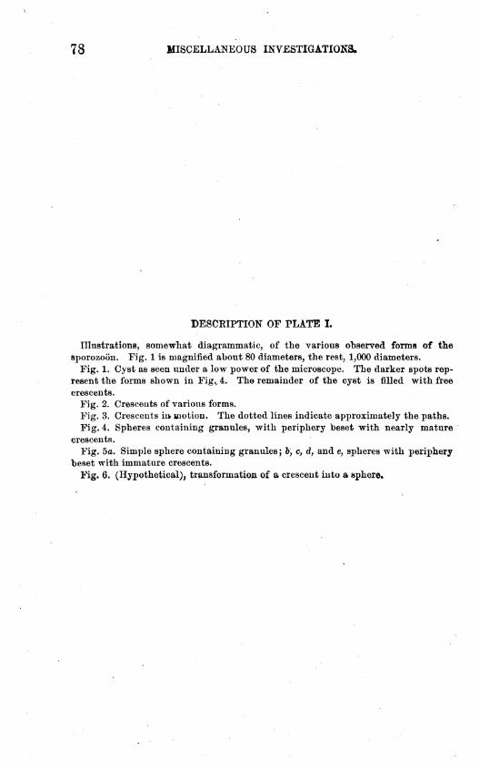
7 8 MISCELLANEOUS INVESTIGATIONS.
DESCRIPTION OF PLATE I.
Illustrations, somewhat diagrammatic, of the various observed forms of thesporozoon. Fig. 1 is magnified about 80 diameters, the rest, 1,000 diameters.
Fig. 1. Cyst as seen under a low power of the microscope. The darker spots rep-resent the forms shown in Figv4. The remainder of the cyst is filled with freecrescents.
Fig. 2. Crescents of various forms.Fig. 3. Crescents in. motion. The dotted lines indicate approximately the paths.Fig. 4. Spheres containing granules, with periphery beset with nearly mature
crescents.Fig. 5a. Simple sphere containing granules; fe, cf d7 and e, spheres with periphery
beset with immature crescents.Fig. 6. (Hypothetical), transformation of a crescent into a sphere.

Bui, 3, Bureau of Animal Industry. PLATE I.
SPOROZOON IN THE INTESTINAL VILLI OF CATTLE.

NOTES ON PARASITES-18: ON THE PRESENCE-OP SARCOSPORIDIA INBIRDS.
By C. W. STILES.
[Plates II and m.]
The first reference I have been able to find in regard to Sarcospo-ridia in birds is an article by J. Kiihn.* This paper is unfortunatelynot at my disposal, but it is evident from the statements of later au-thors that Kiihn found the parasites in chickens.
Eileyt examined a duck whose muscles were infested with parasitesone-fourth to one-fifth of an inch long; they resembled grains of wheatand contained a viscid yellowish substance. Eiley compared them topork measles (Gysticereus cellulosce) and was under the impression thatthey represented the larval stage of some worm which would reachmaturity in the intestine of man. Upon seeing one of the specimens Idescribe below, Eiley stated to me that it was undoubtedly identicalwith his duck measles.
B&LangerJ presented before the Soci6t6 d'Histoire Katurelle deQuibec a case of parasitism in Spatula clypeata Boie. Small " whiteworms" were found by thousands in the muscles. They measured £-1line long, and appeared to belong u& quelques genres voisins destrichines, bien qu'ils ne fussent pas enroules, ni renferm£s dans desenveloppes." This reference, to which Prof. Eiley called my attention,probably refers to the same parasite described below from the same host.
Eivolta§ states:Gli otriceUi psorospermici dei gallinacei, di cui fanno menzione alcuni trattastisti
(R6U ecc.) altro non sono che gli otricelli del Miescher o del Renay, che non si devonoconfondere con i psorbspermi, propriamente detti, dei polli.
On page 398, Z. c, in discussing Enteritepsorospermica, he adds:Alle pareti poi delP intestino si scorgono, situati all' incerca nel sottomucoso, dei
punti bianchicci Bparsi qua et la; quasi tutti della grandezza di uuseme dipapaveroo di un semplice punto. Questi punti bianchicci non sono formati da colonie dipsoro8permi ma bensl da otricelli del Miescher o del Renay pieni di navicelle.
Eivolta also gives two figures of the parasites.*Mittheilungen d. landwirthsch. Instit. zu Halle, 1865, p, 68,tWalsh and Riley. A Measly Wild Duck. The American Entomologist, 1869,
I, p. 89.tLe Naturaliste Canadien, Tome IJ, Mai, 1870, p. 188.}Dei paraaaiti vegetali, Torino, 1873, p. 300, Taf. m, figs. 52, 74.
79

, 8 0 MISCELLANEOUS INVESTIGATIONS.
Leidy* describes some cysts found in the interstices of the musclesof Anas bo8cha8 L. They were oval, white, 2 to 4 mm. long, 0.7 mm.thick, and contained myriads of "fusiform corpuscles'7 17/jt long, re-sembling minute pseudo-navicellse. He adds that "though the Mal-lard is not a fish eater, the bird may have become infected by eatingdiseased fish." Thus it is clear that Leidy was inclined to look uponthese parasites as identical with the Myxosporidia.
Leuckar11 simply states in regard to Sarcosporidia in relation to birds,"selbst das Haushuhn bleibt davon nicht verschont."
Biitschlif refers to the observations by Kiihn and Eivolta as follows:Kiihn§ fand sie auch bei Hiihnem und Eivolta hat auf das Vorkommeu iihnlicher
Parasiten in der Submucosa des Darmes mehrerer Vogel (der Haushiihner, Schwarz-amsel (Turdm merula), des Rabens, etc., aufnierksam gemacht und es scheint auch,dass diesselben mit Eecht den parasitischen Schlauchen der Saugethiere an dieSeite gesetzt werden.
Ziirn|| states that Sarcosporidia (Psorospermienschlauche) have beenreported in the muscles, kidneys, and intestinal walls of various birds.He himself had never found them. The parasites are in some casesmicroscopic, in others visible to the naked eye; they may be 1 or sev-eral mm. long and are surrounded by a resistant but porous membrane.The crescents are 0.01 mm. long and contain one or more vacuoles,together with granules. They are not scattered regularly through theentire cyst, but collected together in balls.
Barrowsfl found a Parula pitiaytimi (Vieill.) whose muscles wereparasitized, and upon examining one of my specimens Mr. Barrowssaid that the parasite appeared identical with those he found, so far asthis could be determined without a microscopical investigation. Hisdescription reads:
One specimen (i. e., Parula pitiayumi) seemed to be abnormal in coloring, show-ing many white feathers in the forehead, and upon skinning the flesh was foundthickly spotted with oval, white lumps about the size of the egg of the commonblow-fly. These were most numerous toward th© surface of the pectoral muscles,but occurred also deeply imbedded in their substance as well as in the muscles ofwings and legs. It was not practicable to examine their structure with the micro-scope until the next day, when decomposition was so far advanced that little couldbe made out. In all probability, however, they were the encysted larvae of someparasitic worm, though whether they had anything to do with the abnormal plumageis an open question.
Barrows' description renders it almost certain that this was a case ofsarcosporidiosis, although it is, of course, impossible to determine
* Psorotperms of Mallard Duck. Proc. Acad. of Science, Phila., 1875, xxvm, p. 125.tDie Parasiten des Menschen, 2. Aufl., 1879, I, p. 254.t Protozoa, I, p. 605. Bronn's Klassen und Ordnungen des Thier-Reichs, 188CM82.$ Sopra i vajuolo dei Columbi e dei Polli. Giornale di Anatomia, Fisiologia e Pata-
logia degli Animali, 1874, vi, p. 257. (N. B.—-The pagination of this journal is in-correct. It should read, p. 321.)
|| Die Krankheiten des Hausgefltlgels, 1882, p. 146.1T Walter B. Barrows. Birds of the Lower Uruguay. Bull, of the Nuttall Ornitho-
logical Club, vin, 1883, p. 87.

NOTES ON PARASITES. ' * 81
Vhether the parasites were identical with any of the forms describedbelow or not. From the size of the animal (" the size of the eggs of thecommon blow-fly", Lucilia ccesar) it is probable that the parasites be-longed to the genus Balbiania.
B. Blanchard* does not believe that the parasites mentioned byEivolta in chickens are Sarcosporidia, but rather that they belong toEimeria.
Mr. Haines, artist, upon seeing my specimens, said that he had noticedthis same parasitized condition of the pectoral muscles in chickens uponseveral occasions, while Prof. 0. Hart Merriam, of this Department, tellsme that he has not infrequently seen similar bodies in the muscles ofvarious birds.
I have mentioned above* all the references I have been able to tracehere at Washington of parasitic Sarcosporidia in birds.- While this list may not be entirely complete, it is quite evidentthat this parasitism, although by no means rare, has attracted verylittle attention, and that we have as yet no exact description of any onespecies of Sarcosporidia in birds. On this account I have deemed itadvisable to place on record several cases of this kind which have re-cently come under my observation, and to give a more or less minutedescription of the animals, so far as the condition of the material athand will permit.
1. The U. S. National Museum, through Prof. Riley, has recently re-ferred to this Bureau a portion of the pectoral muscles of the Shovellerpr Spoon-billed Duck {Spatula clypeata), taken by Walter Brett at ClearLake, California.
2. Prom the XJ. S. Army Medical Museum I have a specimen of para-sitized muscle labelled " Cysticerci (?) from the Shovel-bill Duck." (8p.ehfpeata).
3. In our own collection in the Bureau of Animal Industry we pos-sess a specimen labelled " From ducks. Prof. Liiger, Minnesota, saidthat falciform bodies escaped from cysts like Balbiania gigantea?
4. In Leidy's collection of parasites, which the University of Penn-sylvania has recently sent to me for revision, I have found a bottlecontaining a portion of muscle labelled " Oval, smooth bodies, no limbs.In muscles of Mallard. Anas boschas. Dr. E. Coues. Ex. Jan. 26,1890."
5. Finally, a second bottle referred to me by the U. S. National Mu-seum contains the legs of a Grosbeak (Habia ludoviciana). The labelreads "Explorations in Manitoba, Walter Brett. Habia ludoviciana £(1166). Riding Mountain, June 16,1892."
An examination of the muscle in all cases shows that it containsnumerous elongated cysts lying parallel to the muscle-fibers and per-fectly visible to the naked eye. The parasites found in specimens 1 to
*Note snr Ies Sarcosporidies et sur an Essai de Classification de ces Sporozoaires.Bull. d. 1. Soo. Zool. d. France, 1885.
6311—No. 3 6

8 2 MISCELLANEOUS INVESTIGATIONS.
4 are identical, while two different forms of Sarcosporidia are found inspecimen 5.
1. Balbiania Rileyi sp. n., 1893.
[Plate H, Figs. 1-5.]
The parasites in specimen 1 are in a tolerably good state of preserva-tion, while specimens 2 to 4 are not so well preserved. They measure1 mm. to 6 mm. in length, 0.48 mm. in diameter in their middle. Theyare rather fusiform, but the ends are somewhat blunt. Coloring liquidsdifferentiate the body into two distinct portions—a center core, whichhas the general form of the entire body and is about 0.16 mm. broad inits greatest diameter, and a peripheral portion having about the samethickness as the center core. In some specMens the peripheral zone iseven broader than the central core. The meshes of the central coremay either be empty or contain a granular matter which does not takestain; while the meshes of the peripheral layer are filled with numerouslancet or falciform bodies, to the staining of which is due the differen-tiation of the entire body into the center core and peripheral layer.
The falciform bodies measure 12 to 14// long by 2/* broad. They areslightly more pointed at one end than at the other. They contain adistinct oblong nucleus (about 2//) which stains (Figs. 4,5) very clearlywith methyl blue or acid carmine. On unstained preparations (Fig.5, b) the nucleus appears as a bright refrangent body. Its position isnot constant. The rest of the falciform ,bojiy is granular. Occasionallya small but indistinct vacuole is noticed.
The outer covering of the Sarcosporidiuin is a very thin (2//) cuticu-lar membrane. In this no striation could be discovered. The entireinner portion of the animal is a meshwork or stroma, in the intersticesof which lie the numerous falciform bodies. Although there is no regu-lar arrangement or form to the meshes of this stroma, in general theyradiate from the center to the periphery and are longer (i. e., in a radialdirection) than broad (i. e., in a tangential direction).
The entire stroma can be separated from the cuticle and pressed out,but I was unable to isolate the meshes one from another. Treated witha strong solution of caustic potash the contents of the meshes weredestroyed. The walls, however, remained intact. In nearly all casesthe wall between any two adjacent meshes appeared to be a simplemembrane, common to both lumina, but in one or two cases the wallssplit longitudinally for a short distance, thus giving the impressionthat each lumen possessed its own wall and that the adjacent walls ofthe two meshes were very intimately united. In places wliere threemeshes came together at one point there was frequently a triangularspace between the three membranes.
The parasites lie between the muscle-fibers, surrounded by connect-ive tissue. A very careful examination of a number of microtome sec-tions failed to show any young stages inside of the muscle-fibers.
It is evident from the above that this parasite is very closely allied to

NOTES ON PARASITES. 8 3
Balbiania gigantea Bailliet, and B. mucosa B. Bl. Placing it in thegenus Balbiania, I propose the specific name B. Bileyij dedicating theBpecies to my colleague, Prof. 0. V. Biley, Entomologist of the UnitedStates Department of Agriculture, as he is evidently the first scientistwho examined this parasite.
Biagnosis.—B. Bileyi, sp. n., 1893. 1-6 mm. long by 0.48mm.broad;rather fttsiform, ends not sharply pointed. Cuticle not striated, about2/1 thick. Center core does not color and does not contain falciform
i bodies. Peripheral zone as broad as central core (0.16:0.16 mm.) or evenbroader; colors in various liquids (acid carmine or methyl blue), andcontains numerous falciform bodies. Form of meshes irregular, butelongated radially. Falciform bodies 12 to 14/i long, more pointed atone extremity than at the other; contain a very distinct nucleus (2//)Which stains clearly in acid carmine or methyl blue, and which containsseveral chromatophil granules; vacuole quite indistinct.
Habitat.—Intermuscular connective tissue of ducks (the Shoveller orShovel-bill Duck or Spoon-bill Duck (Spatula clypeata) and the MallardDuck or Tame Duck (Anas boschas).
Development unknown.Geographical distribution.—North America. (?) Philadelphia, Pa.
(Coues-Leidy); St. Louis, Mo. (Eiley); Clear Lake, Cal. (Brett); Min-nesota (Liiger); Quebec (B&anger).
Type material deposited in the U. S. National Museum, in theBureau of Animal Industry, and in Collection of Stiles, Washington,D. C. Specimens are also to be found in the Army Medical Museum,Washington, D. C, and in Collection of Leidy, University of Pennsyl-vania, Philadelphia, Pa.
In conclusion, although "measly duck" is not very appetizing in ap-pearance, there are no grounds for believing that it is dangerous to man.
Specimen 5, mentioned above, contained, as already stated, twodifferent forms of Sarcosporidia, which could easily be seen throughthe skin of the animal. The difference between the two forms, how-ever, could be recognized only in microscopic preparations. One formwas thicker, the cuticle unstriated so far as I could discover, a centralnon-staining core was present, and the parasites were intermuscular.The other form was much thinner, possessed a transversely striatedcuticle, contained no central non-coloring core, and the parasites wereintramuscular, i. e., inside of the muscular fibers.
Blanchard (I. c.) has proposed the following provisional classificationof the Sarcosporidia:
I. Family Miescheridce :Situated in the striated muscles, enveloping membrane—
a. Thin and structureless MietcheHa.ft. Thick and traversed by fine canals Sarcoeystti.
II. Fmnfty Balbianida :Situated in the connective tissue, enveloping membrane thin and
without structure Balbiania.

84 MISCELLANEOUS INVESTIGATIONS.
I have already stated * that I agree with those authors who considerthat the membrane of Sarcocystis is finely striated, instead of possess-ing pores—there can be absolutely no doubt in regard to this point in8. tenella of sheep—so that according to Blanchard's classification, ifwe substitute " finely striated" in place of " traversed by fine canals,"the thicker specimens mentioned above will belong to the genus Balbi-ania, while the thinner form must be placed in the genus Sarcocystis.
Finding, as I have, the two forms lying side by side in the same host,there are two alternatives open to us to explain their presence:
(1) Either we have here a case of infection by two Sarcosporidia ofdifferent genera ; or (2) Sarcoeystis is only a younger intramuscularstage of the intermuscular Balbiania.
Although the second explanation is very tempting to us, yet havingbut a portion of a single animal infested with the two forms now underconsideration, it would be too hazardous for me to state that this is thecorrect explanation, and to attempt to prove it, especially as the fol-lowing facts can not be considered confirmatory of this view:
(1) In the case of B. Rileyi, I have found no intramuscular stage.(2) In the horse, sheep, and hogs, it is a very common occurrence
to find only intramuscular Sarcosporidia.Thus, for the present, I feel compelled to place the two forms now
under consideration in two separate genera, Sarcocystis and Balbiania,respectively, although I will give to both the same specific name, sothat they may more easily be united in case they prove to be twostages of the same species.
2. Balbiania falcatula sp. n.; 1893.[Plate LEI, Figs. 1-2.]
This form appears quite distinct from B. Rileyi, in tbat it islonger in proportion to its breadth; the central non-coloring core ismuch wider in proportion to the peripheral coloring portion; themeshes in the center core are smaller and more regular, while themeshes of the peripheral zone generally have a more regular outline;the falciform bodies are only about half as large (6//) as those of B.Rileyi, and generally more curved; they possess a very prominentnucleus, but no vacuole or portion resembling a vacuole could be dis-covered, except that the middle portion stained more lightly than theends.
Specific diagnosis.—B. falcatula sp. n., 1893. The parasites measure1.3mm.to 3.2mm.longby 0.4mm. to0.42mm. broad; fusiform; cuticle notstriated, about 2pi thick. Center core does not oolo?, and contains nofalciform bodies; meshes quite regular, 8// to 12//. Peripheral zonemuch narrower than centre core (0.096 mm. : 0.224 mm.), colors invarious liquids (acid carmine, for instance) and contains numerous
* Review of recent publications in Medical Zoology. The Journal of Compar-ative Medicine and Veterinary Archives; 1891, p. 693.

NOTES ON PARASITES. 8 5
Jcifonn bodies measuring 5/* to 6/< long by 2// broad; the latter curved,mtaining a distinct nucleus (1.5/*) generally situated at one end; noEtcuole visible (alcohol material).Habitat—In>termuscular connective tissue of the Bose-breastedrosbeak (Habia ludoviciana).Development unknown.Geographical distribution.—Korth America, Manitoba (Brett).Type material deposited in the U. S. National Museum, in the Bu->au of Animal Industry and in Collection of Stiles, Washington, D. 0.
3. Sarcocystls falcatula sp. n., 1893.
[Plate m, Fig. 8.]
These parasites lie within the sarcolemma; they measure on an aver-se 2.4 mm. long by 0.152 mm. broad; the ends are pointed,the membraneH thick) is finely striated; no non-coloring central core could be dis-lguished. At the extremities the divisions or meshes in which theIciform bodies lie are more rounded and distinct, but near the middlethe body it is almost impossible to recognize these divisions. The
Iciform bodies I could measure only on microtome sections. They»peared about 6JA long.Specific diagnosis.—8. falcatula sp. n., 1893. Body 2.4 mm. long byL52mm. thick5 fusiform; cuticle finely striated; meshes at extremitiesLinded and distinct, in middle portion indistinct; falciform bodies 6/*ig.Habitat—Muscle-fibers of Habia ludoviciana.Life history unknown.Geographical distribution.—North America, Manitoba (Brett).rype material deposited in the U. S. National Museum, in the Bu»iu of Animal Industry, and in Collection of Stiles, Washington, D. 0.
Species inquirendse.
L Sarcosporidia of chickens found by Kiihn (1867), at Halle, Ger-my, and by Haines (cited above) in Maryland, TJ. 8. A.5. Sarcosporidia—probably Balbiania sp. found by Barrows in Parulaiayumi, at Ooncepcion del Uruguay, province of Entre Eios, Argen-e Eepublic, South America. .I. 1 Saroosporidia described by Eivolta (1874) from chickens, Turdusrula and crows (cited from Biitschli).

DESCRIPTION OF PLATE II.
Pig. 1. A portion of the pectoral muscles of a duck (Measly duck).Fig. 2. Longitudinal section of Balbiania Bileyi (greatly enlarged).Fig. 3. Transverse section of Balbiania Bileyi (greatly enlarged); ct, connecth
tissue cyst, with numerous nuclei; ou, cuticle of the parasite; m, sections of muscLFig. 4. Microtome section of meshes containing falciform bodies, greatly enlarge*Fig! 5. Falciform bodies of Balbiania Bileyi; a, stained, showing nucleus an
vacuole; b, unstained.All drawings by Mr. Haines, artist, detailed to this Bureau, drawn either fro:
nature or after sketches by author.

Bui. 3, Bureau of Animal Industry.PLATE II.
BALBIANIA RILEYI SP. "•


DESCRIPTION OF PLATE m .
Fig. 1. Falciform bodies of Balbiania falcatula.Fig. 2. Balbiania falcatula, longitudinal section.Fig. 3. SarcocyBtis falcatula, longitudinal section. It will be noticed that the ends
of the parasite are included in a muscle-fiber which has lost part of its transversestriation. In the specimen here drawn the muscle-fiber could not be traced the en-tire length of the parasite.
Fig. da, A portion of the striated cuticle, seen on section.88
o

3ul. 3, Bureau of Animal Industry. PLATE
BALBIANIA FALCATULA sp. n., and SARCOCYSTIS FALCATULA sp. n.


![Basic Concepts in Public Health and Tropical Medicine [Infectious Diseases and Public Health 101] (mainly in relationship to parasitic diseases) Daniel](https://img.dokumen.tips/doc/110x75/56649d3b5503460f94a15bb0/basic-concepts-in-public-health-and-tropical-medicine-infectious-diseases.jpg)

![2005 [Birkh_user Advances in Infectious Diseases BAID] Coronaviruses with Special Emphasis on First Insights Concerning](https://img.dokumen.tips/doc/110x75/613ca6159cc893456e1e7db8/2005-birkhuser-advances-in-infectious-diseases-baid-coronaviruses-with-special.jpg)


