Embed Size (px)
Citation preview

7/28/2019 Computers in Ortho - DR G.S FINAL YEAR
http://slidepdf.com/reader/full/computers-in-ortho-dr-gs-final-year 1/74
COMPUTERS IN ORTHODONTICS
7/28/2019 Computers in Ortho - DR G.S FINAL YEAR
http://slidepdf.com/reader/full/computers-in-ortho-dr-gs-final-year 2/74
•
The computer is basically an automaticelectronic machine that performscalculations or derives results based onthe data fed into it.
• A computer is capable of accepting data,performing operations acc. to instructionsand providing the results of theseoperations in comparatively shorterduration of time and with greateraccuracy.

7/28/2019 Computers in Ortho - DR G.S FINAL YEAR
http://slidepdf.com/reader/full/computers-in-ortho-dr-gs-final-year 3/74
• Orthodontists with their love for technolgy andminiaturization have not remained untouched.
• Orthodontic offices use computers for manypurposes ranging from administrativeapplications, clinical applications to researchapplications.

7/28/2019 Computers in Ortho - DR G.S FINAL YEAR
http://slidepdf.com/reader/full/computers-in-ortho-dr-gs-final-year 4/74
History of Information Technology & Systems
Four Basic Periods of Information Technology
• Pre mechanical• Mechanical• Electromechanical• Electronic

7/28/2019 Computers in Ortho - DR G.S FINAL YEAR
http://slidepdf.com/reader/full/computers-in-ortho-dr-gs-final-year 5/74
The Pre-mechanical Age: 3000 B.C. - 1450 A.D.
First Calculators: The Abacus

7/28/2019 Computers in Ortho - DR G.S FINAL YEAR
http://slidepdf.com/reader/full/computers-in-ortho-dr-gs-final-year 6/74
Alan Mathison Turing Father of Modern Computer Science
Concept of algorithm& computation:
The Turing machine
Designed one of thefirst electronic
programmable digitalcomputers:
THE COLOSSUS
British mathematician& cryptographer

7/28/2019 Computers in Ortho - DR G.S FINAL YEAR
http://slidepdf.com/reader/full/computers-in-ortho-dr-gs-final-year 7/74
The Electronic Age: 1940 - Present
• First Generation (1951-1958) - Punch Cards
•
Second Generation (1959-1963) –Transistors• Third Generation (1964-1979) -Integrated
Circuits
• Fourth Generation (1979-Present)

7/28/2019 Computers in Ortho - DR G.S FINAL YEAR
http://slidepdf.com/reader/full/computers-in-ortho-dr-gs-final-year 8/74
Classification Of Applications
1) Administrative applications
• Patient case records• Recall appointments• Accounts• Patient correspondence• Billing• Inventory lists• Prescription formats• Post-treatment instructions• Insurance claims• Referral information•
Missed appointment follow up.

7/28/2019 Computers in Ortho - DR G.S FINAL YEAR
http://slidepdf.com/reader/full/computers-in-ortho-dr-gs-final-year 9/74
2) Clinical applications
• Patient photographs & radiographs• Patient motivation• Appliance design using CAD CAMs• Growth predictions•
Visual treatment objectives• Generation of pre & post-treatment photographs• Survey information/epidemiological data• Presentations•
Continuing dental/ medical education• Literature reviews• Entertainment

7/28/2019 Computers in Ortho - DR G.S FINAL YEAR
http://slidepdf.com/reader/full/computers-in-ortho-dr-gs-final-year 10/74
3) Miscellaneous applications
• Survey information / epidemiological data• Presentations• Continuing dental / medical education• Literature reviews •
Entertainment

7/28/2019 Computers in Ortho - DR G.S FINAL YEAR
http://slidepdf.com/reader/full/computers-in-ortho-dr-gs-final-year 11/74
CAD / CAM
• CAD Computer AidedDesigning
• CAM
Computer AidedManufacturing
• Occlusal Splints• Planning Surgeries• Bone Implants• Restorations• Designing structures for
FEAnalysis• Appliances and equipment
Application of computers in orthodontics – Research sem1/sb/09-04

7/28/2019 Computers in Ortho - DR G.S FINAL YEAR
http://slidepdf.com/reader/full/computers-in-ortho-dr-gs-final-year 12/74
• Surface geometry of casts - scanned by 3Dlaser surface scanner
• Facial morphology – 3D triangular facets – connecting spatial coordinates – landmarks fromfrontal & lateral cephalograms
• 3D virtual image for surgical simulation
Application of computers in orthodontics – Research/Clinical / Manufacturing sem1/sb/09-04
Procedure for designing a splint

7/28/2019 Computers in Ortho - DR G.S FINAL YEAR
http://slidepdf.com/reader/full/computers-in-ortho-dr-gs-final-year 13/74
• 3D virtual image used to plan the amount anddirection of bone displacement – post surgically
• Surgical splint designed on this image
•
3D graphic image of surgical splint istransferred to a laser lithography unit forpolymerization
Application of computers in orthodontics – Research / Clinical / Manufacturing sem1/sb/09-04

7/28/2019 Computers in Ortho - DR G.S FINAL YEAR
http://slidepdf.com/reader/full/computers-in-ortho-dr-gs-final-year 14/74
CA D/CA M FA B RICA TION OF OCCLUSA LSPLINT FOR ORTHOGNA THIC SURGERY
LASER SCANNINGOF THE CAST
IMAGE OF CASTON MONITOR

7/28/2019 Computers in Ortho - DR G.S FINAL YEAR
http://slidepdf.com/reader/full/computers-in-ortho-dr-gs-final-year 15/74
IMAGES OF STUDY CASTS AND FACIALSKELETON COMBINED FOR SIMULATION
7/28/2019 Computers in Ortho - DR G.S FINAL YEAR
http://slidepdf.com/reader/full/computers-in-ortho-dr-gs-final-year 16/74
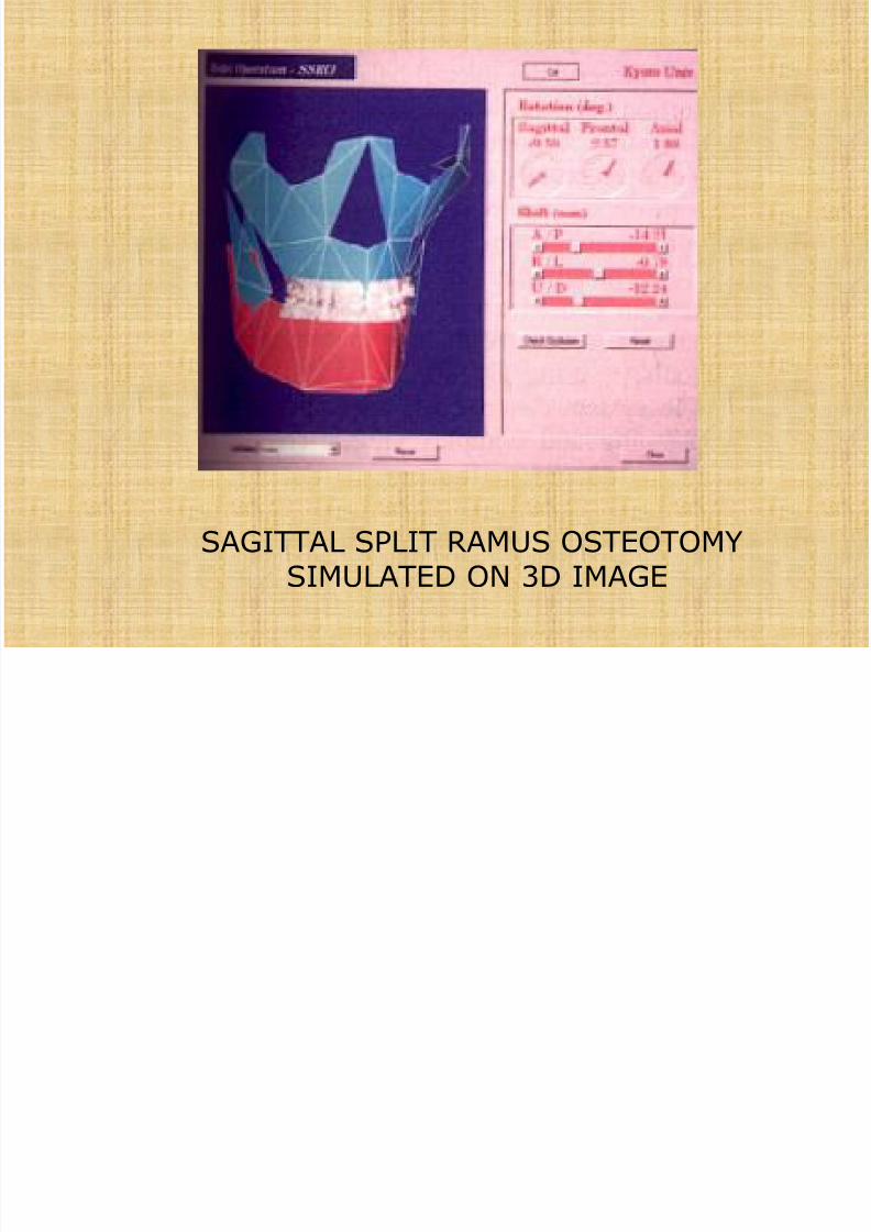
SAGITTAL SPLIT RAMUS OSTEOTOMYSIMULATED ON 3D IMAGE
7/28/2019 Computers in Ortho - DR G.S FINAL YEAR
http://slidepdf.com/reader/full/computers-in-ortho-dr-gs-final-year 17/74
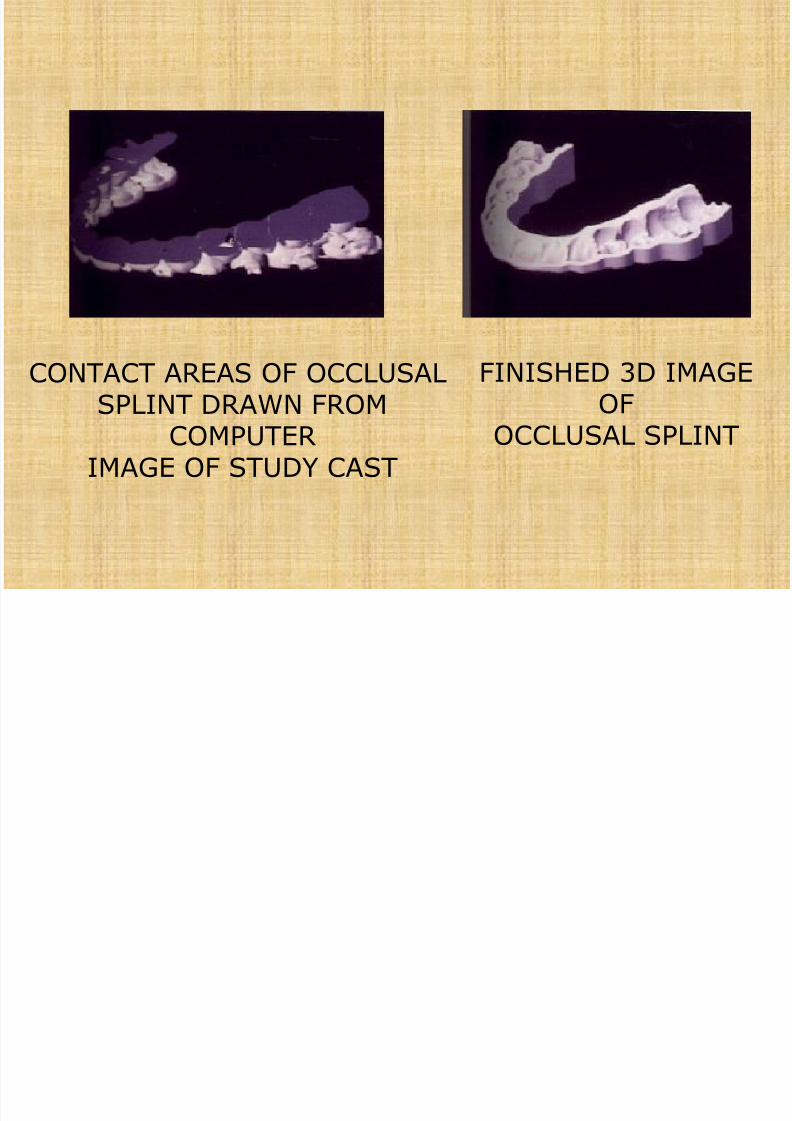
CONTACT AREAS OF OCCLUSALSPLINT DRAWN FROM
COMPUTER IMAGE OF STUDY CAST
FINISHED 3D IMAGEOF
OCCLUSAL SPLINT
7/28/2019 Computers in Ortho - DR G.S FINAL YEAR
http://slidepdf.com/reader/full/computers-in-ortho-dr-gs-final-year 18/74
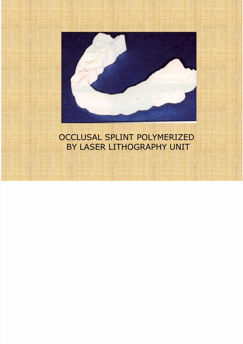
OCCLUSAL SPLINT POLYMERIZEDBY LASER LITHOGRAPHY UNIT
7/28/2019 Computers in Ortho - DR G.S FINAL YEAR
http://slidepdf.com/reader/full/computers-in-ortho-dr-gs-final-year 19/74
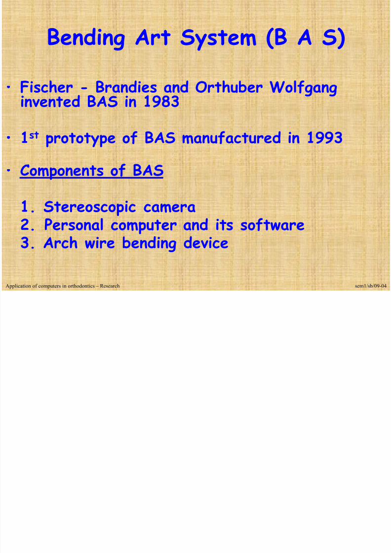
Bending Art System (B A S)
• Fischer - Brandies and Orthuber Wolfganginvented BAS in 1983
•
1st prototype of BAS manufactured in 1993• Components of BAS
1. Stereoscopic camera2. Personal computer and its software3. Arch wire bending device
Application of computers in orthodontics – Research sem1/sb/09-04
7/28/2019 Computers in Ortho - DR G.S FINAL YEAR
http://slidepdf.com/reader/full/computers-in-ortho-dr-gs-final-year 20/74
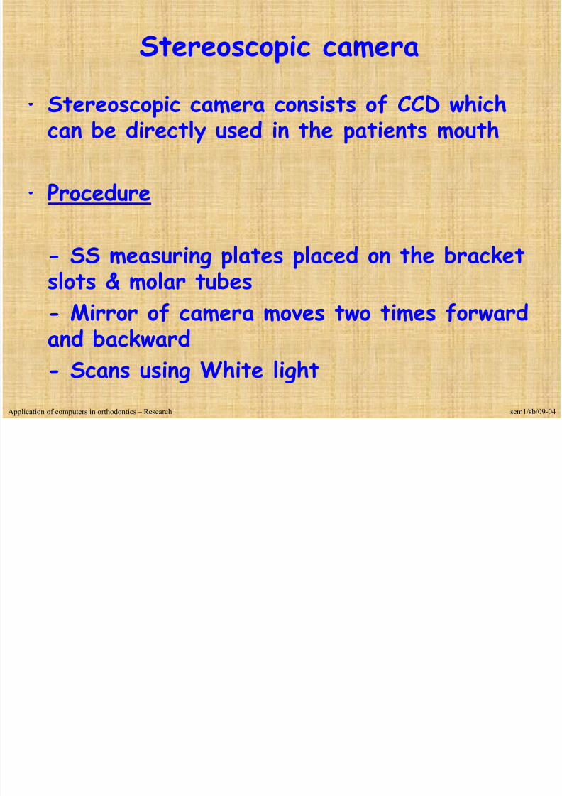
Stereoscopic camera
• Stereoscopic camera consists of CCD whichcan be directly used in the patients mouth
•
Procedure
- SS measuring plates placed on the bracketslots & molar tubes- Mirror of camera moves two times forwardand backward- Scans using White light
Application of computers in orthodontics – Research sem1/sb/09-04

7/28/2019 Computers in Ortho - DR G.S FINAL YEAR
http://slidepdf.com/reader/full/computers-in-ortho-dr-gs-final-year 21/74
Bending Art System (B A S)
Application of computers in orthodontics – Research sem1/sb/09-04

7/28/2019 Computers in Ortho - DR G.S FINAL YEAR
http://slidepdf.com/reader/full/computers-in-ortho-dr-gs-final-year 22/74
Arch Wire Bending Component
• Three components
> Holding cone> Inner cone> Partial cone
Application of computers in orthodontics – Research / Manufacturing sem1/sb/09-04

7/28/2019 Computers in Ortho - DR G.S FINAL YEAR
http://slidepdf.com/reader/full/computers-in-ortho-dr-gs-final-year 23/74
- Round wire and Rectangular wire used
- SS, TMA or Ni-Ti wires
- Bending begins only after all bends aremanipulated
- Approximately takes 5 – 7 min
Application of computers in orthodontics – Research / Manufacturing sem1/sb/09-04

7/28/2019 Computers in Ortho - DR G.S FINAL YEAR
http://slidepdf.com/reader/full/computers-in-ortho-dr-gs-final-year 24/74
Advantages
• Precision arch wires• Rapid fabrication•
Fabricate full sizepassive arch wiresfor surgical cases
• T loop & L loopconstruction
• Utility arches made
Disadvantages
• Time required forthe insertion ofmeasuring plates &their identification
• Clinical judgment isstill vital
•
Steel wire easilydeformed
Application of computers in orthodontics – Research / Manufacturing sem1/sb/09-04
7/28/2019 Computers in Ortho - DR G.S FINAL YEAR
http://slidepdf.com/reader/full/computers-in-ortho-dr-gs-final-year 25/74
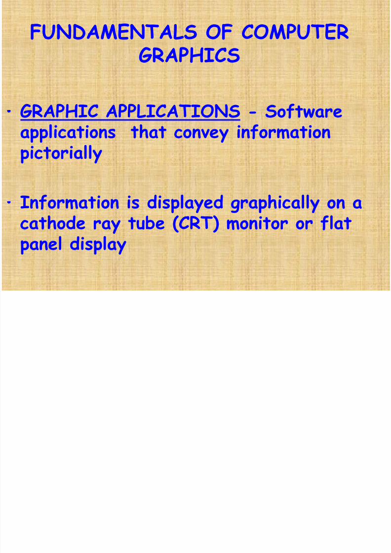
FUNDAMENTALS OF COMPUTERGRAPHICS
• GRAPHIC APPLICATIONS - Software
applications that convey informationpictorially
•
Information is displayed graphically on acathode ray tube (CRT) monitor or flatpanel display

7/28/2019 Computers in Ortho - DR G.S FINAL YEAR
http://slidepdf.com/reader/full/computers-in-ortho-dr-gs-final-year 26/74
Pixel•
The image space on amonitor is made of tinysquare picture elements,called pixels (using thecommon abbreviation"pix" for "picture“)
•
Arranged in the seriesof horizontal lines calledRaster lines

7/28/2019 Computers in Ortho - DR G.S FINAL YEAR
http://slidepdf.com/reader/full/computers-in-ortho-dr-gs-final-year 27/74
Graphic imagesBitmaps
• Digital images are stored in a matrix of rows &columns of pixel values
• Image-editing programs are designed to workprimarily with bitmapped images
• Corel Paint™, Microsoft Photo Editor™ andPhotoshop™ are examples of bitmap editingprograms.

7/28/2019 Computers in Ortho - DR G.S FINAL YEAR
http://slidepdf.com/reader/full/computers-in-ortho-dr-gs-final-year 28/74
Bitmapped image
7/28/2019 Computers in Ortho - DR G.S FINAL YEAR
http://slidepdf.com/reader/full/computers-in-ortho-dr-gs-final-year 29/74
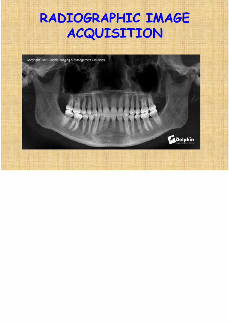
RADIOGRAPHIC IMAGEACQUISITION

7/28/2019 Computers in Ortho - DR G.S FINAL YEAR
http://slidepdf.com/reader/full/computers-in-ortho-dr-gs-final-year 30/74
Denoptix
•
Digital radiographic technique which usesPhosphor imaging plates to capture and storeimages
Advantages
- Alternative to conventional film- Same machine and settings- No dark room required- Environment friendly- no heavy metal
wastage- Can be reused thousands of times
Application of computers in orthodontics – Clinical / Graphic sem1/sb/09-04
I gi g l

7/28/2019 Computers in Ortho - DR G.S FINAL YEAR
http://slidepdf.com/reader/full/computers-in-ortho-dr-gs-final-year 31/74
Imaging cycle
1.Load intraoral or panoramicimaging plate
2. Take X ray
3. Mount imaging plates in carousel 4. Place in scanner & Scan images
5. Erase imaging platesfor reuse
Image on computer

7/28/2019 Computers in Ortho - DR G.S FINAL YEAR
http://slidepdf.com/reader/full/computers-in-ortho-dr-gs-final-year 32/74
CEPHALOMETRIC APPLICATIONS
7/28/2019 Computers in Ortho - DR G.S FINAL YEAR
http://slidepdf.com/reader/full/computers-in-ortho-dr-gs-final-year 33/74
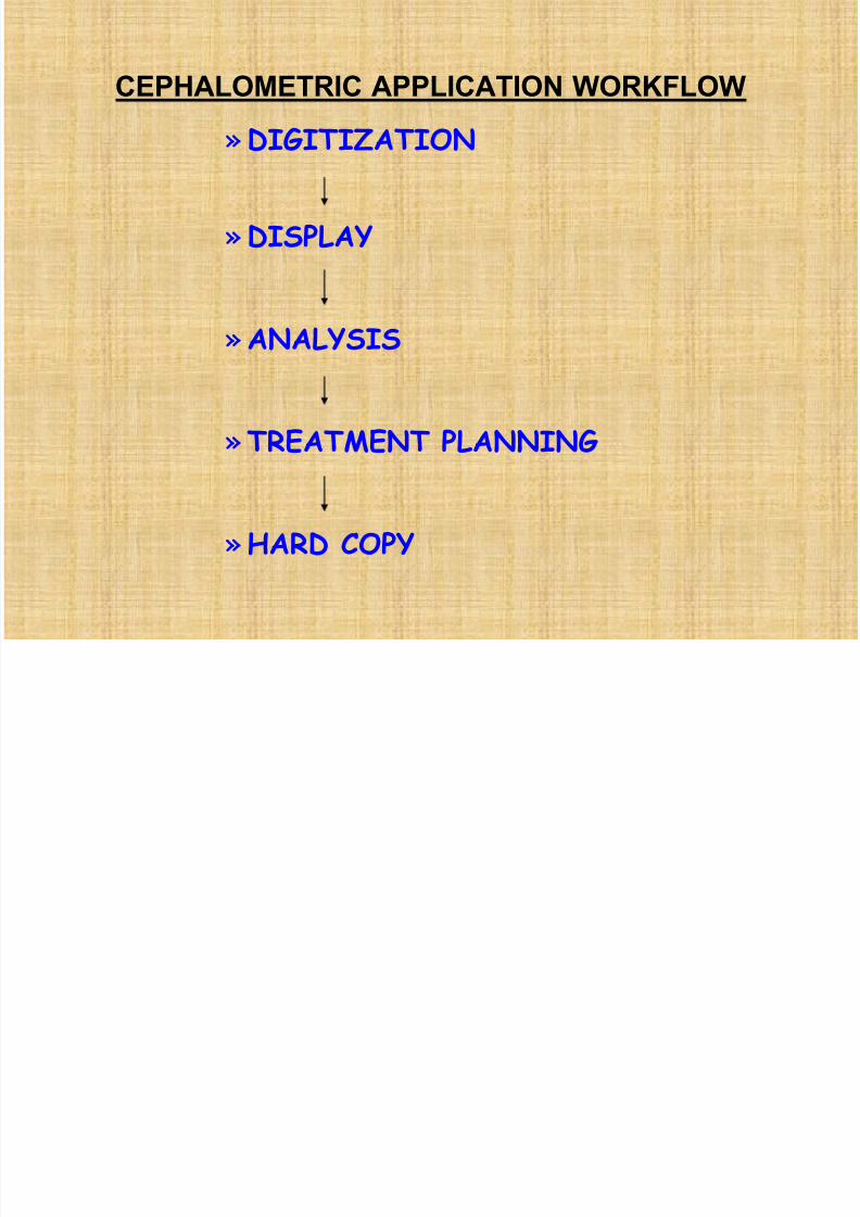
CEPHALOMETRIC APPLICATION WORKFLOW
» DIGITIZATION
» DISPLAY
» ANALYSIS
» TREATMENT PLANNING
» HARD COPY

7/28/2019 Computers in Ortho - DR G.S FINAL YEAR
http://slidepdf.com/reader/full/computers-in-ortho-dr-gs-final-year 34/74
DIGITIZATION•
Is the form by which analog information isconverted to digital form
2 methods used:
• A digitizing table or digitizer with finecross hairs, to locate landmarks &contours on radiographs manually. This istransmitted to computer & recorded for
various cephalometric determinations

7/28/2019 Computers in Ortho - DR G.S FINAL YEAR
http://slidepdf.com/reader/full/computers-in-ortho-dr-gs-final-year 35/74
MODE OF DIGITIZATION
• POINT MODE
discrete location of
individual landmarks

7/28/2019 Computers in Ortho - DR G.S FINAL YEAR
http://slidepdf.com/reader/full/computers-in-ortho-dr-gs-final-year 36/74
• STREAM MODE
a stream of co-ordinatedpairs are recorded asthe user traces the
radiographic contour
7/28/2019 Computers in Ortho - DR G.S FINAL YEAR
http://slidepdf.com/reader/full/computers-in-ortho-dr-gs-final-year 37/74
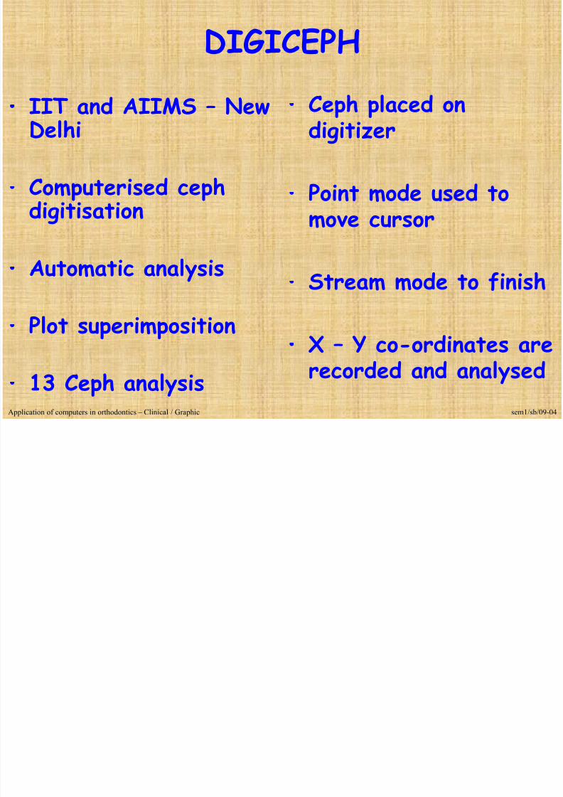
DIGICEPH
• IIT and AIIMS – NewDelhi
•
Computerised cephdigitisation
• Automatic analysis
• Plot superimposition
• 13 Ceph analysis
•
Ceph placed ondigitizer
•
Point mode used tomove cursor
• Stream mode to finish
• X – Y co-ordinates arerecorded and analysed
Application of computers in orthodontics – Clinical / Graphic sem1/sb/09-04

7/28/2019 Computers in Ortho - DR G.S FINAL YEAR
http://slidepdf.com/reader/full/computers-in-ortho-dr-gs-final-year 38/74
Digigraph• Introduced by Dolphin
imaging systems
•
Non radiographicsystem
•
Video imaging is alsopossible
Application of computers in orthodontics – Clinical / Graphic sem1/sb/09-04

7/28/2019 Computers in Ortho - DR G.S FINAL YEAR
http://slidepdf.com/reader/full/computers-in-ortho-dr-gs-final-year 39/74
The Digi-Graph work
station is about 5 feetlong, 3 feet wide and 7feet high.
The main cabinet containsthe electronic circuitry,and the patient sits nextto the cabinet in anadjustable chair similarto those used withcephalometers.

7/28/2019 Computers in Ortho - DR G.S FINAL YEAR
http://slidepdf.com/reader/full/computers-in-ortho-dr-gs-final-year 40/74
The head holder issuspended from a beam,
supported by a verticalcolumn attached to thecabinet.
Ear rods and forehead andposterior head pieces areused to minimize patientmovement.
ear rods can be removed sothat facial and intra-oralimages can be recorded
while the patient is sitting

7/28/2019 Computers in Ortho - DR G.S FINAL YEAR
http://slidepdf.com/reader/full/computers-in-ortho-dr-gs-final-year 41/74
A light box can also beattached to the head holderfor imaging head films, wristfilms and panoramic x-rays.
7/28/2019 Computers in Ortho - DR G.S FINAL YEAR
http://slidepdf.com/reader/full/computers-in-ortho-dr-gs-final-year 42/74
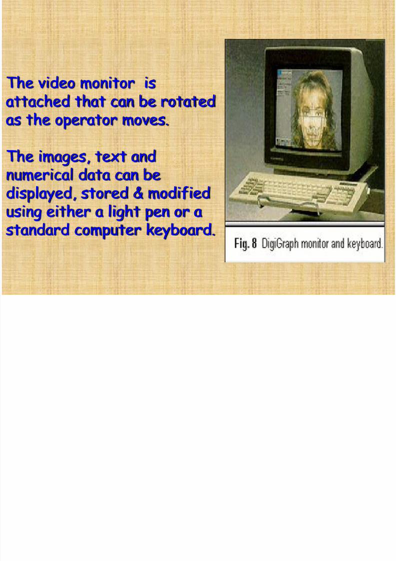
The video monitor isattached that can be rotatedas the operator moves.
The images, text andnumerical data can bedisplayed, stored & modifiedusing either a light pen or astandard computer keyboard.

7/28/2019 Computers in Ortho - DR G.S FINAL YEAR
http://slidepdf.com/reader/full/computers-in-ortho-dr-gs-final-year 43/74
The digitizing handpiece is used torecord cephalometric data
The removable, sterilizable tip of thehandpiece is placed directly on thepatient to record a series of facialand intraoral landmarks. As eachlandmark is located, the handpiecebutton is depressed and the location isrecorded in three-dimensionalcoordinates (x,y,z). Each time the handpiece button is depressed, an audible sound is picked up by an array of four microphones on the beam . Thetime it takes the sound to reach eachof the microphones determines the
landmark location.

7/28/2019 Computers in Ortho - DR G.S FINAL YEAR
http://slidepdf.com/reader/full/computers-in-ortho-dr-gs-final-year 44/74
Any image appearing on the screencan be reproduced instantaneouslywith one of three "hard copy"output devices:
•
Sony video imager: makes 5 " x7 " color prints in 60 seconds• Polaroid freeze-frame cameraproduces Polaroid prints in 10
seconds;• Hewlett Packard Paintjetprinter makes 8 " x 10 " papercolor copies in 4 to 8 minutes.

7/28/2019 Computers in Ortho - DR G.S FINAL YEAR
http://slidepdf.com/reader/full/computers-in-ortho-dr-gs-final-year 45/74
RMO’S JIFFY ORTHODONTIC EVALUATION
»First to provide a computer aidedcephalometric diagnosis to the dentalprofession in the late 1960’s
»Marketed a software packagedescribed as JOE
»JOE generates tracings of lateral orfrontal cephalograms using Ricketts,Jarabak, Sassouni, Steiner &Grummons analyses

7/28/2019 Computers in Ortho - DR G.S FINAL YEAR
http://slidepdf.com/reader/full/computers-in-ortho-dr-gs-final-year 46/74
• JOE can also provide visual representationof normal for comparison to the patienttracings, generate collection ofcephalometric values listed in a logicalorder along the norms and amount ofdeviation from normal and put together a
list of orthodontic problem analyses.
PorDIOS (Purpose on Request Digitizer Input
7/28/2019 Computers in Ortho - DR G.S FINAL YEAR
http://slidepdf.com/reader/full/computers-in-ortho-dr-gs-final-year 47/74
( p q g pOutput System)
•
Is a cephalometric IBM compatible systemwhose development is aimed to provideorthodontists with a user friendly program.
• PorDios works with a digitizer in the standardway and also enables the use of a video orscanner as means of digitization of X-rays.
• Cephalometric analyses used areBjork,Burstone, Coben, Downs, Frontal,McNamara, Profile,Ricketts, Steiner andTweed
• Produces occlusograms from photocopies of

7/28/2019 Computers in Ortho - DR G.S FINAL YEAR
http://slidepdf.com/reader/full/computers-in-ortho-dr-gs-final-year 48/74
Produces occlusograms from photocopies ofdental casts
•
Has built in calculation functions for showingdiscrepancies between the actual mean andits deviation from the norms.
• Main system can automatically alter theorientation of a picture in order to havethe profile looking to the left or right side
of the screen.
• PorDios is multilingual .

7/28/2019 Computers in Ortho - DR G.S FINAL YEAR
http://slidepdf.com/reader/full/computers-in-ortho-dr-gs-final-year 49/74
•
During registration, points can bedeclared as missing or digitized at alater time.
• Drawings can be printed either on amatrix printer as a screen dump, ona laser printer, or on a colourplotter.

7/28/2019 Computers in Ortho - DR G.S FINAL YEAR
http://slidepdf.com/reader/full/computers-in-ortho-dr-gs-final-year 50/74
Dentofacial Planner• Is a computer aided software for diagnostic
and treatment planning in orthodontics andorthognathic surgery
• Works with an IBM compatible processor•
Analyses included are Steiner, McNamara,COGS, Downs, Grummons, Legan & Jarabak.• allows the user to do superimposition,
estimate facial growth, to simulate skeletaland soft tissue effects of orthopedicappliances and to simulate orthodontic toothmovements.
Computer Aided Space Analysis

7/28/2019 Computers in Ortho - DR G.S FINAL YEAR
http://slidepdf.com/reader/full/computers-in-ortho-dr-gs-final-year 51/74
Computer Aided Space Analysis• Chen Hsing Yen
• Bolton ratio
•
Tooth size relation of U & L arches
• Overjet and Overbite
• Posterior intercuspation
•
Arch lengthApplication of computers in orthodontics – Clinical / Graphic sem1/sb/09-04
Computerised Tooth Width Analysis

7/28/2019 Computers in Ortho - DR G.S FINAL YEAR
http://slidepdf.com/reader/full/computers-in-ortho-dr-gs-final-year 52/74
Computerised Tooth Width Analysis• Christopher T.C.Ho
& Terrence.J.Freer• Ho Freer Graphicalanalysis
• Digital callipers ormanual input forWindows
Five Screens
• Patient details• Mesiodistal tooth
width• Tooth width ratio• Tooth width excess• Graphic display
Application of computers in orthodontics – Clinical / Graphic sem1/sb/09-04
Advantages
Convenient, Consistent and Easy to operate
Ali ® T h l

7/28/2019 Computers in Ortho - DR G.S FINAL YEAR
http://slidepdf.com/reader/full/computers-in-ortho-dr-gs-final-year 53/74
Align® Technology
• Align® Technology, Inc. developed theInvisalign appliance for orthodontic toothmovement in the USA in 1998.
• It is an ‘invisible’ way to straighten teethinto a perfect occlusion using thin, clear,overlay sequential appliances.

7/28/2019 Computers in Ortho - DR G.S FINAL YEAR
http://slidepdf.com/reader/full/computers-in-ortho-dr-gs-final-year 54/74
Initial treatment planning with patients’ photographsand radiographs are sent toInvisalign® laboratory
Impressions are convertedinto positive plastermodels & checked forquality.
7/28/2019 Computers in Ortho - DR G.S FINAL YEAR
http://slidepdf.com/reader/full/computers-in-ortho-dr-gs-final-year 55/74
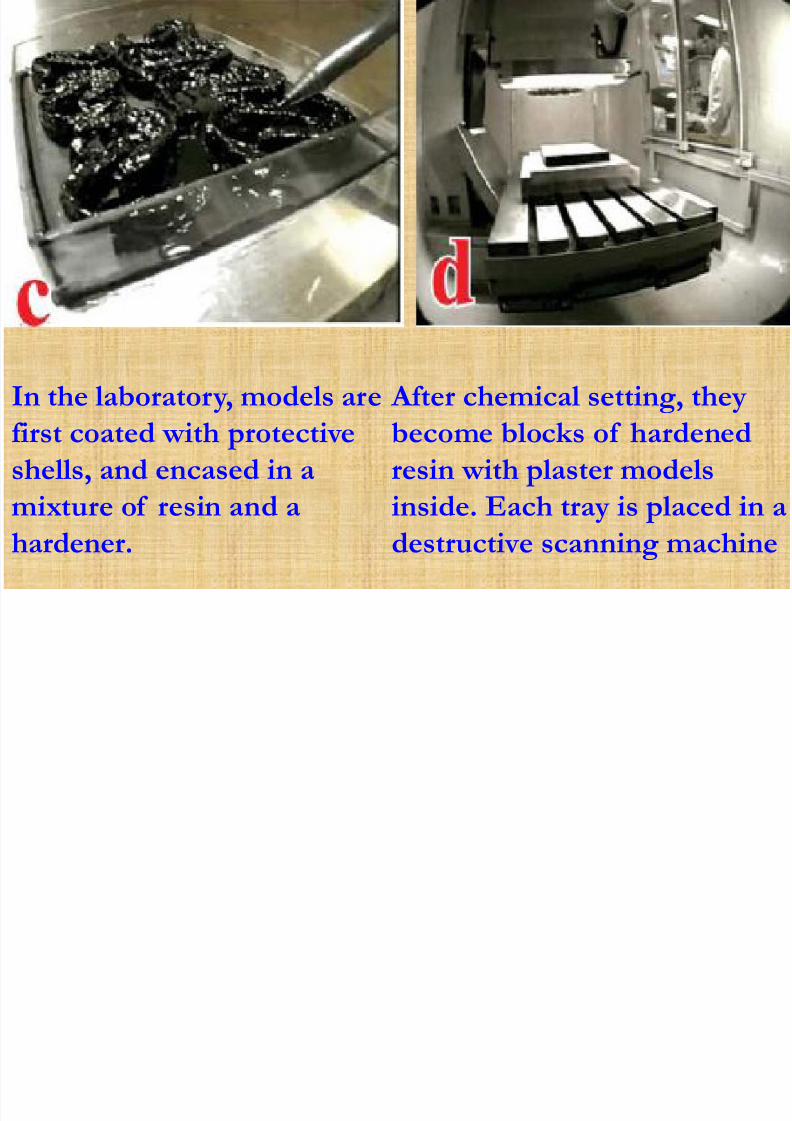
In the laboratory, models arefirst coated with protectiveshells, and encased in amixture of resin and ahardener.
After chemical setting, theybecome blocks of hardenedresin with plaster modelsinside. Each tray is placed in adestructive scanning machine

7/28/2019 Computers in Ortho - DR G.S FINAL YEAR
http://slidepdf.com/reader/full/computers-in-ortho-dr-gs-final-year 56/74
Graphic designers cut outeach tooth and save it as aseparate geometric unit
Once the teeth are separatedand re-assembled back intothe arches, the designerscreate a final set-up of whatthe patient’s teeth will look like when the treatment iscompleted

7/28/2019 Computers in Ortho - DR G.S FINAL YEAR
http://slidepdf.com/reader/full/computers-in-ortho-dr-gs-final-year 57/74
For each stereolithographicconstructed model (whichrepresents a treatmentstage), a clear Invisalign®aligner of 0.030 inchthickness, is created by heat
These aligners are trimmed, polished, cleaned and finallysent to the prescribingorthodontist.

7/28/2019 Computers in Ortho - DR G.S FINAL YEAR
http://slidepdf.com/reader/full/computers-in-ortho-dr-gs-final-year 58/74
• The patient is instructed
to wear each aligner forapproximately 1 –2weeks, and then to moveforward to the next
stage.• A series of evenly
divided 0.15 to 0.25mmmovements are broughtabout at each stage oftreatment.
Advantages :

7/28/2019 Computers in Ortho - DR G.S FINAL YEAR
http://slidepdf.com/reader/full/computers-in-ortho-dr-gs-final-year 59/74
Advantages :
Virtual treatment sequence presents anopportunity to the clinician & the patientfor evaluation of the proposed posttreatment occlusion on screen, before
treatment commences
Proposed treatment can be evaluated by
thorough examination of the entiresequence of tooth movement ,from manyvisual perspectives

7/28/2019 Computers in Ortho - DR G.S FINAL YEAR
http://slidepdf.com/reader/full/computers-in-ortho-dr-gs-final-year 60/74
Contraindications
patients with severe malocclusionsAll children – growing jaws and erupting
teeth make it too complicated for thecomputer to model
Magnetic Resonance Imaging

7/28/2019 Computers in Ortho - DR G.S FINAL YEAR
http://slidepdf.com/reader/full/computers-in-ortho-dr-gs-final-year 61/74
Magnetic Resonance Imaging• Magnetism is a dynamic
invisible phenomenonconsisting of discretefields of forces.
•
Magnetic fields arecaused by movingelectrical charges orrotating electric charges.
• Images generated fromprotons of the hydrogennuclei.

7/28/2019 Computers in Ortho - DR G.S FINAL YEAR
http://slidepdf.com/reader/full/computers-in-ortho-dr-gs-final-year 62/74
Equipment
The Gantry - houses the patientPatient is surrounded bymagnetic coils
Operating console - where theoperator controls thecomputer and scanningprocedure
Computer room network.

7/28/2019 Computers in Ortho - DR G.S FINAL YEAR
http://slidepdf.com/reader/full/computers-in-ortho-dr-gs-final-year 63/74
presence of specific magnetic properties
found within atomic nuclei containingprotons and neutrons.
Inherent property of rotating about theiraxis
Causes a small magnetic field to begenerated around the electrically charged
nuclei.

7/28/2019 Computers in Ortho - DR G.S FINAL YEAR
http://slidepdf.com/reader/full/computers-in-ortho-dr-gs-final-year 64/74
The dipoles exposed within a strong electricfield
Cause Orientation in response to the field
Signal is interpreted and image produced

7/28/2019 Computers in Ortho - DR G.S FINAL YEAR
http://slidepdf.com/reader/full/computers-in-ortho-dr-gs-final-year 65/74
Indications
Assessing diseases of the TMJCleft lip and palateTonsillitis and adenoiditis
Cysts and infectionsTumors

7/28/2019 Computers in Ortho - DR G.S FINAL YEAR
http://slidepdf.com/reader/full/computers-in-ortho-dr-gs-final-year 66/74
CONTRAINDICATIONS
Patients with cardiac pacemakers
Patients with cerebral metallicaneurysm clips. Slight movement ofthe clip could produce bleeding
Stainless steel and other metalsproduce artifacts, obliterate imagedetails of the facial area.

7/28/2019 Computers in Ortho - DR G.S FINAL YEAR
http://slidepdf.com/reader/full/computers-in-ortho-dr-gs-final-year 67/74
Shortcomings
Inability to identify ligament tears orperforations
Dynamics of tissue joint not possible
Cannot be used in patients sufferingfrom Claustrophobia.

7/28/2019 Computers in Ortho - DR G.S FINAL YEAR
http://slidepdf.com/reader/full/computers-in-ortho-dr-gs-final-year 68/74
Computed Tomography
Invented by Sir Godfrey Hounsfield whowas awarded a Nobel prize in 1979
CT is an image display of the anatomyof a thin slice of the body developedfrom multiple x- ray absorptionmeasurements made around the body’speriphery.

7/28/2019 Computers in Ortho - DR G.S FINAL YEAR
http://slidepdf.com/reader/full/computers-in-ortho-dr-gs-final-year 69/74
Parts of the Equipment:
Scanner ( m ovable x
ray table + gantry )
Computer system
A display console

7/28/2019 Computers in Ortho - DR G.S FINAL YEAR
http://slidepdf.com/reader/full/computers-in-ortho-dr-gs-final-year 70/74
Principle:An x-ray source and array of detectors
mounted within the gantry rotatearound the patient during each scan.
Detectors record the attenuation valuesof the beam emerging from thepatient
Information from each traverse is aProfile

7/28/2019 Computers in Ortho - DR G.S FINAL YEAR
http://slidepdf.com/reader/full/computers-in-ortho-dr-gs-final-year 71/74
The tube and detectorsare further angled andanother traverse ismade.
A series of Profiles arebuilt up.
The computer analysesthe data and an image isproduced.

7/28/2019 Computers in Ortho - DR G.S FINAL YEAR
http://slidepdf.com/reader/full/computers-in-ortho-dr-gs-final-year 72/74
Useful in determining
changes in bone density
Primary imaging method
when internal derangementor arthrosis is suspected
Has advantages whenplanning treatment oroperations on jaws and TMJdiseases and deformities.
7/28/2019 Computers in Ortho - DR G.S FINAL YEAR
http://slidepdf.com/reader/full/computers-in-ortho-dr-gs-final-year 73/74
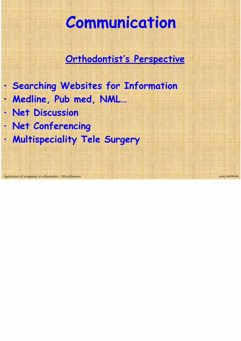
Communication
Orthodontist’s Perspective
•
Searching Websites for Information• Medline, Pub med, NML… • Net Discussion•
Net Conferencing• Multispeciality Tele Surgery
Application of computers in orthodontics – Miscellaneous sem1/sb/09-04

7/28/2019 Computers in Ortho - DR G.S FINAL YEAR
http://slidepdf.com/reader/full/computers-in-ortho-dr-gs-final-year 74/74