Embed Size (px)
Citation preview

RESEARCH ARTICLE
Computational modeling of opioid-induced
synaptic plasticity in hippocampus
Mehdi Borjkhani1, Fariba Bahrami1*, Mahyar Janahmadi2
1 CIPCE, Motor Control and Computational Neuroscience Laboratory, School of ECE, College of
Engineering, University of Tehran, Tehran, Iran, 2 Neuroscience Research Center and Department of
Physiology, School of Medicine, Shahid Beheshti University of Medical Sciences, Tehran, Iran
Abstract
According to a broad range of research, opioids consumption can lead to pathological
memory formation. Experimental observations suggested that hippocampal glutamatergic
synapses play an indispensable role in forming such a pathological memory. It has been
suggested that memory formation at the synaptic level is developed through LTP induction.
Here, we attempt to computationally indicate how morphine induces pathological LTP at hip-
pocampal CA3-CA1 synapses. Then, based on simulations, we will suggest how one can
prevent this type of pathological LTP. To this purpose, a detailed computational model is
presented, which consists of one pyramidal neuron and one interneuron both from CA3, one
CA1 pyramidal neuron, and one astrocyte. Based on experimental findings morphine affects
the hippocampal neurons in three primary ways: 1) disinhibitory mechanism of interneurons
in CA3, 2) enhancement of NMDARs current by μOpioid Receptor (μOR) activation and 3)
by attenuation of astrocytic glutamate reuptake ability. By utilizing these effects, simulations
were implemented. Our results indicate that morphine can induce LTP by all aforementioned
possible mechanisms. Based on our simulation results, attenuation of pathologic LTP
achieved mainly by stimulation of astrocytic glutamate transporters, down-regulation of the
astrocytic metabotropic glutamate receptors (mGlurs) or by applying NMDAR’s antagonist.
Based on our observations, we suggest that astrocyte has a dominant role in forming addic-
tion-related memories. This finding may help researchers in exploring drug actions for pre-
venting relapse.
Introduction
Addiction is a chronic brain disease, in which addicted patients continually exhibit drug-seek-
ing behavior despite its adverse consequences. In an addicted person, cellular and molecular
adaptations in particular brain regions lead to behavioral abnormalities [1, 2]. Drugs induce
plasticity in the functions of neurons and synapses. These modifications can be seen as a form
of "molecular or cellular memory" [3]. In the addiction process, abnormal memories are stored
in specific regions of the brain, including the hippocampus, amygdala, prefrontal cortex, Ven-
tral Tegmental Area, Nucleus Accumbens [2, 3]. As a matter of fact, these abnormal memories
PLOS ONE | https://doi.org/10.1371/journal.pone.0193410 March 7, 2018 1 / 31
a1111111111
a1111111111
a1111111111
a1111111111
a1111111111
OPENACCESS
Citation: Borjkhani M, Bahrami F, Janahmadi M
(2018) Computational modeling of opioid-induced
synaptic plasticity in hippocampus. PLoS ONE
13(3): e0193410. https://doi.org/10.1371/journal.
pone.0193410
Editor: Michel Baudry, Western University of
Health Sciences, UNITED STATES
Received: August 28, 2017
Accepted: February 9, 2018
Published: March 7, 2018
Copyright: © 2018 Borjkhani et al. This is an open
access article distributed under the terms of the
Creative Commons Attribution License, which
permits unrestricted use, distribution, and
reproduction in any medium, provided the original
author and source are credited.
Data Availability Statement: All relevant data and
equations needed to regenerate the results have
been presented in the paper. Additional program
files (codes of simulations) are available from the
Corresponding Author at [email protected].
Funding: The authors received no fundings for this
work.
Competing interests: The authors have declared
that no competing interests exist.

are responsible for addiction syndromes. Drug-induced changes in the functions of glutama-
tergic synapses have been considered to be a significant cause of drug-associated memories
[2–6]. Contextual information of drug consumption is stored in the hippocampus and plays a
vital role in drug-related reward property [6, 7] and the relapse [8].
Opioids that are used for the treatment of pain are intensively addictive. Opioids similar to
other drugs may have long-term effects on the affected neurons through Long-Term Potentia-
tion (LTP) or Long-Term Depression (LTD) mechanisms which are thought to be the cellular
basis of memory formation [1, 2]. Many opioids, including morphine, affect the neurons
through μ opioid receptors, which are G-protein coupled receptors (GPCRs). In the presence
of morphine, μOR activation can lead to induction of LTP/LTD [9]. Therefore, assessing opi-
oid-induced LTP/LTD could be the key to understanding the mechanism by which opioids
induce addiction [2, 10]. Some animal research indicates that morphine affects the hippocam-
pal synapses by inducing LTP in the injected areas, and thereby may trigger the addiction pro-
cess [11, 12].
Opioid-induced induction of LTP in hippocampal neurons is via a disinhibitory mecha-
nism which affects the hippocampal interneurons [13–17]. Opioids cause an inhibition of
GABA release from hippocampal interneurons [16, 17]. So, pyramidal neurons of CA3 receive
less GABA, and inhibitory postsynaptic potentials (IPSPs) in CA3 neurons attenuate [18]. It
seems that inhibition of interneurons by opioids is due to the enhancement of K+ channel
activity, or due to the reduction of Ca2+ channel currents [19]. It has also been shown that inhi-
bition of Ca2+ channels by morphine requires G-proteins activation [20, 21].
On the other side, astrocyte has a dominant role in the induction of LTP and synaptic plas-
ticity [22–27]. Furthermore, astrocyte involves in opioid dependency [28, 29]. It has been
shown that the main player of astrocyte’s contribution to opioids dependency is its important
role in homeostasis, which mainly mediates by astrocytic transporters [28–30].
The goal of this paper is to present a biophysically-induced mathematical model at the syn-
aptic level that can denote some features of the cellular addiction formation by opioids in hip-
pocampus. Based on the experimental evidence, we analyze the effects of opioids on LTP at the
synaptic level due to the activation of μORs using the proposed mathematical model.
In the first part of the paper, neurobiology of opioid signaling in synaptic plasticity and
memory formation will be described. The second part introduces the modeling approach to
describe the opioid signaling process at the synaptic level for the induction of pathological
LTP. In the third part, simulation results are shown for normal and pathological conditions.
Finally, discussion and conclusion of the results are presented.
Neurobiology of opioid signaling in synaptic plasticity and
addiction memory formation
It has been shown that glutamatergic synapses play an indispensable role in the formation of
pathological addiction related memories [5, 31–33]. Furthermore, LTP/LTD induction is sup-
posed to be a key player in synaptic plasticity and memory formation. Here, we attempt to
describe how morphine can modulate the LTP induction in the CA3-CA1 region of the hippo-
campus and how it may contribute to the pathological memory formation (Fig 1).
Morphine activates μORs of hippocampal interneurons [18]. Replacement of Guanosine
Diphosphate to Guanosine Triphosphate occurs in morphine-sensitive GPCRs. In the follow-
ing, the α subunit is disconnected from βγ dimer. Separated subunits are capable of modulat-
ing Ca2+ channels. For example, Gαmodulates calcium channels in a slow and indirect way
through second messengers. On the other side, Gβγ directly affects Ca2+ channels which cause
them to go to a reluctant state [34]. Thus, morphine seems to inhibit the voltage-gated Ca2+
Computational model opioid synpatic plasticity
PLOS ONE | https://doi.org/10.1371/journal.pone.0193410 March 7, 2018 2 / 31

channels of interneurons [19]. Inhibition of Ca2+ channels in turn leads to less Ca2+ influx into
the interneurons. The consequence of less Ca2+ entry is the suppression of GABA release from
interneurons. Thus, GABAergic receptors of CA3 pyramidal neurons receive less GABA,
which causes more neuronal excitation [18]. Further excitation of pyramidal neurons leads to
more glutamate release, which means CA1 pyramidal neurons and astrocytes are going to be
more excited than normal level. Furthermore, opioids decrease astrocytic glutamate trans-
porter1 (GLT1) activation which is responsible for the glutamate uptake from the synaptic
cleft [28, 30]. As a result, reuptake of glutamate is reduced by astrocyte, and therefore more
glutamate can be found in the synaptic cleft. Enhancement of glutamate in synaptic cleft con-
veys to more excitation of the postsynaptic CA1 neurons.
Opioids also affect the CA1 postsynaptic pyramidal neurons. In fact, it removes the block-
ing effect of Mg2+ on N-methyl-D-aspartate (NMDA) receptors and thereby increases the fast
glutamatergic transmission [14, 31, 35]. The outcome of this process is the augmentation of
calcium entry into the postsynaptic neurons [36, 37]. Enhancement of calcium concentration
in the postsynaptic neurons results in phosphorylation of α-amino-3-hydroxy-5-methyl-4-iso-
xazolepropionic acid (AMPA) receptors and Nitric Oxide (NO) synthesis by Ca2+ Calmodu-
lin-dependent protein kinase II (CAMKII) mechanisms [31, 38]. Then, NO retrogradely
diffuses to the presynaptic CA3 pyramidal neuron and increases the binding capability of pre-
synaptic calcium sensors, which may cause more glutamate release and induction of LTP in
the synapse [31, 39, 40].
Fig 1. Neurobiology of opioid signaling studied in this work. The model consists of a pyramidal neuron’s bouton and a GABAergic single compartment
interneuron from CA3 layer, and a spine of a postsynaptic CA1 pyramidal neuron and an astrocyte. The astrocyte monitors the synaptic transmission,
regulates presynaptic glutamate release and is involved in the postsynaptic transmission by releasing gliotransmitters.
https://doi.org/10.1371/journal.pone.0193410.g001
Computational model opioid synpatic plasticity
PLOS ONE | https://doi.org/10.1371/journal.pone.0193410 March 7, 2018 3 / 31

Modeling approach
As mentioned earlier, the goal of this paper is to present a biophysically-driven mathematical
model at the synaptic level in hippocampus in order to describe the cellular addiction forma-
tion by opioids. The main components of the proposed model are: (1) a bouton of a presynap-
tic CA3 pyramidal neuron, which can produce action potentials (APs) by receiving external
stimulations and generates NO which can act as a retrograde signal that enhances the release
of glutamate, (2) a GABAergic single compartment interneuron at the CA3 layer which inhib-
its the CA3 pyramidal neuron, (3) a spine of a postsynaptic CA1 pyramidal neuron which can
produce Excitatory Postsynaptic Potentials (EPSPs) based on the amount of glutamate in the
synaptic cleft; functions of NMDA and AMPA receptors of the postsynaptic neuron are also
considered in this model, and (4) an astrocyte which monitors the synaptic transmission, regu-
lates presynaptic glutamate release and is involved in the postsynaptic transmission by releas-
ing gliotransmitters. The block diagram of different parts of the model is presented in Fig 2.
Fig 2. Block diagram of different parts of the proposed model. External stimulation is followed by action potential generation in the presynaptic pyramidal
neuron and the interneuron. Consequently, level of presynaptic calcium is elevated through two fast and slow mechanisms. Fast calcium oscillations are due to
APs, and slow ones are because of IP3 production. Gliotransmitters activate mGlurs and contribute to slow calcium oscillations by IP3 production. Calcium
enhancement results in glutamate release. Interneuron’s activation modulates GABA receptors in pyramidal neuron and is sensitive to the opioid receptor’s
activation. Released glutamate reaches to the postsynaptic neuron and the astrocyte. The astrocyte releases gliotransmitter which affects presynaptic and
postsynaptic neurons. Functions of AMPARs and NMDARs are considered in the postsynaptic neuron. Activation of the receptors enables CaMKII
phosphorylation process. Exceeding phosphorylated CaMKII from a threshold leads to NO production which retrogrades to the presynaptic neuron. Finally,
postsynaptic opioid receptors modulate NMDARs activation.
https://doi.org/10.1371/journal.pone.0193410.g002
Computational model opioid synpatic plasticity
PLOS ONE | https://doi.org/10.1371/journal.pone.0193410 March 7, 2018 4 / 31

Presynaptic neurons
For modeling the presynaptic CA3 pyramidal cell and interneuron, we have utilized Hodgkin-
Huxley (H-H) type formulation to generate APs in presynaptic neurons [41]:
dVpre
dt¼ fPyrðIapp; INa; IK ; IL; IGABAA
Þ ð1Þ
CdVInt
dt¼ fIntðIapp; INa; IK ; ILÞ ð2Þ
APs can be generated due to externally applied current (Iapp) in the presence of sodium
(INa), potassium (IK) and leak (IL) ionic currents (full description of fPyr and fInt can be found
in Appendix). Besides, we have adapted and added a GABAergic inhibitory current (IGABAA) to
the presynaptic pyramidal neuron similar to the formulation used by Zachariou et al. [42]:
IGABAA¼ 10gðVpre þ 80Þ ð3Þ
In Eq (3), g is the ratio of activated GABAA receptors and is described by [34, 42]:
dgdt¼
g1ðVIntÞ � gtg
; g1ðVIntÞ ¼1
1þ e� ðVInt � 100ð1� CaChÞÞ=5ð4Þ
Here, g1(VInt) is assumed to be a sigmoid function which its half maximum voltage
depends on activation of the interneuron calcium channels (CaCh). τg = 1 ms represents time
constant of GABAA receptor.
Hippocampal opioid receptors which are activated by morphine are mostly μORs. These
receptors belong to GPCRs which are activated by morphine or its agonists. Because of opioids
disinhibitory effect on the synaptic transmission in the hippocampus, it is supposed that pre-
synaptic opioid receptors are on the hippocampal interneurons. Recently, Zachariou and col-
leagues proposed a model for hippocampal cannabinoid receptors (CBRs) [42]. This model is
based on Bertram’s minimal model of G-protein activation [34]. Since μORs and CBRs are
both GPCRs, we have adapted this model to describe the activation of μORs in the hippocam-
pus. The presence of morphine (Morph) results in activation of μORs (MOR) which modulates
calcium channels activation (CaCh). Equations derived from [34, 42] are modified as follows
for opioid receptors:
dðMORÞdt
¼MOR1 � MOR
1000ð5Þ
MOR1 ¼ hMORðMorphÞ ¼2
1þ ð0:1=MorphÞ1:2
ð6Þ
dðCaChÞdt
¼ k� ð1 � CaChÞ � kþCaCh ð7Þ
k� ¼0:3
1þ e� VInt
5
; kþ ¼ 0:0006MOR ð8Þ
Maximum number of activated μORs is described by MOR1. This parameter depends on
morphine concentration (Morph). k_ Shows reluctant to willing transition rate of calcium
channel and depends on interneuron’s voltage (VInt), this parameter reflects the voltage-
Computational model opioid synpatic plasticity
PLOS ONE | https://doi.org/10.1371/journal.pone.0193410 March 7, 2018 5 / 31

dependent dissociation of Gβγ from the channel, k+ is the transition rate of the willing to reluc-
tant state for calcium channel and depends on morphine concentration, this parameter reflects
the concentration of the activated G-proteins. It can be seen that morphine presence
activates μORs which is described in Eqs (5) and (6). Consequently, calcium channels activa-
tion in interneuron changes in Eqs (7) and (8). Therefore, the conductance for inhibitory cur-
rent varies in Eq (3) due to changes in Eq (4). Here, for the sake of simplicity, like Bertram
et al., we assumed no dynamics for the neurotransmitter release from presynaptic interneuron
to the pyramidal postsynaptic neuron [34]. Instead of that, we assumed that activation of
VGCCs in interneuron affects directly the conductance of GABAA receptors in the presynaptic
pyramidal neuron [34].
After APs are generated in presynaptic pyramidal neuron, glutamate will be released. Gluta-
mate release depends on calcium concentration in the neuron. Calcium concentration in the
presynaptic neuron can change through fast (cFast) and slow (cSlow) dynamics which is
described by Eq (9).
ci ¼ cFast þ cSlow ð9Þ
Fast calcium oscillations which occur due to Aps, are governed by:
dcFastdt¼ fFastðICa; IPMCa; IPMleakÞ ð10Þ
Here, ICa represents calcium current through N-type channels, IPMCa denotes calcium cur-
rent due to ATPase pump which pumps extra calcium out of the cell, and IPMleak is the leak
current from extracellular space into Bouton (Details about fFast can be found in the
Appendix).
Slow calcium oscillations occur due to the activation of the extrasynaptic mGlurs. Since
mGlurs activation has an indispensable role in synaptic plasticity governed by opioids, we
have considered its role in the model. Activation of mGlurs is due to the binding of gliotrans-
mitters. The presence of gliotransmitters lead to Inositol trisphosphate (IP3) production in an
intracellular space which enhances calcium concentration in the cell using endoplasmic reticu-
lum (ER) calcium resources. This process is modeled using modified Li-Rinzel model intro-
duced by Tewari and Majumdar [22, 43]:
dcSlowdt¼ fSlowðJchan; JERpump; JERleakÞ ð11Þ
Here Jchan denotes calcium influx from ER into cytosol through the IP3 receptor, JERpump is
the pumped calcium from intracellular space to ER and JERleak indicates leaked calcium ions
from ER into intracellular space (Details about fSlow can be found in the Appendix).
Presynaptic Ca2+ ions can bind to Ca2+ sensors of glutamate vesicles and contribute in vesi-
cle release in the synaptic cleft [22, 23, 44]. The kinetic model for the calcium binding to the
calcium sensor is governed by:
X ����! ����5aci
bXðciÞ1 ����! ����
4aci
2bXðciÞ2 ����! ����
3aci
3bXðciÞ3 ����! ����
2aci
4bXðciÞ4 ����! ����
aci
5bXðciÞ5 ����! ����
g
dXðciÞ
�
5ð12Þ
Here, α is the rate of Ca2+ association constant and β is the rate of Ca2+ dissociation con-
stant. γ, δ and β are constants which describe calcium-independent isomerization. X shows the
Ca2+ sensors on vesicles without any Ca2+ bound. X(ci)1 denotes sensor with one Ca2+ bound,
X(ci)5 describes sensor with five Ca2+ bound and finally XðciÞ�
5is the isomer of X(ci)5 which is
ready to release.
Computational model opioid synpatic plasticity
PLOS ONE | https://doi.org/10.1371/journal.pone.0193410 March 7, 2018 6 / 31

In addition to evoked release due to the APs, the release of vesicles can occur spontaneously
when there is no depolarization in the membrane. This type of release also depends on presyn-
aptic Ca2+ concentration and can occur when the Ca2+ concentration is low in a non-depolar-
ized membrane [44, 45]. Spontaneous release rate due to Ca2+ concentration is modeled by
Nadkarni and Jung [45] using Poisson process and is expressed as follows:
lðCiÞ ¼ a3 1þ expa1 � cia2
� �� �� 1
ð13Þ
Where, a1 = 50μM, a2 = 5μM and a3 = 0.85ms−1 are the Ca2+ concentration at which λ is
halved, slope factor of spontaneous release rate λ, and maximum spontaneous release rate,
respectively. Values for these parameters are determined by Tewari and Majumdar such that,
the frequency of spontaneous release is in the range of 1–3 Hz. Experimental findings indicate
that this is a valid frequency range in the presence of astrocyte [22]. In general, glutamate
release dynamics are depicted as follows [22]:
dRdt¼
Itrec� frR;
dEdt¼ �
Etinacþ frR; I ¼ 1 � R � E ð14Þ
Where, the fraction of ready to release vesicles is denoted by R, effective vesicles in synaptic
cleft is represented by E, and inactive vesicles rate is denoted by I. τinac = 3ms is the time con-
stant of vesicle inactivation and τrec = 800ms is the time constant of vesicle recovery. fr is the
probability of vesicle release and has values (0, 0.5, 1). The total number of vesicles that can be
released is assumed to be two vesicles. When two vesicles get ready to be released, the value of
fr is 1, when one vesicle is ready to release value of fr is 0.5, and finally, when there are not any
vesicles to be released fr is 0. The number of vesicles that get ready to be released can be calcu-
lated by stochastic simulation of Bollmann kinetic model for evoked release (by APs) [44] or
by using Poisson function which refers to spontaneous release [22, 23]. There is also an inacti-
vation time of 6.34 ms for vesicle release (evoked or spontaneous release) [45].
Glutamate concentration in synaptic cleft can be altered due to some factors which are
modeled by Tewari and Majumdar in the CA3-CA1 synapse. The fraction of effective vesicles,
glutamate concentration in vesicles and docked vesicles and also degradation rate of glutamate
in synaptic cleft can be considered as significant factors. Glutamate concentration can be rep-
resented by [22] Eq (15).
dgdt¼ nv:gv:E � gc:g ð15Þ
Here, g is the concentration of glutamate in the synaptic cleft, nv = 2 and gv = 60μM are the
number of docked vesicles and glutamate concentration in vesicles respectively. gc = 10ms−1 is
the degradation rate of glutamate which relates to neurotransmitter reuptake by astrocyte.
Neurotransmitter reuptake is one of the major factors in the processing of addiction associated
LTP formation and would be analyzed in this paper.
Astrocyte
Astrocytes release gliotransmitter, which can affect the synaptic transmission. Gliotransmitter
release by astrocyte depends on the Ca2+ concentration in astrocyte. The Released gliotrans-
mitter can influence the presynaptic neuron through mGlurs, then postsynaptic neuron
through NMDARs. In the proposed model, vesicle release dynamics by astrocyte are based on
the Tewari and Majumdar’s model [22]. Furthermore, It has been shown that during con-
sumption of opioids, the astrocytic ability for reuptake is attenuated [28, 29]. A decrease in
Computational model opioid synpatic plasticity
PLOS ONE | https://doi.org/10.1371/journal.pone.0193410 March 7, 2018 7 / 31
astrocyte’s glutamate reuptake ability is a primary cause for LTP induction that has been
assessed during our simulation studies.
Released glutamate in the synapse, activates mGlurs of astrocyte, which leads to the aug-
mentation of astrocytic Ca2+ concentration. Ca2+ Changes in astrocyte are because of IP3
mechanisms. Ca2+ oscillations in astrocyte is described by [46]:
dcAstrodt¼ fAstroðJchan;a; Jpump;a; Jleak;aÞ ð16Þ
Where cAstro denotes Ca2+ concentration in intracellular space of astrocyte, fAstro denotes the
function which relates calcium currents to the calcium concentration. Jchan,a is Ca2+ flux from
ER to the intracellular space due to IP3 binding to ER’s IP3R, Jpump,a is the Ca2+ removing from
intracellular space by SERCA pump, Jleak,a is the Ca2+ leak from ER to intracellular space
(detailed information about fAstro can be found in the Appendix).
Equations which describe vesicle release dynamics by astrocyte are [22] given by Eq (17).
dRa
dt¼
Iatarec
� YðcAstro � cthreshAstro Þ:Pra:Ra;dEa
dt¼ �
Ea
tainact
þYðcAstro � cthreshAstro Þ:Pra:Ra;
Ia ¼ 1 � Ra � Ea
ð17Þ
Here Ra denotes releasable vesicles in astrocyte, Ea shows effective vesicles, Ia is the inactive
vesicles. Θ is Heaviside function, cthreshAstro ¼ 196:69nM is the threshold for calcium that can con-
tribute to vesicle release, tainact ¼ 3ms denotes to inactivation time constant and ta
rec ¼ 800msshows recovery time constant.
It is suggested that three independent gates are responsible for gliotransmitter release and
each of the gates need a calcium ion to bind [22, 47]. So, at least three calcium ions are needed
for opening of the gates to permit vesicle release from astrocyte. This is described by [47] and
depicted as Eq (18).
dOj
dt¼ kþj :cAstro � ðk
þ
j :cAstro þ k�j Þ:Oj ð18Þ
Here Oj denotes the opening probability of the gate, kþj is the opening rate of the
gate, k�j is the closing rate of the gate and cAstro denotes calcium concentration
(kþ1¼ 3:75 � 10� 3=mM:ms,k�
1¼ 4 � 10� 4=ms,kþ
2¼ 2:5 � 10� 3=mM:ms,k�
2¼ 1 � 10� 3=ms,
kþ3¼ 1:25 � 10� 2=mM:ms,k�
3¼ 1 � 10� 3=ms). So Pra = O1.O2.O3 denotes opening probability
of a vesicle release site. Finally, Gliotransmitter dynamics in synapse is described by [22]:
dgadt¼ nv
a:gva :Ea � gc
a:ga ð19Þ
Here ga indicates gliotransmitter concentration, nva ¼ 12 denotes the number of vesicles,
gva ¼ 20mM represents gliotransmitter concentration in one vesicle and gc
a ¼ 10ðmsÞ� 1shows
clearance rate of glutamate.
Postsynaptic neuron
Spine of the postsynaptic neuron is a passive membrane which can produce EPSPs. Model of
the spine consists of AMPA and NMDA receptors. Binding synaptic glutamate to AMPARs
depolarizes the membrane potential. A depolarized membrane, potentially activates NMDARs
in the presence of neurotransmitter and gliotransmitter. Activation of NMDARs allows the
Ca2+ ions to flow to the neuron. Ca2+ entry precedes to CaMKII phosphorylation and NO
Computational model opioid synpatic plasticity
PLOS ONE | https://doi.org/10.1371/journal.pone.0193410 March 7, 2018 8 / 31

synthesis which can retrograde to the presynaptic neuron. Phosphorylated CaMKII also lead
to AMPAR insertion on the cell membrane, which is one of the signs of memory formation in
synaptic scale. In the presence of morphine, NMDAR’s current enhances because of mor-
phine’s modulation on channel conductance and morphine-induced inhibition of Mg2+ block-
age on NMDARs activity [14, 31, 35]. This enhancement of NMDAR current by morphine is
dose dependent and was considered in the model.
The CA1 pyramidal neuron’s spine modeled using equations [48]:
tpost
dVpost
dt¼ � ðVpost � Vrest
postÞ þ RmIsyn ð20Þ
Here τpost denotes time constant of neuron membrane, Vrestpost shows neuron’s membrane
potential at rest, Rm is the actual resistance of spin, Isyn is synaptic current and can be described
by:
Isyn ¼ � ðIAMPA þ INMDAÞ ð21Þ
Where IAMPA and INMDA are AMPA and NMDA receptors mediated currents respectively.
AMPAR current considered using Destexhe’s model [49]:
IAMPA ¼ fAMPAðgAMPA;mAMPA;VpostÞ ð22Þ
Where VAMPA is reversal potential of the receptor, Vpost denotes membrane potential, mAMPA
represents gating variable of AMPAR. gAMPA is the conductance of AMPAR and changes due
to CaMKII phosphorylation process:
gAMPA ¼ fgAMPAðCaMKIIÞ ð23Þ
Further information about AMPAR phosphorylation can be found in Castellani et all’s
papers [50, 51].
Furthermore, in the proposed model, INMDA modeled using Moradi et all’s formulation
with a slight modification:
INMDA ¼ fNMDAðgNMDA;mNMDA;Mg;VpostÞ ð24Þ
fNMDA represents a function that relates INMDA to the receptor conductance (gNMDA), gating
variables of receptor (mNMDA), Mg2+ blocking (Mg) and postsynaptic membrane potential
(Vpost). Here in the proposed model, Mg2+ blocking depends on morphine concentration
(Morph) and membrane potential (Vpost) through fMg:
Mg ¼ fMgðMorph;VpostÞ ð25Þ
Moreover, NMDAR gating variable is governed by:
dmNMDA
dt¼ fmNMDA
ðaNMDA; bNMDA; gpre; gAstroÞ ð26Þ
Here VNMDA is the reversal potential of NMDAR, αNMDA and βNMDA are opening and clos-
ing rate of receptor respectively, gNMDA is NMDA channels conductance which is described
by:
gNMDA ¼ fgNMDAðMorph; gVDÞ ð27Þ
Which depends on the gVD (voltage dependent conductance) and morphine concentration
through fgNMDA.
Computational model opioid synpatic plasticity
PLOS ONE | https://doi.org/10.1371/journal.pone.0193410 March 7, 2018 9 / 31

Enhancement of postsynaptic calcium is through AMPARs, NMDARs, and voltage-gated
calcium channels and it descends is through calcium pumps. Postsynaptic calcium concentra-
tion can be described by [22]:
dcpostdt¼ fCaPost ðIAMPA; INMDA; ICaL; SPumpÞ ð28Þ
Here, cpost denotes postsynaptic calcium concentration which depends on AMPAR current,
and is denoted by IAMPA, NMDA current is showed by INMDA, voltage-gated calcium channels
activation is described by ICaL and pumped calcium is defined by Spump.
Postsynaptic Ca2+ concentration variation may lead to CaMKII phosphorylation which is
governed by equations [52]:
Ph:CaMKII ¼ fCaMKIIðcPostÞ ð29Þ
Finally enhancement of presynaptic calcium sensors sensitivity due to NOs is governed by a
sigmoidal function:
Ksyt
1þ eð�ðð
P10
1
PiÞ� P1=2Þ
k1=2Þ
ð30Þ
Here, P12¼ 40 mM is the half of total CamKII, ksyt = 0.5% and k1/2 = 0.4 μM are constants.
Sensitivity of calcium sensors in Eq (12) enhances through this sigmoidal function.
Results
Simulation results of the proposed model are presented in this section. Simulations were
implemented in Matlab 2014a software. Forward Euler method with fixed step size of 0.05
msec was used to solve the differential equations. Presynaptic neurons stimulated with a pulse
train with 10 μA/cm2 density (5 Hz with a duration of 4ms). Moreover, simulation time is 60
sec.
Mu-opioid receptor activation
In the first part, μORs activation was simulated. By applying morphine, μORs of interneurons
is activated. Elevation in the morphine dose activates the receptor more. Bourinet et al. showed
that by activating μORs, calcium influx through N-type calcium channels attenuated [20].
Inhibition of calcium channels depends on the amount of μORs activation [21]. Less calcium
entry into the interneuron leads to less inhibitory neurotransmitter release. Therefore, the
Inhibitory Postsynaptic Currents (IPSCs) in the CA3 pyramidal neuron decreases. Capogna
et al. denoted that inhibition of IPSC by morphine in CA3 pyramidal neuron depends on mor-
phine dose. This dependency can be modeled in a hill function form [19]. Simulation result
for activation of μORs with the variety of morphine concentration has been shown in Fig 3.
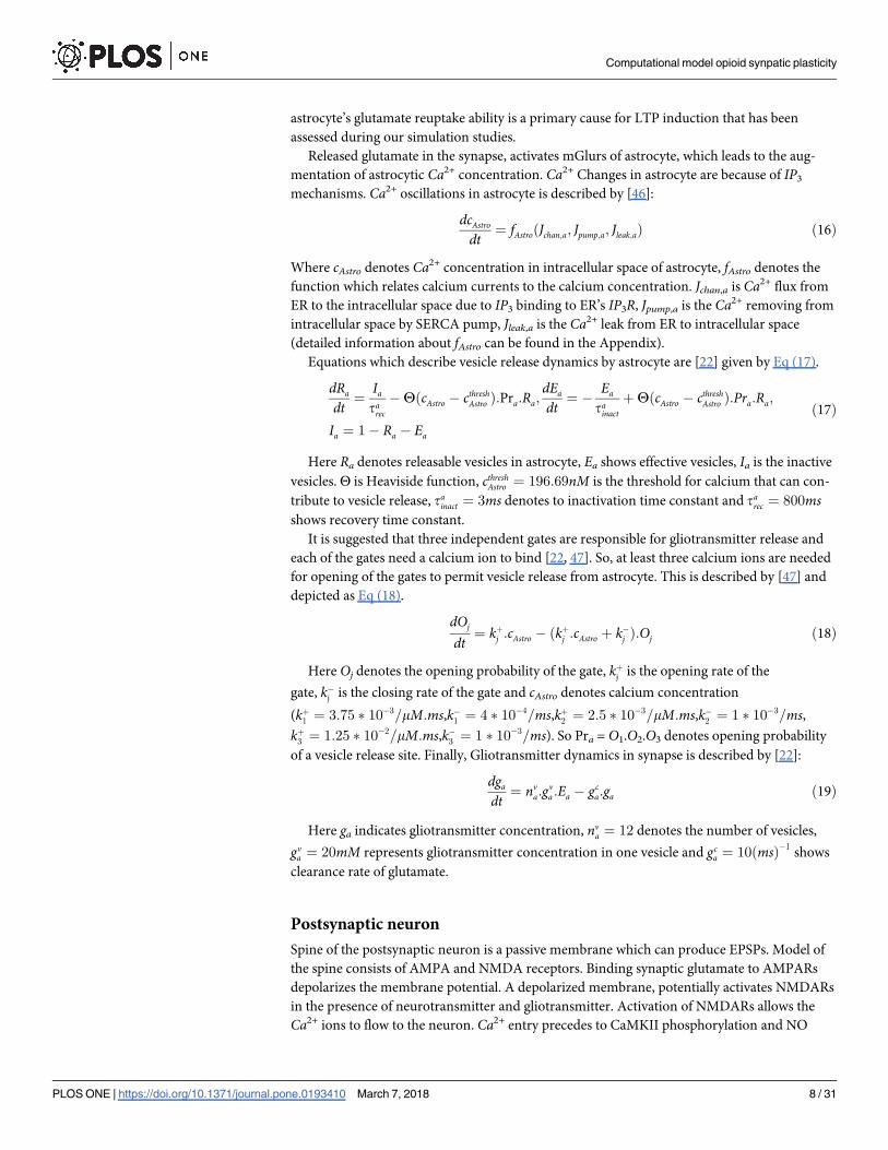
μORs activation leads to inhibition of interneuron calcium channels activation. So, the
amplitude of IPSC in pyramidal neuron attenuates. This process is shown in Fig 4 in response
to different doses of morphine.
Fig 4A demonstrates that when there is not any morphine injection, all VGCCs are in an
active state and the inhibitory current is at its maximum value. By applying 0.1 μM of mor-
phine, VGCCs activation is attenuated to 75%, and IPSCs are decreased to 26 μA/cm^2 (Fig
4B). A further increase in the dose of morphine to 1 μM precedes to 59% activation of VGCCs
and IPSCs are attenuated to approximately 8 μA/cm^2 (Fig 4C).
Computational model opioid synpatic plasticity
PLOS ONE | https://doi.org/10.1371/journal.pone.0193410 March 7, 2018 10 / 31

Comparison of simulation results with the experimental findings for the activation of
VGCCs and inhibition of the IPSC are denoted in Figs 5 and 6. During injection of 0.01 μM
DAMGO (μOR agonist), calcium influx is decreased to 89% of its final value in the experiment
[21]; simulation of the proposed model shows decrease in activated calcium channels to 95%.
By increasing the dose of injected DAMGO to 0.1 μM, activated calcium channels attenuates
to 87% in the experiment [21], and to 75% in the model. Further injection of DAMGO (1 μM)
leads to more reduction in the calcium influx up to 66% in the experiment [21], and inhibition
of activated channels decreases to 59% in the proposed model (Fig 5).
Inhibition of activated calcium channels in an inhibitory neuron can directly inhibit an
inhibitory current on excitatory neurons based on Bertram’s assumption. In this part, inhibi-
tory current on the CA3 pyramidal neuron in simulation compared with experimental data in
Fig 6. Different doses of morphine decrease the inhibitory current in the pyramidal neuron by
75%, 44% and 13% for 1, 0.1 and 0.01 μM injection of morphine in the experimental condition,
respectively [19]. Simulation results for the inhibitory current show decrease by 92%, 57% and
5% With 1, 0.1 and 0.01 μM of morphine, respectively. One can see a similar pattern in simula-
tion results and experiments.
Fig 3. Shows the percentage of activated μORs versus different dose of morphine.
https://doi.org/10.1371/journal.pone.0193410.g003
Computational model opioid synpatic plasticity
PLOS ONE | https://doi.org/10.1371/journal.pone.0193410 March 7, 2018 11 / 31
The model representing opioid receptor activation can relatively mimic the experimental
results reported by Rhim et al. [21] and Capogna et al. [19].
Normal condition
To compare opioid-induced LTP with the typical situation, first, we simulate the model for the
normal state, where there is no contribution of opioid receptors activation in the simulations.
Stimulation of CA3 pyramidal neuron and interneuron with a pulse train leads to the genera-
tion of APs. APs activate N-type VGCCs and the Ca2+ can enter into the neuron. Then, Ca2+
binds to the presynaptic Ca2+ sensors and triggers the glutamate release to the synaptic cleft
(Fig 7A). In addition, the effect of Interneuron activation is an augmentation of gGABA in the
presynaptic pyramidal neuron which exerts a further inhibitory effect.
Synaptic glutamate can bind to astrocytic mGlurs and postsynaptic NMDA and AMPA
receptors. By activation of astrocytic mGlurs, calcium concentration augments through IP3
production in astrocyte. Astrocytic Ca2+ oscillations lead to the gliotransmitter release, which
affects both presynaptic and postsynaptic pyramidal neurons. Gliotransmitter release has been
shown in Fig 7B. Gliotransmitter affects presynaptic mGlurs and leads to IP3 production
which is shown in Fig 7C. Production of presynaptic IP3 leads to the enhancement of calcium
concentration in the synaptic bouton (Fig 7D). Synaptic glutamate can bind to postsynaptic
AMPARs and NMDARs. Activation of AMPARs (Fig 7E) depolarize postsynaptic membrane
potential and convey to remove of NMDARs Mg2+ blocking, so NMDAR mediated current
Fig 4. The activation of the interneuron calcium channel and the amplitude of the IPSC in the pyramidal neuron
are shown for three different doses of morphine (μM). (A) morphine concentration considered to be 0, (B) shows
the results for 0.1 μM concentration of morphine and (C) is the simulation results at 1 μM concentration of morphine.
https://doi.org/10.1371/journal.pone.0193410.g004
Computational model opioid synpatic plasticity
PLOS ONE | https://doi.org/10.1371/journal.pone.0193410 March 7, 2018 12 / 31

flows (Fig 7F). Therefore, Ca2+ influx to the postsynaptic neuron initiates. Postsynaptic Ca2+
oscillations are shown in Fig 7G. Enhancement of Ca2+ in the CA1 pyramidal neuron activates
the CAMKII process which is able to augment AMPAR conductance and NO synthesis. In the
Fig 7H, phosphorylated CaMKII is indicated by the red line and the threshold for the synthesis
of NO with the green line. NO messenger can retrogradely diffuse from postsynaptic neuron
to the presynaptic neuron and increases the sensitivity of Ca2+ sensors in the CA3 pyramidal
neuron. Subsequently, there would be a further glutamate release in the synaptic cleft. This
process acts as a positive feedback to inducing and maintaining the LTP in the synapse. Over-
all, this simulation shows a typical learning process in synaptic level.
Pathological condition
Injection of 1 μM morphine activates presynaptic and postsynaptic μORs. Activation of
presynaptic μORs inhibits the Ca2+ entrance into the interneuron by 40%, which is described
in Fig 5. Therefore, IPSC of CA3 pyramidal neuron decreases to 8 μA/cm^2 due to fewer
GABA releases by interneuron. Reduction of IPSC leads to more neuronal excitability. Conse-
quently, synaptic glutamate increases and stimulates the astrocyte and postsynaptic neuron
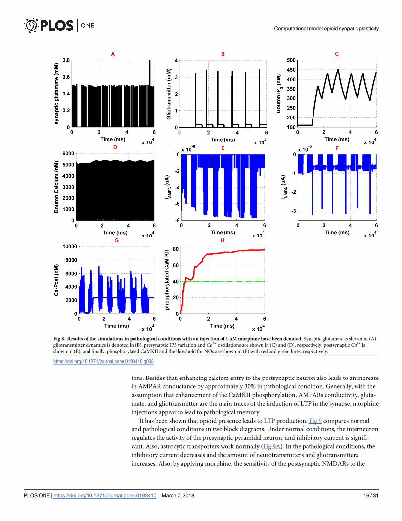
more than its normal condition. Synaptic glutamate has been shown in Fig 8A. As one can see,
Fig 5. Activated VGCCs versus different doses of morphine (μM) in experimental conditions (red line) and simulation setup
(black line) are described here.
https://doi.org/10.1371/journal.pone.0193410.g005
Computational model opioid synpatic plasticity
PLOS ONE | https://doi.org/10.1371/journal.pone.0193410 March 7, 2018 13 / 31

amplitude, frequency and the total amount of released glutamate by the presynaptic neuron is
significantly increased. This increase is about six times larger than its normal condition and
may lead to more excitation of both postsynaptic neuron and astrocyte. Furthermore, glio-
transmitter release by astrocyte is enhanced by 250% of its normal state which is described in
Fig 8B. Gliotransmitter increase is due to further activation of astrocytic mGlurs and enhanc-
ing calcium oscillations of the astrocyte. Binding gliotransmitter to presynaptic mGlurs acts as
a positive feedback and results in IP3 production and increase of presynaptic calcium, which
are denoted in Fig 8C and 8D, respectively. As it has been shown in the Fig 8C, IP3 production
is enhanced in the presynaptic neuron, which can suggest the increase of calcium in synaptic
bouton. Furthermore, since glutamate reuptake is attenuated in the presence of opioids, we
modeled it by decreasing the reuptake ability of glutamate by astrocyte down to 50% of a typi-
cal situation based on experimental observations.
On the other hand, the presence of opioids affects NMDAR activation by inhibiting Mg2+
blocking and by increasing conductance of NMDA receptors. Due to experimental research,
Mg2+ blocking of NMDARs modified in which δ (the relative electrical distance of the binding
site of Mg2+ from the outside of membrane) increased by 11% and K0 (dissociation constant
at 0 mv) augmented by 4.8 fold of its normal value. Besides that, morphine can increase
Fig 6. Inhibition of IPSC in response to different doses of morphine (μM) in experimental study (black line) and simulation
condition (red line) are shown here.
https://doi.org/10.1371/journal.pone.0193410.g006
Computational model opioid synpatic plasticity
PLOS ONE | https://doi.org/10.1371/journal.pone.0193410 March 7, 2018 14 / 31

NMDARs voltage independent conductance by 15% averagely, by Protein Kinase C (PKC)
mechanisms, which has been applied in the model.
Generally, in the pathological conditions, postsynaptic calcium’s entry to the neuron has
been denoted in Fig 8G, which shows a significant increase in comparison to the normal state.
This amplification of calcium oscillations is due to the enhancement of NMDARs activation
(Fig 8F). NMDAR related current increases because it receives more glutamate from the pre-
synaptic neuron and more gliotransmitter from astrocyte. Besides that, activation of the μORs
enhance conductance of the NMDARs through the previously mentioned mechanisms. Cal-
cium entry to the postsynaptic neuron causes phosphorylation of CaMKII which is shown in
Fig 8H. Phosphorylation of CaMKII is followed by NO synthesis. Synthesized NO retrogrades
to the presynaptic neuron and enhances the binding ability of glutamate vesicles to calcium
Fig 7. Simulation results for normal condition has been denoted here. Synaptic glutamate is shown in (A), gliotransmitter dynamics is denoted in (B),
presynaptic IP3 variation and Ca2+ oscillations are shown in (C) and (D), respectively; AMPAR and NMDAR currents are denoted in (E) and (F),
postsynaptic Ca2+ is shown in (G), phosphorylated CaMKII and NOs threshold are shown in (H) by red and green curves, respectively.
https://doi.org/10.1371/journal.pone.0193410.g007
Computational model opioid synpatic plasticity
PLOS ONE | https://doi.org/10.1371/journal.pone.0193410 March 7, 2018 15 / 31
ions. Besides that, enhancing calcium entry to the postsynaptic neuron also leads to an increase
in AMPAR conductance by approximately 30% in pathological condition. Generally, with the
assumption that enhancement of the CaMKII phosphorylation, AMPARs conductivity, gluta-
mate, and gliotransmitter are the main traces of the induction of LTP in the synapse, morphine
injections appear to lead to pathological memory.
It has been shown that opioid presence leads to LTP production. Fig 9 compares normal
and pathological conditions in two block diagrams. Under normal conditions, the interneuron
regulates the activity of the presynaptic pyramidal neuron, and inhibitory current is signifi-
cant. Also, astrocytic transporters work normally (Fig 9A). In the pathological conditions, the
inhibitory current decreases and the amount of neurotransmitters and gliotransmitters
increases. Also, by applying morphine, the sensitivity of the postsynaptic NMDARs to the
Fig 8. Results of the simulations in pathological conditions with an injection of 1 μM morphine have been denoted. Synaptic glutamate is shown in (A),
gliotransmitter dynamics is denoted in (B), presynaptic IP3 variation and Ca2+ oscillations are shown in (C) and (D), respectively, postsynaptic Ca2+ is
shown in (E), and finally, phosphorylated CaMKII and the threshold for NOs are shown in (F) with red and green lines, respectively.
https://doi.org/10.1371/journal.pone.0193410.g008
Computational model opioid synpatic plasticity
PLOS ONE | https://doi.org/10.1371/journal.pone.0193410 March 7, 2018 16 / 31

transmitters enhances. Therefore, more calcium enters into the neuron. The increase in cal-
cium conveys to CaMKII phosphorylation and thus NO is synthesized. Also, AMPAR’s con-
ductance increases due to CaMKII phosphorylation process (Fig 9B).
Comparing the effect of different factors on pathological LTP induction
In the previous simulations, it has been demonstrated that morphine injection conveys to
pathological LTP induction. In this section, the effect of the different factors on the induction
of pathological LTP is considered and significant parameters are specified. Essential elements
can be seen as drug targets to prevent the formation of pathological memory in the use of opi-
oids. The presence of 1 μM morphine in synapse can enhance CaMKII phosphorylation pro-
cess, synaptic glutamate, gliotransmitter release, and AMPAR conductance. These variables
Fig 9. A comparison between the normal (A) and the pathological (B) conditions is shown.
https://doi.org/10.1371/journal.pone.0193410.g009
Computational model opioid synpatic plasticity
PLOS ONE | https://doi.org/10.1371/journal.pone.0193410 March 7, 2018 17 / 31

are considered as monitoring factors for morphine effects on the synapse. Monitoring elements
can be seen as a trace of LTP induction. Fig 10 shows the effects of different parameters on
monitoring factors through the simulations shown in the next section.
Simulation 1. In the presence of 1μM morphine, monitoring factors are shown in Fig 10.
In Simulation 1 morphine affects presynaptic, postsynaptic and astrocyte. Therefore, presyn-
aptic and postsynaptic μORs are in the active state, and astrocytic glutamate transporters coef-
ficient is half of the normal conditions, which indicates the effect of morphine on astrocyte.
Comparing these results with normal conditions (Simulation 13) infers that the average
amount of synaptic glutamate is five times higher than normal. The average amount of glio-
transmitter is 4.5 times the normal state. The mean amount of phosphorylated CaMKII and
AMPARs are 4 and 1.3 times higher than the normal level. Overall, in the presence of mor-
phine, all of the monitoring factors are high, which can be indicative of LTP induction.
Fig 10. Box and whisker plot for different simulations have been described here. Redline shows the median of the signals, ‘+’ sign
represents the mean value of each signal, blue line demonstrates that 25%-75% of data are included in this range, and finally black line
shows the range that 9%-91% of the dada are included. Panel A shows the synaptic glutamate, Panel B denotes the gliotransmitter, Panel
C describes the phosphorylated CaMKII, and Panel D represents the Phosphorylated AMPARs. In the first simulation, all of the synaptic
components are affected by morphine and the simulation of 13th indicates normal mode. In the second simulation, the effect of morphine
on presynaptic neurons has been neglected. In the third simulation, postsynaptic μORs are assumed to be inactive. In the fourth
simulation, morphine only affects astrocyte. In the fifth simulation, the release of the gliotransmitter is considered zero. In the sixth
simulation, it is assumed that despite the effect of morphine on all the components of the synapse, astrocytic GLTs act normal. In the
seventh simulation, under conditions where morphine affects all of the synaptic components, the activity of astrocytic transporters is
stimulated to be 50% greater than normal. In the eighth simulation, the activity of astrocytic mGlurs is considered zero. The ninth
simulation shows that astrocytic mGlurs and postsynaptic μORs are not active. In the tenth simulation, astrocytic transporters are
stimulated 50% more than normal, and astrocytic mGlurs have been deactivated. In the eleventh simulation, by stimulating astrocytic
transporters, NMDA receptors have a normal synaptic effect. In the 12th simulation, astrocytic mGlurs have been deactivated, astrocytic
transporters are stimulated, and NMDARs act as normal.
https://doi.org/10.1371/journal.pone.0193410.g010
Computational model opioid synpatic plasticity
PLOS ONE | https://doi.org/10.1371/journal.pone.0193410 March 7, 2018 18 / 31

Simulation 2. In this simulation, we want to show how the disinhibitory effect of mor-
phine affects monitoring factors. Here, we disable the presynaptic opioid receptors and mor-
phine only affects the postsynaptic μORs and astrocytic GLT1s.
Simulation results denote that despite a significant reduction in synaptic glutamate, other
monitoring factors are still high and pathological LTP is still present. This means that, despite
the importance of disinhibitory mechanism in inducing LTP, preventing it cannot block path-
ological memory.
Simulation 3. In this simulation, the postsynaptic μORs activity is inhibited. however,
presynaptic neurons and astrocyte are under the influence of morphine. The results indicate
that, despite the attenuation of CaMKII phosphorylation and the reduction of AMPAR con-
ductance, the amount of glutamate and gliotransmitter are still highand the probability of for-
mation of pathologic memory is high.
Simulation 4. In this section, morphine is thought to only affect astrocyte. Therefore, pre-
synaptic and postsynaptic μORs are inhibited. In this case, the synaptic glutamate is approach-
ing a level that is normal. In addition, phosphorylation of CaMKII and AMPARs has been
significantly reduced. But it seems that due to the significant release of glutamate in the syn-
apse. LTP occurs. This simulation describes that astrocyte plays a fundamental role in the
induction of pathological LTP, so it is necessary to carefully examine its role in the following
sections.
Simulation 5. In this simulation, it has been assumed that morphine affects all the compo-
nents of the synapse, but astrocyte cannot release the gliotransmitter. Here astrocyte does not
have any effect on presynaptic and postsynaptic neurons. The results suggest that inhibiting
gliotransmitter release cannot disrupt the pathological memory formation.
Simulation 6. In this simulation, the role of astrocytic transporters in pathological mem-
ory induction has been investigated. Therefore, under the conditions of the first simulation, it
is assumed that astrocyte transports are active as normal and their activity is not affected by
the presence of morphine. The results show that normal activity of transporters cannot prevent
the formation of pathologic memory.
Simulation 7. In this section, when morphine affects all the components of the synapse,
the activity of astrocytic transporters enhanced by 50% more than their standard value. In this
case, glutamate and gliotransmitter values have gone to normal, and CaMKII phosphorylation
has been significantly reduced. This simulation shows that up-regulation of astrocytic trans-
porters can be used as an agent to prevent the pathological LTP formation.
Simulation 8. In this simulation, the effect of astrocytic mGlur has been investigated. To
this purpose, astrocytic mGlurs are inhibited in a situation where morphine affects all of the
synaptic components. It can be seen that in this case, with the exception of the gliotransmitter
which is in the normal range, the rest of the monitoring factors are high.
Simulation 9. In this simulation, postsynaptic opioid receptors and astrocytic mGlur are
supposed to be inhibited. The results state that it is still possible to develop pathological
memory.
Simulation 10. In this simulation, astrocytic mGlurs is inhibited and its transporters are
stimulated 50% more than normal. Opioid receptors for presynaptic and postsynaptic neurons
are still active. Results indicate that in spite of a significant drop in glutamate and gliotransmit-
ter, the rest of the monitoring factors were not significantly reduced.
Simulation 11. In this simulation, postsynaptic opioid receptors have been disabled and
astrocytic GLTs are stimulated 50% more than normal. The results show that in the presence
of morphine, all of the monitoring factors are in normal range.
Simulation 12. In this simulation, postsynaptic opioid receptors and astrocytic mGlurs
are supposed to be inhibited and astrocytic transporters are stimulated 50% higher than
Computational model opioid synpatic plasticity
PLOS ONE | https://doi.org/10.1371/journal.pone.0193410 March 7, 2018 19 / 31

normal. In this case, monitoring factors are found to be close to the normal value, and even
take less than normal values.
Simulation 13. This simulates the normal state in which it is assumed that morphine is
not present in the synapse.
Discussion
This paper presented a computational model in CA3-CA1 region of the hippocampus at the
synaptic level. Here we extended and improved previously introduced models by different
researchers [22, 23, 34, 41, 42, 45, 46, 48–53] to represent opioid-induced synaptic plasticity in
the hippocampus. To best of our knowledge, this is the first model that has the ability to be
used for computational analysis of opioid-induced synaptic plasticity. Using computational
models allows researchers to analyze the effect of various parameters simultaneously without
concerning about the interfering effects of different medical agents in experiments.
Our model adequately shows that opioids presence can induce LTP in affected synapses.
Here, it is assumed that amounts of synaptic glutamate, gliotransmitter, phosphorylated CaM-
KII and AMPARs are footprints of LTP induction in the synapse. In fact, these factors are con-
sidered to be parameters which can lead to LTP induction, and we called them as monitoring
factors. To discuss the results of the simulations, the normalized values for monitoring factors
are depicted in Fig 11.
As previously stated, the first simulation shows that morphine affects all the components of
the synapse and the thirteenth simulation indicates the normal mode. It has been shown in
Fig 11. Normalized graph of monitoring factors for different simulations has been described here.
https://doi.org/10.1371/journal.pone.0193410.g011
Computational model opioid synpatic plasticity
PLOS ONE | https://doi.org/10.1371/journal.pone.0193410 March 7, 2018 20 / 31

experiments that the presence of morphine increases the density of AMPARs, synaptic gluta-
mate and calcium signaling in affected neurons [9, 54]. These changes, which describe the
main features of pathological LTP and memory formation, are seen in the results of the first
simulation.
In the second simulation, presynaptic opioid receptors are inhibited. As a result, release of
glutamate is normal, but the traces of pathological LTP is still existed. The results of this simu-
lation indicate that increasing the activity of postsynaptic NMDARs and the effect of morphine
on astrocyte are factors that contribute to the formation of pathologic memory.
In the 3rd simulation, postsynaptic μORs have been inhibited, indicating that postsynaptic
NMDARs exhibit normal activity. In this case, the monitoring factors have significant
amounts. It infers that injection of NMDAR antagonist or inhibition of postsynaptic opioid
receptors is effective in preventing the induction of pathologic LTP, but it is not enough.
In fact, experimental results have also shown that NMDARs play an essential role in induc-
ing opioid-related neural plasticity [38, 55–57]. Furthermore, it has been shown that NMDAR
antagonist injections are effective in relapse weakening; however, it cannot completely elimi-
nate drug-seeking behavior [58, 59]. Based on the results we have obtained in the simulations,
it is true that NMDARs are the main actors in the formation of pathological memories, but the
role of other parameters should not be ignored.
In the fourth simulation, only astrocyte is affected by morphine, and presynaptic and post-
synaptic opioid receptors are inhibited. In this case, glutamate is found to be in normal range,
but the amounts of CaMKII and AMPAR phosphorylation are still significant. As a result, it
can be concluded that, regardless of the role of astrocyte in synapse, the pathologic LTP cannot
be controlled. Some researchers believe that astrocyte dysfunction can lead to addiction [28,
30]. Thus, it can be concluded that without the manipulation of astrocyte function, we may
not be able to inhibit the pathological LTP induction.
In addition, in the 5th and 6th simulations, it was observed that preventing the release of
gliotransmitter and having normal GLTs cannot influence the formation of pathological LTP.
In the 7th simulation, astrocytic transporters have been stimulated by 50% more than nor-
mal. It can be seen that despite the attenuation of synaptic glutamate and gliotransmitter,
other monitoring factors are still high. So, stimulating astrocytic GLTs can be effective, but is
not sufficient for preventing pathological LTP. Now the important question is: is it possible to
up-regulate astrocytic glutamate transporters empirically? The answer is yes. It has recently
been shown that up-regulation of astrocytic transporters is possible and can even undermine
the induction of LTP in the synapse [60].
In the 8th simulation, astrocytic mGlurs are inhibited. This mode describes that the agonist-
dependent IP3 production is inhabited in astrocyte. According to the results obtained, this
mode alone cannot be used as an effective parameter.
In the 9th simulation, astrocytic mGlurs and postsynaptic μORs are simultaneously inhib-
ited. In this case, synaptic glutamate is high, phosphorylated CaMKII is almost halved, and the
amount of phosphorylation of AMPARs and released gliotransmitter are in normal range. It
can be noticed that blocking the agonist-dependent IP3 production and the use of NMDAR
antagonist can undermine the pathological LTP. In this case, the only remaining problem is to
reduce the amount of synaptic glutamate that can also regulate CaMKII phosphorylation.
In the 10th simulation, activity of astrocytic mGlurs is inhibited, and GLTs are stimulated to
be 50% more than normal level. In this case, glutamate and gliotransmitter are in the normal
range, and the other monitoring factors are almost halved. Therefore, it can be concluded that
manipulation in the activity of astrocyte can be effective in preventing the formation of patho-
logical LTP.
Computational model opioid synpatic plasticity
PLOS ONE | https://doi.org/10.1371/journal.pone.0193410 March 7, 2018 21 / 31

The 11th simulation describes the case in which astrocyte transporters are stimulated, and
postsynaptic μORs are inhibited. In this case, all monitoring factors fall into the normal range.
It means that stimulating GLTs and injection of NMDAR antagonists can stop the formation
of pathological LTP.
In the 12th simulation, in addition to inhibiting the activity of postsynaptic opioid receptors
and stimulating GLTs, we also disable astrocytic mGlurs. In this case, monitoring factors are
alose to normal and even some of them have values that are less than normal. It can be con-
cluded that in this case the possibility of the formation of pathological memory is completely
eliminated.
Overall, by summerizing all the simulations, it can be suggested that an optimal method for
coping with the formation of pathological memory can be the stimulation of astrocyte GLTs
and the injection of NMDAR antagonists.
In this paper, some assumptions are made to simplify the modeling process. Dynamics
has not been considered for the release of inhibitor neurotransmitter from the interneuron
to the pyramidal neuron in the presynaptic layer. Such an approach has been implemented
in some computational models. It is also assumed that interneuron and astrocyte do not
interact. Investigation of this interaction and its role in the induction of the synaptic plastic-
ity can be considered in the future works. Also, in this paper, as with some computational
models, the diffusion process for glutamate and gliotransmitter is not considered. It seems
that focusing on this process in models that deal with a large number of neurons (network
level) can be effective, but in our model, it will not have much impact on the results. On
the other hand, we assume that some factors are footprints of LTP induction in the synapse,
such as the concentration of glutamate and gliotransmitter, and phosphorylation of
CaMKII and AMPARs. Considering the effect of LTD in synapse can extend the model.
This goal may be accomplished through the use of the STDP process as a learning rule in
synapse.
Conclusion
Opioids such as morphine can lead to the induction of LTP at the synaptic level, which may
result in an abnormal memory formation process leading to addiction. This pathological
memory can be seen as the key trigger for relapse in addicted patients. Suppression of LTP
induced by opioids may lead to diminishing relapse in withdrawal period in addicted sub-
jects. Since changing different parameters and evaluating various types of effects in memory
formation process at the synaptic level can be difficult and even practically impossible in
experimental conditions, a detailed mathematical model can help us to overcome empirical
obstacles and difficulties to some extent. This paper, presented a biophysically suggested and
supported computational model for opioid-induced memory formation process. The pro-
posed model is at the synaptic level and the role of astrocyte is considered as the third compo-
nent of synapse. The proposed model is capable of describing the pathologic LTP induction
process and can be helpful in addiction-related research. Here, we found that astrocytic
transporters play a fundamental role in the induction of pathological drug-related memories.
Simulation results show that up-regulating astrocytic transporters, omitting astrocytic
mGlurs and applying NMDARs antagonist can completely prevent pathological LTP. We
also suggest that the up-regulation of GLTs and injection of NMDAR antagonist can be used
as an optimal way to prevent the occurrence of pathological LTP. Overall, we suggest that
astrocyte has a prominent role in addiction-related memories and its function must be con-
sidered precisely for preventing abnormal memories. This approach may help researchers to
inhibit relapse in the future.
Computational model opioid synpatic plasticity
PLOS ONE | https://doi.org/10.1371/journal.pone.0193410 March 7, 2018 22 / 31

Appendix
Description of functions
Presynaptic neurons. Presynaptic APs is governed by H-H type formulation:
dVpre
dt¼ fPyrðIapp; INa; IK ; IL; IGABAA
Þ ¼ Iapp � 36n4ðVpre þ 82Þ � 120m3hðVpre � 45Þ � 0:3ðVpre þ 59:4Þ � IGABA
dVInt
dt¼ fIntðIapp; INa; IK ; ILÞ ¼ Iapp � 36n4ðVInt þ 82Þ � 120m3hðVInt � 45Þ � 0:3ðVInt þ 59:4Þ
dxdt¼ axð1 � xÞ � bxx; an ¼
0:01ð� Vpre � 60Þ
expð� Vpre � 60
10Þ � 1
; am ¼0:1ð� Vpre � 45Þ
expð� Vpre � 45
10Þ � 1
; ah ¼ 0:07expð� Vpre � 70
20Þ;
bn ¼ 0:125expð� Vpre � 70
80Þ; bm ¼ 4expð
� Vpre � 70
18Þ; bh ¼
1
expð� Vpre � 40
10Þ þ 1
ðA-1Þ
Here Vpre is presynaptic membrane potential for the pyramidal neuron, VInt is the potential
of presynaptic interneuron, Iapp is the externally applied current, gK, gNa and gL are potassium,
sodium and leak conductance respectively, VN, VK, and VL are sodium, potassium and leak
reversal potentials respectively. Third equation denotes gating variables for ionic currents, x
can be substituted by m, n, h for gating variable of Na+ activation, K+ activation, and Na+ inac-
tivation respectively.
Calcium oscillations can be represented by [22]:
dcFastdt¼ fFastðICa; IPMCa; IPMleak; bnÞ ¼ �
ICaAbtn
zCaFVbtnþ JPMleak �
IPMCaAbtn
zCaFVbtnðA-2Þ
Where calcium current through N-type channels represents by ICa, Abtn is neuron bouton’s
surface area, zCa is the calcium ion valence, Vbtn is the neuron bouton’s volume, F is faraday’s
constant. JPMleak is the leak current from extracellular space into Bouton, IPMCa denotes cal-
cium current due to ATPase pump which pumps extra calcium out of the cell and ICa is the cal-
cium current due to N-type VGCCs and is governed by:
JPMleak ¼ nleakðcext � ciÞ; IPMCa ¼ nPMCac2i
c2i þ K2
PMCa
; ICa ¼ rCa m2
Ca gCaðVpreðtÞ � VCaÞ ðA-3Þ
Here, νleak, cext and ci are the maximum leak of calcium through the membrane, extracellu-
lar calcium concentration, and intracellular calcium concentration respectively. νPMCa is maxi-
mum PMCa current and KPMCa is the calcium concentration at which νPMCa is halved. Here,
ρCa denotes N-type calcium channel density on the membrane of neuron Bouton, gCa is the
single channel’s conductance. In the above equation assumed that N-type channel has two
gates. mCa is the opening probability of a single gate and can be shown by:
dmCa
dt¼
m1Ca � mCa
tmCa
;m1Ca ¼1
1þ expððVmCa� VmÞ=kmCa
Þ;VCa ¼
RTzCaF
lnðcextcresti
Þ ðA-4Þ
Moreover, tmCais the time constant where mCa approaches to m1Ca which is the Boltzmann
function with VmCaand kmCa
as the half-activation voltage of N-type calcium channel and the
slope factor of N-type channel activation respectively. VCa shows reversal potential of Ca2+ the
ion. Here, R is the real gas constant, T denotes absolute temperature, extracellular calcium con-
centration is shown by cext and intracellular calcium concentration at rest is denoted by cresti .
Computational model opioid synpatic plasticity
PLOS ONE | https://doi.org/10.1371/journal.pone.0193410 March 7, 2018 23 / 31

Parameters related to presynaptic calcium dynamics can be found in Tewari and Majumdar’s
paper [22].
Modeling slow calcium oscillations achieved by using Tewari and Majumdar’s modified Li-
Rinzel model which can describe by [22]:
dcSlowdt¼ fSlowðJchan; JERpump; JERleakÞ ¼ � Jchan � JERpump � JERleak ðA-5Þ
Here Jchan denotes calcium influx from ER into cytosol through the IP3 receptor, JERpump is
the pumped calcium from intracellular space to ER and JERleak indicates leaked calcium ions
from ER into intracellular space. Equations that describe these three types of Ca2+ flux are:
Jchan ¼ c1n1m3
1n3
1q3ðci � cERÞ; JERpump ¼
v3c2i
k23þ c2
i
; JERleak ¼ c1n2ðci � cERÞ
dcERdt¼ �
1
c1
dcslowdt
;dpdt¼ ng
g0:7a
k0:7g þ g0:7
a
� tpðp � p0Þ;dqdt¼ aqð1 � qÞ � bqq ðA-6Þ
Where cER is the Ca2+ concentration in ER, c1 is the ratio of ER volume to bouton volume, ν1
denotes maximal flux rate of IP3 receptors, ν2 shows maximal leak of Ca2+ from ER to intracel-
lular space, ν3 is the maximal SERCA (Sarco Endoplasmic Reticulum ATPase) pump rate,
intracellular IP3 concentration is demonstrated by p finally ga and q are Extrasynaptic gluta-
mate concentration and fraction of activated IP3 receptors respectively. Also, m1, n1, αq and
βq can be denoted by:
m1 ¼p
pþ d1
; n1 ¼ci
ci þ d5
; aq ¼ a2d2
pþ d1
pþ d3
; bq ¼ a2ci ðA-7Þ
Here, d1 and d3 are the IP3 dissociation constant, d2 and d5 are the inhibitory Ca2+ dissocia-
tion and activation Ca2+ dissociation constants respectively. a2 is the inhibitory Ca2+ binding
constant. Further information about the equations can be found in Tewari and Majumdar’s
paper [22].
Astrocyte Ca2+ dynamics. Equations which describe Ca2+ oscillations in astrocyte are
[46]:
dcAstrodt¼ fAstroðJchan;a; Jpump;a; Jleak;aÞ ¼ � Jchan;a � Jpump;a � Jleak;a ðA-8Þ
Jchan;a ¼ c1;arcam3
1n3
1h3
1ðcAstro � cER;aÞ; Jpump;a ¼ vER
cAstro2
cAstro2 þ K2ER
; Jleak;a ¼ c1;arLðcAstro � cER;aÞ
Where cAstro denotes Ca2+ concentration in intracellular space of astrocyte, Jchan,a is Ca2+ flux
from ER to the intracellular space due to IP3 binding to ER’sIP3R, Jpump,a is the Ca2+ removing
from intracellular space by SERCA pump, Jleak,a is the Ca2+ leak from ER to intracellular space,
c1,a is the ratio of ER volume to cytosol volume,rca is the maximum rate of Ca2+ flux by IP3R,
m31n31h31
is the opening probability of IP3R vesicles, cER,a shows Ca2+ concentration in ER, νERis the highest rate of Ca2+ uptake by ER, KER denotes intracellular calcium affinity of SERCA
pump and rL shows maximum rate of Ca2+ leak from ER. Equations of IP3 dynamics in
Computational model opioid synpatic plasticity
PLOS ONE | https://doi.org/10.1371/journal.pone.0193410 March 7, 2018 24 / 31

astrocyte can be written as follows:
dpa
dt¼ vbHill g0:7;KR 1þ
Kp
KRHillðC;KpÞ
� �� �
þvd
1þpa
kd
Hillðc2
a;KPLCdÞ
þv3KHillðc4a;KDÞHillðpa;K3Þ � r5pa
pa
dha
dt¼ aha
ð1 � haÞ � bhaha
ðA-9Þ
The first term of above equation denotes agonist-dependent (glutamate) IP3 production;
the second term shows agonist-independent IP3 production which is due to PLCδ signaling
and modulated by Ca2+, the third term shows IP3 degradation rate by IP3 − 3k which is Ca2+
dependent and the last term incorporates degradation of IP3 by IP − 5P. ahaand bha
denote
opening and closing rate of ha respectively. Other parameters in mentioned equation can be
determined using equations:
m1;a ¼ Hillðpa; d1Þ; n1;a ¼ Hillðca; d5Þ;Hillðxn;KÞ ¼xn
xn þ Kn; aha¼ a2d2
pa þ d1
pa þ d3
; bha¼ a2ca ðA-10Þ
Further information about the parameters and their values can be found in De pitta’s paper
[46].
Postsynaptic neuron. AMPAR current governs by the following equations:
IAMPA ¼ fAMPAðgAMPA;mAMPA;VpostÞ ¼ gAMPAmAMPAðVpost � VAMPAÞ ðA-11Þ
Where VAMPA is reversal potential of the receptor, Vpost denotes membrane potential, mAMPA
represents gating variable of AMPAR. Since we assume that both presynaptic and astrocytic
glutamate have effects on postsynaptic neuron, so gating variable of AMPAR can be described
by:
dmAMPA
dt¼ aAMPAða1gpre þ a2gastÞð1 � mAMPAÞ � bAMPAmAMPA ðA-12Þ
Here αAMPA and βAMPA are opening and closing rate of the receptor, gpre and gast denote glu-
tamate concentration released by presynaptic neuron and astrocyte respectively, a1 = 0.42 and
a2 = 0.01 are constants.
In Eq (A-11) AMPAR channel’s conductance (gAMPA) can be varied due to postsynaptic
CaMKII. This process modeled using modified equations [50, 51]:
EP1ðPh:CaMKIIÞ ¼ EP2ðPh:CaMKIIÞ ¼ 1þ30ðPh:CaMKIIÞ2
ð1þ ðPh:CaMKIIÞ2Þ;
EK1ðPh:CaMKIIÞ ¼ EK2ðPh:CaMKIIÞ ¼ 1þ100ðPh:CaMKIIÞ2
ð82 þ ðPh:CaMKIIÞ2ÞðA-13Þ
Computational model opioid synpatic plasticity
PLOS ONE | https://doi.org/10.1371/journal.pone.0193410 March 7, 2018 25 / 31

Here, the activity of protein kinase and protein phosphatase assumed to be a hill function of
CaMKII and have been shown by EK and EP respectively [51].
A ¼AT :EP1:EP2
ðEK2þ EP2ÞðEK1þ EP1Þ;AP1
¼AT :EK1:EP2
ðEK2þ EP2ÞðEK1þ EP1Þ
AP2 ¼AT :EP1:EK2
ðEK2þ EP2ÞðEK1þ EP1Þ;AP2
P1¼
AT :EK1:EK2
ðEK2þ EP2ÞðEK1þ EP1Þ
AT ¼ Aþ Ap1þ AP2 þ AP2
P1
ðA-14Þ
Here, A, Ap1, AP2 and AP2
P1are reaction substrates and products. The conductance of
AMPAR can be determined using the below equation:
gAMPA ¼ fgAMPAðCaMKIIÞ ¼ Aþ 2ðAP1
þ AP2Þ þ 4AP2P1
ðA-15Þ
Further information about AMPAR phosphorylation can be found in Castellani et all’s
papers [50, 51].
Furthermore, in the proposed model, INMDA modeled using Moradi et all’s formulation
with a slight modification:
INMDA ¼ fNMDAðgNMDA;mNMDA;Mg;VpostÞ ¼ gNMDAmNMDAMgðVpost � VNMDAÞ ðA-16Þ
Here, Mg denotes Mg2+ blocking and is governed by:
Mg ¼ fMgðMorph;VpostÞ ¼ 1=ð1þ ½Mg2þ�0ðk0 þ
15:58
1þ ð0:1=MorphÞ1:2Þ� 1
expð� zðdþ0:1
1þ ð0:1=MorphÞ1:2ÞFVpostR
� 1T� 1ÞÞ ðA-17Þ
Moreover, NMDAR gating variable is governed by:
dmNMDA
dt¼ fmNMDA
ðaNMDA; bNMDA; gpre; gAstroÞ
¼ aNMDAðk1gpre þ k2gAstroÞð1 � mNMDAÞ � bNMDA mNMDA ðA-18Þ
Here VNMDA is the reversal potential of NMDAR, αNMDA and βNMDA are opening and clos-
ing rate of receptor respectively, gpre and gAstro are glutamate concentrations released by pre-
synaptic pyramidal neuron and astrocyte respectively. gNMDA is NMDA channels conductance
which is described by:
gNMDA ¼ fgNMDAðMorph; gVDÞ ¼ ðgVI þ
0:15
1þ ð0:1=MorphÞ1:2Þ þ gVD ðA-19Þ
Here gVI is the channel conductance independent of potential and gVD is the channel con-
ductance dependent to the potential which can be written by:
dgVDdt¼ ðgVD;1 � gVDÞ=tg
gVD;1 ¼ kðVpost � V0Þ
ðA-20Þ
Where gVD,1 is the final value of gVD with time constant of τg and has a linear relation with
membrane potential with constant of k. Other parameters and values listed in S1 Table.
Computational model opioid synpatic plasticity
PLOS ONE | https://doi.org/10.1371/journal.pone.0193410 March 7, 2018 26 / 31

Postsynaptic calcium oscillations which introduced in Eq (28) can be determined by [22]:
dcpostdt¼ fCaPost ðIAMPA; INMDA; ICaL; SPumpÞ ¼
f ðcpostÞ1þ y
; f ðcpostÞ ¼ �ðZIAMPA þ gINMDA þ ILÞ
zcaFVspine� spump;
spump ¼ ksðcpost � crestpostÞ; y ¼btKendo
ðKendo þ cpostÞ2; IL ¼ gLBðN;PopenÞðVpost � VLÞ
ðA-21Þ
Here, cpost denotes postsynaptic calcium concentration which depends on AMPAR current,
and is denoted by IAMPA, NMDA current is shown by INMDA, voltage-gated calcium channels
activation is described by IL and pumped calcium defined by spump. Here, η = 0.012 and γ =
0.06 are the constants for AMPAR and NMDAR mediated calcium ions, zca = 2 denotes cal-
cium valence, F = 96487C / mole is the faraday’s constant, Vspine = 0.9048um3 is the volume for
dendrite spin, ks = 100 / s maximum efflux rate of calcium pump, crestpost ¼ 100nM rest value for
postsynaptic calcium concentration, bt = 200uM denotes total endogenous buffer concentra-
tion and Kendo = 10uM shows endogenous buffer calcium affinity. Also, gL = 15ps is the con-
ductance of calcium channel current, B(N, Popen) is a random variable with a binomial
distribution that shows the number of opened channels and VL = 27.4mv describes the reversal
potential.
Postsynaptic Ca2+ concentration variation may lead to CaMKII phosphorylation which is
governed by equations [52]:
dP0
dt¼ � v1 þ v3P1;
dP1
dt¼ v1 � v3P1 � v2P1 þ 2v3P2;
dP2
dt¼ v2P1 � 2v3P2 � 1:8v2P2 þ 3v3P3
dP3
dt¼ 1:8v2P2 � 3v3P3 � 2:3v2P3 þ 4v3P4;
dP4
dt¼ 2:3v2P3 � 4v3P4 � 2:7v2P4 þ 5v3P5
dP5
dt¼ 2:7v2P4 � 5v3P5 � 2:8v2P5 þ 6v3P6;
dP6
dt¼ 2:8v2P5 � 6v3P6 � 2:7v2P6 þ 7v3P7
dP7
dt¼ 2:7v2P6 � 7v3P7 � 2:3v2P7 þ 8v3P8;
dP8
dt¼ 2:3v2P7 � 8v3P8 � 1:8v2P8 þ 9v3P9
dP9
dt¼ 1:8v2P8 � 9v3P9 � v2P9 þ 10v3P10;
dP10
dt¼ v2P9 � 10v3P10
depdt¼ � k3Iep þ k4ðep0 � epÞ;
dIdt¼ � k3Iep þ k4ðep0 � epÞ þ vPKAI0 �
vCaNð½Ca2þ�=KH2Þ3I
1þ ð½Ca2þ�=KH2Þ3
ðA-22Þ
Here, Pi denotes the concentration of the i-fold phosphorylated CaMKII, ep shows the PP1
concentration which not bounded to l1P and can demonstrate active protein phosphatase, the
total concentration of PP1 is denoted by ep0 = 0.1μM, free l1P is shown by I, free l1 concentra-
tion is demonstrated by I0 = 0.1μM. Association and dissociation rate constant of PP1-l1P
complex are denoted by k3 = 1 / μMs and k4 = 10−3 / s respectively. νCaN = 2 / s is the rate of l1P
dephosphorylation due to CaN (calcineurin), vPKA = 0.45μM / s is the phosphorylation rate of
l1 due to PKA, KH2 = 0.7μM is the calcium activation Hill constant of CaN.
The rate of phosphorylation (ν1), auto-phosphorylation (ν2) and dephosphorylation (ν3)
can describe by:
v1 ¼10k1ð½Ca
2þ�=KH1Þ8P0
ð1þ ð½Ca2þ�=KH1Þ4Þ
2; v2 ¼
k1ð½Ca2þ�=KH1Þ
4
1þ ð½Ca2þ�=KH1Þ4; v3 ¼
k2epKM þ
X10
1iPi
ðA-23Þ
Here, k1 = 0.5 / s is the l1 dependent regulation rate of PP1 and KH1 = 4μM is the hill con-
stant of CaMKII for calcium activation. KM = 20μM And k2 = 10 / s are the Michaelis and
Computational model opioid synpatic plasticity
PLOS ONE | https://doi.org/10.1371/journal.pone.0193410 March 7, 2018 27 / 31

catalytic constants respectively. Then, phosphorylated CaMKII can be described by:
Ph:CaMKII ¼ fCaMKIIðcPostÞ ¼X10
i¼1
Pi ðA-24Þ
Supporting information
S1 Table. Postsynaptic neuron parameters.
(DOCX)
Acknowledgments
The authors would like to thank Hadi Borjkhani Ph.D. student at the University of Tehran for
his assistance and comments that greatly improved the manuscript.
Author Contributions
Conceptualization: Mehdi Borjkhani, Fariba Bahrami, Mahyar Janahmadi.
Formal analysis: Mehdi Borjkhani.
Methodology: Mehdi Borjkhani.
Project administration: Fariba Bahrami.
Software: Mehdi Borjkhani.
Supervision: Fariba Bahrami, Mahyar Janahmadi.
Validation: Mahyar Janahmadi.
Visualization: Mahyar Janahmadi.
Writing – original draft: Mehdi Borjkhani.
Writing – review & editing: Fariba Bahrami, Mahyar Janahmadi.
References
1. Nestler EJ. Molecular basis of long-term plasticity underlying addiction. Nature reviews neuroscience.
2001; 2(2):119–28. https://doi.org/10.1038/35053570 PMID: 11252991
2. Kauer JA, Malenka RC. Synaptic plasticity and addiction. Nature reviews neuroscience. 2007; 8
(11):844–58. https://doi.org/10.1038/nrn2234 PMID: 17948030
3. Nestler EJ. Cellular basis of memory for addiction. Dialogues in clinical neuroscience. 2013; 15(4):431.
PMID: 24459410
4. Luscher C. Drug-evoked synaptic plasticity causing addictive behavior. The Journal of Neuroscience.
2013; 33(45):17641–6. https://doi.org/10.1523/JNEUROSCI.3406-13.2013 PMID: 24198356
5. van Huijstee AN, Mansvelder HD. Glutamatergic synaptic plasticity in the mesocorticolimbic system in
addiction. Frontiers in cellular neuroscience. 2015; 8:466. https://doi.org/10.3389/fncel.2014.00466
PMID: 25653591
6. Li YJ, Ping XJ, Qi C, Shen F, Sun LL, Sun XW, et al. Re-exposure to morphine-associated context facili-
tated long-term potentiation in the vSUB-NAc glutamatergic pathway via GluN2B-containing receptor
activation. Addiction biology. 2015.
7. Taepavarapruk P, Phillips AG. Neurochemical correlates of relapse to d-amphetamine self-administra-
tion by rats induced by stimulation of the ventral subiculum. Psychopharmacology. 2003; 168(1–2):99–
108. https://doi.org/10.1007/s00213-002-1337-2 PMID: 12655460
8. Crombag HS, Bossert JM, Koya E, Shaham Y. Context-induced relapse to drug seeking: a review. Phil-
osophical Transactions of the Royal Society B: Biological Sciences. 2008; 363(1507):3233–43.
Computational model opioid synpatic plasticity
PLOS ONE | https://doi.org/10.1371/journal.pone.0193410 March 7, 2018 28 / 31

9. Chartoff EH, Connery HS. It’s MORe exciting than mu: crosstalk between mu opioid receptors and glu-
tamatergic transmission in the mesolimbic dopamine system. Frontiers in pharmacology. 2014; 5.
10. Hyman SE, Malenka RC, Nestler EJ. Neural mechanisms of addiction: the role of reward-related learn-
ing and memory. Annu Rev Neurosci. 2006; 29:565–98. https://doi.org/10.1146/annurev.neuro.29.
051605.113009 PMID: 16776597
11. Hosseinmardi N, Fathollahi Y, Naghdi N, Javan M. Theta pulse stimulation: a natural stimulus pattern
can trigger long-term depression but fails to reverse long-term potentiation in morphine withdrawn hip-
pocampus area CA1. Brain research. 2009; 1296:1–14. https://doi.org/10.1016/j.brainres.2009.08.020
PMID: 19686713
12. Hosseinmardi N, Azimi L, Fathollahi Y, Javan M, Naghdi N. In vivo sodium salicylate causes tolerance
to acute morphine exposure and alters the ability of high frequency stimulation to induce long-term
potentiation in hippocampus area CA1. European journal of pharmacology. 2011; 670(2):487–94.
13. Ga B. Excitatory action of opioid peptides and opiates on cultured hippocampal pyramidal cells. Brain
research. 1980; 194(1):193–203. PMID: 6247031
14. Chen L, Marine L-YMH. Sustained potentiation of NMDA receptor-mediated glutamate responses
through activation of protein kinase C by a μ opioid. Neuron. 1991; 7(2):319–26. PMID: 1678615
15. Akaishi T, Saito H, Ito Y, Ishige K, Ikegaya Y. Morphine augments excitatory synaptic transmission in
the dentate gyrus through GABAergic disinhibition. Neuroscience research. 2000; 38(4):357–63. PMID:
11164562
16. Cohen GA, Doze VA, Madison DV. Opioid inhibition of GABA release from presynaptic terminals of rat
hippocampal interneurons. Neuron. 1992; 9(2):325–35. PMID: 1497896
17. Wimpey TL, Chavkin C. Opioids activate both an inward rectifier and a novel voltage-gated potassium
conductance in the hippocampal formation. Neuron. 1991; 6(2):281–9. PMID: 1993123
18. Caudle R, Chavkin C. Mu opioid receptor activation reduces inhibitory postsynaptic potentials in hippo-
campal CA3 pyramidal cells of rat and guinea pig. Journal of Pharmacology and Experimental Thera-
peutics. 1990; 252(3):1361–9. PMID: 1969476
19. Capogna M, Gahwiler B, Thompson S. Mechanism of mu-opioid receptor-mediated presynaptic inhibi-
tion in the rat hippocampus in vitro. The Journal of physiology. 1993; 470:539. PMID: 8308742
20. Bourinet E, Soong TW, Stea A, Snutch TP. Determinants of the G protein-dependent opioid modulation
of neuronal calcium channels. Proceedings of the National Academy of Sciences. 1996; 93(4):1486–
91.
21. Rhim H, Miller RJ. Opioid receptors modulate diverse types of calcium channels in the nucleus tractus
solitarius of the rat. The Journal of neuroscience. 1994; 14(12):7608–15. PMID: 7996199
22. Tewari S, Majumdar K. A mathematical model for astrocytes mediated LTP at single hippocampal syn-
apses. Journal of computational neuroscience. 2012; 33(2):341–70. https://doi.org/10.1007/s10827-
012-0389-5 PMID: 22454034
23. Tewari SG, Majumdar KK. A mathematical model of the tripartite synapse: astrocyte-induced synaptic
plasticity. Journal of biological physics. 2012; 38(3):465–96. https://doi.org/10.1007/s10867-012-9267-
7 PMID: 23729909
24. Tewari S, Parpura V. A possible role of astrocytes in contextual memory retrieval: an analysis obtained
using a quantitative framework. Frontiers in computational neuroscience. 2013; 7.
25. Henneberger C, Papouin T, Oliet SH, Rusakov DA. Long-term potentiation depends on release of D-
serine from astrocytes. Nature. 2010; 463(7278):232–6. https://doi.org/10.1038/nature08673 PMID:
20075918
26. Min R, Santello M, Nevian T. The computational power of astrocyte mediated synaptic plasticity. Fron-
tiers in computational neuroscience. 2012; 6.
27. Wade JJ, McDaid LJ, Harkin J, Crunelli V, Kelso JS. Bidirectional coupling between astrocytes and neu-
rons mediates learning and dynamic coordination in the brain: a multiple modeling approach. PloS one.
2011; 6(12):e29445. https://doi.org/10.1371/journal.pone.0029445 PMID: 22242121
28. Scofield MD, Kalivas PW. Astrocytic Dysfunction and Addiction Consequences of Impaired Glutamate
Homeostasis. The Neuroscientist. 2014:1073858413520347.
29. Shen H-w, Scofield MD, Boger H, Hensley M, Kalivas PW. Synaptic glutamate spillover due to impaired
glutamate uptake mediates heroin relapse. The Journal of Neuroscience. 2014; 34(16):5649–57.
https://doi.org/10.1523/JNEUROSCI.4564-13.2014 PMID: 24741055
30. Wang H, Wang S, Zhang K, Wang H, Lan L, Ma X, et al. Aquaporin 4 Forms a Macromolecular Complex
with Glutamate Transporter 1 and Mu Opioid Receptor in Astrocytes and Participates in Morphine
Dependence. Journal of Molecular Neuroscience. 2017:1–11.
Computational model opioid synpatic plasticity
PLOS ONE | https://doi.org/10.1371/journal.pone.0193410 March 7, 2018 29 / 31

31. Herman BH, Frankenheim J, Litten RZ, Sheridan PH, Weight FF, Zukin SR. Glutamate and addiction:
Springer; 2003.
32. Harris G, Wimmer M, Byrne R, Aston-Jones G. Glutamate-associated plasticity in the ventral tegmen-
tal area is necessary for conditioning environmental stimuli with morphine. Neuroscience. 2004;
129(3):841–7. https://doi.org/10.1016/j.neuroscience.2004.09.018 PMID: 15541905
33. Mameli M, Bellone C, Brown MT, Luscher C. Cocaine inverts rules for synaptic plasticity of glutamate
transmission in the ventral tegmental area. Nature neuroscience. 2011; 14(4):414–6. https://doi.org/10.
1038/nn.2763 PMID: 21336270
34. Bertram R, Swanson J, Yousef M, Feng Z-P, Zamponi GW. A Minimal Model for G Protein—Mediated
Synaptic Facilitation and Depression. Journal of neurophysiology. 2003; 90(3):1643–53. https://doi.org/
10.1152/jn.00190.2003 PMID: 12724366
35. Chen L, Huang L-YM. Protein kinase C reduces Mg2+ block of NMDA-receptor channels as a mecha-
nism of modulation. 1992.
36. Kow L-M, Commons K, Ogawa S, Pfaff D. Potentiation of the excitatory action of NMDA in ventrolateral
periaqueductal gray by the μ-opioid receptor agonist, DAMGO. Brain research. 2002; 935(1):87–102.
37. Przewlocki R, Parsons KL, Sweeney DD, Trotter C, Netzeband JG, Siggins GR, et al. Opioid enhance-
ment of calcium oscillations and burst events involving NMDA receptors and L-type calcium channels in
cultured hippocampal neurons. The Journal of neuroscience. 1999; 19(22):9705–15. PMID: 10559380
38. Trujillo KA. The neurobiology of opiate tolerance, dependence and sensitization: mechanisms of NMDA
receptor-dependent synaptic plasticity. Neurotoxicity research. 2002; 4(4):373–91. https://doi.org/10.
1080/10298420290023954 PMID: 12829426
39. Shen F, Li Y-J, Shou X-J, Cui C-L. Role of the NO/sGC/PKG signaling pathway of hippocampal CA1 in
morphine-induced reward memory. Neurobiology of learning and memory. 2012; 98(2):130–8. https://
doi.org/10.1016/j.nlm.2012.07.005 PMID: 22820534
40. Shen F, Wang X-W, Ge F-F, Li Y-J, Cui C-L. Essential role of the NO signaling pathway in the hippo-
campal CA1 in morphine-associated memory depends on glutaminergic receptors. Neuropharmacol-
ogy. 2016; 102:216–28. https://doi.org/10.1016/j.neuropharm.2015.11.008 PMID: 26596557
41. Hodgkin AL, Huxley AF. A quantitative description of membrane current and its application to conduc-
tion and excitation in nerve. The Journal of physiology. 1952; 117(4):500. PMID: 12991237
42. Zachariou M, Alexander SP, Coombes S, Christodoulou C. A biophysical model of endocannabinoid-
mediated short term depression in hippocampal inhibition. PloS one. 2013; 8(3):e58926. https://doi.org/
10.1371/journal.pone.0058926 PMID: 23527052
43. Li Y-X, Rinzel J. Equations for InsP 3 receptor-mediated [Ca 2+] i oscillations derived from a detailed
kinetic model: a Hodgkin-Huxley like formalism. Journal of theoretical Biology. 1994; 166(4):461–73.
https://doi.org/10.1006/jtbi.1994.1041 PMID: 8176949
44. Bollmann JH, Sakmann B, Borst JGG. Calcium sensitivity of glutamate release in a calyx-type terminal.
Science. 2000; 289(5481):953–7. PMID: 10937999
45. Nadkarni S, Jung P, Levine H. Astrocytes optimize the synaptic transmission of information. PLoS
computational biology. 2008; 4(5):e1000088. https://doi.org/10.1371/journal.pcbi.1000088 PMID:
18516277
46. De PittàM, Goldberg M, Volman V, Berry H, Ben-Jacob E. Glutamate regulation of calcium and IP3
oscillating and pulsating dynamics in astrocytes. Journal of biological physics. 2009; 35(4):383–411.
https://doi.org/10.1007/s10867-009-9155-y PMID: 19669422
47. Bertram R, Sherman A, Stanley EF. Single-domain/bound calcium hypothesis of transmitter release
and facilitation. Journal of Neurophysiology. 1996; 75(5):1919–31. https://doi.org/10.1152/jn.1996.75.5.
1919 PMID: 8734591
48. Tsodyks MV, Markram H. The neural code between neocortical pyramidal neurons depends on neuro-
transmitter release probability. Proceedings of the National Academy of Sciences. 1997; 94(2):719–23.
49. Destexhe A, Mainen ZF, Sejnowski TJ. Kinetic models of synaptic transmission. Methods in neuronal
modeling. 1998; 2:1–25.
50. Castellani GC, Quinlan EM, Bersani F, Cooper LN, Shouval HZ. A model of bidirectional synaptic plas-
ticity: from signaling network to channel conductance. Learning & Memory. 2005; 12(4):423–32.
51. Castellani GC, Quinlan EM, Cooper LN, Shouval HZ. A biophysical model of bidirectional synaptic plas-
ticity: dependence on AMPA and NMDA receptors. Proceedings of the National Academy of Sciences.
2001; 98(22):12772–7.
52. Zhabotinsky AM. Bistability in the Calmodulin-dependent protein kinase-phosphatase system. Biophysi-
cal journal. 2000; 79(5):2211–21. https://doi.org/10.1016/S0006-3495(00)76469-1 PMID: 11053103
Computational model opioid synpatic plasticity
PLOS ONE | https://doi.org/10.1371/journal.pone.0193410 March 7, 2018 30 / 31

53. Moradi K, Moradi K, Ganjkhani M, Hajihasani M, Gharibzadeh S, Kaka G. A fast model of voltage-
dependent NMDA receptors. Journal of computational neuroscience. 2013; 34(3):521–31. https://doi.
org/10.1007/s10827-012-0434-4 PMID: 23224774
54. Heidari MH, Amini A, Bahrami Z, Shahriari A, Movafag A, Heidari R. Effect of chronic morphine con-
sumption on synaptic plasticity of rat’s hippocampus: a transmission electron microscopy study. Neurol-
ogy research international. 2013; 2013.
55. Martin G, Ahmed SH, Blank T, Spiess J, Koob GF, Siggins GR. Chronic morphine treatment alters
NMDA receptor-mediated synaptic transmission in the nucleus accumbens. The Journal of neurosci-
ence. 1999; 19(20):9081–9. PMID: 10516325
56. Mao J. NMDA and opioid receptors: their interactions in antinociception, tolerance and neuroplasticity.
Brain Research Reviews. 1999; 30(3):289–304. PMID: 10567729
57. Peters J, De Vries TJ. Glutamate mechanisms underlying opiate memories. Cold Spring Harbor per-
spectives in medicine. 2012; 2(9):a012088. https://doi.org/10.1101/cshperspect.a012088 PMID:
22951449
58. Siahposht-Khachaki A, Fatahi Z, Haghparast A. Reduction of the morphine maintenance by blockade of
the NMDA receptors during extinction period in conditioned place preference paradigm of rats. Basic
and Clinical Neuroscience. 2016; 7(4):341. https://doi.org/10.15412/J.BCN.03070407 PMID: 27872695
59. Vengeliene V, Olevska A, Spanagel R. Long-lasting effect of NMDA receptor antagonist memantine on
ethanol-cue association and relapse. Journal of neurochemistry. 2015; 135(6):1080–5. https://doi.org/
10.1111/jnc.13350 PMID: 26342155
60. Sweeney AM, Fleming KE, McCauley JP, Rodriguez MF, Martin ET, Sousa AA, et al. PAR1 activation
induces rapid changes in glutamate uptake and astrocyte morphology. Scientific Reports. 2017; 7.
Computational model opioid synpatic plasticity
PLOS ONE | https://doi.org/10.1371/journal.pone.0193410 March 7, 2018 31 / 31