Embed Size (px)
Citation preview

COMPLICATION OF REGIONAL ANAESTHESIA( IN COLLABORATION WITH SIGRA,
MALAYSIA)
Dr Ling Kwong Ung,Anaesthetist,Department of Anaesthesiology,Sime Darby Medical Centre Subang Jaya,1, Jalan SS 12/1A.47500 Subang Jaya, Selangor , [email protected]

Sime Darby Medical Centre Subang Jaya,Selangor, Malaysia.

Campus: 750 acresHospital: 1,200 beds
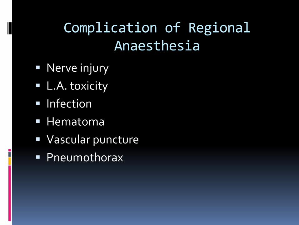
Complication of Regional Anaesthesia
Nerve injury
L.A. toxicity
Infection
Hematoma
Vascular puncture
Pneumothorax
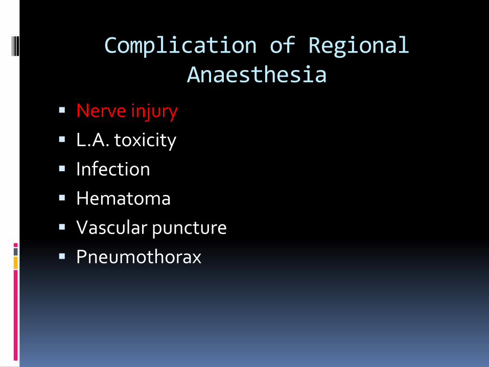
Complication of Regional Anaesthesia
Nerve injury
L.A. toxicity
Infection
Hematoma
Vascular puncture
Pneumothorax
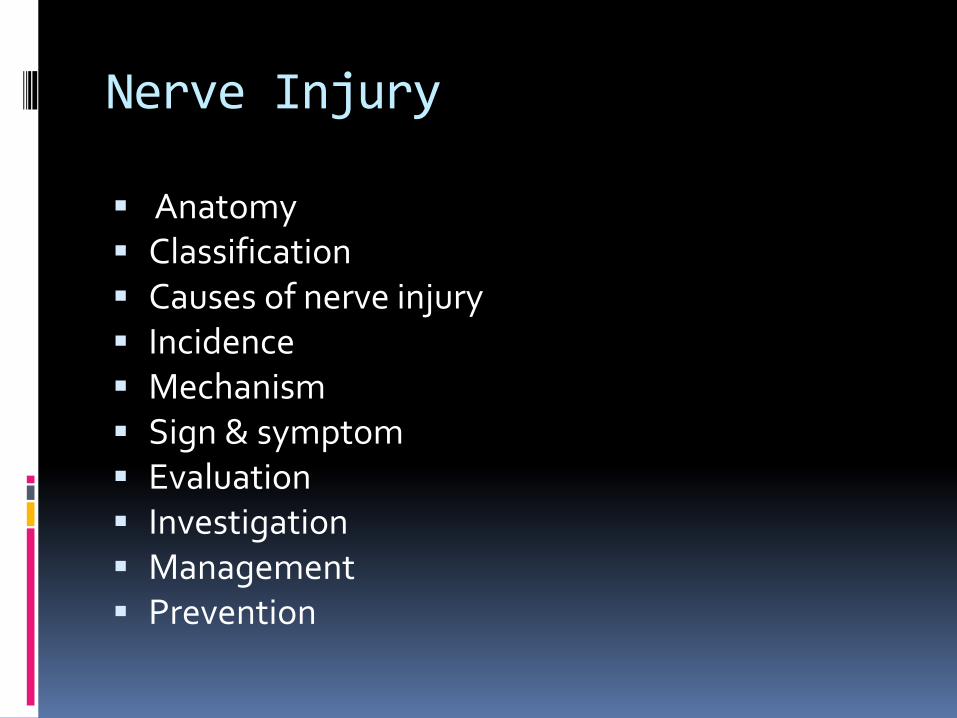
Nerve Injury
Anatomy Classification Causes of nerve injury Incidence Mechanism Sign & symptom Evaluation Investigation Management Prevention
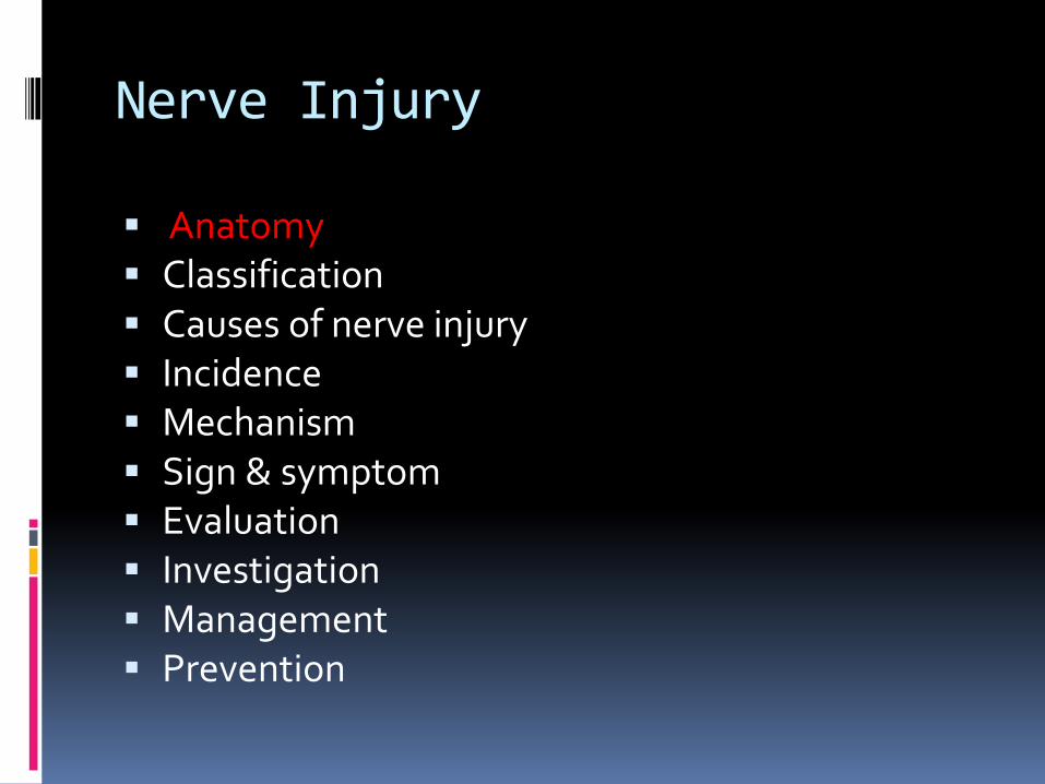
Nerve Injury
Anatomy Classification Causes of nerve injury Incidence Mechanism Sign & symptom Evaluation Investigation Management Prevention
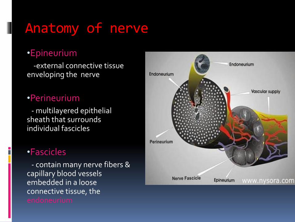
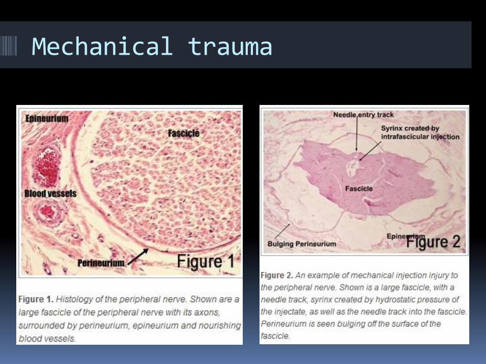
Anatomy of nerve
•Epineurium-external connective tissue
enveloping the nerve
•Perineurium- multilayered epithelial
sheath that surrounds individual fascicles
•Fascicles- contain many nerve fibers &
capillary blood vessels embedded in a loose connective tissue, the endoneurium
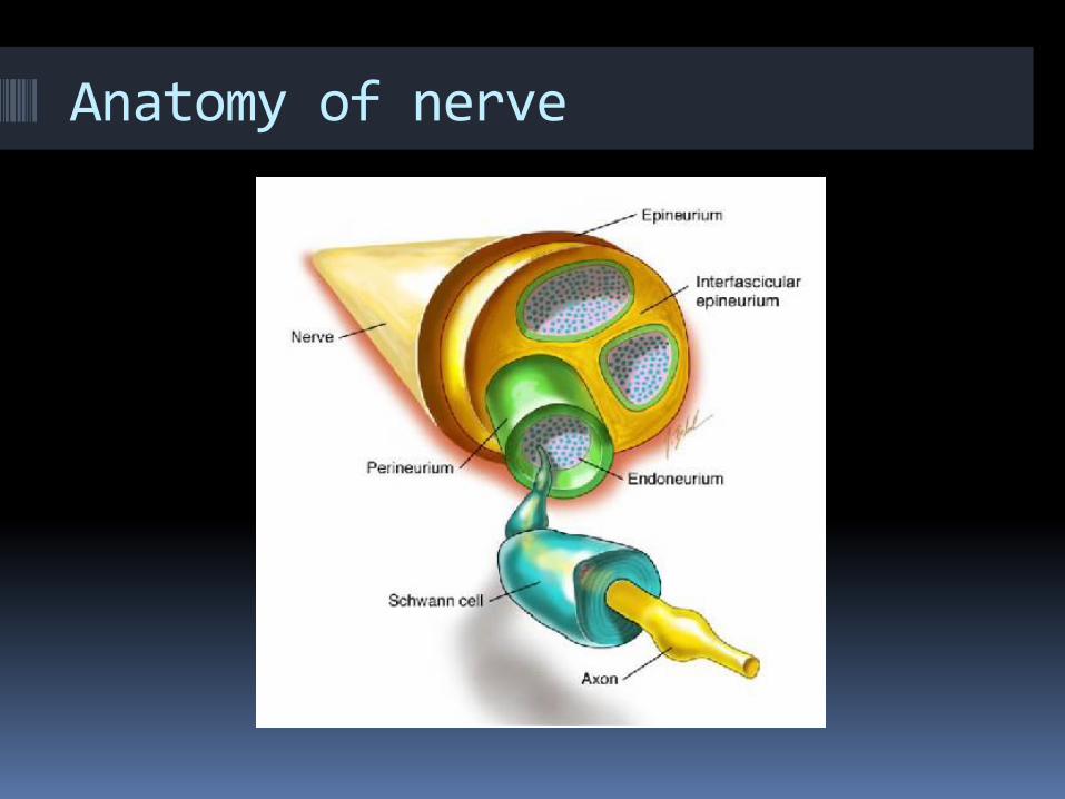
Anatomy of nerve
Nerve Injury
Anatomy Classification Causes of nerve injury Incidence Mechanism Sign & symptom Evaluation Investigation Management Prevention
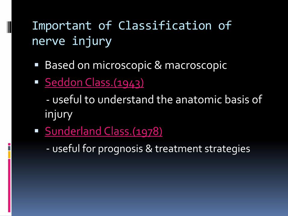
Important of Classification of nerve injury
Based on microscopic & macroscopic
Seddon Class.(1943)
- useful to understand the anatomic basis of injury
Sunderland Class.(1978)
- useful for prognosis & treatment strategies
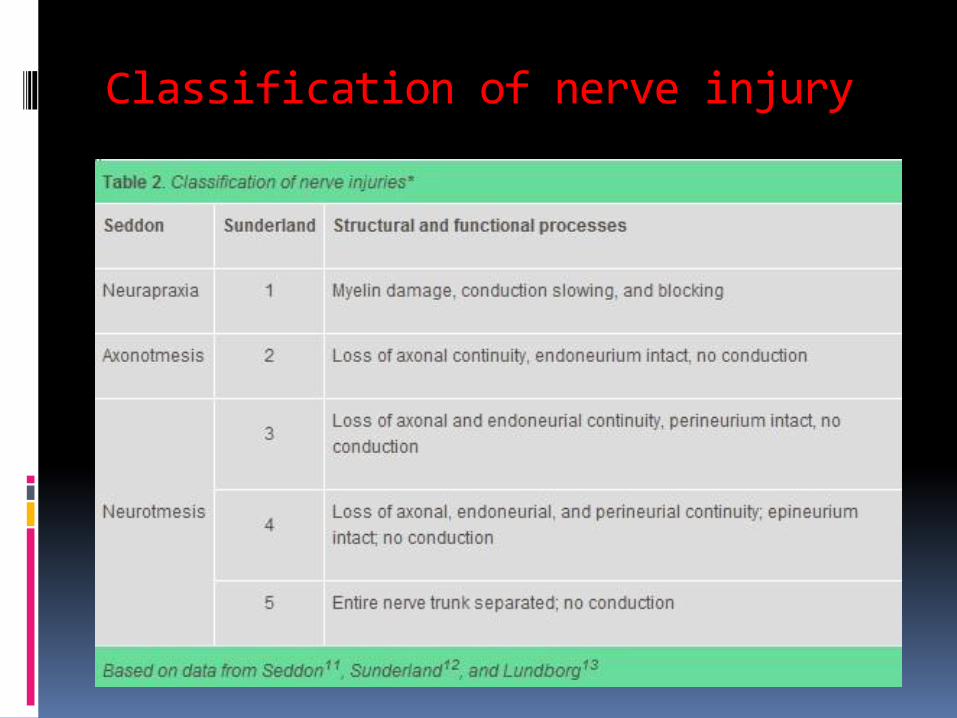
Classification of nerve injury
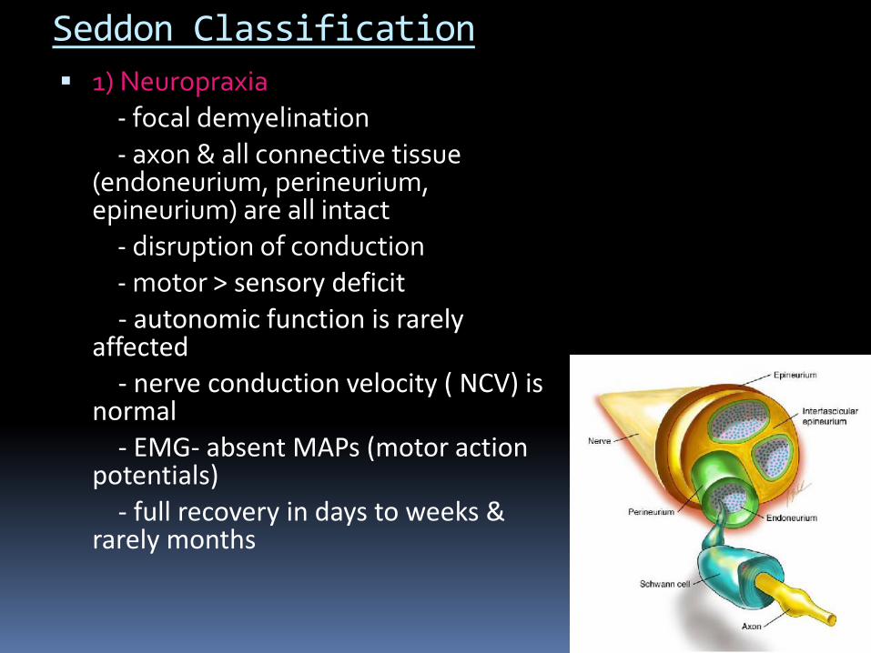
Seddon Classification
1) Neuropraxia- focal demyelination
- axon & all connective tissue (endoneurium, perineurium, epineurium) are all intact
- disruption of conduction- motor > sensory deficit
- autonomic function is rarely affected
- nerve conduction velocity ( NCV) is normal
- EMG- absent MAPs (motor action potentials)
- full recovery in days to weeks & rarely months
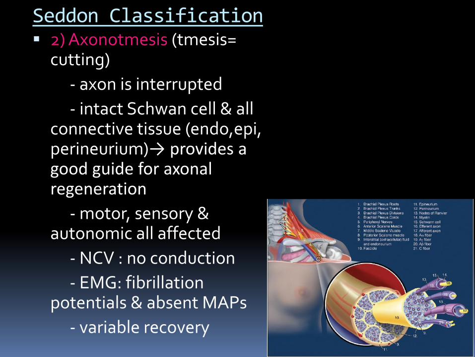
Seddon Classification 2) Axonotmesis (tmesis=
cutting)
- axon is interrupted
- intact Schwan cell & all connective tissue (endo,epi, perineurium)→ provides a good guide for axonal regeneration
- motor, sensory & autonomic all affected
- NCV : no conduction
- EMG: fibrillation potentials & absent MAPs
- variable recovery
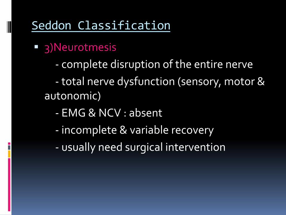
Seddon Classification
3)Neurotmesis
- complete disruption of the entire nerve
- total nerve dysfunction (sensory, motor & autonomic)
- EMG & NCV : absent
- incomplete & variable recovery
- usually need surgical intervention
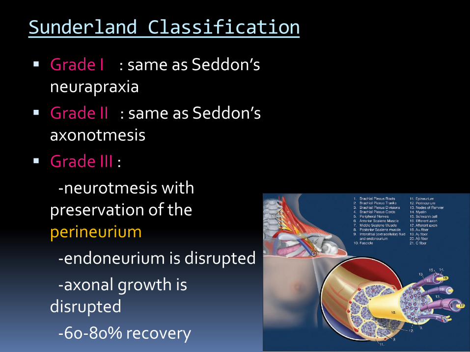
Sunderland Classification
Grade I : same as Seddon’sneurapraxia
Grade II : same as Seddon’saxonotmesis
Grade III :
-neurotmesis with preservation of the perineurium
-endoneurium is disrupted
-axonal growth is disrupted
-60-80% recovery
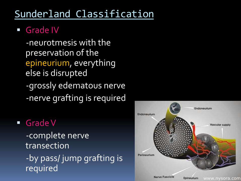
Sunderland Classification
Grade IV
-neurotmesis with the preservation of the epineurium, everything else is disrupted
-grossly edematous nerve
-nerve grafting is required
Grade V
-complete nerve transection
-by pass/ jump grafting is required
Nerve Injury
Anatomy Classification Causes of nerve injury Incidence Mechanism Sign & symptom Evaluation Investigation Management Prevention
Causes of nerve injury
A) ANAESTHETIC TECHNIQUE
Mechanical trauma
- needle trauma
- intraneuronal/ intrafascicular inj.
Neuronal ischemia
Neurotoxicity of L.A.
Wrong drug
Hematoma
Infection
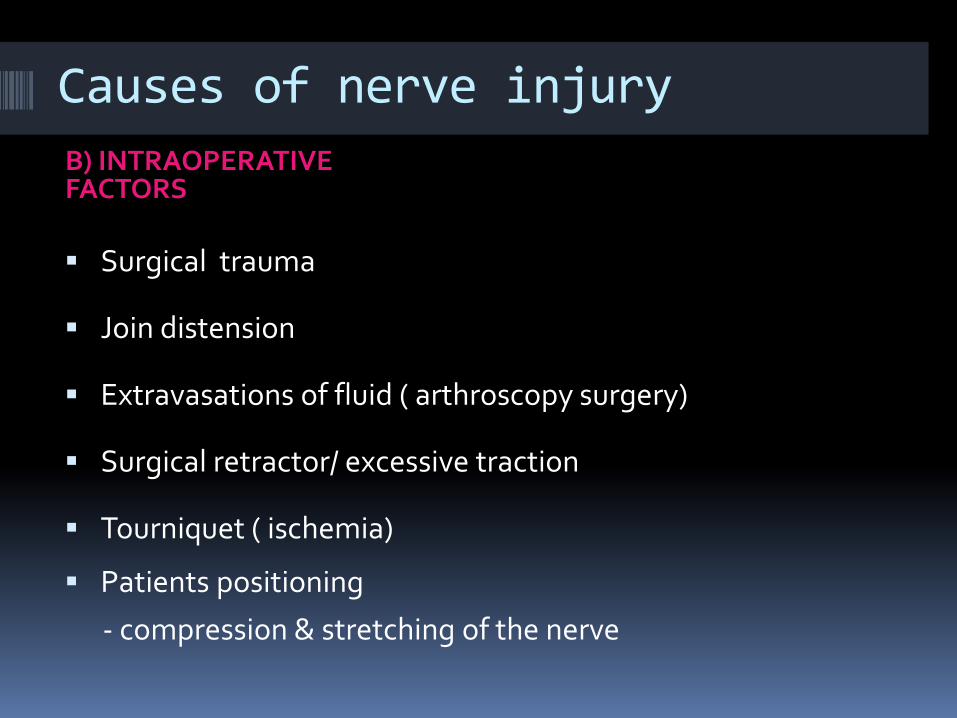
Causes of nerve injury
B) INTRAOPERATIVE FACTORS
Surgical trauma
Join distension
Extravasations of fluid ( arthroscopy surgery)
Surgical retractor/ excessive traction
Tourniquet ( ischemia)
Patients positioning
- compression & stretching of the nerve
Causes of nerve injury
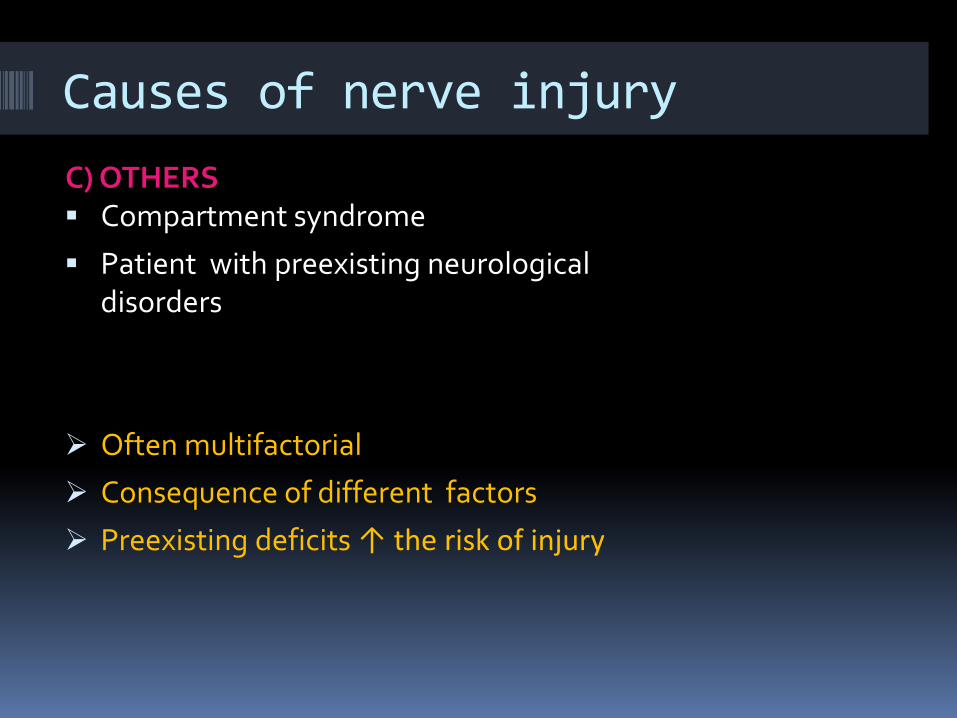
C) OTHERS Compartment syndrome
Patient with preexisting neurological disorders
Often multifactorial
Consequence of different factors
Preexisting deficits ↑ the risk of injury
Nerve Injury
Anatomy Classification Causes of nerve injury Incidence Mechanism Sign & symptom Evaluation Investigation Management Prevention
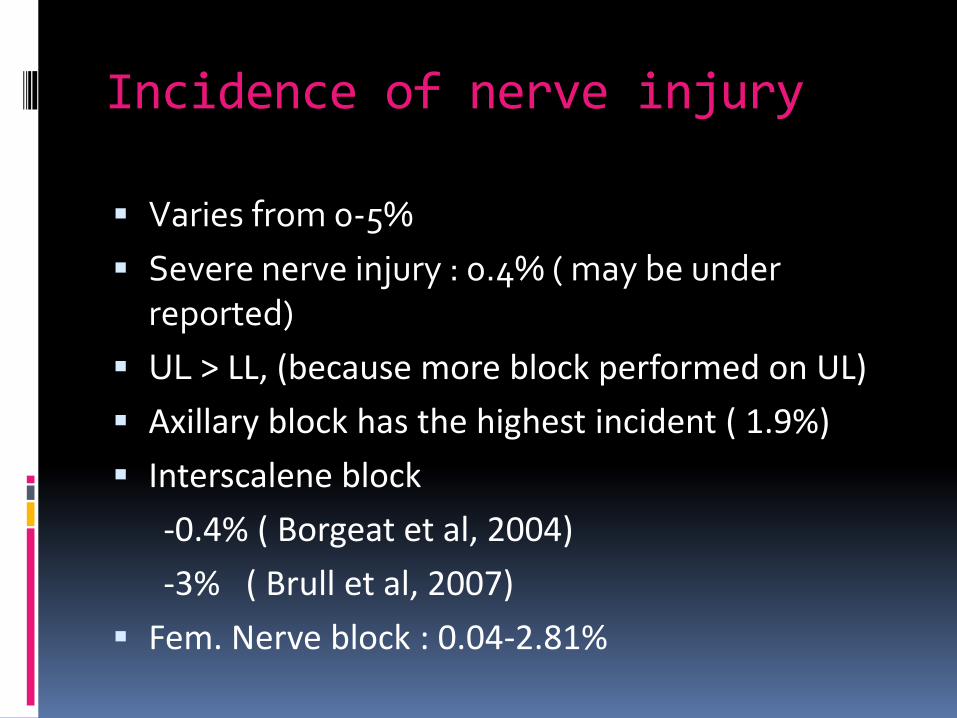
Incidence of nerve injury
Varies from 0-5%
Severe nerve injury : 0.4% ( may be under reported)
UL > LL, (because more block performed on UL)
Axillary block has the highest incident ( 1.9%)
Interscalene block
-0.4% ( Borgeat et al, 2004)
-3% ( Brull et al, 2007)
Fem. Nerve block : 0.04-2.81%
Nerve Injury
Anatomy Classification Causes of nerve injury Incidence Mechanism Sign & symptom Evaluation Investigation Management Prevention
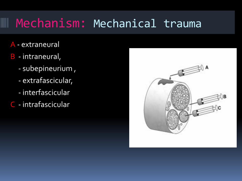
Mechanism: Mechanical trauma
A - extraneural
B - intraneural,
- subepineurium ,
- extrafascicular,
- interfascicular
C - intrafascicular
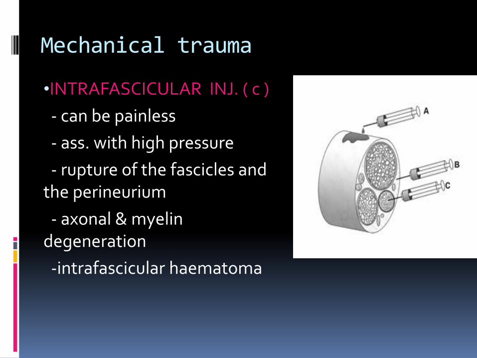
Mechanical trauma
•INTRAFASCICULAR INJ. ( c )
- can be painless
- ass. with high pressure
- rupture of the fascicles and the perineurium
- axonal & myelin degeneration
-intrafascicular haematoma
Mechanical trauma
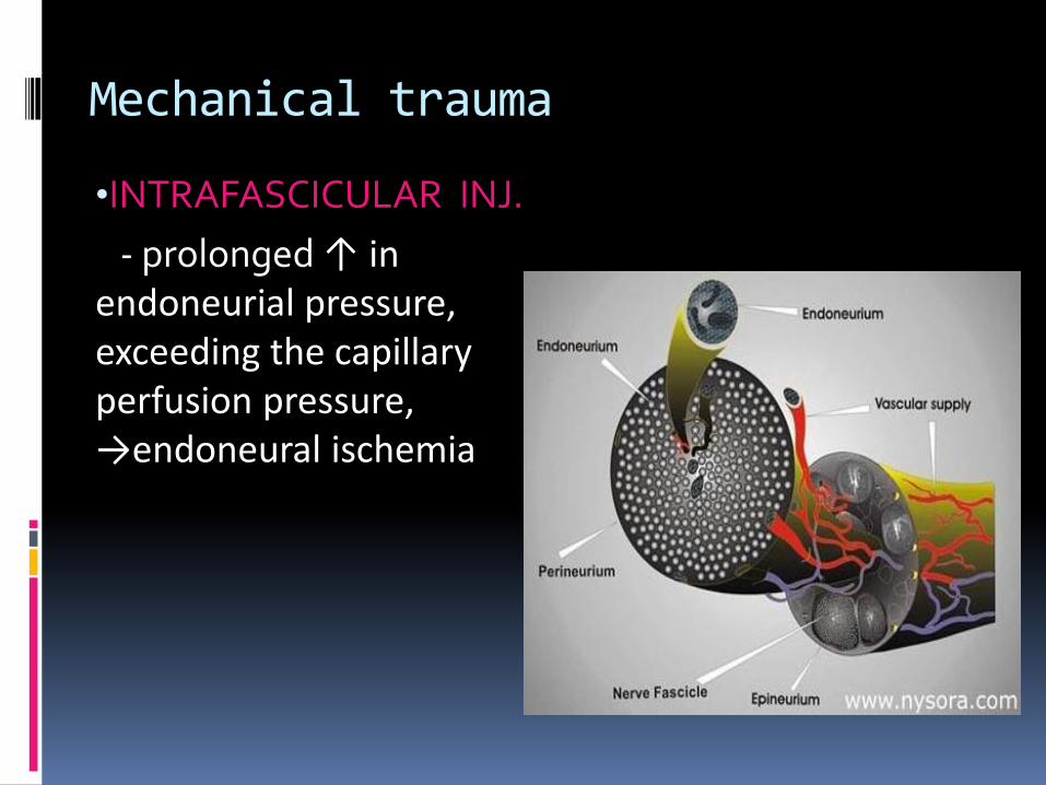
•INTRAFASCICULAR INJ.
- prolonged ↑ in endoneurial pressure, exceeding the capillary perfusion pressure, →endoneural ischemia
Mechanical trauma
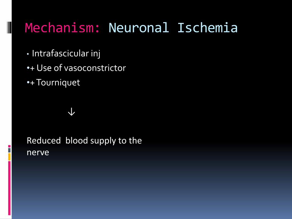
Mechanism: Neuronal Ischemia
• Intrafascicular inj
•+ Use of vasoconstrictor
•+ Tourniquet
↓
Reduced blood supply to the nerve
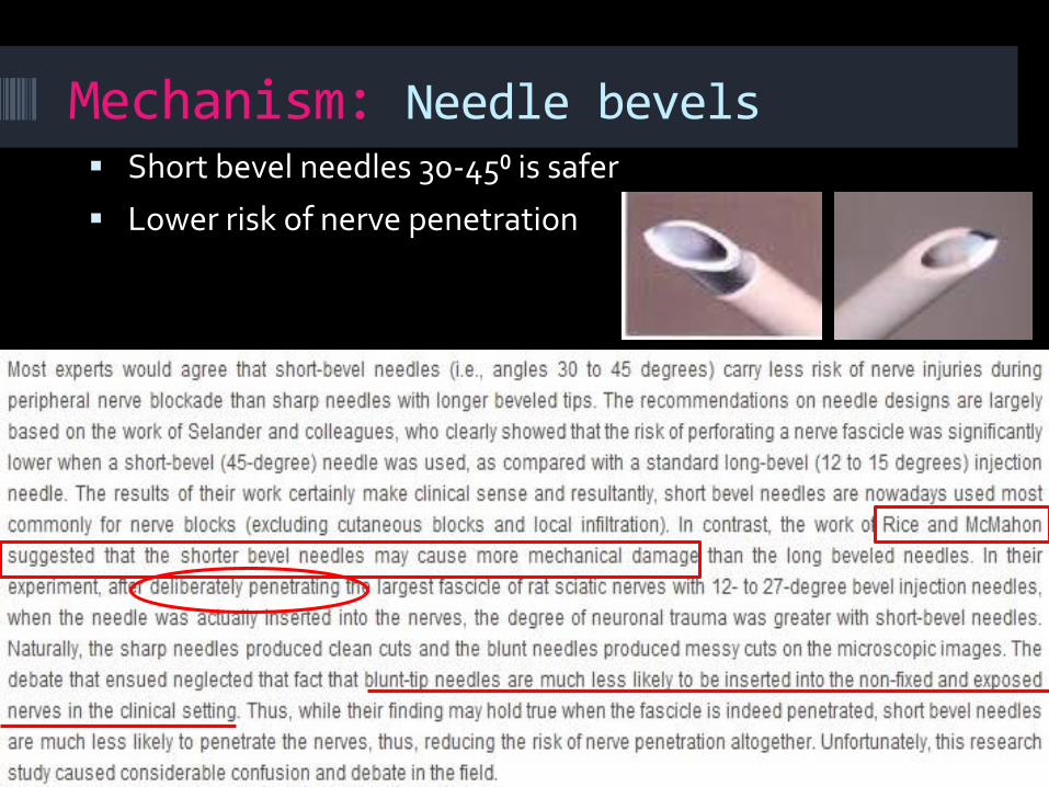
Mechanism: Needle bevels Short bevel needles 30-45⁰ is safer
Lower risk of nerve penetration
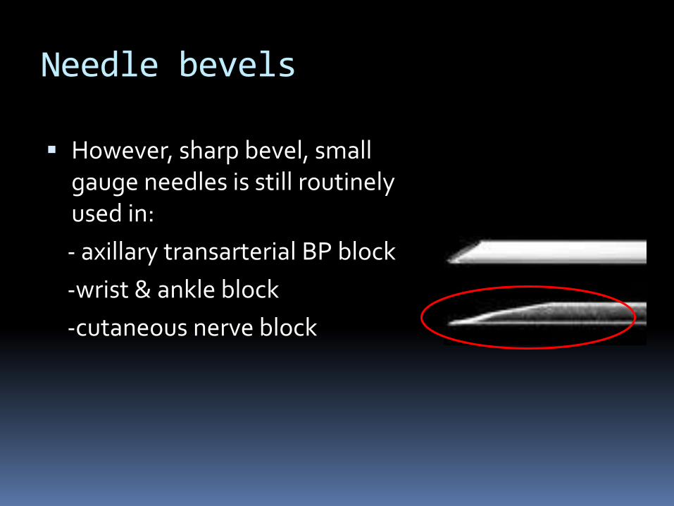
Needle bevels
However, sharp bevel, small gauge needles is still routinely used in:
- axillary transarterial BP block
-wrist & ankle block
-cutaneous nerve block
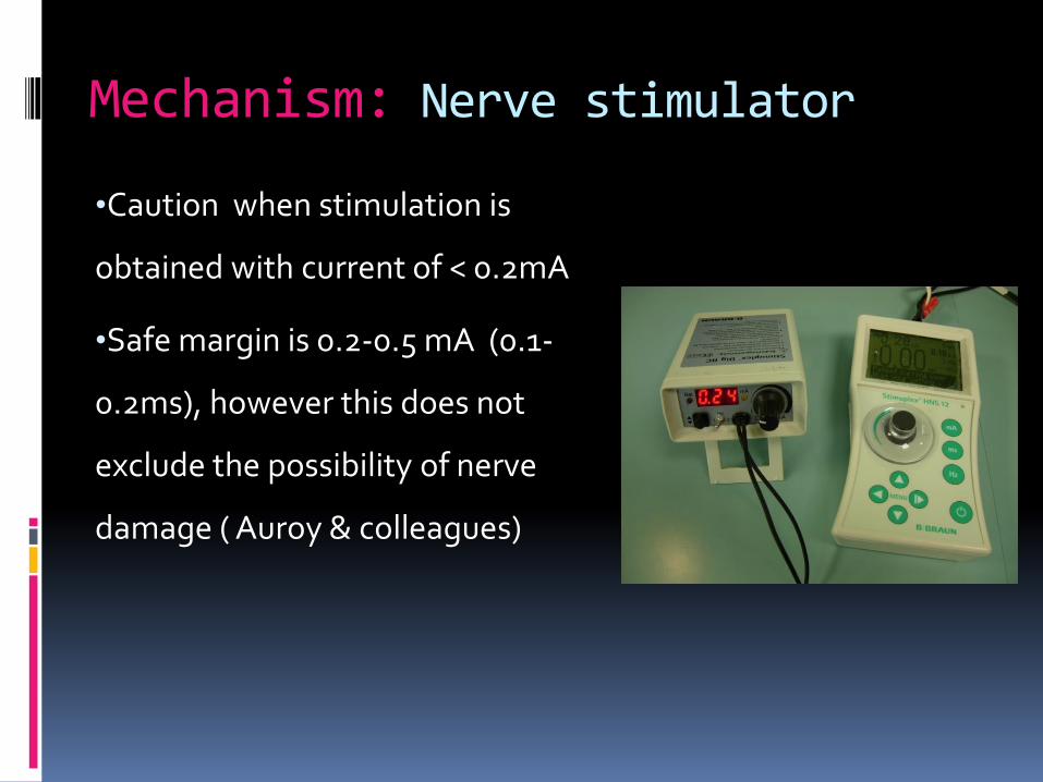
Mechanism: Nerve stimulator
•Caution when stimulation is
obtained with current of < 0.2mA
•Safe margin is 0.2-0.5 mA (0.1-
0.2ms), however this does not
exclude the possibility of nerve
damage ( Auroy & colleagues)
Nerve stimulator
• Motor respond may be absent even when the
needle is inserted intraneurally
-not stimulating on motor nerve fb
-needle tip-nerve fb may has high resistance
• Need accurate & reliable nerve stimulator
• Tested by biomedical dept.
Mechanism: Nerve toxicity of
injected solution / wrong drug
-worst if injected into the fascicle
Most damaging drugs Other drugs
Benzylpenicilin
Diazepam
Paradelhyde ( sedative & antiepileptic)
Antibiotic
Analgesics
Sedatives
Antiemetic
Mechanism: Neurotoxicity of L.A.
L.A. itself produce a variety of cytotoxic effects
Proportionate to the concentration & duration
Usually in intrafascicular inj., but high conc. of
extrafascicular anesthetics may produce axonal injury
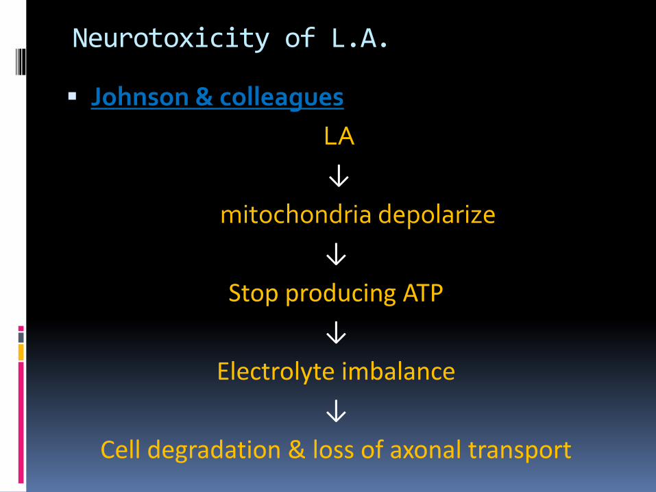
Neurotoxicity of L.A.
Johnson & colleagues
LA
↓
mitochondria depolarize
↓
Stop producing ATP
↓
Electrolyte imbalance
↓
Cell degradation & loss of axonal transport
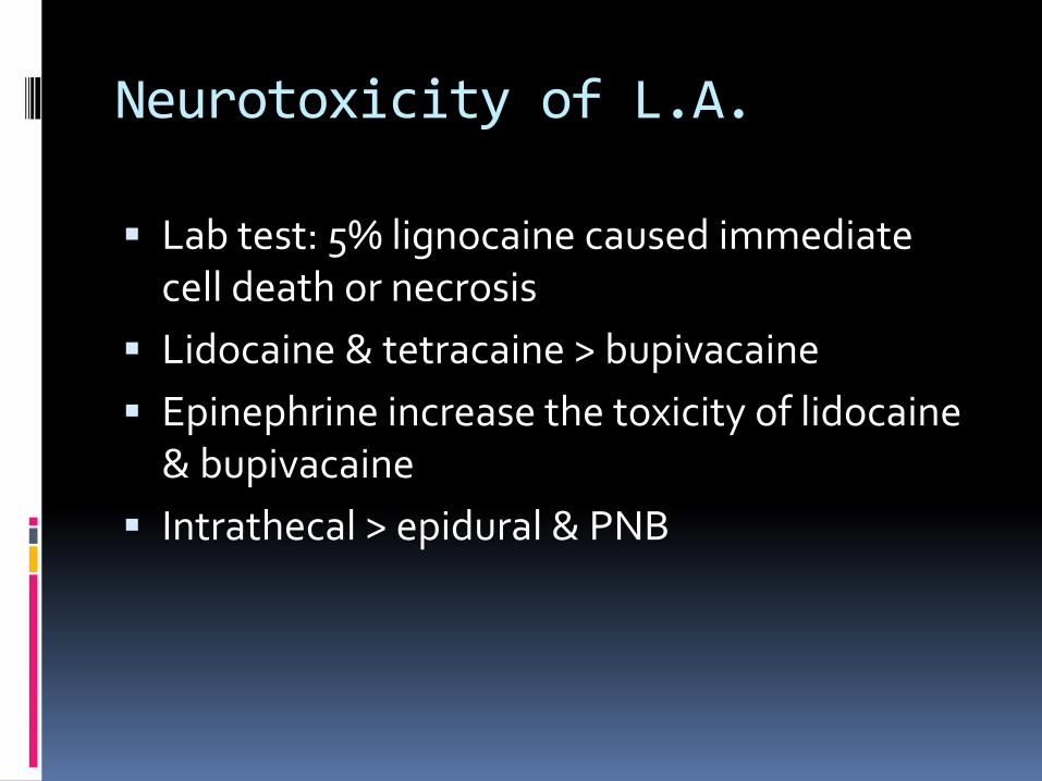
Neurotoxicity of L.A.
Lab test: 5% lignocaine caused immediate cell death or necrosis
Lidocaine & tetracaine > bupivacaine
Epinephrine increase the toxicity of lidocaine& bupivacaine
Intrathecal > epidural & PNB
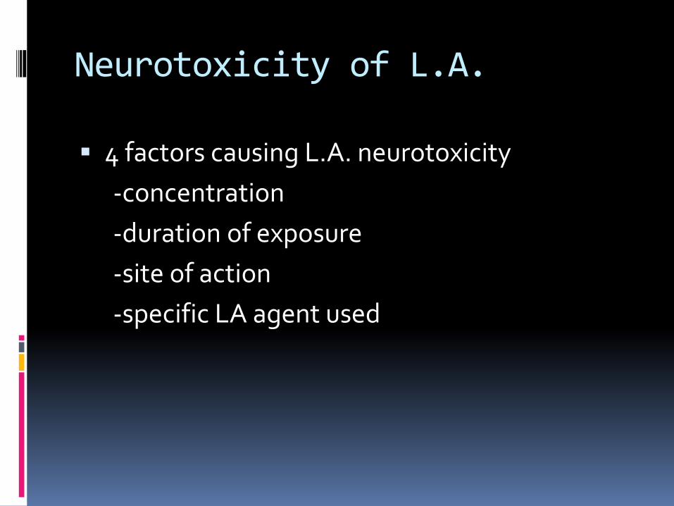
Neurotoxicity of L.A.
4 factors causing L.A. neurotoxicity
-concentration
-duration of exposure
-site of action
-specific LA agent used
Nerve Injury
Anatomy Classification Causes of nerve injury Incidence Mechanism Sign & symptom Evaluation Investigation Management Prevention
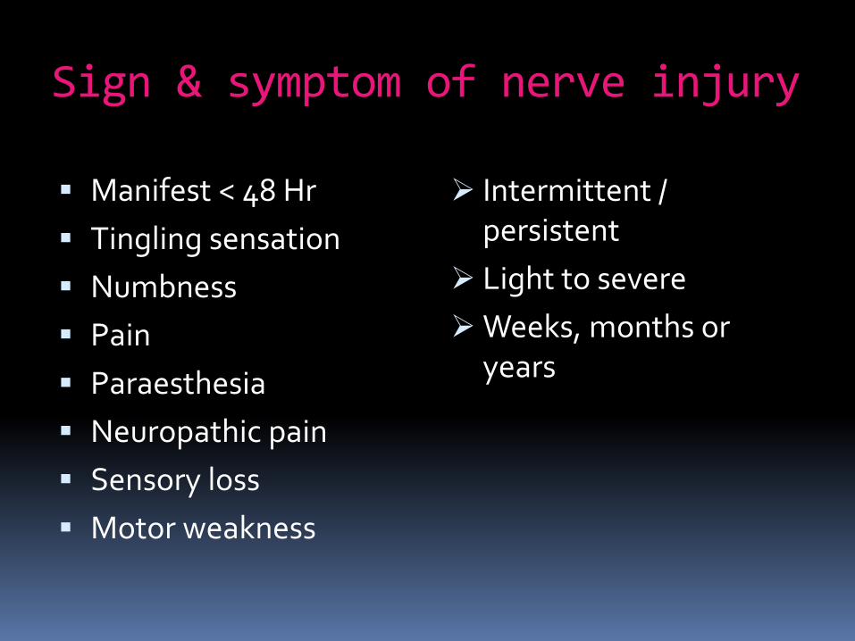
Sign & symptom of nerve injury
Manifest < 48 Hr
Tingling sensation
Numbness
Pain
Paraesthesia
Neuropathic pain
Sensory loss
Motor weakness
Intermittent / persistent
Light to severe
Weeks, months or years
Nerve Injury
Anatomy Classification Causes of nerve injury Incidence Mechanism Sign & symptom Evaluation Investigation Management Prevention
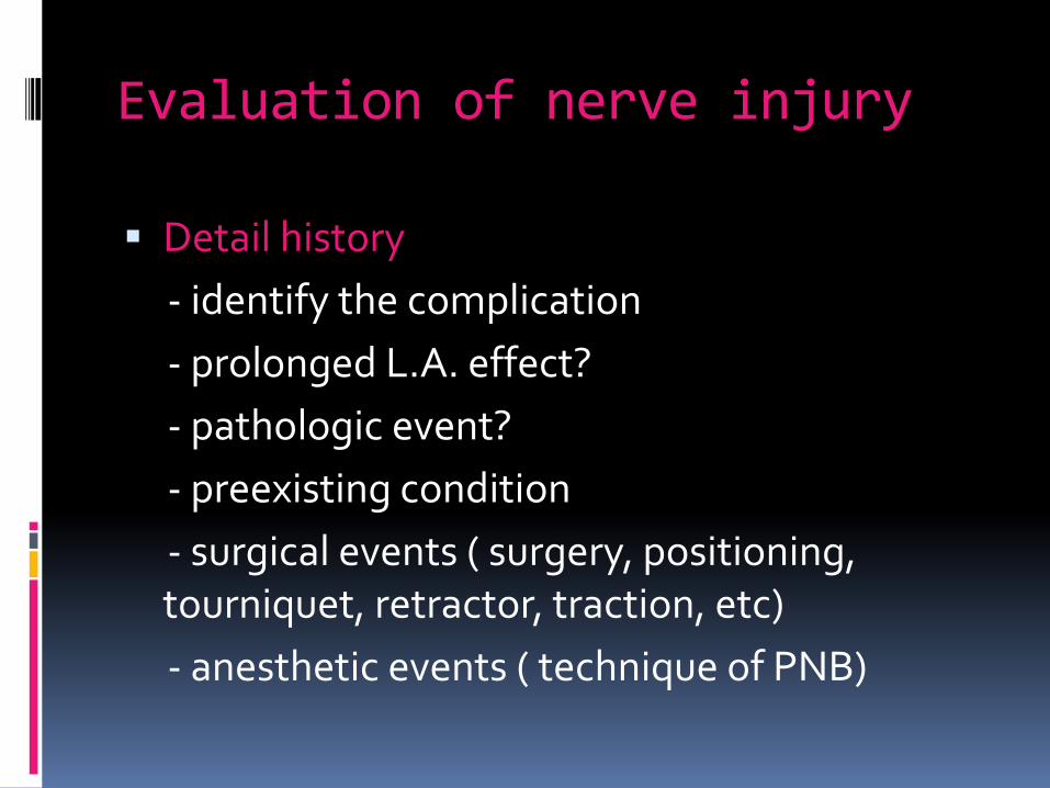
Evaluation of nerve injury
Detail history
- identify the complication
- prolonged L.A. effect?
- pathologic event?
- preexisting condition
- surgical events ( surgery, positioning, tourniquet, retractor, traction, etc)
- anesthetic events ( technique of PNB)
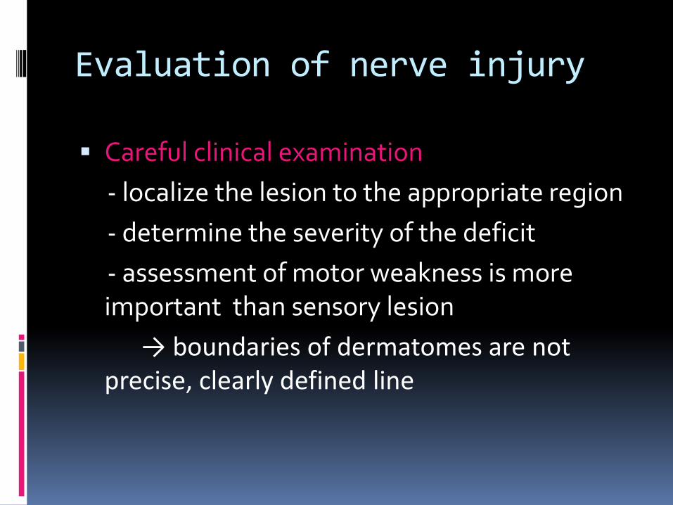
Evaluation of nerve injury
Careful clinical examination
- localize the lesion to the appropriate region
- determine the severity of the deficit
- assessment of motor weakness is more important than sensory lesion
→ boundaries of dermatomes are not precise, clearly defined line
Nerve Injury
Anatomy Classification Causes of nerve injury Incidence Mechanism Sign & symptom Evaluation Investigation Management Prevention
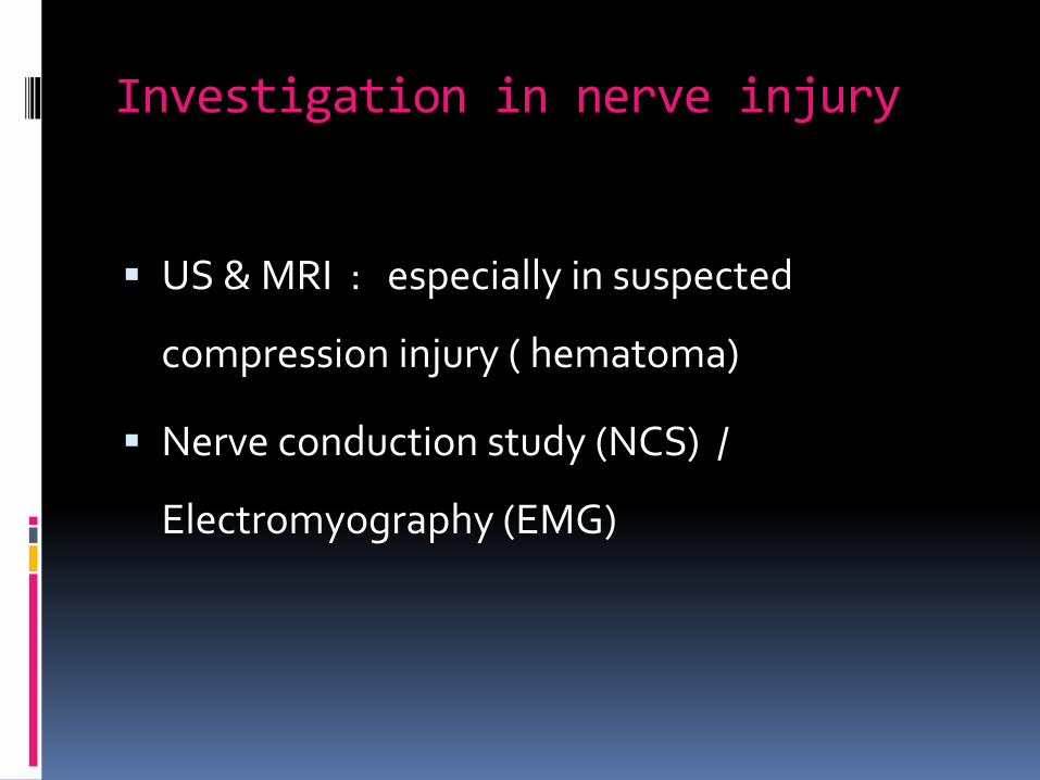
Investigation in nerve injury
US & MRI : especially in suspected
compression injury ( hematoma)
Nerve conduction study (NCS) /
Electromyography (EMG)
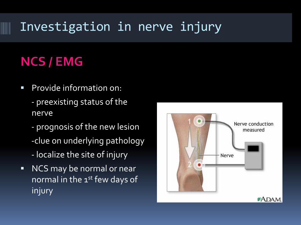
Investigation in nerve injury
NCS / EMG
Provide information on:
- preexisting status of the nerve
- prognosis of the new lesion
-clue on underlying pathology
- localize the site of injury
NCS may be normal or near normal in the 1st few days of injury
Investigation in nerve injury
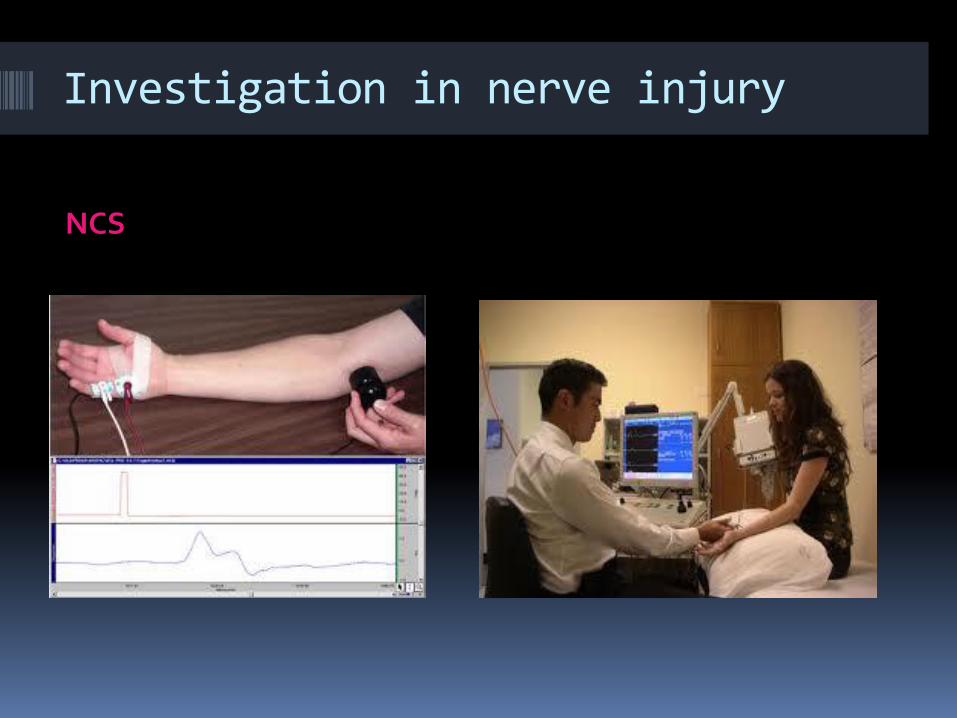
NCS
Investigation in nerve injury
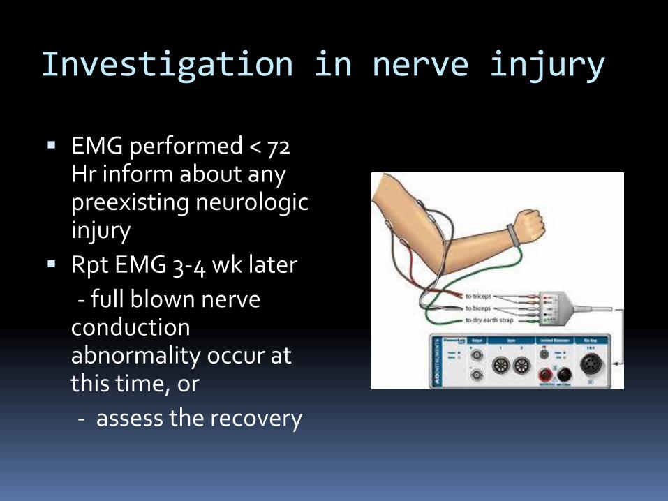
EMG performed < 72 Hr inform about any preexisting neurologic injury
Rpt EMG 3-4 wk later
- full blown nerve conduction abnormality occur at this time, or
- assess the recovery
Nerve Injury
Anatomy Classification Causes of nerve injury Incidence Mechanism Sign & symptom Evaluation Investigation Management Prevention
Management of nerve injury
A) Minor Nerve Deficit
Conservative
Reassurances
Telephone follow up-
weekly / bi-weekly
Management of nerve injuryB) Major Nerve Deficit
Early neurologist/ neurosurgery consultation
Diagnostic test
Early pain treatment to prevent nerve sensitization
Physical & occupational therapy consultation
- strength training
- range of motion exercise
- to minimize contracture , muscle atrophy & prolonged disability
Management of nerve injuryB) Major nerve Deficit
Social services referral
- if patient unable to perform daily activity
Close follow up by neurologist & anaesthetist until
injury is completely resolved or is stable
Repeat EMG at 6 wk, 3 mth & 6 mth
If no improvement after 3-4 months, may consider
neurolysis / neurotization
Management of nerve injury
C) Drug Therapy
Under chronic pain specialist supervision
Multiple drug are needed to treat neurally mediated pain
Need consistent follow up
Management of nerve injuryC) Drugs:
Tricyclic Antidepressants
- amitriptyline 10-25mg O.D upto 100 mg/day
Selective serotonin reuptake inhibitors
- paroxetine
Anticonvulsants
- Gabapentin 300mg O.D upto 1800 mg/day in divided
dose
- carbamazepine
Management of nerve injuryC) Drugs:
Opiods
- oxycodone
- oxycontin
- fentanyl patches
Tramadol
Capsacion ointmemnt
- for cutaneous hyperalgesia
Nerve Injury
Anatomy Classification Causes of nerve injury Incidence Mechanism Sign & symptom Evaluation Investigation Management Prevention
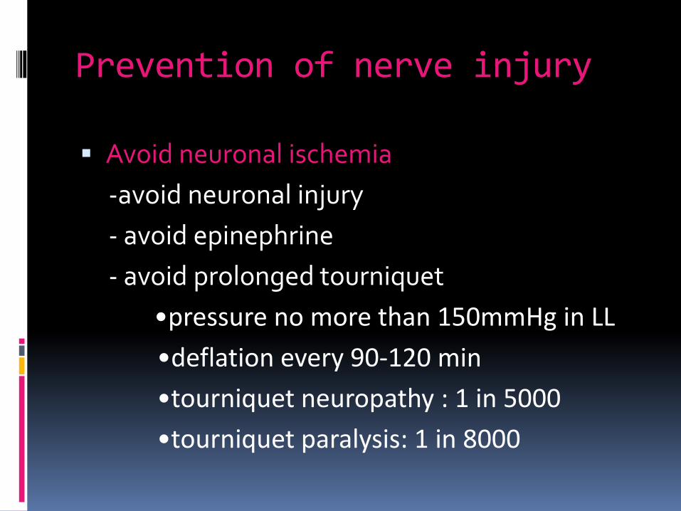
Prevention of nerve injury
Avoid neuronal ischemia
-avoid neuronal injury
- avoid epinephrine
- avoid prolonged tourniquet
•pressure no more than 150mmHg in LL
•deflation every 90-120 min
•tourniquet neuropathy : 1 in 5000
•tourniquet paralysis: 1 in 8000
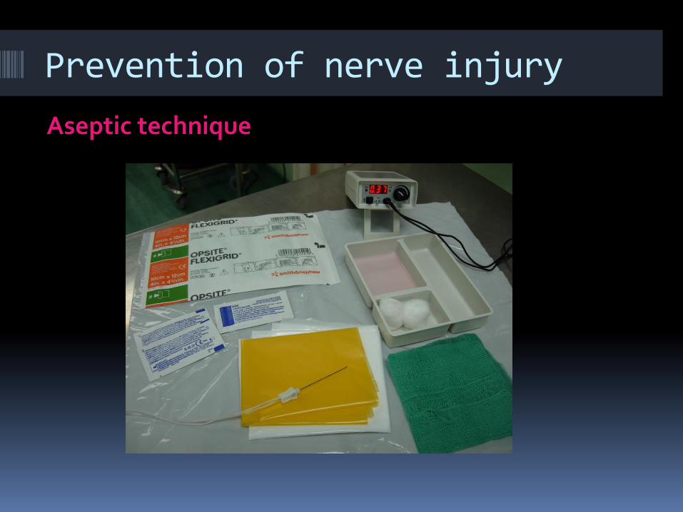
Prevention of nerve injury
Aseptic technique
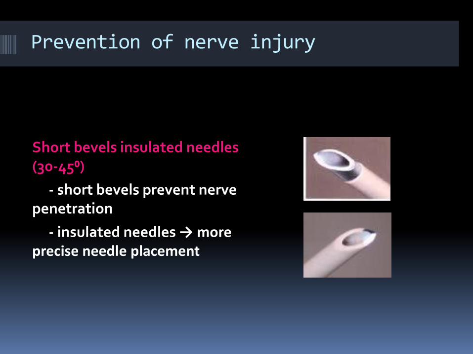
Prevention of nerve injury
Short bevels insulated needles (30-45⁰)
- short bevels prevent nerve penetration
- insulated needles → more precise needle placement
Prevention of nerve injury
Needles of appropriate length for each and every block procedure
Slow needle advancement & withdraw
- fast insertion & withdrawal of the needle may result in failure to stimulate the nerve
Prevention of nerve injury
Fractionated injections
- 3-5 ml with intermittent aspiration
- negative aspiration of blood does not exclude intravascular injection
Accurate nerve stimulator
- operational & correct current
- 0.2-0.5 mA, 0.1-0.2ms, 2 Hz
Prevention of nerve injury
Avoidance of forceful & fast injection
- prevent channeling of L.A. to the unwanted tissue layers, lymphatic vessels or small veins
- avoid intrafascicular injection
- recommended speed : 15- 20 ml/ min
Prevention of nerve injury
Avoidance of injection under high pressure
- intrafascicular needle placement results in higher resistance
- always use the same syringe and needle size to develop a ‘feel’ during injection
- pressure should not exceed 20 psi
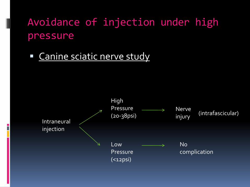
Avoidance of injection under high pressure
Canine sciatic nerve study
Intraneuralinjection
HighPressure(20-38psi)
LowPressure(<12psi)
Nerve injury
Nocomplication
(intrafascicular)
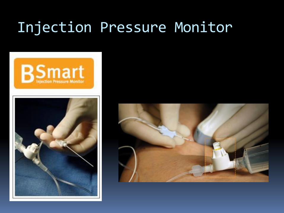
Injection Pressure Monitor
Prevention of nerve injury
Avoidance of paraesthesia on injection
- pain on injection may signify intraneuronalinj.
- however, absence of pain on injection alone does not exclude intraneural inj.
Controversial of Pain as monitor
Most neurologic complication reported after
PNB & even central neuraxial block have not
been associated with pain on injection
- Only 10% of them reported pain on injection
Controversial of Pain as monitor Cheney & coworkers
- when patients reported pain on injection, the anesthesiologist stopped the injection, but patients still went on to develop nerve injury
- in animal models studies, nerve fascicles become injured/ ruptured at the very onset of the injection even with small volume of LA
Controversial of Pain as monitor Pain is difficult to assess
-? Discomfort/paraesthesia on inj, ( which is normal)
- ? Abnormal pain d/t intraneural inj.
- variable patients’ pain thresholds
- patients’ ability to describe the pain sensation
- anesthesiologist’s subjective interpretation of patients’ respond
Controversial of Pain as monitor Borgeat et al
- 21% of the interscalene block reported transient, burning pain, but none dev. nerve injury
Current practice: avoid pain on injection
Prevention of nerve injury
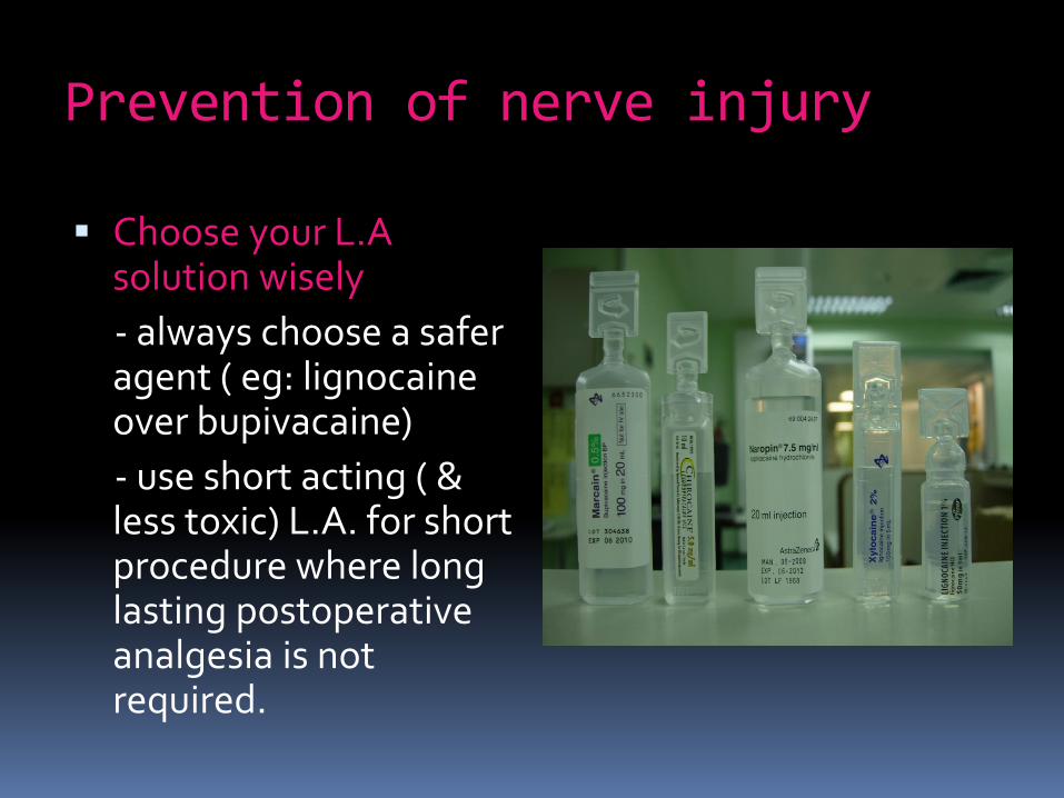
Choose your L.A solution wisely
- always choose a safer agent ( eg: lignocaineover bupivacaine)
- use short acting ( & less toxic) L.A. for short procedure where long lasting postoperative analgesia is not required.
Prevention of nerve injury
Avoid repeating blocks after a failed block
- pain on injection cannot be detected
- when indicated, should be done by experienced hand, & under USG.
Prevention of nerve injury
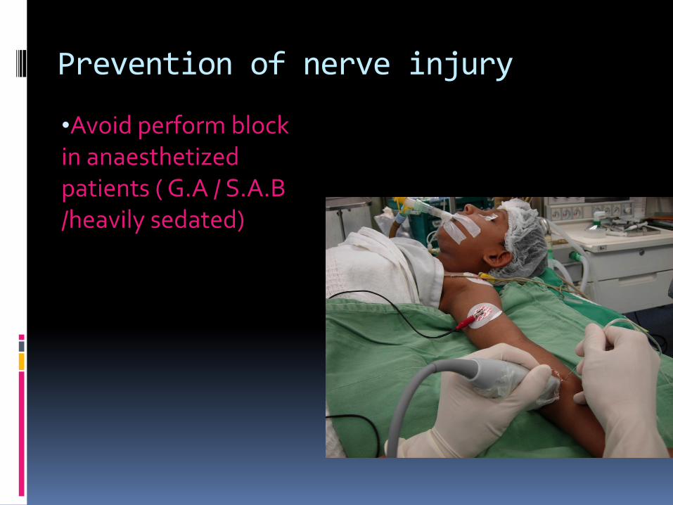
•Avoid perform block in anaesthetized patients ( G.A / S.A.B /heavily sedated)
Controversial of RA in anesthetized patient
General believe:
Awake patients allow monitoring of CNS toxicity
GA/ heavily sedated pt prevent the detection of early sign & symptom of LA toxicity
- however, almost all the LA toxicity cases occur in awake or sedated pt.
- no report of LA toxicity in adult pt under GA
Controversial of RA in anesthetized patient
Possible explanation:
-premedication offers protection because of its anticonvulsion effects
-anesthetized patients who dev LA toxicity may survive better because they already have:
•secured airway
•ventilated or may be hyperventilated
•receiving high conc of O₂
•in an environment that is ideal for resuscitation
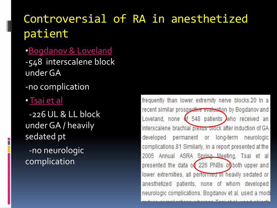
Controversial of RA in anesthetized patient
•Bogdanov & Loveland -548 interscalene block under GA
-no complication
•Tsai et al
-226 UL & LL block under GA / heavily sedated pt
-no neurologic complication
Controversial of RA in anesthetized patient
Pediatric anesthesia- RA is commonly performed under GA/ heavily sedated & the complications are rare
- however, PNBs are not routinely used in pediatric
- & usually performed by senior anesthetist
however, no study has been done to compare awake vs anaesthetized pt.( and it is unlikely that such studies will ever be done)
Controversial of RA in anesthetized patient
The belief of GA predispose to a greater risk of severe systemic LA toxicity is purely theoretical
- no data to firmly support this beliefConclusions Regardless, the practice of RA in anesthetized
pediatric pt is universally accepted Adult:
- in the absence of adequate evidence, blocks in anesthetized patients should still not be a common practice- should be appropriately sedated for block performance and patient acceptance
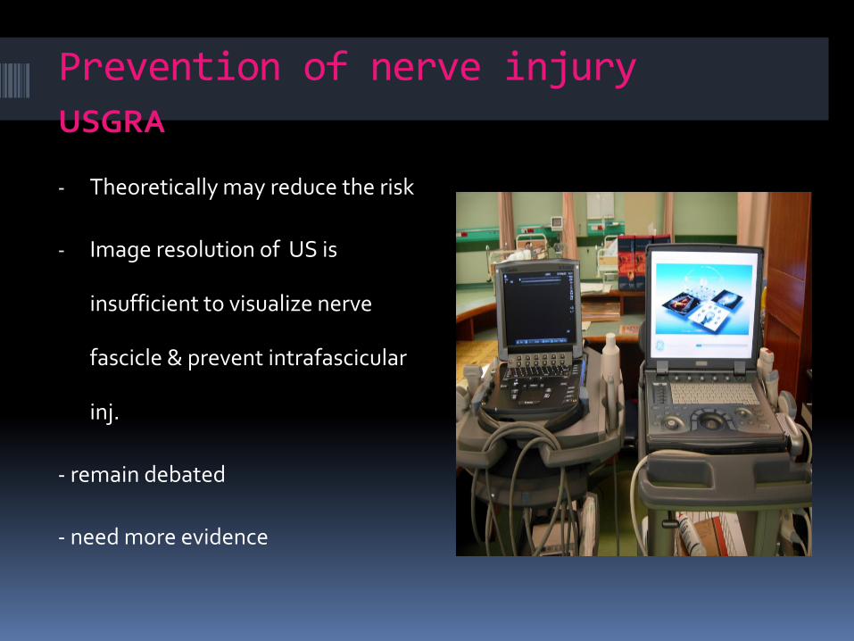
Prevention of nerve injuryUSGRA
- Theoretically may reduce the risk
- Image resolution of US is
insufficient to visualize nerve
fascicle & prevent intrafascicular
inj.
- remain debated
- need more evidence
Prevention of nerve injury
Cautious in anticoagulated patients
- follow the guideline on neuraxialanaesthesia & systemic anticoagulation therapy published by ASRA
Nerve Injury
Remember:
1)Nerve damage after R.A. is unusual &
recovery is generally favorable
2)R.A. is not the 1st cause of nerve damage
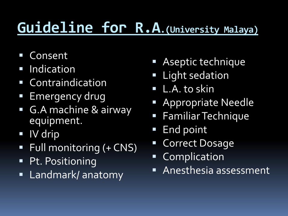
Guideline for R.A.(University Malaya)
Consent Indication Contraindication Emergency drug G.A machine & airway
equipment. IV drip Full monitoring (+ CNS) Pt. Positioning Landmark/ anatomy
Aseptic technique Light sedation L.A. to skin Appropriate Needle Familiar Technique End point Correct Dosage Complication Anesthesia assessment
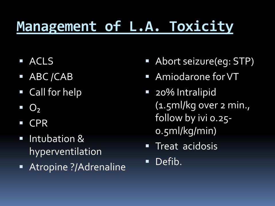
Management of L.A. Toxicity
ACLS
ABC /CAB
Call for help
O₂
CPR
Intubation & hyperventilation
Atropine ?/Adrenaline
Abort seizure(eg: STP)
Amiodarone for VT
20% Intralipid (1.5ml/kg over 2 min., follow by ivi 0.25-0.5ml/kg/min)
Treat acidosis
Defib.
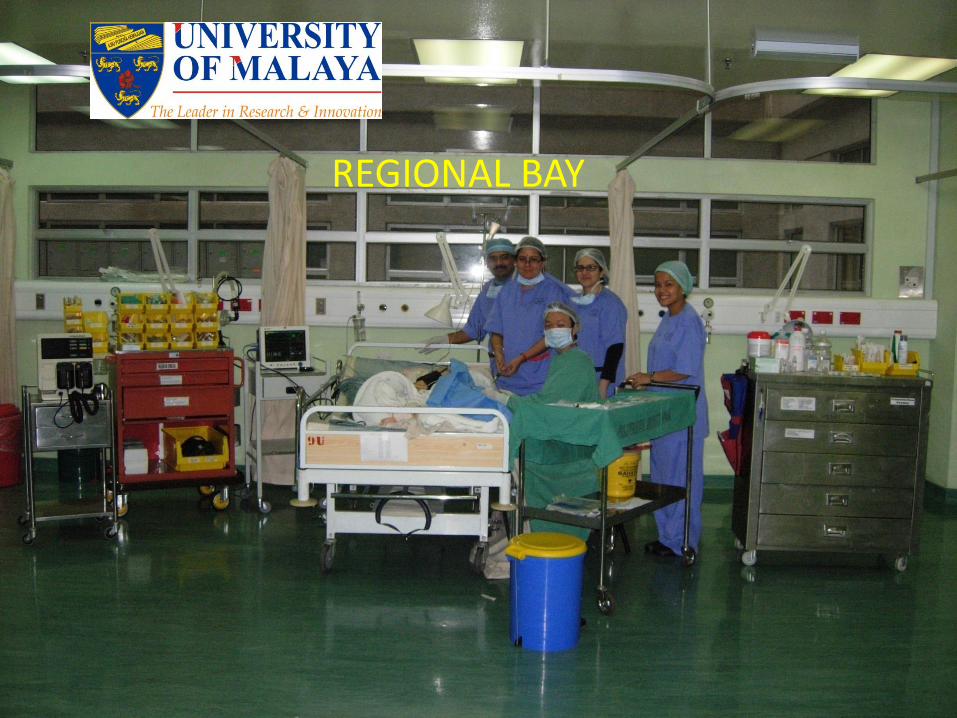
REGIONAL BAY
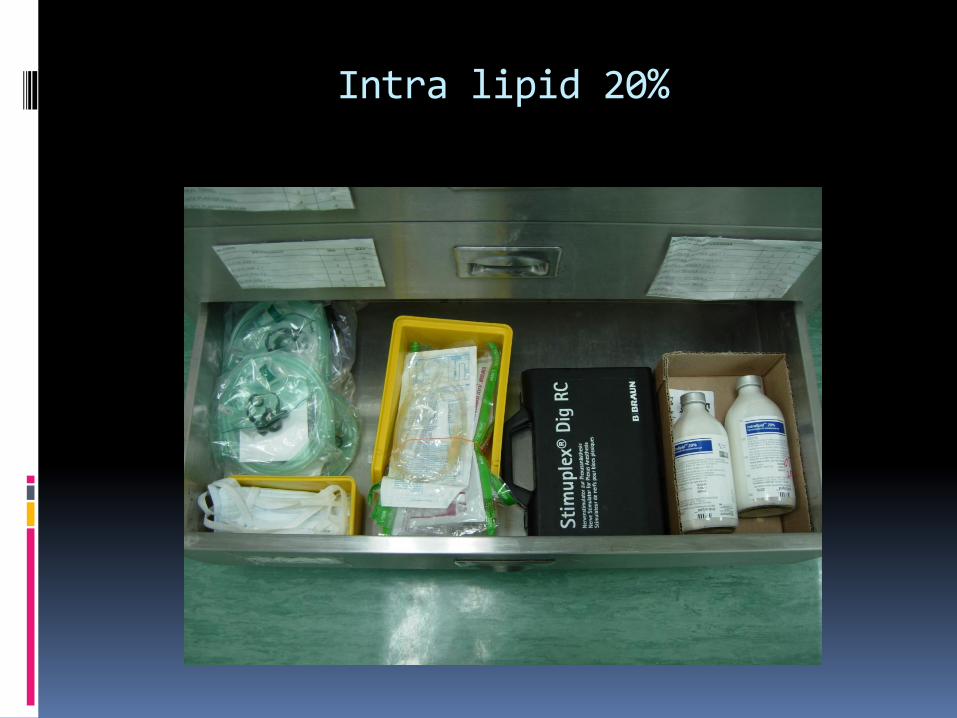
Intra lipid 20%
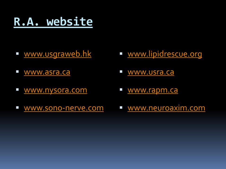
R.A. website
www.usgraweb.hk
www.asra.ca
www.nysora.com
www.sono-nerve.com
www.lipidrescue.org
www.usra.ca
www.rapm.ca
www.neuroaxim.com

Safety First!Lots of Practice!Lots of Patience!
52 Kg Giant Trevalley, off shore of Bintulu, South China Sea, June 2010
NYSORA asia, KL, 2009

NYSORA asia , Bangkok, 2010

NYSORA asia, Bangalore, 2011
References
Mechanism of neurologic complication with peripheral nerve blocks, NYSORA, A.P Admir Hadzic, Alain Borgeat, Stephen Blumenthal.
Practical issues in Regional Anaesthesia: patient cannot move leg20 hour after single dose femoral block. S. Bloc,
USGRA and the prevention of neurologic injury; fact or fiction? Hebl, James R,M.D, Anaesthsiology, Feb 08, vol 108-Issue 2, pg 186-188
Nerve conduction study-Medical disability guideline, www.mdguidelines.com/nerve conduction study, Cachel-Similar
www.arapmi.org/maraa-book-project/chapt25
Morphological and physiological aspect of peripheral nerves and prevention of nerve injury in peripheral nerve blockade, J. De Andres, M.A. Reina, F.Maches
Classification of nerve injury, wiki.cns.org/wiki/index.php/injury-classification
Complication of peripheral nerve blocks, C.L.Jeng, T.M.Torillo and M.A. Rosenblatt, BJA, vol 105,issue suppl 1.

( in collaboration with SIGRA, Malaysia )Dr Ling Kwong Ung,Anaesthetist,Sime Darby medical Centre,Selangor, [email protected]