Embed Size (px)
Citation preview

http://immunol.nature.com • may 2001 • volume 2 no 5 • nature immunology
Pingwei Li1, Daniel L. Morris1, Benjamin E.Willcox2,Alexander Steinle3,*,Thomas Spies3 and Roland K. Strong1
The major histocompatibility complex (MHC) class I homolog, MICA, is a stress-inducible ligand forNKG2D, a C-type lectin–like activating immunoreceptor.The crystal structure of this ligand-receptorcomplex that we report here reveals an NKG2D homodimer bound to a MICA monomer in aninteraction that is analogous to that seen in T cell receptor–MHC class I protein complexes. Similarsurfaces on each NKG2D monomer interact with different surfaces on either the α1 or α2 domains ofMICA. The binding interactions are large in area and highly complementary. The central section ofthe α2-domain helix, disordered in the structure of MICA alone, is ordered in the complex and formspart of the NKG2D interface.The extensive flexibility of the interdomain linker of MICA is shown byits altered conformation when crystallized alone or in complex with NKG2D.
1Division of Basic Sciences, Fred Hutchinson Cancer Research Center, Seattle,WA 98109 USA. 2Division of Biology, California Institute of Technology, Pasadena, CA 91125,USA. 3Division of Clinical Research, Fred Hutchinson Cancer Research Center, Seattle,WA 98109, USA. *Present address: Interfakultäres Institut für Zellbiologie, Abteilung
Immunologie, Auf der Morgenstelle 15, D-72076 Tübingen, Germany. Correspondence should be addressed to R. K. S. ([email protected]).
Complex structure of the activatingimmunoreceptor NKG2D and its MHC
class I–like ligand MICA
T cells detect pathogens through specific interactions between T cellreceptors (TCRs) on their surfaces and complexes of antigenic peptidefragments and polymorphic major histocompatibility complex (MHC)class I molecules on the surfaces of infected cells1. T cell activationrequires an interaction between TCRs and appropriate MHC-peptidecomplexes in the context of appropriate costimulatory signals fromengagement, for example, of the CD28 receptor on T cells with CD80or CD86 ligands2. Diverse cell surface molecules also modulate T cellactivation, including the inhibitory and stimulatory receptors first iden-tified on natural killer (NK) cells that are now recognized as beingexpressed on a range of cell types, including T cells. NK cells regulateinnate and acquired immune responses through the release of variousimmune modulators (such as interferon-γ) or by directly destroyingvirally infected or neoplastic cells. NK cell effector functions are regu-lated by integrating signals across the array of stimulatory and inhibito-ry NK cell surface receptors (NCRs) that are engaged upon interactionwith target-cell surface NCR ligands3. Virally infected cells and tumorcells evade T cell–mediated immune surveillance by down-regulatingthe cell surface expression of MHC class I proteins4. Loss of NK cellinhibitory signals from NCRs that are specific for classical (HLA-A,HLA-B and HLA-C) or nonclassical (HLA-E) MHC class I proteins inthis situation favors NK cell activation by altering the balance betweenstimulatory and inhibitory signals4,5. Thus, NK cells can supplement Tcell immune responses by detecting and eliminating such cells.
NCRs belong to two structural families. These are killer cellimmunoglobulin-like receptors (KIRs), type I transmembrane glyco-proteins containing one to three tandem immunoglobulin domains inthe extracellular moiety, and homo- and heterodimeric type II trans-membrane glycoproteins containing C-type lectin–like NK receptordomains (NKDs)6. NKDs lack the recognizable calcium-binding sites
that are conserved in true C-type lectins7. The NKG2 family of NKDsincludes both activating and inhibitory NCRs: an example of this is therecognition of HLA-E in complex with a fragment of a leader peptideof a classical MHC class I protein by NKG2A-CD94 heterodimericinhibitory receptors8. Crystal structures are available for members ofboth families. The KIR2DL19, KIR2DL210, KIR2DL311 proteins andKIR2DL2 complexed with human MHC class I protein HLA-Cw312
structure are solved, as are human NKD-CD9413 and murine NKD-Ly49A complexed with murine class I protein H-2Dd14.
NKG2D is an activating immunoreceptor that is expressed on mostNK cells, CD8αβ T cells and γδT cells. This makes it one of the mostwidely distributed NCRs currently known15,16. Despite inclusion in theNKG2 family, NKG2D displays only limited sequence similarity toother members of the NKG2 family of NCRs and CD94 (20–30% iden-tical) and forms homodimers, rather than heterodimers, with CD94, asdo other NKG2 NCRs17. NKG2D engagement can be signaled byrecruitment of phosphatidylinositol-3 kinase through the adapter mole-cule DAP1017,18. NKG2D ligands include the human cell-surface pro-teins MICA and MICB. These are distant MHC class I homologs thatdo not function in conventional antigen presentation or form het-erodimers with β2-microglobulin (β2M), as do most other MHC class Iproteins and homologs19–21. NKG2D-MIC recognition events stimulateeffector responses from NK cells and γδ T cells and may positivelymodulate CD8αβ T cell responses16,22. Unlike typical NCR ligands,which are constitutively expressed, MICA and MICB are induced bycellular stress21,22 and their tissue distribution is restricted to intestinalepithelium and epithelium-derived tumors21,23. The uniform cytolyticresponse of diverse human γδ T cell clones (of the Vδ1 family) or NKcell lines against target cells expressing various divergent human ornonhuman primate MIC proteins suggests a promiscuous interaction
ARTICLES
443
©20
01 N
atu
re P
ub
lish
ing
Gro
up
h
ttp
://im
mu
no
l.nat
ure
.co
m© 2001 Nature Publishing Group http://immunol.nature.com

nature immunology • volume 2 no 5 • may 2001 • http://immunol.nature.com
ARTICLES
between MIC proteins and NKG2D22,24. Mapping the sequence conser-vations between these MIC proteins onto the crystal structure of humanMICA25 highlighted potential NKG2D binding sites on the underside ofthe α1α2-platform domain, a surface normally rendered inaccessibleby association with β2M in most MHC class I proteins and homologs.
We show here that, rather than the proposed interaction, an NKG2Dhomodimer is bound to the top of the MICA platform domain in aninteraction that is analogous to αβ TCR recognition of MHC class Iproteins. The main feature of this complex is that the symmetrical sur-faces on both halves of the NKG2D homodimer make near-equal con-tributions to the interaction by binding to distinctly different surfaceson the strictly asymmetric MICA platform.
ResultsTo determine the details of this key immunomodulatory recognitionevent, we determined the crystal structure of the complex formedbetween soluble bacterially expressed forms of the ectodomains ofhuman MICA (allele 001) and human NKG2D using multiwavelength-anomalous dispersion (MAD) phases at a resolution of 2.7 Å (Table 1).Consistent with solution experiments (data not shown), NKG2D formeda homodimer that interacted with a single MICA monomer (Fig. 1a,b).The electron-density map was clear and continuous throughout, except
for residues 47–56 in MICA and also resi-dues 80–88 of molecule A (NKG2D-A) andresidues 80–92 of molecule B (NKG2D-B)in the NKG2D homodimer.
NKG2D structureThe overall fold of NKG2D was similar toother NKDs, with a root-mean-square devi-ation (r.m.s.d.) of <1.3 Å, despite limitedsequence similarity (32–12% identical, Fig.2). Whereas the hallmark secondary struc-ture elements of canonical C-type lectinsincluded two β sheets and two α helices—which are conserved in the murine NKDLy49A and the archetype of the C-typelectin fold family, mannose-binding protein(MBP)26—the fold of NKG2D was moresimilar to that of CD94, which retained onlyone of the α helices (corresponding to α1 inNKG2D, Fig. 2). The most obvious differ-ence between the structures of NKG2D andtrue C-type lectins involved the NH2-termi-nal disulfide bond-containing subdomain(which consists of the NH2-terminal armsand loops and β strands 1 and 2 in NKG2D),which was very similar to a comparablestructure in CD94 but absent in the crystalstructure of MBP. The two canonical βsheets in C-type lectins and NKDs are con-served in NKG2D (1: β1, β2 and β7; 2: β3′,β3, β4, β5′, β5 and β6, Fig. 2), although thesecond was more curved than CD94 or otherNKDs and almost formed a β barrel. InNKG2D, CD94 and Ly49A, the first β sheetspanned the homodimer interfaces (in thecrystal structure, CD94 formed homodimerswhich, it is proposed, recapitulate aspects ofimmunologically relevant CD94-NKG2 het-
erodimers13). NKG2D had an extra β strand (β5′), which is not presentin C-type lectins or other NKDs, and additional β strand–like elementsin one of the extended NH2-terminal arms (residues 93–95) and at thebottom of the homodimer interface (residues 148–150). Even thoughthe loops corresponding to residues 148–150 in CD94 and Ly49A alsoformed part of their respective homodimer interfaces, the interactionwas less extensive than in NKG2D where these residues formed hydro-gen bonds across the homodimer interface. β5′ added a strand to thethird β sheet that, along with a “stirrup” loop formed by residues183–188, had the effect of creating a more saddle-shaped homodimerwhen compared to the relatively flat CD94 or Ly49A homodimers (Fig.2). The canonical α2 helix is absent in CD94 and replaced by a shortfive-residue-long 310 helix in NKG2D (Fig. 2b).
The two NKG2D monomers were very similar in structure andsuperimpose with an r.m.s.d. of 0.66 Å on all common Cαs. The homod-imer symmetry axis of NKG2D was a near-perfect dyad (177°) that wasmarkedly different from that of Ly49A (161°); the homodimer dyadaxis of CD94 is crystallographic. NKG2D contained four intrachaindisulfide bonds and no free thiols (Fig. 2). Whereas the intrachaindisulfide-bonding pattern in the NH2-terminal subdomain is conservedbetween NKG2D and CD94, with the first intrachain disulfide-formingcysteine (Cys96) bonding to the third (Cys105) and the second one (Cys99)
444
Table 1. Data collection and refinement statistics
Data collection and phasing
Data set Native SeMet λ1 SeMet λ2 SeMet λ3
Wavelength 1.100 Å 0.9567 Å 0.9793 Å 0.9794 ÅResolution 2.70 Å 3.25 Å 3.00 Å 3.20 ÅHigh resolution shell 2.80–2.70 Å 3.37–3.25 Å 3.11–3.00 Å 3.31–3.20 ÅUnique reflections 21,938 11,866 15,094 12,396Redundancy 11.7 10.0 10.2 9.85Completeness 99.9 (100.0)% 92.0 (93.2)% 92.8 (95.1)% 92.0 (92.5)%< I/σ(I) > 34.1 (4.1) 22.6 (6.7) 22.7 (4.2) 22.1 (6.0)Rsym 7.60 (42.5)% 11.1 (33.4)% 9.30 (41.4)% 10.3 (32.8)%Anomalous difference 6.69% 7.29% 7.64%Phasing power (centrics/acentrics) 0.93/0.82 1.28/1.23 1.52/1.35
Overall figure-of-merit (20–3.25Å): 53.9%
Refinement
Resolution 30–2.70 ÅReflections (all F>0; working/test) 19,555/1,553Protein/solvent atoms 4,263/47RCryst/RFree 22.7/28.2%Average group B-factor 59.8 Å2
Wilson intercept 68.4 Å2
Geometrya
Bond length 0.007 ÅBond angles 1.4°
Ramachandran statisticsb
Most favored 83.4%Additional allowed 14.7%Generously allowed 1.9%Disallowed 0%
Values for the highest resolution shell are shown in parentheses. Rsym ≡ Σ|I–<I>|/Σ<I> where I is the observedintensity, <I> is the mean intensity of multiple observations of symmetry-related reflections. Phasing power≡<|FH|>/E where FH is the heavy atom structure factor amplitude and E is the residual lack-of-closure error. RCryst,RFree≡ Σ||Fobs|–|Fcalc||/Σ|Fobs| where Fobs and Fcalc are the observed and calculated structure factor amplitudes. RFree is cal-culated from a randomly chosen 7.5% of the reflections excluded from refinement52. aR.m.s.d. from ideality.bAnalyzed by PROCHECK53.
©20
01 N
atu
re P
ub
lish
ing
Gro
up
h
ttp
://im
mu
no
l.nat
ure
.co
m© 2001 Nature Publishing Group http://immunol.nature.com

ARTICLES
http://immunol.nature.com • may 2001 • volume 2 no 5 • nature immunology
bonding to the fourth (Cys110), the spatial arrangement is different, theresult of different spacings between cysteines in the sequences (Fig. 2).The NH2-terminal two disulfide bonds in NKG2D are arranged in a lad-der-like structure, one stacking nearly parallel to the next (with sulfur-to-sulfur distances of 3.5–6.2Å) across the homodimer interface.Despite this proximity, the linkages are clearly intrachain, as deter-mined both by electron-density maps and nonreducing SDS–polyacry-laminde gel electrophoresis (SDS-PAGE) analysis of recombinant pro-tein (data not shown). In CD94, the corresponding disulfide bonds aremore evenly distributed around the homodimer dyad axis (sulfur-to-sul-fur distances of 7.5–12.7 Å). Differences in the spatial placement of thefirst disulfide bond (cysteines 96–105 in NKG2D or 59–70 in CD94)account for most of the alteration. In addition, the physical locations ofthe second disulfide bonds in NKG2D and CD94 (cysteines 99–110 inNKG2D or 61–72 in CD94) overlap well with each other and the firstdisulfide bond in Ly49A (cysteines 145–150) in structural superposi-tions (homologous sulfur-to-sulfur distances within 2.5 Å), even thoughthe arrangement of corresponding cysteines in these three sequences isquite different (Fig. 2).
The altered arrangement of NH2-terminal disulfide bonds in NKG2D
echoes a rearrangement of theNH2-terminal arms, which crossover the homodimer interfaceand contact the neighboringmolecule, as opposed to thecase for CD94, where the NH2-terminal arms run parallel alongthe homodimer interface. TheNH2-terminal arm of NKG2D-B is more ordered than in anyother related structure and isstabilized in the complex struc-ture, in part, by crystal contacts.Because all the cysteine resi-dues in the ectodomain ofNKG2D are accounted for byintrachain disulfide linkages inthe structure, NKG2D homod-imers cannot be stabilized byinterchain disulfide bonds. InCD94, the first cysteine in the
ectodomain (Cys58) participates in a putative interchain disulfide bondwith the corresponding cysteine conserved in other NKG2 family mem-bers. Other members of the NKG2 family of NCRs lack the cysteine cor-responding to the third cysteine in NKG2D (Cys105). This suggests thatthese molecules lack the corresponding intrachain disulfide bond seen inNKG2D, possibly in conjunction with altered conformations of the NH2-terminal arms or subdomains.
Aside from the NH2-terminal arms and subdomains, the largest differ-ences in the backbones of NKG2D and CD94 occurred at the followinglocations: the loop NH2-terminal to the 310 helix (residues 140–143), withan outward movement of ∼ 4 Å in NKG2D; the projecting tip of the β3-β4 sheet, which moves ∼ 6 Å downward toward the stirrup loop inNKG2D; and at the β5′-β5 stirrup loop, which moves ∼ 7 Å downward,closer to the homodimer interface (Fig. 2). The side chains of Phe107 andMet108 in CD94 also intercalated much more deeply into the CD94homodimer interface than the corresponding residues in NKG2D (Leu145
and Leu146). The combination of this and the close approach of the βstrand–like elements (residues 148–150) across the NKG2D interfaceresulted in the NKG2D homodimer closing down by ∼ 13° relative toCD94, bringing the stirrup loops closer together (Fig. 2). The NKG2D
445
Figure 1. The structure of theNKG2D-MICA complex. (a) Sideview and (b) top view (down theNKG2D homodimer dyad axis) ofribbon representations of the struc-ture of the NKG2D-MICA complex.MICA domains are labeled and col-ored by domain (α1, yellow; α2, red;α3, green). NKG2D-A is blue andNKG2D-B is purple. (c) Footprints ofTCR on HLA-A250, NKG2D (A andB) on MICA, Ly49A on H-2Dd (bothsites) and KIR2DL2 on HLA-Cw3 areblue or purple patches on space-fillingrepresentations of the structures ofthe MHC class I homolog (heavychain, gray; β2M, black; peptide, whenpresent, red). Two orientations areshown for each molecule: a side viewon the left and a view from above onthe right.
a
b
c
©20
01 N
atu
re P
ub
lish
ing
Gro
up
h
ttp
://im
mu
no
l.nat
ure
.co
m© 2001 Nature Publishing Group http://immunol.nature.com

nature immunology • volume 2 no 5 • may 2001 • http://immunol.nature.com
ARTICLES
homodimer interface was composed of a series of main chain hydrogenbonds between homodimer-related β1 strands and β strand-like elements(residues 148–150) in NKG2D-A and NKG2D-B sandwiched around apredominately hydrophobic core (Table 2). Additional cross-dimerhydrogen bonds involved residues in the NH2-terminal arms and the bodyof the dimer-related protein. The NKG2D homodimer interface washighly complementary on the basis of the calculated shape correlationstatistic (Sc), a measure of the degree that two contacting surfaces are ageometric match, with an Sc value of 0.69 (where an Sc value of 1.0 indi-cates a theoretically perfect fit)27. The interface was also more extensive(2,170 Å2 buried) than either the CD94 (1,200 Å2) or Ly49A (940 Å2)interfaces, but approached the size of the latter contact areas whenresidues from the NH2-terminal arms (residues 89–95) were excluded(1390 Å2). A proposed carbohydrate-binding site on the surface of
Ly49A14 was absent on NKG2D, the pocket being filled with the sidechains of Glu201 (glycine in Ly49A) and stirrup loop residues. As expect-ed, NKG2D did not retain any of the features that are characteristic of thecalcium-binding sites of C-type lectins (Fig. 2).
MICA structureLike the heavy chain of other MHC class I proteins and homologs, thefold of MICA consisted of two structural domains: the α1α2-platformdomain and the C-type immunoglobulin-like α3 domain. In the crystalstructure of MICA alone25, the platform consisted of four distinct αhelices arranged on an eight-stranded anti-parallel β sheet. These helicesin MICA roughly corresponded to the two helices that define the pep-tide-binding groove in peptide-binding MHC class I proteins andhomologs. The exception was the center section of the helical element
446
Figure 2. Comparisons of NKG2D with other NKDs and C-type lectins. Ribbon representations (left panels) of the homodimer structures of NKG2D, CD94 andLy49A. For comparison purposes, the nonphysiological dimer observed in crystals of MBP, which was crystallized in the absence of its NH2-terminal trimerization domain, isalso shown. Secondary structure elements are delineated (α helices, yellow coils; β strands, green arrows; coil, blue tubes). Numbered arrows indicate features of the NKG2Dstructure discussed: 1 is the β3-β4 loop; 2 is the β5′-β5 stirrup loop. Residues comprising the ligand contact surfaces are mapped to one monomer of either NKG2D or Ly49Aas red spheres, and calcium ions in the MBP structure are yellow spheres.The distances between corresponding features defining the binding surface saddles of NKG2D, CD94and Ly49A are indicated.The views are oriented so that the left-hand monomers in each homodimer are in approximately equivalent orientations and with the homodimerdyad axis of NKG2D lying in the plane of the page.The alignment of the sequences of the ectodomains of human NKG2D, human CD94, murine Ly49A and rat MBP is struc-ture-based (right panel). Dashes indicate gaps introduced to optimize the alignments.The residue numbering is indicated to the right of each sequence and a bar separatesevery ten-residue block. Cysteines are black, MBP cation ligands are purple and putative Ly49A carbohydrate ligands are red. Disulfide bond partners are indicated above eachsequence, numbered, in brackets, by the order of the disulfide bonds in the particular sequence.The CD94 cysteine residue involved in a putative interchain disulfide bond isindicated by a dagger (†) and the free thiol in Ly49A is indicated by an asterisk (*). Secondary structure elements are indicated on the colored bars beneath each sequence (βstrands, green; α helices, yellow; coil, blue; disordered, red dots) and have been numbered as in the original references; NKG2D secondary structure elements have been num-bered sequentially. Homodimer contacts (gray) and NKG2D and Ly49A ligand contacts (black) are indicated below the secondary structure bars.
©20
01 N
atu
re P
ub
lish
ing
Gro
up
h
ttp
://im
mu
no
l.nat
ure
.co
m© 2001 Nature Publishing Group http://immunol.nature.com

ARTICLES
http://immunol.nature.com • may 2001 • volume 2 no 5 • nature immunology
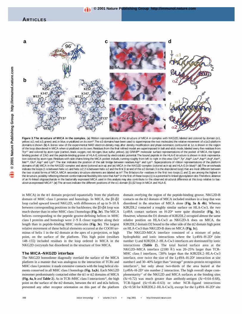
in the α2 domain (which corresponds to helix 2a in MHC class Iproteins) where ten residues in MICA (residues 152–161) were dis-ordered and presumed to form an extended flexible loop25. The rel-ative orientation of the two domains in the structure of free MICAwas different from that of all other MHC class I proteins and relat-ed molecules resulting in an extended structure, with the platform ofMICA flipped by over 110° from the position of the platform inother examples of class I folds.
The largest difference between the structure of MICA in com-plex with NKG2D and free MICA was the rearrangement of theplatform and α3 domains in the two different crystals. When the α3domains were superimposed, the angle between platform domainsin the two forms was over 96°, which confirmed the proposed flex-ibility of the linker (Fig. 3a). We presumed that particular interdo-main orientations seen in different MICA crystal structures in anotherwise extremely flexible molecule were selected by the exten-sive MICA-MICA crystal contacts that occur in these crystals.Despite the absence of an association with β2M, which bridges theplatform and α3 domains in the MHC class I fold, the interdomainrelationship in MICA in the MICA-NKG2D cocrystal structurewas now within approximately 20° of all other MHC class I pro-tein and homolog structures (Fig. 1c).
In a second major structural difference, the disordered loop infree MICA was now ordered, adding almost two turns of helix,which correspond to the canonical MHC class I α2 domain helix 2aand two residues of coil between helix 1 and 2a (Fig. 3b). Orderingof this loop was likely fostered by contacts with NKG2D (Table 2)and created a small pocket (roughly 6 Å wide × 6 Å deep × 14 Ålong) that was similar in size and position to the ligand-bindingpocket of serum Zn-α2-glycoprotein (ZAG, Fig. 3c)28. In the com-plex, the pocket was covered mostly by NKG2D-B, although a thincontinuous channel ran through the complex between the MICA-platform helices and NKG2D. No residues from NKG2D penetrat-ed the pocket. Unlike the ZAG pocket, which was hydrophobic incharacter, the MICA pocket was predominately lined with chargedresidues and limited at one end by a salt bridge between Asp72 andLys152 (Fig. 3c). In comparison, the pocket was just large enough toaccommodate the central segment (P3–P6) of the HLA-E peptide29,but was occupied by only four well-ordered water molecules in theMICA-NKG2D cocrystal structure. There was no electron densitythat was unaccounted for by protein or water molecules that mighthave corresponded to a peptide- or nonpeptide-ligand bound in thispocket. It remained formally possible that this was the binding sitefor some small ligand that was not available in either baculoviral25
or bacterial expression systems. However, steric clashes withNKG2D-B and the small size of the pocket would have severelylimited the size of such a hypothetical ligand.
The r.m.s.d. for the superposition of the free and complexed formsof the MICA-platform domain was 1.00 Å (on 151 Cαs), with thelargest difference occurring at the loop containing helix 1 in the α1domain (residues 45–55, Fig. 3d). These residues were poorlyordered in the complex structure. Other, smaller, differencesoccurred in the conformation of the loops between helix 1 in the α1domain and strand 1 in the α2 domain (residues 82–86) and betweenstrand 4 and helix 1 in the α2 domain (residues 131–134, Fig. 3d).
As noted in the structure of free MICA25, a major differencebetween the structures of the platform domains of MICA and otherMHC class I homologs occurred at two loops at the edge of theplatform α1 domain (Fig. 3e). The loops between β strands 1 and2 (residues 13–21 in MICA) and β strands 3 and 4 (residues 37–40
447
Table 2. Intermolecular interactions within the complex
NKG2D-A MICA Distance (Å) Type MIC substitutions
NKG2D residues common to sites A and B
Tyr152 Lys71 φ (3) GlnTyr152 OH Arg74 NH 2.8 H bond LysTyr152 Met75 φ (2) Arg, LysIle182 His79 φ (1) TyrMet184 O Val18 N 2.8 H bond —Met184 Val18 φ (1) —Met184 Arg74 φ (1) LysMet184 Ala78 φ (2) ThrGln185 Oε Val18 O 3.0 H bond —Lys197 Nζ Asp149 Oδ 3.8 Salt bridge Asn, GluTyr199 Met75 φ (3) Arg, LysTyr199 OH His79 Nε 2.8 H bond TyrAsn207 Nδ Arg38 NH 3.0 H bond —
NKG2D residues unique to site A
Glu183 O Lys81 Nζ 3.1 H bond GlnLys186 Nζ Asp15 O 3.5 H bond —Lys186 N Ser17 Oγ 3.1 H bond —Glu201 Oε Arg74 NH 3.4 Salt bridge LysThr205 Oγ Ser20 Oγ 3.5 H bond —
NKG2D-B MICA Distance (Å) Type MIC substitutionsNKG2D residues common to sites A and B
Tyr152 His156 φ (3) Leu, ∆Tyr152 Ala159 φ (2) Thr, ∆Ile182 Ala162 φ (1) —Ile182 Gln166 φ (1) Trp, LysMet184 His158 φ (2) Arg, ∆Met184 Ala162 φ (1) —Gln185Nε His158 Nε 3.1 H bond Arg, ∆Lys197 Nζ Asp65 Oδ 3.4 Salt bridge —Tyr199 Ala159 φ (4) Thr, ∆Tyr199 OH Asp163 Oδ 2.4 H bond AsnAsn207 Oγ Thr155 Oγ 3.5 H bond Ala
NKG2D residues unique to site B
Lys150 Nζ Ala150 O 3.3 H bond ThrIle181 O Gln166 Nε 3.2 H bond Trp, LysLeu191 Thr155 φ (1) AlaSer195 Oγ Asp163 NH 3.2 H bond Glu, Gly
NKG2D-A NKG2D-B Distance (Å) TypeSymmetrical interactions
Ser94 N Cys99 O 2.9 H bondSer94 O Cys99 N 2.8 H bondTyr95 N Cys96 O 3.5 H bondLys101 O Glu93 N 3.4 H bondCys105 N Cys105 O 2.8 H bondIle104 Tyr106 φ (3)Phe113 Leu148 φ (5)Lys150 N Leu148 O 2.9 H bondLys150 Nζ Ser194 Oγ 3.1 H bond
Asymmetrical interactions
Gly97 O Ser94 O 3.0 H bondIle104 Leu145 φ (1)Gln213 Oε Glu93 Oε 3.4 H bond
Hydrogen bonds (H bonds) are shown for donor-to-receptor distances of 2.4–3.5 Åwith appropriate geometry, salt bridges for partners within 4.0 Å and hydrophobic inter-actions (φ) for carbon-to-carbon distances within 4.0 Å (the number of carbon/carbonpairs is in parentheses). Substitutions of MIC residues that contact NKG2D among thehuman MICA, MICB and nonhuman primate sequences known to bind to humanNKG2D are shown as ∆22,24.
©20
01 N
atu
re P
ub
lish
ing
Gro
up
h
ttp
://im
mu
no
l.nat
ure
.co
m© 2001 Nature Publishing Group http://immunol.nature.com
nature immunology • volume 2 no 5 • may 2001 • http://immunol.nature.com
ARTICLES
in MICA) in the α1 domain projected equatorially from the platformdomain of MHC class I proteins and homologs. In MICA, the β1-β2loop curled upward toward NKG2D, with differences of up to 9–10 Åbetween corresponding positions in the backbones; the β3-β4 loop wasmuch shorter than in other MHC class I homologs (Fig. 3e). The MICAhelices corresponding to the peptide groove-defining helices in MHCclass I proteins and homologs were 2–9 Å closer together along theirlength than in peptide-binding MHC molecules (Fig. 3e). The largestrelative movement of these helical elements occurred at the COOH ter-minus of helix 1 in the α2 domain at the apex of a projection, or highpoint, on the surface of the platform. This high point (residues148–155) included residues in the loop ordered in MICA in theNKG2D cocrystals but disordered in the structure of free MICA.
The MICA–NKG2D interfaceThe NKG2D homodimer diagonally overlaid the surface of the MICAplatform in a manner that was analogous to the interaction of TCRs andMHC class I proteins: it made extensive contacts with the long helical ele-ments conserved in all MHC class I homologs (Fig. 1a,b). Each NKG2Dmonomer predominately contacted either the α1 or α2 domains of MICA(Fig. 4a, b and Table 2). As in TCR–MHC class I interactions30, the highpoint on the surface of the α2 domain, between the α1 and α2a helices,prevented any other receptor orientation on this part of the platform
domain overlying the region of the peptide-binding groove. NKG2D-Bcontacts on the α2 domain of MICA included residues in a loop that wasdisordered in the structure of MICA alone (Fig. 3a & 4b). WhereasKIR2DL2 contacted a roughly similar surface on HLA-Cw3, the twoLy49A contact surfaces on H-2Dd were quite dissimilar (Fig. 1c).However, whereas the D1 domain of KIR2DL2 occupied almost the samerelative position on HLA-Cw3 as NKG2D-A does on MICA, theKIR2DL2 domain D2 bound to the other side of the α2 domain high pointon HLA-Cw3 than NKG2D-B does on MICA (Fig. 1c).
The NKG2D-MICA interface consisted of a mixture of polar,hydrophobic and ionic interactions where the Ly49A–H-2Dd (site number 1) and KIR2DL2–HLA-Cw3 interfaces are dominated by ionicinteractions (Table 2). The total buried surface area at theNKG2D–MICA interface (2180 Å2) was 20–25% larger than TCR-MHC class I interfaces, ∼ 30% larger than the KIR2DL2–HLA-Cw3interface, over twice the size of the Ly49A–H-2Dd interaction at sitenumber1 and 30–40% larger than “average” protein-protein recognitioninterfaces31, but only about two-thirds of the area buried at theLy49A–H-2Dd site number 2 interaction. The high overall shape com-plementarity27 of the NKG2D and MICA surfaces at the binding sites(Sc=0.72) was much greater than antibody-antigen (Sc=0.64–0.68),TCR-ligand (Sc=0.46–0.63) or other NCR–ligand interactions(Sc=0.58 for KIR2DL2–HLA-Cw3), except for the Ly49A–H-2Dd site
448
Figure 3.The structure of MICA in the complex. (a) Ribbon representations of the structure of MICA in complex with NKG2D, labeled and colored by domain (α1,yellow; α2, red; α3, green) and, in blue, crystallized on its own25.The α3 domains have been used to superimpose the two molecules; the relative movement of α1α2-platformdomains is shown. (b) A stereo view of the experimental MAD electron-density map, after density modification and phase extension, contoured at 1σ, is shown in the regionof the loop disordered in MICA when crystallized on its own. Residues from the final refined model are superimposed in ball-and-stick mode, labeled every five residues fromTrp127 and colored by atom type (carbon, black; oxygen, red; nitrogen, blue; sulfur, yellow). (c) GRASP51 molecular surface representations of the pocket of MICA, the ligand-binding pocket of ZAG and the peptide-binding groove of HLA-E, colored by electrostatic potential.The bound peptide in the HLA-E structure is shown in stick representa-tion colored by atom type. Residues with side chains lining the MICA pocket include, running roughly from left to right in this view: Glu167,Tyr7,Asp65, Leu66,Asp163,Arg94,Asn69,Met160, Glu92, Asp72 and Lys152. The star indicates the position of the salt bridge between residues Asp72 and Lys152. Superpositions of ribbon representations of the platformdomains of (d) MICA in the NKG2D complex and alone (colored as in a) and (e) MICA in the NKG2D complex (colored as in a) and HLA-E (in blue)29. (d) The arrowheadsindicate the loops (1 is between helix α1 and helix α2; 2 is between helix α2 and the first β strand of the α2 domain; 3 is the disordered loop) that are most different betweenthe two crystal forms of MICA. MICA secondary structure elements are labeled as in25.The B-factors for residues in the first two loops (1 and 2) are among the highest inthe structure, possibly reflecting inherent conformational flexibility.We note that Asn56 in the first of these loops (1) is a potential N-linked glycosylation site.Therefore, absenceof an N-linked oligosaccharide in the bacterially expressed MICA used in this analysis may also contribute to the observed structural difference at this loop relative to bac-ulovirus-expressed MICA25. (e) The arrows indicate the different positions of the α1 domain β1-β2 loop in MICA and HLA-E.
a
b
c d
e
©20
01 N
atu
re P
ub
lish
ing
Gro
up
h
ttp
://im
mu
no
l.nat
ure
.co
m© 2001 Nature Publishing Group http://immunol.nature.com

ARTICLES
http://immunol.nature.com • may 2001 • volume 2 no 5 • nature immunology
number 1 interaction (Sc=0.78). Unlike the Ly49A–H-2Dd interface,none of the three NKG2D or eight MICA potential N-linked glycosy-lation sites lay near the interface. Thus, we deduced that binding issolely mediated by direct protein-protein contacts. Consistent with thisdeduction, NKG2D failed to retain any recognizable remnant of thesaccharide-binding site of true C-type lectins or the proposed saccha-ride-binding site of Ly49A14 (Fig. 2).
Strength and stability of the MICA–NKG2D interactionTo quantify the affinity and kinetics of NKG2D binding to MICA, weanalyzed the interaction by surface plasmon–resonance (SPR) methods.The resulting equilibrium dissociation constant (KD), 1 µM at 37°, wasone to two orders of magnitude stronger than most NCR and many TCRinteractions with host MHC proteins, where dissociation constants aretypically in the tens-of-micromolar range12,32–37. Kinetic analysis yielded
a dissociation rate constant at 37° of 0.04 s-1
(corresponding to a t1/2 of 17 s), which indi-cated that the NKG2D-MICA interactionmay be relatively more stable than TCR-lig-and and other NCR-ligand complexes. Theassociation rate constant (kon), 4×10⁴–7×10⁴
M-1s-1, was slow relative to many other cellsurface interactions, which typically have kon
≥1×10⁵ M-1s-1 (ref. 38). Also, measurementsat 25 °C and 37 °C indicated that both asso-ciation and dissociation rate constants aresomewhat temperature dependent, whichindicated activation energy barriers impedeassociation and dissociation (Fig. 5). Thesefeatures—both of which are shared by TCR-MHC interactions, where binding is thoughtto be accompanied by a reduction in flexi-bility at the receptor-ligand interface38—were consistent with an extensive highlycomplementary NKG2D-MICA interactionwhere the flexible loop on MICA (residues152–161) must become ordered during com-plex formation. The interdomain flexibilityin MICA was less likely to contribute to thiseffect as neither conformation observed inthe crystal structures nor many of the possi-ble interdomain arrangements would imposesteric hindrances to NKG2D binding.
Higher-order complexesContacts in the crystal were dominated byextensive (3400 Å2 buried) MICA-MICA con-tacts around the crystallographic four-fold
axis and an additional, minor (840 Å2 buried) crystal contact betweenNKG2D and the α3 domain of a MICA molecule in a neighboring com-plex. However, the resultant MICA tetramer is unlikely to form on cell sur-faces because five of the eight asparagines at potential N-linked glycosyla-tion sites in MICA (residues 8, 102, 187, 212 and 239) were buried in thetetramer interface. In addition, the MICA COOH termini were too far awayfrom where the bottom of the tetramer limited approach to the membrane.The NKG2D-MICA α3 domain contact is also unlikely to be physiologi-cally relevant, due to the limited extent of this interaction and the topolog-ical incompatibility between the formation of this complex and the twomembrane surfaces involved. Therefore, we saw no higher-order complex-es in the crystal structure that are likely to be informative about possiblesignaling mechanisms at the cell surface. As in the structure of free MICA,no major contacts were found between domains within a single MICAmolecule that might contribute to stabilizing this particular interdomain
449
Figure 4. Details of the binding sites. Stereoviews of (a) the NKG2D-A–MICA and (b) NKG2D-B–MICA interaction surfaces. Ribbon representa-tions of the NKG2D and MICA backbones, coloredby chain (as in Fig. 1), and side chains of key residues,colored by atom type, are shown. In these twoviews, NKG2D-A and NKG2D-B are shown in iden-tical orientations. (c) A stereo view of the superpo-sition of the MICA binding sites on the two NKG2Dmonomers, colored as above. The backbone ofNKG2D is shown in ribbon representation andbinding site residues are shown in ball-and-stickmode and labeled.
a
b
c
©20
01 N
atu
re P
ub
lish
ing
Gro
up
h
ttp
://im
mu
no
l.nat
ure
.co
m© 2001 Nature Publishing Group http://immunol.nature.com

nature immunology • volume 2 no 5 • may 2001 • http://immunol.nature.com
ARTICLES
conformation.
DiscussionThe NKG2D homodimer bound to MICA through a surface on the sideof the homodimer opposite the NH2-termini, which was comparable tothe ligand-binding surface of Ly49A14, the proposed ligand-binding sur-face of CD9413 and close to the carbohydrate-binding pocket of MBP26.The increased curvature of the binding surface saddle of NKG2D, dueto the stirrup loops and interdomain relationship, precisely comple-mented the decreased interhelical distances of MICA relative to otherMHC class I homologs. Thus, NKG2D was sterically incompatible withother MHC class I protein or homolog platform-domain structures andother NKDs would be a poor match for MICA. Essentially identical,homodimer dyad-related surfaces on NKG2D-A and NKG2D-B madecomparably extensive and complementary contacts with different sur-faces on either the α1 or α2 domains of MICA respectively. This con-trasts with other situations, such as the interactions between otherhomodimeric receptors and monomeric ligands, where one subunit ofthe symmetric receptor dominated the interaction with the asymmetricligand (both Ly49A binding sites on H-2Dd14 and CD8αα binding toHLA-A239 or H-2Kb40), or the binding of multiple copies of receptors toa single asymmetric ligand. The latter is exemplified by the humangrowth hormone receptor–ligand complex41, where interdomain flexibil-ity in the receptor allowed the adoption of different conformations tobind to multiple distinct surfaces on the hormone ligand.
Whereas an α helix constituted the central element of both binding siteson MICA, the side chains that were actually making contacts withNKG2D were essentially completely different (running from one end tothe other of the α1 helix: LysArgMetAlaHisLys versus ThrHisAlaAlaArgGln in the α2 helix). Whereas the structure of the NKG2D-A contactsurface was essentially unchanged in free and complexed forms of MICA,seven residues contacted by NKG2D-B were part of the disordered loopin free MICA. Both NKG2D monomers also made contacts to the otherMICA domain: NKG2D-A contacting Asp149 in α2 and NKG2D-B con-tacting Asp65 and Asp163 in α1. NKG2D-A also interacted with residues15, 17, 18 and 20 in the β1-β2 loop in the α1 domain, which accountedfor its markedly altered conformation relative to other MHC class I
homologs. The NKG2D surfaces,however, were quite similar. Elevenresidues in both NKG2D-A andNKG2D-B contacted MICA, ofwhich seven were common to bothcontact surfaces. These sevenresidues defined a core binding siteon NKG2D that dominated theintermolecular contacts with MICAat both interfaces. However, six ofthese seven residues made differentcontacts at either the NKG2D-A–MICA or the NKG2D-B–MICAinterfaces; the seventh residue,Lys197, participating in a commonsalt bridge to an aspartic acid atboth binding sites (Asp149 or Asp163
in MICA).The recognition of an asymmet-
ric ligand by a symmetric receptorwas achieved by a core-binding sur-face on NKG2D that can specifical-ly interact with two distinct surfaces
on MICA with few conformational changes or substitution of contacts. Thelargest conformational differences occurred at Tyr152 (where the hydroxyloxygen moved almost 6.5 Å and the ring rotated by 90° in the ring planeand 65° perpendicular to the ring), at Lys197 (where the Nζ moved over 4Å) and at Gln185 and Glu201 (where the side chains used different rotamers).There was little induced conformational change in the backbone structureof the two NKG2D monomers; the r.m.s.d. for the superposition of MICAcontacting elements (residues 150–152 and 180–207 in NKG2D) was only0.36 Å on all backbone atoms.
NKG2-CD94 heterodimers bind, in a peptide-specific manner, toHLA-E8. A model for the NKG2-CD94 heterodimer was proposed onthe basis of the structure of the CD94 homodimer. In addition, the inter-action with HLA-E was proposed to be similar to that in a TCR-MHCcomplex, although the orientation of the heterodimer on the HLA-Eplatform could not be given in detail12. We noted that superimposingCD94 onto NKG2D, and the HLA-E platform onto MICA, generated,with only minor adjustments, a reasonable model NKG2-CD94-HLA-E complex. In this complex, CD94 sits on the α1 domain of HLA-E(corresponding to NKG2D-A) and the NKG2 moiety sits on the α2domain, with a small hydrophobic patch on CD94 (Phe114 and Leu162)matching a similar patch on HLA-E (Ile73, Val76 and the side chain ofthe P8 residue in the peptide, Leu in the crystal structure).
As demonstrated by the details of the MICA interaction, NKG2D hasevolved a single binding site that is competent to specifically bind totwo distinct protein surfaces. Reports that human NKG2D binds to var-ious human MICA and MICB alleles and nonhuman primate MIC pro-teins22,24 also demonstrated a remarkable tolerance for sequence substi-tutions in the MIC binding sites. Though no recognizable rodent MIChomologs have been identified, the ectodomains of human and murineNKG2D are highly conserved (69% identical). Murine NKG2D ligandsinclude the distant MHC class I homologs Rae1 and H-6042, which arealso quite unlike MICA. How murine NKG2D interacts with these lig-ands, and how human NKG2D interacts with other potential human lig-ands, remains unclear. This divergence of specificity implies thatNKG2D has evolved mechanisms to recognize a variety of protein sur-faces, apparently without using flexible elements of structure beyondrelatively small changes in a handful of side chain conformations.
450
Figure 5.Affinity and kinetics measurements of the NKG2D–MICA interaction by SPR. (a) Equilibrium binding mea-surements of bacterially expressed NKG2D injected (solid bar) at 5 µl/min over surfaces with either baculovirus-expressedMICA or control protein immobilized. The response is greater over the MICA surface (solid trace) than the control (dottedtrace), which indicates specific binding. (b) A nonlinear curve fit of the 1:1 Langmuir binding isotherm to 37 °C MICA equilibri-um binding data (solid squares).The predicted Rmax=297 resonance units; KD=1 µM. Inset: a Scatchard plot of the same data (KD=1µM). (c) Kinetic measurements are shown, made at 37 °C by injecting NKG2D (50 µl/min) at a range of concentrations overMICA and control surfaces.The data plotted (filled squares) have had the background response subtracted.The results of glob-ally fitting rate equations derived from the 1:1 Langmuir binding model are shown as solid lines and residual errors from the fitsare shown in the bottom panel.The kinetic constants obtained (kon = 6.75×10⁴ M-1s-1, koff=0.039 s-1) predict an affinity of 0.6 µM,similar to that obtained from equilibrium binding analysis.Analogous kinetic measurements at 25 °C yielded a kon of 4.26×10⁴ M-
1s-1 and a koff of 0.013 s-1 (calculated dissociation constant KDcalc was 0.3 µM).
a b c
©20
01 N
atu
re P
ub
lish
ing
Gro
up
h
ttp
://im
mu
no
l.nat
ure
.co
m© 2001 Nature Publishing Group http://immunol.nature.com

ARTICLES
http://immunol.nature.com • may 2001 • volume 2 no 5 • nature immunology
MethodsProtein expression, crystallization and crystallography. The extracellular domains ofhuman MICA (residues 1–276) and human NKG2D (residues 80–216) were expressed inbacteria and refolded as complexes from inclusion bodies. Proper folding was analyzed bysolution monodispersivity of the purified concentrated protein, as determined by size-exclu-sion chromatography (SEC) and dynamic light scattering, and also the presence of a singledisulfide-bonded species by nonreducing PAGE analysis. A selenomethionine (SeMet)derivative of MICA was prepared as described44 and refolded separately; NKG2D-SeMet-MICA complexes were then isolated by SEC. The MICA-NKG2D complex was crystal-lized by sitting-drop vapor diffusion at 22 °C from a solution containing 7.5 mg/ml of pro-tein, 25 mM PIPES (pH 7.0), 1 mM EDTA and 0.02% NaN3 mixed at a ratio of 1:1 with awell solution containing 0.4 M (NH4)2SO4 and 50 mM MES (pH 6.5). The space group ofthe crystals was P4212 (a=b=122.78Å, c=102.82Å) with one complete MICA monomer–NKG2D homodimer complex per asymmetric unit, which resulted in a Vm of 3.0Å3 perDalton. Crystals were cryopreserved by stepwise transfer to mother liquor + 30% glycerol.Diffraction data from native and SeMet crystals were collected at ALS beamline 5.0.2 withthe use of a Quantum-4 CCD detector and reduced with HKL45. All eight selenium sites inMICA were located by automated Patterson search and multiwavelength anomalous dis-persion phases were calculated with the Crystallography & NMR System (CNS) package46.After solvent flipping and phase extension in CNS, the electron-density map was clear andunambiguous. Models of MICA (Protein Data Bank number 1B3J) and CD94 (Protein DataBank number 1B6E) were manually docked into this map and subsequently rebuilt with O47.
The structure was refined in CNS (against all F>0) with overall anisotropic B-factor andbulk solvent corrections applied. Initial torsional simulated-annealing refinement, using themaximum likelihood target function mlhl, was followed by positional refinement using themlf target function. The progress of the refinement was confirmed by the monotonicdecrease in both RCryst and RFree. NCS restraints were not imposed because of the breakdownof symmetry in the NKG2D homodimer at the MICA binding sites and the NH2-termini.Coordinates have been deposited in the Protein Data Bank with accession code 1hyr48.
SPR measurements. Experiments were done in HBS-EP buffer with a Biacore 2000 instrument(Biacore AB, Uppsala, Sweden). To conduct equilibrium-binding measurements at 37 °C, bac-terially expressed NKG2D was injected over either baculovirus-expressed MICA49 or a controlprotein amine-coupled to a CM5 sensor chip. Injections of NKG2D were repeated over a rangeof concentrations and the amount of binding at each concentration (in Fig. 5b) was calculated asthe difference in the response at equilibrium in the MICA and control flow cells. For Scatchardanalysis, the KD value (1 µM) was obtained from the slope of the plot (KD=-1/slope) by linearregression. For kinetic analysis, rate equations derived from the 1:1 Langmuir binding modelwere fitted simultaneously to association and dissociation phases of all three injections (globalfitting) with the use of BIAevaluation 3.0 software. Data collection at progressively higher flowrates (to 100 µl/min) and over flow cells with progressively lower MICA expression immobi-lized excluded mass transport limitations. The slow off-rates obtained (koff=0.013 s-1 at 25 °C)explain the ability to isolate complexes by size-exclusion chromatography, despite the relativelylow affinity. Binding responses over bacterially expressed MICA coupled to chips were similarto those over baculovirally expressed MICA, which suggested that glycosylation in insect cellsdoes not affect the interaction. The activity of NKG2D was judged to be near 100% on the basisof size-exclusion chromatography in the presence of MICA (data not shown). Protein was quan-tified by bicinchoninic (BCA) assay (Pierce, Rockford, IL).
Note added in proof: The structure of human NKG2D in the complex recapitulated all thesalient features of the recently reported structure of murine NKG2D43.
AcknowledgementsWe thank L. Hung, G. McDermott and T. Earnest (Advanced Light Source, LawrenceBerkeley National Laboratory) for assistance with data collection. Supported by NationalInstitutes of Health grants AI42200 (to R. K. S.), CA18221 and AI30581 (to T. S.).
Received 20 February 2001; accepted 23 March 2001.
1. Germain, R. N. & Margulies, D. H.The biochemistry and cell biology of antigen processing and pre-sentation. Ann. Rev. Immunol. 11, 403–450 (1993).
2. Lenschow, D. J.,Walunas,T. L. & Bluestone, J.A. CD28/B7 system of T cell costimulation. Annu. Rev.Immunol. 14, 233–258 (1996).
3. Lanier, L. L.Turning on natural killer cells. J. Exp. Med. 191, 1259–1262 (2000).4. Ravetch, J.V. & Lanier, L. L. Immune inhibitory receptors. Science 290, 84–89 (2000).5. Lee, N. et al. HLA-E is a major ligand for the natural killer inhibitory receptor CD94/NKG2A. Proc.
Natl Acad. Sci. USA 95, 5199–5204 (1998).6. Weis,W. I.,Taylor, M. E. & Drickamer, K.The C-type lectin superfamily in the immune system.
Immunol. Rev. 163, 19–34 (1998).7. Drickamer, K. Ca2+-dependent carbochydrate-recognition domains in animal proteins. Curr. Opin.
Struct. Biol. 3, 393–400 (1993).8. Lopez-Botet, M. & Bellon,T. Natural killer cell activation and inhibition by receptors for MHC class I.
Curr. Opin. Immunol. 11, 301–307 (1999).9. Fan, Q. R. et al. Structure of the inhibitory receptor for human natural killer cells resembles
haematopoietic receptors. Nature 389, 96–100 (1997).10. Snyder, G.A., Brooks,A. G. & Sun, P. D. Crystal structure of the HLA-Cw3 allotype-specific killer cell
inhibitory receptor KIR2DL2. Proc. Natl Acad. Sci. USA 96, 3864–3869 (1999).11. Maenaka, K., Juji,T., Stuart, D. I. & Jones, E.Y. Crystal structure of the human p58 killer cell inhibitory
receptor (KIR2DL3) specific for HLA-Cw3-related MHC class I. Structure 7, 391–398 (1999).
12. Boyington, J. C., Motyka, S.A., Schuck, P., Brooks,A. G. & Sun, P. D. Crystal structure of an NK cellimmunoglobulin-like receptor in complex with its class I MHC ligand. Nature 405, 537–543 (2000).
13. Boyington, J. C. et al. Structure of CD94 reveals a novel C-type lectin fold: implications for the NKcell-associated CD94/NKG2 receptors. Immunity 10, 75–82 (1999).
14. Tormo, J., Natarajan, K., Margulies, D. H. & Mariuzza, R.A. Crystal structure of a lectin-like naturalkiller cell receptor bound to its MHC class I ligand. Nature 402, 623–631 (1999).
15. Houchins, J. P.,Yabe,T., McSherry, C. & Bach, F. H. DNA sequence analysis of NKG2, a family of relat-ed cDNA clones encoding type II integral membrane proteins on human natural killer cells. J. Exp.Med. 173, 1017–1020 (1991).
16. Bauer, S. et al.Activation of NK cells and T cells by NKG2D, a receptor for stress-inducible MICA.Science 285, 727–729 (1999).
17. Wu, J. et al.An activating immunoreceptor complex formed by NKG2D and DAP10. Science 285,730–732 (1999).
18. Wu, J., Cherwinski, H., Spies,T., Phillips, J. H. & Lanier, L. L. DAP10 and DAP12 form distinct, but func-tionally cooperative, receptor complexes in natural killer cells. J. Exp. Med. 192, 1059–1067 (2000).
19. Bahram, S., Bresnahan, M., Geraghty, D. E. & Spies,T.A.A second lineage of mammalian major histo-compatibility complex class I genes. Proc. Natl Acad. Sci. USA 91, 6259–6263 (1994).
20. Bahram, S. & Spies,T.A. Nucleotide sequence of a human MHC class I MICB cDNA. Immunogenetics43, 230–233 (1996).
21. Groh,V. et al. Cell stress-regulated human major histocompatibility complex class I gene expressedin gastrointestinal epithelium. Proc. Natl Acad. Sci. USA 93, 12445–12450 (1996).
22. Groh,V., Steinle,A., Bauer, S. & Spies,T. Recognition of Stress-Induced MHC Molecules by IntestinalEpithelial γδT Cells. Science 279, 1737–1740 (1998).
23. Groh,V. et al. Broad tumor-associated expression and recognition by tumor-derived γ δT cells ofMICA and MICB. Proc. Natl Acad. Sci. USA 96, 6879–6884 (1999).
24. Steinle,A., Groh,V. & Spies,T. Diversification, expression, and γ δT cell recognition of evolutionarilydistant members of the MIC family of major histocompatibility complex class I-related molecules.Proc. Natl Acad. Sci. USA 95, 12510–12515 (1998).
25. Li, P. et al. Crystal Structure of the MHC Class I Homolog MIC-A, a γδT Cell Ligand. Immunity 10,577–584 (1999).
26. Weis,W. I., Kahn, R., Fourme, R., Drickamer, K. & Hendrickson,W.A. Structure of the calcium-depen-dent lectin domain from a rat mannose-binding protein determined by MAD phasing. Science 254,1608–1615 (1991).
27. Lawrence, M. & Colman, P. M. Shape complementarity at protein/protein interfaces. J. Mol. Biol. 234,946–950 (1993).
28. Sanchez, L. M., Chirino,A. J. & Bjorkman, P. J. Crystal structure of human ZAG, a fat-depleting factorrelated to MHC molecules. Science 283, 1914–1919 (1999).
29. O’Callaghan, C.A. et al. Structural features impose tight peptide binding specificity in the nonclassicalMHC molecule HLA-E. Mol. Cell 1, 531–541 (1998).
30. Wilson, I.A. & Garcia, K. C.T-cell receptor structure and TCR complexes. Curr. Opin. Struct. Biol. 7,839–848 (1997).
31. Lo Conte, L., Chothia, C. & Janin, J.The atomic structure of protein-protein recognition sites. J. Mol.Biol. 285, 2177–2198 (1999).
32. Vales-Gomez, M., Reyburn, H.T., Mandelboim, M. & Strominger, J. L. Kinetics of interaction of HLA-Cligands with natural killer cell inhibitory receptors. Immunity 9, 337–344 (1998).
33. Chapman,T. L., Heikema,A. P. & Bjorkman, P. J.The Inhibitory Receptor LIR-1 Uses a CommonBinding Interaction to Recognize Class I MHC Molecules and the Viral Homolog UL-18. Immunity11, 603–613 (1999).
34. Vales-Gomez, M., Reyburn, H.T., Erskine, R.A. & Strominger, J. Differential binding to HLA-C of p50-acti-vating and p58-inhibitory natural killer cell receptors. Proc. Natl Acad. Sci. USA 95, 14326–14331 (1998).
35. Vales-Gomez, M., Reyburn, H.T., Erskine, R.A., Lopez-Botet, M. & Strominger, J. L. Kinetics and pep-tide dependency of the binding of the inhibitory NK receptor CD94/NKG2-A and the activatingreceptor CD94/NKG2-C to HLA-E. EMBO J. 18, 4250–4260 (1999).
36. Maenaka, K. et al. Killer cell immunoglobulin receptors and T cell receptors bind peptide-major his-tocompatibility complex class I with distinct thermodynamic and kinetic properties. J. Biol. Chem.274, 28329–28334 (1999).
37. Davis, M. M. et al. Ligand recognition by αβ T cell receptors. Annu. Rev. Immunol. 16, 523–544 (1998).38. Willcox, B. E. et al.TCR binding to peptide-MHC stabilizes a flexible recognition interface. Immunity
10, 357–365 (1999).39. Gao, G. F. et al. Crystal structure of the complex between human CD8αα and HLA-A2. Nature 387,
630–634 (1997).40. Kern, P. S. et al. Structural basis of CD8 coreceptor function revealed by crystallographic analysis of a
murine CD8αα ectodomain fragment in complex with H-2Kb. Immunity 9, 519–530 (1998).41. de Vos,A. M., Ultsch, M. & Kossiakoff,A.A. Human growth hormone and extracellular domain of its
receptor: crystal structure of the complex. Science 255, 306–312 (1992).42. Cerwenka,A. et al. Retinoic acid early inducible genes define a ligand family for the activating
NKG2D receptor in mice. Immunity 12, 721–727 (2000).43. Wolan, D.W. et al. Crystal structure of the murine NK cell-activating receptor at 1.95 Å. Nature
Immunol. 2, 248–254 (2001).44. Doublié, S. Preparation of Selenomethionyl Proteins for Phase Determination. Meth. Enzymol. 276,
523–531 (1997).45. Otwinowski, Z. & Minor,W. Processing of X-ray diffraction data collected in oscillation mode. Meth.
Enzymol. 276, 307–327 (1997).46. Brünger,A.T. et al. Crystallography & NMR System:A New Software Suite for Macromolecular
Structure Determination. Acta Cryst. D 54, 905–921 (1998).47. Jones,T.A. & Kjeldgaard, M. Electron density map interpretation. Meth. Enzymol. 277, 173–208 (1997).48. Berman, H. M. et al.The Protein Data Bank. Nucleic Acids Res. 28, 235–242 (2000).49. Bauer, S.,Willie, S.T., Spies,T. & Strong, R. K. Expression, purification, crystallization and crystallographic
characterization of the human MHC class I related protein MICA. Acta Cryst. D 54, 451–453 (1998).50. Ding,Y. H. et al.Two human T cell receptors bind in a similar diagonal mode to the HLA-A2/Tax pep-
tide complex using different TCR amino acids. Immunity 8, 403–411 (1998).51. Nicholls,A., Sharp, K.A. & Honig, B. Protein folding and association: insights from the interfacial and
thermodynamic properties of hydrocarbons. Proteins Struct. Funct. Genet. 11, 281–296 (1991).52. Brünger,A.T. Free R value: a novel statistical quantity for assessing the accuracy of crystal structures.
Nature 355, 472–475 (1992).53. Laskowski, R.A., MacArthur, M.W., Hutchinson, E. G. & Thornton, J. M. PROCHECK: a program to
check the stereochemical quality of protein structures. J. Appl. Cryst. 26, 283–291 (1992).
451
©20
01 N
atu
re P
ub
lish
ing
Gro
up
h
ttp
://im
mu
no
l.nat
ure
.co
m© 2001 Nature Publishing Group http://immunol.nature.com