Embed Size (px)
Citation preview
JOURNAL OF BACTERIOLOGY, Sept. 2003, p. 5591–5601 Vol. 185, No. 180021-9193/03/$08.00�0 DOI: 10.1128/JB.185.18.5591–5601.2003Copyright © 2003, American Society for Microbiology. All Rights Reserved.
Complete Genome Sequence of the Oral Pathogenic BacteriumPorphyromonas gingivalis Strain W83
Karen E. Nelson,1* Robert D. Fleischmann,1 Robert T. DeBoy,1 Ian T. Paulsen,1 Derrick E. Fouts,1Jonathan A. Eisen,1 Sean C. Daugherty,1 Robert J. Dodson,1 A. Scott Durkin,1 Michelle Gwinn,1
Daniel H. Haft,1 James F. Kolonay,1 William C. Nelson,1 Tanya Mason,1 Luke Tallon,1Jessica Gray,1 David Granger,1 Herve Tettelin,1 Hong Dong,2 Jamie L. Galvin,2
Margaret J. Duncan,2 Floyd E. Dewhirst,2 and Claire M. Fraser1
The Institute for Genomic Research, Rockville, Maryland 20850,1 and Department ofMolecular Genetics, The Forsyth Institute, Boston, Massachusetts 021152
Received 5 November 2002/Accepted 24 June 2003
The complete 2,343,479-bp genome sequence of the gram-negative, pathogenic oral bacterium Porphyromonasgingivalis strain W83, a major contributor to periodontal disease, was determined. Whole-genome comparativeanalysis with other available complete genome sequences confirms the close relationship between the Cyto-phaga-Flavobacteria-Bacteroides (CFB) phylum and the green-sulfur bacteria. Within the CFB phyla, thegenomes most similar to that of P. gingivalis are those of Bacteroides thetaiotaomicron and B. fragilis. Outside ofthe CFB phyla the most similar genome to P. gingivalis is that of Chlorobium tepidum, supporting the previousphylogenetic studies that indicated that the Chlorobia and CFB phyla are related, albeit distantly. Genomeanalysis of strain W83 reveals a range of pathways and virulence determinants that relate to the novel biologyof this oral pathogen. Among these determinants are at least six putative hemagglutinin-like genes and 36previously unidentified peptidases. Genome analysis also reveals that P. gingivalis can metabolize a range ofamino acids and generate a number of metabolic end products that are toxic to the human host or humangingival tissue and contribute to the development of periodontal disease.
Periodontal diseases are a group of infections that affect thestructures surrounding teeth. If allowed to progress, periodon-tal disease can cause the destruction of supporting connectivetissue and bone, ultimately resulting in tooth loss. Initiationand progression of periodontal diseases is the result of a com-plex interaction between the bacteria colonizing the gingivalcrevice and the hosts’ immune and inflammatory responses.Since the species most strongly implicated in periodontal dis-ease pathogenesis are also usually present in low numbers inhealthy people, the distinction between pathogenic and com-mensal bacteria in the human host is not clearly defined.
The gram-negative anaerobe Porphyromonas gingivalis be-longs to the family Porphyromonadaceae, order Bacteroidales inthe phylum Bacteroidetes, previously known as the Cytophaga-Flavobacteria-Bacteroides (CFB) group (6). The bacterium isa major causative agent in the initiation and progression ofsevere forms of periodontal disease. P. gingivalis is a late orsecondary colonizer of the oral cavity, a process that is facili-tated by other microbial species that provide attachment sites,as well as supply growth substrates, and reduce oxygen tensionto levels optimal for growth of P. gingivalis. Among the earlyplaque organisms that P. gingivalis adheres to are the oralstreptococci (35, 36) and Actinomyces naeslundii (19). Adher-ence is facilitated by a variety of bacterial surface proteins,including fimbriae, hemagglutinins, and proteinases. P. gingi-valis also binds to late colonizers such as Fusobacterium nu-cleatum, Treponema denticola, and Bacteroides forsythus (now
renamed Tanerella forsythensis) (20, 30, 72). The use of a va-riety of metabolic strategies appears to enable the success ofthis microbial community. Once established, P. gingivalis cellsparticipate in intercellular communication networks with otheroral prokaryotic cells, as well as with eukaryotic cells (37).
P. gingivalis is the third oral pathogen (1, 28) and the secondmember of the CFB group to be sequenced. The genome ofP. gingivalis strain W83 (also known as strain HG66) is pre-sented here. Strain W83 was isolated in the 1950s by H. Werner(Bonn, Germany) from an undocumented human oral infec-tion (41, 46) and was brought to The Pasteur Institute byMadeleine Sebald during the 1960s. The strain was subse-quently obtained by Christian Mouton (Quebec, Canada) dur-ing the late 1970s. It is anticipated that the availability of thecomplete genome sequence from this oral pathogen will givetremendous insight into the mechanisms that result in diseaseprogression in an ecological niche deep within the oral cavity.
MATERIALS AND METHODS
Library preparation and random sequencing of P. gingivalis. P. gingivalis strainW83 was obtained from Christian Mouton, Laval University, Quebec City, Que-bec, Canada. Genomic DNA was extracted twice with buffered phenol and oncewith 25:24:1 phenol-chloroform-isoamyl alcohol and precipitated with alcohol.Cloning, sequencing, and assembly were as described previously for genomessequenced by The Institute for Genomic Research (TIGR) (13). One small-insert plasmid library (1.5 to 2.5 kb) was generated by random mechanicalshearing of genomic DNA. One large-insert library was generated by partialTsp5091 digestion and ligation to the �-DASHII/EcoRI vector (Stratagene). Inthe initial random sequencing phase, �8-fold sequence coverage was achievedwith 39,623 sequences (average read length, 534 bases). The plasmid and �sequences were jointly assembled by using TIGR Assembler. Sequences fromboth ends of 506 � clones served as a genome scaffold, verifying the orientation,
* Corresponding author. Mailing address: The Institute for Geno-mic Research, 9712 Medical Center Dr., Rockville, MD 20850. Phone:(301) 838-3565. Fax: (301) 838-0200. E-mail: [email protected].
5591

order, and integrity of the contigs. Sequence gaps were closed by editing the endsof sequence traces and/or primer walking on plasmid clones. Physical gaps wereclosed by direct sequencing of genomic DNA or combinatorial PCR, followed bysequencing of the PCR product. The final molecule has 8.35� sequence redun-dancy.
ORF prediction and gene family identification. An initial set of open readingframes (ORFs) that likely encode proteins was identified with GLIMMER (57),and those shorter than 90 bp, as well as some of those with overlaps, wereeliminated. (For more details on the annotation process that was used to identifyall of the ORFs in the P. gingivalis genome, see reference 70.) A region contain-
ing the likely origin of replication was identified, and bp 1 was designatedadjacent to the dnaA gene that is in this region. ORFs were searched against anonredundant protein database as previously described. Frameshifts and pointmutations were detected and corrected where appropriate as described previ-ously (51). Remaining frameshifts and point mutations are considered authentic,and corresponding regions were annotated as an “authentic frameshift” or an“authentic point mutation,” respectively. ORF prediction and gene family iden-tification was completed by using the methodology described previously (49).Two sets of hidden Markov models (HMMs) were used to determine ORFmembership in families and superfamilies. These included 721 HMMs from
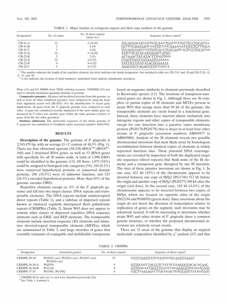
FIG. 1. Circular representation of the P. gingivalis genome. The outer circle shows the predicted coding regions on the plus strand color-codedby role categories as follows: violet, amino acid biosynthesis; light blue, biosynthesis of cofactors, prosthetic groups, and carriers; light green, cellenvelope; red, cellular processes; brown, central intermediary metabolism; yellow, DNA metabolism; light gray, energy metabolism; magenta, fattyacid and phospholipid metabolism; pink, protein synthesis and fate; orange, purines, pyrimidines, nucleosides, and nucleotides; olive, regulatoryfunctions and signal transduction; dark green, transcription; teal, transport and binding proteins; gray, unknown function; salmon, other categories;blue, hypothetical proteins. The second circle shows the predicted coding regions on the minus strand. The third circle presents the �2 analysis ofatypical nucleotide composition; �2 values of �600 are indicated in red. The fourth circle shows the %G�C. The fifth circle shows atypicalnucleotide composition (GC skew). The sixth circle shows the IS elements, indicated by color as follows: orange, ISPg1; light green, ISPg2; magenta,ISPg3; cyan, ISPg4; brown, ISPg5; gold, ISPg6; blue-green, ISPg7; pink, ISPg8; and violet, ISPg9; salmon, ISPg10; olive, ISPg11. The seventh circleshows MITE239 (magenta), MITE700 (cyan), and MITE464 (black). The eighth circle shows Tn4555 (blue), CTn (red), and other transposableelements (green). The ninth circle shows tRNA (green), rRNA (black), and sRNA (red).
5592 NELSON ET AL. J. BACTERIOL.
Pfam v2.0 and 631 HMMs from TIGR ortholog resource. TMHMM (33) wasused to identify membrane-spanning domains in proteins.
Comparative genomics. All genes and predicted proteins from this genome, aswell as from all other completed genomes, were compared by using the basiclocal alignment search tool (BLAST). For the identification of recent geneduplications, all genes from the P. gingivalis genome were compared to eachother. A gene was considered recently duplicated if the most similar gene (asmeasured by P value) was another gene within the same genome (relative togenes from the two other genomes).
Database submission. The nucleotide sequence of the whole genome ofP. gingivalis was submitted to GenBank under accession number AE015924.
RESULTS
Description of the genome. The genome of P. gingivalis is2,343,479 bp, with an average G�C content of 48.3% (Fig. 1).There are four ribosomal operons (5S-23S-tRNAAla-tRNAIle-16S) and 2 structural RNA genes, as well as 53 tRNA geneswith specificity for all 20 amino acids. A total of 1,990 ORFscould be identified in the genome (13). Of these, 1,075 (54%)could be assigned to biological role categories (54), 184 (9.2%)were conserved hypothetical proteins or conserved domainproteins, 208 (10.5%) were of unknown function, and 523(26.3%) encoded hypothetical proteins. More than 85% of thegenome encodes ORFs.
Repetitive elements occupy ca. 6% of the P. gingivalis ge-nome and fall into two major classes: DNA repeats and trans-posable elements. The DNA repeats include uninterrupteddirect repeats (Table 1), and a subclass of dispersed repeatsknown as clustered regularly interspaced short palindromicrepeats (CRISPRs) (Table 2). Strain W83 does not appear tocontain other classes of dispersed repetitive DNA sequenceelements such as ERIC and REP elements. The transposableelements include insertion sequence (IS) elements and minia-ture inverted-repeat transposable elements (MITEs), whichare summarized in Table 3, and large stretches of genes thatresemble remnants of conjugable and mobilizable transposons
based on sequence similarity to elements previously describedin Bacteroides species (11). The locations of transposon-asso-ciated genes are shown in Fig. 1. Although there are 96 com-plete or partial copies of IS elements and MITEs present instrain W83 that occupy more than 94 kb of the genome, thetransposable elements are rarely found in a functional gene.Instead, these elements have inserted almost exclusively intointergenic regions and other copies of transposable elements,except for one insertion into a putative outer membraneprotein (PG0176/PG0178) that is intact in at least four otherstrains of P. gingivalis (accession numbers AB069977 toAB069980). Analysis of the IS elements reveals two possiblechromosomal inversions that most likely arose by homologousrecombination between identical copies of elements at widelyseparated insertion sites. These potential DNA rearrange-ments are revealed by inspection of both the duplicated targetsite sequences (direct repeats) that flank some of the IS ele-ments and a transposon gene disrupted by one IS insertion.The sites of these putative inversions are shown in Fig. 1. Inone case, 821 kb (35%) of the chromosome appears to beinverted between one copy of ISPg2 (PG1746) 512 kb beforethe origin and another copy of ISPg2 (PG0277) 309 kb after theorigin (red dots). In the second case, 103 kb (4.4%) of thechromosome appears to be inverted between two copies ofISPg4, which are located on opposite sides of the origin(PG2194 and PG0050) (green dots). Since inversions about theorigin do not invert the direction of transcription relative toreplication of genes on the segment, such inversions may beselectively neutral. It will be interesting to determine whetherstrain W83 and other strains of P. gingivalis share a commongenetic structure, or whether the proposed chromosomal in-versions are relatively recent events.
There are 21 areas of the genome that display an atypicalnucleotide composition identified by �2 analysis (67) and that
TABLE 1. Major families of contiguous repeats and their copy numbers in the genome
Designationa No. of copies No. of direct repeats(mean no.) Sequence of direct repeatb
CR-41-A 12 2–16 (8) TGCAGGACGCGATTGTCAA�CTGATTCTTGCTTCCTGCACGACR-41-B 1 4 (4) GCTTGCAGGAAGCGATTTCG�GCAAAAAAATTCCTGTTTGCACR-41-C 1 4 (4) TGCAGGAAGCCGTTGTCAC�CTGACAATCACTCCCTGCACGACR-23-A 14 3–14 (9) TATCCTCTCAT�ATGAGACCATAGCR-23-B 5 5 (5) ACTAAACTAT�ATACTTTGGTNNNCR-22-A 32 2–8 (5) CGATTTATA�TATAAATCGAAAACR-22-B 4 4–6 (5) TATTTCCGTAT�ATACGGAAATACR-22-C 2 4–5 (5) AAACGTCT�AGACGTTTCGAGCA
a The number indicates the length of the repetitive element; the letter indicates the family designation. Not included in table are CR-23-C and -D and CR-22-D, -E,-F, -G, and -H.
b A dot indicates the location of dyad symmetry; underlined bases indicate palindromic match(es).
TABLE 2. CRISPRs
Designation Associated gene(s) No. of direct repeats Sequence of direct repeatb
CRISPR-30-36a PG2013 cas2, PG2014 cas1, PG2015 cas4,PG2016 cas3
23 GTTTTAATTCCTGTA�TGGTGCAATTGAAAT
CRISPR-36-30-A PG0338 8 GTTGGATCTACCCTCTAT�TCGAAGGGTACACACAACCRISPR-36-30-B PG1164 8 GTTGGGAATACCCTTAGT�TAGAAGGGTGGAGACAACCRISPR-37-35 PG1981, PG1982 8 GTCTTAATAGCCTTACGGACTGTGTATGTATAGTGAG
a CRISPR-30-36 and cas1 to cas4 were described previously (26).b See Table 1, footnote b.
VOL. 185, 2003 PORPHYROMONAS GINGIVALIS GENOME ANALYSIS 5593
also correspond to regions of higher or lower G�C contentthan the rest of the genome. The areas range in size from 11 to68 kb and range in G�C content from 29.4 to 61.6%. A varietyof genes that could possibly have been acquired by this bacte-rium through lateral gene transfer are encoded in theseregions. The genes include three restriction system proteins(PG0971, most similar to Anabaena sp. strain PCC 7120;PG0968, most similar to Anabaena sp. strain PCC 7120; andPG1469 most similar to Agrobacterium tumefaciens); hemag-glutinin proteins B and C (HagB, PG1972, P. gingivalis specific;and HagC, PG1975, P. gingivalis specific); many capsular bio-synthesis proteins, 20 transposase genes, two large mobileelements (PG1473 to PG1480, resembles only a conjugativeelement of Bacteroides thetaiotaomicron; and PG0868 to PG0875,whose sequence and gene organization most closely resemblesthe antibiotic-resistant mobilizable transposon Tn4555 fromBacteroides fragilis (68); and a thiamine biosynthesis operon(PG2107 to PG2111, which is most similar to the thiaminebiosynthesis operon of Escherichia coli). These atypical regionsin the P. gingivalis genome also encode many hypothetical andconserved hypothetical proteins, which undoubtedly contributeto the unique biology of this organism.
Comparative genomics. Comparison of the predicted pro-teome of P. gingivalis with that of other completely sequencedgenomes confirms the close relationship of P. gingivalis to othermembers of the CFB, including B. fragilis and B. thetaiotaomi-cron. Outside of the CFB phyla, the genome most similar tothat of P. gingivalis is the Chlorobium tepidum genome, sup-porting previous phylogenetic studies that indicated the chlo-robia and CFB phyla are related, albeit distantly. The pro-teomes most similar to that of P. gingivalis (in terms of thenumber of proteins with the best scoring matches) were thoseof B. thetaiotaomicron and B. fragilis with 572 and 437 best-scoring matches (P � 10�5), respectively.
A total of 332 genes were identified as being putativelyduplicated in the P. gingivalis lineage. These duplicated genesare likely an indication that there is some selective evolution-ary advantage to retaining these genes in the genome. Amongthese genes are 10 that encode DNA-binding histone-like pro-
teins that have a distinctive domain architecture comparedto HU and related histone-like proteins. These DNA-bindingproteins have been designated a superfamily (i.e., a set ofproteins that share a given domain architecture; TIGRFAMsfamily TIGR01201). Outside of P. gingivalis, the single knownexample of a DNA-binding histone-like protein is found in thegut bacterium B. fragilis. All members of this superfamily aredistantly related to the bacterial DNA-binding protein HUfamily (Pfam family PF00216, five of which are also found inthe P. gingivalis genome) but differ in architecture, sharingboth an N-terminal extension and a glycine-rich C terminus.HU has been shown, among other DNA-binding functions, toassist the unwinding of oriC DNA by the DNA replicationinitiation protein DnaA (4). Interestingly, all 10 members ofthe TIGR01201 family in P. gingivalis have direct repeats up-stream of their genes that may act as binding sites for theDNA-binding proteins that are encoded by the nearby geneand perhaps regulate their own expression. Alternatively, therepeats may also coordinate expression of the other chromo-somal genes that they flank.
Metabolism and transport. The microbial species that existin supragingival plaque of the oral cavity are exposed to thehost’s dietary intake, and many of these bacteria, including theoral streptococci, ferment carbohydrates to acidic end productssuch as lactic acid for the purpose of energy production. Onthe other hand, anaerobic species in the subgingival plaque areexposed to crevicular fluid and to the host tissue proteins (61).The availability of the complete genome sequence of P. gingi-valis W83 allowed for an analysis of the physiological potentialof this species. Based on this analysis, the range of transportcapabilities and metabolic pathways that could be identified ispresented in Fig. 2.
Genome analysis suggests that P. gingivalis possesses a lim-ited capacity for the uptake and metabolism of organic nutri-ents. Glucose utilization by P. gingivalis is known to be verypoor, and carbohydrates in general do not appear to readilysupport growth (61). Strain W83 does, however, contain puta-tive ORFs for all enzymes of the glycolytic pathway, as well asORFs for a putative glucose/galactose transporter and glucose
TABLE 3. IS elements and MITEs
Elementa Family No. of copies(no. of fragments)b Inverted repeat (length [bp])c Direct repeat lengthd Length
(bp)
ISPg1 IS5 25(7) GAGACCTTTGCA (12) 5 1,334ISPg2 ISAs1 5(11) CAGGGCTGACGCATTAAA (18) 9 1,207ISPg3 IS5 6(1) ACGTCAGTTCGA (12) 7 1,070ISPg4 IS5 10(0) CATTAGTGTCCTAAAAG (17) 9 1,394ISPg5 IS3 11(1) TTGACGTGTACTGAAAAAAGTTGACAGTTGG (31) 3 1,514ISPg6 ISAs1 1(2) CAGGGCAACCGCATCAAA (18) NDe 1,139ISPg7 ISRm7 1(3) GTCGTCCCTTAAgAAtCCTtTT (22) 6 1,428ISPg8 IS5 0(1) Unknown ND NDISPg9 IS5 2(2) GAGACCATTGCA (12) 5 1,326ISPg10 IS256 1 TGaCatTtTtAAAAAAaGAcATGG (24) ND �1,283ISPg11 IS256 (1) Unknown ND NDMITE239 ISPg3 5 ACGysAGTTCGATmTAArsGAA 6–8 239MITE464 ISPg1 13 GAGACTGTTGCA 5 464–1,071MITE700 ISPg3 8 ACGTCAGTTCGA 7 455–717
a ISPg1, ISPg2, and ISPg3 were originally described as IS1126 (44), PGIS2 (69), and IS195 (40).b That is, the number of intact copies with the number of fragments in parentheses.c Lowercase lettering indicates sequence at a position which is not part of the perfect inverted repeat.d Direct repeat or duplicated target site length.e ND, not determined.
5594 NELSON ET AL. J. BACTERIOL.

FIG
.2.
Overview
ofmetabolism
andtransportin
P.gingivalis.Prim
arysubstrates
forenergy
metabolism
arecapitalized
andunderlined.E
ndproducts
offermentation
arehighlighted
byyellow
boxes.Transporters
aregrouped
bysubstrate
specificityand
indicatedby
coloras
follows:inorganic
cations(green),inorganic
anions(m
agenta),organicnutrients
(yellow),and
drugefflux
andother
(black).Arrow
sindicate
directionof
transportfor
substrates(and
couplingions,w
hereappropriate).
VOL. 185, 2003 PORPHYROMONAS GINGIVALIS GENOME ANALYSIS 5595
kinase. Sequence analysis shows that the glucose kinase is en-coded in a split ORF generated by a missense mutation, andthis is a likely explanation for the poor utilization of glucose tosupport growth. Four putative ORFs for the pentose phos-phate pathway were identified, and it is likely that this pathwayplays a role in the generation of precursor metabolites duringanaerobic growth (Fig. 2).
Whole-genome analysis suggests that P. gingivalis can me-tabolize several sugars, including melibiose, galactose, starch,and maltodextrin. The bacterium also possesses enzymes forthe degradation of complex amino sugars in the form of hexoseaminidases. It is still unclear whether these complex sugars aremetabolized, but one possibility is that the removal of aminosugars from host glycoproteins likely renders these proteinsmore susceptible to degradation by bacterial proteinases. Inaddition, at least 11 amino acids may serve as substrates forenergy production (Fig. 2). These amino acids are most likelyderived from the degradation of host tissues (see virulencesection below) or from the breakdown of other bacterial cellsin the oral cavity. Pathways for glutamate and aspartate utili-zation have been characterized by enzyme assays (65), andORFs coding for all of these activities were found in the W83genome. Intracellular glutamate is deaminated to 2-oxoglutat-arate by glutamate dehydrogenase and then decarboxylated tosuccinyl coenzyme A (succinyl-CoA) by a CoA-dependent2-oxoglutarate oxidoreductase. The possession of this activityis somewhat unusual in bacterial species (23, 25). It has beenestablished that two-thirds of the succinyl-CoA produced inthis reaction is converted to butyryl-CoA and then to butyrate.The remaining third may be converted to propionate by apathway that involves the enzymes methylmalonyl-CoA mutaseand acyl-CoA:acetate-CoA transferase, as reported for otherpropionate-producing bacteria (18). This pathway appearsto be unique to P. gingivalis since other anaerobes catabolizeglutamate through the hydroxyglutarate, methylaspartate, and/or the aminobutyrate pathways (5, 17, 18). P. gingivalis did notpossesss activities for three key enzymes of these pathways:hydroxyglutarate dehydrogenase, 3-methylaspartate ammonialyase, and 4-aminobutyrate aminotransferase (65). Peptide-de-rived aspartate is deaminated to fumarate by aspartate ammo-nia lyase and then either oxidized to acetate or reduced topropionate and butyrate (65).
Results from Takahashi et al. (65) suggest that P. gingivalisprefers to utilize arginine and lysine as free amino acids ratherthan in peptide form; thus, carboxy-terminal arginine and ly-sine residues could be released from proteins by carboxypep-tidase activities. Masuda et al. (45) found such an activity inculture supernatants, and an ORF coding for an unspecifiedcarboxypeptidase (PG0232) was identified in the genome. Areport that P. gingivalis produces citrulline and ornithine fromdenatured protein (14) implies that the bacterium degradesarginine through the arginine deiminase pathway. Indeed, agene with homology to arginine deiminase from Bacillus li-cheniformis (43) was identified. In addition, two genes—pyrBand pyrI (PG0357 and PG0358)—were contiguous in the ge-nome and shared homology with aspartate/ornithine transcar-bamylase catalytic and regulatory chains from Vibrio sp. strain2693 and Pyrococcus abyssi, respectively.
The lysine catabolic pathways appear to be very similar tothose found in Clostridium sp. ORFs were identified for the
first steps of both L- and D-lysine catabolism; thus, the isomersare apparently degraded by two different pathways that yieldbutyric acid, acetic acid, and ammonia. Lysine 2,3-amino-mutase (KamA) catalyzes the interconversion of L-lysine andL--lysine, the first step in the lysine degradation pathway inClostridium subterminale SB4 (56). In P. gingivalis W83, kamAwas found clustered with the genes kamD and kamE (PG1070,PG1073, and PG1074) that encode subunits of D-lysine 5,6-aminomutase, the first enzyme of the D-lysine degradativepathway. Genes encoding enzymes for the subsequent conver-sion of lysine to butyrate and acetate were located 3 to kamE.It is not yet known whether these genes are transcribed as anoperon.
Little is known about serine and threonine catabolism inP. gingivalis; however, an ORF was detected with homology toserine dehydratase (PG0084) that hydrolyzes serine to pyru-vate, ammonia, and water. Threonine may be split to glycineand acetaldehyde by the activity of threonine aldolase, forwhich an ORF was detected (PG0474). In summary, P. gingi-valis appears to catabolize amino acids through pathways thatgenerate ammonia. The organism has a growth pH optimum of�7.5, and ammonia generation may have evolved as a strategyto shift the local pH to the favored alkaline range.
Several studies have shown that P. gingivalis preferentiallyuses peptides as sources of carbon and nitrogen (60, 65, 71)and, in addition to the previously described proteinases thatare known to degrade host proteins, a number of peptidasesthat may be involved in the further digestion of protein frag-ments to smaller peptides and amino acids could be identifiedfrom the genome.
There are two carboxylate transporters possibly for lactateand formate, and no sugar transporters other than the afore-mentioned glucose/galactose importer. Although P. gingivalispossesses a broad assortment of secreted peptidases and path-ways for the metabolism of amino acids, the bacterium appearsto rely on two predicted peptide uptake systems and has onlyone amino acid transporter, the characterized sodium ion-driven serine/threonine uptake protein SstT (12). A LysE-typeamino acid efflux protein is present that may protect the or-ganism from toxic concentrations of amino acids.
The major fermentation products that can be producedbased on whole-genome analysis and in vitro end product anal-yses are propionate, butyrate, isobutyrate, isovalerate, acetate,ethanol, and butanol (27). Many of these end products areprobably toxic to human host tissues (see virulence sectionbelow).
Nucleosides and nucleobases may represent a hitherto-un-suspected important nutrient source for P. gingivalis and mightbe used either as building blocks for nucleic acid biosynthesisor may be catabolized as carbon and energy sources. There arethree predicted purine uptake systems, a NupG nucleosideuptake system, and a homolog of the Salmonella enterica se-rovar Typhimurium nicotinamide mononucleotide transporterPnuC. In addition, there are four homologs of E. coli DinF, aDNA damage-induced protein related to sodium ion-drivendrug efflux transporters, that are hypothesized to play a role innucleoside and/or nucleotide efflux (8).
Common to most human pathogens, iron acquisition ap-pears to be an important priority in P. gingivalis, and there aretwo iron chelate ABC uptake systems, two TonB-dependent
5596 NELSON ET AL. J. BACTERIOL.

iron receptors, and two FeoB ferrous iron uptake systems.There is an array of metal ion homeostasis transporters, in-cluding three sodium ion/proton exchangers, which may beimportant since a significant number of P. gingivalis transport-ers are predicted to be sodium ion driven.
Virulence and P. gingivalis. The availability of the completegenome sequence of P. gingivalis facilitates the identification ofputative virulence factors associated with the establishmentand survival of the bacterium in the gingival crevice and sub-sequent penetration into host cells (Table 4). Initially, thebacterium must navigate the oral cavity where, as an obligateanaerobe, it is exposed to limited amounts of oxygen before itestablishes itself in an anaerobic environment. A cluster ofgenes (PG1582 to PG1586) was identified with high levels ofsimilarity to the recently described aerotolerance operon ofB. fragilis (66). These functions promote the survival of B. fra-gilis upon exposure to oxygen, and their presence in P. gingi-valis suggests that this system may also ensure tolerance to oxy-gen in the oral cavity. The genome also encodes a superoxidedismutase (PG1545), genes for an alkyl hydroperoxide reduc-tase (PG0618 and PG0619) (55), a thiol peroxidase (PG1729),and a Dps homolog (PG0090) that is involved in the repair ofoxidatively damaged nucleic acids (33).
The bacterium uses fimbriae to adhere to other bacterialspecies and host tissues. Hemagglutinins and various proteases(gingipains) are also involved in tissue colonization throughadhesion to extracellular matrix proteins (38, 53, 59). Hemag-glutinins in particular may mediate the binding of bacteria toreceptors on human cells (21), and the gene sequences for sixnewly identified putative hemagglutinin-like proteins (PG0411,PG1326, PG1674, PG1427, PG1548, and PG2198) could beidentified. Four of these are recent duplications in the genomeof HagA and HagD adhesin domain-related sequences. A totalof 42 proteinases were identified in the genome sequence thatmay enable adherence of the bacterium to host tissues, as wellas to other bacterial cells, and that may also degrade hostproteins (as discussed above). In vitro experiments have dem-onstrated that proteases attack a range of host proteins, in-cluding extracellular matrix proteins (32, 38, 53, 59) and celladhesion molecules (29), the destruction of which leads to aloss of cell surface receptors (59) and tissue integrity (29).Protease destruction of cytokines (15, 42, 74) and gamma in-terferon (73) can result in disruption of polymorphonuclearleukocyte function (48) and ultimately affect the host immuneresponse.
A single hemolysin for the release of iron and protophorynIX (PG1875) was identified. This sequence has full-lengthhomology only to the characterized hemolysin gene of an-other periodontal pathogen, Prevotella melaninogenica (3).These two hemolysins show absolutely no homology to anyother biochemically characterized hemolysin and have weakhomology to a conserved hypothetical protein/putative hemo-lysin fusion protein sequence from Vibrio cholerae.
In P. gingivalis, metabolic end products from the catabolismof various substrates include short-chained carboxylic acidsthat can affect the host defense system in a variety of ways.When applied directly to healthy human gingiva tissue, short-chain carboxylic acids have been shown to stimulate a gingivalinflammatory response and inflammatory cytokine release(50). Short-chain carboxylic acids have also been shown to alter
cell function and gene expression and may also contribute tothe initiation and prolongation of gingival inflammation (50).
The capsule of P. gingivalis is most likely involved in theevasion of the host response and has been shown to be one ofthe important virulence determinants in this bacterium (34).Whole-genome analysis reveals at least four capsular bio-synthesis gene clusters (PG0106 to PGPG0120, PG0435 toPG0437, PG1140 to PG1149, and PG1560 to PG1565) that arelocated across the genome. Closer investigation of these geneclusters suggests that mannose, glucose, and rhamnose may besome of the sugars that are present in the capsule of P. gingi-valis strain W83. In several pathogens the secretion of viru-lence factors targeted to the host cells is mediated by type IIIprotein secretion systems. The complete genome of P. gingiva-lis was searched for the presence of a cluster of nine genes Sct(Hrc/Ysc) that are known to be components of type III proteinsecretion systems (24). No BLAST matches with these motifswere found. Although several sec gene homologs are present inthe genome, including SecA, SecY, SecD, and SecF, the mainterminal branch of the general secretory pathway (type II)could not be identified, suggesting that this pathway is notfunctional in this bacterium.
DISCUSSION
It is estimated that 35% of the U.S. population has someform of periodontitis (2). Traditional methods for the preven-tion or treatment of periodontal disease include the mechan-ical removal of plaque and the use of antimicrobial agents. Theavailability of the genome sequences of at least three oralpathogens and recent investigations into the microbial compo-sition of the human oral microbiome will afford new opportu-nities to investigate ways to alter the composition of the sub-gingival biofilm. Potential strategies include those that woulddecrease opportunities for biofilm formation, reduce attach-ment among species in the biofilm, or limit the availability ofrequired nutrients. Inhibition of the primary colonizers couldprevent the successful establishment of late colonizers such asP. gingivalis.
Recently, specific bacteria have been found to be associatedwith systemic diseases, e.g., Helicobacter pylori, as etiologicalagent in gastric ulcers, and the tentative association between thepresence of Chlamydia pneumoniae in atherosclerotic plaquesand cardiovascular disease (9). Stimulated by these studies andthe detection of P. gingivalis and other oral pathogens in ath-erosclerotic plaques (10, 62), new research is assessing associ-ations between periodontal infection and cardiovascular dis-ease (22, 47, 52, 58). That the pathogenicity of oral bacteriamay extend beyond their known ecological niche to other or-gan systems introduces an important and exciting new dimen-sion to defining the genetic complement of these organisms.Although the genome sequence has not revealed many of theclassical virulence factors that are associated with pathogens, itis anticipated that the newly described putative virulence fac-tors of this bacterium, as well as from the others that have beenrecently sequenced or whose sequencing is nearing comple-tion, will enable the development of antimicrobial agents thatcan be used against one of the major causative agents of peri-odontal disease. Ultimately, the genome sequence of P. gingi-valis will facilitate an increased understanding of the virulence
VOL. 185, 2003 PORPHYROMONAS GINGIVALIS GENOME ANALYSIS 5597
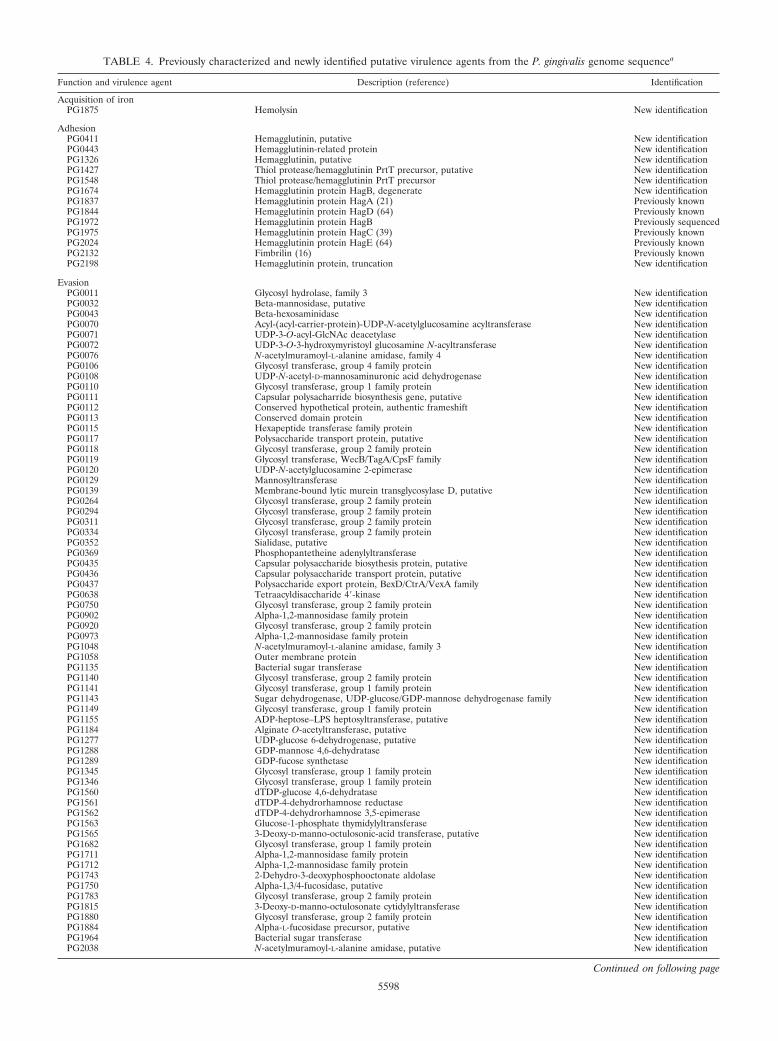
TABLE 4. Previously characterized and newly identified putative virulence agents from the P. gingivalis genome sequencea
Function and virulence agent Description (reference) Identification
Acquisition of ironPG1875 Hemolysin New identification
AdhesionPG0411 Hemagglutinin, putative New identificationPG0443 Hemagglutinin-related protein New identificationPG1326 Hemagglutinin, putative New identificationPG1427 Thiol protease/hemagglutinin PrtT precursor, putative New identificationPG1548 Thiol protease/hemagglutinin PrtT precursor New identificationPG1674 Hemagglutinin protein HagB, degenerate New identificationPG1837 Hemagglutinin protein HagA (21) Previously knownPG1844 Hemagglutinin protein HagD (64) Previously knownPG1972 Hemagglutinin protein HagB Previously sequencedPG1975 Hemagglutinin protein HagC (39) Previously knownPG2024 Hemagglutinin protein HagE (64) Previously knownPG2132 Fimbrilin (16) Previously knownPG2198 Hemagglutinin protein, truncation New identification
EvasionPG0011 Glycosyl hydrolase, family 3 New identificationPG0032 Beta-mannosidase, putative New identificationPG0043 Beta-hexosaminidase New identificationPG0070 Acyl-(acyl-carrier-protein)-UDP-N-acetylglucosamine acyltransferase New identificationPG0071 UDP-3-O-acyl-GlcNAc deacetylase New identificationPG0072 UDP-3-O-3-hydroxymyristoyl glucosamine N-acyltransferase New identificationPG0076 N-acetylmuramoyl-L-alanine amidase, family 4 New identificationPG0106 Glycosyl transferase, group 4 family protein New identificationPG0108 UDP-N-acetyl-D-mannosaminuronic acid dehydrogenase New identificationPG0110 Glycosyl transferase, group 1 family protein New identificationPG0111 Capsular polysacharride biosynthesis gene, putative New identificationPG0112 Conserved hypothetical protein, authentic frameshift New identificationPG0113 Conserved domain protein New identificationPG0115 Hexapeptide transferase family protein New identificationPG0117 Polysaccharide transport protein, putative New identificationPG0118 Glycosyl transferase, group 2 family protein New identificationPG0119 Glycosyl transferase, WecB/TagA/CpsF family New identificationPG0120 UDP-N-acetylglucosamine 2-epimerase New identificationPG0129 Mannosyltransferase New identificationPG0139 Membrane-bound lytic murein transglycosylase D, putative New identificationPG0264 Glycosyl transferase, group 2 family protein New identificationPG0294 Glycosyl transferase, group 2 family protein New identificationPG0311 Glycosyl transferase, group 2 family protein New identificationPG0334 Glycosyl transferase, group 2 family protein New identificationPG0352 Sialidase, putative New identificationPG0369 Phosphopantetheine adenylyltransferase New identificationPG0435 Capsular polysaccharide biosythesis protein, putative New identificationPG0436 Capsular polysaccharide transport protein, putative New identificationPG0437 Polysaccharide export protein, BexD/CtrA/VexA family New identificationPG0638 Tetraacyldisaccharide 4-kinase New identificationPG0750 Glycosyl transferase, group 2 family protein New identificationPG0902 Alpha-1,2-mannosidase family protein New identificationPG0920 Glycosyl transferase, group 2 family protein New identificationPG0973 Alpha-1,2-mannosidase family protein New identificationPG1048 N-acetylmuramoyl-L-alanine amidase, family 3 New identificationPG1058 Outer membrane protein New identificationPG1135 Bacterial sugar transferase New identificationPG1140 Glycosyl transferase, group 2 family protein New identificationPG1141 Glycosyl transferase, group 1 family protein New identificationPG1143 Sugar dehydrogenase, UDP-glucose/GDP-mannose dehydrogenase family New identificationPG1149 Glycosyl transferase, group 1 family protein New identificationPG1155 ADP-heptose–LPS heptosyltransferase, putative New identificationPG1184 Alginate O-acetyltransferase, putative New identificationPG1277 UDP-glucose 6-dehydrogenase, putative New identificationPG1288 GDP-mannose 4,6-dehydratase New identificationPG1289 GDP-fucose synthetase New identificationPG1345 Glycosyl transferase, group 1 family protein New identificationPG1346 Glycosyl transferase, group 1 family protein New identificationPG1560 dTDP-glucose 4,6-dehydratase New identificationPG1561 dTDP-4-dehydrorhamnose reductase New identificationPG1562 dTDP-4-dehydrorhamnose 3,5-epimerase New identificationPG1563 Glucose-1-phosphate thymidylyltransferase New identificationPG1565 3-Deoxy-D-manno-octulosonic-acid transferase, putative New identificationPG1682 Glycosyl transferase, group 1 family protein New identificationPG1711 Alpha-1,2-mannosidase family protein New identificationPG1712 Alpha-1,2-mannosidase family protein New identificationPG1743 2-Dehydro-3-deoxyphosphooctonate aldolase New identificationPG1750 Alpha-1,3/4-fucosidase, putative New identificationPG1783 Glycosyl transferase, group 2 family protein New identificationPG1815 3-Deoxy-D-manno-octulosonate cytidylyltransferase New identificationPG1880 Glycosyl transferase, group 2 family protein New identificationPG1884 Alpha-L-fucosidase precursor, putative New identificationPG1964 Bacterial sugar transferase New identificationPG2038 N-acetylmuramoyl-L-alanine amidase, putative New identification
Continued on following page
5598
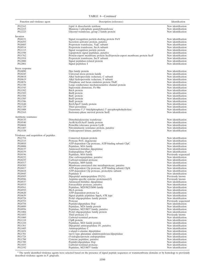
TABLE 4—Continued
Function and virulence agent Description (reference) Identification
PG2162 Lipid A disaccharide synthase New identificationPG2215 Mannose-1-phosphate guanylyltransferase New identificationPG2223 Glycosyl transferase, group 2 family protein New identification
InvasionPG0151 Signal recognition particle-docking protein FtsY New identificationPG0293 Secretion activator protein, putative New identificationPG0485 Preprotein translocase, YajC subunit New identificationPG0514 Preprotein translocase, SecA subunit New identificationPG1115 Signal recognition particle protein New identificationPG1598 Lipoprotein signal peptidase, putative New identificationPG1762 Protein-export membrane protein SecD/protein-export membrane protein SecF New identificationPG1918 Preprotein translocase, SecY subunit New identificationPG2000 Signal peptidase-related protein New identificationPG2001 Signal peptidase I New identification
Stress responsePG0090 Dps family protein New identificationPG0245 Universal stress protein family New identificationPG0618 Alkyl hydroperoxide reductase, C subunit New identificationPG0619 Alkyl hydroperoxide reductase, F subunit New identificationPG0876 Thiophene and furan oxidation protein ThdF New identificationPG1330 Large conductance mechanosensitive channel protein New identificationPG1545 Superoxide dismutase, Fe-Mn New identificationPG1582 BatA protein New identificationPG1583 BatB protein New identificationPG1584 BatC protein New identificationPG1585 BatD protein New identificationPG1586 BatE protein New identificationPG1648 RelA/SpoT family protein New identificationPG1729 Thiol peroxidase New identificationPG1808 Guanosine-3,5-bis(diphosphate) 3-pyrophosphohydrolase New identificationPG2163 Stationary-phase survival protein SurE New identification
Antibiotic resistancePG0135 Dimethyladenosine transferase New identificationPG0540 AcrB/AcrD/AcrF family protein New identificationPG0604 Penicillin tolerance protein LytB New identificationPG1407 Nitroimidazole resistance protein, putative New identificationPG1538 Undecaprenol kinase, putative New identification
Virulence and acquisition of peptidesPG1754 Conserved domain protein New identificationPG2115 Protease PrtT, degenerate New identificationPG0010 ATP-dependent Clp protease, ATP-binding subunit ClpC New identificationPG0088 Peptidase, M16 family New identificationPG0137 Aminoacyl-histidine dipeptidase New identificationPG0159 Endopeptidase PepO New identificationPG0196 Peptidase, M16 family Previously sequencedPG0232 Zinc carboxypeptidase, putative New identificationPG0235 Carboxyl-terminal protease New identificationPG0317 Peptidase, M49 family New identificationPG0383 Membrane-associated zinc metalloprotease, putative New identificationPG0417 ATP-dependent Clp protease, ATP-binding subunit ClpX New identificationPG0418 ATP-dependent Clp protease, proteolytic subunit New identificationPG0445 Peptidase T New identificationPG0503 Dipeptidyl aminopeptidase IV(31) Previously knownPG0506 Arginine-specific cysteine proteinase(63) Previously knownPG0537 Aminoacyl-histidine dipeptidase New identificationPG0553 Extracellular protease, putative New identificationPG0561 Peptidase, M20/M25/M40 family New identificationPG0593 HtrA protein New identificationPG0620 ATP-dependent protease La New identificationPG0639 Signal peptide peptidase SppA, 67K type New identificationPG0724 Prolyl oligopeptidase family protein New identificationPG0753 Protease Previously sequencedPG0758 Peptidyl-dipeptidase Dcp New identificationPG0889 Peptidase, M24 family protein New identificationPG0956 Peptidase, M23/M37 family, putative New identificationPG1004 Prolyl oligopeptidase family protein New identificationPG1055 Thiol protease (7) Previously knownPG1060 Carboxyl-terminal protease New identificationPG1118 ClpB protein New identificationPG1210 Peptidase, M24 family protein New identificationPG1361 Dipeptidyl aminopeptidase IV, putative New identificationPG1605 Aminopeptidase C New identificationPG1654 D-alanyl-D-alanine dipeptidase New identificationPG1701 trp-G type glutamine amidotransferase/dipeptidase New identificationPG1724 O-sialoglycoprotein endopeptidase New identificationPG1788 Cysteine peptidase, putative New identificationPG1789 Peptidyl-dipeptidase Dcp New identificationPG1855 Carboxyl-terminal protease New identificationPG2192 Peptidase, M23/M37 family New identification
a The newly identified virulence agents were selected based on the presence of signal peptide sequences or transmembrane domains or by homology to previouslydescribed virulence agents in P. gingivalis.
5599

of this periodontal pathogen and will enable the developmentof improved diagnostics and therapeutics.
ACKNOWLEDGMENTS
We thank Michael Heaney, Michael Holmes, Vadim Sapiro, andEmmanuel Mongodin for informatics support at TIGR. We thank J. F.Tomb for assistance in the early stages of the project.
This work was supported by the National Institute of Dental andCraniofacial Research at the National Institutes of Health (R01DE-13914).
REFERENCES
1. Ajdic, D., W. M. McShan, R. E. McLaughlin, G. Savic, J. Chang, M. B.Carson, C. Primeaux, R. Tian, S. Kenton, H. Jia, S. Lin, Y. Qian, S. Li, H.Zhu, F. Najar, H. Lai, J. White, B. A. Roe, and J. J. Ferretti. 2002. Genomesequence of Streptococcus mutans UA159, a cariogenic dental pathogen.Proc. Natl. Acad. Sci. USA 99:14434–14439.
2. Albandar, J. M., J. A. Brunelle, and A. Kingman. 1999. Destructive peri-odontal disease in adults 30 years of age and older in the United States,1988–1994. J. Periodontol. 70:13–29.
3. Allison, H. E., and J. D. Hillman. 1997. Cloning and characterization of aPrevotella melaninogenica hemolysin. Infect. Immun. 65:2765–2771.
4. Bahloul, A., F. Boubrik, and J. Rouviere-Yaniv. 2001. Roles of Escherichiacoli histone-like protein HU in DNA replication: HU-beta suppresses thethermosensitivity of dnaA46ts. Biochimie 83:219–229.
5. Barker, H. A. 1981. Amino acid degradation by anaerobic bacteria. Annu.Rev. Biochem. 50:23–40.
6. Boone, D. R., and R. W. Castenholtz (ed.). 2001. Bergey’s manual of sys-tematic bacteriology, 2nd ed., vol. 1. Springer-Verlag, New York, N.Y.
7. Bourgeau, G., H. Lapointe, P. Peloquin, and D. Mayrand. 1992. Cloning,expression, and sequencing of a protease gene (tpr) from Porphyromonasgingivalis W83 in Escherichia coli. Infect. Immun. 60:3186–3192.
8. Brown, M. H., I. T. Paulsen, and R. A. Skurray. 1999. The multidrug effluxprotein NorM is a prototype of a new family of transporters. Mol. Microbiol.31:394–395.
9. Byrne, G. I., S. I. Skarlotos, C. Grunfeld, M. V. Kalayoglu, P. Libby, P.Saikku, J. T. Summersgill, and P. Wyrick. 2000. Collaborative multidisci-plinary workshop report: interface of lipid metabolism, atherosclerosis, andChlamydia infection. J. Infect. Dis. 181(Suppl. 3):S490–S491.
10. Chiu, B. 1999. Multiple infections in carotid atherosclerotic plaques. Am.Heart J. 138:S534–S536.
11. Churchward, G. 2002. Conjugative transposons and related elements, p.177–191. In N. Craig, R. Craigie, M. Gellert, and A. Lambowitz (ed.), MobileDNA II. ASM Press, Washington, D.C.
12. Dashper, S. G., L. Brownfield, N. Slakeski, P. S. Zilm, A. H. Rogers, andE. C. Reynolds. 2001. Sodium ion-driven serine/threonine transport in Por-phyromonas gingivalis. J. Bacteriol. 183:4142–4148.
13. Delcher, A. L., D. Harmon, S. Kasif, O. White, and S. L. Salzberg. 1999.Improved microbial gene identification with GLIMMER. Nucleic Acids Res.27:4636–4641.
14. Endo, J., M. Otsuka, E. Ohara, M. Sato, and R. Nakamura. 1989. Cleavageaction of a trypsin-like protease from Bacteroides gingivalis 381 on reducedegg-white lysozyme. Arch. Oral Biol. 34:911–916.
15. Fletcher, J., K. Reddi, S. Poole, S. Nair, B. Henderson, P. Tabona, and M.Wilson. 1997. Interactions between periodontopathogenic bacteria and cyto-kines. J. Periodontal Res. 32:200–205.
16. Fujiwara, T., S. Morishima, I. Takahashi, and S. Hamada. 1993. Molecularcloning and sequencing of the fimbrilin gene of Porphyromonas gingivalisstrains and characterization of recombinant proteins. Biochem. Biophys.Res. Commun. 197:241–247.
17. Gharbia, S. E., and H. N. Shah. 1991. Pathways of glutamate catabolismamong Fusobacterium species. J. Gen. Microbiol. 137(Pt. 5):1201–1206.
18. Gottschalk, G. 1986. Bacterial fermentations, p. 208–282. In G. Gottschalk(ed.), Bacterial metabolism, 2nd ed. Springer-Verlag, New York, N.Y.
19. Goulbourne, P. A., and R. P. Ellen. 1991. Evidence that Porphyromonas(Bacteroides) gingivalis fimbriae function in adhesion to Actinomyces viscosus.J. Bacteriol. 173:5266–5274.
20. Grenier, D. 1992. Demonstration of a bimodal coaggregation reaction be-tween Porphyromonas gingivalis and Treponema denticola. Oral Microbiol.Immunol. 7:280–284.
21. Han, N., J. Whitlock, and A. Progulske-Fox. 1996. The hemagglutinin geneA (hagA) of Porphyromonas gingivalis 381 contains four large, contiguous,direct repeats. Infect. Immun. 64:4000–4007.
22. Herzberg, M. C., and M. W. Meyer. 1998. Dental plaque, platelets, andcardiovascular diseases. Ann. Periodontol. 3:151–160.
23. Hoffman, P. S., A. Goodwin, J. Johnsen, K. Magee, and S. J. Veldhuyzen vanZanten. 1996. Metabolic activities of metronidazole-sensitive and -resistantstrains of Helicobacter pylori: repression of pyruvate oxidoreductase andexpression of isocitrate lyase activity correlate with resistance. J. Bacteriol.178:4822–4829.
24. Hueck, C. J. 1998. Type III protein secretion systems in bacterial pathogensof animals and plants. Microbiol. Mol. Biol. Rev. 62:379–433.
25. Hughes, N. J., C. L. Clayton, P. A. Chalk, and D. J. Kelly. 1998. Helicobacterpylori porCDAB and oorDABC genes encode distinct pyruvate:flavodoxinand 2-oxoglutarate:acceptor oxidoreductases which mediate electron trans-port to NADP. J. Bacteriol. 180:1119–1128.
26. Jansen, R., J. D. Embden, W. Gaastra, and L. M. Schouls. 2002. Identifica-tion of genes that are associated with DNA repeats in prokaryotes. Mol.Microbiol. 43:1565–1575.
27. Kaczmarek, F. S., and A. L. Coykendall. 1980. Production of phenylaceticacid by strains of Bacteroides asaccharolyticus and Bacteroides gingivalis (sp.nov.). J. Clin. Microbiol. 12:288–290.
28. Kapatral, V., I. Anderson, N. Ivanova, G. Reznik, T. Los, A. Lykidis, A.Bhattacharyya, A. Bartman, W. Gardner, G. Grechkin, L. Zhu, O. Vasieva,L. Chu, Y. Kogan, O. Chaga, E. Goltsman, A. Bernal, N. Larsen, M. D’Souza,T. Walunas, G. Pusch, R. Haselkorn, M. Fonstein, N. Kyrpides, and R.Overbeek. 2002. Genome sequence and analysis of the oral bacterium Fu-sobacterium nucleatum strain ATCC 25586. J. Bacteriol. 184:2005–2018.
29. Katz, J., V. Sambandam, J. H. Wu, S. M. Michalek, and D. F. Balkovetz.2000. Characterization of Porphyromonas gingivalis-induced degradation ofepithelial cell junctional complexes. Infect. Immun. 68:1441–1449.
30. Kinder, S. A., and S. C. Holt. 1989. Characterization of coaggregation be-tween Bacteroides gingivalis T22 and Fusobacterium nucleatum T18. Infect.Immun. 57:3425–3433.
31. Kiyama, M., M. Hayakawa, T. Shiroza, S. Nakamura, A. Takeuchi, Y.Masamoto, and Y. Abiko. 1998. Sequence analysis of the Porphyromonasgingivalis dipeptidyl peptidase IV gene. Biochim. Biophys. Acta 1396:39–46.
32. Kontani, M., H. Ono, H. Shibata, Y. Okamura, T. Tanaka, T. Fujiwara, S.Kimura, and S. Hamada. 1996. Cysteine protease of Porphyromonas gingi-valis 381 enhances binding of fimbriae to cultured human fibroblasts andmatrix proteins. Infect. Immun. 64:756–762.
33. Krogh, A., B. Larsson, G. von Heijne, and E. L. Sonnhammer. 2001. Pre-dicting transmembrane protein topology with a hidden Markov model: ap-plication to complete genomes. J. Mol. Biol. 305:567–580.
34. Laine, M. L., and A. J. van Winkelhoff. 1998. Virulence of six capsularserotypes of Porphyromonas gingivalis in a mouse model. Oral Microbiol.Immunol. 13:322–325.
35. Lamont, R. J., S. Gil, D. R. Demuth, D. Malamud, and B. Rosan. 1994.Molecules of Streptococcus gordonii that bind to Porphyromonas gingivalis.Microbiology 140(Pt. 4):867–872.
36. Lamont, R. J., G. W. Hsiao, and S. Gil. 1994. Identification of a moleculeof Porphyromonas gingivalis that binds to Streptococcus gordonii. Microb.Pathog. 17:355–360.
37. Lamont, R. J., and H. F. Jenkinson. 2000. Subgingival colonization by Por-phyromonas gingivalis. Oral Microbiol. Immunol. 15:341–349.
38. Lantz, M. S., R. D. Allen, L. W. Duck, J. L. Blume, L. M. Switalski, and M.Hook. 1991. Identification of Porphyromonas gingivalis components that me-diate its interactions with fibronectin. J. Bacteriol. 173:4263–4270.
39. Lepine, G., and A. Progulske-Fox. 1996. Duplication and differential expres-sion of hemagglutinin genes in Porphyromonas gingivalis. Oral Microbiol.Immunol. 11:65–78.
40. Lewis, J. P., and F. L. Macrina. 1998. IS195, an insertion sequence-likeelement associated with protease genes in Porphyromonas gingivalis. Infect.Immun. 66:3035–3042.
41. Loos, B. G., D. W. Dyer, T. S. Whittam, and R. K. Selander. 1993. Geneticstructure of populations of Porphyromonas gingivalis associated with peri-odontitis and other oral infections. Infect. Immun. 61:204–212.
42. Madianos, P. N., P. N. Papapanou, and J. Sandros. 1997. Porphyromonasgingivalis infection of oral epithelium inhibits neutrophil transepithelial mi-gration. Infect. Immun. 65:3983–3990.
43. Maghnouj, A., T. F. de Sousa Cabral, V. Stalon, and C. Vander Wauven.1998. The arcABDC gene cluster, encoding the arginine deiminase pathwayof Bacillus licheniformis, and its activation by the arginine repressor argR.J. Bacteriol. 180:6468–6475.
44. Maley, J., and I. S. Roberts. 1994. Characterisation of IS1126 from Porphy-romonas gingivalis W83: a new member of the IS4 family of insertion se-quence elements. FEMS Microbiol. Lett. 123:219–224.
45. Masuda, K., H. Hayashi, M. Yoshioka, and R. Nakamura. 1995. Presence ofan arginine carboxypeptidase in culture supernatants of Porphyromonas gin-givalis. J. Dent. Res. 74:974.
46. Menard, C., and C. Mouton. 1995. Clonal diversity of the taxon Porphyromo-nas gingivalis assessed by random amplified polymorphic DNA fingerprint-ing. Infect. Immun. 63:2522–2531.
47. Meyer, D. H., and P. M. Fives-Taylor. 1998. Oral pathogens: from dentalplaque to cardiac disease. Curr. Opin. Microbiol. 1:88–95.
48. Nakayama, K., T. Kadowaki, K. Okamoto, and K. Yamamoto. 1995. Con-struction and characterization of arginine-specific cysteine proteinase(Arg-gingipain)-deficient mutants of Porphyromonas gingivalis: evidence forsignificant contribution of Arg-gingipain to virulence. J. Biol. Chem. 270:23619–23626.
49. Nelson, K. E., R. A. Clayton, S. R. Gill, M. L. Gwinn, R. J. Dodson, D. H.Haft, E. K. Hickey, J. D. Peterson, W. C. Nelson, K. A. Ketchum, L. Mc-
5600 NELSON ET AL. J. BACTERIOL.
Donald, T. R. Utterback, J. A. Malek, K. D. Linher, M. M. Garrett, A. M.Stewart, M. D. Cotton, M. S. Pratt, C. A. Phillips, D. Richardson, J. Hei-delberg, G. G. Sutton, R. D. Fleischmann, J. A. Eisen, C. M. Fraser, et al.1999. Evidence for lateral gene transfer between Archaea and bacteria fromgenome sequence of Thermotoga maritima. Nature 399:323–329.
50. Niederman, R., J. Zhang, and S. Kashket. 1997. Short-chain carboxylic-acid-stimulated, PMN-mediated gingival inflammation. Crit. Rev. Oral Biol. Med.8:269–290.
51. Nierman, W. C., T. V. Feldblyum, M. T. Laub, I. T. Paulsen, K. E. Nelson,J. A. Eisen, J. F. Heidelberg, M. R. Alley, N. Ohta, J. R. Maddock, I. Potocka,W. C. Nelson, A. Newton, C. Stephens, N. D. Phadke, B. Ely, R. T. DeBoy,R. J. Dodson, A. S. Durkin, M. L. Gwinn, D. H. Haft, J. F. Kolonay, J. Smit,M. B. Craven, H. Khouri, J. Shetty, K. Berry, T. Utterback, K. Tran, A. Wolf,J. Vamathevan, M. Ermolaeva, O. White, S. L. Salzberg, J. C. Venter, L.Shapiro, and C. M. Fraser. 2001. Complete genome sequence of Caulobactercrescentus. Proc. Natl. Acad. Sci. USA 98:4136–4141.
52. Offenbacher, S., P. N. Madianos, C. M. Champagne, J. H. Southerland,D. W. Paquette, R. C. Williams, G. Slade, and J. D. Beck. 1999. Periodontitis-atherosclerosis syndrome: an expanded model of pathogenesis. J. Periodon-tal Res. 34:346–352.
53. Pike, R. N., J. Potempa, W. McGraw, T. H. Coetzer, and J. Travis. 1996.Characterization of the binding activities of proteinase-adhesin complexesfrom Porphyromonas gingivalis. J. Bacteriol. 178:2876–2882.
54. Riley, M. 1993. Functions of the gene products of Escherichia coli. Microbiol.Rev. 57:862–952.
55. Rocha, E. R., and C. J. Smith. 1999. Role of the alkyl hydroperoxide reduc-tase (ahpCF) gene in oxidative stress defense of the obligate anaerobeBacteroides fragilis. J. Bacteriol. 181:5701–5710.
56. Ruzicka, F. J., K. W. Lieder, and P. A. Frey. 2000. Lysine 2,3-aminomutasefrom Clostridium subterminale SB4: mass spectral characterization of cyano-gen bromide-treated peptides and cloning, sequencing, and expression of thegene kamA in Escherichia coli. J. Bacteriol. 182:469–476.
57. Salzberg, S. L., A. L. Delcher, S. Kasif, and O. White. 1998. Microbial geneidentification using interpolated Markov models. Nucleic Acids Res. 26:544–548.
58. Scannapieco, F. A., and R. J. Genco. 1999. Association of periodontal infec-tions with atherosclerotic and pulmonary diseases. J. Periodontal Res. 34:340–345.
59. Scragg, M. A., S. J. Cannon, M. Rangarajan, D. M. Williams, and M. A.Curtis. 1999. Targeted disruption of fibronectin-integrin interactions in hu-man gingival fibroblasts by the RI protease of Porphyromonas gingivalis W50.Infect. Immun. 67:1837–1843.
60. Seddon, S. V., H. N. Shah, J. M. Hardie, and J. P. Robinson. 1988. Chem-ically defined and minimal media for Bacteroides gingivalis. Curr. Microbiol.17:147–149.
61. Shah, H. N., and R. A. D. Williams. 1987. Utilization of glucose and aminoacids by Bacteroides intermedius and Bacteroides gingivalis. Curr. Microbiol.15:241–246.
62. Shelburne, C. E., N. P. VanPoperin, R. M. Gleason, A. Prabhu, and D. E.
Lopatin. 2000. Assay of bacterial DNA in atherosclerotic lesions by quanti-tative PCR. J. Dent. Res. 79:226.
63. Slakeski, N., P. S. Bhogal, N. M. O’Brien-Simpson, and E. C. Reynolds.1998. Characterization of a second cell-associated Arg-specific cysteine pro-teinase of Porphyromonas gingivalis and identification of an adhesin-bindingmotif involved in association of the prtR and prtK proteinases and adhesinsinto large complexes. Microbiology 144(Pt. 6):1583–1592.
64. Slakeski, N., S. M. Cleal, P. S. Bhogal, and E. C. Reynolds. 1999. Charac-terization of a Porphyromonas gingivalis gene prtK that encodes a lysine-specific cysteine proteinase and three sequence-related adhesins. Oral Mi-crobiol. Immunol. 14:92–97.
65. Takahashi, N., T. Sato, and T. Yamada. 2000. Metabolic pathways for cyto-toxic end product formation from glutamate- and aspartate-containing pep-tides by Porphyromonas gingivalis. J. Bacteriol. 182:4704–4710.
66. Tang, Y. P., M. M. Dallas, and M. H. Malamy. 1999. Characterization of theBatl (Bacteroides aerotolerance) operon in Bacteroides fragilis: isolation of aB. fragilis mutant with reduced aerotolerance and impaired growth in in vivomodel systems. Mol. Microbiol. 32:139–149.
67. Tettelin, H., K. E. Nelson, I. T. Paulsen, J. A. Eisen, T. D. Read, S. Peterson,J. Heidelberg, R. T. DeBoy, D. H. Haft, R. J. Dodson, A. S. Durkin, M.Gwinn, J. F. Kolonay, W. C. Nelson, J. D. Peterson, L. A. Umayam, O. White,S. L. Salzberg, M. R. Lewis, D. Radune, E. Holtzapple, H. Khouri, A. M.Wolf, T. R. Utterback, C. L. Hansen, L. A. McDonald, T. V. Feldblyum, S.Angiuoli, T. Dickinson, E. K. Hickey, I. E. Holt, B. J. Loftus, F. Yang, H. O.Smith, J. C. Venter, B. A. Dougherty, D. A. Morrison, S. K. Hollingshead,and C. M. Fraser. 2001. Complete genome sequence of a virulent isolate ofStreptococcus pneumoniae. Science 293:498–506.
68. Tribble, G. D., A. C. Parker, and C. J. Smith. 1999. Genetic structure andtranscriptional analysis of a mobilizable, antibiotic resistance transposonfrom Bacteroides. Plasmid 42:1–12.
69. Wang, C. Y., V. C. Bond, and C. A. Genco. 1997. Identification of a secondendogenous Porphyromonas gingivalis insertion element. J. Bacteriol. 179:3808–3812.
70. White, O. Bacterial genome annotation at The Institute for Genomic Re-search. In C. M. Fraser, T. D. Read, and K. E. Nelson (ed.), Microbialgenomics, in press. Humana Press, Inc., New York, N.Y.
71. Wyss, C. 1992. Growth of Porphyromonas gingivalis, Treponema denticola,T. pectinovorum, T. socranskii, and T. vincentii in a chemically defined me-dium. J. Clin. Microbiol. 30:2225–2229.
72. Yao, E. S., R. J. Lamont, S. P. Leu, and A. Weinberg. 1996. Interbacterialbinding among strains of pathogenic and commensal oral bacterial species.Oral Microbiol. Immunol. 11:35–41.
73. Yun, P. L., A. A. DeCarlo, and N. Hunter. 1999. Modulation of majorhistocompatibility complex protein expression by human gamma interferonmediated by cysteine proteinase-adhesin polyproteins of Porphyromonas gin-givalis. Infect. Immun. 67:2986–2995.
74. Zhang, J., H. Dong, S. Kashket, and M. J. Duncan. 1999. IL-8 degradationby Porphyromonas gingivalis proteases. Microb. Pathog. 26:275–280.
VOL. 185, 2003 PORPHYROMONAS GINGIVALIS GENOME ANALYSIS 5601