Embed Size (px)
Citation preview
Biology of Human Tumors
Competitive Binding Between Id1 and E2F1 toCdc20 Regulates E2F1 Degradation andThymidylate Synthase Expression to PromoteEsophageal Cancer ChemoresistanceBin Li1,2,3,WenWenXu1,2, Xin YuanGuan3,4,YanRuQin5, Simon Law3,6, Nikki Pui Yue Lee3,6,Kin Tak Chan6, Pui Ying Tam6, Yuk Yin Li1,2, Kwok Wah Chan2,3,7, Hiu Fung Yuen8,Sai Wah Tsao1,3, Qing Yu He9, and Annie L.M. Cheung1,2,3
Abstract
Purpose: Chemoresistance is a major obstacle in cancer ther-apy. We found that fluorouracil (5-FU)-resistant esophagealsquamous cell carcinoma cell lines, established through exposureto increasing concentrations of 5-FU, showed upregulation of Id1,IGF2, and E2F1. We hypothesized that these genes may play animportant role in cancer chemoresistance.
Experimental Design: In vitro and in vivo functional assayswere performed to study the effects of Id1–E2F1–IGF2 signal-ing in chemoresistance. Quantitative real-time PCR, Westernblotting, immunoprecipitation, chromatin immunoprecipita-tion, and dual-luciferase reporter assays were used to investi-gate the molecular mechanisms by which Id1 regulates E2F1and by which E2F1 regulates IGF2. Clinical specimens, tumortissue microarray, and Gene Expression Omnibus datasets wereused to analyze the correlations between gene expressions andthe relationships between expression profiles and patient sur-vival outcomes.
Results: Id1 conferred 5-FU chemoresistance through E2F1-dependent induction of thymidylate synthase expression inesophageal cancer cells and tumor xenografts. Mechanistically,Id1 protects E2F1 protein from degradation and increases itsexpression by binding competitively to Cdc20, whereas E2F1mediates Id1-induced upregulation of IGF2 by binding directlyto the IGF2 promoter and activating its transcription. The expres-sion level of E2F1 was positively correlated with that of Id1 andIGF2 in human cancers. More importantly, concurrent highexpression of Id1 and IGF2 was associated with unfavorablepatient survival in multiple cancer types.
Conclusions: Our findings define an intricate E2F1-depen-dent mechanism by which Id1 increases thymidylate synthaseand IGF2 expressions to promote cancer chemoresistance.The Id1–E2F1–IGF2 regulatory axis has important impli-cations for cancer prognosis and treatment. Clin Cancer Res;22(5); 1243–55. �2015 AACR.
IntroductionChemotherapy, alone or in combination with other treatment
modalities, is widely used in cancer treatment. However, develop-ment of resistance to chemotherapeutic drugs remains a seriouschallenge in the management of human cancer because this mayresult in disease recurrence andmore aggressive tumor phenotypes.A better understanding of the genetic alterations and molecularmechanisms responsible for cancer chemoresistance aswell as novelstrategies to improve treatment outcome are urgently needed.
We recently succeeded in establishing cell line models ofacquired chemoresistance by treating esophageal cancer cells withincreasing concentrations of 5-fluorouracil (5-FU) up to 80 mmol/Lfor 18 months. Besides upregulation of thymidylate synthase (TS;ref. 1), which is an essential enzyme for de novo synthesis ofthymidylates and a critical target of 5-FU (2, 3) and activation ofAKT (4), we have obtained novel evidence in the present studythat there was significant increase in the expression of E2F1,inhibitor of DNA binding 1 (Id1), and insulin-like growth factor2 (IGF2) proteins in these 5-FU–resistant (FR) cell lines. The incre-ase of E2F1 in the FR cell lines was not surprising because E2F1 hasbeen reported to increase the resistance of cancer cells to 5-FU andto directly induce the transcription and expression of thymidylate
1School of Biomedical Sciences, Li Ka Shing Faculty of Medicine, TheUniversity of Hong Kong, Pokfulam, Hong Kong SAR, China. 2TheUniversity of Hong Kong-Shenzhen Institute of Research and Innova-tion (HKU-SIRI), China. 3Centre for Cancer Research,TheUniversity ofHongKong,Pokfulam,HongKongSAR,China. 4DepartmentofClinicalOncology, Li Ka Shing Faculty of Medicine, The University of HongKong, Pokfulam, Hong Kong SAR, China. 5Department of ClinicalOncology, FirstAffiliatedHospital, ZhengzhouUniversity, Zhengzhou,China. 6Department of Surgery The University of Hong Kong, Pok-fulam, Hong Kong SAR, China. 7Department of Pathology, Li Ka ShingFaculty of Medicine, The University of Hong Kong, Pokfulam, HongKong SAR, China. 8Institute of Molecular and Cell Biology, Agency forScience,Technology, and Research (A�STAR), Singapore. 9Institute ofLife and Health Engineering, Jinan University, Guangzhou, China.
Note: Supplementary data for this article are available at Clinical CancerResearch Online (http://clincancerres.aacrjournals.org/).
Current address for B. Li is Institute of Life and Health Engineering, JinanUniversity, Guangzhou, China.
B. Li and W.W. Xu contributed equally to this article.
Corresponding Author: Annie L.M. Cheung, The University of Hong Kong, Li KaShing Faculty of Medicine, 21 Sassoon Road, Pokfulam, Hong Kong SAR, China.Phone: 852-3917-9293; Fax: 852-2817-0857; E-mail: [email protected]
doi: 10.1158/1078-0432.CCR-15-1196
�2015 American Association for Cancer Research.
ClinicalCancerResearch
www.aacrjournals.org 1243
on May 19, 2018. © 2016 American Association for Cancer Research. clincancerres.aacrjournals.org Downloaded from
Published OnlineFirst October 16, 2015; DOI: 10.1158/1078-0432.CCR-15-1196
synthase (5, 6). However, the functions of Id1 and IGF2 in 5-FUresistance have not been reported. Our previous study showed thatId1 overexpression upregulates IGF2 in a variety of cancer cells andthat blockade of insulin-like growth factor type 1 receptor (IGF1R),which is the main receptor that mediates the biologic functions ofIGF2, can inhibit the PI3K/AKT pathway and sensitize esophagealcancer cells to 5-FU treatment (1). Whether there is a causal linkbetween increased Id1/IGF2 and E2F1 upregulation in 5-FU che-moresistance warrants investigation.
As a transcription factor, E2F1 is capable of directly binding toDNA consensus sequences to exert transcriptional effects. Recently,the anaphase-promoting complex/cyclosome (APC/C)-associatedprotein Cdc20 (cell division cycle protein 20), which is an inter-action partner of Id1 (7), was found to target E2F1 for degradation(8), but the significance and regulation of this mechanism incancer are yet unknown. We therefore hypothesize that there iscompetitive binding between Id1 and E2F1 to Cdc20 in cancercells, so that increased Id1 in FR cells may stabilize E2F1 proteinand protect it from degradation. To test this hypothesis, weinvestigated whether Id1 modulates E2F1 protein stability andwhether this mechanism regulates thymidylate synthase expres-sion and 5-FU chemoresistance. In addition, gain- and loss-of-function experiments were carried out to demonstrate the effect ofIGF2 on thymidylate synthase expression and the significance ofIGF2 in acquired chemoresistance in esophageal squamous cellcarcinoma (ESCC) cells. We also aim to decipher the mechanismby which Id1 regulates IGF2 and to determine whether E2F1mediates the regulation of IGF2 by Id1.
Materials and MethodsCell lines
Human ESCC cell lines KYSE150, KYSE270, KYSE410 (DSMZ)(9), T.Tn (JCRB Cell Bank; ref. 10), human colon carcinoma cellline Caco-2 (ATCC), and human hepatocarcinoma cell lineSMMC-7721 (CAMS) were maintained in RPMI-1640 (Sigma)supplementedwith 10%FBS (Invitrogen) at 37�C in 5%CO2. The293 phoenix cells (ATCC) were maintained in DMEM (Sigma)
supplemented with 10% FBS. All cell lines were authenticated byshort tandem repeat profiling.
Primary tumor tissues and tissue microarrayHuman ESCC tumors and the corresponding adjacent normal
esophageal tissues were collected with informed consent andInstitutional Review Board approval from 50 patients undergoingsurgical resection of primary esophageal tumor at Queen MaryHospital in Hong Kong from 2011 to 2014 and at the FirstAffiliated Hospital, Zhengzhou University in Zhengzhou, China,from 2008 to 2010. All specimens were snap-frozen in liquidnitrogen and stored at �80�C. Total RNA isolated from anothercohort of human ESCC tumors with complete patient clinicaldata, collected from 35 patients at Queen Mary Hospital from2003 to 2007, was used for survival correlation analysis. A tissuemicroarray (TMA) containing 35 cases of human ESCC in dupli-cated cores (Catalogue no. ES802, Biomax) was also used toevaluate the correlation between E2F1 and IGF2.
In vitro BrdUrd cell proliferation, migration, Western blotting,ELISA, quantitative real-time PCR, chromatinimmunoprecipitation, immunoprecipitation, and luciferasereporter assays
Cell proliferation was determined on the basis of BrdUrdincorporation. Transwell chambers (Millipore) were used toexamine cellmigration (11). Preparation of cell and tumor lysatesand details of immunoblotting were described previously (12).More detailed experimental procedures can be found in theSupplementary Materials and Methods.
In vivo tumorigenicity in nude miceFemale BALB/c nude mice aged 6 to 8 weeks were maintained
under standard conditions according to the institutional guide-lines for animal care. All the animal experiments were approvedby the Committee on the Use of Live Animals in Teaching andResearch of the University of Hong Kong. The tumorigenicityexperiments were performed as described previously (4).
Immunohistochemistry and evaluation of stainingAfter antigen retrieval and blocking with normal serum, the
slides were incubated overnight at 4�C with the primary antibodyagainst E2F1 (#SC-251, Santa Cruz Biotechnology) followed bybiotinylated secondary antibodies and peroxidase-conjugated avi-din–biotin complex. Immunostaining was visualized using 3,30-diaminobenzidine (DAKO) as chromogen, and then the sectionswere counterstainedwith hematoxylin. The E2F1 immunostainingin the TMAwas assessed using a grading system on the basis of thepercentage of positive nuclei (13): 0, no nuclear staining; 1, <10%positive staining; 2, 10%–50%; 3, >50%. Immunostaining of IGF2was performed with an anti-human IGF2 antibody (#AF-292-NA)from R&D Systems and evaluated as described previously (1).Specimens assigned scores of 0 to 1were consideredweak, whereasscores 2 to 3 were considered strong.
Analysis of gene expression and survival data from cancerpatient datasets
Microarraygeneexpressionandsurvivaldataofcohortsofpatientswith ESCC (14), esophageal adenocarcinoma (EAC; ref. 15, 16),colon cancer (17,18), hepatocellular carcinoma(HCC; ref. 19), lungcancer (20), and breast cancer (21, 22), were downloaded fromthe GEO database (accession numbers GSE23400, GSE47404,GSE13898, GSE37203, GSE28000, GSE28722, GSE10141,
Translational Relevance
Esophageal cancer ranks as the sixth most frequent cause ofcancer death in the world. Neoadjuvant or adjuvant chemo-therapy is widely used in treatment of esophageal cancer, butdevelopment of chemoresistance can compromise treatmentefficacy or even result in recurrence. A better understanding ofthe molecular mechanisms and development of novel strategiesto improve treatment outcome is urgently needed. This studyprovides the first evidence that Id1 confers 5-fluorouracil (5-FU)chemoresistance through E2F1-dependent induction of IGF2and thymidylate synthase, a critical target of anticancer drugsespecially 5-FU. Analysis of gene expressions, clinical data, andmultiple Gene Expression Omnibus (GEO) datasets revealsthat concurrent high expression of Id1 and IGF2 is associatedwith poor survival in esophageal, colon, liver, lung, and breastcancers. By providing solid evidence on the importance of theId1–E2F1-IGF2 regulatory axis in promoting chemoresistance,our study offers new insights into developing novel therapeuticinterventions and prognostic strategies for esophageal cancer.
Li et al.
Clin Cancer Res; 22(5) March 1, 2016 Clinical Cancer Research1244
on May 19, 2018. © 2016 American Association for Cancer Research. clincancerres.aacrjournals.org Downloaded from
Published OnlineFirst October 16, 2015; DOI: 10.1158/1078-0432.CCR-15-1196
GSE45436,GSE54236,GSE3141,GSE7849,GSE50948).Rscriptingwas used to extract the expression values of genes of interests andclinical data from the data matrices as described by Yuen andcolleagues (23, 24). Gene expressions were further divided intohigh and low levels using median expression level as the cutoffpoint for Kaplan–Meier survival analyses.
Statistical analysisThe data were expressed as the mean � SD and compared using
ANOVA. The expression level of Id1, E2F1, and IGF2 in tumor sam-ples and matched normal samples was compared using paired orunpaired t test. CorrelationbetweenE2F1 and Id1or IGF2 expressionin the frozen tissues and TMA was assessed using Pearson rank
correlation coefficient and Fisher exact tests, respectively. The asso-ciation between the expression level and patient survival was plottedusing the Kaplan–Meier method, and statistical differences werecompared using the log-rank test. P < 0.05 was deemed significant.All in vitro experiments and assays were repeated at least three times.
ResultsUpregulation of Id1, IGF2, and E2F1 in 5-FU–chemoresistantesophageal cancer cell subpopulation and significance of E2F1in 5-FU chemoresistance
The PI3K/AKT pathway is one of the most important pathwaysinvolved in the development of chemoresistance. Because our
C
Actin
Id1
IGF2
E2F1
0
20
40
60
80
100
KYSE150 KYSE410
hIG
F2 (n
g/m
L)Parental cellsFR cells**
***
D
***
020406080
100120140
KYSE410 KYSE410FR
(% o
f DM
SO
-tre
ated
tum
ors)
DMSO 5-FUB
Tum
or v
olum
e
5 -FU 5-FU
ESCCparental cells
Most of cellswere killed
Surviving cellsreplicated
Continuous treatment with increasing concentrations of 5-FU (up to 80 μmol/L) for 18 months
5-FU–resistant (FR) cells
A
012345678
Rel
ativ
e Id
1 m
RN
A ex
pres
sion Parental
KYSE150 KYSE410
***
**
0
1
2
0123456789
10
Rel
ativ
e IG
F2 m
RN
A ex
pres
sion
*****
KYSE150 KYSE410 KYSE150 KYSE410
FR ParentalFR
Rel
ativ
e E
2F1
mR
NA
expr
essi
on ParentalFR
E
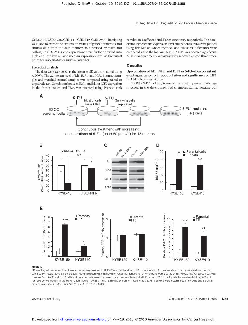
Figure 1.FR esophageal cancer sublines have increased expression of Id1, IGF2 and E2F1 and form FR tumors in vivo. A, diagram depicting the establishment of FRsublines from esophageal cancer cells. B, nudemice bearing KYSE410FR- or KYSE410-derived tumor xenograftswere treatedwith 5-FU (20mg/kg) twiceweekly for3 weeks (n ¼ 6). C and D, FR cells and parental cells were compared for expression levels of Id1, IGF2, and E2F1 in cell lysate by Western blotting (C) andfor IGF2 concentration in the conditioned medium by ELISA (D). E, mRNA expression levels of Id1, E2F1, and IGF2 were determined in FR cells and parentalcells by real-time RT-PCR. Bars, SD. �� , P < 0.01; ��� , P < 0.001.
Id1 Regulates E2F1 Degradation and Cancer Chemoresistance
www.aacrjournals.org Clin Cancer Res; 22(5) March 1, 2016 1245
on May 19, 2018. © 2016 American Association for Cancer Research. clincancerres.aacrjournals.org Downloaded from
Published OnlineFirst October 16, 2015; DOI: 10.1158/1078-0432.CCR-15-1196
previous study showed that PI3K/AKT can be activated by Id1-induced IGF2 in cancer cells (1), we hypothesized that Id1 andIGF2 may have a role in 5-FU resistance. Furthermore, as it was
reported that E2F1 expression can increase the resistance offibrosarcoma cells to 5-FU (5), we speculated that E2F1 proteinmay also be differentially expressed upon acquisition of 5-FU
Actin
Id1
TS
E2F1
KYSE150 KYSE410 KYSE150FR
Actin
Id1
TS
E2F1
KYSE410FRC
pBab
e +
shCO
NId
1 +
shCO
NId
1 +
shE2
F1#1
Id1
+ sh
E2F1
#2pB
abe
+ sh
CON
Id1
+ sh
CON
Id1
+ sh
E2F1
#1Id
1 +
shE2
F1#2
shCO
N +
pcDN
A3sh
Id1
+ pc
DNA3
shId
1 +
E2F1
shCO
N +
pcDN
A3sh
Id1
+ pc
DNA3
shId
1 +
E2F1
pcDNA3-Id1 (μg) 0 0.4 0.8 0 0.4 0.8
Id1 overexpressionKYSE150
Actin
Id1
E2F1
KYSE410
TS
0 0.4 0.8shId1 (μg)
0 0.4 0.8 siId1 (μmol/L)
Id1 knockdown
0 0.4 0.8 shId1 (μg)
0 0.4 0.8 siId1 (μmol/L)
Actin
Id1
E2F1
TS
KYSE150FR KYSE410FR
A B
0
0.5
1
1.5
2
2.5
0 0.5 1 1.5 2 2.5
Rel
ativ
e E
2F1
expr
essi
on
Relative Id1 expression
n = 100r = 0.51
P < 0.001
D
Id1
E2F1
Actin
N T N T N T N T N T N T#1 #2 #3 #4 #5 #6
****
***
0
50
100
150
Tum
or v
olum
e (%
of D
MS
O-tr
eate
d tu
mor
s)
0
40
80
120
160
Tum
or v
olum
e(%
of D
MS
O-tr
eate
d tu
mor
s)
KYSE410-pBabe-shCON
KYSE410-Id1-shCON
KYSE410-Id1-shE2F1
KYSE410FR-shCON-pcDNA3
KYSE410FR-shId1-pcDNA3
KYSE410FR-shId1-E2F1
DMSO 5-FU DMSO 5-FUF
0
20
40
60
80
100
KYSE150 KYSE410
Cel
l pro
lifer
atio
n (%
of D
MS
O-tr
eate
d ce
lls)
pBabe + shCONId1 + shCONId1 + shE2F1#1Id1 + shE2F1#2
*
*
0
20
40
60
80
100
120
KYSE150FR KYSE410FR
Cel
l pro
lifer
atio
n
(% o
f DM
SO
-trea
ted
cells
)
shCON + pcDNA3shId1 + pcDNA3shId1 + E2F1
** **
5-FU treatment 5-FU treatmentE
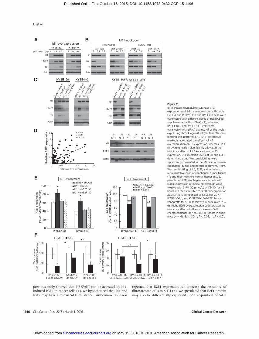
Figure 2.Id1 increases thymidylate synthase (TS)expression and 5-FU chemoresistance throughE2F1. A and B, KYSE150 and KYSE410 cells weretransfected with different doses of pcDNA3-Id1supplemented with pcDNA3 (A), whereasKYSE150FR and KYSE410FR cells weretransfected with siRNA against Id1 or the vectorexpressing shRNA against Id1 (B), then Westernblotting was performed. C, E2F1 knockdownmarkedly abrogated the effects of Id1overexpression on TS expression, whereas E2F1re-overexpression significantly alleviated theinhibitory effects of Id1 knockdown on TSexpression. D, expression levels of Id1 and E2F1,determined using Western blotting, weresignificantly correlated in the 50 pairs of humanesophageal tumor and normal specimens. Right,Western blotting of Id1, E2F1, and actin in sixrepresentative pairs of esophageal tumor tissues(T) and their matched normal tissues (N). E,parental and FR esophageal cancer cells withstable expression of indicated plasmids weretreated with 5-FU (10 mmol/L) or DMSO for 48hours and then subjected to BrdUrd incorporationassay. F, left, comparison of KYSE410-CON,KYSE410-Id1, and KYSE410-Id1-shE2F1 tumorxenografts for 5-FU sensitivity in nude mice (n ¼6). Right, E2F1 overexpression counteracted theinhibitory effect of Id1 knockdown on 5-FUchemoresistance of KYSE410FR tumors in nudemice (n ¼ 6). Bars, SD. � , P < 0.05; �� , P < 0.01.
Li et al.
Clin Cancer Res; 22(5) March 1, 2016 Clinical Cancer Research1246
on May 19, 2018. © 2016 American Association for Cancer Research. clincancerres.aacrjournals.org Downloaded from
Published OnlineFirst October 16, 2015; DOI: 10.1158/1078-0432.CCR-15-1196
E
A
0
20
40
60
80
100
120
0 2 4 6 8
E2F
1 pr
otei
n (%
)
Time after CHX treatment (h)
KYSE150-pBabeKYSE150-Id1
0
20
40
60
80
100
120
0 2 4 6 8
E2F
1 pr
otei
n (%
)
Time after CHX treatment (h)
KYSE410-pBabeKYSE410-Id1
Actin
Id1
E2F1
KYSE150-pBabe KYSE150-Id1CHX (h) 0 1 2 4 6 8 0 1 2 4 6 8
Actin
Id1
E2F1
KYSE410-pBabe KYSE410-Id1CHX (h) 0 1 2 4 6 8 0 1 2 4 6 8
E2F1 E2F1
E2F1
Cdc20
Cdc20Id1
Id1 Id1
Degradation Function
IGF2 IGF2
OFF ON
Without Id1 With Id1
B C
1 2 3 4 5 6 7 8 9
Flag-Cdc20 + + – + + –HA-Id1 – + + – + +
Input IP: Flag
WB: Flag (Cdc20)
WB: HA (Id1)
+ + –– + +
IP: IgG
D
1 2 3 4 5 6
Flag -Cdc20 + + + + + +HA-E2F1 + + + + + +
Input IP: Flag
WB: Flag (Cdc20)
WB: HA (Id1)
HA-Id1 – + – +
WB: HA (E2F1)
– +
IP: IgG
Flag-Cdc20 + + – + + –HA-E2F1 – + + – + +
Input IP: Flag
WB: Flag (Cdc20)
WB: HA (E2F1)
1 2 3 4 5 6 7 8 9
+ + –– + +
IP: IgG
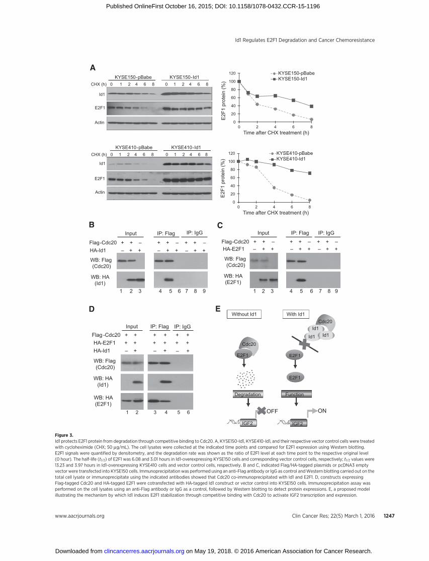
Figure 3.Id1 protects E2F1 protein from degradation through competitive binding to Cdc20. A, KYSE150-Id1, KYSE410-Id1, and their respective vector control cells were treatedwith cycloheximide (CHX; 50 mg/mL). The cell lysates were collected at the indicated time points and compared for E2F1 expression using Western blotting.E2F1 signals were quantified by densitometry, and the degradation rate was shown as the ratio of E2F1 level at each time point to the respective original level(0 hour). The half-life (t1/2) of E2F1 was 6.08 and 3.01 hours in Id1-overexpressing KYSE150 cells and corresponding vector control cells, respectively; t1/2 values were13.23 and 3.97 hours in Id1-overexpressing KYSE410 cells and vector control cells, respectively. B and C, indicated Flag/HA-tagged plasmids or pcDNA3 emptyvector were transfected into KYSE150 cells. Immunoprecipitationwas performed using an anti-Flag antibody or IgG as control andWestern blotting carried out on thetotal cell lysate or immunoprecipitate using the indicated antibodies showed that Cdc20 co-immunoprecipitated with Id1 and E2F1. D, constructs expressingFlag-tagged Cdc20 and HA-tagged E2F1 were cotransfected with HA-tagged Id1 construct or vector control into KYSE150 cells. Immunoprecipitation assay wasperformed on the cell lysates using an anti-Flag antibody or IgG as a control, followed by Western blotting to detect protein expressions. E, a proposed modelillustrating the mechanism by which Id1 induces E2F1 stabilization through competitive binding with Cdc20 to activate IGF2 transcription and expression.
Id1 Regulates E2F1 Degradation and Cancer Chemoresistance
www.aacrjournals.org Clin Cancer Res; 22(5) March 1, 2016 1247
on May 19, 2018. © 2016 American Association for Cancer Research. clincancerres.aacrjournals.org Downloaded from
Published OnlineFirst October 16, 2015; DOI: 10.1158/1078-0432.CCR-15-1196
pcDNA3-E2F1 (μg) 0 0.4 0.8 0 0.4 0.8
E2F1 overexpressionKYSE150 KYSE410
E2F1 overexpression E2F1 knockdown
0 0.4 0.8 (μg) shE2F1#1
0 0.4 0.8 shE2F1#2
E2F1 knockdownKYSE270
0 0.4 0.8 shE2F1#1
0 0.4 0.8 shE2F1#2
T.Tn
Actin
E2F1
IGF2
(μg) (μg)
E2F1
IGF2
Actin
A
0
1
2
3
4
5
6
7
Rel
ativ
e IG
F2 m
RN
A ex
pres
sion
*
*****
**
pcDNA3-E2F1 (μg) 0 0.4 0.8 0 0.4 0.8
KYSE150 KYSE410
0
20
40
60
80
100
120
Rel
ativ
e IG
F2 m
RN
A ex
pres
sion KYSE270
0 0.4 0.8 shE2F1#1 (μg)
*******
**
0
20
40
60
80
100
120
0 0.4 0.8 shE2F1#2 (μg)
0 0.4 0.8 shE2F1#1 (μg)
0 0.4 0.8 shE2F1#2 (μg)
*
*****
**
T.Tn
C
E
0.0
1.0
2.0
3.0
4.0
5.0
BS1 BS2 BS3
% in
put
IgGAnti-E2F1
(μg)
Actin
Id1
IGF2
E2F1
KYSE150 KYSE410 T.Tn
Actin
Id1
IGF2
E2F1
KYSE270
**
**R
elat
ive
IGF2
mR
NA
expr
essi
on
0
1
2
3
4
5
6
7
WT Mutant 1 Mutant 2 Mutant 3
Rel
ativ
e lu
cife
rase
act
ivity
pcDNA3E2F1*** * **
WT GGCGCGGA
GGCGCTGTACATTTCCA
AGATATCGA
GGCGCGGGGG
GGCCCGCGGG
BS1 BS2 BS3 Luc
Mutation
B
D
IGF2
pro
mot
er b
indi
ng
pcDNA3-E2F1 (μg)
F
TranscriptionE2F1 E2F1 E2F1
TSSBS1 BS2 BS3
Li et al.
Clin Cancer Res; 22(5) March 1, 2016 Clinical Cancer Research1248
on May 19, 2018. © 2016 American Association for Cancer Research. clincancerres.aacrjournals.org Downloaded from
Published OnlineFirst October 16, 2015; DOI: 10.1158/1078-0432.CCR-15-1196
chemoresistance. We therefore made use of 5-FU–resistant sub-lines (designated KYSE150FR and KYSE410FR) which were estab-lished from ESCC cell lines KYSE150 and KYSE410 throughcontinuous treatment with increasing doses of 5-FU (from 1.25to 80 mmol/L) for over 18 months (Fig. 1A) as cell models to testour hypothesis. The proliferation rate and migration ability of FRcells were similar or slightly higher than parental cells (Supple-mentary Fig. S1). Tumor xenografts thatwerederived fromFR cellswere confirmed to exhibit robust resistance to 5-FU in vivo (Fig.1B). Comparison of the FR cell lines and their parental cell linesshowed upregulation of Id1, IGF2, and E2F1 protein expression(Fig. 1C), as well as increased secretion of IGF2 in the FR cells (Fig.1D). Increased mRNA expression levels of Id1 and IGF2, but notE2F1, were observed in the FR cells (Fig. 1E). ESCC cells with E2F1overexpression or knockdown were treated with 5-FU and thencell proliferationwasmeasured. As expected, ectopic expressionofE2F1 increased thymidylate synthase expression and 5-FU che-moresistance, whereas repressed expression of E2F1 had theopposite effects (Supplementary Fig. S2). These findings stronglysupport the rationale of using these FR sublines as cell models foridentifying chemoresistance-associated genes and for studying theroles of Id1 and IGF2 in regulating 5-FU chemoresistance in ESCC.
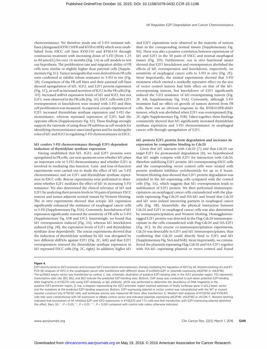
Id1 confers 5-FU chemoresistance through E2F1-dependentinduction of thymidylate synthase expression
Having established that Id1, IGF2, and E2F1 proteins wereupregulated in FR cells, our next questions werewhether Id1 playsan important role in 5-FU chemoresistance and whether E2F1 isinvolved in mediating this function. Gain- and loss-of-functionexperiments were carried out to study the effect of Id1 on 5-FUchemoresistance and on E2F1 and thymidylate synthase expres-sion in ESCC cells. Rescue experiments were performed to deter-mine whether E2F1 mediates the effect of Id1 in increasing 5-FUresistance. We also determined the clinical relevance of Id1 andE2F1 by analyzing their protein levels in 50 pairs of primary ESCCtumors and tumor-adjacent normal tissues by Western blotting.The in vitro experiments showed that ectopic Id1 expressionsignificantly enhanced the resistance of esophageal cancer cellsto 5-FU (Supplementary Fig. S3A). Conversely, knockdown of Id1expression significantly restored the sensitivity of FR cells to 5-FU(Supplementary Fig. S3B and S3C). Interestingly, we found thatId1 overexpression induced (Fig. 2A), whereas Id1 knockdownreduced (Fig. 2B), the expression levels of E2F1 and thymidylatesynthase dose dependently. The rescue experiments showed thatthe induction of thymidylate synthase by Id1 was abrogated bytwo different shRNAs against E2F1 (Fig. 2C, left) and that E2F1overexpression restored the thymidylate synthase expression inId1-repressed ESCC cells (Fig. 2C, right). In addition, higher Id1
and E2F1 expressions were observed in the majority of tumorsthan in the corresponding normal tissues (Supplementary Fig.S4). There was also a positive correlation between expressions ofId1 and E2F1 in the 50 pairs of ESCC and normal esophagealtissues (Fig. 2D). Furthermore, our in vitro functional assaysshowed that E2F1 knockdown and overexpression abolished theeffects of Id1 overexpression and knockdown, respectively, onsensitivity of esophageal cancer cells to 5-FU in vitro (Fig. 2E).More importantly, the animal experiments showed that 5-FUtreatment which exerted a markedly repressive effect on the sizeof vector control tumors had little effect on that of the Id1-overexpressing tumors, but knockdown of E2F1 significantlyreduced the 5-FU resistance of Id1-overexpressing tumors (Fig.2F, left; Supplementary Fig. S5A). Conversely, although 5-FUtreatment had no effect on growth of tumors derived from FRcells, there was an obvious response in the KYSE410FR-shId1tumors, which was abolished when E2F1 was overexpressed (Fig.2F, right; Supplementary Fig. S5B). Taken together, these findingsconsistently showed that Id1 significantly increased thymidylatesynthase expression and 5-FU chemoresistance in esophagealcancer cells through upregulation of E2F1.
Id1 protects E2F1 protein from degradation and increases itsexpression by competitive binding to Cdc20
Given that Id1 interacts with Cdc20 (7) and that Cdc20 cantarget E2F1 for proteasomal degradation (8), we hypothesizedthat Id1 might compete with E2F1 for interaction with Cdc20,therefore stabilizing E2F1 protein. Id1-overexpressing ESCC cellsand the corresponding vector control cells were treated withprotein synthesis inhibitor cycloheximide for up to 8 hours.Western blotting data showed that E2F1 protein degradation wasretarded in the Id1-expressing cells compared with the controlcells (Fig. 3A), which suggests that Id1 overexpression leads tostabilization of E2F1 protein. We then performed immunopre-cipitation on esophageal cancer cells cotransfected with the plas-mids expressing Flag-Cdc20 and HA-Id1 and found that Cdc20and Id1 were indeed interacting partners in esophageal cancercells (Fig. 3B). Meanwhile, the physical interaction betweenCdc20 and E2F1 in esophageal cancer cells was also determinedby immunoprecipitation and Western blotting. Hemagglutinin-tagged E2F1 protein was detected in the Flag-Cdc20 immunopre-cipitate in the cells cotransfected with Flag-Cdc20 and HA-E2F1(Fig. 3C). In the reverse co-immunoprecipitation experiments,Cdc20 was detectable in E2F1 and Id1 immunoprecipitates, thusconfirming that Cdc20 could directly bind to E2F1 and Id1(Supplementary Fig. S6A andS6B).More importantly,we cotrans-fected the plasmids expressing Flag-Cdc20 and HA-E2F1 togetherwith HA-Id1–expressing plasmid or vector control and found
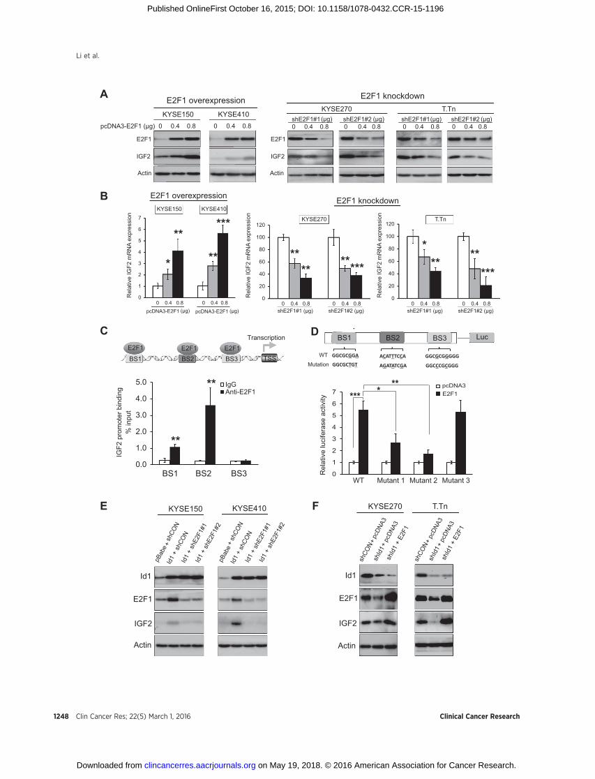
Figure 4.E2F1 directly binds to IGF2 promoter and increases IGF2 transcription and expression, thereby mediating the regulation of IGF2 by Id1. Western blotting (A) and RT-PCR (B) analyses of IGF2 in the esophageal cancer cells transfected with different doses of pcDNA3-E2F1 or plasmids expressing shE2F1#1 or shE2F1#2.The pcDNA3 empty vector was transfected as control. C, top, schematic illustration of putative E2F1-binding sites in the IGF2 promoter region. TSS representstranscription start site. BS1, BS2, and BS3 indicate the predicted E2F1-binding sites. Bottom, ChIP assay was conducted to pull-down potential E2F1-bindingDNA fragments in KYSE270 cells using E2F1 antibody or IgG antibody. qPCR was performed to determine the abundance of DNA fragments in theputative IGF2 promoter region. D, top, a diagram representing the IGF2 promoter region inserted upstream of firefly luciferase gene in pGL2-basic vectorand the mutations at the predicted E2F1-binding sequences. Bottom, E2F1-expressing plasmid or vector control was cotransfected with the WT or mutantreporter construct into KYSE150 cells, and luciferase activity was measured 48 hours after transfection. E, Western blot analyses of KYSE150 and KYSE410cells that were cotransfected with Id1 expression or pBabe control vector and indicated plasmids expressing shE2F1#1, shE2F1#2 or shCON. F, Western blottingindicated that knockdown of Id1 inhibited E2F1 and IGF2 expressions in KYSE270 and T.Tn cells and that transfection with E2F1-expressing plasmid abolishedthis effect. Bars, SD. � , P < 0.05; �� , P < 0.01; ��� , P < 0.001 compared with control cells unless otherwise indicated.
Id1 Regulates E2F1 Degradation and Cancer Chemoresistance
www.aacrjournals.org Clin Cancer Res; 22(5) March 1, 2016 1249
on May 19, 2018. © 2016 American Association for Cancer Research. clincancerres.aacrjournals.org Downloaded from
Published OnlineFirst October 16, 2015; DOI: 10.1158/1078-0432.CCR-15-1196
CE2F1 IGF2
Case 1
Case 2
–2–1
01234567
Log
2 IG
F2 e
xpre
ssio
n (n
orm
aliz
ed to
nor
mal
tiss
ue)
0
0.5
1
1.5
2
2.5
3
3.5
Normal Tumor
IGF2
exp
ress
ion
B
0
0.5
1
1.5
2
2.5
3
0 0.5 1 1.5 2 2.5
IGF2
exp
ress
ion
E2F1 expression
n = 100r = 0.64P < 0.001
Actin
E2F1
IGF2
N T N T N T N T N T N T#1 #2 #3 #4 #5 #6
80.0% (40/50)Paired t-test, P = 6.8e-10
n = 50P < 0.001
Weak StrongWeak 9 5 14Strong 4 17 21
13 22 35
E2F1Total
IGF2
Total( P = 0.012)
Immunostaining intensity of E2F1 and IGF2 in ESCC TMA
4
6
8
10
12
14
16
18
5 7 9 11 13
IGF2
exp
ress
ion
E2F1 expression
n = 124r = 0.658P < 0.001
4
6
8
10
12
14
5 6 7 8 9 10
IGF2
exp
ress
ion
n = 112r = 0.225P < 0.05
E2F1 expression
ESCC patients EAC patientsD
A
E2F1 expression
Colon cancer patients
–6
–4
–2
0
2
4
–3 –2 –1 0 1
n = 211r = 0.459P < 0.001
IGF2
exp
ress
ion
2
5
8
11
14
17
20
3 5 7 9 11 13 15
IGF2
exp
ress
ion
n = 256r = 0.226P < 0.001
E2F1 expression
HCC patients
2
4
6
8
10
12
14
16
6 7 8 9 10
IGF2
exp
ress
ion
n = 111r = 0.216P < 0.05
E2F1 expression
Lung cancer patients
2
4
6
8
10
12
14
3 5 7 9
IGF2
exp
ress
ion
n = 234r = 0.748
P < 0.001
E2F1 expression
Breast cancer patients
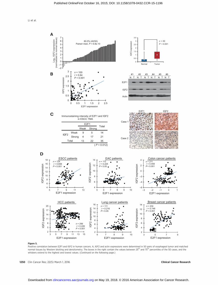
Figure 5.Positive correlation between E2F1 and IGF2 in human cancers. A, IGF2 and actin expressions were determined in 50 pairs of esophageal tumor and matchednormal tissues by Western blotting and densitometry. The boxes in the right contain the values between 25th and 75th percentiles of the 50 cases, and thewhiskers extend to the highest and lowest values. (Continued on the following page.)
Clin Cancer Res; 22(5) March 1, 2016 Clinical Cancer Research1250
Li et al.
on May 19, 2018. © 2016 American Association for Cancer Research. clincancerres.aacrjournals.org Downloaded from
Published OnlineFirst October 16, 2015; DOI: 10.1158/1078-0432.CCR-15-1196
significantly lower E2F1 level in the Flag-Cdc20 immunoprecip-itate of the Id1 transfectants (Fig. 3D, lane 4 vs. lane 3), indicatingthat Id1–Cdc20 interaction inhibited the association betweenCdc20 and E2F1. Similar results were observed when the cellswere treated with 5-FU (Supplementary Fig. S6C). On the otherhand, immunoprecipitation assay failed to reveal any interactionbetween Id1 and E2F1 in either ESCC parental cells or FR cells(Supplementary Fig. S7). Our results collectively demonstratedthat Id1 could protect E2F1 protein degradation and increase itsexpression by competitive binding to Cdc20, as illustratedin Fig. 3E.
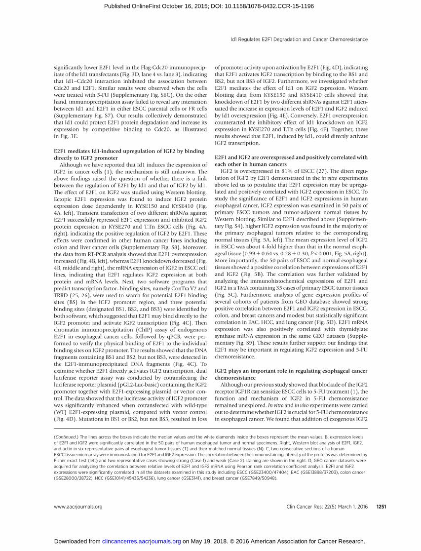
E2F1 mediates Id1-induced upregulation of IGF2 by bindingdirectly to IGF2 promoter
Although we have reported that Id1 induces the expression ofIGF2 in cancer cells (1), the mechanism is still unknown. Theabove findings raised the question of whether there is a linkbetween the regulation of E2F1 by Id1 and that of IGF2 by Id1.The effect of E2F1 on IGF2 was studied using Western blotting.Ectopic E2F1 expression was found to induce IGF2 proteinexpression dose dependently in KYSE150 and KYSE410 (Fig.4A, left). Transient transfection of two different shRNAs againstE2F1 successfully repressed E2F1 expression and inhibited IGF2protein expression in KYSE270 and T.Tn ESCC cells (Fig. 4A,right), indicating the positive regulation of IGF2 by E2F1. Theseeffects were confirmed in other human cancer lines includingcolon and liver cancer cells (Supplementary Fig. S8). Moreover,the data from RT-PCR analysis showed that E2F1 overexpressionincreased (Fig. 4B, left), whereas E2F1 knockdown decreased (Fig.4B, middle and right), the mRNA expression of IGF2 in ESCC celllines, indicating that E2F1 regulates IGF2 expression at bothprotein and mRNA levels. Next, two software programs thatpredict transcription factor–binding sites, namely ConTra V2 andTRRD (25, 26), were used to search for potential E2F1-bindingsites (BS) in the IGF2 promoter region, and three potentialbinding sites (designated BS1, BS2, and BS3) were identified byboth software, which suggested that E2F1may bind directly to theIGF2 promoter and activate IGF2 transcription (Fig. 4C). Thenchromatin immunoprecipitation (ChIP) assay of endogenousE2F1 in esophageal cancer cells, followed by qPCR, were per-formed to verify the physical binding of E2F1 to the individualbinding sites on IGF2 promoter. The results showed that the DNAfragments containing BS1 and BS2, but not BS3, were detected inthe E2F1-immunoprecipitated DNA fragments (Fig. 4C). Toexamine whether E2F1 directly activates IGF2 transcription, dualluciferase reporter assay was conducted by cotransfecting theluciferase reporter plasmid (pGL2-Luc-basic) containing the IGF2promoter together with E2F1-expressing plasmid or vector con-trol. The data showed that the luciferase activity of IGF2 promoterwas significantly enhanced when cotransfected with wild-type(WT) E2F1-expressing plasmid, compared with vector control(Fig. 4D). Mutations in BS1 or BS2, but not BS3, resulted in loss
of promoter activity upon activation by E2F1 (Fig. 4D), indicatingthat E2F1 activates IGF2 transcription by binding to the BS1 andBS2, but not BS3 of IGF2. Furthermore, we investigated whetherE2F1 mediates the effect of Id1 on IGF2 expression. Westernblotting data from KYSE150 and KYSE410 cells showed thatknockdown of E2F1 by two different shRNAs against E2F1 atten-uated the increase in expression levels of E2F1 and IGF2 inducedby Id1 overexpression (Fig. 4E). Conversely, E2F1 overexpressioncounteracted the inhibitory effect of Id1 knockdown on IGF2expression in KYSE270 and T.Tn cells (Fig. 4F). Together, theseresults showed that E2F1, induced by Id1, could directly activateIGF2 transcription.
E2F1 and IGF2 are overexpressed and positively correlated witheach other in human cancers
IGF2 is overexpressed in 81% of ESCC (27). The direct regu-lation of IGF2 by E2F1 demonstrated in the in vitro experimentsabove led us to postulate that E2F1 expression may be upregu-lated and positively correlated with IGF2 expression in ESCC. Tostudy the significance of E2F1 and IGF2 expressions in humanesophageal cancer, IGF2 expression was examined in 50 pairs ofprimary ESCC tumors and tumor-adjacent normal tissues byWestern blotting. Similar to E2F1 described above (Supplemen-tary Fig. S4), higher IGF2 expression was found in the majority ofthe primary esophageal tumors relative to the correspondingnormal tissues (Fig. 5A, left). The mean expression level of IGF2in ESCC was about 4-fold higher than that in the normal esoph-ageal tissue (0.99� 0.64 vs. 0.28� 0.30; P < 0.001; Fig. 5A, right).More importantly, the 50 pairs of ESCC and normal esophagealtissues showed a positive correlation between expressions of E2F1and IGF2 (Fig. 5B). The correlation was further validated byanalyzing the immunohistochemical expressions of E2F1 andIGF2 in a TMA containing 35 cases of primary ESCC tumor tissues(Fig. 5C). Furthermore, analysis of gene expression profiles ofseveral cohorts of patients from GEO database showed strongpositive correlation between E2F1 and IGF2 expression in ESCC,colon, and breast cancers and modest but statistically significantcorrelation in EAC, HCC, and lung cancer (Fig. 5D). E2F1 mRNAexpression was also positively correlated with thymidylatesynthase mRNA expression in the same GEO datasets (Supple-mentary Fig. S9). These results further support our findings thatE2F1 may be important in regulating IGF2 expression and 5-FUchemoresistance.
IGF2 plays an important role in regulating esophageal cancerchemoresistance
Although our previous study showed that blockade of the IGF2receptor IGF1R can sensitize ESCC cells to 5-FU treatment (1), thefunction and mechanism of IGF2 in 5-FU chemoresistanceremained unexplored. In vitro and in vivo experiments were carriedout todeterminewhether IGF2 is crucial for 5-FU chemoresistancein esophageal cancer. We found that addition of exogenous IGF2
(Continued.) The lines across the boxes indicate the median values and the white diamonds inside the boxes represent the mean values. B, expression levelsof E2F1 and IGF2 were significantly correlated in the 50 pairs of human esophageal tumor and normal specimens. Right, Western blot analysis of E2F1, IGF2,and actin in six representative pairs of esophageal tumor tissues (T) and their matched normal tissues (N). C, two consecutive sections of a humanESCC tissuemicroarraywere immunostained for E2F1 and IGF2 expression. The correlation between the immunostaining intensity of the proteinswas determined byFisher exact test (left) and two representative cases showing strong (Case 1) and weak (Case 2) staining are shown in the right. D, GEO cancer datasets wereacquired for analyzing the correlation between relative levels of E2F1 and IGF2 mRNA using Pearson rank correlation coefficient analysis. E2F1 and IGF2expressions were significantly correlated in all the datasets examined in this study including ESCC (GSE23400/47404), EAC (GSE13898/37203), colon cancer(GSE28000/28722), HCC (GSE10141/45436/54236), lung cancer (GSE3141), and breast cancer (GSE7849/50948).
www.aacrjournals.org Clin Cancer Res; 22(5) March 1, 2016 1251
Id1 Regulates E2F1 Degradation and Cancer Chemoresistance
on May 19, 2018. © 2016 American Association for Cancer Research. clincancerres.aacrjournals.org Downloaded from
Published OnlineFirst October 16, 2015; DOI: 10.1158/1078-0432.CCR-15-1196
A
Actin
Caspase-3
Cleaved caspase-3
DMSOIGF2
5-FUKYSE150FR
DMSOIGF2
5-FUKYSE410FR
0
20
40
60
80
100
120
140
Tum
or v
olum
e(%
of D
MS
O-tr
eate
d tu
mor
s)
KYSE410FR-shCON
KYSE410FR-shIGF2#2
DMSO 5-FU**
KYSE150FR KYSE410FR
Actin
p-AKT
IGF2
AKT
TS
B
D
C
Id1 E2F1 IGF2 AKT TS Increased 5-FU chemoresistance
Lung cancer patients
Log Rank = 9.853P = 0.002
Cum
ulat
ive
surv
ival
rate
Months
Log Rank = 4.880P = 0.027
Cum
ulat
ive
surv
ival
rate
Months
ESCC patients
Breast cancer patients
Log Rank = 4.945P = 0.026
Cum
ulat
ive
surv
ival
rate
Log Rank = 6.534P = 0.011
Colon cancer patients
Months
Months
HCC patients
Log Rank = 4.594P = 0.032
Cum
ulat
ive
surv
ival
rate
Cum
ulat
ive
surv
ival
rate
Months
E F
Id1low/ww IGF2low
Id1high/h IGF2high
Id1low/ww IGF2low Id1low/ww IGF2low
Id1low/ww IGF2low
Id1low/ww IGF2low
Id1high/hh IGF2high Id1high/h IGF2high
Id1high/h IGF2high
Id1high/h IGF2high
0
20
40
60
80
100
120
KYSE150FR KYSE410FR C
ell p
rolif
erat
ion
(%
of D
MS
O-tr
eate
d ce
lls)
** ******
0
20
40
60
80
100
KYSE150 KYSE410
Cel
l pro
lifer
atio
n
(% o
f DM
SO-tr
eate
d ce
lls)
0
20
40
60
80
100
120
KYSE150 KYSE410
Cel
l pro
lifer
atio
n
(% o
f DM
SO-tr
eate
d ce
lls)
pBabe + shCONId1 + shCONId1 + shIGF2#2Id1 + IGF2 Ab
pcDNA3 + shCONE2F1 + shCONE2F1 + shIGF2#2E2F1 + IGF2 Ab
* **
* **
** **
** **
5-FU treatment 5-FU treatment
5-FU treatmentshCONshIGF2#1shIGF2#2 shIGF2#1 + IGF2shIGF2#2 + IGF2
Clin Cancer Res; 22(5) March 1, 2016 Clinical Cancer Research1252
Li et al.
on May 19, 2018. © 2016 American Association for Cancer Research. clincancerres.aacrjournals.org Downloaded from
Published OnlineFirst October 16, 2015; DOI: 10.1158/1078-0432.CCR-15-1196
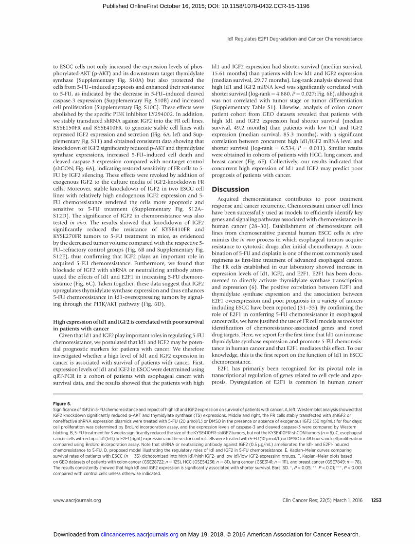
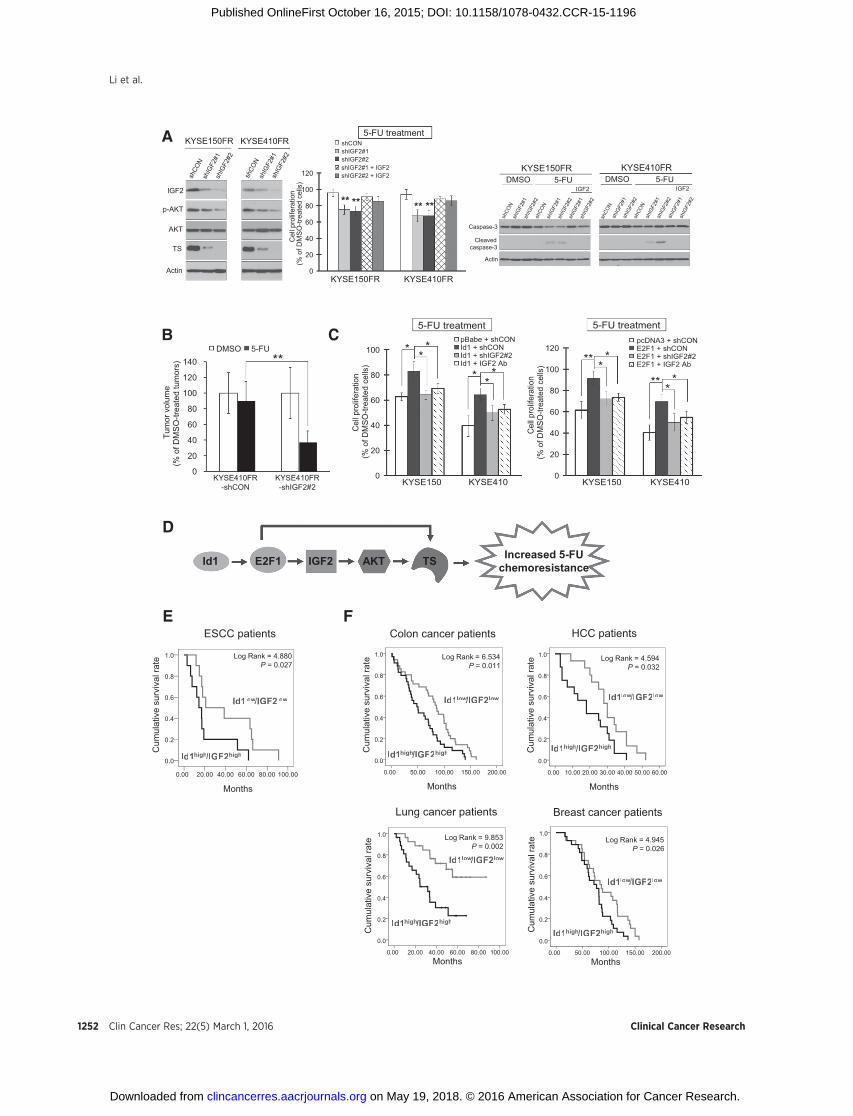
to ESCC cells not only increased the expression levels of phos-phorylated-AKT (p-AKT) and its downstream target thymidylatesynthase (Supplementary Fig. S10A) but also protected thecells from 5-FU–induced apoptosis and enhanced their resistanceto 5-FU, as indicated by the decrease in 5-FU–induced cleavedcaspase-3 expression (Supplementary Fig. S10B) and increasedcell proliferation (Supplementary Fig. S10C). These effects wereabolished by the specific PI3K inhibitor LY294002. In addition,we stably transduced shRNA against IGF2 into the FR cell lines,KYSE150FR and KYSE410FR, to generate stable cell lines withrepressed IGF2 expression and secretion (Fig. 6A, left and Sup-plementary Fig. S11) and obtained consistent data showing thatknockdown of IGF2 significantly reduced p-AKT and thymidylatesynthase expressions, increased 5-FU–induced cell death andcleaved caspase-3 expression compared with nontarget control(shCON; Fig. 6A), indicating restored sensitivity of FR cells to 5-FU by IGF2 silencing. These effects were revoked by addition ofexogenous IGF2 to the culture media of IGF2-knockdown FRcells. Moreover, stable knockdown of IGF2 in two ESCC celllines with relatively high endogenous IGF2 expression and 5-FU chemoresistance rendered the cells more apoptotic andsensitive to 5-FU treatment (Supplementary Fig. S12A–S12D). The significance of IGF2 in chemoresistance was alsotested in vivo. The results showed that knockdown of IGF2significantly reduced the resistance of KYSE410FR andKYSE270FR tumors to 5-FU treatment in mice, as evidencedby the decreased tumor volume compared with the respective 5-FU–refractory control groups (Fig. 6B and Supplementary Fig.S12E), thus confirming that IGF2 plays an important role inacquired 5-FU chemoresistance. Furthermore, we found thatblockade of IGF2 with shRNA or neutralizing antibody atten-uated the effects of Id1 and E2F1 in increasing 5-FU chemore-sistance (Fig. 6C). Taken together, these data suggest that IGF2upregulates thymidylate synthase expression and thus enhances5-FU chemoresistance in Id1-overexpressing tumors by signal-ing through the PI3K/AKT pathway (Fig. 6D).
High expressionof Id1 and IGF2 is correlatedwithpoor survivalin patients with cancer
Given that Id1 and IGF2play important roles in regulating 5-FUchemoresistance, we postulated that Id1 and IGF2 may be poten-tial prognostic markers for patients with cancer. We thereforeinvestigated whether a high level of Id1 and IGF2 expression incancer is associated with survival of patients with cancer. First,expression levels of Id1 and IGF2 in ESCCwere determined usingqRT-PCR in a cohort of patients with esophageal cancer withsurvival data, and the results showed that the patients with high
Id1 and IGF2 expression had shorter survival (median survival,15.61 months) than patients with low Id1 and IGF2 expression(median survival, 29.77 months). Log-rank analysis showed thathigh Id1 and IGF2 mRNA level was significantly correlated withshorter survival (log-rank¼ 4.880, P¼ 0.027; Fig. 6E), although itwas not correlated with tumor stage or tumor differentiation(Supplementary Table S1). Likewise, analysis of colon cancerpatient cohort from GEO datasets revealed that patients withhigh Id1 and IGF2 expression had shorter survival (mediansurvival, 49.2 months) than patients with low Id1 and IGF2expression (median survival, 85.3 months), with a significantcorrelation between concurrent high Id1/IGF2 mRNA level andshorter survival (log-rank ¼ 6.534, P ¼ 0.011). Similar resultswere obtained in cohorts of patients with HCC, lung cancer, andbreast cancer (Fig. 6F). Collectively, our results indicated thatconcurrent high expression of Id1 and IGF2 may predict poorprognosis of patients with cancer.
DiscussionAcquired chemoresistance contributes to poor treatment
response and cancer recurrence. Chemoresistant cancer cell lineshave been successfully used as models to efficiently identify keygenes and signaling pathways associated with chemoresistance inhuman cancer (28–30). Establishment of chemoresistant celllines from chemosensitive parental human ESCC cells in vitromimics the in vivo process in which esophageal tumors acquireresistance to cytotoxic drugs after initial chemotherapy. A com-bination of 5-FU and cisplatin is one of themost commonly usedregimens as first-line treatment of advanced esophageal cancer.The FR cells established in our laboratory showed increase inexpression levels of Id1, IGF2, and E2F1. E2F1 has been docu-mented to directly activate thymidylate synthase transcriptionand expression (6). The positive correlation between E2F1 andthymidylate synthase expression and the association betweenE2F1 overexpression and poor prognosis in a variety of cancersincluding ESCC have been reported (31–33). By confirming therole of E2F1 in conferring 5-FU chemoresistance in esophagealcancer cells, we have justified the use of FR cell models as tools foridentification of chemoresistance-associated genes and noveldrug targets. Here, we report for the first time that Id1 can increasethymidylate synthase expression and promote 5-FU chemoresis-tance in human cancer and that E2F1 mediates this effect. To ourknowledge, this is the first report on the function of Id1 in ESCCchemoresistance.
E2F1 has primarily been recognized for its pivotal role intranscriptional regulation of genes related to cell cycle and apo-ptosis. Dysregulation of E2F1 is common in human cancer
Figure 6.Significance of IGF2 in 5-FU chemoresistance and impact of high Id1 and IGF2 expression on survival of patientswith cancer. A, left,Western blot analysis showed thatIGF2 knockdown significantly reduced p-AKT and thymidylate synthase (TS) expressions. Middle and right, the FR cells stably transfected with shIGF2 ornoneffective shRNA expression plasmids were treated with 5-FU (20 mmol/L) or DMSO in the presence or absence of exogenous IGF2 (50 ng/mL) for four days;cell proliferation was determined by BrdUrd incorporation assay, and the expression levels of caspase-3 and cleaved caspase-3 were compared by Westernblotting. B, 5-FU treatment for 3weeks significantly reduced the size of theKYSE410FR-shIGF2 tumors, but not theKYSE410FR-shCON tumors (n¼6). C, esophagealcancer cellswith ectopic Id1 (left) or E2F1 (right) expressionand the vector control cellswere treatedwith 5-FU (10mmol/L) orDMSO for 48hours andcell proliferationcompared using BrdUrd incorporation assay. Note that shRNA or neutralizing antibody against IGF2 (0.5 mg/mL) ameliorated the Id1- and E2F1-inducedchemoresistance to 5-FU. D, proposed model illustrating the regulatory roles of Id1 and IGF2 in 5-FU chemoresistance. E, Kaplan–Meier curves comparingsurvival rates of patients with ESCC (n ¼ 35) dichotomized into high Id1/high IGF2- and low Id1/low IGF2-expressing groups. F, Kaplan–Meier plots basedon GEO datasets of patients with colon cancer (GSE28722; n¼ 125), HCC (GSE54236; n¼ 81), lung cancer (GSE3141; n¼ 111), and breast cancer (GSE7849; n¼ 78).The results consistently showed that high Id1 and IGF2 expression is significantly associated with shorter survival. Bars, SD. � , P < 0.05; �� , P < 0.01; ��� , P < 0.001compared with control cells unless otherwise indicated.
www.aacrjournals.org Clin Cancer Res; 22(5) March 1, 2016 1253
Id1 Regulates E2F1 Degradation and Cancer Chemoresistance
on May 19, 2018. © 2016 American Association for Cancer Research. clincancerres.aacrjournals.org Downloaded from
Published OnlineFirst October 16, 2015; DOI: 10.1158/1078-0432.CCR-15-1196
including esophageal cancer (34), but amplification of E2F1 incancer is rare. As in the case formany transcription factors, E2F1 ismainly regulated by posttranslational modification. The pRbprotein, which functionally inactivates E2F1 on one hand butprotects it from degradation on the other, was thought to be themost crucial regulator of E2F1 (35). However, after dissociationfrom pRb, interaction with other proteins may be vital for thestability of E2F1 protein. In this study, the gain- and loss-of-function experiments showed that ectopic Id1 expressioninduced, whereas Id1 knockdown reduced, the expression ofE2F1 in multiple cancer cell lines, thus strongly suggesting thatId1 can regulate E2F1. Our results from cycloheximide chase andimmunoprecipitation experiments give novel insight into theregulation of E2F1 by providing the first evidence that Id1 com-petes with E2F1 for Cdc20 binding, thereby protecting E2F1 fromCdc20-mediated degradation. As discussed below, our data alsorevealed that this mechanism plays an important role in upregu-lating IGF2 in esophageal cancer.
Overexpression of IGF2 and its clinical significance in humancancer is well documented (36–38). Increased IGF2 expression inTaxol-resistant ovarian cancer cell line and the feasibility of IGF2as a potential therapeutic target in Taxol-resistant ovarian cancerhave been validated recently (39–41), but the functional role ofIGF2 in 5-FU chemoresistance has not been elucidated. We foundfor the first time that IGF2 can significantly increase, whereasknockdown of IGF2 can decrease, thymidylate synthase expres-sion. E2F1 is an important target of chemotherapeutic drugs, andaberrant expression of thymidylate synthase is significantly asso-ciated with the resistance of tumors to chemotherapy (42, 43).Our data showed that both intrinsic and acquired 5-FU chemore-sistance of ESCC cells could be achieved by knocking down IGF2to reduce thymidylate synthase expression. In addition, our in vitroand in vivo data from gain- and loss-of-function experimentsprovide novel evidence to support that IGF2 plays an importantrole in mediating the effects of Id1 in regulating the sensitivity ofcancer cells to 5-FU. We recently reported that Id1 induces IGF2expression and secretion (1), but the molecular mechanisms bywhich Id1 regulates IGF2 are still unknown. In this study, usingChIP, dual luciferase reporter, and rescue assays, we show for thefirst time that E2F1 mediates the positive regulation of Id1 onIGF2 by directly binding to the IGF2 promoter, thereby activatingIGF2 transcription and expression.
Overall, our results suggest that besides directly inducing thetranscription and expression of thymidylate synthase, there existsa parallel mechanism in which Id1 and E2F1 can indirectlyupregulate thymidylate synthase by transcriptional activation ofIGF2, thus engaging the PI3K/AKT pathway in mediating 5-FUchemoresistance. The strong positive correlation between Id1 andE2F1 andbetween E2F1 and IGF2 protein expressions observed inesophageal tumor tissues, as well as between Id1 and IGF2mRNAexpressions in esophageal cancer and a variety of other cancertypes further suggest that this regulatory mechanism has clinicalsignificance in human cancer. More importantly, analysis of gene
expression profiles of multiple cancer types indicated that simul-taneous high Id1 and IGF2 expression in the tumors is signifi-cantly correlated with shorter survival of patients with cancer.Taken together, this study suggests that dysregulation of E2F1 andIGF2 due to Id1 overexpression is important in cancer progressionand that the Id1–E2F1–IGF2 regulatory axis may be a valid geneexpression signature for prognostic prediction and a target for newtreatment strategies.
Disclosure of Potential Conflicts of InterestNo potential conflicts of interest were disclosed.
Authors' ContributionsConception and design: B. Li, Y.R. Qin, K.W. Chan, S.W. Tsao, A.L.M. CheungDevelopment ofmethodology:B. Li,W.W. Xu, K.T. Chan, K.W.Chan, S.W. TsaoAcquisition of data (provided animals, acquired and managed patients,provided facilities, etc.): B. Li, W.W. Xu, X.Y. Guan, S. Law, N.P.Y. Lee, K.T.Chan, P.Y. Tam, K.W. Chan, A.L.M. CheungAnalysis and interpretation of data (e.g., statistical analysis, biostatistics,computational analysis): B. Li, W.W. Xu, Y.Y. Li, K.W. Chan, S.W. Tsao, A.L.M.CheungWriting, review, and/or revision of the manuscript: B. Li, S. Law, A.L.M.CheungAdministrative, technical, or material support (i.e., reporting or organizingdata, constructing databases): S. Law, K.T. Chan, P.Y. Tam, Y.Y. Li, H.F. Yuen,Q.Y. HeStudy supervision: S.W. Tsao, A.L.M. Cheung
AcknowledgmentsThe authors thank Professor Yutaka Shimada (University of Toyama,
Toyama, Japan) and DSMZ for the KYSE cell lines and Dr. Hitoshi Kawamata(DokkyoUniversity School ofMedicine, Tochigi, Japan) for the T.Tn cell line;Dr.Joan Massague (Memorial Slocan-Kettering Cancer Center, New York) for Id1-overexpressing and -knockdown plasmids; Dr. Pomila Singh (The University ofTexas Medical Branch, Galveston, TX) for human IGF2 promoter-luciferaseconstruct; Dr. Douglas Cress (Moffitt Cancer Center, Tampa, FL) and Dr. MariaAlvarado-Kristensson (Lund University, Malmo, Sweden) for E2F1 overexpres-sion and knockdown plasmids; Dr. Michele Pagano (New York UniversitySchool of Medicine, New York) for Flag-tagged Cdc20 expressing construct;Dr. Patrick Ling (QueenslandUniversity of Technology, Brisbane, Australia) andFredDick (University ofWestern Ontario, London, ON, Canada) for HA-taggedplasmids expressing Id1 and E2F1, respectively; Dr. Stefan Broos and Dr. PieterDe Bleser (Ghent University, Ghent, Belgium) for help in ConTra V2 analysis.
Grant SupportThe studywas supported byGeneral Research Fund from the ResearchGrants
Council of the Hong Kong SAR, China (Project Nos. HKU 762610M, HKU763111M, and HKU 17103814); The University of Hong Kong CRCG SmallProject Funding (Project No. 201409176113) and the SRT Cancer researchprogram; and National Natural Science Foundation of China (Project No.81472790).
The costs of publication of this articlewere defrayed inpart by the payment ofpage charges. This article must therefore be hereby marked advertisement inaccordance with 18 U.S.C. Section 1734 solely to indicate this fact.
Received May 27, 2015; revised September 9, 2015; accepted September 15,2015; published OnlineFirst October 16, 2015.
References1. Li B, Tsao SW, Chan KW, Ludwig DL, Novosyadlyy R, Li YY, et al. Id1-
induced IGF-II and its autocrine/endocrine promotion of esophagealcancer progression and chemoresistance–implications for IGF-II andIGF-IR-targeted therapy. Clin Cancer Res 2014;20:2651–62.
2. Jakob C, Aust DE, Meyer W, Baretton GB, Schwabe W, Hausler P, et al.Thymidylate synthase, thymidine phosphorylase, dihydropyrimidine
dehydrogenase expression, and histological tumour regression after 5-FU-based neo-adjuvant chemoradiotherapy in rectal cancer. J Pathol2004;204:562–8.
3. Lee SY, McLeod HL. Pharmacogenetic tests in cancer chemotherapy:what physicians should know for clinical application. J Pathol 2011;223:15–27.
Clin Cancer Res; 22(5) March 1, 2016 Clinical Cancer Research1254
Li et al.
on May 19, 2018. © 2016 American Association for Cancer Research. clincancerres.aacrjournals.org Downloaded from
Published OnlineFirst October 16, 2015; DOI: 10.1158/1078-0432.CCR-15-1196
4. Li B, Li J, Xu WW, Guan XY, Qin YR, Zhang LY, et al. Suppression ofesophageal tumor growth and chemoresistance by directly targeting thePI3K/AKT pathway. Oncotarget 2014;5:11576–87.
5. Banerjee D, Schnieders B, Fu JZ, Adhikari D, Zhao SC, Bertino JR. Role ofE2F-1 in chemosensitivity. Cancer Res 1998;58:4292–6.
6. DeGregori J, Kowalik T, Nevins JR. Cellular targets for activation by theE2F1 transcription factor include DNA synthesis- and G1/S-regulatorygenes. Mol Cell Biol 1995;15:4215–24.
7. Wang X, Di K, Zhang X, Han HY, Wong YC, Leung SC, et al. Id-1 promoteschromosomal instability through modification of APC/C activity duringmitosis in response to microtubule disruption. Oncogene 2008;27:4456–66.
8. PeartMJ, PoyurovskyMV, Kass EM, Urist M, Verschuren EW, SummersMK,et al. APC/C(Cdc20) targets E2F1 for degradation in prometaphase. CellCycle 2010;9:3956–64.
9. Shimada Y, ImamuraM,Wagata T, Yamaguchi N, Tobe T. Characterizationof 21 newly established esophageal cancer cell lines. Cancer 1992;69:277–84.
10. Kawamata H, Furihata T, Omotehara F, Sakai T, Horiuchi H, Shinagawa Y,et al. Identification of genes differentially expressed in a newly isolatedhuman metastasizing esophageal cancer cell line, T.Tn-AT1, by cDNAmicroarray. Cancer Sci 2003;94:699–706.
11. Li B, Li YY, Tsao SW, Cheung AL. Targeting NF-kappaB signaling pathwaysuppresses tumor growth, angiogenesis, and metastasis of human esoph-ageal cancer. Mol Cancer Ther 2009;8:2635–44.
12. Li B, Tsao SW, Li YY, Wang X, Ling MT, Wong YC, et al. Id-1 promotestumorigenicity and metastasis of human esophageal cancer cells throughactivationof PI3K/AKT signaling pathway. Int J Cancer 2009;125:2576–85.
13. Saiz AD, Olvera M, Rezk S, Florentine BA, McCourty A, Brynes RK.Immunohistochemical expression of cyclinD1, E2F-1, andKi-67 in benignand malignant thyroid lesions. J Pathol 2002;198:157–62.
14. Su H, Hu N, Yang HH, Wang C, Takikita M, Wang QH, et al. Global geneexpression profiling and validation in esophageal squamous cell carcino-ma and its association with clinical phenotypes. Clin Cancer Res 2011;17:2955–66.
15. Kim SM, Park YY, Park ES, Cho JY, Izzo JG, Zhang D, et al. Prognosticbiomarkers for esophageal adenocarcinoma identifiedby analysis of tumortranscriptome. PLoS One 2010;5:e15074.
16. Silvers AL, Lin L, Bass AJ, Chen G, Wang Z, Thomas DG, et al. Decreasedselenium-binding protein 1 in esophageal adenocarcinoma results fromposttranscriptional and epigenetic regulation and affects chemosensitivity.Clin Cancer Res 2010;16:2009–21.
17. Jovov B, Araujo-Perez F, Sigel CS, Stratford JK, McCoy AN, Yeh JJ, et al.Differential gene expression between African American and EuropeanAmerican colorectal cancer patients. PLoS One 2012;7:e30168.
18. Loboda A,NebozhynMV,Watters JW, Buser CA, ShawPM,Huang PS, et al.EMT is the dominant program in human colon cancer. BMC Med Geno-mics 2011;4:9.
19. Hoshida Y, Villanueva A, KobayashiM, Peix J, ChiangDY, Camargo A, et al.Gene expression in fixed tissues and outcome in hepatocellular carcinoma.N Engl J Med 2008;359:1995–2004.
20. Bild AH, Yao G, Chang JT, Wang Q, Potti A, Chasse D, et al. Oncogenicpathway signatures in human cancers as a guide to targeted therapies.Nature 2006;439:353–7.
21. Anders CK, Acharya CR, Hsu DS, Broadwater G, Garman K, Foekens JA,et al. Age-specific differences in oncogenic pathway deregulation seen inhuman breast tumors. PLoS One 2008;3:e1373.
22. Prat A, Bianchini G, Thomas M, Belousov A, Cheang MC, Koehler A, et al.Research-based PAM50 subtype predictor identifies higher responses andimproved survival outcomes in HER2-positive breast cancer in the NOAHstudy. Clin Cancer Res 2014;20:511–21.
23. YuenHF, Chan KK, Grills C,Murray JT, Platt-Higgins A, EldinOS, et al. Ranis a potential therapeutic target for cancer cells with molecular changes
associated with activation of the PI3K/Akt/mTORC1 and Ras/MEK/ERKpathways. Clin Cancer Res 2012;18:380–91.
24. YuenHF,GunasekharanVK,ChanKK, Zhang SD, Platt-Higgins A,Gately K,et al. RanGTPase: a candidate for Myc-mediated cancer progression. J NatlCancer Inst 2013;105:475–88.
25. Broos S, Hulpiau P, Galle J, Hooghe B, Van RF, De BP. ConTra v2: a tool toidentify transcription factor binding sites across species, update 2011.Nucleic Acids Res 2011;39:W74–8.
26. Heinemeyer T, Wingender E, Reuter I, Hermjakob H, Kel AE, Kel OV, et al.Databases on transcriptional regulation: TRANSFAC, TRRD and COMPEL.Nucleic Acids Res 1998;26:362–7.
27. Chava S, Mohan V, Shetty PJ, Manolla ML, Vaidya S, Khan IA, et al.Immunohistochemical evaluation of p53, FHIT, and IGF2 gene expressionin esophageal cancer. Dis Esophagus 2012;25:81–7.
28. Zhou Y, Tozzi F, Chen J, Fan F, Xia L, Wang J, et al. Intracellular ATP levelsare a pivotal determinant of chemoresistance in colon cancer cells. CancerRes 2012;72:304–14.
29. Sharma SV, Lee DY, Li B, Quinlan MP, Takahashi F, Maheswaran S, et al. Achromatin-mediated reversible drug-tolerant state in cancer cell subpopu-lations. Cell 2010;141:69–80.
30. Vidal SJ, Rodriguez-Bravo V,Quinn SA, Rodriguez-Barrueco R, LujambioA,Williams E, et al. A targetable GATA2-IGF2 axis confers aggressiveness inlethal prostate cancer. Cancer Cell 2015;27:223–39.
31. Kasahara M, Takahashi Y, Nagata T, Asai S, Eguchi T, Ishii Y, et al.Thymidylate synthase expression correlates closely with E2F1 expressionin colon cancer. Clin Cancer Res 2000;6:2707–11.
32. Huang CL, Liu D, Nakano J, Yokomise H, Ueno M, Kadota K, et al. E2F1overexpression correlates with thymidylate synthase and survivin geneexpressions and tumor proliferation in non small-cell lung cancer. ClinCancer Res 2007;13:6938–46.
33. Ebihara Y, Miyamoto M, Shichinohe T, Kawarada Y, Cho Y, Fukunaga A,et al. Over-expression of E2F-1 in esophageal squamous cell carcinomacorrelates with tumor progression. Dis Esophagus 2004;17:150–4.
34. Xanthoulis A, Tiniakos DG. E2F transcription factors and digestive systemmalignancies: how much do we know? World J Gastroenterol 2013;19:3189–98.
35. Dyson N. The regulation of E2F by pRB-family proteins. Genes Dev1998;12:2245–62.
36. Zha J, Lackner MR. Targeting the insulin-like growth factor receptor-1Rpathway for cancer therapy. Clin Cancer Res 2010;16:2512–7.
37. Gallagher EJ, LeRoith D. Minireview: IGF, insulin, and cancer. Endocri-nology 2011;152:2546–51.
38. Brouwer-Visser J, Huang GS. IGF2 signaling and regulation in cancer.Cytokine Growth Factor Rev 2015;26:371–7.
39. Huang GS, Brouwer-Visser J, Ramirez MJ, Kim CH, Hebert TM, Lin J, et al.Insulin-like growth factor 2 expression modulates Taxol resistance and is acandidate biomarker for reduced disease-free survival in ovarian cancer.Clin Cancer Res 2010;16:2999–3010.
40. Brouwer-Visser J, Lee J, McCullagh K, Cossio MJ, Wang Y, Huang GS.Insulin-like growth factor 2 silencing restores taxol sensitivity in drugresistant ovarian cancer. PLoS One 2014;9:e100165.
41. Beltran PJ, Calzone FJ, Mitchell P, Chung YA, Cajulis E, Moody G, et al.Ganitumab (AMG 479) inhibits IGF-II-dependent ovarian cancer growthand potentiates platinum-based chemotherapy. Clin Cancer Res 2014;20:2947–58.
42. Salonga D, Danenberg KD, Johnson M, Metzger R, Groshen S, Tsao-WeiDD, et al. Colorectal tumors responding to 5-fluorouracil have low geneexpression levels of dihydropyrimidine dehydrogenase, thymidylatesynthase, and thymidine phosphorylase. Clin Cancer Res 2000;6:1322–7.
43. Metzger R, Danenberg K, Leichman CG, Salonga D, Schwartz EL, Wadler S,et al. High basal level gene expression of thymidine phosphorylase (plate-let-derived endothelial cell growth factor) in colorectal tumors is associatedwith nonresponse to 5-fluorouracil. Clin Cancer Res 1998;4:2371–6.
www.aacrjournals.org Clin Cancer Res; 22(5) March 1, 2016 1255
Id1 Regulates E2F1 Degradation and Cancer Chemoresistance
on May 19, 2018. © 2016 American Association for Cancer Research. clincancerres.aacrjournals.org Downloaded from
Published OnlineFirst October 16, 2015; DOI: 10.1158/1078-0432.CCR-15-1196
2016;22:1243-1255. Published OnlineFirst October 16, 2015.Clin Cancer Res Bin Li, Wen Wen Xu, Xin Yuan Guan, et al. Promote Esophageal Cancer ChemoresistanceE2F1 Degradation and Thymidylate Synthase Expression to Competitive Binding Between Id1 and E2F1 to Cdc20 Regulates
Updated version
10.1158/1078-0432.CCR-15-1196doi:
Access the most recent version of this article at:
Material
Supplementary
http://clincancerres.aacrjournals.org/content/suppl/2015/10/16/1078-0432.CCR-15-1196.DC1
Access the most recent supplemental material at:
Cited articles
http://clincancerres.aacrjournals.org/content/22/5/1243.full#ref-list-1
This article cites 43 articles, 17 of which you can access for free at:
Citing articles
http://clincancerres.aacrjournals.org/content/22/5/1243.full#related-urls
This article has been cited by 1 HighWire-hosted articles. Access the articles at:
E-mail alerts related to this article or journal.Sign up to receive free email-alerts
Subscriptions
Reprints and
To order reprints of this article or to subscribe to the journal, contact the AACR Publications Department at
Permissions
Rightslink site. Click on "Request Permissions" which will take you to the Copyright Clearance Center's (CCC)
.http://clincancerres.aacrjournals.org/content/22/5/1243To request permission to re-use all or part of this article, use this link
on May 19, 2018. © 2016 American Association for Cancer Research. clincancerres.aacrjournals.org Downloaded from
Published OnlineFirst October 16, 2015; DOI: 10.1158/1078-0432.CCR-15-1196


![Ubiquitination of Cdc20 by the APC Occurs through an ... · the activation of Esp1 and the separation of sister chromatids throughcleavageofcohesion[12].InlatemitosisandG1,Cdh1 associates](https://img.dokumen.tips/doc/110x75/606812e6ceded05016463120/ubiquitination-of-cdc20-by-the-apc-occurs-through-an-the-activation-of-esp1.jpg)