Embed Size (px)
Citation preview

[CANCER RESEARCH 46, 744-756, February 1986]
Comparison of Triazines as Inhibitors of L1210 Dihydrofolate ReducÃaseand ofL1210 Cells Sensitive and Resistant to Methotrexate1
Cynthia Dias Selassie, Cynthia D. Strong, Corwin Mansch,2 Tavner J. Delcamp, J. H. Freisheim, and
Tasneem A. Khwaja
Department oÃChemistry, Pomona College, Claremont, California 97777 [C. D. S., C. D. S., C. H.]; Department of Biochemistry, Medical College of Ohio, C.S. 10008,Toledo, Ohio 43699 [T. J. D., J. H. F.]; and Department of Pathology and Comprehensive Cancer Center, University of Southern California School of Medicine,Los Angeles, California 90033 ¡T.A. K.]
ABSTRACT
The inhibition of dihydrofolate redactase from L1210 leukemiacells as well as the inhibition of intact L1210 cells, both sensitiveand resistant, to methotrexate by over 100, 4,6-diamino-2,2-dihydro - 2,2 - dimethyl -1 - (X - phenyl) - s - triazines was studied.Quantitative structure-activity relationships were derived for the
three systems. These equations, based on a set of congenershaving a range in lipophilicity of about 700,000,000 on theoctanol-water scale, delineate the inhibitory potency of the tria
zines in relation to their hydrophobicity. The data demonstratethat there is a close parallel between the way isolated dihydrofolate reducÃaseand methotrexate sensitive cells respond to thetriazines. However, the resistant L1210 cells behave in an entirelydifferent manner, which suggests that the passive diffusion oftriazines into the cells dominates the structure-activity relation
ship. The optimum lipophilicity (ir0) of triazine substituents forpurified L1210 dihydrofolate reducÃaseis 1.76 to 2.11; for sensitive cells, it is 1.45 to 1.83, and for resistant cells, it is ~6.
INTRODUCTION
A general trend in recent years in drug research has been tobegin drug development at the molecular level when possible.More recently another step toward more fundamental levels hasbeen made in the use of X-ray crystallography to define thethree-dimensional structures of various drugs and receptor proteins (1-4).
Since the pioneering work of Hitchings and Roth (5) and Baker(6), we have become adept at the design of enzyme inhibitorsonce some biochemical information about their substrates hasbeen elucidated. However, success in finding clinically usefuldrugs starting with studies of purified enzymes has been limited.There are formidable obstacles to overcome in designing inhibitors which can reach their sites of action in vivo with minimaltoxicity and without undergoing extensive metabolism, elimination, or distribution in the wrong compartments. The development of effective in vitro inhibitors into useful drugs is a multi-
variate problem of huge proportions in which computerizedstructure-activity analysis offers some hope of disentangling themany variables involved (2-4, 7, 8).
However, while it is clear that mathematical SAR3 can be
Received 4/3/85; revised 8/16/85; accepted 10/16/85.'This research was supported by Grants GM-30362 (C. H.), CA-11666
(J. H. F.), and CA-14089 (T. A. K.) from the National Institutes of Health.2To whom requests for reprints should be addressed.3The abbreviations used are: SAR, structure activity relationships; DHFR,
dihydrofolate reducÃase;MIX, methotrexate; OSAR, quantitative structure activityrelationships; MR. molar refraction; IDW concentration of drug that halves thegrowth rate relative to the untreated controls; CMS, central nervous system.
formulated for inhibitors acting on isolated enzymes (9), it is notreadily apparent what such relationships portend for the actionof inhibitors in vivo. Thus, jt seems that the first step toward abetter understanding of drug SAR in vivo might be to makesystematic and extensive comparative structure-activity studies
of the action of inhibitors on isolated, purified enzymes with theirSAR on cell cultures. There is a surprising shortage of suchcomprehensive studies in the literature. Preliminary work onbacterial (10, 11) and mammalian cell systems (12-14), which
involved comparison of inhibition of purified, isolated DHFR withthat of cell growth, has been encouraging. QSAR has revealedsimilarities as well as distinct differences in drug action on thetwo distinct types of systems.
A problem which limits the effectiveness of cancer chemotherapy is the spontaneous selection and overgrowth of permanentdrug resistant, neoplastia populations (15). Insight into this matter can also be obtained by comparing the action of a welldesigned set of inhibitors on isolated, purified, DHFR enzymewith the action on intact cells resistant to methotrexate (12,13).
The toxic effect of MTX in mammalian cells is attributed to aninhibition of DHFR. DHFR catalyzes the NADPH dependentreduction of 7,8-dihydrofolate to 5,6,7,8-tetrahydrofolate. Reduced folate cofactors are required for the transfer of one-carbonunits in both thymidylate, purine, and amino acid biosynthesis(16). Decreases in the levels of thymidylate lead to diminishedcellular levels of thymidine triphosphate (17). It is of interest tonote that inhibition of DNA synthesis by MTX and related antagonists is also accompanied by inhibition of RNA and proteinsynthesis (18).
While remarkably similar SAR have been found for triazines Iinhibiting DHFR from L5178Y tumor cells and sensitive L5178Ycell culture, the SAR for L5178Y cells resistant to MTX wereextremely different. The intriguing results obtained from theL5178Y system prompted the present study on the clinicallymuch more important L1210 mouse leukemia. In this report, acomparison is made of the potency of a large number of triazinesI (>100) acting on purified DHFR from L1210 leukemia cells(L1210/R71 ) with the inhibition of growth of L1210 cells sensitiveto MTX (L1210/0) and L1210 cells resistant to MTX (L1210/R71).
MATERIALS AND METHODS
Chemicals. The triazines used in this study have been synthesizedpreviously (13). Dihydrofolic acid (90%) and /3-NADPH (95-97%) were
obtained from Sigma Chemical Company (St. Louis, MO). MTX was agenerous gift from the National Cancer Institute, as were Bakers AntifolsI and II.
Enzymes. DHFR from L1210/R71 cells was utilized for the enzyme
CANCER RESEARCH VOL. 46 FEBRUARY 1986
744
on March 30, 2020. © 1986 American Association for Cancer Research.cancerres.aacrjournals.org Downloaded from
COMPARISON OF TRIAZINES AND MTX SENSITIVE AND RESISTANT L1210 CELLS
inhibition studies, in 0.1 M Tris buffer at pH 7.50. The specific activitywas approximately 14.4 units/mg. DHFR activity, with and without addedinhibitor, was determined by the spectrophotometric method, whichmeasures the oxidation of NADPH to NADP+ and the reduction ofdihydrofolic to tetrahydrofolic acid at 340 nm using a Durrum stoppedflow spectrophotometer. The Ki and the 95% confidence limits and logl/K, values were determined by using the jackknife procedure (19).
Cell Growth Inhibition Studies. The L1210/R71 cells, which wereoriginally developed by R. C. Jackson, are resistant to methotrexate, atleast in part by virtue of 100-fold elevated levels of DHFR (20). For
routine passage and during dose response experiments, L1210/0 andL1210/R71 murine leukemia cells were maintained in asynchronouslogarithmic growth at 37°C in RPMI 1640 medium supplemented with
either 10% (v/v) fetal bovine serum and 1% (v/v) Pen Strep (L1210/0) or5% (v/v) fetal bovine serum, 1% (v/v) Pen Strep, and 3 MMMTX (L1210/R71). The population doubling times were 12-13 h for the L1210/0 cellsand 12-14 h for the L1210/R71 line. Every 48 h, cells in the mid- to latelogarithmic stage of growth were spun down and passed at 1 x 10⁴
cells/ml, in order to maintain a portion of the cell stock, in the logarithmicstage of growth at all times. All stock solutions and necessary dilutionswere made in unsupplemented RPMI 1640 medium. Before each doseresponse experiment, the L1210/R71 cells were spun down, and theexcess medium was removed. The cells were washed twice with freshmedium (without 3 MMMTX) and allowed to grow for approximately 2 hbefore proceeding with the actual dose response experiments.
Cell cultures were seeded at 2-5 x 10⁴ cells/ml in duplicate for each
inhibitor concentration in a microtiter plate (0.18 ml/well). The triazines(0.02 ml) were then added to the cell cultures in 1:10 dilution in order toachieve the desired concentration. Each inhibitor was tested at a minimum of 8 concentrations. The range of concentrations of the triazinesused in the L1210/0 and the L1210/R71 cell lines was IO"5 to 10"9 Mand 10~3 to 1Q~7M, respectively. After 48 h of continuous drug exposure
in a humidified incubator containing 5% CO2 in air, the cells werealiquoted into a measured volume of isotonic saline (Isoton) and countedusing a Coulter counter, Model B (Coulter Electronics, Hialeah, FL). Acontrol untreated set of cultures and four duplicate sets of methotrexatetreated cells were included for each separate dose-response experiment.
Duplicate counts were taken on each well and were usually in agreementwith each other (10%). Cell viability was determined by trypan blueexclusion and was usually greater than 90%.
From the data, a dose response curve was drawn, and the IDso wascalculated as in our previous studies (12). The confidence limits on log1/IDso and w0were calculated by utilizing the jackknife procedure (19).
Physicochemical Constants. The overall w constants of most ofthese substituents have been well documented, while the followingexceptions were calculated (21). For all alkyl substituents an incrementof 0.54 was used for every additional CH2.
= 1.55
ir ((CH2)sCH3) = 1.55 + 3 (0.54)
= 3.17
For all alkoxy substituents, the same procedure was applied, e.g.,
* (O(CH2)2CH3) = 1.05
JT(O(CH2)10CH3)= 1.05 + 8(0.54) = 5.37
ir (OCHuCsHa—3' ,4'—CI2) = ir(OCH2C6H5) + (Cfe)
= 1.66 + 1.42 = 3.08
IT (OCH;radmantyl) = *-(OCH2) + rr(adamantyl)
= -0.30 + 3.37 = 3.07
For the enzymatic OSAR the n values are truncated as follows to yieldir' values.
TT' = -Kfor all small substituents
= ir of CH2ZC6H5 for all CH2ZC6H4—Y or ZCHsCeH«—Y
irrespective of the nature of Y and where Z = O, S, Se, NH
= 1.68 for ail OfCHüfeOCeH«—Y where Y = H, CF3
= 2.71 for all OfCHükOCeHU—Y where Y = H, CF3
= 0.0 for ail alkoxy substituents
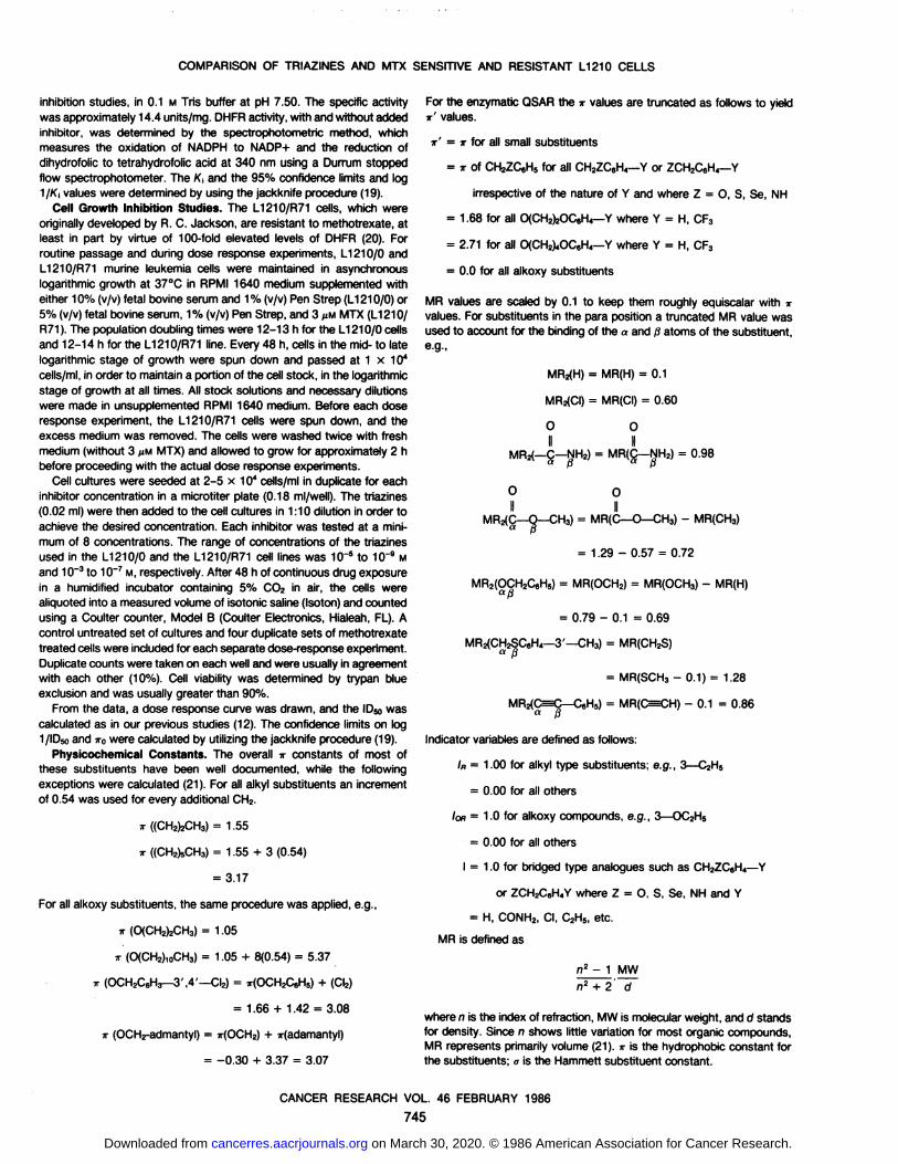
MR values are scaled by 0.1 to keep them roughly equiscalar with irvalues. For substituents in the para position a truncated MR value wasused to account for the binding of the a and ßatoms of the substituent,e.g.,
= MR(H) = 0.1
MR2(CI) = MR(CI) = 0.60
O O
II IIMR2(-Ç-NH2) = MR(Ç-NH2)= 0.98
O OII II
MRj(C—O—CH3) = MR(C—O—CH3) - MR(CH3)
= 1.29 - 0.57 = 0.72
MR2(OCH2C6H5) = MR(OCH2) = MR(OCH3) - MR(H)aß
= 0.79 - 0.1 = 0.69
'—CH3) = MR(CH2S)
= MR(SCH3 - 0.1) = 1.28
MRz(C=X>-C6H5) = MR(C=3») - 0.1 = 0.86a ß
Indicator variables are defined as follows:
IK = 1.00 for alkyl type substituents; e.g., 3—C2H5
= 0.00 for all others
/OH= 1.0 for alkoxy compounds, e.g., 3—OC2H5
= 0.00 for ail others
I = 1.0 for bridged type analogues such as CH2ZC6H4—Y
or ZCH2C6H4Y where Z = O, S, Se, NH and Y
= H, CONH2, Cl, C2H5, etc.
MR is defined as
n2- 1 MWn2 + 2' d
where n is the index of refraction, MW is molecular weight, and d standsfor density. Since n shows little variation for most organic compounds,MR represents primarily volume (21). ir is the hydrophobic constant forthe substituents; a is the Hammett substituent constant.
CANCER RESEARCH VOL. 46 FEBRUARY 1986
745
on March 30, 2020. © 1986 American Association for Cancer Research.cancerres.aacrjournals.org Downloaded from

COMPARISON OF TRIAZINES AND MTX SENSITIVE AND RESISTANT L1210 CELLS
(B)
(C)
The structure of the triazines used is illustrated in Fig. 1.
RESULTS
From the data in Table 1 for the inhibition of purified DHFRfrom L1210 leukemia cells, the following QSAR have been derived.
Inhibition of Purified L1210 DHFR by 3-X-Triazines
log 1/Ki = 0.22(±0.10)ir' + 6.31 (±0.17) (A)
n = 58, r = 0.532, s = 0.488, F,.« = 22.10
log 1/K¡= 0.90(±0.14)7T' - 1.10(±0.20)log(|8-10'' + 1)
+ 6.26(±0.10)
n = 58, r = 0.879, s = 0.280, F2.M = 58.27,
TO= 1.67(±0.25), log ß= -1.00
tog 1/Ki = 0.98^0.14)*' - 1.14(±0.20)log(/MO*' + 1)
+ 0.79(±0.57)<r+ 6.12(±0.14)
n = 58, r = 0.900, s = 0.264, F,,53 = 6.65,
TO= 1.76(±0.28), log ß= -0.979
Poorly fit congeners:
3—CN(+0.74), 3—CHOHC6HS(-0.73) 3—OCH2-1-adamantyl,
(-0.72) 3—C2H5(+0.65)
In the above expression, n represents the number of datapoints, r is the correlation coefficient, s is the standard deviation,and F is the F-statistic. The collinearity between variables is nothigh; r2 = 0.29 for TT'versus a.
In these equations K¡is the apparent inhibition constant(19), and the prime with the hydrophobic parameter ir' signifies
that for 3-substituents of the type 3—CH2ZC6H4—Y or3—ZCH2C6H4—Y(Z = O, S, Se), irY is set equal to zero (i.e.,3—CH2ZC6H4—Y= 7r3—CH2ZC6H5).Since the substituents
containing Y appear to have essentially the same effect on K¡regardless of hydrophobicity or size, it is assumed that they donot contact the enzyme. Of the above poorly fit substituents, thefirst three were not used in the derivation of equations A to C.For all the alkoxy substituents, ir' has been set to zero. In the
stepwise development of equation C, both equations A (Fi.se =22.10) and B are significant (F2f54= 58.27).
The parameter warepresents the optimum value for the hydro-
phobic effect of substituents, and the figures in parenthesis arefor construction of the 95% confidence limits. In equations B andC, the bilinear model of Kubinyi (22) has been used to accountfor the hydrophobic effect of substituents. In this model, activityfirst rises with a slope of about 1 until the point of *-0 where it
;:HeN
\i- ^ *•
/VÃŽTfI
Table 1Physicochemical parameters used to derive equations A-C, for the inhibition of
DHFR from U 210 cells by triazines IThe inhibition of the isolated, purified enzyme from MTX-resistant L1210/R71
cells was assayed spectrophotometrically. The apparent inhibition constants andthe physicochemical constants of the analogues were determined as described in"Materials and Methods."
Ha Triazine I
Fig. 1. Structure of the triazines used in this study.
<- tog1//C-»No.
XObserved81.
H 6.42 ±0.03e2.
3—SO2NH2 4.68±0.033.3—CONH2 5.03±0.044.3—COCH3 5.68±0.035.3—OH 5.60±0.046.3—CF3 6.93±0.077.3—F 6.43±0.058.3—CI 7.01 ±0.069.3—Br 7.31 ±0.0410.3- 1 7.04 +0.0711.3—NO, 6.64 +0.0512.3—CM3 6.73±0.0513.
3—CH3 6.78±0.0414.3—CH2CH3 7.36 +0.0515.3—(CHsfeCHs 6.81±0.0616.3—<CH2),CH3 6.24±0.0517.3—(CH2)„CH3 6.21 ±0.0418.3—CICHafe 6.47±0.0319.3—D,L-CH(OH)CeH5tà 5.74 ±0.0320.
3—OCH3 6.02 +0.0421.3—OCH2CH3 6.00±0.0222.S-OfCH^CHa 5.91 ±0.0323.3—OfCHïfeCHa 6.16 +0.0424.3—O(CH2)4CH3 6.08±0.0425.3—OfCHzJsCHa 5.98 +0.0526.3—O(CH2),CH3 6.16±0.0627.3—OfCH^oCHa 6.23 ±0.0828.3—O(CH2)„CH3 6.24±0.0829.3—OÕCH^jCHa 6.19 ±0.0830.3—O(CH2)13CH3 5.50±0.0731.3—O(CH2)2—OC.HS 6.82±0.0632.3—O(CH2)2—OC,H4—3'—CF3 6.68 ±0.0733.
3—O(CH2)4—OC.HS 6.60±0.0634.3—O(CH2)4—OC.H«—3'—CF3 6.63 ±0.0835.
3—OCH2C6H5 6.92±0.0436.3—OCH2C,H3—3' ,4'—CI2 6.53 +0.0337.3—OCH2C,H4—4'—CONH2 6.97 +0.0438.3—OCHz—1—adamantyl" 6.10±0.0739.
3—CHzO—CYCLO—C.H,, 6.58 ±0.0640.3-CH2OC.H5 7.06±0.0441.3—CH2OC«H4—3'—CI 7.04 ±0.0742.3—CH2OC.H4—3'—CN 6.24 ±0.0743.3—CH2OC«H4—3'—OCH3 7.23 ±0.0644.3—CH2OC.H4—3'—CH2OH 7.08 ±0.0545.3—CHÎOC.H,—3'—CH3 7.00 ±0.0746.3- CHîOCom—3'—CH2CH3 6.72 ±0.0847.3—CH2OC,H«—3'—CHfCH^ 6.62 ±0.0648.3—CH2OC«H«—3'—C(CH3)3 6.44 +0.0649.3—CH2OC,H4—3'—(VU 7.04 ±0.0950.3—CH2OC,H«—3'— 7.06 ±0.05NHCOCH351.
3—CHiOCtH,— 3'— 6.76 +0.06NHCSNH252.
3—CH2OC,H4—3'—7.11+0.05NHCONH253.
3—CH2OC,H4—4'— 6.56 +0.06(CH2)4CH354.
3—CH20—2'—naphthyl 7.1 8±0.0655.3—CHzO—1'—naphthyl 7.01 ±0.0856.
3—CH2SC6H5 7.1 2±0.0957.3—CHzSCoH«—3'-CH3 6.85 ±0.0958.
3—CHuSeCeHs 7.23 +0.0559.3—SCH2C,H6 7.31 ±0.0560.3—SCH2C,H<—4'—CI 7.22 ±0.0761.3—CH20C.HΗ2',4',5'— CI3 6.59 ±0.05
62. Methotrexate 8.82±0.11895% confidenceintervals.6Calculated using equationC.cMean ±95% confidencelimits."Not used in the derivation of equations.Calculated"6.074.694.875.865.547.036.466.906.997.076.385.996.466.706.666.416.156.806.476.176.156.156.156.156.156.156.156.156.156.156.966.966.876.876.966.966.966.826.916.936.936.936.936.936.936.936.936.936.936.936.936.936.936.936.936.896.896.896.876.876.93ff
T'0.00
0.000.46-1.820.28-1.490.38-0.550.12-0.670.430.880.340.140.370.710.390.860.351.120.71-0.280.56
-0.57-0.070.56-0.071.03-0.083.17-0.084.79-0.086.41-0.101.98-0.04
0.540.120.000.100.000.100.000.100.000.100.000.100.000.100.000.100.000.100.000.100.000.100.000.101.680.101.680.102.710.102.710.101.660.101.660.101.660.103.070.061.430.061.660.061.660.06
1.660.061.660.061.660.061.660.061.660.061.660.061.660.061.660.061.660.06
1.660.06
1.660.06
1.660.06
1.660.061.660.062.300.062.300.062.370.032.300.032.300.06
1.66
CANCER RESEARCH VOL 46 FEBRUARY 1986
746
on March 30, 2020. © 1986 American Association for Cancer Research.cancerres.aacrjournals.org Downloaded from

COMPARISON OF TRIAZINES AND MTX SENSITIVE AND RESISTANT L1210 CELLS
(E)
(F)
(G)
levels off. That is, the slope of the right hand side of equation Cis 1.14 - 0.98 = 0.16. It is assumed that this flat part of the
curve means that portions of the larger substituents do notcontact the enzyme. Evidence from molecular graphics studiesbased on X-ray crystallographic coordinates supports this contention in the case of chicken liver DHFR (2). The disposableparameter ßis evaluated via an iterative procedure. The inhibitionof growth in intact L1210/0 cells by 3-X-triazines I was deter
mined and is shown in Table 2.
Inhibition of L1210 Cells Sensitive to MTX by 3-X Triazines
tog 1/C = 0.04<±0.07)ir + 6.74(±0.19) (D)
n = 61, r = 0.147, s = 0.500, FLOT= 1.31
log 1/C = 0.98(±0.25)ir - 1.KX±0.28)log(/3-10' + 1)
+ 6.87(±0.14)
n = 61, r = 0.724, s = 0.355, Ftsl = 30,
Tro= 1.29(±0.51), log ß= -0.371
log 1/C = 0.88(±0.20)ir - 1.02(±0.24)log(/MO' + 1)
+ 0.55(±0.27)//, + 6.75(±0.12)
n = 61, r = 0.794, s = 0.315, Fi.se = 16.3,
TO = 1.32(±0.74), tog ß= -0.516
log 1/C = 0.98(±0.17)îr- 1.10(±0.20)log08.10- + 1) +
0.78(±0.25)/fl + 1.00(±0.41)<7+ 6.51(±0.14)
n = 61, r = 0.862, s = 0.266, F,.ss = 23.8,
io = 1.45(±0.62), tog ß= -0.546
log 1/C = 1.13(±0.18)ir - 1.20(±0.21)log(/3.10* + 1)
+ 0.66(±0.23)/fl+ 0.94(±0.37)«T (H)
- 0.32(±0.17)/oB + 6.72(±0.13)
n = 61, r = 0.890, s = 0.241, F,.M = 12.8,
TO = 1.45(±0.43), log ß= -0.274
Poorly fit points:
3—qCHafe, (-0.84) 3—CHOHCsHs, (-1.23)
3—OCHzCoHs—3',4'—dz(-0.59)
Correlation between variables (r ):
IR a lo« T
IR 1 0.15 0.03 0.02a 0.15 1 0.01 0.14IOR 0.03 0.01 1 0.11i 0.02 0.14 0.11 1
Equation H has one term (/„),which takes the value of 1 foralkyl groups, not found to be of significance in equation C. In theleukemia cells the alkyl groups are about 5 times as active, onthe average, as one would expect from the QSAR without the IRterm. This is in contrast to the halogens or CF3, which are alsohydrophobic groups binding in the same region but which arewell predicted on the basis of T and a alone.
Table 2Physicochemical parameters used to derive equations D-G for the inhibition of
growth in L1210/0 cells by 3-X-triazines I
The L1210/0 cells were incubated in the presence of the drugs at eight differentconcentrations; the number of cells was counted after 48 h, and the IDM valueswere determined. The physicochemical parameters were obtained as described in•Materialsand Methods.*
-logi/iD«,^No.1.2.3.4.5.6.7.8.9.10.11.12.13.14.15.16.17.18.19.20.21.22.23.24.25.26.27.28.29.30.31.32.33.34.35.36.37.38.39.40.41.42.43.44.45.46.47.48.49.50.51.52.53.54.55.56.57.58.59.60.61.62.63.64.abcdXH3
—SOjNH23—CONHj3—COCHs3—OH3—CF33—F3-CI3—
Br3—I3—NOz3—CN3—CH33—CrbCH33—(CH2)sCHj3—(CHj)8CH33
(CHj)iiCH3S-QCH,),"S-O.L-ChKOHXVHs"3—
OCH33—OCHzCHj3
—0(CH2)2CH33—O(CH2)3CH33—0(CH2)4CH33—
CHCHzJsCHa3—O(CH2),CH33-0(CH2),oCH33—
O(CH2),,CH33-O(CH2)12CH33-0(CH2)13CH33
—O(CH2)2—OC6H53—OfCHzJz-OCVU— 3'—CF33-0(CH2),-OC.H53—
O(CH2)4—OCJV- 3'—CF,3—
OCHZC«H53—OCH2C«H3—3',4'-Cba3—OCH2Q,H4—4'—CONH23—
OCHrl-adamantyl3—CHjO—CYCLO—C.H,,3—
CrbOC.Hs3—CH2OCsH4-3'-CI3
—CHjOCeH, —3' —CN3—CHjOC.rU— 3'—OCH33—CHzOC^U— 3'—CHïOH3
—CHjOCJH, —3' —CH33—CHzOCoH«—3'—CHîCHa3—CHzOC^i,— 3'—CHfCHafe3
—CH2OCcri4—3 —«(CHsJs3—CH2OC,H4—3'—C6H53—CH2OC«H4—3'—NHCOCH33—CH2OC,H4—3'—NHCSNH23—CH2OC.H,—3'—NHCONH23—
Cri2OCgH4—4^"{CHj^CHs3—CrbO—2'—naphthyl3—CrbO— 1'—naphthyl3
—CH^CaHs3—CH2SC.H,—3'—CH33
—CH2SeC6H53—SCH^HS3—
SCHAH,— 4'—a3-CH2OC.H2—2',4',5'-Cl,3,5—
Cb3,5—(CH3)jS.SHCFjfe95%
confidenceinterval.Calculatedby using equationH.Mean
±95% confidence limits.Observed86.67
±0.05C4.94
±0.265.37±0.066.22±0.216.27±0.077.1
7±0.077.08±0.087.47±0.037.27±0.067.26±0.076.86±0.076.73±0.177.24±0.077.69±0.047.58±0.087.28±0.066.86±0.136.62±0.055.51±0.146.62+0.126.59±0.066.37±0.086.43±0.116.54±0.126.67±0.066.89±0.056.70
±0.106.20±0.216.06
±0.106.44±0.086.57±0.036.98±0.076.49±0.077.11
±0.067.00±0.056.32±0.046.19±0.036.44±0.086.67±0.087.10±0.057.13±0.056.53±0.027.16±0.036.74±0.047.23±0.056.93±0.036.95±0.076.41±0.076.84±0.076.71±0.036.47±0.246.75±0.076.74±0.077.20±0.027.10
±0.047.14+0.057.02±0.057.20±0.087.13±0.097.02±0.066.45±0.087.60±0.087.48±0.077.69±0.09Calcu
lated6.505.105.306.396.027.276.917.187.237.226.946.547.397.147.407.287.157.466.756.286.506.676.686.666.626.506.426.386.346.307.006.956.946.877.006.916.706.596.966.966.926.956.966.876.936.906.866.836.836.886.726.776.786.886.886.936.896.926.906.856.827.607.437.71/„0000000000001111110000000000000000000000000000000000000000000010a0.000.460.280.380.120.430.340.370.390.350.710.56-0.07-0.07-0.08-0.08-0.08-0.10-0.040.120.100.100.100.100.100.100.100.100.100.100.100.100.100.100.100.100.100.100.060.060.060.060.060.060.060.060.060.060.060.060.060.060.060.060.060.060.060.060.030.030.060.74-0.140.86/OB0000000000000000000111111111110000000100000000000000000000000000n0.00-1.82-1.49-0.55-0.670.880.140.710.861.12-0.28-0.570.561.033.174.796.411.980.54-0.020.381.051.592.132.674.295.375.916.456.991.682.562.713.591.663.080.173.071.431.662.371.091.640.632.222.683.193.643.690.690.260.364.332.982.982.302.862.372.303.013.791.421.121.76Not
used in the derivation of equations.
CANCER RESEARCH VOL. 46 FEBRUARY 1986
747
on March 30, 2020. © 1986 American Association for Cancer Research.cancerres.aacrjournals.org Downloaded from
COMPARISON OF TRIAZINES AND MIX SENSITIVE AND RESISTANT L1210 CELLS
In equations D to H the overall TTof the substituents has beenused because in membrane penetration the substituents mustmake hydrophobic contact. Since Y does not make contact withthe enzyme in the case of isolated DHFR, as opposed to thesmaller 3-X groups, it seems that some kind of correction isneeded. Attempts to use either * in the bilinear model or jry(which we thought might appear as a term with a negativecoefficient to correct for its simple hydrophobic effect), did notyield an improved correlation. w0 for the sensitive cells (equation I) is 1.45, which is close to 1.76 for equation H. Thus therole for hydrophobic effects of substituents with the isolatedDHFR and the MTX sensitive cells is essentially the same. Onecould conclude that quasi-equilibrium conditions exist in the cell
culture study.The use of the indicator variables IORand /n is a rather crude
means for dealing with the aberrant behavior of alkoxy and alkylgroups; however, this method of "molecular bookkeeping" does
keep in mind special behavior of certain classes of substituentsso that these features of the SAR are readily apparent to anyonestudying the data for the first time.
The alkoxy substituted drugs show their own independent,parabolic SAR starting with a low for the first two members ofthe series, increasing to a maximum at OC9 and then decliningto Oda, but as a group they are less active than expected byabout a factor of two.
Of the substituents poorly fit by both equations C and H onlythe 3—CHOHCeHs is badly fit by both equations. The 3—CN
which, as usual, is poorly fit by the enzyme equation, is well fitby equation H. This does suggest a difference between DHFRenzyme in its isolated state and in situ. The poor activity of thealkoxy groups is somewhat mitigated in the cell culture study,as shown by the small coefficient with /on in equation H. The aterm plays about the same role in isolation as in vitro. The resultsof the inhibition of growth of L1210/R71 cells by 3-X-triazines
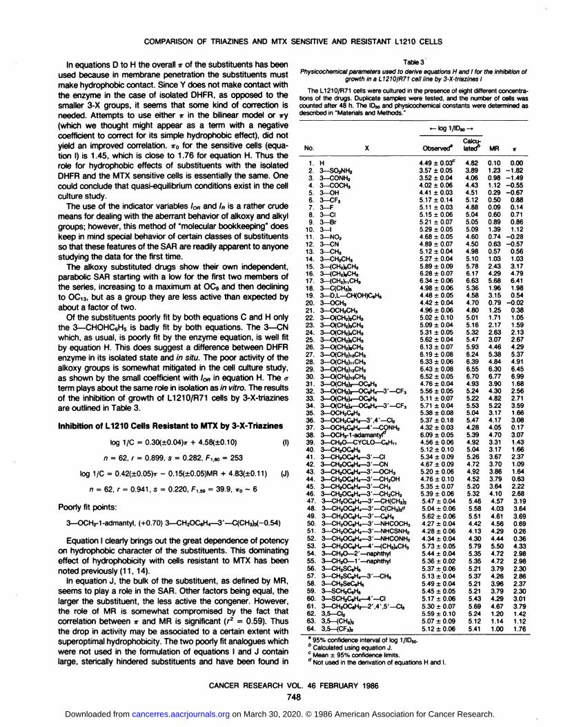
are outlined in Table 3.
Inhibition of L1210 Cells Resistant to MTX by 3-X-Triazines
log 1/C = 0.30(±0.04)ir + 4.58<±0.10) (I)
n = 62, r = 0.899, s = 0.282, F,.«,= 253
log 1/C = 0.42(±0.05)7T- 0.15(±0.05)MR + 4.83(±0.11) (J)
fi = 62, r = 0.941, s = 0.220, F,.69 = 39.9, TTO~ 6
Poorly fit points:
3—OCHü-1-admantyl,(+0.70) 3—CH2OC6H4—3'—C(CH3)3(-0.54)
Equation I clearly brings out the great dependence of potencyon hydrophobic character of the substituents. This dominatingeffect of hydrophobicity with cells resistant to MTX has beennoted previously (11,14).
In equation J, the bulk of the substituent, as defined by MR,seems to play a role in the SAR. Other factors being equal, thelarger the substituent, the less active the congener. However,the role of MR is somewhat compromised by the fact thatcorrelation between * and MR is significant (r2 = 0.59). Thus
the drop in activity may be associated to a certain extent withsuperoptimal hydrophobicity. The two poorly fit analogues whichwere not used in the formulation of equations I and J containlarge, sterically hindered substituents and have been found in
Table 3
Physicochemical parameters used to derive equations H and I for the inhibition ofgrowth in a L1210/R71 cell line by 3-X-triazines I
The L1210/R71 cells were cultured in the presence of eight different concentrations of the drugs. Duplicate samples were tested, and the number of cells wascounted after 48 h. The ID»and physicochemical constants were determined asdescribed in 'Materials and Methods."
No.1.2.3.4.5.6.7.8.9.10.11.12.13.14.15.16.17.18.19.20.21.22.23.24.25.26.27.28.29.30.31.32.33.34.35.36.37.38.39.40.41.42.43.44.45.46.47.48.49.50.51.52.53.54.55.56.57.58.59.60.61.62.63.64.XH3—
SO2NH23—CONH23—COCHa3—OH3—CF33—F3-CI3—
Br3—13—
N023—CN3-CH33—
CH2CH33—(CH2)sCHa3-(CH2).CH33-(CH2),,CH33
—CfCH^3—D,L—CH(OH)C,H53—
OCH33—OCH2CH3S-CXCHzfcCH,3
—OlCH^CHs3—O(CH2)4CH33—CXCHjfeCHa3—O(CH2),CH33—O(CH2),0CH33—O(CH2),,CH33—0(CH2),2CH33—O(CH2),3CH3S-OfCHjJj-OC.Hs3-O(CH2)2—
OC«H4—3'—CF33—
OfCHîV—OC,H53-0(CH2)4—OC.H4—3'—CF33—
OCH2C«H53—OCHAH,— 3' .4'—CI23—OCH2C,H4—4'—CONH23—
OCH2-1-adamanty^3—CH2O—CYCLO—C.H,,3—
CH2OC,H53—CH2OC«H4—3'—CI3—CHjOCeH,—3'—CN3—CH2OC,H4—3'—OCH33—CH2OC,H4—3'—CH2OH3—CH2OC«H4—3'—CH33-CH2OC.H4—
3'—CH2CH33—CH2OC«H4—3'—CH(CH3)j3
—CH2OCeH4—3' —CÃCHa)^3—CH2OC«H4—3'—C,H53—CH2OC«H4—3'—NHCOCH33—CH2OC»H4—3'—NHCSNH23—CH2OC,H4—3'—NHCONH23—CH2OC,H4—4'—(CH^CHa3—CH2O—2'—naphthyl3—CH2O—1'—naphthyl3
—CH^CjHs3—CHZSC.H*—3'—CH33
—CHjSeCeHs3—SCH2C,H53—
SCH2C,H4—4'—CI3—CH2OC«H2—2', 4', 5' —CI33,5—
C\,3,5—(CHsfc3,5—
(CFafe•—log
1/lCObserved84.49
±0.03C3.57
±0.053.52+0.044.02±0.064.41±0.035.17+0.145.11
±0.035.15±0.065.21
±0.075.29±0.054.68±0.054.89±0.075.12
±0.045.27±0.045.89+0.096.28±0.076.34±0.064.98±0.064.48±0.054.42±0.044.96±0.065.02+0.105.09+0.045.31±0.055.62+0.046.1
3±0.076.19±0.086.33
±0.066.43±0.086.52±0.054.76±0.045.56±0.055.11
±0.075.71±0.045.38±0.085.37±0.184.32±0.036.09±0.054.56±0.065.12
±0.105.34±0.094.67±0.095.20±0.064.76
±0.105.35±0.075.39±0.065.47±0.045.04±0.065.62±0.064.27±0.044.28±0.064.34±0.045.73±0.055.44±0.045.36±0.025.37±0.065.13+0.045.49±0.045.45+0.055.1
7±0.065.30±0.075.59+0.105.07±0.095.12+ 0.06>»-Calcu
lated"4.823.894.064.434.515.124.885.045.055.094.604.504.985.105.786.176.635.364.584.704.805.015.165.325.475.936.246.396.556.704.935.245.225.535.045.474.285.394.925.045.264.724.924.525.205.325.465.585.514.424.134.305.795.355.355.215.375.215.215.435.695.245.125.41MR0.101.230.981.120.290.500.090.600.891.390.740.630.571.032.434.295.681.963.150.791.251.712.172.633.074.465.384.846.306.773.904.304.825.223.174.174.054.703.313.173.673.703.863.793.644.104.574.034.614.564.294.445.504.724.723.794.263.963.794.294.671.201.141.00T0.00-1.82-1.49-0.55-0.670.880.140.710.861.12-0.28-0.570.561.033.174.796.411.980.54-0.020.381.051.592.132.674.295.374.916.456.991.682.562.713.591.663.080.173.071.431.662.371.091.640.632.222.683.193.643.690.690.260.364.332.982.982.302.862.372.303.013.791.421.121.76
" 95% confidence interval of log I/ID»." Calculated using equation J.c Mean + 95% confidence limits." Not used in the derivation of equations H and I.
CANCER RESEARCH VOL. 46 FEBRUARY 1986
748
on March 30, 2020. © 1986 American Association for Cancer Research.cancerres.aacrjournals.org Downloaded from

COMPARISON OF TRIAZINES AND MTX SENSITIVE AND RESISTANT L1210 CELLS
other instances to be poorly predicted by QSAR.The optimum TTvalue, estimated to be about 6, cannot be
derived mathematically, since there are no compounds the activity of which has started to decrease as TTincreases. This valueof 6 is far removed from the value of about 1.8 found for isolatedDHFR and sensitive cells. This difference of 4.2 log units is themost remarkable aspect of the resistant cell QSAR and, whencombined with the fact that similar results were found for L5178Yand Lactobacillus case; cells, it means that more lipophilic drugsare needed for MTX resistant tumors.
The structure-activity relationships of the 4-X-triazines aresufficiently different from the 3-X-triazines so that they are best
treated as a separate group. The stepwise development of theessential QSAR, as outlined in Table 4, is as follows.
Table 4Physicochemical parameters used to derive equations K-N, for the inhibition of
L1210 DHFR by 4-X-triazines I
The inhibition of L1210 DHFR was assayed spectrophotometrically. The apparent inhibition constants and the physicochemical constants of analogues weredetermined as described in "Materials and Methods."
*-log1/Ki-^No.
X1.
H2.4—SO2NH23.4—S02CH34.4—CONH25.4—COCH36.4—COOCH/7.4—COOCH2CH38.4—OH9.4—NH210.4—NHCOCH311.4—CF312.4—F13.
4—Ci14.4—Br15.4—I16.
4—0^174—OCH2CN(CH2CH2)20<'18.
4—0(CH2)j—OC6H4--r—NH219.4—CH320.4—(CH^CHs21.4—(CH2).CH322.4—C(CHs)323.4—C=CH<*24.4—C=C—SilCHafe"25.4—O^C—CsHs"26.
4—OCH327.4—OCH2CH328.4—OfCH^CHs29.4—OCH2CH=CH230.4—OfCH^CHs31.4—O(CH2)5CH332.4—O(CH2)rCH333.4—O(CH2),CH334.4—0(CH2)10CH335.4—OCH2C,H536.4—OCHzCgHs—3 ,4 —Clz37.4—OCH2C«H4—4'—SO2NH238.4—OCH2C6H4—4'—CONH239.4—OCH2C6H4—4'—CH2OH40.
4—CH2SC„H541.4—CH2SC„H4—2'—CH342.4-CH2SC„H4—3'—CH343.
4—SCH2C«H644.4—SCH2C6H4—4'—CIObserved86.42
±0.03C4.62
±0.045.17±0.044.61
±0.025.31±0.054.98±0.044.98±0.035.87±0.065.42±0.045.10
±0.046.59±0.056.57±0.046.89±0.056.72±0.076.39±0.044.95±0.036.75±0.037.25±0.056.49±0.036.69±0.046.59±0.046.25±0.035.71±0.065.67±0.035.86±0.036.26±0.045.74±0.036.04±0.036.57±0.035.97±0.036.17±0.046.53±0.056.48±0.096.29±0.087.22±0.067.15+0.087.98
±0.127.65±0.057.57±0.047.71±0.097.31±0.087.46±0.057.45±0.057.48±0.08Calcu
lated66.414.614.704.965.606.156.495.855.335.466.776.516.656.616.515.785.156.926.586.836.666.426.336.376.376.176.176.176.176.176.176.176.176.177.617.617.617.617.617.397.397.397.397.39MR2/TT'0.10
0.000.001.230.00-1.821.340.00-1.630.980.00-1.491.120.00-0.550.720.00-0.010.72
0.000.510.290.00-0.670.540.00-1.230.700.00-0.970.50
0.000.880.090.000.140.600.000.710.890.000.861.390.001.120.630.00-0.570.690.00-1.390.69
0.002.020.570.000.560.930.002.090.930.004.791.960.001.980.960.000.400.860.000.400.860.000.400.690.000.000.690.000.000.690.000.000.690.000.000.690.000.000.690.000.000.690.000.000.690.000.000.690.000.000.691.001.660.691.001.660.691.001.660.691.001.660.691.001.661.281.002.301.281.002.301.281.002.301.281.002.301.281.00 2.30
" 95% confidence interval on log 1/K,." Calculated by using equation N.c Mean ±SD." Excluded in the derivation of the equations.
Inhibition of Purified L1210 DHFR by 4-X-Triazines
log 1/K, = 0.48(±0.13)ir' + 6.14(±0.20) (K)
n = 37, r = 0.794, s = 0.529, F1i35= 59.6
log l/K, = 0.81(±0.14)7r' - 1.14(±0.37)log(/MO*' + 1)
+ 6.19(±0.14)
n = 37, r = 0.912, s = 0.368, F2.33= 19.7, TTO= 2.14, log ß= -1.75
log 1/K, = 0.95(±0.20)ir' - 1.00(±0.29)log(/MO"' + 1)
+ 0.78(±0.26) / + 6.35(±0.15)
n = 37, r = 0.949, s = 0.287, FI.& = 22.4,
»o= 1.72(±0.54), log ß= -0.50
log l/K, = 0.76(±0.15)ir' - 0.83(±0.28)log(tf-10" + 1)
+ 0.70(±0.25)/ - 0.40(±0.26)MR2 + 6.47(±0.22)
n = 37, r = 0.959, s = 0.263, F,.31 = 6.96, TTO= 2.11, log ß= -1.10
(L)
(M)
(N)
Poorly fit points:
4—COOCHsM.17), 4—COOC2Hs(-1.51),4—CN(-0.83),
4—OCH2CO—morpholine(+1.60), 4—C=CH(-0.62),
4—C^C—Si(CH3)3(-0.70),4—C=CC6H5(-0.51)
The correlation between variables is shown in the followingsquared correlation matrix.
T' / MR2ir' 1.0 0.30 0.08
/ 0.30 1.0 0.06MR2 0.08 0.06 1.00
The prime with * in equations K to N implies that for sub-stituents of the type 4—Z—CH2C6H4—Yor 4—CH2ZC6H4—Y(Z = 0 or S) T for Y is set to zero as in equations A to C. Forsubstituents of the type 4—C=C—fî,irfi has been assigned a
value of zero, since fl, regardless of size, seems to have noeffect on log 1/K¡.For alkoxy substituents TT'has been set to
zero.An analysis of K( values in our previous studies indicates the
presence of a steric effect of substituents in the para position ofthe phenyl group of the triazines (3). A graphics study of DHFRfrom chicken liver makes it clear that 4-substituents encounteran unfavorable interaction with lle-60. a residue which appears
to be conserved in L1210 DHFR. The interaction is only importantfor about the first two atoms («and ß)of the groups in the paraposition of phenyl ring: this effect has thus been parameterizedby MR2, which is the sum of MR for these two atoms. Althoughthis constitutes a crude approximation it does help to quantitatethis steric effect. The results of the inhibition of growth of theL1210/0 cells by 4-X-triazines are outlined in Table 5.
Inhibition of L1210 Cells Sensitive to MTX by 4-X-Triazines
log 1/C = 0.14(±0.08K+ 6.45(±0.21) (O)
n = 42, r = 0.479, s = 0.565, F,i40=11.91
CANCER RESEARCH VOL. 46 FEBRUARY 1986
749
on March 30, 2020. © 1986 American Association for Cancer Research.cancerres.aacrjournals.org Downloaded from

COMPARISON OF TRIAZINES AND MIX SENSITIVE AND RESISTANT L1210 CELLS
TablesPhysicochemicalparametersused to derive equations O-R for the inhibition of
growth in L1210/0 cells by 4-X-triazines I
The L1210/0 cells were incubated in the presenceof the drugs at eight differentconcentrations; the numbers of cells were counted after 48 h, and the ID»valueswere determined.
(R)
No.1.2.3.4.5.6.7.8.9.10.11.12.13.14.15.16.17.18.19.20.21.22.
23.24.25.26.27.28.29.30.31.32.33.34.35.36.37.38.39.40.41.42.43.44.45.46.47.48.XH4—
S02NH24—S02CH34—CONH24—COCH34—OH4—NH24—NHCOCH34—CF34—F4—CI4—Br4—14—
CN4—OCH2CON(CH2CH2)204—
0(CH2)2—OC8H4—4'—NH24—
CH34—(CH^CHs4-<CH2).CH34
CiCHglg4-C^CHd4—
C=C—SKCHafe"4—
OCH3*4—
OCH2CH34—O(CH2)2CH34—
OCH2CH=CH24-0(CH2)3CH34—
O(CH2)4CH34-0(CH2)sCH34—
O(CH2)7CH34—O(CH2),CH34—0(CH2)10CH34—C^CH^nCHs4—
O(CH2),3CH34—OCH2C6H54—
OCH2C.H3—3' ,4'—CI24—OCH2C6H4—4'—SO2NH24—OCH2C»H4—4'—CONH24—OCH2C«H4—4'—CH2OH4
—CHuSC8H54—CH2SC«H4—2'—CH34—CHzSCeH»—3'—CH34—
SCH2C«H54—SCH2C6H4—4'—CIMethotrexate0Bakers
AntifolfBakersAntifol lld*
—locj 1/IDObserved86.67
±0.05C4.65
±0.075.13±0.075.01
±0.056.15±0.036.42
±0.066.10±0.045.67
±0.087.23±0.067.24+0.067.24+0.067.26+0.037.25±0.036.25±0.095.88±0.057.05
±0.116.85±0.067.16
±0.147.23±0.086.55±0.076.06±0.056.14±0.04
5.73 ±0.066.47+0.086.30±0.036.47±0.046.83±0.036.58±0.066.81±0.067.00±0.077.11
±0.086.88±0.066.34
±0.106.49±0.086.47±0.067.27±0.057.19+0.066.64
±0.116.94±0.097.03+0.097.08±0.057.06+0.057.02±0.057.37+0.076.72±0.098.22±0.076.95±0:096.92±0.0950^Calcu
lated66.994.915.035.326.086.365.755.917.307.107.197.116.956.295.547.027.137.227.056.756.877.25
7.226.216.486.766.696.836.836.806.736.706.626.596.517.337.276.606.857.117.057.027.027.057.01MR20.101.231.350.981.120.290.540.700.500.090.600.891.390.630.690.690.570.930.931.960.960.860.860.690.690.690.690.690.690.690.690.690.690.690.690.690.690.690.690.691.281.281.281.281.28V0.00-1.82-1.63-1.49-0.55-0.67-1.23-0.970.880.140.710.861.12-0.57-1.390.450.562.094.791.980.402.062.65-0.020.381.050.811.552.132.673.754.295.374.916.991.663.08-0.160.170.632.302.862.862.303.01/<*0.000.000.000.000.000.000.000.000.000.000.000.000.000.000.000.000.000.000.000.000.000.000.001.001.001.001.001.001.001.001.001.001.001.001.000.000.000.000.000.000.000.000.000.000.00
log 1/C = 0.90(±0.11)ir - 0.97(±0.14)log(/3-10* + 1)
- 0.45(±0.20)MR2 - 0.50(±0.18)/on + 7.11(±0.19)
n = 42, r = 0.949, s = 0.215, F,.» = 12.7, *•„= 1.83, log ß= -0.706
Poorly fit congeners:
4—CssCH(-0.81),4—C=C—C6H5(-1.49),
Squared correlation matrix:
(ORT
MR2
/OR
10.220.05
0.2210.01
MR,
0.050.011
95% confidence interval of log I/ID».0 Calculatedusing equation R.c Mean + SD.d Not used in the derivation of equations.
log 1/C = 1.18(±0.2G> - 1.27(±0.24)log(/M 0' + 1)
+ 7.08(±0.16)
n = 42, r = 0.896, s = 0.294, F2.w = 55.1,
*o = 1.22(±0.41), log ß= -0.079
log 1/C = 1.27(±0.18)ir - 1.31 (±0.21)log(j3-10" + 1)
(Q)- 0.39(±0.19)/ofl + 7.22(±0.15)
n = 42, r = 0.930, s = 0.247, F,^ = 17.0,
As for the 3-X-congeners acting in cell culture, we have used
ir for the complete substituent. The parameters of the hydrophobic effects of the 4-substituents, including v0, are close to thosefound for 3-substituents. The role of other structural features
has been changed. The important / variable in equations M andN does not show up in equation R, which suggests a change inthe receptor in vivo. The steric effect MR2 does attain significancein equation R when the /on is also included. Both of these termsare weak.
The IORterm does not occur in equation N for isolated DHFR,where the setting of w' to zero for alkoxy groups accounts for
the essentially constant value of the alkoxy set of substituants.However it does occur in equations Q and R, where the overallhydrophobicity is important for membrane penetration. Thus thealkoxy groups do show some hydrophobic effect in the cells, butthe negative sign with the /on term indicates that they still do notcontact the receptor the way other hydrophobic substituents do.
The 4—OCH2CO—morpholino analogue, which is often 10
times or more active than predicted with isolated enzyme, is only2.5 times more active than expected in the sensitive cells. Thetwo esters 4—COOCH3 and 4—COOC2H5were not tested because previous erratic test results suggested that hydrolysis wasoccurring in the cell culture medium.
Treating the acetylenic derivatives like the others by usingtheir complete v values results in all of them being poorly fit, norare they well fit by IT'. The steric effect of the rigid acetylenic
group is out of line with the other more flexible 4-substituents.The results of the inhibition of growth of L1210/R71 cells by 4-X-triazines are outlined in Table 6.
Inhibition of L1210 Cells Resistant to MTX by 4-X-Triazines
log 1/C = 0.39(±0.05)ir + 4.32(±0.14) (S)
n = 41, r = 0.918, s = 0.359, F,.-»= 209
tog 1/C = 0.46(±0.07)ir - 0.47(±0.31)log08-10" + 1)
+ 4.29(±0.13)
n = 41, r = 0.935, s = 0.330, F2.37= 9.09, *-„= 6.18, tog,8 = -4.54
Poorly fit congeners:
4—CN (-0.86), 4—CssCSi(CH3)3,
(-1.49), 4—OCH2C6H4—4'—CONH2(0.89),
4—OCHîCsH,—4'—CH2OH(0.94)
There is a 3000-fold range in 1/C, and equation S shows thatir0 = 1.52(±0.52),log ß= 0.000 activity is correlated within a factor of about ±2.Of the acetylenic
CANCER RESEARCH VOL. 46 FEBRUARY 1986
750
(P) (T)
on March 30, 2020. © 1986 American Association for Cancer Research.cancerres.aacrjournals.org Downloaded from

COMPARISON OF TRIAZINES AND MTX SENSITIVE AND RESISTANT L1210 CELLS
TablesPhysicochemicalparametersused to derive equations S and T for the inhibition of
L1210/R71 cells by 4-X-triazines I
The L1210/R71 cells were grown in the presence of eight different concentrations of each drug. Duplicate samples were tested for each concentration,and thenumber of cells was counted after 48 h in order to determine the ID»values.
NO.1.2.3.4.5.6.7.8.9.10.11.12.13.14.15.16.17.18.19.20.21.22.23.24.25.26.27.28.29.30.31.32.33.34.35.36.37.38.39.40.41.42.43.44.45.46.47.48.XH4—
S02NH24—S02CH34—CONH24—COCH34—OH4—NH24—NHCOCH34—CF34—F4—CI4—Br4—14
CN0A
—OC^CONfCH^H^O4-O(CH2)2—OC,H4—4'—NH24—
CH34—(CH2)sCH34-<CH2),CH34
—C(CH3)s4—C^CH4__c=c
—SHCH^4—C=C—C.HS4—
OCH34—OCH2CH34-0(CH2)2CHa4—
OCH2CH=CH24—OfCH^CHa4-0(CH2)4CH34—
CKCH^CHa4-0(CH2)7CH34—
0(CH2),CH34—O(CH2)1oCHa4—
O(CH2)„CH34—0(CH2)13CH34—OCH2C,H54—
OCH2C.H3—3' ,4'—Ct4—OCH2C,H4—4'—SO2NH24—OCH2C«H4—4'—CONH2"4—OCHjCsHi— 4'—CH2OHrf4—
CH2SC,HS4—CHzSCeH,—2'—CH34—CH2SC,H4—3'—CH34—
SCH2C«H54—SCH2C,H4—4'—CIMethotrexateBakers
Antifol\aBakersAntifol II"«-
log1/IDObserved"4.49
±0.03C2.98
+0.053.28±0.033.64±0.073.91+0.054.07±0.053.94±0.073.23±0.034.91±0.084.87±0.085.02±0.095.11
±0.085.11±0.083.17
±0.033.92+0.074.45±0.075.05±0.065.31
±0.126.50±0.115.39
+0.104.02±0.033.74+0.055.20+0.094.1
8±0.073.97+0.064.32±0.044.34±0.044.52+0.075.1
8±0.065.63±0.086.06±0.096.16+0.136.20
±0.106.24±0.136.39±0.105.33
±0.125.23±0.094.95+0.185.26±0.085.52±0.065.37+0.075.44±0.045.49±0.045.56±0.255.52
±0.115.15±0.064.69
±0.064.61±0.0950
—*Calcu
lated"4.293.463.553.614.043.983.733.854.694.354.614.684.804.033.664.494.545.246.275.194.475.235.494.284.464.774.665.005.265.505.976.156.326.346.335.055.694.224.374.585.345.595.595.345.65T0.001.821.631.49-0.55-0.67-1.23-0.970.880.140.710.861.12-0.57-1.390.450.562.094.791.980.402.062.65-0.020.381.050.811.552.132.673.754.295.375.916.991.663.08-0.160.170.632.302.862.862.303.01
* 95% confidence interval.b Calculatedusing equation T.c Mean ±95% confidence limits.d Not used in the derivation of equations S and T.
compounds 23-25, only the 4—C=C—Si(CH3)3 is seriously
misfit, and as is normally the case with isolated DHFR fromvarious sources, the 4—CN derivative is much less active than
expected (2). Interesting results were obtained with congeners39-42, having the structure 4—OCH2C6H4—Y.Although these
congeners are fairly well fit by equation N for the isolated DHFRand by equation R for sensitive cells, they are poorly fit by theresistant cell equations. This too suggests a slight difference inthe DHFR or its immediate environment which affects thesesubstituents.
In resistant cells, the 4-X triazines behave in an analogousfashion to the 3-X triazines. A simple equation S, linear in n-,
explains most of the variance in log 1/C (84%). Although equationT only accounts for an additional 3% it allows us to ascertain avalue of 6.2 for w0. In the equations for both 3- and 4-substit-
uents, in L1210/R71 cells the hydrophobic effect overwhelmsother substituent effects and suggests that the process of thedrug traversing the membrane to the active site of the enzymeis of paramount importance.
DISCUSSION
To clearly delineate the role of structure on the various processes by studying drug action in whole animals is exceedinglydifficult. Therefore, it seems reasonable to start at the simplestlevel, that of the receptor, and systematically study levels ofincreasing complexity. In attempting to design more effectiveantifolate, antitumor drugs, several steps in drug action whichare strongly dependent on chemical structure must be considered. Critical steps might be factored as follows: (a) interactionof inhibitor with receptor; (b) movement of inhibitor through andpartitioning among the various lipophilic and hydrophilic compartments of the cell; (c) interaction of the inhibitor with amacromolecular transport system necessary for cell entry (incertain cases); (d) transport from the site of application in theanimal to the target cells; and (e) losses via metabolic pathwaysand elimination in the animal. The first three steps in this integrated approach will now be considered.
Questions on which we hope to gain information are: (a) Howclosely will the SAR for the inhibition of purified enzyme parallelthe inhibition in cell culture? (b) What factors determine thedifference in sensitivity of sensitive and resistant cells to a largeset of potent inhibitors? (c) Are there structural features whichcan be exploited to devise inhibitors that are more effectiveagainst resistant cells? and (d) Is it practical to study inhibitorson purified enzyme and maximize receptor fit with the goal ofusing this information to design better therapeutic agents ingeneral?
Comparison of Enzyme and Cell QSAR. From equations A-
C, it is clear that the hydrophobicity of the substituents is theparameter of major importance, since it accounts for 77% of thevariance in equation B, while a accounts for slightly less than4% of the variance. The main feature of the SAR is that inhibitorpotency increases linearly with hydrophobicity with a slope ofabout 1.0 until *-0of approximately 1.8 is reached, and from thispoint there is a slight decrease (descending slope = 0.16) in
potency with further increases in hydrophobicity.Several special features of u-' must be kept in mind. Since *'
has been set to zero for all alkoxy groups from OCH3 to OC^H»and, except for OCi4H29, all of these congeners, regardless oftheir overall hydrophobicity, show essentially the same activity,it is assumed that they do not contact the enzyme (with theexception of the oxygen). If they do contact the enzyme the gainin potency must somehow be compensated by a steric effect.This is difficult to visualize. Log 1/K¡for 3-H is 6.42, and the
average value for the alkoxy congeners is 6.04. This is in directcontrast to the alkyl groups, where activity rises from 6.42 for Hto 6.78 for methyl and 7.63 for ethyl before falling to 6.81 forhexyl and 6.24 for dodecyl. The long hydrophobic hexyl anddodecyl chains may promote some kind of wrong way bindingto the enzyme and hence lower their effectiveness in the activesite. The other special feature of w' is that for substituents of
CANCER RESEARCH VOL. 46 FEBRUARY 1986
751
on March 30, 2020. © 1986 American Association for Cancer Research.cancerres.aacrjournals.org Downloaded from

COMPARISON OF TRIAZINES AND MIX SENSITIVE AND RESISTANT L1210 CELLS
the type CH2ZC6H4-Y and ZCH2C6H4-Y, Try is set to zero. This
group of over 20 congeners is reasonably well fit by such anassumption. In fact, TT'for the group is 1.66-2.30, which is close
to the optimum TTOof 1.8.Although the bilinear model for the treatment of hydrophobic
effects developed by Kubinyi (22) was not designed for theinteraction of ligands with purified enzymes, we have found it tobe very useful, with only one apparent limitation. For elongatedsubstituants, part of which do not contact the enzyme, thosesubstituents having TTvalues more positive than 7r0have theireffect truncated by the negative bilinear term. Provided the regionin which the substituents are binding is of essentially uniformcharacter, one can expect and appears to get a reasonable fit ofthe data. However, if the CH2ZC6H4-Y moiety falls in hydrophobicspace and Y has a negative - value but does not actually contact
the enzyme, the bilinear model will not account for this. Usingthe overall ir value for such substituents would produce a verylow log 1/Ki value. Unless the X-ray crystallographic structure of
the receptor site is known one cannot anticipate such discontinuities in receptor-ligand interactions. In fact, no general model
can achieve this; thus such effects must be deduced from thestudy of well designed sets of ligands.
The 3-cyano analogue is one of the most interesting marker
groups. It is more active than expected with DHFR from thefollowing sources: bovine (+0.91); chicken (+0.71); human(+0.42); L case/ (+1.19); rat (+1.04); L5178Y (+0.97); andL1210 (+0.60). Note that the residuals in parentheses are in logunits. In the cell culture QSAR, however, it is rather well predicted: sensitive L5178Y (-0.12); resistant L5178Y (+0.07);
sensitive L1210 (+0.19); and resistant L1210 (+0.39). It is notwell predicted in L case/ cell culture sensitive to MTX (+1.19) orresistant to MTX (+0.57).
Since the enzymic space where 3-substituents bind is notsensitive to steric effects and the 3-cyano is small in size, we
are inclined to disavow this result in terms of a positive stericeffect. In general, the cyano group is well behaved in systemswhere hydrophobic effects are important, and hence it is believedthat the polar cyano group probably enhances enzymic activityvia an electronic effect such as a dipole-dipole interaction. A
slight change in DHFR enzyme conformation in cellular systemscould nullify such interaction.
In addition to the 3-cyano group, another substituent which
usually shows considerable difference between its activity onisolated DHFR and intact cells is the 4-OCH2CO-morpholinoanalogue. Like the 3-cyano analogue it is much more active on
isolated DHFR [chicken (+1.47), L case/ (+0.77), human (+1.46),and L1210 (+1.60)] than is predicted by QSAR. It is adequatelypredicted by cell culture QSAR [sensitive L5178Y (+0.49), resistant L5178Y (+0.25), sensitive L1210 (+0.35), and resistantL1210 (+0.26)]. As with the 3-cyano group we tend to view
these differences in the values of the deviations as being due toa difference in a polar interaction of the morpholino group whichoccurs with purified enzyme but which is mitigated when theenzyme is present in its cellular environment.
As we have noted previously, the most important structuralfeature producing the variation in activity of these triazines is thehydrophobic character of the substituents. Consequently thefact that the QSAR for L1210 DHFR and sensitive cells eachhave coefficients of about 1 with the TTterm and a TTOof about1.5 implies similarity between the DHFR receptor in isolation and
in the sensitive cells. Similar results were found for L5178Ytumor cells and DHFR (13).
The results may be summarized as follows: (a) the QSAR forpurified DHFR and sensitive cells are similar; and (b) the QSARfor MTX resistant cells is extremely different from that of purifiedDHFR (or sensitive cells) despite the fact that the enzyme wasisolated from the resistant cells.
On the Role of Lipophilicity. After the discovery (23,24), that"lipophilic" 2,4-diaminopyrimidine antifolates were effective in
vitro and in vivo against MTX resistant tumors various researchgroups have experimented with "lipophilic" antifolates (25). However, the meaning of the term "lipophilic" has not been delineatedclearly, nor have enough "lipophilic" congeners been studied to
be certain that features other than lipophilicity are not alsoinvolved. If lipophilicity is highly desirable, no systematic efforthas been made to deduce what is the upper limit to the beneficialeffect of lipophilicity. In order to establish such limits one mustbe able to selectively define lipophilic as well as electronic andsteric contributions in drug action.
In recent years considerable evidence has accumulated toshow that in the complex problems of cancer chemotherapy(e.g., in animal model studies) the octanol/water partition coefficient (log P) for drugs or ir values for their substituents on drugscan be used to separate the role of hydrophobicity from electronicand steric factors (9, 26-28).
Greco and Hakala (25) have reviewed the earlier studies whichdemonstrated that lipophilic antifolates are taken up rapidly bytumor cells and that they can be effective antitumor agents inmice. They view transport of the lipophilic 2,4-diaminopyrimidinesas primarily a physical process. For the compounds, 2,4-diamino-5-(1 '-adamantyl)-6-methylpyrimidine (2.64), 2,4-diamino-5-(3'-4'-dichlorophenyl)-6-methylpyrimidine (2.56), and 2,4-diamino-5-(4'-chlorophenyl)-6-ethylpyrimidine (2.44), they found a linear
relationship between uptake and octanol/water log P values (inparentheses). However, the range in log P is very small (0.20)for these congeners, and relatively little insight could be obtainedabout the role of hydrophobicity. The range of hydrophobicity ofthe set of triazines in our report is 8.8 log units or a factor ofabout 6 x 10" on the octanol/water scale. Sirotnak ef a/. (29)
have also noted that murine tumors sensitive to MTX are insensitive to lipophilic antifolates and that tumors which are relativelyunresponsive to MTX are highly sensitive to lipophilic agents.
A study of their own results and those of other investigatorshas led Greco and Hakala (25) to note that lipophilic antifolatesbind to plasma proteins and that such binding may facilitate theiraccumulation in cells, which can be 1 to 2 orders of magnitudegreater than the extracellular concentrations. While considerableevidence has mounted to show that, other factors being equal,organic compounds bind to lipophilic sites in proteins such asserum albumin (30) and hemoglobin (31) in proportion to theiroctanol/water partition coefficients, this does not imply that thereis a linear relationship between log P/TTand the drug concentration in the cell. Higuchi and Davis (32) have shown that evenunder equilibrium conditions of drug partitioning in multicompart-
ments, a linear relationship between drug concentration and logP was not to be expected in all compartments. This has longbeen apparent for nonequilibrium conditions (33).
All of our equations for purified DHFR and L1210/0 cell systems clearly show a cutoff point to be at TTOof about 1.8 orlower. It is only up to ir0 or log P0 that one can expect linearity
CANCER RESEARCH VOL. 46 FEBRUARY 1986
752
on March 30, 2020. © 1986 American Association for Cancer Research.cancerres.aacrjournals.org Downloaded from

COMPARISON OF TRIAZINES AND MIX SENSITIVE AND RESISTANT L1210 CELLS
between lipophilicity and activity, provided that all other factorsare held constant or accounted for. The upper limit on hydropho-
bicity for the resistant cells of about 6.0 is many orders ofmagnitude different from that of the DHFR enzyme and sensitivecells and is a most important finding which must be addressed.
An important difference in the hydrophobic effect is the coefficient with IT.For purified DHFR obtained from various sources,we have found the following slopes for the 3-X triazines: chicken,1.01; human, 1.07; L case/', 0.74; bovine, 1.50; L5178Y, 1.19;
and L1210, 0.98. For cells sensitive to MTX, the coefficients areas follows: L5178Y, 1.18; L1210/0, 1.13; and L case/, 0.80.Except for the L case; enzyme and L case/' (sensitive) cells, the
coefficients all have slopes that approach unity. In the case ofthe resistant cells, the role of the hydrophobic effects of thesubstituents is remarkably different: L5178Y, 0.61; L1210/R71,0.42; and L case/', 0.45. In the last three examples the slope of
the term is about one-half of that found for the sensitive cells.
Thus it is not just the range of the hydrophobic effect which isdrastically different but its very quality.
Similar results have been obtained with other bacterial systemssuch as Staphylococcus aureus and Escherichia coli which lackactive transport systems (11,34-36). The coefficients with ?rare
about 0.5, and the optimum hydrophobicity requirements aregenerally high. Thus the OSAR for cells which lack an activetransport system for folates resembles the QSAR for cells whichare resistant to MTX. The above results support the generalview (25,29,37-40) that MTX resistant cells which are sensitive
to hydrophobic drugs lack an effective active transport system.Other factors which could confer phenotypic resistance on
MTX resistant cells include the presence of significant amountsof MTX-resistant DHFR, increased cellular content of the enzyme
which is in excess of the amount inactivated by the drug, andalterations in the cell membrane.
Methotrexate resistance in the hyperproducing cell lines results from a selective amplification of the dhfr gene in homoge-
nously staining regions of the chromosomes or in double minutechromosomes (41-44, 45). It is of interest to note that eventhough certain resistant colonies contain 100-fold more high
affinity binding sites (DHFR), they only take up 5% as much MTXas do sensitive cells (46).
There has been speculation and evidence that differences inthe glycoprotein composition and the structure of the membraneor cell surface could influence the influx of antifolates (47, 48).However, these differences are small and do not explain thelarge differences in the QSARs for the sensitive and resistantcells.
The differences in the QSAR for the sensitive and resistantcells could be attributed to the presence of an altered form ofthe DHFR in the resistant cells. There are a number of reportson the isolation of altered forms of DHFR in other cell lines (49-
53). However significant amounts of MTX insensitive DHFR werenot discernible in the L1210/R71 cell line.
It is of interest to note that in both of the murine leukemia celllines (L5178Y/R and L1210/R71), as the lipophilicity of thesubstituents increases their activities in the sensitive and resistant cell lines begin to converge. It is with the hydrophilic congeners that the differences are magnified, as can be seen in Table7.
This convergence of potency with increased hydrophobicity inthe resistant cells cannot be explained in terms of different forms
Table 7
Comparison of the inhibition of growth of four cell lines and their hydrophobicities
«-logl/ID.,0-
X3—
oc,¡HZ,3—OCizH»3—OCtJH»4—
OC,4H293—SO2NH23—CONH24—SO2NH24—
SO2CH3T5.375.916.996.99-1.82-1.49-1.82-1.63L5178YS6.776.426.175.616.105.976.28L5178YR6.135.976.493.133.783.113.12A0.640.45-0.322.482.322.863.16L1210/06.706.206.446.474.945.374.655.13L1210/R716.196.336.526.393.573.522.983.28A0.51-0.13-0.080.081.371.851.671.85
of DHFR, especially in view of the gross differences in activity,of the hydrophilic congeners.
It has been shown previously that antineoplastic agents suchas hydroxyurea, puromycin, daunomycin, Adriamycin, actino-mycin D, mitomycin C, 1-0-o-arabinofuranosylcytosine, and cis-
platinum displayed no selectivity for the two types of L5178Ycells (14). However, in the case of other antifolates like meto-
prine, etoprine, trimethoprim, tetroxoprim, and BW201U, thelipophilic factor was of paramount importance. This behaviortends to obviate changes in composition or structure of themembrane as being responsible for the difference in activitybetween sensitive and resistant tumor cells.
Although all of the above reviewed factors may play some partin causing the different response to the triazines by the sensitiveand resistant cells, the difference in active transport may constitute the single most important variable. It has been documentedthat Bakers "triazinate" utilizes the same carrier system as MTX
and reduced folates in intact L1210 cells, although the level ofuptake is rather poor (37). It is proposed that the major differencein behavior between MTX sensitive and resistant tumor cells totriazines is the difference in the mode of cell entry. The QSARhave implications about the nature of the transport carrier.
There is now considerable evidence that a protein serves asa carrier for the transport of folates and structurally similarcompounds into mammalian cells (54, 55). The great similaritybetween QSAR for isolated DHFR and QSAR for MTX sensitivecells strongly suggests that the active site on the protein responsible for MTX transport must look very similar to the active siteon DHFR or that the active site on the carrier molecule mustmake very few structural demands on its substrates. Only recognition of the 2,4-diaminopyrimidine moiety or its 2-amino-4hydroxy analogue could be necessary. X-ray cristallographie
and molecular graphics analysis of the enzyme shows that itsactive site is complex and convoluted (3). From this knowledgeit is clear that an angular molecule such as MTX or the folateswould need a complimentary type receptor or a very simplereceptor which would not place restraints on the large bent MTXstructure. It is quite feasible that the transport protein could havean active site similar to that of DHFR. It is also possible that thereduced folate carrier system is not limited to structural orstereochemical analogues of folie acid as substrates. This pieceof data provides further evidence that this carrier is capable oftransporting dissimilar compounds as has been suggested recently (56, 57).
One important conclusion reached from a comparison ofQSAR based on over 100 triazine inhibitors acting on twosensitive types of tumor cells and one type of bacterial cell andon the corresponding purified DHFR is that there are distinct and
CANCER RESEARCH VOL. 46 FEBRUARY 1986
753
on March 30, 2020. © 1986 American Association for Cancer Research.cancerres.aacrjournals.org Downloaded from

COMPARISON OF TRIAZINES AND MIX SENSITIVE AND RESISTANT L1210 CELLS
Fig.2. Structures and log P values of two /*^ /f ~CH3
triazines now in clinicalstudies. HeN N CHz
Baker's Antifol I
log P = - 1.84
,CON(CH3)2 NHs £l
-GHs
Baker's Antifol II
log P = 2.42
minor differences in the way specific inhibitors react with isolatedenzymes and in intact cells. In general the QSAR for the twosystems are quite similar so that the results with isolated enzymeadequately predict sensitive cell results. We have also found thisto be true for a small number of inhibitors acting on two otherenzymes, alcohol dehydrogenase and cholinesterase (15).
This augers well for those who use purified enzymes as thestarting point in drug design. It is clear that the problems of druguptake through membranes, as well as their partitioning intoother lipophilic/hydrophilic compartments, does not obscure thestructure-activity relationship in vitro. The hydrophobic terms in
the QSAR for the site of loss would seem to be linear enoughso that the combination of them with the hydrophobic terms forinteraction with the enzyme in situ yields a reasonable correlation. It seems likely that steric terms and electronic terms in theQSAR for the sites of loss must be of negligible importance. Thisis in line with what has been learned about nonspecific bindingof drugs to serum proteins (58). There has been increasingconcern about the role of hydrophobicity in the general designof chemotherapeutic antifolates for resistant cells, for CNS chemotherapy as well as as in mitigating the problem of neurotoxicity(40, 59-63). While it has long been appreciated that "lipophilic"
drugs penetrate the CNS, it is only in recent years that it hasbecome clear that increasing lipophilicity beyond log P0 cangreatly hinder the penetration of drugs into the CNS (64, 65).Drugs which are too lipophilic sequester in other lipophilic sitesbefore they have the opportunity to cross the blood-brain barrier.
Another serious problem encountered with highly lipophilic drugsis their fast absorption and metabolism by liver microsomes (66).
It is now abundantly clear that designing an antifolate or anyother drug with the optimum degree of lipophilicity is in no waya simple problem but is instead an important challenge that canonly be solved by systematic research. One must have somekind of numerical reference scale in mind when discussing theadvantages of lipophilic" drugs.
In our present study it is clear that as far as sensitive tumorcells are concerned there is little to be gained by having triazineswith substituents with STT> 1.5. Since log P for the parentcompounds is -3.0, this would mean that a log P of -1.5 is near
optimum, while for resistant cells an ideal log P would be near3. However, there are no data available for determining log P0for the triazines in animal systems. Log P has been determinedfor many of the antitumor drugs now in use, and it is of interestto note that although the range of values extends from -2.13for 1-0-D-arabinofuranosylcytosine to 3.72 for vinblastine, mostof the clinically used drugs have values <1 (67). However thesedrugs were largely developed from studies on leukemia and havebeen found to be rather ineffective against solid tumors.
Two triazines, now in clinical studies, have the log P valuesillustrated in Fig. 2. Over the years evidence has accumulated
that for neutral compounds, a log P of 2 is ideal for movementthrough animals, but since the triazines are protonated at physiological pH, this benchmark may not be relevant. Compared toantifol I, antifol II would appear to be very lipophilic; however,the SO2F substituent is hydrolyzed in vivo. Whether or not thishappens before the drug reaches its site of action is not clear.The hydrolyzed form containing the SO3~ group would have anestimated log P of -1.6. Baker's antifol II is of particular interest
since it appears to show activity against solid tumors, butbecause of the presence of SO2F function the true or effectivehydrophobicity is not calculable. Baker had hoped that the SO?Ffunction would enable the triazine to bind covalently with thetumor DHFR and thus be an irreversible inhibitor of the enzyme.While this has been demonstrated to occur with chicken liverDHFR (68), it has not yet been shown for DHFR from tumorcells. Both of Baker's antifols have higher log P values than does
MTX (-2.6).
Despite the growing belief in the role of hydrophobicity in thedesign of antitumor agents, discussion has often been carriedon without any scale of reference. Statements such as "hydro-phobic drugs are effective against resistant tumors" or "hydro-phobic drugs are active against CNS cancer" have no value
unless there is a general consensus for the definition of hydrophobicity and an agreement that the hydrophobicity contributionbe separated from steric and electronic properties of the drugs.Moreover, one cannot expect the effectiveness of drugs toincrease linearly with hydrophobicity (or any other property)indefinitely. It is believed that the results in this paper clearlydefine the role of hydrophobicity and set the upper limits fortriazines at the receptor level (DHFR) in sensitive and in resistanttumor cells. The next step is to determine the optimum hydrophobicity in animals.
REFERENCES
1. Horn, A. S. and De Ranter, C. J. (eds). X-Ray Crystallography and Drug Action.Oxford, England: Clarendon Press, 1984.
2. Mansch, C., Hathaway, B. A., Quo, Z. R., Selassie, C. D., Dietrich, S. W.,Blaney, J. M., Langridge, R., Volz, K. W., and Kaufman, B. T. Crystallography,quantitative structure activity relationships, and molecular graphics in a comparative analysis of the inhibition of dihydrofolate reducÃasefrom chicken liverand Lactobacillus case/ by 4,6-diamino-1,2-dihydro-2,2-dimethyl-1-(substi-tuted-phenyl)-s-triazines. J. Med. Chem., 27:129-143,1984.
3. Blaney, J. M., Hansen, C., Silipo, C., and Vittoria, A. Structure-activity relationships of dihydrofolate reducÃaseinhibitors. Chem. Rev., 84: 333-407, 1984.
4. Hansch, C., McClarin, J., Klein, T., and Langridge, R. A QSAR and moleculargraphics study of carbonic anhydrase inhibitors. Mol. Pharmacol., 27: 493-498,1985.
5. Hitchings, A. H., and Roth, B. Dihydrofolate reductases as targets for selectiveinhibitors. In: M. Sandler (ed.), Enzyme Inhibitors as Drugs, pp. 263-280.London and Basingstoke: Macmillan Press Ltd., 1980.
6. Baker, B. R. Design of active-site-directed irreversible enzyme inhibitors. NewYork: Wiley, 1967.
7. Martin, Y. C. Quantitative Drug Design. New York: Marcel Dekker, 1978.8. Topliss, J., (ed.). Quantitative structure activity relationships of drugs. New
York: Academic Press. Inc., 1983.
CANCER RESEARCH VOL. 46 FEBRUARY 1986
754
on March 30, 2020. © 1986 American Association for Cancer Research.cancerres.aacrjournals.org Downloaded from

COMPARISON OF TRIAZINES AND MTX SENSITIVE AND RESISTANT L1210 CELLS
9. Mansch,C. and Blaney, J. M. The new look to QSAR. In: G. Jdles and K. R.H. Wooldridge (eds.), Drug Design: Fact or Fantasy,pp. 185-208. New York:Academic Press, Inc., 1984.
10. Coats, E. A., Genther, C. S., Dietrich, S. W., Guo, Z. R., and Mansch, C.Comparison of the inhibitionof methotrexate-sensitiveand resistant Lactoba-cillus case/ cell cultures with purified Lactobacillus case/ dihydrofolate reducÃaseby 4,6-diamino-1,2-dihydro-2,2-dimethyl-1-(3-substiluted-phenyl)-s-lria-zines: use of quantitative structure activity relationships in making inferencesabout the mechanismsof resistance and the structure of the enzyme in situcompared with the enzyme In vitro. J. Med. Chem.,24: 1422-1429,1981.
11. Coats, E. A., Genther, C. S., Selassie, C. D., Strong, C. D., and Mansch,C.QSAR of antifolate inhibitionof bacteria cell cultures resistant and sensitive tomethotrexate. J. Med. Chem., in press, 1985.
12. Selassie, C. D., Quo, Z. R., Mansch,C., Khwaja, T. A., and Pentecost, S. Acomparisonof the inhibitionof growth of methotrexate-resistantand sensitiveleukemia cells in culture by triazines: evidence for a new mechanismof cellresistance to methotrexate. J. Med. Chem., 25:157-161,1982.
13. Selassie, C. D., Mansch, C., Khwaja, T. A., Dias, C. B., and Pentecost, S.Comparative structure activity relationships of antifolate triazines inhibitingmurine tumor cells sensitive and resistant to methotrexate. J. Med. Chem.,27: 347-357,1984.
14. Cornell, N. W., Mansch, C., Kim, K. H., and Henegar, K. The inhibition ofalcohol dehydrogenase in vitro and in isolated hepatocytes by 4-substitutedpyrazoles. Arch. Biochem. Biophys., 227: 81-90, 1983.
15. Goldie, J. H. and Goldman, A. J. The genetic origin of drug resistance inneoplasms: implications for systemic therapy. Cancer Res., 44: 3643-3653,1984.
16. Taylor, I. W. and Tattersall, M. M. N. Methotrexate cytotoxicity in culturedhuman leukemiccells studied by flow cytometry. CancerRes.,41:1549-1558,1981.
17. Tattersall, M. H. N., Jackson, R. C., Jackson, S. T. M., and Harrap, K. R.Factors determining cell sensitivity to methotrexate: studies of folate anddeoxyribonucleoside triphosphate pools in five mammaliancell lines. Eur. J.Cancer, 10: 819-826,1974.
18. Hakala, M. T. Homofolate and tetrahydrohomofolate inhibitors of purine synthesis. Cancer Res., 37: 813-816,1971.
19. Dietrich, S. W., Blaney, J. M., Reynolds, M. A., Jow, P. Y. C., and Mansch,C.Quantitative structure-selectivity relationships:comparison of the inhibition ofEscherichia coli and bovine liver dihydrofolate reducÃaseby 5-(substituted-benzyl)-2,4-diaminopyrimidines.J. Med. Chem.,23:1205-1212,1980.
20. Susten, S. S., Kempton, R. J., Black, A. M., and Freisheim,J. H. A fluorescentanalogueof methotrexate as a probe for folate antagonist molecularreceptors.Biochem. Pharmacol.,33:1957-1962,1984.
21. Mansch, C., and Leo, A. Substituent constants for correlation analysis. In:Chemistry and Biology. New York: Wiley-lnterscience,1979.
22. Kubinyi, H. Lipophilicity and biological activity: drug transport and drug distribution in model systems and in biological systems. Arzneim. Forsch., 29:1067-1080,1979.
23. Clarke, D. A., Buckley, S. M., Sternberg, S. S., Stock, C. C., Rhoads, C. P.,and Hitchings, G. H. Effects of 2,4-diaminopyrimidineson mouse sarcoma180. Cancer Res., 12:255,1952.
24. Burchenal,J. H., Goetchius, S. K., Stock, C. C., and Hitchings,G. M. Diaminodichlorophenylpyrimidinesin mouse leukemia.Cancer Res., 12: 251,1952.
25. Greco, W. R.. and Hakala, M. T. Cellular pharmacokineticsof lipophilicdiami-nopyrimidineantifolates. J. Pharmacol.Exp. Ther., 2)2: 39-46,1980.
26. Denny, W. A., Cain, B. F., Atwell, G. J., Mansch,C., Panthananickal,A., andLeo, A. Potential antitumor agents, 36: quantitative relationships betweenexperimentalantitumor activity, toxicity, and structure for the generalclass of9-anilinoacridineantitumor agents. J. Med. Chem.,25: 276-315,1982.
27. Panthananickal,A., Mansch,C., and Leo, A. Structure-activity relationship ofaniline mustards acting against B-16 melanoma in mice. J. Med. Chem., 22:1267-1269,1979.
28. Hansch, C., Leo, A., Schmidt, C., Jow, P. Y. C., and Montgomery, J. A.Antitumor structure activity relationships:nitrosoureas vs. L1210 leukemia.J.Med. Chem., 23:1095-1101,1980.
29. Sirotnak, F. M., Moccio, D. M., Goûtas,L. J., Kelleher,L. E., and Montgomery,J. A. Biochemical correlates of responsiveness and collateral sensitivity ofsome methotrexate-resistant murine tumors to the lipophilicantifolate, meto-prine. Cancer Res., 42: 924-928,1982.
30. Vendenbelt,J. M., Hansch, C., and Church, C. Binding of apolar moleculesbyserum albumin.J. Med. Chem., 15: 787-789,1972.
31. Kiehs, K., Hansch, C., and Moore, L. The role of hydrophobic bonding in thebindingof organic compounds by bovine hemoglobin.Biochemistry,5: 2602-2605,1966.
32. Higuchi, T. and Davis, S. S. Thermodynamic analysis of structure activityrelationships of drugs: prediction of optimal structure. J. Pharm. Sci., 59:1376-1383,1970.
33. Hansch, C. and Clayton, J. M. Lipophilic character and biological activity ofdrugs. II: the paraboliccase. J. Pharm.Sci., 62:1-21,1973.
34. Henderson, G. B., Zevely, E. M.. and Hunrtekens, F. M. Purification andpropertiesof a membrane-associated,folate-bindingprotein from Lactobacilluscase/. J. Biol. Chem.,252: 3760-3765,1977.
35. Wooldridge, K. R. H. A rational substituents set for structure activity studies.Eur. J. Med. Chem., 15: 63-66,1980.
36. Dietrich,S. W., Smith, R. N., Brendler, S., and Hansch,C. The use of triazineinhibitors in mappingthe active site region of Lactobacilluscase/ dihydrofolatereducÃase.Arch. Biochem. Biophys., 194: 612-619,1979.
37. Berlino, J. R. and Lindquist, C. New approaches lo Ihe design of highlyselective folate anlagonisls. Adv. Cancer Chemother., 155-165,1978.
38. McCormick, J. I., Susten, S. S., and Freisheim,J. H. Characterization of themethotrexate transport defect in a resistant L1210 lymphoma cell line. Arch.Biochem. Biophys., 272: 311-318,1981.
39. Kamen, B. A., Eible, B., Cashmore,A., and Berlino, J. Uptake and efficacy oftrimetrexate (TMQ, 2,4-diamino-5-melhyl-6-[(3,4,5-lrimelhoxyanilino)melhyl]quinazoline,a non-classicalanlifolafe in methotrexale-resistant leukemiacellsin vitro. Biochem. Pharmacol.,33:1694-1699,1984.
40. Rosowsky, A., Lazarus, H., Yuan, G. C., Beltz, W. R., Mangini, L., Ahelson,H. T., Modest, E. J., and Frei, E. III. Effects of metholrexate esters and otherlipophilicantifolates on methotrexate-resistanthuman leukemic lymphoblasls.Biochem. Pharmacol.,29: 648-652,1980.
41. Schimke, R. T. Gene amplification,drug resistance, and cancer. CancerRes.,44:1735-1742,1984.
42. Dolnick, B. J., Bevenson,R. J., Berlino, J. R., Kaufman, R. J., Nunberg,J. H.,and Schimke, R. T. Correlationof dihydrofolate reducÃaseelevation with geneamplification in a homogeneously staining chromosomal region in L5178Ycells. J. Cell Bid., 83:394-402,1979.
43. Rath, H., Tlsty, T., and Schimke, R. T. Rapid emergence of melholrexateresistance in cultured mouse cells. Cancer Res., 44: 3303-3306,1984.
44. Schimke, R. T., Alt, F. W., Keltems, R. E., Kaufman, R., and Benino. J. R.Amplification of dihydrofolate reductase genes in metholrexate resistan! cultured mousecells.Cold SpringHarborSymp.Quant Biol.,42:649-657,1978.
45. Baskan, F., Rosenberg, R. N., and Vailhilingham, D. Correlation of doubleminute chromosomeswilh unstable multidrug cross-resislance in uptake mu-tanls of neuroblastoma cells. Proc. Nati. Acad. Sci. USA, 78: 3654-3658,1981.
46. Kamen, B. A., Cashmore,A. R., Dreyer, R. N., Moroson, B. A., Hsieh, P., andBerlino, J. R. Effecl of [3H]methotrexate impurities on apparent transport of
melholrexate by a sensitive and resistant L1210 cell line. J. Biol. Chem.,255:3254-3257,1980.
47. Bums, C. P., Lutlenegger, D. G., Dudley, D. E., Buettner, G. R., and Speclor,A. A. Effecl of modification of plasma membrane fatty acid composition onfluidity and metholrexate Iransport in L1210 murine leukemia cells. CancerRes., 39: 1726-1732,1979.
48. Ling, V., Kartner, N., Sudo, T., Siminovilch, L., and Rtordan,J. R. Multidrugresistanl phenolype in Chinese hamster ovary cells. Cancer Treat. Rep., 67:869-874,1983.
49. Dedhar, S., Freisheim,J. H., Hynes, J. B., and Goldie. J. H. Inhibition of ametholrexale-insensilive dihydrofolate reducÃasefrom L5178Y cells by substituted triazines and quinazolines.Biochem. Pharmacol.,32: 922-924,1983.
50. Haber, D. A., Beverly, S. M. Kiely,M. L., and Schimke, R. T. Propertiesof analtereddihydrofolatereductaseencodedby amplifiedgenes in cultured mousefibroblasto. J. Biol. Chem.,256: 9501-9510,1981.
51. Melera, P. W., Hession, C. A., Davide, J. P., Scollo, K. W.. Biedler, J. L.,Meyers, M. B., and Shanske, S. Anlifolale-resislanl Chinesehamster cells. J.Biol. Chem.,257: 12939-12949,1982.
52. Dedhar,S. and Goldie,J. H. Overproductionof two antigenicallydislincl formsof dihydrofolatereductase in a highly melholrexate resistanl mouse leukemiacell line. Cancer Res., 43: 4863-4871,1983.
53. Duffy,T. H., Beckman,S. B., and Hunennekens,F. M. Multipleforms of L1210dihydrofolatereduclasedifferinginaffinityfor melholrexate. Biochem.Biophys.Res. Commun., 7?9: 352-358,1984.
54. Henderson,G. B., Grzelakowska-Szlabert,B., Zevely,E. M., and Huennekens,F. M. Binding properties of the 5-methyllelrahydrofolale/melhotrexate transport system in L1210 cells. Arch. Biochem. Biophys.,202: 144-149, 1980.
55. Henderson,G. B. and Zevely, E. M. Affinity labellingof the S-methyttelrahydro-folate/methotrexate transport protein of L1210 cells by treatmenl with an N-hydroxysuccinimide ester of [3H]metholrexate. J. Biol. Chem., 259: 4558-
4562, 1984.56. Fry, D. W., Besserer, J. A., and Borilzke, T. J. Transport of Ihe anlilumor
antibioticCI-920into L1210 leukemiacells by Ihe reducedfolate carriersystem.Cancer Res., 44: 3366-3370,1984.
57. Henderson,G. B. andZevely,E. M. Struclural requiremenlfor aniónsubslralesof Ihe melholrexate Iransport system in L1210 cells.Arch. Biochem.Biophys..227:438-446,1983.
58. Hansch, C. Quantitative structure-activity relationships in drug design, in E.J. Ariens (éd.),Drug Design, pp. 271-342. New York: Academic Press, Inc.,1971.
59. Skeel, R. T., Cashmore,A. R., Sawicki, W. L., and Berlino, J. R. Clinical andpharmacologicalevaluation of Iriazinate in humans. Cancer Res., 36: 48-54,1976.
60. Nichol, C. A., Cavallito, J. C., Woolley, J. L., and Sigel, C. W. Lipid-solublediaminopyrimidineinhibitors of dihydrofolate reduclase. Cancer Treal. Rep.,67: 559-564,1977.
61. Serano, R. D., Sigel, C. W., Nichol, C. A., and Bigner, D. D. Evaluationof the
CANCER RESEARCH VOL. 46 FEBRUARY 1986
755
on March 30, 2020. © 1986 American Association for Cancer Research.cancerres.aacrjournals.org Downloaded from

COMPARISON OF TRIAZINES AND MTX SENSITIVE AND RESISTANT L1210 CELLS
iipid-soluble diaminopyrimidines,metopnne and etoprine, in the avian carci- hypnotics. J. Meo. Chem., 11:1-11,1967.noma virus rat glioma model. CancerTreat. Rep., 66: 99-106,1982. 65. Mansch, C. The physicochemical approach to drug design and discovery
62. Harwell, M. R. Inhibitionof dihydrofolate reducÃaseand cell growth by antifo- (OSAR).Drug Dev. Res., 1:267-309,1981.lates in a methotrexate resistant cell line. Oncology (Basel), 41: 343-348, 66. Mansch,C. Quantitative relationships between lipophilic character and drug1984. metabolism. Drug. Met. Rev., 7:1-13,1972.
63. Browman, G. P. and Lazarus, H. Combinationantifol therapy: observationson 67. Mansch,C. OSAR in cancer chemotherapy. Farmaco,34: 89-104,1979.the developmentof resistant L1210 leukemiacells in vivo. CancerTreat. Rep., 68. Kumar, A. A., Mangum, J. H., Blankenship,D. T., and Freisheim,Ãœ.M.Affinity64: 231-236,1980. labelingof chicken liver dihydrofolate reducÃaseby a substituted 4.6-diamino-
64. Mansch,C., Steward, A. R., Anderson, S. M., and Bentley, D. The parabolic dihydrotriazinebearinga terminal sulfonyl fluoride. J. Btol.Chem.,256:8970-dependenceof drug action upon lipophiliccharacter as revealedby a study of 8976,1981.
CANCER RESEARCH VOL. 46 FEBRUARY 1986
756
on March 30, 2020. © 1986 American Association for Cancer Research.cancerres.aacrjournals.org Downloaded from

1986;46:744-756. Cancer Res Cynthia Dias Selassie, Cynthia D. Strong, Corwin Hansch, et al. MethotrexateReductase and of L1210 Cells Sensitive and Resistant to Comparison of Triazines as Inhibitors of L1210 Dihydrofolate
Updated version
http://cancerres.aacrjournals.org/content/46/2/744
Access the most recent version of this article at:
E-mail alerts related to this article or journal.Sign up to receive free email-alerts
Subscriptions
Reprints and
To order reprints of this article or to subscribe to the journal, contact the AACR Publications
Permissions
Rightslink site. Click on "Request Permissions" which will take you to the Copyright Clearance Center's (CCC)
.http://cancerres.aacrjournals.org/content/46/2/744To request permission to re-use all or part of this article, use this link
on March 30, 2020. © 1986 American Association for Cancer Research.cancerres.aacrjournals.org Downloaded from