Embed Size (px)
Citation preview
Comparison of Standard and Riemannian Fluid
Registration for Tensor-Based Morphometry inHIV/AIDS
Caroline Brun1, Natasha Lepore1, Xavier Pennec2, Yi-Yu Chou1,Oscar L. Lopez3, Howard J. Aizenstein4, James T. Becker4, Arthur W. Toga1,
and Paul M. Thompson1
1 Laboratory of Neuro Imaging, UCLA, Los Angeles, CA 90095, USA2 Asclepios Research Project, INRIA, 06902 Sophia-Antipolis Cedex, France
3 Department of Psychiatry, University of Pittsburgh, Pittsburgh, PA 15213 USA4 Department of Neurology, University of Pittsburgh, Pittsburgh, PA 15213 USA
Abstract. Tensor-based morphometry (TBM) is an analysis approachthat can be applied to structural brain MRI scans to detect group dif-ferences or changes in brain structure. TBM uses nonlinear image reg-istration to align a set of images to a common template or atlas. De-tection sensitivity is crucial for clinical applications such as drug trials,but few studies have examined how the choice of deformation model(regularizer or Bayesian prior) affects sensitivity. Here we tested a newregistration algorithm based on a fluid extension of Riemannian Elastic-ity [17], which penalizes deviations from zero strain in a log-Euclideantensor framework, but has the desirable property of enforcing one-to-onemappings. We compared it to a standard large-deformation continuum-mechanical registration approach based on hyperelasticity. To comparethe sensitivity of the two models, we studied corpus callosum morphol-ogy in 26 HIV/AIDS patients and 12 matched healthy controls. We an-alyzed the spatial gradients of the deformation fields in a multivariateLog-Euclidean framework [1] [12] to map the profile of systematic groupdifferences. In cumulative p-value plots, the Riemannian prior detecteddisease-related atrophy with greater signal-to-noise than the standardhyperelastic approach. Riemannian priors regularize the full multivari-ate deformation tensor, yielding statistics on deformations that are unbi-ased in the associated Log-Euclidean metrics. Compared with standardcontinuum-mechanical registration, these Riemannian fluid models maymore sensitively detect disease effects on the brain.
1 Introduction
Non-rigid image registration has numerous medical applications, including align-ment of multi-subject functional and structural images, and multi-modality atlasconstruction. One increasingly popular application is tensor-based morphome-
try, which registers a set of structural brain images to a common template oratlas, and statistically analyzes the deformations. TBM can detect morphome-tric differences associated with disease, development or cognitive performance.
Time-dependent changes induced by treatment and disease progression may alsobe localized and visualized. This method has led to a better understanding ofbrain growth during normal and abnormal development [21] [6] [10] [18] as wellas neurodegenerative diseases such as HIV/AIDS [3] and semantic dementia [20].
Most nonlinear registration algorithms optimize a measure of image simi-larity between a deforming and a target image, such as the squared intensitydifferences, cross-correlation or information-theoretic measures such as normal-ized mutual information or the Jensen-Renyi divergence [3]. A second term, theregularizer, is optimized along with the intensity similarity to enforce desirableproperties such as smoothness, invertibility or inverse-consistency [5] [2].
Early registration models used elastic [7] or fluid [4] regularizers, in which reg-istration forces obeyed a continuum-mechanical law. Those forces were appliedto the deforming image. Grenander’s pattern theory [9] recast these problems ina Bayesian setting, by enforcing statistical prior distributions on the deforma-tions using stochastic PDEs of the form Lu = e, where L is a self-adjoint 2ndorder differential operator, and u and e are the vector-valued displacement fieldand driving force, respectively. Large deformation diffeomorphic mappings [15]extend this work by constructing energies on velocity fields whose extrema aregeodesic paths on groups of diffeomorphisms [23].
Recently, Pennec [16] proposed a deformation prior based on Riemannianelasticity. He defined a corresponding metric using the full deformation tensorsΣ = JT J = (Id + ∇u)T (Id + ∇u), where u is the displacement and J theJacobian matrix of the transformation, so the approach regularizes all the mul-tivariate information in the tensors. Using the standard Euclidean metric here isnot ideal as the deformation tensors Σ live on a conical submanifold of the vectorspace of matrices with the usual operations. Instead, the log-Euclidean distanceis used (see [1]) and incorporated into an hyperelastic registration algorithm.It is based on the distance from these symmetric positive-definite deformationmatrices to the identity (the reference point where the deformation is rigid).The resulting method regulates the local anisotropy and orientation changes ina deformation, on top of local expansion factors. The standard Euclidean elasticenergy based on the Saint-Venant Kirchhoff elasticity is replaced in the Rieman-nian framework by the log-Euclidean Riemannian elasticity:
RegSV KE(u) =
∫µ
4Tr((Σ − Id)2) +
λ
8Tr(Σ − Id)2 (1)
RegLERE(u) =1
4dist2Eucl(log(Σ), log(Id)) =
1
4
∫|| log(Σ)||2 (2)
which measures differences among tensors accommodating the curvature of theassociated manifold of symmetric positive-definite matrices.
Good detection power is crucial for clinical applications, but few studieshave examined how the deformation model depending on the regularizer affectsdisease detection sensitivity in TBM. Most TBM studies create a spatial mapof the deformation measures. Statistics are performed on a voxel-by-voxel basisby examining the determinant of J , or more recently, the square root of the
deformation tensors√
Σ [12]. These depend on the regularizer, but the effectsof different regularizers on the sensitivity of the statistics is not well knownin real empirical cases. For instance, [11] found that some deformation priorsdrawn from information theory can remove several sources of statistical bias(e.g., skewness). [12] showed that HIV/AIDS-related atrophy was detected morepowerfully by examining multivariate statistics of the deformation tensors ina log-Euclidean space as compared to the commonly used univariate statisticson detJ . A statistical prior on the logarithm of the deformation tensors (ratherthan the scalar logarithm of their determinants) may therefore improve detectionpower and reduce bias in morphometric studies.
Here, we aim to demonstrate the empirical advantages of the Riemannianelasticity prior proposed in [16] over the standard Euclidean elastic one. TheRiemannian prior penalizes deformations directly in the log-Euclidean space ofthe log-transformed deformation tensors (see [17]), where we later compute ourstatistics. As a novel contribution, we also extended both of these image registra-tion models to a large-deformation (fluid) approach by applying the priors to thedeformation velocity field v, i.e. the derivative of u. In [4], it was shown that thisachieves large image deformations, while guaranteeing a smooth invertible map-ping. To better emphasize the role of the regularizer, we focus on binary imagesas the information is only located at the edges. The similarity criterion we chooseis the sum-of-squared intensity differences, which is reasonable for binary images(we do not consider the intensity cost further in this work, but the formulationhere could readily accommodate others as only the body force term would beaffected). We compared the results of the Riemannian and Euclidean registra-tions for the morphometric analysis of 26 HIV/AIDS patients and 12 matchedhealthy controls. To avoid bias in comparing approaches due to segmentationerrors, the corpus callosum of each subject was manually segmented and treatedas a binary image that was then nonlinearly registered to one of the controls. Weuse a single-subject as the target rather than the minimum mean-squared tem-plate estimation, as the latter depends on the deformation prior, complicatingthe interpretation of the results (because the templates would not be the samefor different registration methods). To analyze the deformation fields, we used amultivariate statistical method based on the Log-Euclidean metric (see [12]).
2 Methods
2.1 Subjects and Image Acquisition
Twenty-six HIV/AIDS patients (age: 47.2±9.8 yr; 25M/1F; CD4+ T-cell count:299.5 ± 175.7/µl; log10 viral load: 2.57 ± 1.28 RNA copies/ml of blood plasma)and twelve HIV-seronegative controls (age: 37.6 ± 12.2 yr; 8M/6F) underwent3D T1-weighted MRI scanning; the same scans were analyzed in a prior corticalthickness study [22], which also presents the subjects’ neuropsychiatric data. Allpatients met Center for Disease Control criteria for AIDS, stage C and/or 3 andnone had HIV-associated dementia. AIDS patients with recent traumatic braininjury, CNS opportunistic infections, lymphoma, or stroke were excluded.
All subjects received 3D spoiled gradient echo (SPGR) anatomical brain MRIscans (256x256x124 matrix, TR = 25 ms, TE = 5ms; 24-cm field of view; 1.5-mmslices, zero gap; flip angle = 40o) as part of a comprehensive neurobehavioralevaluation. Each subject’s brain MRI was co-registered with scaling (9-parametertransformation) to the ICBM53 average brain template, after removing extrac-erebral tissues (e.g., scalp, meninges, brainstem and cerebellum).
2.2 Elastic versus Fluid Registration
There are two primary classes of methods for registering one image to another.In standard elastic registration (which remains diffeomorphic only for small im-age deformations), the deformation energy between two images is given by thesum of a similarity measure and a regularization measure that each depend onthe displacement u. The Navier-Lame equation was initially used for anatomi-cal image registration. By contrast, fluid registration algorithms regularize thevelocity field v rather than the displacement u, and this guarantees one-to-onemappings when the velocity field is integrated in time to generate the displace-ment. [4] [8] considered the deforming image as embedded in a Navier-Poissonfluid:
F + µ∇2v(x, t) + (λ + µ)∇∇T
v(x, t) = 0 (3)
F is the body force that drives the transformation, µ and λ are viscosity coeffi-cients chosen by the user, and
v(x, t) =du(x, t)
dt=
∂u(x, t)
∂t+ v(x, t) · ∇u (4)
Here we use a cost function that minimizes the squared intensity difference be-tween the two images. Its gradient yields the body force:
F (x, u(x, t)) = −[T (x− u(x, t)) − S(x)]∇T |x−u(x,t) (5)
2.3 Regularizer
Instead of the Navier-Poisson fluid formulation, here we directly regularize thedeformation tensors, as they are ultimately the measures that are analyzed inTBM. We build on [17], where a regularizer is defined in a Riemannian frameworkon the Σ’s to quantify the amount of deformation. This penalty (2) can bemade more complex either by considering the anisotropic (non-homogeneous)case or by requiring that the norm be globally inverse-consistent. Here, we donot consider these variants, and evaluate the new regularizer in the isotropiccase. Using ||Σ||2 = Tr(Σ2), the formula for the regularizer takes the simpleform
RegILERE(u) =
∫µ
4Tr((log(Σ)2) +
λ
8Tr(log(Σ))2 (6)
In the fluid case, we regularize v rather than u during the registration. Thus weimplemented the term log((∇v + Id)T (∇v + Id)) instead of log(Σ) in our fluidregistration algorithm. Eq. (6) becomes
RegRiem(v, t) =∫
µ4 Tr(log((∇v + Id)T (∇v + Id))2)
+λ8 Tr(log((∇v + Id)T (∇v + Id)))2 (7)
Thus, our final fluid equation is
dv(x, t)
dt= F + ∇Reg(v, t) (8)
Here Reg is RegRiem(v, t) or the standard Euclidean hyperelastic regularizerRegEucl(v, t) =
∫µ4 Tr((∇v + Id)2) + λ
8 Tr(∇v + Id)2.
2.4 Numerical Solution
Registration aims to find a displacement field mapping the study onto the tem-plate. As our regularizing functionals contain nonlinear terms depending on bothu and v, computing u is not straightforward. First, a multiresolution algorithmis used to solve the Partial Differential Equation (PDE). The computed veloc-ity field is considered as a Lagrangian velocity given that the time steps areinfinitesimal. At each time t, a force is calculated depending upon the previousdisplacement. The velocity is then found using a gradient-descent method basedon Levenberg-Marquardt optimization, and integrated to find the displacement.This approach is termed a ’greedy’ algorithm; other regularizers could be usedinstead that generate geodesics on the space of diffeomorphisms by defining en-ergies on the full space-time path of the deformation. A supplementary step isneeded to prevent singularities. If the Jacobian falls below a threshold (here 0.5),a regriding step is performed [4]. The algorithm is as follow:
1. Define a grid on the template and an initial resolution; initialize t = 0 andu(x, t = 0) = 0
2. Calculate the force, i.e. the gradient of the mean square difference eqn. 5 atthis given resolution.
3. Solve the PDE to find the velocity at the same resolution, at each point inthe grid, using gradient descent. We chose v0 = ηG ◦ F with η = 0.1 andG is a Gaussian function. vn+1 = vn − ǫ(vn − F + α∇RegRiem) (α is theweight given to the regularizer)
4. Find a time step that is consistent with the maximal flow allowed in defor-mation.
5. Integrate v to find u, with this time step.
6. Compute the Jacobian of the displacements. If the Jacobian determinantfalls below 0.5, then re-grid the template and return to Step 4.
7. Obtain the new displacement field once the Jacobian value is acceptable.
3 Results
Figures 1 a. and b. show different registrations with O- and C- shaped geome-tries and gray scale phantoms. Boundaries are accurately matched by both reg-istration methods even when large deformations and gray scale modification arerequired. Fig 1c. shows thresholded statistical maps of local differences in thecorpus callosum between the HIV/AIDS group and controls. These are based onmultivariate (Hotelling’s T-squared) statistics on the full deformation tensors,which incorporate information on local orientations, directional scaling and arealdifferences. Green colors show regions for which p < 0.05 locally. The Rieman-nian prior arguably outperforms the Euclidean one as it can detect significantdifferences that are undetected with the Euclidean prior. This makes sense asthe Riemannian prior regularizes the tensor-valued quantity used to computethe statistics. To emphasize the difference between the distributions, in eachcase we plotted the cumulative distribution function of the p-values against thecorresponding p-value that would be expected under the null hypothesis (of nogroup difference). For a null distribution, this plot falls along the line x = y,as represented by the dotted line. Steeper upward inflections of this curve areassociated with significant signal and greater effect sizes (Fig 1.c.) (see [14]).This CDF approach has been used in [13] to compare effect sizes in TBM, and isbased on the False Discovery Rate concept used in imaging statistics for multiplecomparison correction [19].
4 Discussion
Using two different registration methods to warp the images, and the same mul-tivariate statistical analysis, we showed that the deformation model (regularizer)greatly influences the sensitivity for detecting anatomical findings in TBM. InHIV/AIDS, previous studies using parametric mesh methods showed an anatom-ically distributed profile of differential atrophy (reduced thickness) in the corpuscallosum [22]. The Riemannian fluid prior confirmed these results, while theEuclidean hyperelastic prior showed a subtle and more anatomically restrictedalteration in the corpus callosum. Although the prior obviously has an impact onthe statistical analysis, this study is one of surprisingly few TBM studies thathave examined its effect. There are two caveats regarding our analysis. First,the extent of atrophy in HIV is strictly speaking unknown, although patholog-ical studies support the notion of regional atrophy. Future studies will aim tofind the optimal prior in a predictive statistical design (e.g. predicting futureclinical deterioration), where ground truth is known, and the relative power ofany detected signal can be independently established. Second, our current dis-ease sample could not be matched precisely for age or sex with the controls, whowere slightly younger on average, so we cannot rule out that age effects mightcontribute to the effects mapped here. Future studies will address the etiology ofthe signals, but it is clear now that different priors detect tensor differences withdifferent levels of power, motivating future empirically-driven and theoreticalwork on priors in deformation morphometry.
References
1. Arsigny V. et al., Log-Euclidean metrics for fast and simple calculus on diffusiontensors, Mag Res Med 56, (2006) 411-421
2. Cachier P. et al., Symmetrization of the Non-Rigid Registration Problem usingInversion-Invariant Energies: Application to Multiple Sclerosis, MICCAI, Pittsburgh,PA, USA, (2000) 472-481
3. Chiang MC et al., 3D pattern of brain atrophy in HIV/AIDS visualized using tensor-based morphometry, Neuroimage (2007)
4. Christensen GE et al., Deformable templates using large deformation kinematics,IEEE Trans. Image Process. 5, (1996) 1435-1447
5. Christensen GE et al., Consistent Image Registration, IEEE TMI 20, (2001) 568-5826. Chung MK et al., A Unified Statistical Approach to Deformation-Based Morphom-
etry, Neuroimage, 14, (2006) 595-6067. Davatzikos C. et al., A computerized approach for morphological analysis of the
corpus callosum, JCAT 20, (1996) 88-978. Gramkow C., Registration of 2D and 3D medical images, Master’s thesis, Danish
Technical University, Copenhagen, Denmark (1996)9. Grenander U. et al, Computational anatomy: An emerging discipline, Quart. of App.
Maths 56, (1998) 617-69410. Lee AD et al., 3D Pattern of Brain Abnormalities in Fragile X Syndrome Visualized
using Tensor-Based Morphometry, Neuroimage, (2006)11. Leow AD et al., Statistical properties of Jacobian maps and inverse-consistent de-
formations in non-linear image registration IEEE TMI 26, (2007) 822-83212. Lepore N. et al., Multivariate Statistics of the Jacobian Matrices in Tensor-Based
Morphometry and their application to HIV/AIDS, MICCAI, Copenhagen, Denmark(2006)
13. Lepore N. et al., Mean template for Tensor-Based Morphometry using deformationtensors, MICCAI, Brisbane, Australia (2007)
14. Manly K. et al., Genomics, Prior Probability, and Statistical Tests of MultipleHypotheses, Genome Research 14, (2004) 997-1001
15. Miller MI, Computational anatomy: shape, growth and atrophy comparison via dif-feomorphisms, Neuroimage 23, (2004) 19-33
16. Pennec X. et al., Riemannian elasticity: A statistical regularization framework fornon-linear registration, MICCAI, Palm Springs, CA, USA, (2005) 943-950
17. Pennec X., Left-invariant Riemannian elasticity: a distance on shape diffeomor-phisms?, MFCA, Copenhagen, Denmark, (2006) 1-13
18. Rueckert D. et al., Diffeomorphic Registration using B-Splines, MICCAI, Copen-hagen, Denmark (2006)
19. Storey JD, A Direct Approach to False Discovery Rates, J.R. Stat. Soc. B 64,(2002) 479-498
20. Studholme C. et al., Deformation tensor morphometry of semantic dementia withquantitative validation, Neuroimage 21, (2004) 1387-1398
21. Thompson PM et al., Growth Patterns in the Developing Brain Detected By UsingContinuum-Mechanical Tensor Maps, Nature 404, (2000) 190-193
22. Thompson PM et al., Thinning of the cerebral cortex visualized in HIV/AIDSreflects CD4+ T-lymphocyte decline, PNAS 102, (2005) 15647-15652
23. Wang L et al., Large deformation diffeomorphism and momentum based hippocam-pal shape discrimination in dementia of the Alzheimer type, IEEE TMI 26, (2007)462-470
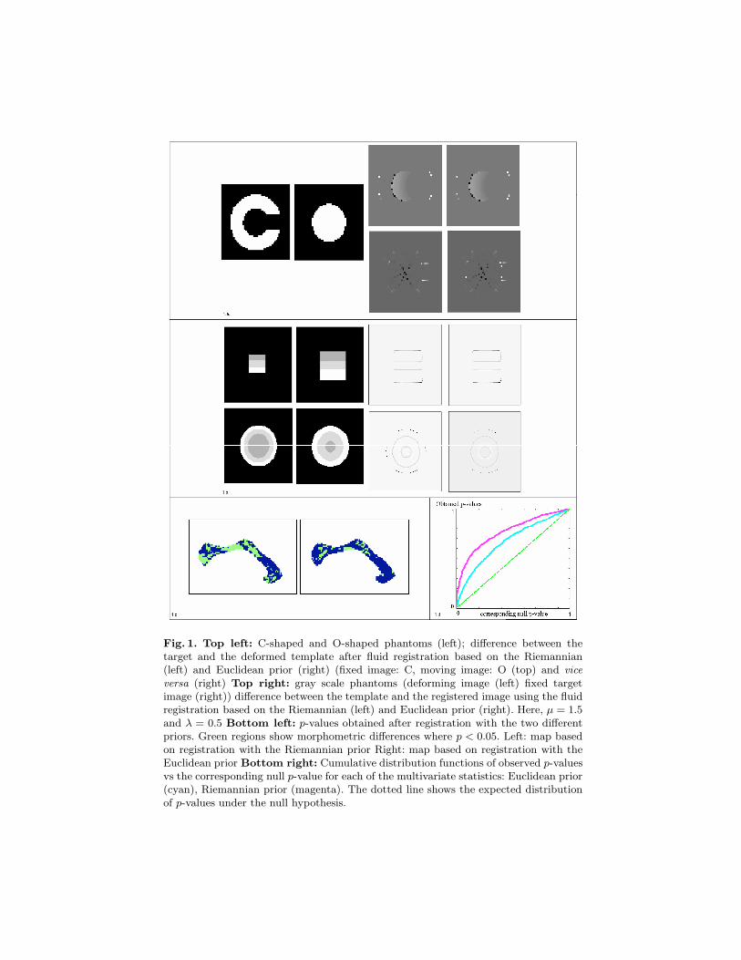
Fig. 1. Top left: C-shaped and O-shaped phantoms (left); difference between thetarget and the deformed template after fluid registration based on the Riemannian(left) and Euclidean prior (right) (fixed image: C, moving image: O (top) and viceversa (right) Top right: gray scale phantoms (deforming image (left) fixed targetimage (right)) difference between the template and the registered image using the fluidregistration based on the Riemannian (left) and Euclidean prior (right). Here, µ = 1.5and λ = 0.5 Bottom left: p-values obtained after registration with the two differentpriors. Green regions show morphometric differences where p < 0.05. Left: map basedon registration with the Riemannian prior Right: map based on registration with theEuclidean prior Bottom right: Cumulative distribution functions of observed p-valuesvs the corresponding null p-value for each of the multivariate statistics: Euclidean prior(cyan), Riemannian prior (magenta). The dotted line shows the expected distributionof p-values under the null hypothesis.


![Riemannian GeometryRiemannian Geometry Dr Emma Carberry Semester 2, 2015 Lecture 15 [Riemannian Geometry – Lecture 15]Riemannian Geometry – Lecture 15 Riemann Curvature Tensor](https://img.dokumen.tips/doc/110x75/5eaeb2e645c7213d450b3d20/riemannian-riemannian-geometry-dr-emma-carberry-semester-2-2015-lecture-15-riemannian.jpg)


![[SfN] Agreement between the white matter connectivity via tensor-based morphometry and the volumetric white matter parcellations](https://img.dokumen.tips/doc/110x75/55ae666e1a28ab306b8b4655/sfn-agreement-between-the-white-matter-connectivity-via-tensor-based-morphometry-and-the-volumetric-white-matter-parcellations.jpg)