Embed Size (px)
Citation preview
COMPARISON OF QUADRICEPS INACTIVATION BETWEEN NERVE ANDMUSCLE STIMULATIONNICOLAS PLACE, PhD,1 NICOLA CASARTELLI, MSc,2 JULIA F. GLATTHORN, MSc,2 and NICOLA A. MAFFIULETTI, PhD2
1 Institute of Movement Sciences and Sport Medicine, University of Geneva, Geneva, Switzerland2Neuromuscular Research Laboratory, Schulthess Clinic, Lengghalde 2, Zurich 8008, Switzerland
Accepted 19 April 2010
ABSTRACT: We evaluated the use of direct muscle stimula-tion for quantifying quadriceps inactivation at different contrac-tion levels as opposed to conventional twitch interpolation usingnerve stimulation. Fourteen healthy volunteers were tested.Paired stimuli were delivered to the femoral nerve or to thequadriceps muscle belly during voluntary contractions rangingfrom 20% to 100% of maximum, and the amplitude of thesuperimposed doublet was quantified to investigate inactivation.Superimposed doublet for muscle and nerve stimulation,respectively between the range of 60% to 100% of maximum(e.g., at 100%, muscle stimulation was 14 6 5 Nm and nervestimulation was 15 6 6 Nm). Despite higher current doses,muscle stimulation was associated with less discomfort thannerve stimulation (P < 0.05). Collectively, our data suggest thatdirect muscle stimulation could be used to assess quadricepsinactivation at maximal and quasi-maximal contraction levels asa valid alternative to motor nerve stimulation.
Muscle Nerve 42: 894–900, 2010
The level of voluntary drive during an effort canbe assessed in vivo using the twitch interpolationtechnique,1,2 which consists of superimposition ofone or more supramaximal stimuli during maximalvoluntary contraction (MVC); the amplitude of thesuperimposed response is generally believed toreflect the level of muscle inactivation.3 Submaxi-mal contractions can also be used to evaluate theextent of inactivation, which is extrapolated fromthe voluntary/superimposed torque relation.3,4
The twitch interpolation technique is reliable,5,6
and therefore it is widely used to assess the level ofinactivation of fresh and fatigued human muscles,particularly the quadriceps.7–10
Supramaximal stimulation of the femoral nerve(hereby referred to as nerve stimulation) is the‘‘gold standard’’ for assessment of quadriceps inac-tivation,11 as the superimposed stimulus theoreti-cally recruits all the motor units simultaneously.However, several concerns can be raised againstthe use of femoral nerve stimulation: (i) it is asso-ciated with discomfort, possibly leading to incom-plete voluntary activation,12 and its use can there-fore be limited in current-sensitive subjects and inpatients; (ii) the stimulating electrode is likely tobe pushed away from the femoral nerve during vol-untary contractions because of the nearby tendon,
so that delivery of a supramaximal stimulus isuncertain; and (iii) it could also recruit sartoriusmotor units, and this will inevitably reduce the am-plitude of the superimposed response and thusunderestimate inactivation. Furthermore, trunknerves are not always accessible, and therefore theassessment of inactivation is limited to muscleswith superficial motor nerves.
Therefore, it may be that direct muscle stimula-tion (hereafter referred to as muscle stimulation)would be more appropriate than nerve stimulationfor quantifying quadriceps inactivation. The use oflarge electrodes over the quadriceps is likely tominimize subjective discomfort,13 which can justifythe use of this stimulation modality for the assess-ment of inactivation in clinical populations14–17
Nevertheless, muscle stimulation results in incom-plete, random,18,19 and presumably superficialmotor unit recruitment,20 as no plateau in twitchtorque has been consistently obtained in previousstudies by gradually increasing stimulation inten-sity.5,21,22 Therefore, the validity of this modalityfor assessment of inactivation is questionable.
The main aim of this study was to evaluate theuse of muscle stimulation for quantifying quadri-ceps inactivation over a range of contraction levelsas opposed to the conventional twitch interpola-tion technique based on nerve stimulation. Wecompared quadriceps contractile properties, super-imposed doublet amplitude (i.e., inactivation), anddiscomfort scores between the two stimulationmodalities.
METHODS
Subjects. Fourteen healthy subjects (6 women),without any known orthopedic or neuromuscularproblems, volunteered to participate in this study.Their mean (6SD) age, height, and body masswere 27 6 5 years, 176 6 9 cm, and 69 6 11 kg,respectively. The local ethics committee approvedthe study, and all procedures were conductedaccording to the Declaration of Helsinki. Subjectsgave their written informed consent and wereasked not to take part in vigorous physical activityfor 2 days prior to their test date.
Experimental Setup and Protocol. All assessmentswere completed on the right quadriceps muscle
Abbreviations: ANOVA, analysis of variance; EMG, electromyography;MVC, maximal voluntary contraction; VAS, visual analog scale
Correspondence to: N.A. Maffiuletti; e-mail: [email protected]
VC 2010 Wiley Periodicals, Inc.Published online 6 October 2010 in Wiley Online Library (wileyonlinelibrary.com). DOI 10.1002/mus.21776
Key words: contractile properties, M-wave, MVC, potentiated doublet,vastus lateralis
894 Twitch Interpolation Technique MUSCLE & NERVE December 2010
under isometric conditions. Subjects were posi-tioned in the chair of an isokinetic device with thethighs parallel to the floor, the knee at 70� of flex-ion (0� ¼ full extension), and the trunk inclined15� with respect to the vertical to facilitate accessto the femoral nerve. Isometric knee extension tor-que was recorded using an isokinetic dynamometer(Biodex, Shirley, New York), with a lever armattached by a strap, 2–3 cm above the lateral mal-leolus. The torque signal was fed directly from thedynamometer into a 16-bit A/D converter, theninto a computer sampling at 1 kHZ using Acq-Knowledge software (BIOPAC Systems, Goleta, Cal-ifornia). Torque resolution was �0.3 Nm, and tem-poral resolution was 1 ms. The weight of the testedlimb was consistently quantified to allow torque tobe corrected for that resistance. Extraneous move-ments of the upper body were limited by two cross-over shoulder harnesses and a belt across theabdomen.
After electrode positioning for both muscleand nerve stimulation as well as for electromyo-graphic (EMG) activity recordings (see later), thesite of stimulation (muscle or nerve) used first wasrandomized by a coin flip. For each of the stimula-tion sites, once stimulation intensity was deter-mined (see later), the three main steps of the ex-perimental protocol consisted of: (i) singlestimulations (singlets) of increasing intensity(20%, 40%, 60%, 80%, and 100% of the predeter-mined stimulation intensity) under resting condi-tions to produce the recruitment curve of the peaktwitch torque; (ii) singlets and doublets under rest-ing conditions at the predetermined stimulationintensity to examine contractile properties; and(iii) doublets (100 HZ) superimposed on voluntarycontractions of different intensities (20%, 40%,60%, 80%, and 100% of the MVC) to investigatequadriceps inactivation as the size of the superim-posed doublet (Fig. 1A). These stimulations wereconsistently delivered 2–3 s after contraction onset(i.e., over the isometric torque plateau) and 1 s af-ter the end of the effort to obtain a potentiateddoublet response. We used superimposed doubletsinstead of singlets, because the force produced bythe former is greater and less variable than for asingle twitch (fibers that are in the refractory pe-riod at the arrival of the first stimulus will be acti-vated at least one time by the second stimulus).23
For both muscle and nerve stimulation, rectangu-lar pulses of 1 ms were produced via a constant-current stimulator (Model DS7-A; Digitimer, Hert-fordshire, UK).
A standardized warm-up, consisting of 8–10 sub-maximal voluntary isometric contractions of thequadriceps, was always completed before the super-imposed trials. Subjects were asked to complete
three MVCs followed by four randomly performedsubmaximal trials. For the MVC trials, subjectswere instructed to produce their maximal torquewithout any concern for the rate of torque devel-opment. Visual feedback and consistent verbalencouragement were provided throughout the vol-untary contractions. The duration of these contrac-tions was approximately 5 s, and 60 s of rest wereinterspersed between trials.
Immediately after each stimulation series(except for the recruitment curve), subjects wereasked to place a vertical mark on a horizontal line(100 mm) to rate the intensity of discomfort,which is known as a visual analog scale (VAS). Thescale therefore ranges from 0 to 100 mm, where0 mm ¼ ‘‘no discomfort’’ and 100 mm ¼ ‘‘worstpossible discomfort.’’
The EMG activity was recorded from the vastuslateralis muscle (as a surrogate for the quadricepsmuscle)6 during nerve stimulation at rest, to re-cord the M-wave response. Two pairs of silver-chlo-ride surface electrodes were positioned on a linetwo-thirds of the distance from the anterosuperioriliac spine to the lateral side of the patella, with aninterelectrode distance of 25 mm. Low resistancebetween the two electrodes (impedance <10 kX)was obtained by light abrasion of the skin and
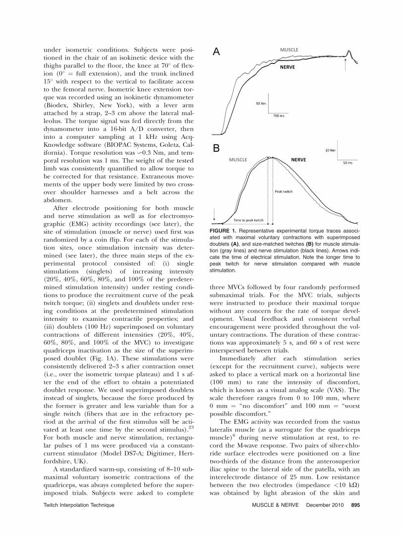
FIGURE 1. Representative experimental torque traces associ-
ated with maximal voluntary contractions with superimposed
doublets (A), and size-matched twitches (B) for muscle stimula-
tion (gray lines) and nerve stimulation (black lines). Arrows indi-
cate the time of electrical stimulation. Note the longer time to
peak twitch for nerve stimulation compared with muscle
stimulation.
Twitch Interpolation Technique MUSCLE & NERVE December 2010 895
cleaning with alcohol. The ground electrode wasfixed over the patella. EMG signals were amplifiedwith a bandwidth frequency ranging from 10 HZ to500 HZ (gain: 1000), digitized online at a samplingfrequency of 2 kHZ, and recorded by the system(MP150; BIOPAC, Goleta, California).
Muscle Stimulation. The quadriceps muscle wasstimulated using two large (12.7 � 7.6 cm) self-ad-hesive electrodes (Versastim; Conmed, Utica, NewYork) placed 5–10 cm below the inguinal creaseand 5–10 cm above the superior border of the pa-tella over the muscle belly of vastus lateralis, rectusfemoris, and vastus medialis. After electrodepositioning, current intensity was progressivelyincreased in an attempt to reach a plateau in peaktwitch torque. However, similar to other investiga-tors,5,21,22 we failed to observe any plateau, even atthe maximal stimulator output (100 mA). There-fore, 100 mA was arbitrarily considered to be themaximal intensity for muscle stimulation. Therecruitment curve was then constructed using 20,40, 60, 80, and 100 mA for all subjects, and 100-mA current intensity was used thereafter.
Nerve Stimulation. The femoral nerve was stimu-lated using a circular (diameter: 5.08 cm) self-ad-hesive electrode (Dermatrode; American Imex,Irvine, California) positioned in the femoral trian-gle, 3–5 cm below the inguinal ligament. A large(5 � 10 cm) rectangular self-adhesive electrode(Compex, Ecublens, Switzerland), was fixed overthe gluteal fold to close the stimulation currentloop. Current intensity was progressively increasedfrom 0 mA to full motor unit recruitment, as veri-fied by M-wave and peak twitch torque recordings,and this was further increased by 20–25% to pro-
vide supramaximal stimuli.6 The recruitment curvewas constructed using 20%, 40%, 60%, 80%, and100% of the supramaximal intensity, and the lattercurrent level was used thereafter.
Data Analysis. For both stimulation modalities,peak twitch torque was calculated from recruit-ment curve trials (20–100 mA for muscle stimula-tion and 20–100% of the supramaximal intensityfor nerve stimulation). For nerve stimulation, anM-wave (peak-to-peak amplitude) recruitmentcurve was also constructed. Moreover, we quanti-fied peak torque and time to peak torque for rest-ing responses (singlet, non-potentiated, and poten-tiated doublets). We also compared time to peaktorque for ‘‘matched’’ twitch sizes (i.e., twitcheswith similar peak torques), obtained respectively at100 mA for muscle stimulation and at 60% of thesupramaximal intensity for nerve stimulation (Fig.1B). The average response of all voluntary andevoked trials was consistently retained.
Statistics. After checks for normality, differencesin contractile properties and discomfort levelsbetween muscle stimulation and nerve stimulationwere examined using Student’s paired t-tests (two-tailed). Two-way repeated measures analyses ofvariance (ANOVAs) followed by least-significant-difference post hoc comparisons were also used toinvestigate the effect of stimulation site and inten-sity on recruitment curve characteristics and inacti-vation. An alpha level of P � 0.05 was consideredsignificant.
RESULTS
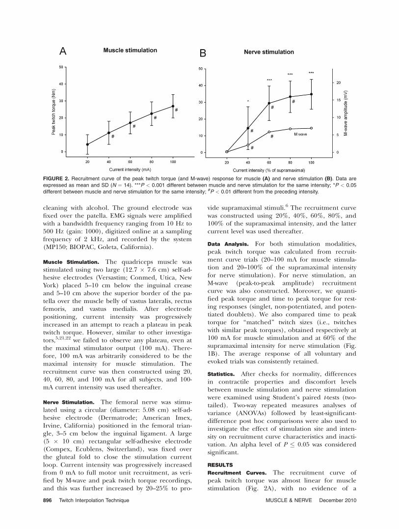
Recruitment Curves. The recruitment curve ofpeak twitch torque was almost linear for musclestimulation (Fig. 2A), with no evidence of a
FIGURE 2. Recruitment curve of the peak twitch torque (and M-wave) response for muscle (A) and nerve stimulation (B). Data are
expressed as mean and SD (N ¼ 14). ***P < 0.001 different between muscle and nerve stimulation for the same intensity; *P < 0.05
different between muscle and nerve stimulation for the same intensity; #P < 0.01 different from the preceding intensity.
896 Twitch Interpolation Technique MUSCLE & NERVE December 2010
plateau, whereas quasi-stable peak twitch torqueand M-wave responses were observed for nerve stim-ulation at current intensities of >80% (Fig. 2B).Despite lower stimulation intensities with nervestimulation (71 6 19 mA) compared with musclestimulation (100 6 0 mA) (P < 0.001), peak twitchtorque of differences was consistently higher in theformer condition (range: 3.4–12.4 Nm; P < 0.05),except at the lowest stimulation intensity (i.e., 20mA for muscle stimulation and 20% of supramaxi-mal intensity for nerve stimulation).
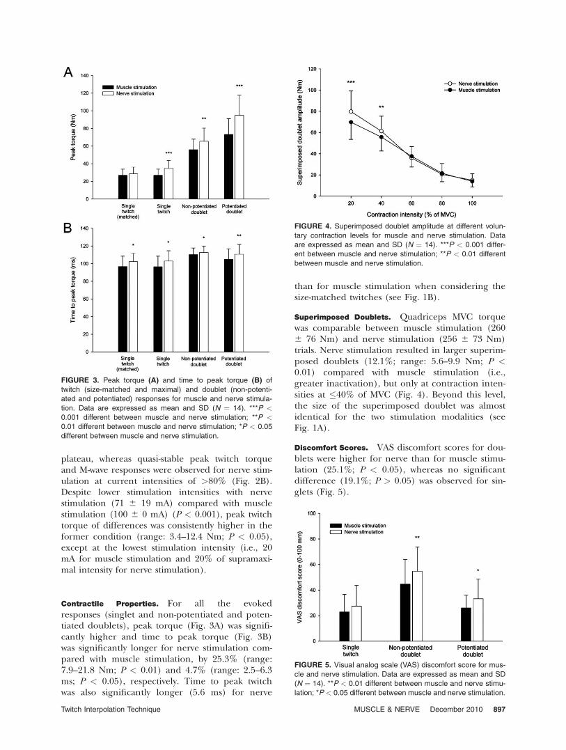
Contractile Properties. For all the evokedresponses (singlet and non-potentiated and poten-tiated doublets), peak torque (Fig. 3A) was signifi-cantly higher and time to peak torque (Fig. 3B)was significantly longer for nerve stimulation com-pared with muscle stimulation, by 25.3% (range:7.9–21.8 Nm; P < 0.01) and 4.7% (range: 2.5–6.3ms; P < 0.05), respectively. Time to peak twitchwas also significantly longer (5.6 ms) for nerve
than for muscle stimulation when considering thesize-matched twitches (see Fig. 1B).
Superimposed Doublets. Quadriceps MVC torquewas comparable between muscle stimulation (2606 76 Nm) and nerve stimulation (256 6 73 Nm)trials. Nerve stimulation resulted in larger superim-posed doublets (12.1%; range: 5.6–9.9 Nm; P <0.01) compared with muscle stimulation (i.e.,greater inactivation), but only at contraction inten-sities at �40% of MVC (Fig. 4). Beyond this level,the size of the superimposed doublet was almostidentical for the two stimulation modalities (seeFig. 1A).
Discomfort Scores. VAS discomfort scores for dou-blets were higher for nerve than for muscle stimu-lation (25.1%; P < 0.05), whereas no significantdifference (19.1%; P > 0.05) was observed for sin-glets (Fig. 5).
FIGURE 3. Peak torque (A) and time to peak torque (B) of
twitch (size-matched and maximal) and doublet (non-potenti-
ated and potentiated) responses for muscle and nerve stimula-
tion. Data are expressed as mean and SD (N ¼ 14). ***P <
0.001 different between muscle and nerve stimulation; **P <
0.01 different between muscle and nerve stimulation; *P < 0.05
different between muscle and nerve stimulation.
FIGURE 4. Superimposed doublet amplitude at different volun-
tary contraction levels for muscle and nerve stimulation. Data
are expressed as mean and SD (N ¼ 14). ***P < 0.001 differ-
ent between muscle and nerve stimulation; **P < 0.01 different
between muscle and nerve stimulation.
FIGURE 5. Visual analog scale (VAS) discomfort score for mus-
cle and nerve stimulation. Data are expressed as mean and SD
(N ¼ 14). **P < 0.01 different between muscle and nerve stimu-
lation; *P < 0.05 different between muscle and nerve stimulation.
Twitch Interpolation Technique MUSCLE & NERVE December 2010 897
DISCUSSION
The main findings of this study were that quadri-ceps muscle stimulation and femoral nerve stimula-tion resulted in similar inactivation levels (as wit-nessed by superimposed doublet amplitude) forcontraction intensities �60% of MVC, despite theuse of submaximal current intensities (maximalstimulator output) and weaker (resting) contractileresponse in the former condition. Muscle stimula-tion resulted in lower discomfort scores and fasterevoked responses (shorter time to peak torque)than nerve stimulation.
The two stimulation modalities (muscle vs.nerve) certainly lead to different motor unitrecruitments. The femoral nerve contains axonsfrom all a-motoneurons that innervate the quadri-ceps muscle; therefore, theoretically, the torqueresponse induced by maximal stimulation of thefemoral nerve reflects the synchronous response ofall motor units, independent of their location(deep or superficial). In contrast, excitation of theterminal branches of the motoneuron using mus-cle stimulation would result in incomplete motorunit recruitment,24,25 as also confirmed by the cur-rent resting peak torque differences between mus-cle and nerve stimulation (25%). Indeed, musclefibers innervated by motor nerve endings locatedunder the stimulating electrodes are primarily acti-vated by muscle stimulation; that is, recruitment israndom18,19 and mostly,20 although not exclusively,superficial.25
Our data show that time to peak torque oftwitch and doublet responses was shorter for mus-cle than for nerve stimulation, which suggests thepossibility of a preferential recruitment of the su-perficial, faster contracting motor units in the for-mer condition. The shorter time to peak torquefor the maximal twitch obtained with muscle stim-ulation compared with the submaximal size-matched twitch evoked by nerve stimulation (seeFig. 3) strengthens this speculation and excludesany twitch amplitude effect on the observedresults. Interestingly, it has previously been demon-strated that larger motor units are mainly locatedin superficial regions of the vastus lateralis mus-cle.26 Taken together, these facts suggest that,under the current experimental conditions, therelative proportion of the fast motor units acti-vated by the electrical current would have beenhigher for muscle than for nerve stimulation,because of limited current diffusion to the deepest(mainly small)26 motor units in the former condi-tion. However, this assumption may not necessarilybe generalized to muscle groups other than thequadriceps femoris.
The different recruitment pattern for muscle(incomplete, superficial, and random) vs. nerve
stimulation is also evidenced by the absence of aplateau in peak twitch torque with increasing stim-ulation intensity, as already reported.5,21,22 It istherefore tempting to suggest that quadriceps con-tractile properties obtained with muscle stimula-tion under resting conditions may not be represen-tative of the whole quadriceps muscle.Nevertheless, it may be that the use of an electricalstimulator with a maximal output >100 mA couldhave led to a plateau of peak twitch force; it wouldthen be of interest to verify whether the plateaucould be achieved without excessive discomfortassociated with the stimulation.
Surprisingly, such an evident limitation of mus-cle stimulation to characterize contractile proper-ties at rest had no or limited effect on superim-posed responses at high torque levels (�60%MVC), because of the possibility of activating thosefast muscle fibers that are not or partially activatedby the ongoing voluntary contraction. This repre-sents the main interest of muscle stimulation inthe context of twitch interpolation, even if thesuperimposed stimuli are not necessarily supramax-imal (lack of plateau). Moreover, the use of sub-maximal current intensities limits stimulus spreadto adductor and antagonist muscles, which is oneof the main pitfalls of supramaximal trunk nervestimulation. Our results of similar superimposedresponses at high torque levels between the twostimulation modalities are compatible with previ-ous findings. In fact, Rutherford et al.27 and New-man et al.28 demonstrated similar activation scoresbetween maximally tolerated muscle stimulationand supramaximal stimulation of the femoralnerve, either electrical or magnetic. The use ofpaired stimuli in our study certainly improved thesensitivity of the twitch interpolation technique ascompared with use of singlets in previous stud-ies.23,27,28 This may explain, at least in part, thelack of difference in superimposed responsesbetween muscle and nerve stimulation at low MVClevels (20–60%), as demonstrated by Rutherfordet al.27
However, nerve stimulation also has limitationsfor twitch interpolation experiments, which canlimit its clinical implementation. First, the positionof the electrode–nerve interface may change dur-ing voluntary contractions because of the nearbytendon, so that supramaximal stimulation, as veri-fied at rest, remains uncertain. Second, femoralnerve stimulation could possibly activate the sarto-rius muscle, which is an antagonist of the quadri-ceps muscle, and this would inevitably tend toreduce the size of the superimposed response.Third, the current results confirm that singlets anddoublets delivered over the trunk nerve result inrelatively high discomfort levels in healthy subjects,
898 Twitch Interpolation Technique MUSCLE & NERVE December 2010

thus the use of submaximal muscle stimulationcould be valuable for the evaluation of muscleinactivation in frail or current-sensitive subjects.
Transcutaneous electrical stimulation–induceddiscomfort, which varies considerably betweenindividuals,29 is mainly mediated by Ad and Cfibers.30 Ab fibers are recruited at very low levelsof stimulation, such as at detection threshold,contrary to smaller Ad and C fibers, which arerecruited at higher, more uncomfortable, stimula-tion intensities.31 The use of superimposed elec-trical stimuli may also induce discomfort,32 andthe expectation of a noxious stimulus may reducevoluntary effort in unaccustomed subjects.12 Thehigher discomfort with nerve stimulation com-pared with muscle stimulation (despite lower stim-ulation intensities) may thus be attributed tolarger and/or more densely packed Ad and C no-ciceptor axons in the trunk nerve rather than inthe skin.33 In addition, a greater pulse-chargedensity (current dose per stimulated area) cer-tainly contributes to this altered perception, as asimple calculation indicates that pulse-charge den-sity was three times higher under the stimulatingelectrode with nerve stimulation compared withmuscle stimulation. In fact, one study showedthat pain associated with stimulation over thefemoral nerve was lower with large than withsmall electrodes.13
Further studies are required to extend theseresults to muscles other than the quadriceps andalso to patient populations. Knowing the differ-ence in evoked muscle response and related dis-comfort between different stimulation modalities(including magnetic stimulation) could be valuableto clinicians who may want to use the twitch inter-polation technique for evaluating muscle inactiva-tion. In addition, comparison of muscle vs. nervestimulation under fatigue conditions would requirefurther investigation, although inactivation mightbe overestimated with the twitch interpolationtechnique.34
Within the limits of the study, our findingslegitimize the use of muscle stimulation fortwitch interpolation applications, but only forstrong voluntary contractions (�60% MVC).Because the choice of stimulation procedures isparamount to the success of the twitch interpola-tion technique, we suggest that electrical stimula-tion over the quadriceps muscle belly is a validalternative to femoral nerve stimulation. Ourfindings also provide further insights into theneurophysiological mechanisms that underlieelectrical stimulation of intact human muscles ortrunk nerves. Finally, we do not recommend theuse of muscle stimulation for assessment of quad-riceps contractile properties under resting condi-
tions, because motor unit recruitment is incom-plete and nonselective.
The authors thank Severin Stahli for help with some of theexperiments.
REFERENCES
1. Merton PA. Voluntary strength and fatigue. J Physiol 1954;123:553–564.
2. Gandevia SC. Spinal and supraspinal factors in human musclefatigue. Physiol Rev 2001;81:1725–1789.
3. Shield A, Zhou S. Assessing voluntary muscle activation with thetwitch interpolation technique. Sports Med 2004;34:253–267.
4. Folland JP, Williams AG. Methodological issues with the interpolatedtwitch technique. J Electromyogr Kinesiol 2007;17:317–327.
5. Behm DG, St-Pierre DM, Perez D. Muscle inactivation: assessment ofinterpolated twitch technique. J Appl Physiol 1996;81:2267–2273.
6. Place N, Maffiuletti NA, Martin A, Lepers R. Assessment of the reli-ability of central and peripheral fatigue after sustained maximal vol-untary contraction of the quadriceps muscle. Muscle Nerve 2007;35:486–495.
7. Place N, Lepers R, Deley G, Millet GY. Time course of neuromuscu-lar alterations during a prolonged running exercise. Med Sci SportsExerc 2004;36:1347–1356.
8. Nybo L. CNS fatigue and prolonged exercise: effect of glucose sup-plementation. Med Sci Sports Exerc 2003;35:589–594.
9. Suter E, Herzog W, Huber A. Extent of motor unit activation in thequadriceps muscles of healthy subjects. Muscle Nerve 1996;19:1046–1048.
10. Behm DG, Whittle J, Button D, Power K. Intermuscle differences inactivation. Muscle Nerve 2002;25:236–243.
11. Maffiuletti NA. Assessment of hip and knee muscle function in or-thopedic practice. J Bone J Surg 2010;92:220–229.
12. Button DC, Behm DG. The effect of stimulus anticipation on theinterpolated twitch technique. J Sports Sci Med 2008;7:520–524.
13. Alon G. High voltage stimulation. Effects of electrode size on basicexcitatory responses. Phys Ther 1985;65:890–895.
14. Urbach D, Nebelung W, Becker R, Awiszus F. Effects of reconstruc-tion of the anterior cruciate ligament on voluntary activation ofquadriceps femoris a prospective twitch interpolation study. J BoneJoint Surg [Br] 2001;83:1104–1110.
15. Molloy CB, Al-Omar AO, Edge KT, Cooper RG. Voluntary activationfailure is detectable in some myositis patients with persisting quadri-ceps femoris weakness: an observational study. Arthritis Res Ther2006;8:R67.
16. Horemans HL, Beelen A, Nollet F, Jones DA, Lankhorst GJ. Repro-ducibility of maximal quadriceps strength and its relationship tomaximal voluntary activation in postpoliomyelitis syndrome. ArchPhys Med Rehabil 2004;85:1273–1278.
17. Mizner RL, Stevens JE, Snyder-Mackler L. Voluntary activation anddecreased force production of the quadriceps femoris muscle aftertotal knee arthroplasty. Phys Ther 2003;83:359–365.
18. Jubeau M, Gondin J, Martin A, Sartorio A, Maffiuletti NA. Randommotor unit activation by electrostimulation. Int J Sports Med 2007;28:901–904.
19. Gregory CM, Bickel CS. Recruitment patterns in humanskeletal muscle during electrical stimulation. Phys Ther 2005;85:358–364.
20. Vanderthommen M, Depresseux JC, Dauchat L, Degueldre C, Crois-ier JL, Crielaard JM. Spatial distribution of blood flow in electricallystimulated human muscle: a positron emission tomography study.Muscle Nerve 2000;23:482–489.
21. Hanchard NC, Williamson M, Caley RW, Cooper RG. Electrical stim-ulation of human tibialis anterior: (A) contractile properties are sta-ble over a range of submaximal voltages; (B) high- and low-frequency fatigue are inducible and reliably assessable at submaximalvoltages. Clin Rehabil 1998;12:413–427.
22. Mannion AF, Dolan P, Adam GG, Adams MA, Cooper RG. Can maxi-mal back muscle strength be predicted from submaximal efforts? JBack Musculoskel Rehabil 1997;9:49–51.
23. Duchateau J. Stimulation conditions can improve the validity of theinterpolated twitch technique. J Appl Physiol 2009;107:361.
24. Hultman E, Sjoholm H, Jaderholm-Ek I, Krynicki J. Evaluation ofmethods for electrical stimulation of human skeletal muscle in situ.Pflugers Arch 1983;398:139–141.
25. Adams GR, Harris RT, Woodard D, Dudley GA. Mapping ofelectrical muscle stimulation using MRI. J Appl Physiol 1993;74:532–537.
26. Knight CA, Kamen G. Superficial motor units are larger than deepermotor units in human vastus lateralis muscle. Muscle Nerve 2005;31:475–480.
Twitch Interpolation Technique MUSCLE & NERVE December 2010 899
27. Rutherford OM, Jones DA, Newham DJ. Clinical and experimentalapplication of the percutaneous twitch superimposition techniquefor the study of human muscle activation. J Neurol Neurosurg Psy-chiatry 1986;49:1288–1291.
28. Newman SA, Jones G, Newham DJ. Quadriceps voluntary activationat different joint angles measured by two stimulation techniques.Eur J Appl Physiol 2003;89:496–499.
29. Delitto A, Strube MJ, Shulman AD, Minor SD. A study of discomfortwith electrical stimulation. Phys Ther 1992;72:410–421.
30. Julius D, Basbaum AI. Molecular mechanisms of nociception. Nature2001;413:203–210.
31. Collins WR Jr, Nulsen FE, Randt CT. Relation of peripheral nervefiber size and sensation in man. Arch Neurol 1960;3:381–385.
32. Miller M, Downham D, Lexell J. Effects of superimposed electricalstimulation on perceived discomfort and torque increment size andvariability. Muscle Nerve 2003;27:90–98.
33. Sang CN, Max MB, Gracely RH. Stability and reliability of detectionthresholds for human A-beta and A-delta sensory afferents determinedby cutaneous electrical stimulation. J Pain Sympt Manage 2003;25:64–73.
34. Place N, Yamada T, Bruton JD, Westerblad H. Interpolated twitchesin fatiguing single mouse muscle fibres: implications for the assess-ment of central fatigue. J Physiol 2008;586:2799–2805.
900 Twitch Interpolation Technique MUSCLE & NERVE December 2010
![Surface neuromuscular electrical stimulation for ...doras.dcu.ie/19651/1/dpom4.pdf · [Intervention Review] Surface neuromuscular electrical stimulation for quadriceps strengthening](https://img.dokumen.tips/doc/110x75/5f36ebff4193e847ed61bb54/surface-neuromuscular-electrical-stimulation-for-dorasdcuie196511dpom4pdf.jpg)