Embed Size (px)
Citation preview

RESEARCH ARTICLE Open Access
Comparison of percutaneous curvedkyphoplasty and bilateral percutaneouskyphoplasty in osteoporotic vertebralcompression fractures: a randomizedcontrolled trialChen Wang1,2†, Yu Zhang2†, Wang Chen2, Shi-Lei Yan2, Kai-Jin Guo1,2* and Shuo Feng2*
Abstract
Background: Here we compared the clinical efficacy of bilateral percutaneous kyphoplasty (PKP) and percutaneouscurved kyphoplasty (PCKP) in the treatment of osteoporotic vertebral compression fractures (OVCF).
Methods: Seventy-two patients with single-level thoracolumbar OVCF were randomly divided into 2 groups (36patients in each) and were subjected to either PCKP or bilateral PKP. The intraoperative fluoroscopy time, totalsurgical time, bone cement injection volume, bone cement leakage, preoperative and postoperative anteriorvertebral height, Cobb angles, visual analog scales (VAS) and oswestry disability index questionnaire (ODI) wererecorded.
Results: Both groups of patients had a trend towards improvements in VAS and ODI scores 24 h and 6monthsafter surgery, when compared to preoperative results, despite lack of statistical significance. The total surgical andintraoperative fluoroscopy times and intraoperative bone cement injection volume were significantly decreased inthe PCKP group than those in the PKP group. The anterior edge height and Cobb angle of the injured vertebrawere similarly improved after operation in both groups.
Conclusion: PCKP is safer, less invasive and quicker than traditional bilateral PKP despite similar short-term effectsfor the treatment of OVCF.
Trial registration: ChiCTR, ChiCTR2100042859. Registered 25 January 2021- Retrospectively registered.
Keywords: Osteoporosis, Vertebral compression fractures, Percutaneous curved kyphoplasty, Kyphoplasty
© The Author(s). 2021 Open Access This article is licensed under a Creative Commons Attribution 4.0 International License,which permits use, sharing, adaptation, distribution and reproduction in any medium or format, as long as you giveappropriate credit to the original author(s) and the source, provide a link to the Creative Commons licence, and indicate ifchanges were made. The images or other third party material in this article are included in the article's Creative Commonslicence, unless indicated otherwise in a credit line to the material. If material is not included in the article's Creative Commonslicence and your intended use is not permitted by statutory regulation or exceeds the permitted use, you will need to obtainpermission directly from the copyright holder. To view a copy of this licence, visit http://creativecommons.org/licenses/by/4.0/.The Creative Commons Public Domain Dedication waiver (http://creativecommons.org/publicdomain/zero/1.0/) applies to thedata made available in this article, unless otherwise stated in a credit line to the data.
* Correspondence: [email protected]; [email protected]†Chen Wang and Yu Zhang contributed equally to this work.1Department of Orthopedic Surgery, Nanjing Medical University, Nanjing,Jiangsu, China2Department of Orthopedic Surgery, Affiliated Hospital of Xuzhou MedicalUniversity, 99 Huaihai Road, Xuzhou 221002, Jiangsu, China
Wang et al. BMC Musculoskeletal Disorders (2021) 22:588 https://doi.org/10.1186/s12891-021-04469-1

BackgroundOsteoporotic vertebral compression fractures (OVCF)[1] are common clinical fractures, especially in the eld-erly. The loss of bone height and vertebral body com-pression fractures lead to kyphotic deformities in thevertebral spine of patients, causing long-term back painand seriously affecting their quality of life. A conserva-tive approach using pharmacological treatment is appro-priate for specific populations [2].At present, percutaneous kyphoplasty (PKP) is
widely performed clinically to treat OVCF [3–5]. Bal-loon expansion can be used to correct kyphoplastyand make a cavity, so that bone cement can beinjected at a relatively low pressure to reduce leakage,and postoperative pain can be significantly relieved.The commonly used puncture approaches include aunilateral straight approach and a bilateral transpedi-cular approach, both of which can improve the backpain of patients [3, 5]. However, in the traditionalunilateral straight approach, bone cement can oftenonly fill the ipsilateral vertebral bone, leaving thecontralateral vertebral bone poorly filled [5]. The bi-lateral transpedicular approach also has disadvantages,such as a long operation time, need of several punc-tures and a large amount of radiation, which can leadto the “binocular” phenomenon that occurs when thebone cement injected from both sides do not fuse [3].Therefore we have focused our research on achievinga uniform distribution of bone cement in the verte-bral body through a unilateral approach. In recentyears, the introduction of percutaneous curved kypho-plasty (PCKP) provided a new approach for the treat-ment of OVCF [6].Thus, the present prospective study compared the
clinical efficacy of PCKP with that of PKP in OVCF todefine the optimal surgical treatment.
MethodsStudy designThis is a prospective and controlled study that includedresearchers who analyzed patient data and were trainedin the study methods but did not know the patientpopulation. This study was conducted in our orthopedicDepartment from January 2019 to January 2020. Thisstudy was approved by the ethics Committee of our hos-pital, and its design conforms to the Regulations on theManagement of Medical Institutions. This trial is regis-tered at Chinese Clinical Trials Registry, numberChiCTR2100042859. The CONSORT flow diagram isprovided in Fig. 1.
Inclusion and exclusion criteriaThe inclusion criteria for this study were patients withOVCF who were able to tolerate surgery and cooperate
with postoperative functional exercise in our hospital.Patients in the study were informed of treatment plansand surgical risks and signed informed consent forms.Therefore, the inclusion criteria were: (1) patients aged> 65 years, both men and women; (2) painful OVCF and(3) granted informed consent to enroll in the trial.The exclusion criteria were (1) vertebra tuberculosis
and bacterial infection; (2) bleeding and clotting dys-functions that could not be corrected or patients with ableeding tendency; (3) extensive and largely incompletevertebral posterior margin bone destruction; (4) > 70%compression degree of vertebral bodies; (5) two or morevertebral bodies compression fractures; (6) participationin other drug or medical device clinical trials within 30days prior to screening and (7) the researcher judgedthat the patient had poor compliance and could notcomplete the study as required.
Patient randomization and sample size calculationA total of 78 patients were initially identified. Two pa-tients did not meet inclusion criteria, 2 patients refusedto participate, 2 patients were lost during follow-up,leaving a total of 72 patients included. There were 22males and 50 females, aged 66–87 years (average, 76.04years). Preoperative routine examination consisted ofblood routine (Hb, HBC, WBC and lymphocyte count),coagulation routine (TT, APTT and PT) and lower limbcolor Doppler ultrasonography. At the time of admis-sion, patients were numbered consecutively from 1 to72. The patients were randomly divided into a studygroup (n = 36) and a control group (n = 36) using a ran-dom distribution software. The selection process of pa-tients in the two groups is shown in Fig. 1.To calculate the sample size, the observational cohort
study was powered to detect bone cement infusion vol-ume as the minimum mean difference of significance,and calculated standardized difference (0.953) usingstandard deviation (1.202) based on an earlier report byCheng et al. [6]. It was estimated that 32 participantswould be required to enable detection of significant dif-ferences, at the 5% significance level, with 95% power.
Surgical proceduresPCKP (percutaneous curved kyphoplasty) groupPercutaneous curved kyphoplasty puncture needle andbone cement high-pressure perfusion instrument werepurchased from Ningbo Huarun Biotechnology Co.(China), while acrylic resin bone cement was purchasedfrom StrykerInstruments (France) (Fig. 2).The patient was placed in a prone position, with a soft
padded chest. The C-arm machine was used for fluoros-copy, to locate the damaged vertebral body and to deter-mine and mark the pedicle puncture needlepoint. Afterlocal anesthesia, a unilateral pedicle puncture was
Wang et al. BMC Musculoskeletal Disorders (2021) 22:588 Page 2 of 9

performed, and the puncture needle was placed at theposterior 1/3 of the vertebral body. After the core of thepuncture needle was pulled out, the curved catheter wasplaced into the vertebra along the straight cannula. Theanteroposterior fluoroscopy showed that the end of thecurved catheter crossed the midline of the vertebra toreach the opposite side, while the lateral fluoroscopyreached 1/3 of the anterior middle vertebra. The work-ing channel at the bending angle of the vertebral bodywas established. The vacuum-extracted vertebral dilationballoon catheter was inserted into the vertebral bodythrough the working channel. The balloon had com-pletely entered the vertebral body when the proximalblack mark entered the puncture channel, upon which itwas expanded and the expected position of the vertebralbody was observed through fluoroscopy. The bone ce-ment was mixed evenly and injected into the establishedexpansion channel through the injection delivery system.Then the bone cement was slowly injected into the
Fig. 2 Percutaneous curved kyphoplasty puncture needle (NingboHuarun Biotechnology Co., LTD., China)
Fig. 1 Flow chart of the analysis. After exclusions, a total of 72 patients were followed up
Wang et al. BMC Musculoskeletal Disorders (2021) 22:588 Page 3 of 9
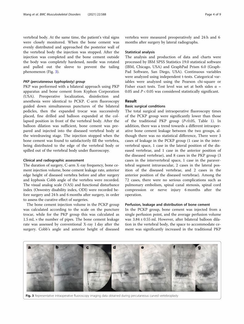
vertebral body. At the same time, the patient’s vital signswere closely monitored. When the bone cement wasevenly distributed and approached the posterior wall ofthe vertebral body the injection was stopped. After theinjection was completed and the bone cement outsidethe body was completely hardened, needle was rotatedand pulled out the sleeve to prevent the tailingphenomenon (Fig. 3).
PKP (percutaneous kyphoplasty) groupPKP was performed with a bilateral approach using PKPapparatus and bone cement from Kyphon Corporation(USA). Preoperative localization, disinfection andanesthesia were identical to PCKP. C-arm fluoroscopyguided down simultaneous punctures of the bilateralpedicles, then the expanded trocar was successivelyplaced, fine drilled and balloon expanded at the col-lapsed position in front of the vertebral body. After theballoon dilation was sufficient, bone cement was pre-pared and injected into the diseased vertebral body atthe wiredrawing stage. The injection stopped when thebone cement was found to satisfactorily fill the vertebra,being distributed to the edge of the vertebral body orspilled out of the vertebral body under fluoroscopy.
Clinical and radiographic assessmentThe duration of surgery, C-arm X-ray frequency, bone ce-ment injection volume, bone cement leakage rate, anterioredge height of diseased vertebra before and after surgeryand kyphosis Cobb angle of the vertebra were recorded.The visual analog scale (VAS) and functional disturbanceindex (Oswestry disability index, ODI) were recorded be-fore surgery and 24 h and 6months after surgery, in orderto assess the curative effect of surgeries.The bone cement injection volume in the PCKP group
was calculated according to the scale on the puncturetrocar, while for the PKP group this was calculated as1.5 mL × the number of pipes. The bone cement leakagerate was assessed by conventional X-ray 1 day after thesurgery. Cobb’s angle and anterior height of diseased
vertebra were measured preoperatively and 24 h and 6months after surgery by lateral radiographs.
Statistical analysisThe analysis and production of data and charts wereprocessed by IBM SPSS Statistics 19.0 statistical software(IBM, Chicago, USA) and GraphPad Prism 6.0 (Graph-Pad Software, San Diego, USA). Continuous variableswere analyzed using independent t-tests. Categorical var-iables were analyzed using the Pearson chi-square orFisher exact tests. Test level was set at both sides α =0.05 and P < 0.05 was considered statistically significant.
ResultBasic surgical conditionsThe total surgical and intraoperative fluoroscopy timesof the PCKP group were significantly lower than thoseof the traditional PKP group (P<0.05, Table 1). Inaddition, there was a trend towards a different intraoper-ative bone cement leakage between the two groups, al-though there was no statistical difference, There were 3cases of leakage in the PCKP group (1 case in the inter-vertebral space, 1 case in the lateral position of the dis-eased vertebrae, and 1 case in the anterior position ofthe diseased vertebrae), and 8 cases in the PKP group (3cases in the intervertebral space, 1 case in the paraver-tebral segment intravascular, 2 cases in the lateral pos-ition of the diseased vertebrae, and 2 cases in theanterior position of the diseased vertebrae). Among the72 cases, there were no serious complications such aspulmonary embolism, spinal canal stenosis, spinal cordcompression or nerve injury 6 months after theoperation.
Perfusion, leakage and distribution of bone cementIn the PCKP group, bone cement was injected from asingle perfusion point, and the average perfusion volumewas 3.84 ± 0.55 ml. However, after bilateral balloon dila-tion in the vertebral body, the space to accommodate ce-ment was significantly increased in the traditional PKP
Fig. 3 Representative intraoperative fluoroscopy imaging data obtained during percutaneous curved vertebroplasty
Wang et al. BMC Musculoskeletal Disorders (2021) 22:588 Page 4 of 9

group. Therefore, the traditional PKP group had a higherbone cement perfusion volume, with an average perfu-sion volume of 4.78 ± 0.67 ml, compared with that of thePCKP group (P<0.05, Table 1). In the traditional PKPgroup, there was only 1 case of small paravertebral seg-ment intravascular leakage and no distant intravascular
leakage. Three cases of bone cement leakage occurred inthe PCKP group in the paravertebral body without vas-cular leakage. When the puncture needle reached theideal position in the vertebral body, bone cement couldbe distributed in the anterior and middle part of the ver-tebral body in PCKP group (Fig. 3), while the bone ce-ment injected by traditional PKP after bilateral balloondilation was mainly distributed in the sides of the verte-bral body.
Radiographic resultsThe anterior height of the vertebral body in the PCKPgroup and traditional PKP group were respectively21.93 ± 4.05 mm and 20.95 ± 3.34 mm before the oper-ation, and were increased by 2.79 mm (final 24.72 ± 3.47mm) and 3.01 mm (final 23.96 ± 3.36 mm) respectivelyafter the operation(P<0.05) (Fig. 4). However, there wasno statistically significant difference in the height of theanterior vertebral body between the two groups beforeand after surgery (P>0.05, Table 2).Cobb’s angles in the PCKP group were 16.44° ± 9.06°,
while that of the traditional PKP group was 18.01° ± 12.00°before operation. After the operation, Cobb’s angles
Fig. 4 Comparison of the preoperative and postoperative anterior height of diseased vertebrae and cobb angles between the two groups
Table 1 Comparison of basic data between the two groups
classification PCKP group PKP group P values
Number(n) 36 36
Sex (male/female) 10/26 12/24 0.609
Age (years) 75.55 ± 6.11 76.52 ± 6.24 0.506
BMI kg/m2) 22.99 ± 2.06 23.19 ± 1.97 0.674
BMD 2.55 ± 0.65 2.62 ± 0.78 0.684
Comorbidity
Hypertension 9 11 0.599
Diabetes 7 6 0.759
operative time (min) 39.30 ± 7.87 48.19 ± 9.00 0.000
X-ray frequency (n) 19.97 ± 4.70 29.66 ± 5.98 0.000
Infusion volume (ml) 3.84 ± 0.55 4.78 ± 0.67 0.000
Cement leakage (n) 3 8 0.101
Wang et al. BMC Musculoskeletal Disorders (2021) 22:588 Page 5 of 9

decreased to 10.76° ± 10.10° and 12.35° ± 13.53° for PCKPand PKP groups respectively (P<0.05). There was no sig-nificant difference in Cobb’s angle between the two groupsbefore and after operation (P>0.05, Table 2).
Clinical resultsThe VAS score and ODI of PCKP and traditional PKPgroups were significantly improved 1 day after the oper-ation (P<0.05). Six months after surgery, the VAS scoreand ODI of both groups were also significantly improvedcompared with that of 24 h after surgery (P<0.05). How-ever, there was no difference in VAS score and ODI
between PCKP and traditional PKP groups at any timepoint (Table 2).
Postoperative complicationsIn this study, a small amount of bone cement leakageoccurred in some patients during surgery. There were 3patients in the PCKP group (total leakage rate 8.3%,Fig. 5), and 8 patients in the traditional PKP group (totalleakage rate 22.2%). Most of the bone cement leakagewas characterized by leakage along the fracture line,while 1 case in the traditional PKP group had paraver-tebral segment intravascular leakage, because the bone
Table 2 Comparison of pain and functional efficacy between the two groups
classification PCKP group PKP group P values
VAS score (score)
Pre-surgery 7.58 ± 1.30(6 ~ 10) 7.39 ± 0.96 (6 ~ 10) 0.473
24 h post-surgery 2.11 ± 0.57 (1 ~ 3) 2.00 ± 0.58 (1 ~ 3) 0.419
6-months post-surgery 1.33 ± 0.47 (1 ~ 2) 1.27 ± 0.45 (1 ~ 2) 0.615
ODI
Pre-surgery 70.75 ± 10.02 72.63 ± 9.35 0.411
24 h post-surgery 34.27 ± 7.06 35.88 ± 7.45 0.350
6-months post-surgery 23.52 ± 4.45 23.97 ± 4.43 0.672
Anterior height of diseased vertebrae (mm)
Pre-surgery 21.93 ± 4.05 20.95 ± 3.34 0.268
24 h post-surgery 24.72 ± 3.47 23.96 ± 3.36 0.353
6-months post-surgery 24.50 ± 3.42 23.67 ± 3.18 0.290
Cobb angles(°)
Pre-surgery 16.44 ± 9.06 18.01 ± 12.00 0.532
24 h post-surgery 10.76 ± 10.10 12.35 ± 13.53 0.576
6-months post-surgery 10.91 ± 10.05 12.51 ± 13.45 0.571
Fig. 5 Postoperative bone cement leakage occurred in PCKP group, the patient had no symptoms
Wang et al. BMC Musculoskeletal Disorders (2021) 22:588 Page 6 of 9

cement did not enter the drawing stage and was injectedprematurely (Table 3).In this study, during the perfusion of bone cement, all
cases that have used an X-ray real-time dynamic moni-tor were able to stop the perfusion when the leakage wasobserved.
DiscussionOsteoporotic fracture of the vertebral body is very com-mon in the elderly, and traditional treatment requireslong-term bed rest, fixation and drug treatment. Due toreduced activity, osteoporosis is further aggravated inpatients, and fractures occur repeatedly. Meanwhile,spending a long time in bed leads to bedsore, deep ven-ous thrombosis and other complications [7]. Osteopor-otic fractures of vertebral bodies can seriously affect thequality of life of patients and threaten their physical andmental health [8]. Therefore, pain relief, early activityand spinal stabilization are the key points in the treat-ment of thoracolumbar osteoporotic compression frac-tures [9]. PKP can reconstruct the vertebral body height,increase the stiffness of vertebral bodies, immediatelystabilize the vertebral body, quickly relieve back pain, re-duce the complications in bed, improve cardiopulmo-nary function, improve the quality of life of elderlypatients and is currently the preferred treatment ofOVCF [10, 11].In recent years many scholars have pro-posed the use of a unilateral pedicle puncture in PKP [5,6, 12, 13]. Compared with bilateral PKP, unilateral PKPproduced advantages such as a shorter surgery time,smaller dosage of cement, lower risk of cement leakage,and relieved a higher degree of intractable pain at short-term follow-up after surgery [14]. Indeed, unilateralpuncture PKP can reduce both the operation time andcomplications of bilateral punctures [12]. However, itmay cause an uneven distribution of bone cement onboth sides of the vertebral body, thus resulting in wedgeformation of the non-punctured vertebral body, althoughthis is still controversial [15–18]. Therefore, in recentyears, increasing attention has been paid to comparativestudies on the filling effect of single versus double punc-turing cement [4, 19–21].The unilateral approach has obvious advantages in
terms of the operation time, radiation exposure and
device cost [6], but it is often necessary to increase theangle of the puncture, thus leading to the penetration ofthe inner wall of the vertebral pedicle and an increasedrisk of spinal cord and nerve root injuries. The bilateralapproach, on the other hand, has a higher operationtime and puncture risk. Some studies [22, 23] haveshown that unilateral percutaneous vertebroplasty forOVCF can achieve the same clinical effect as the trad-itional bilateral approach by grasping the intraoperativeinsertion angle and using the method of multiple push-ing while backing.The advantage of the unilateral bending vertebroplasty
is that it does not need to overemphasize the inclinationangle. One only needs to master the basic technique oftranspedicle puncture to achieve a symmetrical distribu-tion of bone cement, ensure the continuity of bone ce-ment distribution in the midline area, and providestronger sagittal plane stress to support spinal injuries[6]. Compared with the traditional direct unilateral ap-proach, which uses “single point and single time” perfu-sion, the angle type of bone cement injection can notonly ensure the uniform distribution of bone cement,but also reduce the injection pressure, thus helping toreduce the leakage rate of bone cement. Performing PKPwith a unilateral puncture and a bending angle can leadto a uniform distribution of bone cement on both sides,achieving a similar effect to that of performing a bilateralpuncture. Meanwhile, in terms of operation time, punc-ture risk and X-ray exposure, PCKP also has the sameadvantages of the unilateral approach.At present, the results from comparative studies of
single and double punctures are not uniform. Tohmehet al. [17] and Steinmann et al. [18], through in vitromechanical experiments, found that unilateral PKP waseffective in reconstructing the stiffness and strength ofinjured vertebrae and there was no significant differencecompared with bilateral puncture. Kim et al. [24] sug-gested that unilateral puncture PKP was not as effectiveas a bilateral puncture in restoring vertebral stability.Authors argue that there is an unbalance of the piercingcement filling and a possible mechanical deflection. Ithas been reported [5] that when unilateral pedicle punc-ture PKP was performed, bone cement filling was limitedto the semi-vertebral body and could restore the axial
Table 3 Comparison of postoperative adverse events between the two groups
Classification PCKP group PKP group P values
Bone cement leakage 3 8 0.101
Intervertebral space 1 3
Lateral position of vertebrae 1 2
Anterior position of vertebrae 0 2
Posterior position of vertebrae 1 0
Paravertebral segment intravascular 0 1
Wang et al. BMC Musculoskeletal Disorders (2021) 22:588 Page 7 of 9

compression strength of the vertebral body. But under alateral pressure load, the stiffness of the non-puncturedside was significantly lower than that of the puncturedside. When the bone cement filling crossed the midline,the stiffness of both sides of the vertebral body can bemore evenly enhanced, so as to achieve a balanced en-hancement of the vertebral physicochemical perform-ance and reduce the risk of postoperative vertebralphysicochemical deflection and wedge fractures on thenon-punctured side [25]. In this study, unilateral anglepuncture was used for PCKP. When the puncture needlereached the ideal position in the vertebral body, the bal-loon expanded, and bone cement was dispersed in thefront and middle of the vertebral body, which was sig-nificantly different from that of PKP after bilateral bal-loon expansion, in which the bone cement was mainlydistributed on the sides of the vertebral body. OVCFwere mainly at the collapse of the anterior, middle andendplate of the vertebral body. The volume of bonecement inpoured into the PCKP group was lowerthan that of the traditional PKP group, but the bonecement in the anterior and middle of the vertebralbody was more in line with the biomechanics of thefractured vertebral body.Bone cement leakage is a serious complication of verteb-
roplasty. Previous studies [11, 26] have suggested that thefracture of the perivertebral wall or endplate, the pressureof bone cement perfusion and the volume of bone cementperfused are the main causes of bone cement leakage.Throughout this study, we suggest that the direction of in-jection is also an influencing factor of bone cement leak-age. Conventional PKP is required to correct kyphoticdeformities of an injured vertebra by injecting bone ce-ment at the point where the puncture needle tip reaches1/3 of the front of the vertebra. At this point, when thepuncture needle is injected with bone cement toward theanterior edge of the vertebra, leakage in the front and sideof the bone cement is likely to occur. However, in thisstudy, when the elbow cannula entered the front 1/3 ofthe vertebral body, the distal end of the cannula was to-ward the rear side, so the bone cement injection spacewas large and the bone cement injection pressure was low,which prevented from causing leakage. Indeed, bone ce-ment leakage occurred in only 3 of the 36 vertebral bodiesof patients from the PCKP group (8.3%), far lower thanthat reported in previous literature (about 14.6%) [11]. In-traoperative real-time X-ray fluoroscopy monitoring canreduce the amount of bone cement leakage, thus avoidingpulmonary embolism, spinal canal stenosis, spinal cordcompression, nerve injury and other serious complica-tions.There are several limitations to our study. The re-sults of this study may be limited by the relatively shortfollow-up time (6months), the relatively small number ofparticipants, and the fact that this was a single-center
study. Therefore, the conclusions drawn from this studyremain to be validated by larger prospective randomizedcontrolled clinical trials and longer follow-up.
ConclusionIn conclusion, PCKP has similar short-term effects astraditional bilateral PKP in the treatment of senile osteo-porotic compression fractures, while both approachescan obtain satisfactory clinical efficacy, significantly re-lieve pain and improve the life quality of patients. How-ever, compared with traditional bilateral PKP, PCKP isless invasive, has a shorter operation time, needs fewerX-rays, and has a lower bone cement leakage rate, mak-ing PCKP worthy of widespread clinical application.
AbbreviationsPKP: Percutaneous kyphoplasty; PCKP: Percutaneous curved kyphoplasty;OVCFs: Osteoporotic vertebral compression fractures; DI: Oswestry disabilityindex questionnaire; VAS: Visual analogue scale
AcknowledgementsWe would like to acknowledge Ningbo Huakerun Biological Co., LTD.(China) for its support in data and materials and the helpful comments onthis paper received from our reviewers.
Authors’ contributionsCW and YZ did the study, analyzed the data, and wrote the manuscript. WC,SLY, KJG, SF were involved in the design, data management, and analysis ofthe study. KJG, SF were involved in the study design, and data analysis. Allauthors read and approved the final manuscript.
FundingThe author(s) received no financial support for the research, authorship, and/or publication of this article.
Availability of data and materialsWe do not wish to share our data due to individual privacy, and accordingto the policy of our hospital, the data should not be shared to otherswithout permission.
Declarations
Ethics approval and consent to participateThis study was approved by the Ethics Committee of the Affiliated Hospitalof Xuzhou Medical University and conducted in accordance with thestandards of the National Research Council. Written informed consent wasobtained from all participants.
Consent for publicationNot applicable.
Competing interestsThe authors declare that they have no competing interests.
Received: 31 January 2021 Accepted: 10 June 2021
References1. Li HM, Zhang RJ, Gao H, Jia CY, Zhang JX, Dong FL, et al. New vertebral
fractures after osteoporotic vertebral compression fracture between balloonkyphoplasty and nonsurgical treatment PRISMA. Medicine. 2018;97(40):e12666.
2. Iolascon G, Moretti A, Toro G, Gimigliano F, Paoletta M. Pharmacologicaltherapy of osteoporosis: What’s new? Clinical interventions in. Aging. 2020;15:485–91.
Wang et al. BMC Musculoskeletal Disorders (2021) 22:588 Page 8 of 9

3. Liang C, Huilin Y, Tiansi T. Unilateral versus bilateral balloon kyphoplasty formultilevel osteoporotic vertebral compression fractures: a prospective study.Spine. 2011;36:534–40.
4. Sun G, Jin P, Li FD, et al. Preliminary study on a single balloon cross-midlineexpansion via unipedicular approach in kyphoplasty. Chin Med J. 2008;121(18):1811–4. https://doi.org/10.1097/00029330-200809020-00011.
5. Chen B, Li Y, Xie D, Yang X, Zheng Z. Comparison of unipedicular andbipedicular kyphoplasty on the stiffness and biomechanical balance ofcompression fractured vertebrae. Eur Spine J. 2011;20(8):1272–80. https://doi.org/10.1007/s00586-011-1744-3.
6. Cheng Y, Liu Y. Percutaneous curved vertebroplasty in the treatment ofthoracolumbar osteoporotic vertebral compression fractures. J Int Med Res.2019;47(6):2424–33. https://doi.org/10.1177/0300060519836917.
7. Martikos K, Greggi T, Faldini C, Vommaro F, Scarale A. Osteoporoticthoracolumbar compression fractures: long-term retrospective comparisonbetween vertebroplasty and conservative treatment. Eur Spine J. 2018;27(Suppl 2):244–7. https://doi.org/10.1007/s00586-018-5605-1.
8. Johnell O, Kanis JA. An estimate of the worldwide prevalence and disabilityassociated with osteoporotic fractures. Osteoporos Int. 2006;17(12):1726–33.https://doi.org/10.1007/s00198-006-0172-4.
9. Wood KB, Li W, Lebl DR, Ploumis A. Management of thoracolumbar spine fractures.Spine J. 2014;14(1):145–64. https://doi.org/10.1016/j.spinee.2012.10.041.
10. Wang F, Wang LF, Miao DC, Dong Z, Shen Y. Which one is more effectivefor the treatment of very severe osteoporotic vertebral compressionfractures: PVP or PKP? J Pain Res. 2018;11:2625–31.
11. Chang X, Lv YF, Chen B, Li HY, Han XB, Yang K, et al. Vertebroplasty versuskyphoplasty in osteoporotic vertebral compression fracture: a meta-analysisof prospective comparative studies. Int Orthop. 2015;39(3):491–500. https://doi.org/10.1007/s00264-014-2525-5.
12. Tang J, Guo WC, Hu JF, Yu L. Unilateral and bilateral percutaneousKyphoplasty for thoracolumbar osteoporotic compression fractures. J CollPhysicians Surg Pak. 2019;29(10):946–50. https://doi.org/10.29271/jcpsp.2019.10.946.
13. Yang S, Chen C, Wang H, Wu Z, Liu L. A systematic review of unilateralversus bilateral percutaneous vertebroplasty/percutaneous kyphoplasty forosteoporotic vertebral compression fractures. Acta Orthop Traumatol Turc.2017;51(4):290–7. https://doi.org/10.1016/j.aott.2017.05.006.
14. Tan G, Li F, Zhou D, Cai X, Huang Y, Liu F. Unilateral versus bilateralpercutaneous balloon kyphoplasty for osteoporotic vertebral compressionfractures: A systematic review of overlapping meta-analyses. Medicine. 2018;97(33):e11968.
15. Knavel EM, Rad AE, Thielen KR, Kallmes DF. Clinical outcomes withhemivertebral filling during percutaneous vertebroplasty. AJNR Am JNeuroradiol. 2009;30(3):496–9. https://doi.org/10.3174/ajnr.A1416.
16. Molloy S, Riley LH 3rd, Belkoff SM. Effect of cement volume and placementon mechanical-property restoration resulting from vertebroplasty. AJNR AmJ Neuroradiol. 2005;26(2):401–4.
17. Tohmeh AG, Mathis JM, Fenton DC, Levine AM, Belkoff SM. Biomechanicalefficacy of unipedicular versus bipedicular vertebroplasty for themanagement of osteoporotic compression fractures. Spine. 1999;24(17):1772–6.
18. Steinmann J, Tingey CT, Cruz G, Dai Q. Biomechanical comparison ofunipedicular versus bipedicular kyphoplasty. Spine. 2005;30(2):201–5.
19. Ryu KS, Park CK, Kim MK, Kim DH. Single balloon kyphoplasty using far-lateral extrapedicular approach: technical note and preliminary results. JSpinal Disord Tech. 2007;20(5):392–8. https://doi.org/10.1097/BSD.0b013e31802da846.
20. Chung HJ, Chung KJ, Yoon HS, Kwon IH. Comparative study of balloonkyphoplasty with unilateral versus bilateral approach in osteoporoticvertebral compression fractures. Int Orthop. 2008;32(6):817–20. https://doi.org/10.1007/s00264-007-0439-1.
21. Rebolledo BJ, Gladnick BP, Unnanuntana A, Nguyen JT, Kepler CK, Lane JM.Comparison of unipedicular and bipedicular balloon kyphoplasty for thetreatment of osteoporotic vertebral compression fractures: a prospectiverandomised study. Bone Joint J. 2013;95-b(3):401–6.
22. Sun H, Li C. Comparison of unilateral and bilateral percutaneousvertebroplasty for osteoporotic vertebral compression fractures: a systematicreview and meta-analysis. J Orthop Surg Res. 2016;11(1):156. https://doi.org/10.1186/s13018-016-0479-6.
23. Yang XM, Wu TL, Xu HG, Wang H, Liu P, Wang LT, et al. Modified unilateraltranspedicular percutaneous vertebroplasty for treatment of osteoporotic
vertebral compression fractures. Orthop Surg. 2011;3(4):247–52. https://doi.org/10.1111/j.1757-7861.2011.00154.x.
24. Kim JM, Shin DA, Byun DH, Kim HS, Kim S, Kim HI. Effect of bone cementvolume and stiffness on occurrences of adjacent vertebral fractures aftervertebroplasty. J Korean Neurosurg Soc. 2012;52(5):435–40. https://doi.org/10.3340/jkns.2012.52.5.435.
25. He S, Zhang Y, Lv N, Wang S, Wang Y, Wu S, et al. The effect of bonecement distribution on clinical efficacy after percutaneous kyphoplasty forosteoporotic vertebral compression fractures. Medicine. 2019;98(50):e18217.
26. Yang S, Liu Y, Yang H, Zou J. Risk factors and correlation of secondaryadjacent vertebral compression fracture in percutaneous kyphoplasty. Int JSurg. 2016;36(Pt A):138–42.
Publisher’s NoteSpringer Nature remains neutral with regard to jurisdictional claims inpublished maps and institutional affiliations.
Wang et al. BMC Musculoskeletal Disorders (2021) 22:588 Page 9 of 9


![PERCUTANEOUS VERTEBROPLASTY AND KYPHOPLASTY FOR … · (p\.001) and were 37% less likely to die [adjusted hazard ratio (HR) = 0.63, p\.001]. Gerling et al. performed in a group of](https://img.dokumen.tips/doc/110x75/5fc95050477aed4fc225a79e/percutaneous-vertebroplasty-and-kyphoplasty-for-p001-and-were-37-less-likely.jpg)