Embed Size (px)
Citation preview

Int.J.Curr.Microbiol.App.Sci (2015) 4(6): 696-707
696
Original Research Article
Comparison of Antibacterial Activities of Fermented with those of Unfermented Annona muricata (L) Fruit Extracts
Robert B. D. Otto1*, Maureen Nankwanga2 and Duncan Sesaazi2
1Unit of Pharmaceutical Microbiology and Biologicals Studies, Department of Pharmacy, School of Health Sciences, College of Health Sciences, Makerere University,
P.O. Box 7072, Kampala, Uganda 2Department of Pharmacy, Faculty of Medicine, Mbarara University of Science and
Technology, Mbarara, Uganda
*Corresponding author
A B S T R A C T
Introduction
The growing resistance of microorganisms to conventional antibiotics is becoming a serious concern to microbiologists and health care practitioners all over the world. As a result, efforts are being made todevelop antimicrobial agents from local sources for better chemotherapeutic effect but with less adverse effects (Oyeleke et al., 2008; Ismail
et al., 2011).With increasing realization of the health hazards and toxicity associated with the indiscriminate use of synthetic drugs and antibiotics, interest in the use of biogenic drugs has revived throughout the world (Nalawadeet al., 2003).There is therefore a general call for newer antimicrobial agents that possess low
ISSN: 2319-7706 Volume 4 Number 6 (2015) pp. 696-707 http://www.ijcmas.com
Fermentation is known to enhance antibacterial activities of some plant products. The research aimed to compare the antibacterial activities of the fermented with those of unfermented fruits of Annona muricata, obtained from eastern Uganda. Ripe fruits were blended wholly and fermented for a period of one week, immediately after; fresh ripe fruits were blended in the same way. Cold maceration for extraction were used on the fermented and unfermented blends to obtain hexane, ethyl acetate and methanol extracts, which were tested against standard strains of Escherichia Coli (ATCC 25922), Pseudomonas aeruginosa (ATCC 27853), Staphylococcus aureus(ATCC 25923)and Streptococcus pyogenes (ATCC 19615), using modified agar diffusion technique. The micro-dilution method was applied for the determination of the minimal inhibitory concentrations (MICs). With the exception of hexane extracts, all the other extracts inhibited the growth of all the bacteria, but the corresponding fermented extracts inhibited greater. The study showed both the fermented and unfermented fruits of Annona muricata have antibacterial activity against both gram negative and gram positive bacteria, but the potency is higher with the fermented fruits.
K e y w o r d s
Annona muricata, Antibacterial activities, Plantextracts, Fermented fruits, Unfermented fruits

Int.J.Curr.Microbiol.App.Sci (2015) 4(6): 696-707
697
toxicityto patients but with high selective toxicity to infectious agents (Prescottet al., 2008). It is worth noting that natural products play a major role as active substances; model molecules for the discovery and validation of drug targets. In fact, about 50% of the drugs introduced into the market during the last 20 years are derived directly or indirectly from small biogenic molecules (Vuorela et al., 2004).
Annona muricata
The plant, Annona muricata L., belongs to the family of Annonaceae. It is a native of central and South America and the Carribean, but is now wide spread pantropical distribution. It is proudly known as corossol, soursop, graviola, sirsak, guanabana, guyabano, because of its effectiveness in treating various health conditions (Weleet al., 2004).The plant has been used for centuries by medicine men in South America to treat a number of ailments, including hypertension, influenza, rashes, neuralgia, arthritis, rheumatism, high blood pressure, diarrhea, nausea, dyspepsia, ulcers, ringworm, scurvy, malaria, dysentery, palpitations, nervousness, insomnia, fever, boils and muscle spasms. (Rojas et al., 2002; Rojas et al., 2003; Sawantand Dongre, 2014).All parts, bark, leaves, roots, fruits and seeds, of the Graviola (Annona) tree are used in natural medicine in the tropics. Different properties and uses are attributed to the different parts of the tree (Georgeand Pamplona, 1999).
Why this study?
The fruit of Annona muricatais used, albeit not widely, as medicine for treatment of many diseases including bacterialpneumonia, diarrhoea, urinary tract infection and even some skin diseases; therefore became a good candidate for the
investigation. Though various parts of Annona muricata, have been evaluated many times for the antibacterial activity, the fruit has not been widely examined for the same purpose. There are no preclinical studies that have been done on the plant. Investigations have demonstrated that a number of ecological factors such as geographic location (Fisher et al., 1995; Collado et al., 2001; Gajalakshmi et al., 2012), differences in site (Okaneet al., 1997) and microclimate (Johnson and Whitney, 1989), anthropological modifications (Sieber, 1989), the age and specificity of the planttissue (Bills and Polishook, 1991; Sahashiet al., 2000) could greatly influence the type of metabolites as well as their activities. Anearobic vegetable fermentation involves controlling microorganisms known as endophytes for the production of metabolites. The metabolic activity tends to differ depending on the length of fermentation.
Organisms inherited from the plant (endophytes), have been observed to differ from the ones in the fresh juice as fermentation goes on; for example in Anambra and Delta States in Nigeria, the mycoflora associated with the different parts of fresh and rotten fruits of soursop (Annona muricata) were shown to differ in terms of both species and load. (Okigboand Obire, 2009).Yet these organismsmay influence or contribute to the medicinal property of the plant. Organisms isolated from natural fermentation of fruit juices of Annonamuricata from Edo State, Nigeria includedB. polymixa and Penicillium sp.The latter is known for production of penicillin, the antibiotic (Imadeet al., 2013.).If the endophytes are the factor or source of the antibacterial metabolites in the juice, the fresh and the fermented juices may vary in antibacterial activity.

Int.J.Curr.Microbiol.App.Sci (2015) 4(6): 696-707
698
This study was laboratory-based, to ascertain in-vitro antibacterial activity of the fruit from this plant growing in Tororo district in Eastern Uganda. It checked the activities in both fresh and naturally fermented fruits. To determine the broadness of the antibacterial activity of the juices, the few bacteria species chosen in this study represented both gram negative and gram positive bacteria.
Materials and Methods
Preparation of fruit extracts
Sample collection and plant identification
The ripe healthy fruits of Annona muricata were collected collectedfrom Bison in Tororo district, eastern part of Uganda in December 2012 at 5:30pm,and then transported immediately by road for about 450km to Mbarara University of Science and Technology (MUST) for continuation of the study, as fermented fruits. The fruits were identified and authenticated by the herbarium in Biology department at Mbarara University of Science and Technology. A sample is deposited at the herbarium under reference number (001 NANKWANGA). These were subjected to anaerobic fermentation to produce Juice.
Fresh healthy fruits of Annonamuricata were collected, identified and authenticated in the same herbarium after a week for extraction without fermentation. These were used to prepare the unfermented juice.
Preparation of fermented fruit blend
A day after the collection, the fruits were gently washed with running tap water to remove dust and debris, then successively surface sterilized by washing with sterile distilled water, dipping in 70% ethanol for
1min then in 2.5% sodium hypochlorite for 15 min and again in 70% ethanol for 1 min, then washed again with plenty of fresh distilled sterile water at room temperature (Schulz et al., 1993). All glass ware involved in the fermentation and other materials were wrapped in aluminium foil and then autoclaved to have them sterilized. Those which could not be autoclaved were washed with ordinary detergents, distilled water and 70% ethanol.
The fruits were then cut, whilein the laminar-flow hood, into smaller pieces, transferred into the cleaned and sterilized blender and blended. They were then poured into the sterilized beaker and the seeds were manually removed using a previously heat-sterilized stainless steel spatula, before transferring into the amber-colored glass bottle,up to about halfway full. The bottle was tightly covered and left to ferment in a room that allows entry of light for a period of one week at room temperature, according to Okigbo and Obire, 2009.
Preparation of unfermented fruit blend
The fruits for the preparation of unfermented blend, were surface sterilized, cut, blended and seeds removed in the same way the ones for preparation of fermented juice. These were extracted as explained below.
Preparation of extracts
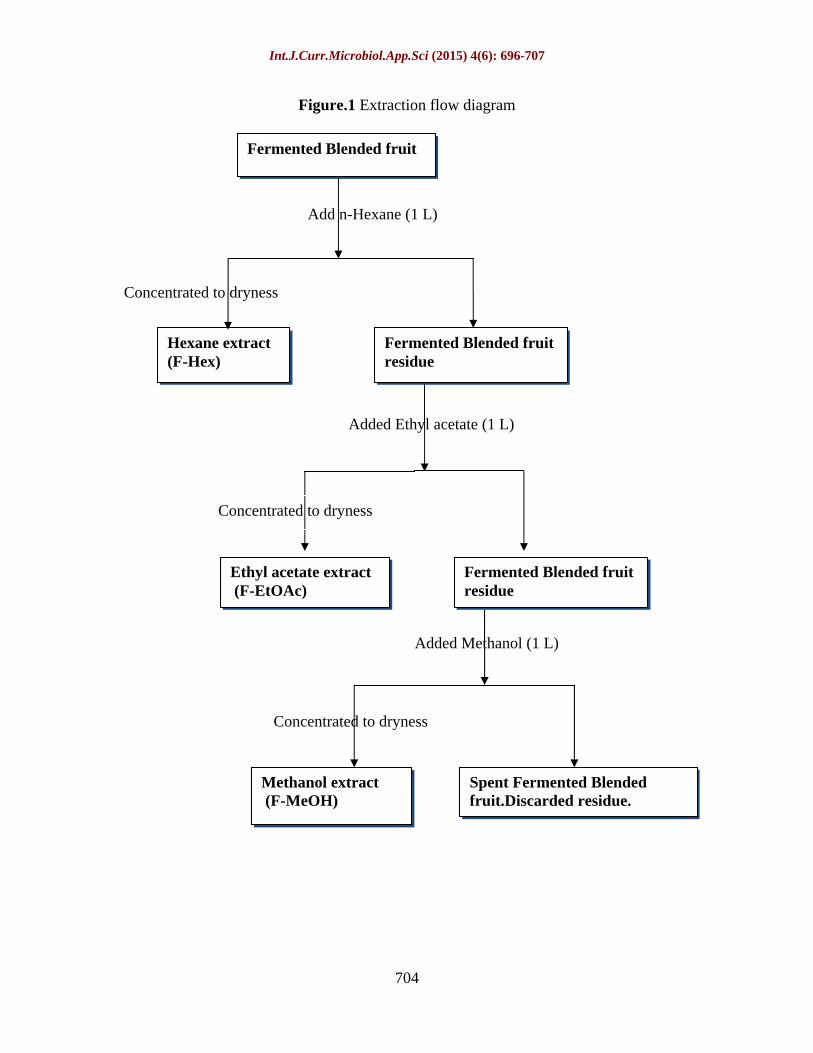
The extraction was done according to the method of (Jagessar et al., 2008), sequentially starting from the least polar solvent n-hexane (hex) through ethyl acetate (EtOAc) to the most polar methanol (MeOH).
To carry out cold maceration, 1 kg of the blended unfermented fruits was weighed into a separate (extraction) glass bottle, to

Int.J.Curr.Microbiol.App.Sci (2015) 4(6): 696-707
699
which, 1 L of n-hexane (hex) was added to and left to macerate at room temperature with intermittent shaking for a period of 48 Hrs. The mixture was filtered, the residue kept and the filtrate was concentrated using a rotary evaporator at 40OC to obtain the n-hexane extract (UF-Hex) that was kept in a refrigerator at 2
6OC. The residue was similarly extracted sequentially with EtOAc and MeOH to obtain the corresponding UF-EtOAc and UF-MeOH extracts.
The above procedure was repeated for the fermented blend to obtain the corresponding F-Hex, F-EtOAc and F-MeOH extracts. A flow diagram summarizing the procedureis Figure 1.
Antibacterial activity assays
Bacterial strains and preparation of inocula
The species of bacterial organisms that were used for the study were standard strains of Escherichia coli (ATCC 25922), Pseudomonas aeruginosa (ATCC 27853), Staphylococcus aureus(ATCC 25923) and Streptococcus pyogenes (ATCC 19615), obtained from Microbiology Laboratory, Mbarara University of Science and Technology. The cultures of these bacteria were maintained on double strength Mueller-Hinton agar (MHA) slants at 4OC. Each species wasby sub-cultured onto a fresh Mueller Hintonbroth (MHB) for 24 h at 37OC. 0.2 mL aliquot of the broth culture was dispensed inanother sterilized 20mL Mueller-Hintonbroth and incubated for 3-5 h. 1 mL portion from the final broth was expected to be 0.5McFarland standard (1.6x10⁸cfu/mL) according to (Oyelekeet al.,2008). The turbidity was checked and logically adjusted to 0.5 McFarland Standard (Pro-Lab) (1.5x10⁸cfu/mL),using isotonic sodium chloride solution and
against the McFarland Standards. This was done for each of the species and the final adjusted broth cultures used for inoculations.
Inoculation and antibacterial activity testing using agar diffusion (well) method
35ml of freshly prepared molten MuellerHinton agar (MHA) was dispensed into 90 mm-Petri dishes and allowed to set. By using sterile cotton swabs, inoculum of the of each of bacterial strains was then plated on to2 MHA Petri dishes, where5 uniformly spaced 5 mm wells were bored using a sterilized gel borer. For each extract, 100 µl of the test extract dissolved in DMSO (40%, v/v) were pipetted into the wells of two petri dishes corresponding to (0.78, 1.56, 3.13, 6.25, 12.5, 25, 50 and 100mg/well). Ceftriaxone (100 µL at a concentration of 10mg/mL,equivalent to 1 mg/well)was used as positive control;and 100 µL of DMSO (40%, v/v) as negative control, to check sterility of the solvent and the process. The Petri dishes were pre-incubated for 3 h at room temperature, allowing for complete diffusion of the samples (Möller, 1966; Das et al., 2010) and, then, incubated at37OC for 45 h. The antibacterial activity was determined by measuring of inhibition zone diameters (mm) and was evaluated according to the parameters suggested by (Alveset al., 2000): inhibition zones <9 mm, inactive; 9 12 mm, less active; 13 18 mm, active; >18 mm, very active.
Determination of minimal inhibitory concentrations (MICs)
The evaluation of MICs was performed for the extracts that inhibited growth in the antibacterial activity testing, using the micro-dilution methodology described by the Clinical and Laboratory Standards Institute (CLSI, 2009b). Alves et al., 2000 criteria for selecting which extract to

Int.J.Curr.Microbiol.App.Sci (2015) 4(6): 696-707
700
undergo MIC determination was dropped, since many other studies have not used it too (Adegbehingbe and Bello, 2014; Priya and Ravindhran, 2015; Rupapara et al., 2015).
The crude extracts dissolved in DMSO (40%, V/V) were two-fold serially diluted with freshly prepared MHB. The dilutions were mixed with equal volumes(1 mL) of 0.5 McFarland Scale (1.5 X 10⁸cfu/mL) bacterial suspensions in MHB, to give the following final concentrations (mg/mL): 1000, 500, 250, 125,62.5, 31.3, 15.6and 7.8 in test tubes. These test tubes were incubated at 37OC for 48 h and assessed for turbidity for growth or no-growth by the naked eye. Three controls were run in parallel: the extract in DMSO (40 %V/V) + MuellerHinton broth to make 1000 mg/mL concentration, without inoculation to check sterility of the extract and the process; DMSO (40%, V/V) + Mueller Hinton broth, without inoculation to check sterility of the medium; DMSO (40%, V/V) + MuellerHinton broth, inoculated to check growth support of the medium. The MIC value was determined as being the lowest extract concentrations that preventedthe bacteria to grow.
Result and Discussion
Antibacterial activity
Agar diffusion techniques have been widely used to assay antimicrobial activity of plant extracts (Perezet al., 1990; Rojas et al., 2006; Das et al., 2010). The use of this technique in the present study screened for bacterial growth inhibition of the extracts. The controls behaved as expected; the negative control did not inhibit any bacterial growth, whilst the positive control markedly inhibited all the bacterial growth (by >18 mm), therefore validated the assay.
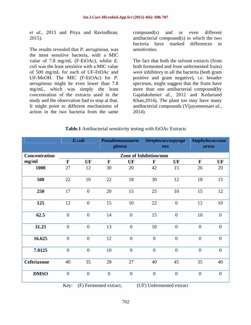
Both fermented and unfermented MeOHand EtOAc extracts showed activity against the tested gram positive bacteria (Staph.aureus andStrep. pyogenes) and gram negative bacteria (E.coli and P.aeruginosa) (Zones of inhibition in Tables 1 & 2). The fermented fruit extracts had bigger zones of inhibition compared to their corresponding unfermented fruit extracts. Generally the EtOAc extracts had the higher activity against almost all the micro-organisms tested. None of the hexane extracts inhibited growth of any bacteria even at the highest concentration of 1000 mg/mL and were therefore not subjected to micro-dilution technique for determination of MICs. The lack of antibacterial activity by hexane extracts in relation to EtOAc and MeOH extracts is not uncommon as has been observed in other studies (Martins et al., 2013; Priya and Ravindhran, 2015; Rupaparaet al., 2015).
For both solvent extracts that showed activity, the zones of inhibition widened with increasing concentration. The plateaus of the width of zone of inhibition probably had not been reached yet at the highest concentration of 1000 mg/mL, as the widths might have still been increasing at that highest concentration. According to the parameters suggested by Alveset al. (2000), of evaluating the antibacterial activity,theactivity of both the fermented and unfermented fruit extracts of each of the MeOHand EtOAcsolvents reached up to very active (> 18 mm), against each of the bacteria, as concentration increased, except forUF-EtOAc,against E. coli that only achieved less activity (9 12 mm), at that highest concentration. All the extracts were least active against E. coli, compared to the other bacteria, starting activity at a higher concentration of 125 mg/ml and 500 mg/ml for fermented and unfermented extracts respectively for both solvents. The widest

Int.J.Curr.Microbiol.App.Sci (2015) 4(6): 696-707
701
zone of inhibition (42 mm)by the extracts was observed in F-EtOAc (1000 mg/mL)on Strep.pyogenes.
Ceftriaxone at the concentration used (10 mg/ml, equivalent to 1,000 µg/well) was superior to all the extracts, even at the highest concentration, except against P. aeruginosa, where the F-EtOAc, at the highest concentration inhibited slightly wider (30 mm) than it (28 mm).
Determination of minimal inhibitory concentrations (MICs)
Since the F-EtOAc, UF-EtOAc, F-MeOH, and UF-MeOHfractions showed antibacterial activity against the tested Gram-positive and Gram-negative bacteria strains, the real extend of their inhibitory activity was evaluated by determining MIC values, which are shown in Fig.2. The controls(in Table 3) which were not inoculated did not show turbidity, confirming that the medium, the solvent and the process were sterile; whilst the ones which were without the extracts but inoculated showed turbidity, confirming that the medium, the solvent and the process were not inhibitory, thereby validated the assay.
As can be seen (Fig. 2), the MIC values varied for each sample; from 7.8mg/ml (the lowest concentration used) to 500mg/mL.For F-EtOAc fraction, from 7.8 to 125 mg/mL.For F-MeOH, from 62.5 to 125 mg/mL. MICs were identical for both UF-EtOAc and UF-MeOH, from 125 to 500 mg/mL, proposing that MeOH could be a better extracting solvent than EtOAc, because the former solvent was used on the residue left after extraction by the latter;which gave chance to the EtOAc to extract more than the MeOH could have. It could also be that the two solvents when it
comes to unfermented A. muricata fruits, extract different compounds or the same compounds in different quantity profiles (Sen and Batra, 2012).
It was also observed that all the MICs of the fermented extracts were lower than their corresponding unfermented extracts. This trend was similar with what was obtained in the antibacterial activity assay using the agar diffusion technique, pointing to a possibility that the antibacterial compound(s) and mechanism(s) of action might be the same in both the fermented and unfermented extracts, but the difference in potency could have come from the fermentation process which increased the concentration of these active compound(s). A similar observation was gotten when cabbage was fermented (Gogoet al., 2010) and when whey was fermented (Adegbehingbe and Bello, 2014).The possibility of the two solvents extracting different compounds, in the unfermented fruits, could also be extrapolated to fermented extracts; the MIC for F-MeOH on E. coli was lower than the corresponding F-EtOAc, whereas the MICs for P. aeruginosa and Strep. pyogenes were higher with F-MeOH. The MICs for Staph.aureus were identical for both F-EtOAc and F-MeOH. These differences are probably because the solvents extracted different compounds qualitatively and/or extracted the same compounds differently quantitatively (Sen and Batra, 2012).
Generally EtOAc extracts gave greater difference in sets of corresponding MICs of the fermented and unfermented extracts than those of MeOH. These should not lead to a conclusion that EtOAc is a better solvent in extracting the antibacterial compound(s) from the fermented fruits than MeOH; as it might be due to the sequential extraction as explained earlier. But MeOH is known to be a good extracting solvent at times (Martins
Int.J.Curr.Microbiol.App.Sci (2015) 4(6): 696-707
702
et al., 2013 and Priya and Ravindhran, 2015).
The results revealed that P. aeruginosa, was the most sensitive bacteria, with a MIC value of 7.8 mg/mL (F-EtOAc), whilst E. coli was the least sensitive with a MIC value of 500 mg/mL for each of UF-EtOAc and UF-MeOH. The MIC (F-EtOAc) for P. aeruginosa might be even lower than 7.8 mg/mL, which was simply the least concentration of the extracts used in the study and the observation had to stop at that. It might point to different mechanisms of action in the two bacteria from the same
compound(s) and or even different antibacterial compound(s) to which the two bacteria have marked differences in sensitivities.
The fact that both the solvent extracts (from both fermented and from unfermented fruits) were inhibitory to all the bacteria (both gram positive and gram negative), i.e. broader spectrum, might suggest that the fruits have more than one antibacterial compound(by Gajalakshmiet al., 2012 and Kedariand Khan,2014), The plant too may have many antibacterial compounds (Vijayameenaet al., 2014).
Table.1 Antibacterial sensitivity testing with EtOAc Extracts
E.coli Pseudomonasaeruginosa
Streptococcuspyogenes
Staphylococcusaureus
Zone of Inhibition/mm Concentrationmg/ml F UF F UF F UF F UF
1000 27 12 30 20 42 15 26 20
500 22 10 22 18 30 12 18 15
250 17 0 20 15 25 10 15 12
125 12 0 15 10 22 0 12 10
62.5 0 0 14 0 15 0 10 0
31.25 0 0 13 0 10 0 0 0
16.625 0 0 12 0 0 0 0 0
7.8125 0 0 10 0 0 0 0 0
Ceftriaxone 40 35 28 27 40 45 35 40
DMSO 0 0 0 0 0 0 0 0
Key: (F) Fermented extract, (UF) Unfermented extract

Int.J.Curr.Microbiol.App.Sci (2015) 4(6): 696-707
703
Table.2 Antibacterial sensitivity testing with MeOH Extract
E.coli Pseudomonas
aeruginosa Streptococcus
pyogenes Staphylococcus
Aureus
Zone of Inhibition/mm Concentrationmg/ml F UF F UF F UF F UF 1000
22 15 20 20 24 20 30 20
500
18 10 16 18 18 14 20 15
250
15 0 14 15 12 10 18 14
125
12 0 12 10 10 0 15 12
62.5
0 0 10 0 10 0 10 0
31.25
0 0 0 0 0 0 0 0
15.625 0 0 0 0 0 0 0 0
7.8125 0 0 0 0 0 0 0 0
Ceftriaxone 40 40 28 22 40 40 30 32
DMSO 0 0 0 0 0 0 0 0
Key: (F) Fermented extract, (UF) Unfermented extract
Table.3 Growth or no-growth in Negative Controls
Ethylacetate Extracts MethanolExtracts
Control F-EtOAc UF-EtOAc F-MeOH
UF-MeOH 1 1000 mg/mL extract in DMSO (40
%V/V) + MHB. Without inoculation - - - -
2 DMSO (40%, V/V) + MHB. Without inoculation.
- - - -
3 DMSO (40%, V/V) + MHB. Inoculated.
+ + + +
Key: (- ) No growth of any of the four bacteria, (+) Growth of all the four bacteria
Int.J.Curr.Microbiol.App.Sci (2015) 4(6): 696-707
704
Figure.1 Extraction flow diagram
Fermented Blended fruit
Methanol extract (F-MeOH)
Spent Fermented Blended fruit.Discarded residue.
Concentrated to dryness
Added Methanol (1 L)
Ethyl acetate extract (F-EtOAc)
Fermented Blended fruit residue
Added Ethyl acetate (1 L)
Fermented Blended fruit residue
Hexane extract (F-Hex)
Concentrated to dryness
Concentrated to dryness
Add n-Hexane (1 L)

Int.J.Curr.Microbiol.App.Sci (2015) 4(6): 696-707
705
Figure.2 Bar Chart showing the MICs of the extracts for each of the four bacteria
References
Adegbehingbe K. T., Bello, M. (2014).Antibacterial activities of fermented whey on some
selected Enteropathogenic bacteria.Int. J. Curr. Microbiol. App. Sci.3(9): 152161.
Alves, T.M.A., Silva, A.F., Brandão, M., Grandi, T.S.M., Smânia, E.F.A., Smânia Jr., A.,
Zani, C.L.(2000). Biological screening of Brazilian medicinal plants. Mem. Inst. Oswaldo Cruz.95(3): 367 373.
Bills, G. F., Polishook, J. D. (1991) Microfungi from Carpinus caroliniana. Can. J. Bot. 69: 14771482.
CLSI
Clinical and Laboratory Standards Institute.(2009b). Methods for Dilution
Antimicrobial Susceptibility Tests for Bacteria that Grow Aerobically, (17th edn). Approved Standard. Document M07-A8, CLSI, Wayne, PA.
Collado, J., Platas, G., Pelaez, F.(2001). Identification of an Endophytic Nodulisporium sp. from
Quercus ilex in Central Spain as the Anamorph of Biscogniauxia mediterranea by rDNA Sequence Analysis and Effect of Different Ecological Factors on Distribution of the Fungus. Mycologia.93(5): 875886.
Das, K., Tiwari, R.K. S., Shrivastava, D.K. (2010). Techniques for evaluation of medicinal plant products as antimicrobial agent: Current methods and future trends.Journal of Medicinal Plants Research. 4(2): 104 111.
Fisher, P. J., Graf, F., Petrini, L. E., Sutton, B. C., Wookey, P. A. (1995). Fungal endophytes of
Dryas octopetala from a high arctic semidesert and from the Swiss alps. Mycologia.87: 319 323.
Gajalakshmi, S., Vijayalakshmi, S., Rajeswari, D. V. (2012). Phytochemical and
Pharmacological Properties of Annona muricata: A Review. Int. J. Pharm. Pharm. Sci.4(2): 3 6.

Int.J.Curr.Microbiol.App.Sci (2015) 4(6): 696-707
706
George, D., Pamplona, R. (1999).
Encyclopedia of medical plants. Editional Safelize Spain. 1:381.
Gogo, L. A., Shitandi, A. A., Lokuruka, M. N. I., Sang, W. (2010). Antimicrobial Effect of Juice Extract From Fermented Cabbage Against Select Food-Borne Bacterial Pathogens. J. Appl. Sci. Res. 6(11): 1807 1813.
Imade, E. E., Ikenebomeh, M. J., Obayagbona, O. N., Igiehon, O. N.(2013). Evaluation of Changes in the Microbial Profile, Physico-Chemical and Nutritional Attributes During the Bioconversion of Soursop (Annona muricata) Must to Wine. Nig J. Biotech. Vol. 25: 1 11.
Ismail, M., Rahman, S., Muhammad, N., Mohani, N., Khan, M. A., Barkatullah Hussain, J. (2011). Pharmacognostic and phytochemical investigation of the stem bark of Pistacia integerrima Stewart exb. J. Med. Plants Res. 5: 3891 3895.
Jagessar, R. C., Mohamed A.,, Gomes, G. (2008). An evaluation of the Antibacterial and Antifungal activity of leaf extracts of Momordica Charantia against Candida albicans, Staphylococcus aureus and Escherichia coli. Nature and Science.6(1): 1 14.
Johnson, J. A., Whitney, N. J. (1989). An investigation of needle endophyte colonization patterns with respect to height and compass direction in a single crown of balsam fir (Abies balsamea). Can. J. Bot. 67: 723 725.
Kedari, T. S., Khan, A. A. (2014). Guyabano (Annona muricata): A review of its traditional uses phytochemistry and pharmacology. American Journal of Research Communication.2(10): 247 268.
Martins, S., Amorim,E. L.C., Sobrinho,T. J. S. P., Saraiva, A. M., Pisciottano, M.
N.C., Aguilar, C. N., Teixeira, J. A., Mussatto, S. I. (2013). Antibacterial activity of crude methanolic extract and fractions obtained from Larrea tridentata leaves. Industrial Crops and Products.41: 306 311.
Möller, A.J. R.(1966). Microbiological examination of root canals and perapical tissues of human teeth. Odontolgisk Tidskrift.74: 1 38.
Nalawade, S.M., Sagare, A.P., Lee, C. Y., Kao, C. L., Tsay, H. S.(2003). Studies on tissue culture of Chinese medicinal plant resources in Taiwan and their sustainable utilization. Botanical Bulletin of Academia Sinica. 44(2): 79 98.
Okane, I., Nakagiri, A., Ito, T. (1997). Endophytic fungi in leaves of ericaceous plant. Can. J. Bot. 76: 657 663.
Okigbo, R. N., Obire, O. (2009). Mycoflora and production of wine from fruits of soursop (Annona Muricata L.). Internat. J. Wine Res. 1: 1 9.
Oyeleke, S. B., Dauda, B. E. N., Boye, O. A. (2008). Antibacterial activity of Ficus capensis. Afr. J.Biotechnol.7 (10):1414 1417.
Perez, C., Pauli, M., Bazevque, P. (1990). An antibiotic assay by the agar well diffusion method. Acta Biologiae et Medecine Experimentalis.15: 113115.
Prescott, L. M., Harley, J. P., Klein, D. A. (2008). Medical Microbiology (6th
edition), McGraw Hill, New York. Pp: 573 736.
Priya, S. E., Ravindhran, R. (2015). Phytochemical Analysis and Antimicrobial Properties of Extracts from Aerial Parts of Phyla nodiflora (L) Greene. Int.J.Curr.Microbiol.App.Sci.4(2): 347 358.

Int.J.Curr.Microbiol.App.Sci (2015) 4(6): 696-707
707
Rojas, R., Bustamante, B., Bauer, J.,
Fernandez, I., Alban, J., Lock, O.(2003). Antimicrobial activity of Selected Peruvian medicinal plants. J. Ethnopharmacol.88(2 3): 199 204.
Rojas, J.J., Ochoa, V.J., Ocampo, S.A., Mu noz, J.F. (2006). Screening for antimicrobial activity of ten medicinal plants used in Colombian folkloric medicine: a possible alternative in the treatment of non-nosocomial infections. BMC Complementary and Alternative Medicine.6: 2.
Rupapara, K. V., Joshi, N.H., Vyas, K.G. (2015). Evaluation of Antimicrobial Activity of Crude Extracts of Seaweed Sargassum johnstonii. Int. J. Curr. Microbiol. App. Sci.4(2): 300304.
Sahashi, N. Y., Miyasawa, T., Kubano, S., Ito, T. (2000). Colonization of beech leaves by two endophytic fungi in northern Japan. Forest Pathol. 30: 77 86.
Sawant, T. P., Dongre, R. S. (2014). Bio-Chemical Compositional Analysis of Annona muricata: A Miracle Fruit s Review. International Journal of Universal Pharmacy and Bio Sciences. 3(2): 82 104.
Sen, A., Batra, A. (2012). Evaluation of antimicrobial activity of different solvent extracts of medicinal plant: Melia azedarach L.Int. J. Curr. Pharm. Res.Vol 4, Issue 2: 67-73.
Sieber, T. N. (1989). Endophytic fungi in twigs of healthy and diseased Norway spruce and white fir. Mycol. Res. 92: 322 326.
Schulz, B., Wanke, U., Draeger, S., Aust, H. J.(1993). Endophytes from herbaceous plants and shrubs, effectiveness of surface sterilization methods. Mycological Research.97: 1447 1450.
Vijayameena, C., Subhashini, G., Loganayagi, M., Ramesh, B. (2013). Phytochemical screening and assessment of antibacterial activity for the bioactive compounds in Annona muricata. Int. J. Curr. Microbiol. App. Sci.2(1): 1 8.
Vuorela, P., Leinonen, M., Saikku, P., Tammela, P., Rauha, J. P., Wennberg, T., Vuorela, H. (2004). Natural products in the process of finding new drug candidates. Current Medicinal Chemistry 11: 1375 1389.
Wele, A., Zhang, Y., Caux, C., Brouard, J. P., Pousset, J. L., Bodo, B. (2004). Annomuricatin C, anovel cyclohexapeptide from the seeds of Annona muricata. C. R. Chimie.7(1011): 981 988.


![Milk, Fermentation, And Fermented and Non-fermented [Compatibility Mode]](https://img.dokumen.tips/doc/110x75/55cf85df550346484b923a09/milk-fermentation-and-fermented-and-non-fermented-compatibility-mode.jpg)
