Embed Size (px)
Citation preview

www.elsevier.com/locate/procbio
Process Biochemistry 42 (2007) 995–1002
Comparative studies of loblolly pine biodegradation and
enzyme production by Argentinean white rot fungi
focused on biopulping processes
L. Levin a,*, L. Villalba b, V. Da Re a, F. Forchiassin a, L. Papinutti a
a Laboratorio de Micologıa Experimental, Departamento de Biodiversidad y Biologıa Experimental, Facultad de Ciencias Exactas y Naturales,
Universidad de Buenos Aires, Pabellon II, 4to piso, C1428EHA Ciudad Universitaria, Ciudad Autonoma de Buenos Aires, Buenos Aires, Argentinab Programa de Investigacion en Celulosa y Papel, Facultad de Ciencias Exactas, Quımicas y Naturales, Universidad Nacional de Misiones,
Felix de Azara 1552, N3300LQH Posadas, Misiones, Argentina
Received 24 November 2006; received in revised form 5 March 2007; accepted 19 March 2007
Abstract
The ability of eight white rot fungi: Coriolopsis rigida, Coriolus versicolor var. antarcticus, Peniophora sp., Phanerochaete sordida,
Pycnoporus sanguineus, Steccherinum sp., Trametes elegans and Trametes villosa to selectively delignify loblolly pine (Pinus taeda) chips was
studied. They were selected among 34 basidiomycetes from Argentina because of their capacity to decolorize Poly R-478 and Azure B. Fungal
pretreatment caused changes in wood chemical composition as well as in physical structure. The present study allowed the identification of a new
strain, potentially a candidate for use in softwoods biopulping processes. Results showed that P. sanguineus was able to reduce lignin content in
11% in 14 days of treatment, but also that P. taeda wood suffered notable structural changes of lignin and hemicelluloses during the treatment, as
revealed from 13C CP-MAS NMR spectra. An increase of 15% in porosity of decayed wood confirmed physical changes due to fungal attack.
# 2007 Elsevier Ltd. All rights reserved.
Keywords: Biopulping; Lignocellulolytic enzymes; Loblolly wood degradation; White rot fungi
1. Introduction
Wood cell walls are made up mostly of cellulose,
hemicellulose and lignin. The tensile strength of wood fibers
is primarily determined by cellulose and hemicelluloses, while
lignin mediates adhesion among the fibers. White rot fungi
(WRF) have all the necessary enzymes for the complete
degradation of wood components. Some fungal species remove
lignin more efficiently than other wood components; such
degradation pattern is known as selective lignin degradation or
delignification [1]. This effect decreases the dependence on
chemicals in the pulping processes and it is the most useful for
biopulping. Under particular conditions extensive differential
degradation of lignin was observed, an interesting example of
such process is ‘‘palo podrido’’, where up to 90% of lignin
degradation was achieved [2].
* Corresponding author.
E-mail address: [email protected] (L. Levin).
1359-5113/$ – see front matter # 2007 Elsevier Ltd. All rights reserved.
doi:10.1016/j.procbio.2007.03.008
The high capability of WRF to degrade all wood
components is based principally on the activity of different
complexes of extracellular enzymes. These fungi secrete
hydrolytic enzymes such as cellulases, pectinases and
xylanases, which are typically induced by their substrates.
On the other hand, lignin (a polymer of phenylpropane units
connected by different C–C and C–O–C linkages) is oxidized
and degraded by a ligninase system made up of at least three
enzyme activities: lignin peroxidase (LiP), manganese perox-
idase (MnP) and laccase. In addition, several findings provided
information about low weight molecules that participate in the
initial attack of lignocellulose. Fenton reaction produces �OH,
this radical is the strongest oxidant found in white rot fungi
[3,4]; another reactive molecules involved in lignin attack are
the peroxyl radicals rendered from lipid peroxidation [5]. The
same unique non-specific enzymes and mechanisms that give
these fungi the ability to degrade lignin also allow them to
degrade a wide range of pollutants. Among such pollutants a
wide range of dyes are substrates of ligninases rendering as a
product a colourless substance. Thus, dye decolorization may
be used as a screening method to select potentially promissory
L. Levin et al. / Process Biochemistry 42 (2007) 995–1002996
strains. Poly R-478 and Azure B were used in many previous
reports as useful tools to detect ligninolytic activity in WRF [6].
Cellulose is a linear polymer of glucose units, which can be
hydrolyzed by the action of endoglucanases, cellobiohydro-
lases and b-glucosidases. Hemicellulose is a heterogeneous,
branched polymer. The backbone of the polymer is built up by
sugar monomers like xylose, in this case, the enzymes involved
in its degradation are named xylanases. Since the structure of
xylans is more complex and variable, a more complex assembly
of enzymes is required than for cellulose hydrolysis. Similar to
cellulases, the xylanases can act synergistically to achieve
hydrolysis, predominant enzymes within this system are endo
1,4-b-xylanases which attack the polysaccharide backbone,
and b-xylosidases, which hydrolyze short xylooligosaccharides
to xylose [7].
WRF and their enzymes (particularly ligninases and
xylanases) are considered for the treatment of wood chips
prior to pulping. While ligninases attack the lignin, xylanases
degrade hemicelluloses and make the pulp more permeable for
the removal of residual lignin. Termed ‘‘biopulping’’, this
process removes not only lignin but also some of the wood
extractives, thus reducing the pitch content and effluent
toxicity. When biological treatment is followed by mechanical
pulping, there is as much as 30% energy saving, whereas when
it is followed by sulfite or kraft pulping, the cooking time is
dramatically reduced. The paper strength properties have also
been found to improve after biopulping. However, this process
is still in its beginnings and no full-scale biopulping mills are in
operation at the moment [8,9].
The studies on enzyme production by wood rot fungi are
usually carried out using chemically defined liquid medium,
under conditions able to induce the production of a particular
enzyme [10–12]. However, in natural environments and in
solid-state fermentation, these fungi grow on woody substrates
under quite different conditions from those of submerged
cultures, the patterns of enzyme production being consequently
different. Much research work is still necessary to fully
understand the degradation process, and particularly the
enzymes and other metabolites secreted by the fungi during
wood decay. Additional findings on this field could help to
elucidate the biochemical mechanisms of wood decay by fungi
and as a result facilitate the fungal strain selection for
biopulping and for other industrial applications [13–15].
Many previous studies have focused on the lignin-degrading
enzymes of Phanerochaete chrysosporium, Trametes versico-
lor and Ceriporiopsis subvermispora. Recently however, there
has been a growing interest in studying the lignin-modifying
enzymes of a wider array of WRF, not only from the standpoint
of comparative biology but also with the prospect of finding
better lignin-degrading systems for use in biopulping and other
biotechnological applications [16–18]. Fungi from Misiones
province (Argentina) usually grow subject to extremely high
temperatures and humidity. Therefore, they represent an
interesting source in the search of thermo-tolerant enzymes
for biotechnological applications. In this work 33 strains of
WRF from Misiones, were evaluated for their ability to
decolorize Poly R-478. Eight isolates that displayed the fastest
decoloration in Poly R-478 medium were screened for their
ability to decolorize Azure B, and inoculated on loblolly (Pinus
taeda) wood chips, to evaluate their potential for biopulping
processes. Several relevant hydrolytic enzymes (endoglucanase
and endoxylanase) and oxidative enzymes (laccase and Mn-
peroxidase) present in the culture extracts were studied. To
explore the relationship between the enzyme equipment of each
fungus and its corresponding pattern of degradation, wood
physical structure and chemical composition were also
determined.
2. Materials and methods
2.1. Fungal strains
Pure cultures of 34 white rot fungi strains from the Culture Collection
(BAFC) of Universidad de Buenos Aires were used in the present study. Stock
cultures were maintained on malt extract (1.2%), agar (2%) slants at 4 8C with
periodic transfer.
2.2. Culture conditions
All the strains were inoculated on agar plates (90 mm in diameter,
20 cm3 medium/Petri dish) containing malt extract (12.7 g dm�3), glucose
(10 g dm�3) and agar (20 g dm�3) (MEA) supplemented with Poly R-478
(0.02%) or Azure B (50 mM). Inoculum consisted of a 25-mm2 surface agar
plug from a 7-day-old culture grown on MEA. Uninoculated plates served as
controls for abiotic decoloration. The plates were incubated at 28 8C for 21–28
days. Fungal growth was followed by measuring radial extension of the
mycelium. Average growth rates (cm day�1) were calculated. A decolorized
zone appeared when the fungus degraded the dye. Weekly measurements of the
colonies and the decolorized zones (if any) were performed for each strain.
Wood degradation experiments were carried out with 20 g (dry weight) of
wood chips (5 cm � 2 cm � 0.5 cm) and 20 cm3 of aqueous solution of corn
steep liquor 1% (C1), corn steep liquor 3% (C3), malt extract 3% (M) or malt
extract 5% plus peptone 1.5% (MP), which were sterilized and inoculated with
the fungus. Inoculum was grown in 150 cm3 Erlenmeyer flasks containing
25 cm3 of liquid medium (shaken at 100 rpm) with the following composition:
1% glucose and 1.3% malt extract. Inoculation was carried out using 5 cm3
(55% final moisture content of wood chips) of homogenized pellets from 7-day-
old shaken cultures. The fungi were incubated at 28 8C for 14 days. After the
given period of incubation, wood chips were cut in small fragments (ca.
2.5 cm � 0.4 cm � 0.5 cm), then soluble proteins and sugars were extracted
by adding 225 cm3 of distilled water, stirring for 20 min, followed by filtration
and centrifugation. Crude extracts were used for all the determinations of
enzyme activities and reducing sugars content. All the steps for crude extraction
were performed at room temperature. The supernatants were stored at �20 8Cuntil needed. The experiments were carried out in triplicate parallel cultures.
2.3. Enzyme assays
All enzyme activities were assayed at 30 8C. Laccase activity was determined
by oxidation of ABTS [2,20azinobis(3-ethylbenzthiazoline-6-sulphonate)]
(e420 = 36 mM�1 cm�1). The reaction mixture contained 0.1 M sodium acetate
buffer, pH 3.6 and 5 mM ABTS [19]. MnP activity was determined by oxidation of
phenol red (0.01%). The reaction product was measured at 610 nm
(e610 = 22 mM�1 cm�1). The reaction mixture contained 0.05 M succinate buffer
pH 4.8, 0.1 mM MnSO4, 0.1 mM H2O2 [20]. Endoglucanase and endoxylanase
were determined measuring the reducing sugars produced after hydrolysis of the
substrate by the Somogyi–Nelson method [21]. Measurements were made in
0.1 M sodium acetate buffer, pH 4.8, using the following substrates: carbox-
ymethylcellulose (CMC) 0.5% for endoglucanase and xylan from oat spelts 0.2%
for endoxylanase. Standard curve with glucose was made to calculate enzyme
activities. Enzyme activity has been expressed in International Units (U), as the
amount of enzyme needed to release 1 mmol of product in 1 min.
L. Levin et al. / Process Biochemistry 42 (2007) 995–1002 997
2.4. Analysis of wood composition
Chemical composition of non-treated and bio-treated wood chips was
determined. Extractive-free wood samples were prepared according to T204
om-88 ‘‘Solvent extractives of wood and pulp’’ using ethanol–benzene or water
as solvent. The solvent extractable material may be considered to consist
primarily of resin and fatty acids and their esters, waxes, and unsaponifiable
substances. Soluble materials or extractives in wood consist of those compo-
nents that are soluble in neutral organic solvents. These compounds include
waxes, fats, resins, photosterols and non-volatile hydrocarbons, low-molecular-
weight carbohydrates, salts, and other water-soluble substances. Klason lignin
was determined in accordance with Tappi T-222 om-88, ‘‘Acid-insoluble Lignin
in Wood and Pulp’’. To determine acid-insoluble lignin in wood and pulp, the
carbohydrates in wood are hydrolyzed and solubilized by sulfuric acid (72%);
the acid-insoluble lignin is filtered off, dried, and weighed. Wood contains from
about 20 to 30% lignin; removal of which is a main objective of pulping and
bleaching processes. Determination of lignin content in wood and pulps
provides information for evaluation and application of pulping and bleaching
processes respectively. Cellulose content was determined by the Seifert method.
The solvolytic method involving treatment of wood meal in a boiling mixture of
acetylacetone–dioxane–hydrochloric acid gave the most accurate cellulose
values and produced the purest cellulose residues [22]. Hemicellulose content
was calculated by difference (100% � (total extractives + lignin + cellulose)).
Lignin and cellulose analysis were determined on extractive-free wood. Pore
size analysis was conducted by mercury porosimetry in a Poresizer 9320.
2.5. Analysis of NMR spectra
The non-treated and bio-treated with Pycnoporus sanguineus BAFC 2126
samples of P. taeda wood were analysed by 13C CP-MAS NMR as a fine
powder. 13C solid-state NMR spectra were recorded at 100.6 MHz (9.4 T) on a
Bruker Avance 400 spectrometer. A 7-mm double bearing Bruker rotor was
spun in air at 5.0 kHz. In all experiments the 1H and 13C 908 pulses were ca.
4 ms. The CP-MAS spectra were recorded with a 5 s recycle delay and a 2 ms
contact time.
3. Results and discussion
In this work 33 strains of WRF from Misiones province
(Argentina) were examined for the decolorization of plates with
the lignin model substrate Poly R-478. Their decolorizing
abilities were compared with a strain of Coriolus versicolor var.
antarcticus, which proved to be a very efficient dye-degrading
microorganism [6]. The capability of Poly R-478 decoloriza-
tion is indicative of peroxidase activity. Such activity includes
the combined activity of H2O2-producing oxidases and
peroxidases, and is correlated with the ability of WRF to
degrade the tricyclic aromatic hydrocarbon anthracene [23].
According to de Koker et al. [24] fungi decolorizing Poly R-478
could be placed in different groups: fungi with strong LiP and
MnP activity, fungi with strong MnP activity and fungi with
strong laccase activity. On the other hand, LiP activity is
responsible for Azure B degradation, therefore plates contain-
ing this dye were used to assess this activity [25]. All of the
fungal strains tested were able to grow on MEA supplemented
with Poly R-478 (Table 1), but 13 of them did not decolorize the
dye after 21 days of incubation. We found inter-genera, inter-
specific and intra-specific differences in the ratio between the
growth halo and the decolorization halo, suggesting physio-
logical differences. When surveying Poly R-478 decolorization
abilities of wood-inhabiting fungi, similar results were obtained
by Freitag and Morrell [26] and Levin et al. [6]. In most cases,
when produced, all the decolorized zones were smaller than the
diameters of the corresponding colonies, consistent with the
decolorization being a secondary metabolic activity of the older
mycelium. Similar results were obtained by Levin et al. [6]
when screening WRF for their ability to decolorize various
dyes. Consistent with previous findings, in our study P.
sanguineus and Peniophora strains were also included among
the most rapid Poly R-478 decolorizing agents [6,26,27]. The
eight isolates that displayed the fastest decoloration in Poly R-
478 medium were tested for their ability to decolorize Azure B.
Growth rates observed in this medium were higher than those
observed in cultures supplemented with Poly R-478. All of the
strains tested in this second screening step were able to
decolorize Azure B, showing similar values of decolorization
halo diameter than those observed in Poly R-478 medium.
The selected fungi were inoculated on P. taeda wood chips
to evaluate their potential for biopulping processes. Growth was
assessed visually, and while P. sanguineus displayed the fastest
growth and a dense mycelial mat covered the chips after 7 days,
only scatter hyphae were seen on the chips colonized by the
other fungi evaluated after 2 weeks of treatment.
Quantification of extracellular enzymes is limited by the not
exhaustive extraction method, therefore a preliminary study
was conducted to determine the optimum of extraction time.
Wood chips did not render a significantly higher enzyme
activity by increasing the extraction time from 20 min to 4 h
(data not shown). Extraction time used onwards (20 min) was
similar to others reported previously [28,29]. Nevertheless, the
extraction procedure may have not been appropriate to
guarantee the total recovery of the enzymes adsorbed on the
wood substrate. Thus, data reported are not comparable to other
works where enzyme secreted in solid-state fermentation were
measured.
Hydrolytic (endoglucanase and endoxylanase) and oxidative
(MnP and laccase) extracellular enzymes were measured after
14 days (Table 2). Regarding ligninases, the highest laccase
activity (5560 mU g�1) was produced by Trametes villosa in M
supplemented cultures. This fungus produced also a high MnP
value (217 mU g�1) similar to that obtained in P. sanguineus
(332 mU g�1). A negligible laccase activity and no MnP
activity were detected in Trametes elegans as well as in
Steccherinum sp. which produce very low titres of MnP and no
laccase activity. Taking into account that in MnP determination
lactate or other Mn-chelating agents were not included, MnP
levels may be underestimated [30]. Maximal hydrolytic
enzyme secretion was observed in Peniophora sp. and P.
sanguineus, highest endoglucanase titre (332 mU g�1) was
produced by P. sanguineus on wood chips supplemented with
C3, while Peniophora sp. produced the highest endoxylanase
activity (660 mU g�1) on MP.
All dry weight loss values ranged from 2 to 4.5%, after 2
weeks of treatment. The biopulping effect can be observed at
very low weight losses (<5%) for sulfite pulping process.
Biopulping may not require degradation of nonphenolic
structures; modification and weakening of lignin rather than
bulk removal of lignin appears to be sufficient to give the
biopulping effect. Then, good biopulping is observed when the

Table 1
Growth and solid-plate dye decolorization on media containing glucose (10 g dm�3), malt extract (12.7 g dm�3), agar (20 g dm�3), supplemented with either Poly R-
478 (0.02%) or Azure B (50 mM)
Strain Order Family Growth in
Poly R
(cm day�1)
Decol. of
Poly Ra
(cm day�1)
Growth in
Azure B
(cm day�1)
Decolorization
of Azure Bb
(cm day�1)
Skeletocutis nivea var. diluta (BAFC 2347) Stereales Grammotheleaceae 0.114 0
Peniophora sp. (BAFC 633) Stereales Peniophoraceae 1.286 0.957 1.286 1.086
Steccherinum sp. (BAFC 1171) Hericiales Stecherinaceae 1.286 0.814 1.286 0.614
Phanerochaete sordida (BAFC 2122) Telephorales Meruliaceae 1.286 0.571 1.142 Diffuse
Ganoderma applanatum (BAFC 1168) Ganodermatales Ganodermataceae 0.450 0.111
G. applanatum (BAFC 1172) Ganodermatales Ganodermataceae 0.450 0.356
Coriolus pinsitus (BAFC 663) Poriales Coriolaceae 0.714 0.271
C. pinsitus (BAFC 667) Poriales Coriolaceae 1.186 0
Coriolus pavonius (BAFC 752) Poriales Coriolaceae 0.643 0.322
C. pavonius (BAFC 753) Poriales Coriolaceae 0.500 0.414
C. pavonius (BAFC 760) Poriales Coriolaceae 0.643 0.536
Coriolopsis rigida (BAFC 2101) Poriales Coriolaceae 1.014 0.686 0.95 0.772
Fomitopsis sp. (BAFC 746) Poriales Coriolaceae 0.536 0.273
Hexagona papyracea (BAFC 1173) Poriales Coriolaceae 0.636 0
Lenzites sp. (BAFC 669) Poriales Coriolaceae 0.554 0.143
Pycnoporus sanguineus (BAFC 2126) Poriales Coriolaceae 1.286 0.943 1.143 0.643
Rigidoporus ulmarius (BAFC 1160) Poriales Coriolaceae 0.471 0
Tyromyces caesius (BAFC 2283) Poriales Coriolaceae 1.286 0.570
Trametes elegans (BAFC 2127) Poriales Coriolaceae 1.286 0.943 1.286 0.757
Trametes villosa (BAFC 2755) Poriales Coriolaceae 1.286 1.000 1.286 0.444
Lentinus afın villosus (BAFC 1743) Polyporales Lentinaceae 1.171 0
Schizophyllum commune (BAFC 583) Polyporales Schizophyllaceae 1.214 0
Pleurotus ostreatus (BAFC 215) Polyporales Lentinaceae 0.714 0.553
Pleurotus pulmonarius (BAFC 0076/2002) Polyporales Lentinaceae 0.818 0.464
Polyporus tenuiculus (BAFC 1020) Polyporales Polyporaceae 0.364 0
P. tenuiculus (BAFC 1154) Polyporales Polyporaceae 0.657 0
Polyporus sp. (BAFC 665) Polyporales Polyporaceae 1.286 0.182
Polyporus sp. (BAFC 739) Polyporales Polyporaceae 1.071 0
Polyporus sp. (BAFC 2452) Polyporales Polyporaceae 0.686 0
Phellinus calcitratus (BAFC 2152) Hymenochaetales Hymenochaetaceae 0.364 0
Scytinostroma sp. (BAFC 2740) Lachnocladiales Lachnocladiaceae 1.286 0.167
Eichleriella leveilliana (BAFC 670) Tremellales Exidiaceae 0.179 0
Auricularia polytricha (BAFC 2095) Tremellales Exidiaceae 0.545 0
Coriolus versicolor var. antarcticus (BAFC 266) Poriales Coriolaceae 1.286 1.286 1.286 0.857
The plates were incubated for 3 weeks at 28 8C.a Ligninolytic activity (Poly R-478 solid-plate decoloration rate (cm day�1)). The values are the mean of three replications, S.D. < 5%.b LiP activity (Azure B solid-plate decoloration rate (cm day�1)). The values are the mean of three replications, S.D. < 5%.
L. Levin et al. / Process Biochemistry 42 (2007) 995–1002998
major cell wall components: lignin, cellulose and hemicellulose
are largely intact [31].
Biotreatment of P. taeda wood chips with four of the
selected fungi (Steccherinum sp., C. versicolor var. antarcticus,
P. sanguineus and T. elegans) resulted in a decrease of Klason
lignin content. While incubation with Steccherinum reduced
Klason lignin content/total wood weight components (%, w/w)
from 29 (control) to 26.4 (2.6%), up to 3.1% decrease in lignin
content was registered when using P. sanguineus, after 2 weeks
(Table 3). These results compared favorably with previously
findings by Guerra et al., that demonstrated a weight loss of
only 3% after 30 days biotreatment of P. taeda wood chips with
C. subvermispora [32] accompanied by a lignin content
decrease from 28.2 to 26.1% [33]. P. sanguineus was selected
for additional studies, taking into account that it caused the
highest reduction in loblolly wood lignin content among the
fungi evaluated. The extractive fraction, a group of non-
structural components of wood that usually contains resin
acids, fatty acids, terpenes and phenolic compounds, increased
with fungal treatment. Previous studies attributed this raise to
biodegradation products [34]. Cellulose content of treated
wood with C. versicolor, P. sanguineus and T. elegans was
higher than the control, showing thus selective delignification.
Lignin removal was also accompanied by hemicellulose
degradation in these fungi. In selective delignification
hemicelluloses and lignin are preferentially attacked, especially
in early stages [35].
Nevertheless, the high levels of hydrolytic and oxidative
enzymatic activities detected after 14 days of decay (Table 2)
did not correlate with the visualized wood colonization or with
the extent of initial wood weight or component losses (i.e. T.
elegans grew on wood even giving very low titers of oxidative
enzymes, but diminished its lignin content in 3%). Similar
results were obtained by Ferraz et al. [36], when analyzing
wood biodegradation and enzyme production by C. sub-
vermispora during solid-state fermentation of Eucalyptus
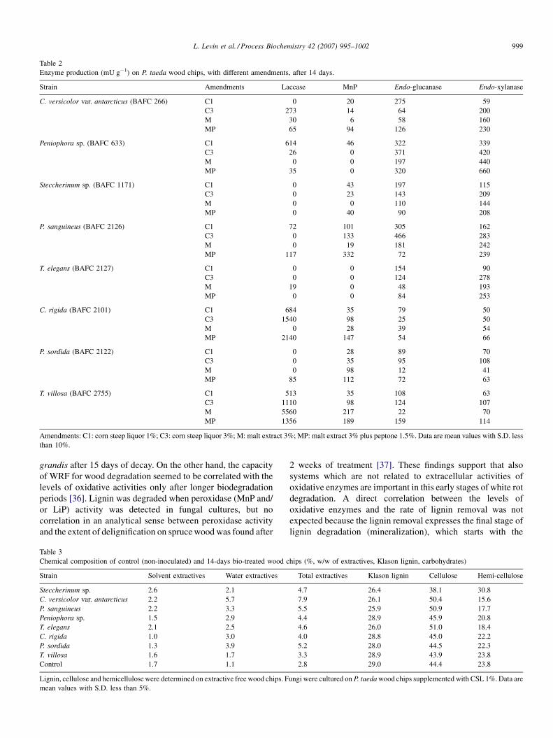
Table 2
Enzyme production (mU g�1) on P. taeda wood chips, with different amendments, after 14 days.
Strain Amendments Laccase MnP Endo-glucanase Endo-xylanase
C. versicolor var. antarcticus (BAFC 266) C1 0 20 275 59
C3 273 14 64 200
M 30 6 58 160
MP 65 94 126 230
Peniophora sp. (BAFC 633) C1 614 46 322 339
C3 26 0 371 420
M 0 0 197 440
MP 35 0 320 660
Steccherinum sp. (BAFC 1171) C1 0 43 197 115
C3 0 23 143 209
M 0 0 110 144
MP 0 40 90 208
P. sanguineus (BAFC 2126) C1 72 101 305 162
C3 0 133 466 283
M 0 19 181 242
MP 117 332 72 239
T. elegans (BAFC 2127) C1 0 0 154 90
C3 0 0 124 278
M 19 0 48 193
MP 0 0 84 253
C. rigida (BAFC 2101) C1 684 35 79 50
C3 1540 98 25 50
M 0 28 39 54
MP 2140 147 54 66
P. sordida (BAFC 2122) C1 0 28 89 70
C3 0 35 95 108
M 0 98 12 41
MP 85 112 72 63
T. villosa (BAFC 2755) C1 513 35 108 63
C3 1110 98 124 107
M 5560 217 22 70
MP 1356 189 159 114
Amendments: C1: corn steep liquor 1%; C3: corn steep liquor 3%; M: malt extract 3%; MP: malt extract 3% plus peptone 1.5%. Data are mean values with S.D. less
than 10%.
L. Levin et al. / Process Biochemistry 42 (2007) 995–1002 999
grandis after 15 days of decay. On the other hand, the capacity
of WRF for wood degradation seemed to be correlated with the
levels of oxidative activities only after longer biodegradation
periods [36]. Lignin was degraded when peroxidase (MnP and/
or LiP) activity was detected in fungal cultures, but no
correlation in an analytical sense between peroxidase activity
and the extent of delignification on spruce wood was found after
Table 3
Chemical composition of control (non-inoculated) and 14-days bio-treated wood c
Strain Solvent extractives Water extractives
Steccherinum sp. 2.6 2.1
C. versicolor var. antarcticus 2.2 5.7
P. sanguineus 2.2 3.3
Peniophora sp. 1.5 2.9
T. elegans 2.1 2.5
C. rigida 1.0 3.0
P. sordida 1.3 3.9
T. villosa 1.6 1.7
Control 1.7 1.1
Lignin, cellulose and hemicellulose were determined on extractive free wood chips. F
mean values with S.D. less than 5%.
2 weeks of treatment [37]. These findings support that also
systems which are not related to extracellular activities of
oxidative enzymes are important in this early stages of white rot
degradation. A direct correlation between the levels of
oxidative enzymes and the rate of lignin removal was not
expected because the lignin removal expresses the final stage of
lignin degradation (mineralization), which starts with the
hips (%, w/w of extractives, Klason lignin, carbohydrates)
Total extractives Klason lignin Cellulose Hemi-cellulose
4.7 26.4 38.1 30.8
7.9 26.1 50.4 15.6
5.5 25.9 50.9 17.7
4.4 28.9 45.9 20.8
4.6 26.0 51.0 18.4
4.0 28.8 45.0 22.2
5.2 28.0 44.5 22.3
3.3 28.9 43.9 23.8
2.8 29.0 44.4 23.8
ungi were cultured on P. taeda wood chips supplemented with CSL 1%. Data are

Fig. 1. 13C CP-MAS NMR spectra of sound Pinus taeda wood (bottom
spectrum) and wood bio-treated with Pycnoporus sanguineus (top spectrum).
151–152 ppm: C-3/C-5 in syringyl (S) units, C-3 in 5-50 and 4-O-50 units, C-4 in
structures with Ca O and vinyl moieties; 144 ppm: C-4 in phenolic structures;
60 ppm: b-O-4 structures (Cg); 55.6 ppm: carbon resonance in OCH3.
Fig. 2. Pore size distribution of P. taeda wood after 2-week treatment with P.
sanguineus. Control, lower curve; bio-treated wood, upper curve.
L. Levin et al. / Process Biochemistry 42 (2007) 995–10021000
depolymerization steps. However it has previously been shown
that the amount of aryl-ether linkages of lignin decreases and
lignin is extensively fragmented at the early stages of
cultivation before being mineralized by C. subvermispora
[38]. These modifications of the lignin structure by the fungus
may be beneficial for the downstream pulping processes. In
addition, a selective removal of lignin apparently correlates
with beneficial biomechanical pulping performance [39]. In our
work, P. taeda wood suffered notable structural changes of
lignin and hemicelluloses during treatment with P. sanguineus
as revealed from 13C CP-MAS NMR spectra (Fig. 1). Thus, a
group of signals at 151–152 ppm, assigned to C-3/C-5 in
syringyl (S) units, C-3 in 5-50 and 4-O-50 units and C-4 in
structures with Ca O and vinyl moieties [1], decreased
remarkably in the spectrum of biotreated wood. Simulta-
neously, the resonance at 144 ppm (C-4 in phenolic structures)
[1] increased remarkably showing clearly the cleavage of lignin
interunit linkages. The depolymerization of lignin may include
also the cleavage of b-O-4 structures since a shoulder at around
60 ppm, assigned to Cg in those structures, decreased notably
after wood biotreatment. Previously, degradation of b-O-4
structures in lignin of P. taeda wood during biotreatment with
white rot fungus C. subvermispora was suggested based on wet
chemistry and NMR analyses [40]. Additionally, the decrease
of resonance at 55.6 ppm (carbon resonance in OCH3) in the
spectrum of biotreated wood, when compared to starting woody
material, indicated the lignin demethoxylation. The biotreat-
ment of P. taeda wood with P. sanguineus affected also the
hemicelluloses, which was evidenced from the significant
decrease of resonance at around 21 ppm assigned to methyl
carbon in acetyl groups, belonging mostly to galactogluco-
mannan [41]. The degradation of the last one was also
confirmed by the decrease of resonance at 61.8 ppm (C-6 in
hexoses of galactoglucomannan) in the biotreated wood.
Fig. 2 shows pore size distribution after the incubation
during 14 days with P. sanguineus. Pore size increased in
treated wood chips in the range 10 and 100 mm compared to
non-treated wood chips. The more remarkable increase was
observed around pore size 100 mm, which reveals a high
amount of such pore size, resulting from fungal degradation.
Average pore size values of treated and non-treated wood chips
were 0.3025 and 0.2377 mm, respectively; on the other hand,
the porosity was 83% for treated wood chips while in the
control was 68%. The resulting effect is a more open structure
which enhances chemical impregnation. A previous report
showed an increase in the porosity of the secondary wall during
incipient stages of decay [42]. Pore size in Pinus resinosa wood
fibers was also enlarged after treatment with Phlebiopsis
gigantea [43].
4. Conclusions
Understanding the mechanism of wood decay under
biopulping simulated conditions could be useful for improving
this technological procedure. Results from this work allowed
the identification of a new strain with potential for loblolly
wood biopulping. P. sanguineus was able to reduce total Klason
lignin content in 11% in 14 days of treatment, but also P. taeda
wood suffered notable structural changes of lignin and
hemicelluloses during the treatment, as revealed from 13C
CP-MAS NMR spectra. Similarly, porosity of treated wood
increased 15%. In addition to the chemical modification caused
by selective delignification, physical changes, as the increase in
pore size, affects directly the penetration of cooking chemicals.
Both combined effects increase reaction’s rate, leading to
shorter cooking times or the possibility of decreasing chemical
charge reducing in consequence the environmental impact of
the process [31].
L. Levin et al. / Process Biochemistry 42 (2007) 995–1002 1001
Acknowledgements
This work was supported by CONICET (Consejo Nacional
de Investigaciones Cientıficas y Tecnicas) Argentina and
Universidad de Buenos Aires. The authors thank the Chemistry
Department (University of Aveiro) and Chemical Engineering
Department (University of Coimbra) for porosity and NMR
analyses.
References
[1] Hawkes GE, Smith CZ, Utley JHP, Vargas RR, Viertler H. A comparison
of solution and solid-state 13C NMR spectra of lignins and lignin model
compounds. Holzforschung 1993;47:302–12.
[2] Dill I, Kraepelin G. Palo podrido: model for extensive delignification of
wood by Ganoderma applanatum. Appl Environ Microbiol 1986;52:
1305–12.
[3] Forney LJ, Reddy CA, Tien M, Aust SD. The involvement of hydroxyl
radical derived from hydrogen peroxide in lignin degradation by the white
rot fungus Phanerochaete chrysosporium. J Biol Chem 1982;257:11455–
62.
[4] Backa S, Gierer J, Reitberger T, Nilsson T. Hydroxyl radical activity
associated with the growth of white-rot fungi. Holzforschung 1993;47:
181–7.
[5] Kapich AN, Jensen KA, Hammel KE. Peroxyl radicals are potential agents
of lignin biodegradation. FEBS Lett 1999;461:115–9.
[6] Levin L, Papinutti L, Forchiassin F. Evaluation of Argentinean white rot
fungi for their ability to produce lignin-modifying enzymes and decolorize
industrial dyes. Bioresour Technol 2004;94:169–76.
[7] Eriksson KEL, Blanchette RA, Ander P. Microbial and enzymatic degra-
dation of wood and wood components. Berlin: Springer-Verlag; 1990. p.
416.
[8] Bajpai P. Application of enzymes in the pulp and paper industry. Bio-
technol Prog 1999;15:147–57.
[9] Ali M, Sreekrishnan TR. Aquatic toxicity from pulp and paper mill
effluents: a review. Adv Environ Res 2001;5:175–81.
[10] Boer GC, Obici L, Marques De Souza CG, Peralta RM. Purification and
some properties of Mn peroxidase from Lentinula edodes. Process Bio-
chem 2006;41:1203–7.
[11] Eichlerova I, Homolka L, Frantisek N. Ability of industrial dyes decolor-
ization and ligninolytic enzymes production by different Pleurotus species
with special attention on Pleurotus calyptratus, strain CCBAS 461.
Process Biochem 2006;41:941–6.
[12] Vasconcelos AFD, Barbosa AM, Dekker RFH, Scarminio IS, Rezende MI.
Optimization of laccase production by Botryosphaeria sp. in the presence
of veratryl alcohol by the response surface method. Process Biochem
2000;35:1131–8.
[13] Machuca A, Ferraz A. Hydrolytic and oxidative enzymes produced by
white- and brown-rot fungi during Eucalyptus grandis decay in solid
medium. Enzyme Microb Technol 2001;29:386–91.
[14] Houa H, Zhoua J, Wang J, Dua C, Yan B. Enhancement of laccase
production by Pleurotus ostreatus and its use for the decolorization of
anthraquinone dye. Process Biochem 2004;39:1415–9.
[15] Lechner BE, Papinutti L. Production of lignocellulosic enzymes during
growth and fruiting of the edible fungus Lentinus tigrinus on wheat straw.
Process Biochem 2006;41:594–8.
[16] Levin L, Forchiassin F, Viale A. Ligninolytic enzyme production and dye
decolorization by Trametes trogii: application of the Plackett–Burman
experimental design to evaluate nutritional requirements. Process Bio-
chem 2005;40:1381–7.
[17] Silva EM, Machuca A, Milagres AM. Evaluating the growth and enzyme
production from Lentinula edodes strains. Process Biochem 2005;40:161–
4.
[18] Trejo-Hernandez MR, Lopez-Munguia A, Quintero-Ramırez R. Residual
compost of Agaricus bisporus as a source of crude laccase for enzymic
oxidation of phenolic compounds. Process Biochem 2001;36:635–9.
[19] Bourbonnais R, Paice MG, Reid ID, Lanthier P, Yaguchi M. Lignin
oxidation by laccase isozymes from Trametes versicolor and role
of the mediator 2,20-azinobis(3-ethylbenzthiazoline-6-sulfonate) in
kraft lignin depolymerization. Appl Environ Microbiol 1995;61:
1876–80.
[20] Glenn JK, Gold MH. Purification and characterisation of an extracellular
Mn(II)-dependent peroxidase from the lignin degrading basidiomycete
Phanerochaete chrysosporium. Arch Biochem Biophys 1985;242:329–
41.
[21] Nelson NJ. A photometric adaptation of the Somogyi method for the
determination of glucose. J Biol Chem 1944;153:375–80.
[22] Seifert K. Zur frage der cellulose-schnellbestimmung nach der acetyla-
ceton-methode. Papier 1960;14:104–6.
[23] Field JA, de Jong E, Feijoo Costa G, de Bont JAM. Screening for
ligninolytic fungi applicable to the biodegradation of xenobiotics. Tibtech
1993;11:44–9.
[24] de Koker TH, Zhao J, Allsop SF, Janse BJH. Isolation and enzymic
characterisation of South African white-rot fungi. Mycol Res 2000;104:
820–4.
[25] Archibald FS. A new assay for lignin-type peroxidases employing the dye
Azure B. Appl Environ Microbiol 1992;58:3110–6.
[26] Freitag M, Morrell JJ. Decolorization of the polymeric dye Poly R-478 by
wood-inhabiting fungi. Can J Microbiol 1992;38:811–22.
[27] Kiiskinen LL, Ratto M, Kruus K. Screening for novel laccase-producing
microbes. J Appl Microbiol 2004;97:640–6.
[28] Heck JX, Hertz PF, Ayub MAZ. Extraction optimization of xylanases
obtained by solid-state cultivation of Bacillus circulans BL53. Process
Biochem 2005;40:2891–5.
[29] Patidar P, Agrawal D, Banerjee T, Patil S. Optimisation of process
parameters for chitinase production by soil isolates of Penicillium chry-
sogenum under solid substrate fermentation. Process Biochem 2005;40:
2962–7.
[30] Wariishi H, Valli K, Gold MH. Manganese(II) oxidation by manganese
peroxidase from the basidiomycete Phanerochaete chrysosporium.
Kinetic mechanism and role of chelators. J Biol Chem 1992;267:
23688–95.
[31] Messner K, Koller K, Wall MB, Akhtar M, Scott GM. Fungal treatment of
wood chips for chemical pulping. In: Young R, Akhtar M, editors.
Environmentally friendly technologies for the Pulp and Paper Industry.
New York: Wiley; 1998. p. 385–419.
[32] Guerra A, Mendonca R, Ferraz A. Molecular weight distribution of wood
components extracted from Pinus taeda biotreated by Ceriporiopsis
subvermispora. Enzyme Microb Technol 2003;33:12–8.
[33] Guerra A, Mendonca R, Ferraz A. Characterization of the residual lignins
in Pinus taeda biodegraded by Ceriporiopsis subvermispora by using in
situ CuO oxidation and DFRC methods. Holzforschung 2002;56:157–60.
[34] Oriaran TP, Labosky Jr TP, Royce D. Lignin degradation capabilities of
Pleurotus ostreatus, Lentinula edodes and Phanerochaete chrysosporium.
Wood Fiber Sci 1989;21:183–92.
[35] Worall JJ, Anagnost SE, Zabel RA. Comparison of wood decay among
diverse lignicolous fungi. Mycologia 1997;89:199–219.
[36] Ferraz A, Cordoba AM, Machuca A. Wood biodegradation and enzyme
production by Ceriporiopsis subvermispora during solid-state fermenta-
tion of Eucalyptus grandis. Enzyme Microb Technol 2003;32:59–63.
[37] Fackler K, Gradinger C, Hinterstoisser B, Messner K, Schwanninger M.
Lignin degradation by white rot fungi on spruce wood shavings during
short-time solid-state fermentations monitored by near infrared spectro-
scopy. Enzyme Microb Technol 2006;39:1476–83.
[38] Ferraz A, Guerra A, Souza-Cruz PB, Mendonca R. Attempts to correlate
biopulping benefits with changes in the chemical structure of wood
components and enzymes produced during the wood biotreatment with
Ceriporiopsis subvermispora. In: Viikari L, Lantto R, editors. Proceedings
of the Eighth ICBPPI Meeting on Biotechnology in the Pulp and Paper
Industry. Amsterdam: Elsevier; 2002. p. 273–80.
[39] Hakala TK, Maijala P, Konn J, Hatakka A. Evaluation of novel wood-
rotting polypores and corticioid fungi for the decay and biopulping of
Norway spruce (Picea abies) wood. Enzyme Microb Technol 2004;34:
255–63.
L. Levin et al. / Process Biochemistry 42 (2007) 995–10021002
[40] Guerra A, Mendonca R, Ferraz A, Lu F, Ralph J. Structural char-
acterization of lignin during Pinus taeda wood treatment with Cer-
iporiopsis subvermispora. Appl Environ Microbiol 2004;70:
4073–8.
[41] Capek P, Alfodi J, Liscova D. An acetylated galactoglucomannan from
Picea abies L. Karst. Carbohydr Res 2002;337:1033–7.
[42] Blanchette RA, Krueger EW, Haight JE, Akhtar M, Akin DE. Cell wall
alterations in loblolly pine wood decayed by the white-rot fungus,
Ceriporiopsis subvermispora. J Biotechnol 1997;53:203–13.
[43] Behrendt ChJ, Blanchette RA. Biological processing of pine logs for pulp
and paper production with Phlebiopsis gigantea. Appl Environ Microbiol
1997;63:1995–2000.


![PINUS TAEDA - Inter Link SAS · PINUS TAEDA [Loblolly Pine] Growing zones and origin The loblolly pine is native to the Southeastern United States. Tree profile The evergreen loblolly](https://img.dokumen.tips/doc/110x75/5c486c1993f3c31f4f7b23c2/pinus-taeda-inter-link-sas-pinus-taeda-loblolly-pine-growing-zones-and-origin.jpg)