Embed Size (px)
Citation preview

Comparative Complications of Amniocentesis, Chorionic
Villus Sampling and Cordocentesis
Prof. Dr. Merih Bayram
Gazi University Faculty of Medicine
28-29.04.2017 İzmir

Miscarriage rate -without any invasive procedure
N US (weeks) Miscarriage (Weeks)
Miscarrage Rate (%)
Wilson et al.
918 12 <20 2.3
Gilmore et al
806 12 <28 2.1
Liu et al
1068 12 <28 1.5
Tabor et al
2304 16 <28 0.7

Maternal age ≥ 35 years 57.7%
Previous child with chromosome abnormality 3.4%
Parent carrier of chromosome abnormality 1.1%
Chromosome abnormality in the family 5.0%
Mental retardation in a previous child or in the family 2.0%
Increased risk of monogenic disease 3.3%
Increased risk of open fetal malformation i.E. Neural tube defect or abdominal wall defect
2.1%
Increased risk of chromosomal abnormality following a triple test,
6.1%
Ultrasound scan (2nd tri) 3.0%
Combined first-trimester risk assessment 3.8%
Malformation on ultrasound (1st tri) 3.9%
Miscellaneous 8.5%
The indications for amniocentesis and CVS:

Amniocentesis
• At or beyond 15 + 0 completed weeks of gestation
• 20 – 22-G needle • Transabdominally under
continuous ultrasound guidance • Avoidance of the placenta is
preferable, especially in Rhesus-negative women
• To minimize contamination with maternal cells, the first 2mL of fluid should be discarded

Technique
20 – 22-G needle transabdominally under continuous ultrasound guidance
• comparing 20-G and 22-G intrauterine bleeding rates were similar (4/100 vs 8/100)
• A retrospective study (n = 793) reported similar fetal loss rates with 20-G (1.57%), 21-G (1.47%) and 22-G (1.61%) needles

Timing – Early (< 14 + 0 weeks) vs midtrimester (> 15 + 0
weeks) amniocentesis
• higher rate of total fetal losses (7.6% vs 5.9%),
• fetal talipes (1.3% vs 0.1%) and
• post-procedure amniotic fluid leakage (3.5% vs 1.7%)
Fetal Diagn Ther 2010;27:1–7

Laboratory aspects
• Failure of amniocyte culture is reported after 0.1% of procedures.
• Blood-stained amniotic fluid and late gestational age at amniocentesis increase the risk of culture failure
• Amniotic cell mosaicism is seen in 0.25% of procedures
• A retrospective study of amniocentesis after 28 gestational weeks reported a 9.7% culture failure rate

Complications
• Fetal Loss
–Procedure-related risk of miscarriage for amniocentesis is 0.11% (95% CI, −0.04 to 0.26%)
–A review from Denmark of 147 987 invasive procedures, published in 2016,
• miscarriage of 0.56% within 28 days
• a risk of stillbirth of 0.09% within 42 days after amniocentesis

Randomised trial of AC vs no procedure
• 4606 women aged 25-34 yrs
• No risk factor for spontaneous abortion
Study group A/S
N: 2302
Control group No proc. N: 2304
Fetal Loss Rate (%)
1.7 0.7
Procedure related loss rate 1.0% (95% CI 0.3-1.5%)
Tabor et al 1986

Tabor et al 1986

Ultrasound Obstet Gynecol 2015; 45: 16–26.

Complications
• Amniotic fluid leakage
–Its occurrence is reported to vary between 1 and 2%
–Leakage rate increases with decreasing gestational age
–Spontaneous sealing of the membranes is commonly observed

Complications
• Chorioamnionitis – The risk of chorioamnionitis and uterine infection
after genetic amniocentesis is low (< 0.1%)
• Needle injury – The occurrence of needle injury to the fetus is
extremely rare
• Maternal complications – Severe maternal complications related to
amniocentesis, including sepsis or even death, have been reported in a very small number of cases

Complications
• Risk factors for complications
– Lower fetal loss rates have been documented if more than 100 procedures are performed per annum
– A higher number of attempts (three or more punctures) increases the risk of fetal loss. If more than two punctures are necessary, it has been suggested to delay the procedure by 24 hours
Complications
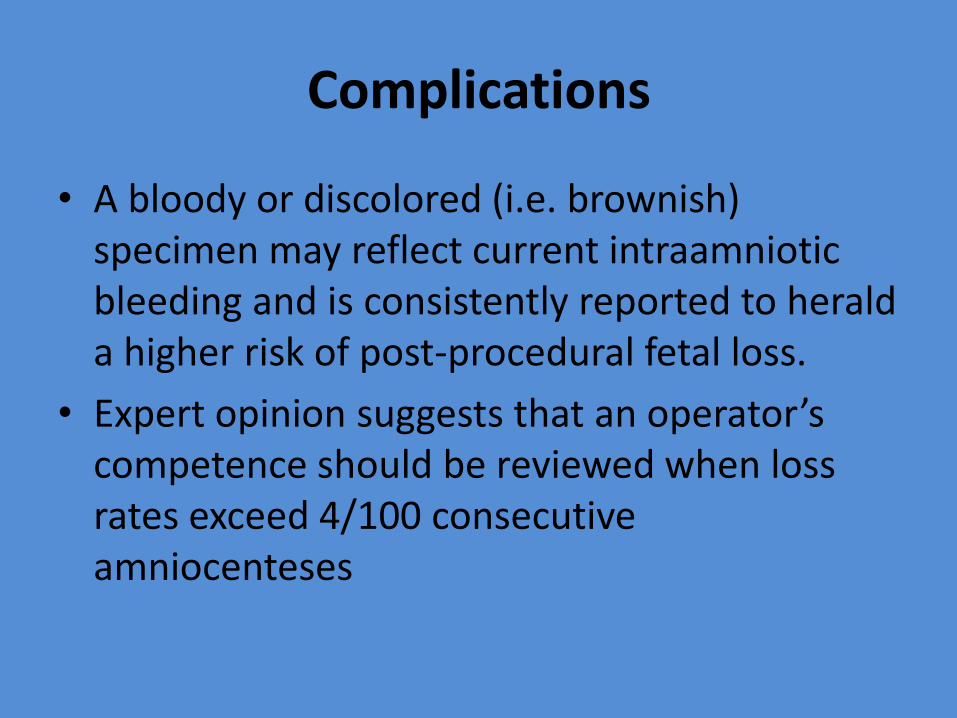
• A bloody or discolored (i.e. brownish) specimen may reflect current intraamniotic bleeding and is consistently reported to herald a higher risk of post-procedural fetal loss.
• Expert opinion suggests that an operator’s competence should be reviewed when loss rates exceed 4/100 consecutive amniocenteses

Risk factors
• Uterine fibroids • Mullerian malformations • Chorioamniotic separation • Retrochorionic hematoma • Previous or current maternal bleeding • Maternal body mass index > 40 kg/m2 • Multiparity (>3 births) • Manifest vaginal infection • History of three or more miscarriages
Risk

Chorionic Villus Sampling (CVS) • Performed after 10 + 0 gestational
weeks • Transabdominally or
transcervically • Under continuous ultrasound
guidance • A single needle of 17 – 20 G or a
two-needle set of outer 17/19 G and inner 19/20 G may be used
• A minimum amount of 5 mg villi in each sample is required to achieve a valid result

Chorionic Villus Sampling (CVS)
Transcervical approach. • Biopsy forceps are inserted
transvaginally through the cervical canal to the trophoblastic area, or a catheter with plastic or metal stylet under syringe aspiration may be used
• The amount of villi obtained in the sample must be checked visually.
• A minimum amount of 5 mg villi in each sample is required to achieve a valid result.
• Sampling failure is reported to occur in 2.5–4.8% of procedures
Chorionic Villus Sampling (CVS)
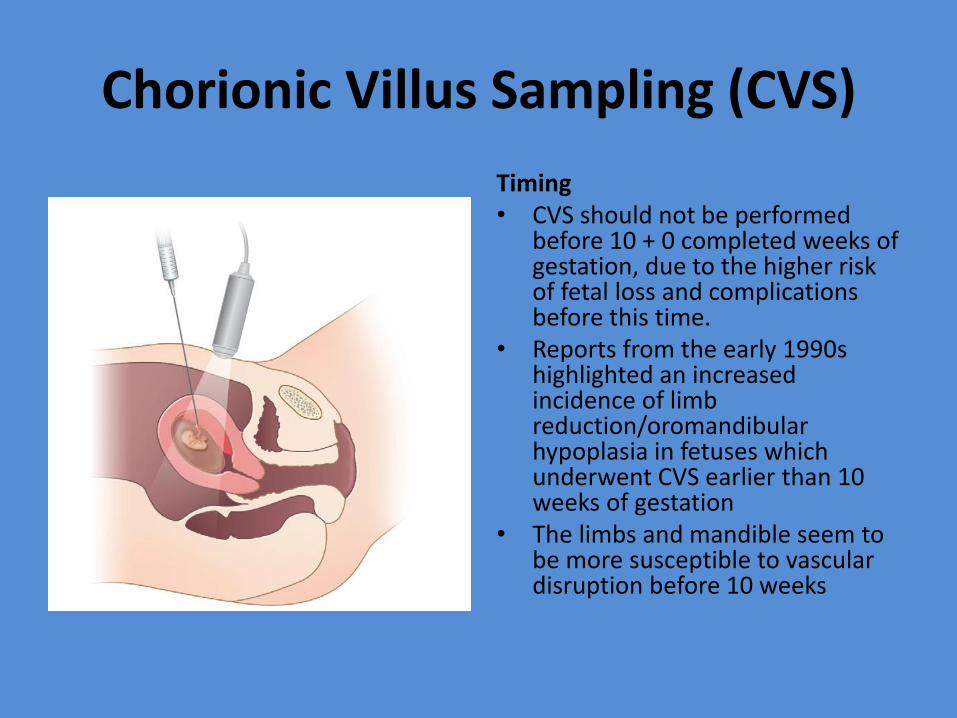
Timing • CVS should not be performed
before 10 + 0 completed weeks of gestation, due to the higher risk of fetal loss and complications before this time.
• Reports from the early 1990s highlighted an increased incidence of limb reduction/oromandibular hypoplasia in fetuses which underwent CVS earlier than 10 weeks of gestation
• The limbs and mandible seem to be more susceptible to vascular disruption before 10 weeks

Chorionic Villus Sampling (CVS)
Laboratory aspects
• Failure of the cytotrophoblastic culture < 0.5% of procedures
• Placental cell mosaicism is seen in 1% of procedures.
• In these cases, genetic counseling is recommended and amniocentesis may be indicated to differentiate true fetal mosaicism from confined placental mosaicism

Complications
Fetal loss
• Between 0.2% and 2%
• This risk appears to be lower in experienced centers and to decrease with increasing experience, ranging between 1/150 and 1/500
• The fetal loss rate after transcervical CVS was reported to be 2.5%

Ultrasound Obstet Gynecol 2015; 45: 16–26.
Complications
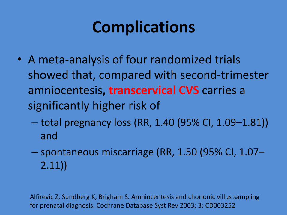
• A meta-analysis of four randomized trials showed that, compared with second-trimester amniocentesis, transcervical CVS carries a significantly higher risk of
– total pregnancy loss (RR, 1.40 (95% CI, 1.09–1.81)) and
– spontaneous miscarriage (RR, 1.50 (95% CI, 1.07–2.11))
Alfirevic Z, Sundberg K, Brigham S. Amniocentesis and chorionic villus sampling for prenatal diagnosis. Cochrane Database Syst Rev 2003; 3: CD003252

Complications
• Vaginal bleeding – Vaginal bleeding is reported to occur in 10% of
cases. more frequent after the transcervical (up to 30% of cases).
• Uncommon complications – The risk of amniotic fluid leakage following CVS is
exceedingly rare, occurring after < 0.5% of procedures
– The risk of chorioamnionitis and uterine infection after CVS is extremely small (1–2/3000).

Complications
• There have been some reports associating CVS with development of preeclampsia later in pregnancy, possibly due to placental damage, but these findings have not been consistent across studies and a meta-analysis failed to show an association.
• Similarly, a case–control study did not detect an association between CVS and impaired fetal growth;

Fetal Blood Sampling (FBS, Cordocentesis)
• Transabdominally after 18 + 0 weeks, using a 20 – 22-G needle under ultrasound guidance.
• If the placenta is anterior, a puncture of the cord at the level of placental insertion is suggested;
• if the placenta is posterior, a free loop of the cord or the intra-abdominal portion of the umbilical vein is sampled

Indications

Complications
• Fetal Loss
–The risk of fetal loss after FBS is between 1% and 2%
–Factors associated with increased risk of fetal loss after FBS include fetal anomalies, IUGR and gestational age < 24 weeks.

Am J Obstet Gynecol 2001;184:719-23
Fetal Loss Rate Associated With Cordocentesis At Midgestation

Summary -Amniocentesis

Summary -CVS

Amniocentesis and chorionic villus sampling for prenatal diagnosis
Cochrane Database of Systematic Reviews 21 JUL 2003 DOI: 10.1002/14651858.CD003252 http://onlinelibrary.wiley.com/doi/10.1002/14651858.CD003252/full#CD003252-fig-00320

Tabor et al., Ultrasound Obstet Gynecol 2009; 34: 19–24

Tabor et al., Ultrasound Obstet Gynecol 2009; 34: 19–24

Tabor et al., Ultrasound Obstet Gynecol 2009; 34: 19–24

Prenat Diagn 2002; 22: 598–604.
Long Term Follow-up for CVS and AC

Cochrane Database of Systematic Reviews 2012, Issue 8. Art. No.: CD008678
• For amniocentesis three interventions were evaluated - intramuscular progesterone, hexoprenaline and selecting high or low puncture sites for late 'blind' procedure - each intervention in a single small study. There was no conclusive evidence of benefit for any of them.
• The same applies for terbutaline tocolysis and use of continuous vacuum aspiration during CVS.
![[PPT]PRENATAL TANI - WordPress.com · Web viewInvasiveproceduresused in prenatal diagnosis: Amniocentesis, Chorionic villus sampling, and Fetal blood sampling Preimplantation genetic](https://img.dokumen.tips/doc/110x75/5c8b3d5e09d3f22c4e8bb28d/pptprenatal-tani-web-viewinvasiveproceduresused-in-prenatal-diagnosis-amniocentesis.jpg)