Embed Size (px)
Citation preview
Comparative Analysis of Salivary Gland Transcriptomesof Phlebotomus orientalis Sand Flies from Endemic andNon-endemic Foci of Visceral LeishmaniasisMichaela Vlkova1, Michal Sima1, Iva Rohousova1, Tatiana Kostalova1, Petra Sumova1, Vera Volfova1,
Erin L. Jaske2, Kent D. Barbian2, Teshome Gebre-Michael3, Asrat Hailu4, Alon Warburg5,
Jose M. C. Ribeiro6, Jesus G. Valenzuela7*, Ryan C. Jochim7¤*, Petr Volf1*
1 Department of Parasitology, Faculty of Science, Charles University, Prague, Czech Republic, 2 Genomics Unit, Research Technologies Section, Rocky Mountain
Laboratories, Hamilton, Montana, United States of America, 3 Aklilu Lemma Institute of Pathobiology, Addis Ababa University, Addis Ababa, Ethiopia, 4 Department of
Microbiology, Immunology & Parasitology, Faculty of Medicine, Addis Ababa University, Addis Ababa, Ethiopia, 5 Department of Parasitology, The Kuvin Centre for the
Study of Infectious and Tropical Diseases, Hadassah Medical School, The Hebrew University of Jerusalem, Jerusalem, Israel, 6 Vector Biology Section, Laboratory of Malaria
and Vector Research, National Institute of Allergy and Infectious Diseases, National Institutes of Health, Rockville, Maryland, United States of America, 7 Vector Molecular
Biology Section, Laboratory of Malaria and Vector Research, National Institute of Allergy and Infectious Diseases, National Institutes of Health, Rockville, Maryland, United
States of America
Abstract
Background: In East Africa, Phlebotomus orientalis serves as the main vector of Leishmania donovani, the causative agent ofvisceral leishmaniasis (VL). Phlebotomus orientalis is present at two distant localities in Ethiopia; Addis Zemen where VL isendemic and Melka Werer where transmission of VL does not occur. To find out whether the difference in epidemiology ofVL is due to distant compositions of P. orientalis saliva we established colonies from Addis Zemen and Melka Werer,analyzed and compared the transcriptomes, proteomes and enzymatic activity of the salivary glands.
Methodology/Principal Findings: Two cDNA libraries were constructed from the female salivary glands of P. orientalis fromAddis Zemen and Melka Werer. Clones of each P. orientalis library were randomly selected, sequenced and analyzed. In P.orientalis transcriptomes, we identified members of 13 main protein families. Phylogenetic analysis and multiple sequencealignments were performed to evaluate differences between the P. orientalis colonies and to show the relationship withother sand fly species from the subgenus Larroussius. To further compare both colonies, we investigated the humoralantigenicity and cross-reactivity of the salivary proteins and the activity of salivary apyrase and hyaluronidase.
Conclusions: This is the first report of the salivary components of P. orientalis, an important vector sand fly. Our studyexpanded the knowledge of salivary gland compounds of sand fly species in the subgenus Larroussius. Based on thephylogenetic analysis, we showed that P. orientalis is closely related to Phlebotomus tobbi and Phlebotomus perniciosus,whereas Phlebotomus ariasi is evolutionarily more distinct species. We also demonstrated that there is no significantdifference between the transcriptomes, proteomes or enzymatic properties of the salivary components of Addis Zemen(endemic area) and Melka Werer (non-endemic area) P. orientalis colonies. Thus, the different epidemiology of VL in theseEthiopian foci cannot be attributed to the salivary gland composition.
Citation: Vlkova M, Sima M, Rohousova I, Kostalova T, Sumova P, et al. (2014) Comparative Analysis of Salivary Gland Transcriptomes of Phlebotomus orientalisSand Flies from Endemic and Non-endemic Foci of Visceral Leishmaniasis. PLoS Negl Trop Dis 8(2): e2709. doi:10.1371/journal.pntd.0002709
Editor: Mary Ann McDowell, University of Notre Dame, United States of America
Received September 3, 2013; Accepted January 7, 2014; Published February 27, 2014
This is an open-access article, free of all copyright, and may be freely reproduced, distributed, transmitted, modified, built upon, or otherwise used by anyone forany lawful purpose. The work is made available under the Creative Commons CC0 public domain dedication.
Funding: The research was supported by Bill and Melinda Gates Foundation Global Health Program (grant number OPPGH5336). This study was partially fundedby EU grant FP7-261504 EDENext and is catalogued by the EDENext Steering Committee as EDENext176 (http://www.edenext.eu). The contents of this publicationare the sole responsibility of the authors and do not necessarily reflect the views of the European Commission. The Czech team was also supported by CzechScience Foundation (13-05292S), by Grant Agency of Charles University (GAUK-675012/2012), and by UNCE (204017). JMCR and JGV were supported by theIntramural Research Program of the Division of Intramural Research, National Institute of Allergy and Infectious Diseases. The funders had no role in study design,data collection and analysis, decision to publish, or preparation of the manuscript.
Competing Interests: The authors have declared that no competing interests exist.
* E-mail: [email protected] (JGV); [email protected] (RCJ); [email protected] (PV)
¤ Current address: Entomology Branch, Walter Reed Army Institute of Research, Silver Spring, Maryland, United States of America.
Introduction
Protozoan parasites belonging to the genus Leishmania are the
pathogenic agents causing a broad range of diseases commonly
known as leishmaniasis. Sand fly vectors (Diptera: Phlebotominae)
spread leishmaniasis among the vertebrate hosts during the
bloodfeeding when infected sand fly females eject parasites into
the wound along with their saliva. Salivary compounds possess
powerful anti-hemostatic and immunomodulatory properties
(reviewed in [1]); nonetheless, the salivary proteins are highly
antigenic. As the repeated exposure to sand fly bites was shown to
be protective against leishmaniasis (e.g. [2]), the immune profiles
elicited by single salivary proteins are of major scientific interest.
PLOS Neglected Tropical Diseases | www.plosntds.org 1 February 2014 | Volume 8 | Issue 2 | e2709
To date, the intensive investigation of salivary proteins in
certain sand fly species has allowed the generation of individual
recombinant salivary proteins that have been employed as reliable
markers of exposure to sand fly bites [3–5] or as the protective
agent against cutaneous and visceral leishmaniases (CL and VL,
respectively) under laboratory conditions [6–13]. However, most
of the experiments were performed using New World VL vector
Lutzomyia longipalpis. As the composition of salivary glands and the
protective effect conferred by sand fly saliva is species-specific [14–
19], it is vital to continue with detailed characterization of the
salivary proteins with a special focus on sand fly species causing
lethal VL.
Phlebotomus orientalis is a member of the subgenus Larroussius and
represents the main sand fly species transmitting Leishmania donovani
within the countries of East Africa (reviewed in [20]) as well as in
Saudi Arabia [21] and Yemen [22]. At two distinct localities in
Ethiopia, Addis Zemen and Melka Werer, we observed different
epidemiology of VL, although P. orientalis was present in both
places. While in Addis Zemen, human VL caused by Le. donovani
with high mortality rate was reported [23], Melka Werer is
considered to be a non-endemic area with no human cases. A
recently published study compared various molecular aspects of
colonies from both foci and showed that the susceptibility of Addis
Zemen and Melka Werer colonies to Le. donovani infection was
identical [24]. As Warburg et al. described the possible connection
of the salivary gland composition with varying pathologies of CL
[25] and sand fly saliva is known to play a crucial role in
transmission of Leishmania spp. (e.g. [2]), we hypothesized that the
composition of salivary glands may explain the different epidemi-
ology in these Ethiopian foci. Therefore, we studied the
transcriptomes, proteomes and the enzymatic activities (apyrase
and hyaluronidase) in the saliva of female sand flies from Addis
Zemen (VL endemic) and from Melka Werer (non-endemic).
Furthermore, we characterized the main salivary antigens in both
colonies and determined the level of glycosylation of P. orientalis
salivary proteins. Importantly, we compared our data with other
sand fly species from the subgenus Larroussius, whose cDNA
libraries have already been constructed [26–28], and used
sequences of the New World sand fly species L. longipalpis as an
outgroup.
Methods
Ethics statementBALB/c mice were maintained and handled in the animal
facility of Charles University in Prague in accordance with
institutional guidelines and Czech legislation (Act No. 246/1992
coll. on Protection of Animals against Cruelty in present statutes at
large). The experiments were approved by the Committee on the
Ethics of Animal Experiments of the Charles University in Prague
(Permit Number: 24773/2008-10001) and were performed under
the Certificate of Competency (Registration Numbers: CZU 934/
05, CZU 307/09) in accordance with the Examination Order
approved by Central Commission for Animal Welfare of the
Czech Republic.
Sand flies and salivary gland dissectionsTwo colonies of P. orientalis were established; one from a non-
endemic lowland area in central Ethiopia, Melka Werer (MW)
(altitude of 800 m), the later one from an endemic focus of VL in
the highlands of Northwest Ethiopia, Addis Zemen (AZ) (altitude
of 1800–2000 m), and then transferred to Czech Republic. Both
sand fly colonies were kept in the insectary of Charles University in
Prague and were reared under standard conditions as described in
[29]. For the experiments, the sand flies from F5–F6 generation
were used. Salivary glands of 1-day old adult females were
dissected; mRNA was extracted and stored in RNA later
(Ambion). For proteome analysis, western blot, affinity blot, and
hyaluronidase assay, salivary glands from 5- to 8-day old P.
orientalis adult females were dissected and stored in Tris buffer
(20 mM Tris, 150 mM NaCl, pH 7.7). For the apyrase assay, 8-
day old adult female salivary glands were dissected into Tris buffer
containing 0.005% Triton X-100 and stored at 280uC.
Construction of salivary gland cDNA librariesSalivary gland mRNA was isolated separately from 45 pairs
each of MW and AZ glands using Micro-FastTrack mRNA
isolation kit (Invitrogen). Both cDNA libraries were constructed
following the manufacturer’s instructions for SMART cDNA
Library Construction Kit (BD Clontech) with some modifications
as described in [30]. Each library was fractionated into large,
medium, and small cDNA fragments. Gigapack III Gold
Packaging Extract (Stratagene) was used for packaging the phage.
Both libraries were then plated by infecting log-phase XL-1 blue
Escherichia coli (Clontech). Transfected plaques were randomly
selected and a PCR reaction with vector primers flanking the
inserted cDNA was made. The presence of recombinants was
checked by visualization the PCR products on 1.1% agarose gel
with SYBR Safe (Invitrogen). Inserts were sequenced as previously
described [31] using a ABI 3730XL DNA Sequencer (Applied
Biosystems).
BioinformaticsDetailed description of the bioinformatics analysis can be found
elsewhere [28]. Briefly, expression sequence tags (ESTs) were
analyzed using a customized program based on the Phred
algorithm [32,33]. Sequences with Phred quality scores lower
than 25 were removed, as well as vector sequences and primers.
Resulting sequences were grouped based on nucleotide homology
of 90% identity over 100 residues and aligned into consensus
transcript sequences (contigs) using the CAP3 sequence assembly
program. BLAST programs were used to compare contigs and
Author Summary
Phlebotomus orientalis is the vector of visceral leishman-iasis (VL) caused by Leishmania donovani in NortheastAfrica. Immunization with sand fly saliva or with individualsalivary proteins has been shown to protect againstleishmaniasis in different hosts, warranting the intensivestudy of salivary proteins of sand fly vectors. In our study,we characterize the salivary compounds of P. orientalis,thereby broadening the repertoire of salivary proteins ofsand fly species belonging to the subgenus Larroussius. Inorder to find out whether there is any connection betweenthe composition of P. orientalis saliva and the epidemiol-ogy of VL in two distinct Ethiopian foci, Addis Zemen andMelka Werer, we studied the transcriptomes, proteomes,enzymatic activities, and the main salivary antigens in twoP. orientalis colonies originating from these areas. We didnot detect any significant difference between the saliva offemale sand flies originating in Addis Zemen (endemicarea) and Melka Werer (non-endemic area). Therefore, thedifferent epidemiology of VL in these Ethiopian foci cannotbe related to the distant salivary gland protein composi-tion. Identifying the sand fly salivary gland compounds willbe useful for future research focused on characterizingsuitable salivary proteins as potential anti-Leishmaniavaccine candidates.
Phlebotomus orientalis Saliva Transcriptome
PLOS Neglected Tropical Diseases | www.plosntds.org 2 February 2014 | Volume 8 | Issue 2 | e2709
singletons (contigs with a single sequence) to the non-redundant
protein database of the NCBI, the Gene Ontology database (GO)
[34], to COG conserved domains database [35], Protein Family
database (Pfam) [36], SimpleModular Architecture Tool database
(SMART) [37], and to rRNA Nucleotide Sequences, and
Mitochondrial and Plastid Sequence (MITPLA) databases avail-
able from NCBI. The three frame translations of each dataset were
submitted to the SignalP server [38] to find signal sequences. The
grouped and assembled sequences, BLAST results, and SignalP
results, combined by dCAS software [39] in an Excel spreadsheet,
were manually verified and annotated. N- and O-Glycosylation
sites on the proteins were predicted using NetNGlyc 1.0 and
NetOGlyc 3.1 software (www.cbs.dtu.dk/services/NetNGlyc,
www.cbs.dtu.dk/services/NetOGlyc) [40].
Phylogenetic analysisProtein sequences were aligned using ClustalX (version 2.0) [41]
and manually refined in BioEdit 7.1.3.0 editing software. For each
alignment, best substitution matrix was determined by ProtTest
software 2.0 [42]. This matrix was subsequently used by TREE-
PUZZLE 5.2 [43] to reconstruct maximum likelihood phyloge-
netic trees from the protein alignments using quartet puzzling with
1000 puzzling steps in each phylogenetic analysis. Resulting trees
were visualized in MEGA 4 [44].
Proteome analysisFor mass spectrometry analysis, salivary glands of both AZ and
MW P. orientalis colonies were dissolved in non-reducing sample
buffer and electrophoretically separated in 12.5% SDS gel.
Proteins within the gel were visualized by staining with Coomassie
Brilliant Blue R-250 (Serva). The individual bands were cut and
incubated with 10 mM dithiothreitol (Sigma) and then treated
with 55 mM iodoacetamide (Sigma). Washed and dried bands
were digested with trypsin (Promega). The tryptic peptides were
separated by liquid chromatography using an Ultimate 3000
HPLC system (Dionex). The peptide samples diluted in 0.3%
trichloroacetic acid (TCA) with 10% acetonitrile (ACN) were
loaded onto a PepMap 100 C18 RP column (Dionex) at a flow rate
of 300 nl per minute. The peptides were eluted by a 45-min linear
gradient of 5–80% (v/v) ACN in 0.1% (v/v) TCA over a period of
20 min. The eluent was mixed 1:3 with matrix solution (20 mg/ml
a-cyano-4-hydroxycinnamic acid in 80% ACN) and subsequently
spotted onto MALDI target plates using a Probot microfraction
collector (Dionex). Spectra were acquired on 4800 Plus MALDI
TOF/TOF analyzer (Applied Biosystems/MDS Sciex) equipped
with a Nd: YAG laser (355 nm, firing rate 200 Hz) as described in
detail in [28].
Hyaluronidase activity analysisHyaluronidase activity in salivary glands of both P. orientalis
colonies was quantified using a sensitive assay in microtitration
plates coupled with biotinylated hyaluronic acid (bHA). Salivary
glands were homogenized by three freeze-thaw cycles and salivary
gland extract (SGE) was obtained by centrifugation at 17000 g
(5 min, 2uC). Biotinylated HA, prepared as described in [45], was
immobilized onto Covalink NH microtiter plates (NUNC) using
the method by Frost and Stern [46] modified by [28] at a final
concentration of 1 mg/well bHA. The plates were incubated
overnight at 4uC and washed three times in PBS, pH 7.2
containing 2 M NaCl and 50 mM MgSO4. The plates with
immobilized bHA were coated for 45 min with 1% BSA in PBS,
then washed and equilibrated with assay buffer (0.1 M acetate
buffer, pH 5.0, 0.1 M NaCl, 0.1% Triton X-100) to adjust the pH
for optimum sand fly salivary hyaluronidase activity. Four SGE
samples for each colony were pipetted into the plates in triplicate
at a final concentration of 0.5 salivary gland per well and
incubated for 45 min at 37uC. To obtain a standard curve ranging
from 0.5 to 7.86103 rTRU, hyaluronidase from bovine testes
(Sigma), at a concentration of 0.01 TRU/ml, was diluted by two-
fold serial dilution in 0.1 M acetate buffer, pH 4.5, 0.1 M NaCl,
0.1% Triton X-100. Wells without bHA or enzyme were used as
controls. The reaction was terminated by the addition of 200 ml/
well of 6 M guanidine. After washing, avidin-peroxidase (Sigma,
2 mg/ml) was added at a final concentration of 0.2 mg/well and
incubated for 30 min at room temperature. Color reaction was
developed with o-phenylenediamine substrate in 0.1 M citrate-
phosphate buffer, pH 5.5. Absorbance was measured at 492 nm
using Infinite M 200 fluorometer (Schoeller Instruments). Raw
data were evaluated by Measurement Parameters Editor Magellan
6 (Tecan) and the standard curve created using a 4-parameter
logistic fit.
Apyrase activity analysisApyrase activity was determined using the Fiske and Subbarow
method for measuring inorganic phosphate (Pi) released from ADP
or ATP [47], with some modifications. Salivary glands were
homogenized by one freeze-thaw cycle combined with a mechan-
ical homogenization. Two ml of salivary gland homogenate (SGH)
diluted 1:25 in assay buffer (50 mM TRIS 150 mM NaCl, pH 8.5
with 5 mM CaCl2 or 5 mM MgCl2) were mixed in wells with
78 ml of assay buffer and 20 ml of substrate to obtain a final
concentration of 2 mM ATP or ADP and 1/25 of gland pair per
well. SGH samples were pipetted into the microtiter plate in series
of six. Wells containing only assay buffer were used as negative
controls. Plates were incubated for 15 min at 37uC. Then the
enzymatic reaction was stopped by addition of 25 ml of 1.25%
ammonium molybdate in 1.25 M sulfuric acid and 5 ml of Fiske-
Subbarow reducer (25 mg/ml, F5428 Sigma) per well. The
colorimetric reaction was read after 15 min by Tecan Infinite M
200 fluorometer (Schoeller Instruments) at 665 nm. The amount
of Pi released from substrate was determined using potassium
dihydrogen phosphate as a standard. The study of pH optimum
was carried out within a range of pH 6.0–9.5. Salivary glands of P.
papatasi, the species with previously described apyrase activity [48],
were used as a positive control. Amount of proteins within SGHs
was determined using Bio-Rad DC Protein Assay with BSA as a
standard according to the manufacturer’s instructions.
Western blottingSalivary glands of both P. orientalis colonies were separated by
SDS-PAGE on 10% gel under non-reducing conditions using
Mini-Protean III apparatus (Biorad). Salivary proteins were
transferred from gel to nitrocellulose membrane (NC) by Semi-
Phor equipment (Hoefer Scientific Instruments) and cut into strips.
The strips were then blocked with 5% low fat dry milk in Tris-
buffered saline with 0.05% Tween 20 (TBS-Tw) and subsequently
incubated with BALB/c mice sera (AZ – mice bitten 18 times in a
week interval; MW – mice bitten 17 times in a week interval),
diluted 1:100 in TBS-Tw, for 1 hour. After the washing with TBS-
Tw, the strips were incubated for 1 hour with peroxidase-
conjugated goat anti-mouse IgG (Serotec) diluted 1:1000 in
TBS-Tw. The chromogenic reaction was developed using a
substrate solution containing diaminobenzidine and H2O2.
Affinity blottingAffinity blotting was performed using salivary glands from MW
P. orientalis colony separated by SDS-PAGE as described above.
After transfer, free binding sites on NC membrane were blocked
Phlebotomus orientalis Saliva Transcriptome
PLOS Neglected Tropical Diseases | www.plosntds.org 3 February 2014 | Volume 8 | Issue 2 | e2709
with 5% bovine serum albumin in 20 mM TBS-Tw overnight at
4uC. The strips were then incubated for 1.5 hour on the shaker at
room temperature with biotinylated lectins from Dolichos biflorus
(DBA, Vector), Glycine max (SBA, Vector), Ulex europaeus (UEA-I,
Vector), Tetragonolobus purpureus (LTA, Sigma), Canavalia ensiformis
(ConA, Sigma), and Pisum sativum (PSA, Vector). Based on the
preliminary experiments with different lectin concentrations, the
lectins were diluted: 5 mg/ml, 10 mg/ml, 10 mg/ml, 0.2 mg/ml,
0.1 mg/ml and 10 mg/ml in TBS-Tw, respectively. To control the
reaction specificity the aforementioned lectins were pre-incubated
for 30 min with the appropriate saccharide inhibitors (Sigma) as
follows: 0.25 M N-acetyl-D-galactosamine for DBA and SBA,
0.5 M L-fucose for UEA-I and LTA, 0.5 M methyl-a-D-
mannopyranoside for ConA and PSA, and subsequently applied
on the strips. After the washing with TBS-Tw, streptavidin-
peroxidase (Sigma) was added to strips at a final concentration of
1 mg/ml and incubated for 1 h on the shaker at room
temperature. The chromogenic reaction was developed as
mentioned above.
Results and Discussion
Sequencing of P. orientalis salivary gland cDNA librariesTwo cDNA libraries were constructed from salivary glands of P.
orientalis colonies originating in Addis Zemen and Melka Werer,
Ethiopia. For each cDNA library, 940 clones were randomly
selected and sequenced, which resulted in 835 and 749 high
quality sequences from AZ and MW, respectively. Based on
nucleotide homology, sequences were clustered into contigs,
analyzed using the dCAS cDNA annotation software [39] and
subsequently verified by manual annotation. From the AZ cDNA
library, sequences were assembled into 263 contigs, where 185 of
them were singletons (one sequence per contig). From the MW
cDNA library, we obtained 242 contigs, including 171 singletons.
In accordance with previously published cDNA libraries from sand
fly salivary glands, the most abundant transcripts were those
coding for putative salivary proteins (607 out of 835 in AZ; 567 out
of 749 in MW). Of the nucleotide sequences encoding putative
salivary proteins, 574 (AZ) and 506 (MW) salivary transcripts
encoded a predicted signal peptide sequence. Those that did not
possess sequences encoding a signal peptide were truncated at the
59 end. Most of the contigs coding for putative salivary proteins
were comprised of more than one sequence (averaging 7.14
sequences per contig in AZ and 6.23 in MW), whereas
housekeeping proteins or proteins with unknown function were
mostly represented by singletons. All obtained ESTs were
deposited in the NCBI dbEST database under accession numbers
JZ479238–JZ480094 for AZ colony and JZ480095–JZ480885 for
MW colony.
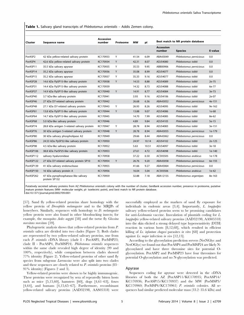
Members of 13 main protein families were found among the
putative salivary proteins of the two P. orientalis colonies: apyrase,
yellow-related protein, antigen 5-related protein, odorant-binding
proteins (D7-related and PpSP15-like proteins), hyaluronidase,
endonuclease, phospholipase, pyrophosphatase, amylase, PpSP32-
like protein, ParSP25-like protein, SP16-like protein, and Lufaxin
(SP34-like protein). Detailed descriptions of each protein family
are listed in the following paragraphs. Interestingly, we did not
detect any sequences coding for adenosin deaminase in either P.
orientalis cDNA library. Thus, we expect that P. orientalis saliva
contains adenosin and ADP/AMP; leaving only P. duboscqi, L.
longipalpis, and L. intermedia [49–52] as the sand fly species identified
to produce adenosine deaminase, to date.
BLAST comparison of translated nucleotide sequences with the
non-redundant (NR) protein database showed high similarity with
salivary proteins of P. perniciosus and P. tobbi (both subgenus
Larroussius). Sporadically, the best match was found with salivary
proteins of P. arabicus (subgenus Adlerius) or P. argentipes (subgenus
Euphlebotomus). Representative sequences of putative salivary
proteins from both P. orientalis colonies that were deposited into
NCBI GenBank database are listed in Table 1 and Table 2. Both
tables include GenBank accession numbers, the predicted molec-
ular weight, isoelectric point, best match to the NR database, the
sand fly species with the highest homology, and presence in the
proteome.
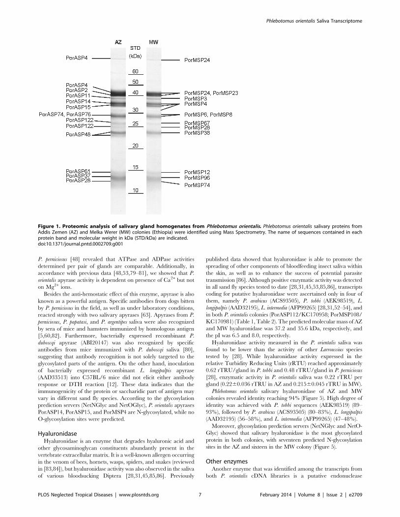
Proteome analysisSalivary proteins presented in the proteome were identified by
mass spectrometry and are shown in Figure 1. In both cDNA
libraries, 12 salivary proteins were determined to be present in
proteome. In Addis Zemen colony, the identified proteins were two
yellow-related proteins (PorASP2/KC170933; PorASP4/KC
170934), three apyrases (PorASP11/KC170935; PorASP14/
KC170936; PorASP15/KC170937), two D7-related proteins (Por
ASP48/KC170943; PorASP122/KC170954), two antigen 5-relat-
ed proteins (PorASP74/KC170947; PorASP76/KC170948), and
three PpSP15-like proteins (PorASP28/KC170938; PorASP37/
KC170940; PorASP61/KC170944) (Figure 1). In Melka Werer
colony, the identified proteins were two yellow-related proteins
(PorMSP23/KC170966; PorMSP24/KC170967), two apyrases
(PorMSP3/KC170960; PorMSP4/KC170961), three D7-related
proteins (PorMSP28/KC170969; PorMSP38/KC170970; Por
MSP67/KC170973), two antigen 5-related proteins (PorMSP6/
KC170962; PorMSP8/KC170963), and three PpSP15-like proteins
(PorMSP12/KC170964; PorMSP74/KC170974; PorMSP96/
KC170978) (Figure 1). Except for apyrase, none of the salivary
enzymes identified in P. orientalis transcriptomes were detected in
proteome analysis, even though all of the nucletoide sequences
coding for these salivary proteins possessed signal peptides. It might
be explained by the fact that extremely active enzymes do not need
a huge amount of protein to be effective.
Yellow-related proteinsYellow-related proteins are abundantly expressed in the sand fly
salivary glands and have been detected in the saliva of all sand fly
species tested, to date [7,26–28,31,50,52–56]. Two yellow-related
proteins were found in the cDNA library of the AZ (PorASP2/
KC170933; PorASP4/KC170934) as well as the MW (PorMSP23/
KC170966; PorMSP24/KC170967) P. orientalis colony). All four P.
orientalis yellow-related proteins had similar predicted molecular
mass (41.5–42.3 kDa) and wide range of pI (6.1–8.1) (Table 1,
Table 2). All obtained sequences contained the entire major royal
jelly protein (MRJP) domain, which is characteristic for the yellow-
related proteins. Some advances have been also made in describing
the function of sand fly yellow-related proteins. It was shown that
recombinant yellow-related proteins from L. longipalpis saliva
(AAD32198, AAS05318) act as high affinity binders of prohemo-
static and proinflammatory biogenic amines such as serotonin,
catecholamines and histamine [12]. Similarly, the amino acid motif
present in the ligand binding pocket of L. longipalpis (T-x(52,63)-Y-
Q-x(85,90)-[FY]-x(44,46)-F-x(54)-[IVL]-x(45,46)-[FY]-x-[TS]-D-
x(13)-[NT]-x-[QHFL]) was discovered in the yellow-related
proteins of L. ayacuchensis (BAM69111, BAM69185, BAM69109,
BAM69110) [56] and L. intermedia (AFP99235) [52], but also in P.
orientalis and other sand fly species from the subgenus Larroussius
tested (Figure 2). These findings suggest similar anti-inflammatory
function of these salivary proteins in other Lutzomyia and Phlebotomus
sand fly species [12] and could potentially explain the lectin-like
properties of 42 kDa yellow-related protein from P. duboscqi saliva
Phlebotomus orientalis Saliva Transcriptome
PLOS Neglected Tropical Diseases | www.plosntds.org 4 February 2014 | Volume 8 | Issue 2 | e2709
[57]. Sand fly yellow-related proteins share homology with the
yellow protein of Drosophila melanogaster and to the MRJPs of
honeybees. Similarly, sequences with homology to D. melanogaster
yellow protein were also found in other bloodsucking insects; for
example, the mosquito Aedes aegypti [58] and the tsetse fly Glossina
morsitans morsitans [59].
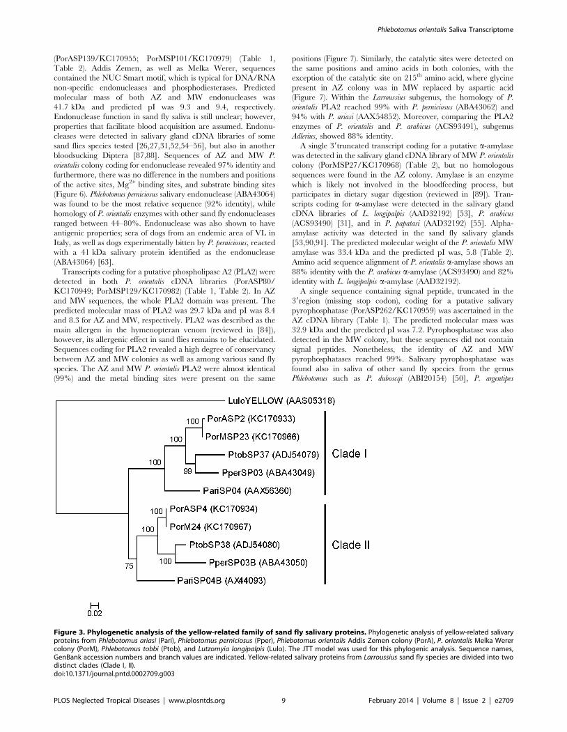
Phylogenetic analysis shows that yellow-related proteins from P.
orientalis saliva are divided into two clades (Figure 3). Both clades
are represented by two yellow-related salivary proteins, one from
each P. orientalis cDNA library (clade I - PorASP2, PorMSP23;
clade II - PorASP4, PorMSP24). Phlebotomus orientalis sequences
within the same clade revealed high degree of identity (99 and
100%, respectively), while comparison between clades showed
77% identity (Figure 2). Yellow-related proteins of other sand fly
species from subgenus Larroussius were also split into two clades
and these sequences are closely related to P. orientalis proteins (83–
91% identity) (Figures 2 and 3).
Yellow-related proteins were shown to be highly immunogenic.
These proteins were recognized by sera of repeatedly bitten hosts
such as mice [5,15,60], hamsters [60], dogs [4,61–63], foxes
[4,64], and humans [4,15,65–67]. Furthermore, recombinant
yellow-related salivary proteins (AAD32198, AAS05318) were
successfully employed as the markers of sand fly exposure for
individuals in endemic areas [3,4]. Importantly, L. longipalpis
salivary yellow-related proteins seem to be promising candidates
for anti-Leishmania vaccine. Inoculation of plasmids coding for L.
longipalpis yellow-related salivary proteins (AAD32198, AAS05318)
into the skin elicited a strong delayed type hypersensitivity (DTH)
reaction in various hosts [8,12,68], which resulted in efficient
killing of Le. infantum chagasi parasites in vitro [68] and protection
against Le. major infection in vivo [12,13].
According to the glycosylation prediction servers (NetNGlyc and
NetOGlyc) we found out that PorASP4 and PorMSP24 are likely N-
glycosylated and have three threonine sites for potential O-
glycosylation. PorASP2 and PorMSP23 have four threonines for
potential O-glycosylation and no N-glycosylation was predicted.
ApyraseSequences coding for apyrase were detected in the cDNA
libraries of both the AZ (PorASP11/KC170935; PorASP14/
KC170936; PorASP15/KC170937) and the MW (PorMSP3/
KC170960; PorMSP4/KC170961) P. orientalis colonies. All se-
quences had similar predicted molecular mass (33.2–35.6 kDa) and
Table 1. Salivary gland transcripts of Phlebotomus orientalis – Addis Zemen colony.
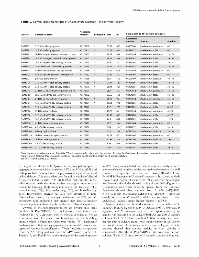
Cluster Sequence nameAccessionnumber Proteome MW pI Best match to NR protein database
Accessionnumber Species E-value
PorASP2 42 kDa yellow-related salivary protein KC170933 Y 41.54 6.09 ABA43049 Phlebotomus perniciosus 0.0
PorASP4 42.6 kDa yellow-related salivary protein KC170934 Y 42.31 8.07 ADJ54080 Phlebotomus tobbi 0.0
PorASP11 35.5 kDa salivary apyrase KC170935 Y 35.53 9.95 ABB00906 Phlebotomus perniciosus 0.0
PorASP14 35.2 kDa salivary apyrase KC170936 Y 35.08 8.99 ADJ54077 Phlebotomus tobbi 0.0
PorASP15 35.2 kDa salivary apyrase KC170937 Y 35.33 9.16 ADJ54077 Phlebotomus tobbi 0.0
PorASP28 14.6 kDa PpSP15-like salivary protein KC170938 Y 14.53 8.88 ADJ54089 Phlebotomus tobbi 2e-75
PorASP31 14.4 kDa PpSP15-like salivary protein KC170939 14.32 8.73 ADJ54088 Phlebotomus tobbi 6e-77
PorASP37 14.9 kDa PpSP15-like salivary protein KC170940 Y 14.91 8.77 ADJ54084 Phlebotomus tobbi 3e-73
PorASP40 3.7 kDa-like salivary protein KC170941 3.93 9.16 ADJ54106 Phlebotomus tobbi 2e-07
PorASP46 27 kDa D7-related salivary protein KC170942 26.68 6.36 ABA43052 Phlebotomus perniciosus 4e-151
PorASP48 27.1 kDa D7-related salivary protein KC170943 Y 26.93 8.26 ADJ54095 Phlebotomus tobbi 9e-162
PorASP61 13.8 kDa PpSP15-like salivary protein KC170944 Y 13.88 9.07 ADJ54086 Phlebotomus tobbi 1e-68
PorASP64 14.7 kDa PpSP15-like salivary protein KC170945 14.70 7.99 ADJ54085 Phlebotomus tobbi 8e-62
PorASP68 5.0 kDa-like salivary protein KC170946 4.89 9.84 ADJ54105 Phlebotomus tobbi 5e-15
PorASP74 28.8 kDa antigen 5-related salivary protein KC170947 Y 28.78 8.94 ADJ54083 Phlebotomus tobbi 3e-151
PorASP76 30 kDa antigen 5-related salivary protein KC170948 Y 28.78 8.94 ABA43055 Phlebotomus perniciosus 1e-179
PorASP80 30 kDa salivary phospholipase A2 KC170949 29.66 8.44 ABA43062 Phlebotomus perniciosus 0.0
PorASP86 24.53 kDa PpSP32-like salivary protein KC170950 24.97 10.14 ADJ54102 Phlebotomus tobbi 2e-125
PorASP98 4.5 kDa-like salivary protein KC170952 5.63 10.51 ADJ54097 Phlebotomus tobbi 3e-18
PorASP106 38.8 kDa ParSP25-like salivary protein KC170953 27.61 4.72 ADJ54098 Phlebotomus tobbi 1e-140
PorASP112 salivary hyaluronidase KC170958 37.22 6.50 ACS93505 Phlebotomus arabicus 1e-178
PorASP122 27 kDa D7-related salivary protein SP10 KC170954 Y 26.76 9.20 ABA43058 Phlebotomus perniciosus 6e-155
PorASP139 41 kDa salivary endonuclease KC170955 41.66 9.27 ABA43064 Phlebotomus perniciosus 0.0
PorASP150 16 kDa salivary protein A KC170956 16.04 5.04 ACS93506 Phlebotomus arabicus 1e-42
PorASP262 47 kDa pyrophosphatase-like salivaryprotein SP132
KC170959 32.88 7.18 ABA12155 Phlebotomus argentipes 8e-163
Putatively secreted salivary proteins from AZ Phlebotomus orientalis colony with the number of cluster, GenBank accession number, presence in proteome, putativemature protein features (MW- molecular weight, pI- isoelectric point), and best match to NR protein database.doi:10.1371/journal.pntd.0002709.t001
Phlebotomus orientalis Saliva Transcriptome
PLOS Neglected Tropical Diseases | www.plosntds.org 5 February 2014 | Volume 8 | Issue 2 | e2709
pI ranged from 8.8 to 10.0. Apyrase is the principal anti-platelet
aggregation enzyme which hydrolyses ATP and ADP to AMP and
orthophosphate, thereby blocks the physiological signal of damaged
cells and tissues. This enzyme has been found in the saliva of all sand
fly species tested, to date [7,26–28,31,50,52–56], but also in the
saliva of other medically important hematophagous insect such as
triatomine bugs (e.g. [69]), mosquitoes (e.g. [70]), fleas (e.g. [71]),
tsetse flies (e.g. [72]), biting midges (e.g. [73]), and horseflies (e.g.
[74]). Interestingly, apyrase has also been described in non-
bloodsucking insects; for example, Helicoverpa zea [75] and D.
melanogaster [76], indicating that apyrase may have a broader
functional potential than only the facilitation of blood acquisition.
Apyrases of the bloodfeeding insect are divided into three
families: GTPase/CD-39, 59- nucleotidase, and Cimex type
(reviewed in [77]). Apyrases from P. orientalis colonies, as well as
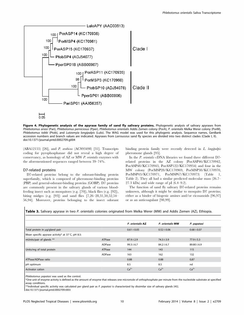
from other sand fly species, are homologous to the bed bug
apyrase which defined the Cimex type family [78]. Phylogenic
analysis showed that salivary apyrases from P. orientalis colonies are
separated into two clades (Figure 4). Clade I includes two apyrases
from the AZ colony and one from the MW colony (PorASP14,
PorASP15, and PorMSP4), as the analogue of the second apyrase
in MW colony was excluded from the phylogenetic analysis due to
absence of signal peptide and the low quality of sequence. Clade II
contains two apyrases, one from each colony (PorASP11 and
PorMSP3). Sequences of P. orientalis apyrase within the same clade
revealed high degree of identity (95–99%), whereas the compar-
ison between the clades showed an identity of 66% (Figure S1).
Comparison with other sand fly species from the subgenus
Larroussius showed that apyrases from P. tobbi (ADJ54077,
ADJ54078) and P. perniciosus (ABB00906, ABB00907) saliva are
closely related to P. orientalis, while apyrase from P. ariasi
(AAX56357) saliva is more distinct (Figures 4 and S1).
Apyrase activity has been demonstrated in the saliva of L.
longipalpis [53], P. papatasi [48,79], P. duboscqi [80], P. perniciosus, P.
argentipes, and P. colabaensis [48]. In our experiments, apyrase
activity was measured in the saliva of both AZ and MW P. orientalis
colonies (Table 3). ATPase as well as ADPase activity, determined
per the pair of salivary glands, was slightly higher in AZ colony,
but recalculation of enzymatic activity per milligram of total
proteins showed that apyrase activity in both colonies is
comparable. Also, the ATPase/ADPase ratio was equal in both
colonies (Table 3). Comparison of P. orientalis apyrase activity with
Table 2. Salivary gland transcripts of Phlebotomus orientalis – Melka Werer colony.
Cluster Sequence nameAccessionnumber Proteome MW pI Best match to NR protein database
Accessionnumber Species E-value
PorMSP3 35.5 kDa salivary apyrase KC170960 Y 35.63 8.83 ABB00906 Phlebotomus perniciosus 0.0
PorMSP4 35.2 kDa salivary apyrase KC170961 Y 33.22 8.89 ADJ54077 Phlebotomus tobbi 0.0
PorMSP6 30 kDa antigen 5-related salivary protein KC170962 Y 28.78 8.94 ABA43055 Phlebotomus perniciosus 1e-179
PorMSP8 28.8 kDa antigen 5-related salivary protein KC170963 Y 28.78 8.94 ADJ54083 Phlebotomus tobbi 3e-151
PorMSP12 14.9 kDa PpSP15-like salivary protein KC170964 Y 14.9 8.77 ADJ54084 Phlebotomus tobbi 3e-73
PorMSP15 24.53 kDa PpSP32-like salivary protein KC170965 25.02 10.24 ADJ54102 Phlebotomus tobbi 1e-127
PorMSP23 42 kDa yellow-related salivary protein KC170966 Y 41.59 6.09 ABA43049 Phlebotomus perniciosus 0.0
PorMSP24 42.6 kDa yellow-related salivary protein KC170967 Y 42.31 8.07 ADJ54080 Phlebotomus tobbi 0.0
PorMSP27 putative alpha-amylase KC170968 33.4 5.75 ACS93490 Phlebotomus arabicus 4e-178
PorMSP28 27.0 kDa D7-related salivary protein KC170969 Y 27.27 7.53 ADJ54096 Phlebotomus tobbi 1e-156
PorMSP38 27.1 kDa D7-related salivary protein KC170970 Y 26.94 8.26 ADJ54095 Phlebotomus tobbi 3e-162
PorMSP43 27 kDa D7-related salivary protein SP04B KC170971 26.7 6.71 ABA43052 Phlebotomus perniciosus 1e-151
PorMSP65 38.8 kDa ParSP25-like salivary protein KC170972 27.56 4.78 ADJ54098 Phlebotomus tobbi 6e-140
PorMSP67 27 kDa D7-related salivary protein KC170973 Y 26.76 9.2 ABA43058 Phlebotomus perniciosus 6e-155
PorMSP74 13.8 kDa PpSP15-like salivary protein KC170974 Y 13.92 9.18 ADJ54086 Phlebotomus tobbi 4e-70
PorMSP75 14.7 kDa PpSP15-like salivary protein KC170975 14.7 7.99 ADJ54085 Phlebotomus tobbi 8e-62
PorMSP78 33 kDa salivary lufaxin KC170976 18.78 8.4 ABA43054 Phlebotomus perniciosus 4e-99
PorMSP90 14.4 kDa PpSP15-like salivary protein KC170977 14.32 8.73 ADJ54088 Phlebotomus tobbi 6e-77
PorMSP96 14.6 kDa PpSP15-like salivary protein KC170978 Y 14.5 8.88 ADJ54089 Phlebotomus tobbi 1e-74
PorMSP101 41 kDa salivary endonuclease KC170979 41.7 9.44 ABA43064 Phlebotomus perniciosus 0.0
PorMSP104 4.5 kDa-like salivary protein KC170980 5.63 10.97 ADJ54097 Phlebotomus tobbi 3e-18
PorMSP108 salivary hyaluronidase KC170981 35.6 7.98 ACS93505 Phlebotomus arabicus 2e-163
PorMSP129 30 kDa salivary phospholipase A2 KC170982 29.72 8.31 ABA43062 Phlebotomus perniciosus 0.0
PorMSP162 16 kDa salivary protein A KC170983 15.97 5.04 ACS93506 Phlebotomus arabicus 1e-41
PorMSP169 3.7 kDa-like salivary protein KC170984 3.93 9.16 ADJ54106 Phlebotomus tobbi 2e-7
PorMSP196 5.0 kDa-like salivary protein KC170985 4.97 10.18 ADJ54105 Phlebotomus tobbi 2e-14
Putatively secreted salivary proteins from MW Phlebotomus orientalis colony with the number of cluster, GenBank accession number, presence in proteome, putativemature protein features (MW- molecular weight, pI- isoelectric point), and best match to NR protein database.doi:10.1371/journal.pntd.0002709.t002
Phlebotomus orientalis Saliva Transcriptome
PLOS Neglected Tropical Diseases | www.plosntds.org 6 February 2014 | Volume 8 | Issue 2 | e2709
P. perniciosus [48] revealed that ATPase and ADPase activities
determined per pair of glands are comparable. Additionally, in
accordance with previous data [48,53,79–81], we showed that P.
orientalis apyrase activity is dependent on presence of Ca2+ but not
on Mg2+ ions.
Besides the anti-hemostatic effect of this enzyme, apyrase is also
known as a powerful antigen. Specific antibodies from dogs bitten
by P. perniciosus in the field, as well as under laboratory conditions,
reacted strongly with two salivary apyrases [63]. Apyrases from P.
perniciosus, P. papatasi, and P. argentipes saliva were also recognized
by sera of mice and hamsters immunized by homologous antigen
[5,60,82]. Furthermore, bacterially expressed recombinant P.
duboscqi apyrase (ABI20147) was also recognized by specific
antibodies from mice immunized with P. duboscqi saliva [80],
suggesting that antibody recognition is not solely targeted to the
glycosylated parts of the antigen. On the other hand, inoculation
of bacterially expressed recombinant L. longipalpis apyrase
(AAD33513) into C57BL/6 mice did not elicit either antibody
response or DTH reaction [12]. These data indicates that the
immunogenicity of the protein or saccharidic part of antigen may
vary in different sand fly species. According to the glycosylation
prediction servers (NetNGlyc and NetOGlyc), P. orientalis apyrases
PorASP14, PorASP15, and PorMSP4 are N-glycosylated, while no
O-glycosylation sites were predicted.
HyaluronidaseHyaluronidase is an enzyme that degrades hyaluronic acid and
other glycosaminoglycan constituents abundantly present in the
vertebrate extracellular matrix. It is a well-known allergen occurring
in the venom of bees, hornets, wasps, spiders, and snakes (reviewed
in [83,84]), but hyaluronidase activity was also observed in the saliva
of various bloodsucking Diptera [28,31,45,85,86]. Previously
published data showed that hyaluronidase is able to promote the
spreading of other components of bloodfeeding insect saliva within
the skin, as well as to enhance the success of potential parasite
transmission [86]. Although positive enzymatic activity was detected
in all sand fly species tested to date [28,31,45,53,85,86], transcripts
coding for putative hyaluronidase were ascertained only in four of
them, namely P. arabicus (ACS93505), P. tobbi (AEK98519), L.
longipalpis (AAD32195), L. intermedia (AFP99265) [28,31,52–54], and
in both P. orientalis colonies (PorASP112/KC170958; PorMSP108/
KC170981) (Table 1, Table 2). The predicted molecular mass of AZ
and MW hyaluronidase was 37.2 and 35.6 kDa, respectively, and
the pI was 6.5 and 8.0, respectively.
Hyaluronidase activity measured in the P. orientalis saliva was
found to be lower than the activity of other Larroussius species
tested by [28]. While hyaluronidase activity expressed in the
relative Turbidity Reducing Units (rRTU) reached approximately
0.62 rTRU/gland in P. tobbi and 0.48 rTRU/gland in P. perniciosus
[28], enzymatic activity in P. orientalis saliva was 0.22 rTRU per
gland (0.2260.036 rTRU in AZ and 0.21560.045 rTRU in MW).
Phlebotomus orientalis salivary hyaluronidase of AZ and MW
colonies revealed identity reaching 94% (Figure 5). High degree of
identity was achieved with P. tobbi sequences (AEK98519) (89–
93%), followed by P. arabicus (ACS93505) (80–83%), L. longipalpis
(AAD32195) (56–58%), and L. intermedia (AFP99265) (47–48%).
Moreover, glycosylation prediction servers (NetNGlyc and NetO-
Glyc) showed that salivary hyaluronidase is the most glycosylated
protein in both colonies, with seventeen predicted N-glycosylation
sites in the AZ and sixteen in the MW colony (Figure 5).
Other enzymesAnother enzyme that was identified among the transcripts from
both P. orientalis cDNA libraries is a putative endonuclease
Figure 1. Proteomic analysis of salivary gland homogenates from Phlebotomus orientalis. Phlebotomus orientalis salivary proteins fromAddis Zemen (AZ) and Melka Werer (MW) colonies (Ethiopia) were identified using Mass Spectrometry. The name of sequences contained in eachprotein band and molecular weight in kDa (STD/kDa) are indicated.doi:10.1371/journal.pntd.0002709.g001
Phlebotomus orientalis Saliva Transcriptome
PLOS Neglected Tropical Diseases | www.plosntds.org 7 February 2014 | Volume 8 | Issue 2 | e2709
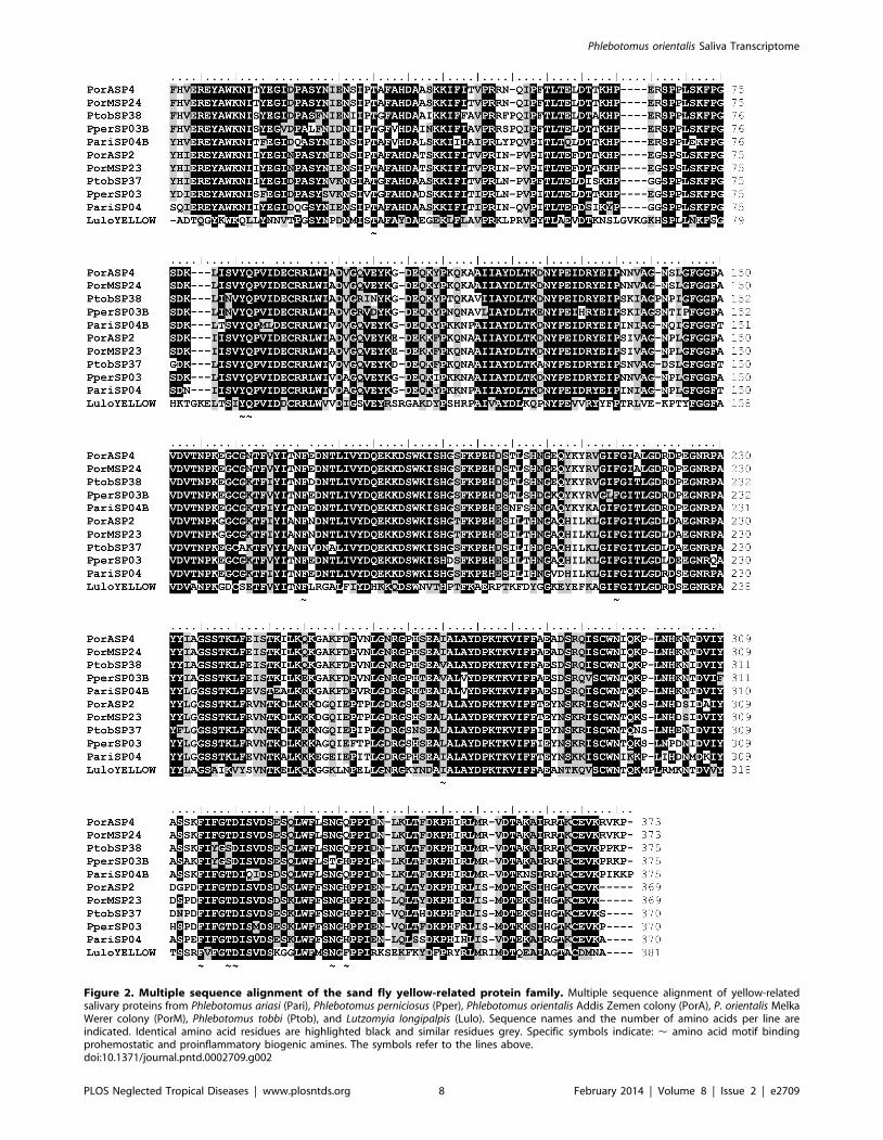
Figure 2. Multiple sequence alignment of the sand fly yellow-related protein family. Multiple sequence alignment of yellow-relatedsalivary proteins from Phlebotomus ariasi (Pari), Phlebotomus perniciosus (Pper), Phlebotomus orientalis Addis Zemen colony (PorA), P. orientalis MelkaWerer colony (PorM), Phlebotomus tobbi (Ptob), and Lutzomyia longipalpis (Lulo). Sequence names and the number of amino acids per line areindicated. Identical amino acid residues are highlighted black and similar residues grey. Specific symbols indicate: , amino acid motif bindingprohemostatic and proinflammatory biogenic amines. The symbols refer to the lines above.doi:10.1371/journal.pntd.0002709.g002
Phlebotomus orientalis Saliva Transcriptome
PLOS Neglected Tropical Diseases | www.plosntds.org 8 February 2014 | Volume 8 | Issue 2 | e2709
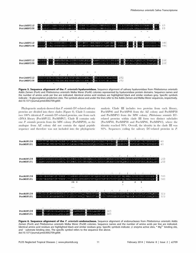
(PorASP139/KC170955; PorMSP101/KC170979) (Table 1,
Table 2). Addis Zemen, as well as Melka Werer, sequences
contained the NUC Smart motif, which is typical for DNA/RNA
non-specific endonucleases and phosphodiesterases. Predicted
molecular mass of both AZ and MW endonucleases was
41.7 kDa and predicted pI was 9.3 and 9.4, respectively.
Endonuclease function in sand fly saliva is still unclear; however,
properties that facilitate blood acquisition are assumed. Endonu-
cleases were detected in salivary gland cDNA libraries of some
sand flies species tested [26,27,31,52,54–56], but also in another
bloodsucking Diptera [87,88]. Sequences of AZ and MW P.
orientalis colony coding for endonuclease revealed 97% identity and
furthermore, there was no difference in the numbers and positions
of the active sites, Mg2+ binding sites, and substrate binding sites
(Figure 6). Phlebotomus perniciosus salivary endonuclease (ABA43064)
was found to be the most relative sequence (92% identity), while
homology of P. orientalis enzymes with other sand fly endonucleases
ranged between 44–80%. Endonuclease was also shown to have
antigenic properties; sera of dogs from an endemic area of VL in
Italy, as well as dogs experimentally bitten by P. perniciosus, reacted
with a 41 kDa salivary protein identified as the endonuclease
(ABA43064) [63].
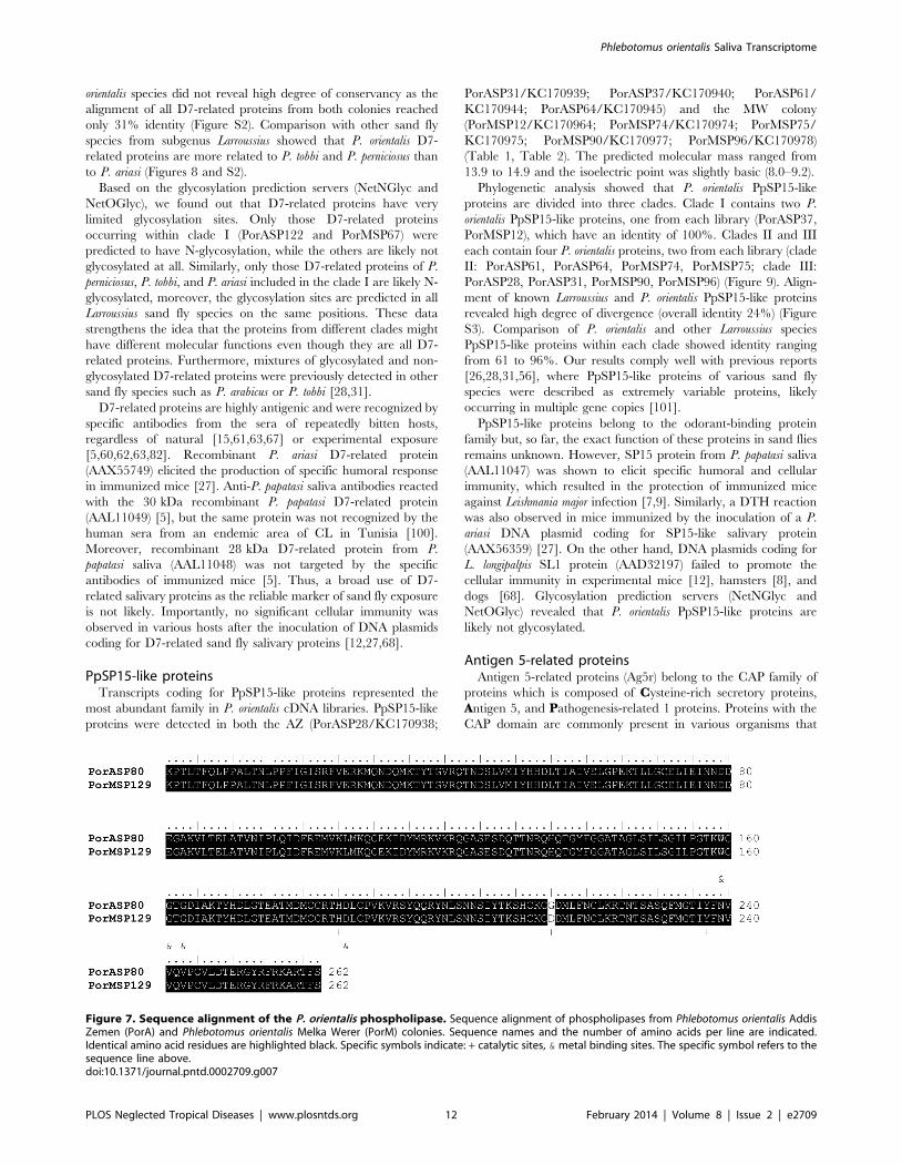
Transcripts coding for a putative phospholipase A2 (PLA2) were
detected in both P. orientalis cDNA libraries (PorASP80/
KC170949; PorMSP129/KC170982) (Table 1, Table 2). In AZ
and MW sequences, the whole PLA2 domain was present. The
predicted molecular mass of PLA2 was 29.7 kDa and pI was 8.4
and 8.3 for AZ and MW, respectively. PLA2 was described as the
main allergen in the hymenopteran venom (reviewed in [84]),
however, its allergenic effect in sand flies remains to be elucidated.
Sequences coding for PLA2 revealed a high degree of conservancy
between AZ and MW colonies as well as among various sand fly
species. The AZ and MW P. orientalis PLA2 were almost identical
(99%) and the metal binding sites were present on the same
positions (Figure 7). Similarly, the catalytic sites were detected on
the same positions and amino acids in both colonies, with the
exception of the catalytic site on 215th amino acid, where glycine
present in AZ colony was in MW replaced by aspartic acid
(Figure 7). Within the Larroussius subgenus, the homology of P.
orientalis PLA2 reached 99% with P. perniciosus (ABA43062) and
94% with P. ariasi (AAX54852). Moreover, comparing the PLA2
enzymes of P. orientalis and P. arabicus (ACS93491), subgenus
Adlerius, showed 88% identity.
A single 39truncated transcript coding for a putative a-amylase
was detected in the salivary gland cDNA library of MW P. orientalis
colony (PorMSP27/KC170968) (Table 2), but no homologous
sequences were found in the AZ colony. Amylase is an enzyme
which is likely not involved in the bloodfeeding process, but
participates in dietary sugar digestion (reviewed in [89]). Tran-
scripts coding for a-amylase were detected in the salivary gland
cDNA libraries of L. longipalpis (AAD32192) [53], P. arabicus
(ACS93490) [31], and in P. papatasi (AAD32192) [55]. Alpha-
amylase activity was detected in the sand fly salivary glands
[53,90,91]. The predicted molecular weight of the P. orientalis MW
amylase was 33.4 kDa and the predicted pI was, 5.8 (Table 2).
Amino acid sequence alignment of P. orientalis a-amylase shows an
88% identity with the P. arabicus a-amylase (ACS93490) and 82%
identity with L. longipalpis a-amylase (AAD32192).
A single sequence containing signal peptide, truncated in the
39region (missing stop codon), coding for a putative salivary
pyrophosphatase (PorASP262/KC170959) was ascertained in the
AZ cDNA library (Table 1). The predicted molecular mass was
32.9 kDa and the predicted pI was 7.2. Pyrophosphatase was also
detected in the MW colony, but these sequences did not contain
signal peptides. Nonetheless, the identity of AZ and MW
pyrophosphatases reached 99%. Salivary pyrophosphatase was
found also in saliva of other sand fly species from the genus
Phlebotomus such as P. duboscqi (ABI20154) [50], P. argentipes
Figure 3. Phylogenetic analysis of the yellow-related family of sand fly salivary proteins. Phylogenetic analysis of yellow-related salivaryproteins from Phlebotomus ariasi (Pari), Phlebotomus perniciosus (Pper), Phlebotomus orientalis Addis Zemen colony (PorA), P. orientalis Melka Werercolony (PorM), Phlebotomus tobbi (Ptob), and Lutzomyia longipalpis (Lulo). The JTT model was used for this phylogenic analysis. Sequence names,GenBank accession numbers and branch values are indicated. Yellow-related salivary proteins from Larroussius sand fly species are divided into twodistinct clades (Clade I, II).doi:10.1371/journal.pntd.0002709.g003
Phlebotomus orientalis Saliva Transcriptome
PLOS Neglected Tropical Diseases | www.plosntds.org 9 February 2014 | Volume 8 | Issue 2 | e2709
(ABA12155) [26], and P. arabicus (ACS93498) [31]. Transcripts
coding for pyrophosphatase did not reveal a high degree of
conservancy, as homology of AZ or MW P. orientalis enzymes with
the aforementioned sequences ranged between 39–74%.
D7-related proteinsD7-related proteins belong to the odorant-binding protein
superfamily, which is composed of pheromone-binding proteins
(PBP) and general-odorant-binding proteins (GOBP). D7 proteins
are commonly present in the salivary glands of various blood-
feeding insect such as mosquitoes (e.g. [70]), black flies (e.g. [92]),
biting midges (e.g. [93]) and sand flies [7,26–28,31,50,52,54–
56,94]. Moreover, proteins belonging to the insect odorant
binding protein family were recently detected in L. longipalpis
pheromone glands [95].
In the P. orientalis cDNA libraries we found three different D7-
related proteins in the AZ colony (PorASP46/KC170942,
PorASP48/KC170943, PorASP122/KC170954) and four in the
MW colony (PorMSP28/KC170969, PorMSP38/KC170970,
PorMSP43/KC170971, PorMSP67/KC170973) (Table 1,
Table 2). They all had a similar predicted molecular mass (26.7–
27.3 kDa) and wide range of pI (6.4–9.2).
The function of sand fly salivary D7-related proteins remains
unknown, although it might be similar to mosquito D7 proteins;
either as a binder of biogenic amines and/or eicosanoids [96,97]
or as an anticoagulant [98,99].
Figure 4. Phylogenetic analysis of the apyrase family of sand fly salivary proteins. Phylogenetic analysis of salivary apyrases fromPhlebotomus ariasi (Pari), Phlebotomus perniciosus (Pper), Phlebotomus orientalis Addis Zemen colony (PorA), P. orientalis Melka Werer colony (PorM),Phlebotomus tobbi (Ptob), and Lutzomyia longipalpis (Lulo). The WAG model was used for this phylogenic analysis. Sequence names, GenBankaccession numbers and branch values are indicated. Apyrases from Larroussius sand fly species are divided into two distinct clades (Clade I, II).doi:10.1371/journal.pntd.0002709.g004
Table 3. Salivary apyrase in two P. orientalis colonies originated from Melka Werer (MW) and Addis Zemen (AZ), Ethiopia.
P. orientalis AZ P. orientalis MW P. papatasi
Total protein in mg/gland pair 0.6160.05 0.5260.06 0.6860.07
Mean specific apyrase activity* at 37uC, pH 8.5:
mUnits/pair of glands ** ATPase 87.962.9 74.363.9 77.965.3
ADPase 99.366.7 84.266.7 89.8066.9
Units/mg of total protein ATPase 144 143 115
ADPase 163 162 132
ATPase/ADPase ratio 0.88 0.88 0,87
pH optimum 8.5 8.5 nd
Activator cation Ca2+ Ca2+ Ca2+
Phlebotomus papatasi was used as the control.*One unit of enzyme activity is defined as the amount of enzyme that releases one micromole of orthophosphate per minute from the nucleotide substrate at specifiedassay conditions.**Individual specific activity was calculated per gland pair as P. papatasi is characterized by dissimilar size of salivary glands [45].doi:10.1371/journal.pntd.0002709.t003
Phlebotomus orientalis Saliva Transcriptome
PLOS Neglected Tropical Diseases | www.plosntds.org 10 February 2014 | Volume 8 | Issue 2 | e2709
Phylogenetic analysis showed that P. orientalis D7-related salivary
proteins are divided into three clades (Figure 8). Clade I contains
two 100% identical P. orientalis D7-related proteins, one from each
cDNA library (PorASP122, PorMSP67). Clade II contains only
one P. orientalis protein from the MW colony (PorMSP28), as the
analogue from AZ colony did not contain the signal peptide
sequence and therefore was not included into the phylogenetic
analysis. Clade III includes two proteins from each library,
PorASP46 and PorASP48 from the AZ colony and PorMSP38
and PorMSP43 from the MW colony. Phlebotomus orientalis D7-
related proteins within clade III form two distinct subclades
(PorASP48, PorMSP38 and PorASP46, PorMSP43), where the
identity reached 99%. Overall, the identity in the clade III was
92%. Sequences coding for salivary D7-related proteins in P.
Figure 5. Sequence alignment of the P. orientalis hyaluronidase. Sequence alignment of salivary hyaluronidase from Phlebotomus orientalisAddis Zemen (PorA) and Phlebotomus orientalis Melka Werer (PorM) colonies represented by hyaluronidase protein domains. Sequence names andthe number of amino acids per line are indicated. Identical amino acid residues are highlighted black and similar residues grey. Specific symbolsindicate: - N-glycosylation prediction sites. The symbols above and under the lines refer to the Addis Zemen and Melka Werer sequences, respectively.doi:10.1371/journal.pntd.0002709.g005
Figure 6. Sequence alignment of the P. orientalis endonuclease. Sequence alignment of endonucleases from Phlebotomus orientalis AddisZemen (PorA) and Phlebotomus orientalis Melka Werer (PorM) colonies. Sequence names and the number of amino acids per line are indicated.Identical amino acid residues are highlighted black and similar residues grey. Specific symbols indicate: # enzyme active sites, * Mg2+ binding site,and ‘ substrate binding sites. The specific symbol refers to the sequence line above.doi:10.1371/journal.pntd.0002709.g006
Phlebotomus orientalis Saliva Transcriptome
PLOS Neglected Tropical Diseases | www.plosntds.org 11 February 2014 | Volume 8 | Issue 2 | e2709
orientalis species did not reveal high degree of conservancy as the
alignment of all D7-related proteins from both colonies reached
only 31% identity (Figure S2). Comparison with other sand fly
species from subgenus Larroussius showed that P. orientalis D7-
related proteins are more related to P. tobbi and P. perniciosus than
to P. ariasi (Figures 8 and S2).
Based on the glycosylation prediction servers (NetNGlyc and
NetOGlyc), we found out that D7-related proteins have very
limited glycosylation sites. Only those D7-related proteins
occurring within clade I (PorASP122 and PorMSP67) were
predicted to have N-glycosylation, while the others are likely not
glycosylated at all. Similarly, only those D7-related proteins of P.
perniciosus, P. tobbi, and P. ariasi included in the clade I are likely N-
glycosylated, moreover, the glycosylation sites are predicted in all
Larroussius sand fly species on the same positions. These data
strengthens the idea that the proteins from different clades might
have different molecular functions even though they are all D7-
related proteins. Furthermore, mixtures of glycosylated and non-
glycosylated D7-related proteins were previously detected in other
sand fly species such as P. arabicus or P. tobbi [28,31].
D7-related proteins are highly antigenic and were recognized by
specific antibodies from the sera of repeatedly bitten hosts,
regardless of natural [15,61,63,67] or experimental exposure
[5,60,62,63,82]. Recombinant P. ariasi D7-related protein
(AAX55749) elicited the production of specific humoral response
in immunized mice [27]. Anti-P. papatasi saliva antibodies reacted
with the 30 kDa recombinant P. papatasi D7-related protein
(AAL11049) [5], but the same protein was not recognized by the
human sera from an endemic area of CL in Tunisia [100].
Moreover, recombinant 28 kDa D7-related protein from P.
papatasi saliva (AAL11048) was not targeted by the specific
antibodies of immunized mice [5]. Thus, a broad use of D7-
related salivary proteins as the reliable marker of sand fly exposure
is not likely. Importantly, no significant cellular immunity was
observed in various hosts after the inoculation of DNA plasmids
coding for D7-related sand fly salivary proteins [12,27,68].
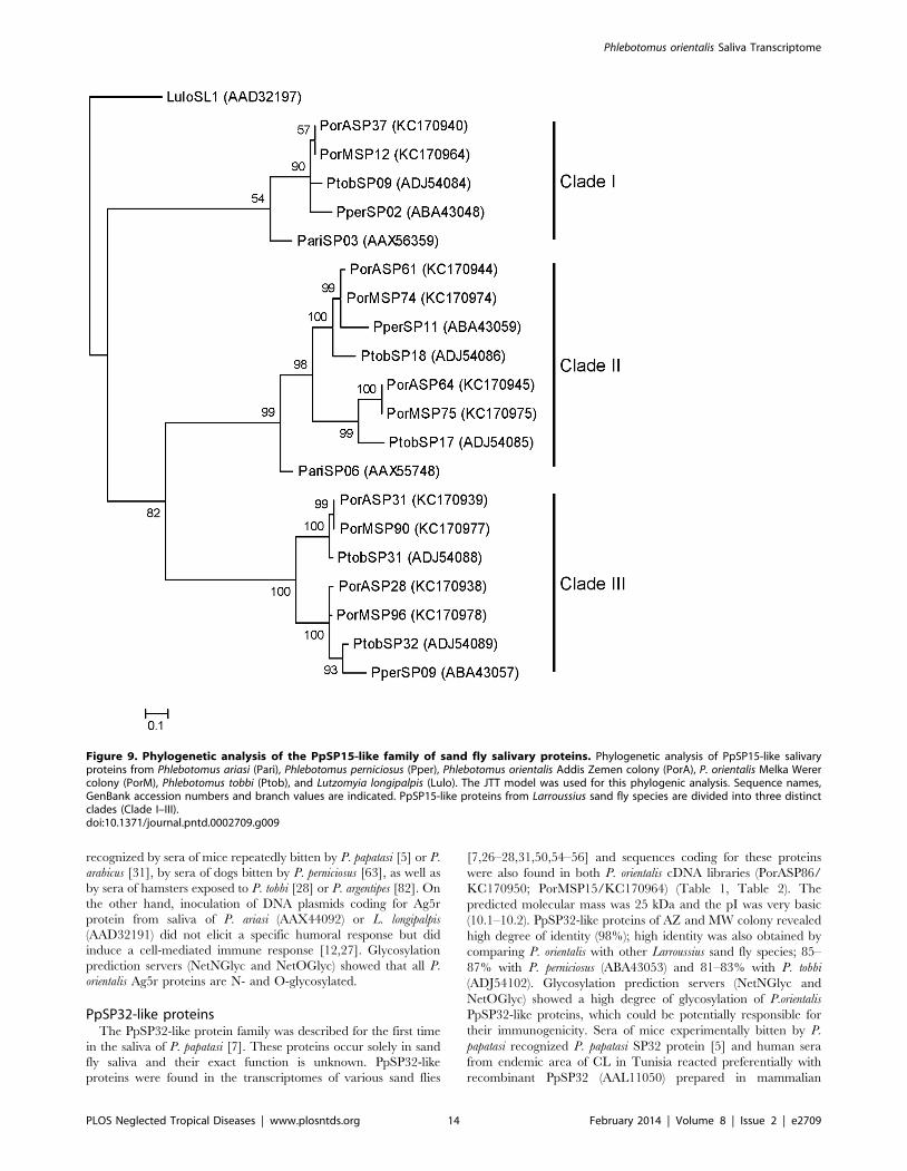
PpSP15-like proteinsTranscripts coding for PpSP15-like proteins represented the
most abundant family in P. orientalis cDNA libraries. PpSP15-like
proteins were detected in both the AZ (PorASP28/KC170938;
PorASP31/KC170939; PorASP37/KC170940; PorASP61/
KC170944; PorASP64/KC170945) and the MW colony
(PorMSP12/KC170964; PorMSP74/KC170974; PorMSP75/
KC170975; PorMSP90/KC170977; PorMSP96/KC170978)
(Table 1, Table 2). The predicted molecular mass ranged from
13.9 to 14.9 and the isoelectric point was slightly basic (8.0–9.2).
Phylogenetic analysis showed that P. orientalis PpSP15-like
proteins are divided into three clades. Clade I contains two P.
orientalis PpSP15-like proteins, one from each library (PorASP37,
PorMSP12), which have an identity of 100%. Clades II and III
each contain four P. orientalis proteins, two from each library (clade
II: PorASP61, PorASP64, PorMSP74, PorMSP75; clade III:
PorASP28, PorASP31, PorMSP90, PorMSP96) (Figure 9). Align-
ment of known Larroussius and P. orientalis PpSP15-like proteins
revealed high degree of divergence (overall identity 24%) (Figure
S3). Comparison of P. orientalis and other Larroussius species
PpSP15-like proteins within each clade showed identity ranging
from 61 to 96%. Our results comply well with previous reports
[26,28,31,56], where PpSP15-like proteins of various sand fly
species were described as extremely variable proteins, likely
occurring in multiple gene copies [101].
PpSP15-like proteins belong to the odorant-binding protein
family but, so far, the exact function of these proteins in sand flies
remains unknown. However, SP15 protein from P. papatasi saliva
(AAL11047) was shown to elicit specific humoral and cellular
immunity, which resulted in the protection of immunized mice
against Leishmania major infection [7,9]. Similarly, a DTH reaction
was also observed in mice immunized by the inoculation of a P.
ariasi DNA plasmid coding for SP15-like salivary protein
(AAX56359) [27]. On the other hand, DNA plasmids coding for
L. longipalpis SL1 protein (AAD32197) failed to promote the
cellular immunity in experimental mice [12], hamsters [8], and
dogs [68]. Glycosylation prediction servers (NetNGlyc and
NetOGlyc) revealed that P. orientalis PpSP15-like proteins are
likely not glycosylated.
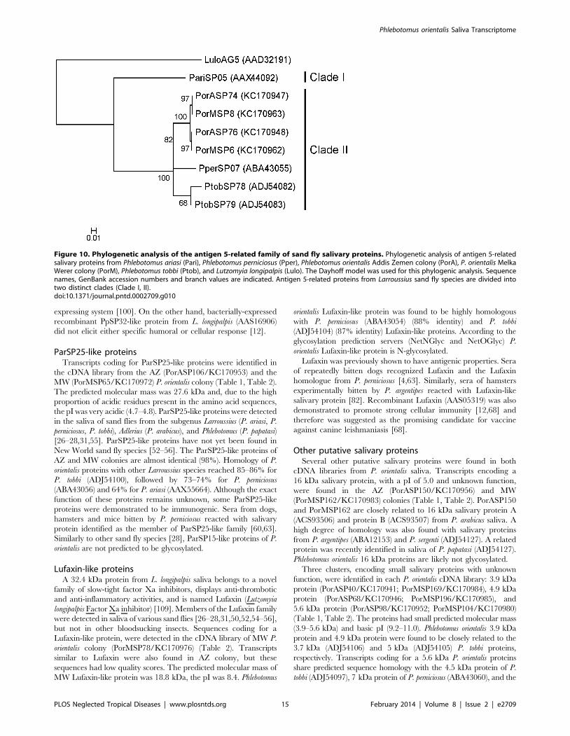
Antigen 5-related proteinsAntigen 5-related proteins (Ag5r) belong to the CAP family of
proteins which is composed of Cysteine-rich secretory proteins,
Antigen 5, and Pathogenesis-related 1 proteins. Proteins with the
CAP domain are commonly present in various organisms that
Figure 7. Sequence alignment of the P. orientalis phospholipase. Sequence alignment of phospholipases from Phlebotomus orientalis AddisZemen (PorA) and Phlebotomus orientalis Melka Werer (PorM) colonies. Sequence names and the number of amino acids per line are indicated.Identical amino acid residues are highlighted black. Specific symbols indicate: + catalytic sites, & metal binding sites. The specific symbol refers to thesequence line above.doi:10.1371/journal.pntd.0002709.g007
Phlebotomus orientalis Saliva Transcriptome
PLOS Neglected Tropical Diseases | www.plosntds.org 12 February 2014 | Volume 8 | Issue 2 | e2709
include prokaryotes and non-vertebrate eukaryotes [102,103].
Ag5r proteins were described from the venom of ants, wasps and
other Hymenoptera [104–106], but were also found in salivary
glands of various bloodsucking insects, including sand flies [7,26–
28,31,50,52–56]. The exact function of Ag5r in sand flies is still
unknown although biological properties of other proteins from the
same family may give us some clue. The X-ray structure of NA-
ASP-2 protein (pathogenesis –related 1 protein) from the human
hookworm, Necator americanus, reveals structural and charge
similarities to chemokines, suggesting that these proteins could
potentially modulate the host immune response [107]; more
recently, a triatomine salivary member of the family was shown to
have superoxide dismutase activity and to exert anti-neutrophil
activity [108].
Sequences coding for salivary Ag5r proteins were found in
cDNA library from the AZ (PorASP74/KC170947; PorASP76/
KC170948) and the MW (PorMSP6/KC170962; PorMSP8/
KC170963) P. orientalis colonies (Table 1, Table 2). The predicted
molecular weight was 28.8 kDa and pI was slightly basic (8.9).
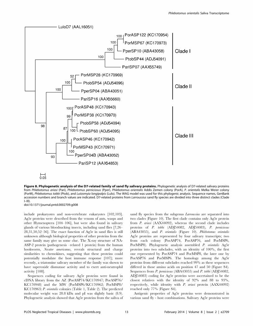
Phylogenetic analysis showed that Ag5r proteins from the saliva of
sand fly species from the subgenus Larroussius are separated into
two clades (Figure 10). The first clade contains only Ag5r protein
from P. ariasi (AAX44092), whereas the second clade includes
proteins of P. tobbi (ADJ54082, ADJ54083), P. perniciosus
(ABA43055), and P. orientalis (Figure 10). Phlebotomus orientalis
Ag5r proteins are represented by four salivary transcripts; two
from each colony (PorASP74, PorASP76, and PorMSP6,
PorMSP8). Phylogenetic analysis assembled P. orientalis Ag5r
proteins into two subclades, with an identity of 100%, the first
one represented by PorASP74 and PorMSP8, the later one by
PorASP76 and PorMSP6. The homology among the Ag5r
proteins from different subclades reached 99% as these sequences
differed in those amino acids on position 47 and 50 (Figure S4).
Sequences from P. perniciosus (ABA43055) and P. tobbi (ADJ54082,
ADJ54083) coding for Ag5r proteins were ascertained to be the
closest relatives with the identity of 92% and 88 to 93%,
respectively, while identity with P. ariasi protein (AAX44092)
reached only 77% (Figure S4).
Antigenic properties of Ag5r proteins were demonstrated in
various sand fly - host combinations. Salivary Ag5r proteins were
Figure 8. Phylogenetic analysis of the D7-related family of sand fly salivary proteins. Phylogenetic analysis of D7-related salivary proteinsfrom Phlebotomus ariasi (Pari), Phlebotomus perniciosus (Pper), Phlebotomus orientalis Addis Zemen colony (PorA), P. orientalis Melka Werer colony(PorM), Phlebotomus tobbi (Ptob), and Lutzomyia longipalpis (Lulo). The WAG model was used for this phylogenic analysis. Sequence names, GenBankaccession numbers and branch values are indicated. D7-related proteins from Larroussius sand fly species are divided into three distinct clades (CladeI–III).doi:10.1371/journal.pntd.0002709.g008
Phlebotomus orientalis Saliva Transcriptome
PLOS Neglected Tropical Diseases | www.plosntds.org 13 February 2014 | Volume 8 | Issue 2 | e2709
recognized by sera of mice repeatedly bitten by P. papatasi [5] or P.
arabicus [31], by sera of dogs bitten by P. perniciosus [63], as well as
by sera of hamsters exposed to P. tobbi [28] or P. argentipes [82]. On
the other hand, inoculation of DNA plasmids coding for Ag5r
protein from saliva of P. ariasi (AAX44092) or L. longipalpis
(AAD32191) did not elicit a specific humoral response but did
induce a cell-mediated immune response [12,27]. Glycosylation
prediction servers (NetNGlyc and NetOGlyc) showed that all P.
orientalis Ag5r proteins are N- and O-glycosylated.
PpSP32-like proteinsThe PpSP32-like protein family was described for the first time
in the saliva of P. papatasi [7]. These proteins occur solely in sand
fly saliva and their exact function is unknown. PpSP32-like
proteins were found in the transcriptomes of various sand flies
[7,26–28,31,50,54–56] and sequences coding for these proteins
were also found in both P. orientalis cDNA libraries (PorASP86/
KC170950; PorMSP15/KC170964) (Table 1, Table 2). The
predicted molecular mass was 25 kDa and the pI was very basic
(10.1–10.2). PpSP32-like proteins of AZ and MW colony revealed
high degree of identity (98%); high identity was also obtained by
comparing P. orientalis with other Larroussius sand fly species; 85–
87% with P. perniciosus (ABA43053) and 81–83% with P. tobbi
(ADJ54102). Glycosylation prediction servers (NetNGlyc and
NetOGlyc) showed a high degree of glycosylation of P.orientalis
PpSP32-like proteins, which could be potentially responsible for
their immunogenicity. Sera of mice experimentally bitten by P.
papatasi recognized P. papatasi SP32 protein [5] and human sera
from endemic area of CL in Tunisia reacted preferentially with
recombinant PpSP32 (AAL11050) prepared in mammalian
Figure 9. Phylogenetic analysis of the PpSP15-like family of sand fly salivary proteins. Phylogenetic analysis of PpSP15-like salivaryproteins from Phlebotomus ariasi (Pari), Phlebotomus perniciosus (Pper), Phlebotomus orientalis Addis Zemen colony (PorA), P. orientalis Melka Werercolony (PorM), Phlebotomus tobbi (Ptob), and Lutzomyia longipalpis (Lulo). The JTT model was used for this phylogenic analysis. Sequence names,GenBank accession numbers and branch values are indicated. PpSP15-like proteins from Larroussius sand fly species are divided into three distinctclades (Clade I–III).doi:10.1371/journal.pntd.0002709.g009
Phlebotomus orientalis Saliva Transcriptome
PLOS Neglected Tropical Diseases | www.plosntds.org 14 February 2014 | Volume 8 | Issue 2 | e2709
expressing system [100]. On the other hand, bacterially-expressed
recombinant PpSP32-like protein from L. longipalpis (AAS16906)
did not elicit either specific humoral or cellular response [12].
ParSP25-like proteinsTranscripts coding for ParSP25-like proteins were identified in
the cDNA library from the AZ (PorASP106/KC170953) and the
MW (PorMSP65/KC170972) P. orientalis colony (Table 1, Table 2).
The predicted molecular mass was 27.6 kDa and, due to the high
proportion of acidic residues present in the amino acid sequences,
the pI was very acidic (4.7–4.8). ParSP25-like proteins were detected
in the saliva of sand flies from the subgenus Larroussius (P. ariasi, P.
perniciosus, P. tobbi), Adlerius (P. arabicus), and Phlebotomus (P. papatasi)
[26–28,31,55]. ParSP25-like proteins have not yet been found in
New World sand fly species [52–56]. The ParSP25-like proteins of
AZ and MW colonies are almost identical (98%). Homology of P.
orientalis proteins with other Larroussius species reached 85–86% for
P. tobbi (ADJ54100), followed by 73–74% for P. perniciosus
(ABA43056) and 64% for P. ariasi (AAX55664). Although the exact
function of these proteins remains unknown, some ParSP25-like
proteins were demonstrated to be immunogenic. Sera from dogs,
hamsters and mice bitten by P. perniciosus reacted with salivary
protein identified as the member of ParSP25-like family [60,63].
Similarly to other sand fly species [28], ParSP15-like proteins of P.
orientalis are not predicted to be glycosylated.
Lufaxin-like proteinsA 32.4 kDa protein from L. longipalpis saliva belongs to a novel
family of slow-tight factor Xa inhibitors, displays anti-thrombotic
and anti-inflammatory activities, and is named Lufaxin (Lutzomyia
longipalpis Factor Xa inhibitor) [109]. Members of the Lufaxin family
were detected in saliva of various sand flies [26–28,31,50,52,54–56],
but not in other bloodsucking insects. Sequences coding for a
Lufaxin-like protein, were detected in the cDNA library of MW P.
orientalis colony (PorMSP78/KC170976) (Table 2). Transcripts
similar to Lufaxin were also found in AZ colony, but these
sequences had low quality scores. The predicted molecular mass of
MW Lufaxin-like protein was 18.8 kDa, the pI was 8.4. Phlebotomus
orientalis Lufaxin-like protein was found to be highly homologous
with P. perniciosus (ABA43054) (88% identity) and P. tobbi
(ADJ54104) (87% identity) Lufaxin-like proteins. According to the
glycosylation prediction servers (NetNGlyc and NetOGlyc) P.
orientalis Lufaxin-like protein is N-glycosylated.
Lufaxin was previously shown to have antigenic properties. Sera
of repeatedly bitten dogs recognized Lufaxin and the Lufaxin
homologue from P. perniciosus [4,63]. Similarly, sera of hamsters
experimentally bitten by P. argentipes reacted with Lufaxin-like
salivary protein [82]. Recombinant Lufaxin (AAS05319) was also
demonstrated to promote strong cellular immunity [12,68] and
therefore was suggested as the promising candidate for vaccine
against canine leishmaniasis [68].
Other putative salivary proteinsSeveral other putative salivary proteins were found in both
cDNA libraries from P. orientalis saliva. Transcripts encoding a
16 kDa salivary protein, with a pI of 5.0 and unknown function,
were found in the AZ (PorASP150/KC170956) and MW
(PorMSP162/KC170983) colonies (Table 1, Table 2). PorASP150
and PorMSP162 are closely related to 16 kDa salivary protein A
(ACS93506) and protein B (ACS93507) from P. arabicus saliva. A
high degree of homology was also found with salivary proteins
from P. argentipes (ABA12153) and P. sergenti (ADJ54127). A related
protein was recently identified in saliva of P. papatasi (ADJ54127).
Phlebotomus orientalis 16 kDa proteins are likely not glycosylated.
Three clusters, encoding small salivary proteins with unknown
function, were identified in each P. orientalis cDNA library: 3.9 kDa
protein (PorASP40/KC170941; PorMSP169/KC170984), 4.9 kDa
protein (PorASP68/KC170946; PorMSP196/KC170985), and
5.6 kDa protein (PorASP98/KC170952; PorMSP104/KC170980)
(Table 1, Table 2). The proteins had small predicted molecular mass
(3.9–5.6 kDa) and basic pI (9.2–11.0). Phlebotomus orientalis 3.9 kDa
protein and 4.9 kDa protein were found to be closely related to the
3.7 kDa (ADJ54106) and 5 kDa (ADJ54105) P. tobbi proteins,
respectively. Transcripts coding for a 5.6 kDa P. orientalis proteins
share predicted sequence homology with the 4.5 kDa protein of P.
tobbi (ADJ54097), 7 kDa protein of P. perniciosus (ABA43060), and the
Figure 10. Phylogenetic analysis of the antigen 5-related family of sand fly salivary proteins. Phylogenetic analysis of antigen 5-relatedsalivary proteins from Phlebotomus ariasi (Pari), Phlebotomus perniciosus (Pper), Phlebotomus orientalis Addis Zemen colony (PorA), P. orientalis MelkaWerer colony (PorM), Phlebotomus tobbi (Ptob), and Lutzomyia longipalpis (Lulo). The Dayhoff model was used for this phylogenic analysis. Sequencenames, GenBank accession numbers and branch values are indicated. Antigen 5-related proteins from Larroussius sand fly species are divided intotwo distinct clades (Clade I, II).doi:10.1371/journal.pntd.0002709.g010
Phlebotomus orientalis Saliva Transcriptome
PLOS Neglected Tropical Diseases | www.plosntds.org 15 February 2014 | Volume 8 | Issue 2 | e2709
5 kDa protein of P. ariasi (AAX55658). Based on the glycosylation
prediction servers (NetNGlyc and NetOGlyc) we found that all P.
orientalis small salivary proteins are likely O-glycosylated.
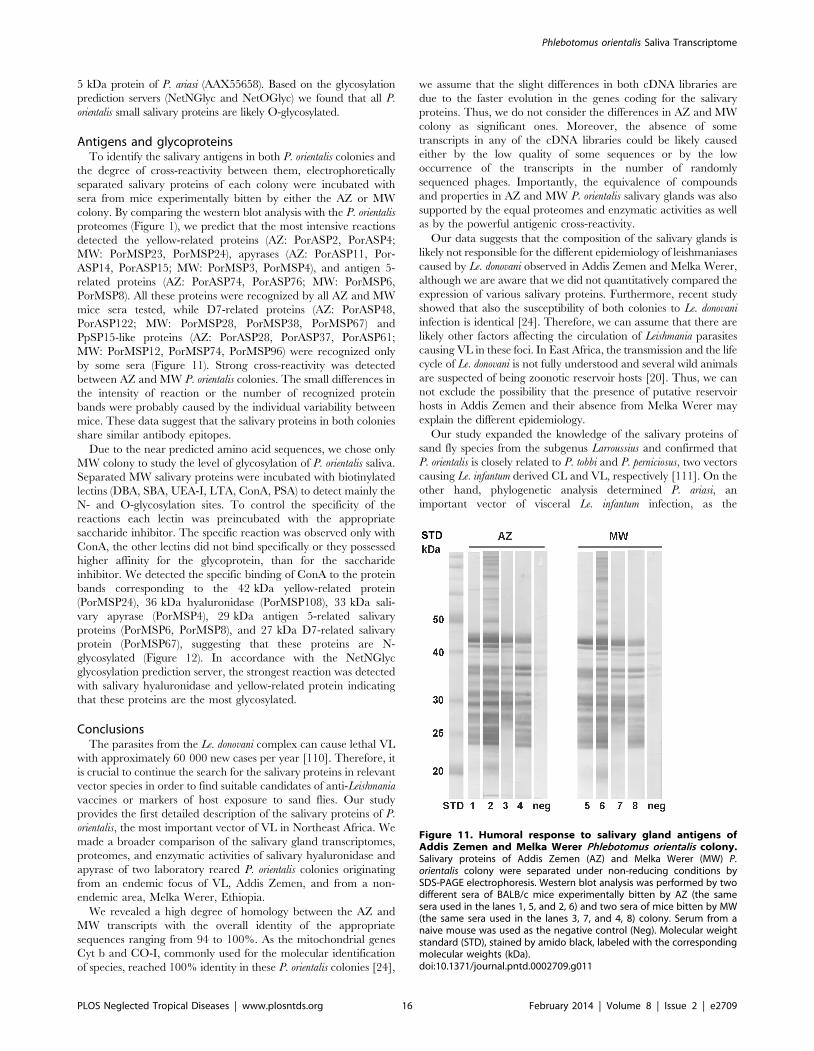
Antigens and glycoproteinsTo identify the salivary antigens in both P. orientalis colonies and
the degree of cross-reactivity between them, electrophoretically
separated salivary proteins of each colony were incubated with
sera from mice experimentally bitten by either the AZ or MW
colony. By comparing the western blot analysis with the P. orientalis
proteomes (Figure 1), we predict that the most intensive reactions
detected the yellow-related proteins (AZ: PorASP2, PorASP4;
MW: PorMSP23, PorMSP24), apyrases (AZ: PorASP11, Por-
ASP14, PorASP15; MW: PorMSP3, PorMSP4), and antigen 5-
related proteins (AZ: PorASP74, PorASP76; MW: PorMSP6,
PorMSP8). All these proteins were recognized by all AZ and MW
mice sera tested, while D7-related proteins (AZ: PorASP48,
PorASP122; MW: PorMSP28, PorMSP38, PorMSP67) and
PpSP15-like proteins (AZ: PorASP28, PorASP37, PorASP61;
MW: PorMSP12, PorMSP74, PorMSP96) were recognized only
by some sera (Figure 11). Strong cross-reactivity was detected
between AZ and MW P. orientalis colonies. The small differences in
the intensity of reaction or the number of recognized protein
bands were probably caused by the individual variability between
mice. These data suggest that the salivary proteins in both colonies
share similar antibody epitopes.
Due to the near predicted amino acid sequences, we chose only
MW colony to study the level of glycosylation of P. orientalis saliva.
Separated MW salivary proteins were incubated with biotinylated
lectins (DBA, SBA, UEA-I, LTA, ConA, PSA) to detect mainly the
N- and O-glycosylation sites. To control the specificity of the
reactions each lectin was preincubated with the appropriate
saccharide inhibitor. The specific reaction was observed only with
ConA, the other lectins did not bind specifically or they possessed
higher affinity for the glycoprotein, than for the saccharide
inhibitor. We detected the specific binding of ConA to the protein
bands corresponding to the 42 kDa yellow-related protein
(PorMSP24), 36 kDa hyaluronidase (PorMSP108), 33 kDa sali-
vary apyrase (PorMSP4), 29 kDa antigen 5-related salivary
proteins (PorMSP6, PorMSP8), and 27 kDa D7-related salivary
protein (PorMSP67), suggesting that these proteins are N-
glycosylated (Figure 12). In accordance with the NetNGlyc
glycosylation prediction server, the strongest reaction was detected
with salivary hyaluronidase and yellow-related protein indicating
that these proteins are the most glycosylated.
ConclusionsThe parasites from the Le. donovani complex can cause lethal VL
with approximately 60 000 new cases per year [110]. Therefore, it
is crucial to continue the search for the salivary proteins in relevant
vector species in order to find suitable candidates of anti-Leishmania
vaccines or markers of host exposure to sand flies. Our study
provides the first detailed description of the salivary proteins of P.
orientalis, the most important vector of VL in Northeast Africa. We
made a broader comparison of the salivary gland transcriptomes,
proteomes, and enzymatic activities of salivary hyaluronidase and
apyrase of two laboratory reared P. orientalis colonies originating
from an endemic focus of VL, Addis Zemen, and from a non-
endemic area, Melka Werer, Ethiopia.
We revealed a high degree of homology between the AZ and
MW transcripts with the overall identity of the appropriate
sequences ranging from 94 to 100%. As the mitochondrial genes
Cyt b and CO-I, commonly used for the molecular identification
of species, reached 100% identity in these P. orientalis colonies [24],
we assume that the slight differences in both cDNA libraries are
due to the faster evolution in the genes coding for the salivary
proteins. Thus, we do not consider the differences in AZ and MW
colony as significant ones. Moreover, the absence of some
transcripts in any of the cDNA libraries could be likely caused
either by the low quality of some sequences or by the low
occurrence of the transcripts in the number of randomly
sequenced phages. Importantly, the equivalence of compounds
and properties in AZ and MW P. orientalis salivary glands was also
supported by the equal proteomes and enzymatic activities as well
as by the powerful antigenic cross-reactivity.
Our data suggests that the composition of the salivary glands is
likely not responsible for the different epidemiology of leishmaniases
caused by Le. donovani observed in Addis Zemen and Melka Werer,
although we are aware that we did not quantitatively compared the
expression of various salivary proteins. Furthermore, recent study
showed that also the susceptibility of both colonies to Le. donovani
infection is identical [24]. Therefore, we can assume that there are
likely other factors affecting the circulation of Leishmania parasites
causing VL in these foci. In East Africa, the transmission and the life
cycle of Le. donovani is not fully understood and several wild animals
are suspected of being zoonotic reservoir hosts [20]. Thus, we can
not exclude the possibility that the presence of putative reservoir
hosts in Addis Zemen and their absence from Melka Werer may
explain the different epidemiology.
Our study expanded the knowledge of the salivary proteins of
sand fly species from the subgenus Larroussius and confirmed that
P. orientalis is closely related to P. tobbi and P. perniciosus, two vectors
causing Le. infantum derived CL and VL, respectively [111]. On the
other hand, phylogenetic analysis determined P. ariasi, an
important vector of visceral Le. infantum infection, as the
Figure 11. Humoral response to salivary gland antigens ofAddis Zemen and Melka Werer Phlebotomus orientalis colony.Salivary proteins of Addis Zemen (AZ) and Melka Werer (MW) P.orientalis colony were separated under non-reducing conditions bySDS-PAGE electrophoresis. Western blot analysis was performed by twodifferent sera of BALB/c mice experimentally bitten by AZ (the samesera used in the lanes 1, 5, and 2, 6) and two sera of mice bitten by MW(the same sera used in the lanes 3, 7, and 4, 8) colony. Serum from anaive mouse was used as the negative control (Neg). Molecular weightstandard (STD), stained by amido black, labeled with the correspondingmolecular weights (kDa).doi:10.1371/journal.pntd.0002709.g011
Phlebotomus orientalis Saliva Transcriptome
PLOS Neglected Tropical Diseases | www.plosntds.org 16 February 2014 | Volume 8 | Issue 2 | e2709
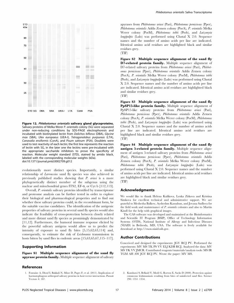
evolutionarily more distinct species. Importantly, a similar
relationship of Larroussius sand fly species was also achieved in
previously published studies showing that P. ariasi is a more
phylogenetically distinct member of the subgenus using the
nuclear and mitochondrial genes ITS2, EF-a, or Cyt b [112,113].
Overall, P. orientalis salivary proteins identified by transcriptome
and proteome analysis can be further tested in order to explore
their biological and pharmacological properties and to find out
whether these salivary proteins could, in the recombinant form, be
the suitable vaccine candidates. The identification of the antigenic
properties of salivary proteins in several sand fly species would also
indicate the feasibility of cross-protection between closely related
and more distant sand fly species as promisingly demonstrated by
[11,12]. Furthermore, the humoral immune response elicited by
the powerful salivary antigens would allow us to predict the
intensity of exposure to sand fly bites [5,15,62,63,114] and,
consequently, to estimate the risk of Leishmania transmission in
hosts bitten by sand flies in endemic areas [15,63,65,67,115–117].
Supporting Information
Figure S1 Multiple sequence alignment of the sand flyapyrase protein family. Multiple sequence alignment of salivary
apyrases from Phlebotomus ariasi (Pari), Phlebotomus perniciosus (Pper),
Phlebotomus orientalis Addis Zemen colony (PorA), P. orientalis Melka
Werer colony (PorM), Phlebotomus tobbi (Ptob), and Lutzomyia
longipalpis (Lulo) was performed using Clustal X 2.0. Sequence
names and the number of amino acids per line are indicated.
Identical amino acid residues are highlighted black and similar
residues grey.
(TIF)
Figure S2 Multiple sequence alignment of the sand flyD7-related protein family. Multiple sequence alignment of
D7-related salivary proteins from Phlebotomus ariasi (Pari), Phlebot-
omus perniciosus (Pper), Phlebotomus orientalis Addis Zemen colony
(PorA), P. orientalis Melka Werer colony (PorM), Phlebotomus tobbi
(Ptob), and Lutzomyia longipalpis (Lulo) was performed using Clustal
X 2.0. Sequence names and the number of amino acids per line
are indicated. Identical amino acid residues are highlighted black
and similar residues grey.
(TIF)
Figure S3 Multiple sequence alignment of the sand flyPpSP15-like protein family. Multiple sequence alignment of
PpSP15-like salivary proteins from Phlebotomus ariasi (Pari),
Phlebotomus perniciosus (Pper), Phlebotomus orientalis Addis Zemen
colony (PorA), P. orientalis Melka Werer colony (PorM), Phlebotomus
tobbi (Ptob), and Lutzomyia longipalpis (Lulo) was performed using
Clustal X 2.0. Sequence names and the number of amino acids
per line are indicated. Identical amino acid residues are
highlighted black and similar residues grey.
(TIF)
Figure S4 Multiple sequence alignment of the sand flyantigen 5-related protein family. Multiple sequence align-
ment of antigen 5-related salivary proteins from Phlebotomus ariasi
(Pari), Phlebotomus perniciosus (Pper), Phlebotomus orientalis Addis
Zemen colony (PorA), P. orientalis Melka Werer colony (PorM),
Phlebotomus tobbi (Ptob), and Lutzomyia longipalpis (Lulo) was
performed using Clustal X 2.0. Sequence names and the number
of amino acids per line are indicated. Identical amino acid residues
are highlighted black and similar residues grey.
(TIF)
Acknowledgments
We would like to thank Helena Kulikova, Lenka Zitkova and Kristina
Simkova for excellent technical and administrative support. We are
grateful to Meshesha Balkew, Aysheshm Kassahun, and Jovana Sadlova for
the field work and maintenance of P. orientalis colonies and also to Martin
Kindl for the help with graphical images.
The CAS software was developed and maintained at the Bioinformatics
and Scientific IT Program (BSIP), Office of Technology Information
Systems (OTIS), National Institute of Allergy and Infectious Diseases
(NIAID) in Bethesda, MD, USA. The software is freely available for
download at http://exon.niaid.nih.gov.
Author Contributions
Conceived and designed the experiments: JGV RCJ PV. Performed the
experiments: MV MS TK PS VV ELJ KDB RCJ. Analyzed the data: MV
MS TK VV JMCR. Contributed reagents/materials/analysis tools: MS IR
TGM AH AW JGV RCJ PV. Wrote the paper: MV MS.
References
1. Fontaine A, Diouf I, Bakkali N, Misse D, Pages F, et al. (2011). Implication of
haematophagous arthropod salivary proteins in host-vector interactions. Parasit
Vectors 4: 187.
2. Kamhawi S, Belkaid Y, Modi G, Roeton E, Sacks D (2000). Protection against
cutaneous leishmaniasis resulting from bites of uninfected sand flies. Science
290: 1351–1354.
Figure 12. Phlebotomus orientalis salivary gland glycoproteins.Salivary proteins of Melka Werer P. orientalis colony (SG) were separatedunder non-reducing conditions by SDS-PAGE electrophoresis andincubated with biotinylated lectin from Dolichos biflorus (DBA), Glycinemax (SBA), Ulex europaeus (UEA-I), Tetragonolobus purpureas (LTA),Canavalia ensiformis (ConA), and Pisum sativum (PSA). Doublets wereused to test reactivity of each lectin; the first line represents the reactionof lectin with SG, in the later one the lectins were pre-incubated withthe appropriate saccharide inhibitors to prove the specificity ofreaction. Molecular weight standard (STD), stained by amido black,labeled with the corresponding molecular weights (kDa).doi:10.1371/journal.pntd.0002709.g012
Phlebotomus orientalis Saliva Transcriptome
PLOS Neglected Tropical Diseases | www.plosntds.org 17 February 2014 | Volume 8 | Issue 2 | e2709
3. Souza AP, Andrade BB, Aquino D, Entringer P, Miranda JC, et al. (2010).Using recombinant proteins from Lutzomyia longipalpis saliva to estimate human
vector exposure in visceral leishmaniasis endemic areas. PLoS Negl Trop Dis 4:
e649.
4. Teixeira C, Gomes R, Collin N, Reynoso D, Jochim R, et al. (2010). Discoveryof markers of exposure specific to bites of Lutzomyia longipalpis, the vector of
Leishmania infantum chagasi in Latin America. PLoS Negl Trop Dis 4: e638.
5. Vlkova M, Rohousova I, Hostomska J, Pohankova L, Zidkova L, et al. (2012).Kinetics of antibody response in BALB/c and C57BL/6 mice bitten by
Phlebotomus papatasi. PLoS Negl Trop Dis 6: e1719.
6. Morris RV, Shoemaker CB, David JR, Lanzaro GC, Titus RG (2001). Sand flymaxadilan exacerbates infection with Leishmania major and vaccinating against it
protects against L. major infection. J Immunol 167: 5226–5230.
7. Valenzuela JG, Belkaid Y, Garfield MK, Mendez S, Kamhawi S, et al. (2001).Toward a defined anti-Leishmania vaccine targeting vector antigens: character-
ization of a protective salivary protein. J Exp Med 194: 331–342.
8. Gomes R, Teixeira C, Teixeira MJ, Oliveira F, Menezes MJ, et al. (2008).Immunity to a salivary protein of a sand fly vector protects against the fatal
outcome of visceral leishmaniasis in a hamster model. Proc Natl Acad Sci U S A
105: 7845–7850.
9. Oliveira F, Lawyer PG, Kamhawi S, Valenzuela JG (2008). Immunity to
distinct sand fly salivary proteins primes the anti-Leishmania immune response
towards protection or exacerbation of disease. PLOS Negl Trop Dis 2: e226.
10. da Silva RA, Tavares NM, Costa D, Pitombo M, Barbosa L, et al. (2011). DNAvaccination with KMP11 and Lutzomyia longipalpis salivary protein protects
hamsters against visceral leishmaniasis. Acta Trop 120: 185–190.
11. Tavares NM, Silva RA, Costa DJ, Pitombo MA, Fukutani KF, et al. (2011).Lutzomyia longipalpis saliva or salivary protein LJM19 protects against Leishmania
braziliensis and the saliva of its vector, Lutzomyia intermedia. PLoS Negl Trop Dis5: e1169.
12. Xu X, Oliveira F, Chang BW, Collin N, Gomes R, et al. (2011). Structure and
function of a ‘‘yellow’’ protein from saliva of the sand fly Lutzomyia longipalpis
that confers protective immunity against Leishmania major infection. J Biol Chem
286: 32383–32393.
13. Gomes R, Oliveira F, Teixeira C, Meneses C, Gilmore DC, et al. (2012).Immunity to sand fly salivary protein LJM11 modulates host response to
vector-transmitted Leishmania conferring ulcer-free protection. J Invest Derma-
tol 132: 2735–2743.
14. Volf P, Rohousova I (2001). Species-specific antigens in salivary glands ofphlebotomine sandflies. Parasitology 122: 37–41.
15. Rohousova I, Ozensoy S, Ozbel Y, Volf P (2005). Detection of species-specific
antibody response of humans and mice bitten by sand flies. Parasitology 130:493–499.
16. Thiakaki M, Rohousova I, Volfova V, Volf P, Chang KP, et al. (2005). Sand fly
specificity of saliva-mediated protective immunity in Leishmania amazonensis-BALB/c mouse model. Microbes Infect 7: 760–766.
17. Wahba M, Riera C (2006). Salivary gland composition of some Old World
vector sand fly. J Egypt Soc Parasitol 36: 289–296.
18. Drahota J, Lipoldova M, Volf P, Rohousova I (2009). Specificity of anti-salivaimmune response in mice repeatedly bitten by Phlebotomus sergenti. Parasite
Immunol 31: 766–770.
19. Rohousova I, Volfova V, Nova S, Volf P (2012a). Individual variability ofsalivary gland proteins in three Phlebotomus species. Acta Trop 122: 80–86.
20. Elnaiem DE (2011). Ecology and control of the sand fly vectors of Leishmania
donovani in East Africa, with special emphasis on Phlebotomus orientalis. J VectorEcol 36: S23–31.
21. Doha SA, Samy AM (2010). Bionomics of phlebotomine sand flies (Diptera:
Psychodidae) in the province of Al-Baha, Saudi Arabia. Mem Inst OswaldoCruz 105: 850–856.
22. Daoud W, Rioux JA, Delalbre-Belmonte A, Dereure J, Rageh HA (1989). Eco-
epidemiology of visceral and cutaneous leishmaniasis in the Arab Republic ofYemen. III. Inventory and dynamics of Phlebotomus. Bull Soc Pathol Exot
Filiales 82: 669–677.
23. Herrero M, Orfanos G, Argaw D, Mulugeta A, Aparicio P, et al. (2009).
Natural history of a visceral leishmaniasis outbreak in highland Ethiopia.Am J Trop Med Hyg 81: 373–377.
24. Seblova V, Volfova V, Dvorak V, Pruzinova K, Votypka J, et al. (2013).
Phlebotomus orientalis sand flies from two geographically distant Ethiopianlocalities: biology, genetic analyses and susceptibility to Leishmania donovani.
PLoS Negl Trop Dis 7: e2187.
25. Warburg A, Saraiva E, Lanzaro GC, Titus RG, Neva F (1994). Saliva ofLutzomyia longipalpis sibling species differs in its composition and capacity to
enhance leishmaniasis. Philos Trans R Soc Lond B Biol Sci 345: 223–230.
26. Anderson JM, Oliveira F, Kamhawi S, Mans BJ, Reynoso D, et al. (2006).Comparative salivary gland transcriptomics of sandfly vectors of visceral
leishmaniasis. BMC Genomics 7: a.n. 52.
27. Oliveira F, Kamhawi S, Seitz AE, Pham VM, Guigal PM, et al. (2006). Fromtranscriptome to immunome: Identification of DTH inducing proteins from a
Phlebotomus ariasi salivary gland cDNA library. Vaccine 24: 374–390.
28. Rohousova I, Subrahmanyam S, Volfova V, Mu J, Volf P, et al. (2012b).Salivary gland transcriptomes and proteomes of Phlebotomus tobbi and Phlebotomus
sergenti, vectors of leishmaniasis. PLoS Negl Trop Dis 6: e1660.
29. Volf P, Volfova V (2011). Establishment and maintenance of sand fly colonies.
J Vector Ecol 36: S1–S9.
30. Chmelar J, Anderson JM, Mu J, Jochim RC, Valenzuela JG, et al. (2008).
Insight into the sialome of the castor bean tick, Ixodes ricinus. BMC Genomics 9:
233.
31. Hostomska J, Volfova V, Mu JB, Garfield M, Rohousova I, et al. (2009).
Analysis of salivary transcripts and antigens of the sand fly Phlebotomus arabicus.
BMC Genomics 10: 282.
32. Ewing B, Hillier L, Wendl MC, Green P (1998). Base-calling of automated
sequencer traces using phred. I. Accuracy assessment. Genome Res 8: 175–
185.
33. Ewing B, Green P (1998). Base-calling of automated sequencer traces using
phred. II. Error probabilities. Genome Res 8: 186–194.
34. Ashburner M, Ball CA, Blake JA, Botstein D, Butler H, et al. (2000). Gene
Ontology: tool for the unification of biology. Nat Genet 25: 25–29.
35. Tatusov RL, Fedorova ND, Jackson JD, Jacobs AR, Kiryutin B, et al. (2003).
The COG database: an updated version includes eukaryotes. BMC Bioinfor-
matics 4: 4.
36. Bateman A, Birney E, Durbin R, Eddy SR, Howe KL, et al. (2000). The Pfam
protein families database. Nucleic Acids Res 28: 263–266.
37. Schultz J, Copley RR, Doerks T, Ponting CP, Bork P (2000). SMART: a web-
based tool for the study of genetically mobile domains. Nucleic Acids Res 28:
231–234.
38. Bendtsen JD, Nielsen H, von Heijne G, Brunak S (2004). Improved prediction
of signal peptides: SignalP 3.0. J Mol Biol 340: 783–795.
39. Guo YJ, Ribeiro JMC, Anderson JM, Bour S (2009). dCAS: a desktop
application for cDNA sequence annotation. Bioinformatics 25: 1195–1196.
40. Julenius K, Mølgaard A, Gupta R, Brunak S (2005). Prediction, conservation
analysis, and structural characterization of mammalian mucin-type O-
glycosylation sites. Glycobiology 15: 153–164.
41. Larkin MA, Blackshields G, Brown NP, Chenna R, McGettigan PA, et al.
(2007). Clustal W and clustal X version 2.0. Bioinformatics 23: 2947–2948.
42. Abascal F, Zardoya R, Posada D (2005). ProtTest: selection of best-fit models of
protein evolution. Bioinformatics 21: 2104–2105.
43. Schmidt HA, Strimmer K, Vingron M, von Haeseler (2002). TREE-PUZZLE:
maximum likelihood phylogenetic analysis using quartets and parallel
computing. Bioinformatics 18: 502–504.
44. Tamura K, Dudley J, Nei M, Kumar S (2007). MEGA4: Molecular
evolutionary genetics analysis (MEGA) software version 4.0. Mol Biol Evol
24: 1596–1599.
45. Cerna P, Mikes L, Volf P (2002). Salivary gland hyaluronidase in various
species of phlebotomine sand flies (Diptera : Psychodidae). Insect Biochem MolBiol 32: 1691–1697.
46. Frost GI, Stern R (1997). A microtiter-based assay for hyaluronidase activity
not requiring specialized reagents. Anal Biochem 251: 263–269.
47. Marinotti O, deBrito M, Moreira CK (1996). Apyrase and alpha-glucosidase in
the salivary glands of Aedes albopictus. Comp Biochem Physiol 113B: 675–679.
48. Ribeiro JMC, Modi GB, Tesh RB (1989). Salivary apyrase activity of some Old
World phlebotomine sand flies. Insect Biochemistry 19: 409–412.
49. Charlab R, Rowton ED, Ribeiro JM (2000). The salivary adenosine deaminase
from the sand fly Lutzomyia longipalpis. Exp Parasitol 95: 45–53.
50. Kato H, Anderson JM, Kamhawi S, Oliveira F, Lawyer PG, et al. (2006). High
degree of conservancy among secreted salivary gland proteins from two
geographically distant Phlebotomus duboscqi sandflies populations (Mali and
Kenya). BMC Genomics 7: 226.
51. Kato H, Jochim RC, Lawyer PG, Valenzuela JG (2007). Identification and
characterization of a salivary adenosine deaminase from the sand fly Phlebotomus
duboscqi, the vector of Leishmania major in sub-Saharan Africa. J Exp Biol 210:
733–740.
52. de Moura TR, Oliveira F, Carneiro MW, Miranda JC, Clarencio J, et al.
(2013). Functional transcriptomics of wild-caught Lutzomyia intermedia salivary
glands: identification of a protective salivary protein against Leishmania
braziliensis infection. PLoS Negl Trop Dis 7: e2242.
53. Charlab R, Valenzuela JG, Rowton ED, Ribeiro JMC (1999). Toward an
understanding of the biochemical and pharmacological complexity of the saliva
of a hematophagous sand fly Lutzomyia longipalpis. Proc Natl Acad Sci U S A 96:
15155–15160.
54. Valenzuela JG, Garfield M, Rowton ED, Pham VM (2004). Identification of
the most abundant secreted proteins from the salivary glands of the sand fly
Lutzomyia longipalpis, vector of Leishmania chagasi. J Exp Biol 207: 3717–3729.
55. Abdeladhim M, Jochim RC, Ben Ahmed M, Zhioua E, Chelbi I, et al. (2012).
Updating the salivary gland transcriptome of Phlebotomus papatasi (Tunisian
Strain): The search for sand fly-secreted immunogenic proteins for humans.
PLoS One 7: e47347.
56. Kato H, Jochim RC, Gomez EA, Uezato H, Mimori T, et al. (2012). Analysis
of salivary gland transcripts of the sand fly Lutzomyia ayacuchensis, a vector of
Andean-type cutaneous leishmaniasis. Infect Genet Evol 13C: 56–66.
57. Volf P, Skarupova S, Man P (2002). Characterization of the lectin from females
of Phlebotomus duboscqi sand flies. Eur J Biochem 269: 6294–6301.
58. Johnson JK, Li J, Christensen BM (2001). Cloning and characterization of a
dopachrome conversion enzyme from the yellow fever mosquito, Aedes aegypti.
Insect Biochem Mol Biol 31: 1125–1135.
59. Alves-Silva J, Ribeiro JMC, Van Den Abbeele J, Attardo G, Hao ZR, et al.
(2010). An insight into the sialome of Glossina morsitans morsitans. BMC
Genomics 11: 213.
Phlebotomus orientalis Saliva Transcriptome
PLOS Neglected Tropical Diseases | www.plosntds.org 18 February 2014 | Volume 8 | Issue 2 | e2709
60. Martin-Martin I, Molina R, Jimenez M (2012). An insight into the Phlebotomus
perniciosus saliva by a proteomic approach. Acta Trop123: 22–30.61. Bahia D, Gontijo NF, Leon IR, Perales J, Pereira MH, et al. (2007). Antibodies
from dogs with canine visceral leishmaniasis recognise two proteins from the
saliva of Lutzomyia longipalpis. Parasitol Res 100: 449–454.62. Hostomska J, Rohousova I, Volfova V, Stanneck D, Mencke N, et al. (2008).
Kinetics of canine antibody response to saliva of the sand fly Lutzomyia
longipalpis. Vector Borne Zoonotic Dis 8: 443–450.
63. Vlkova M, Rohousova I, Drahota J, Stanneck D, Kruedewagen EM, et al. (2011).
Canine antibody response to Phlebotomus perniciosus bites negatively correlates withthe risk of Leishmania infantum transmission. PLoS Negl Trop Dis 5: e1344.
64. Gomes RB, Mendonca IL, Silva VC, Ruas J, Silva MB, et al. (2007).Antibodies against Lutzomyia longipalpis saliva in the fox Cerdocyon thous and the
sylvatic cycle of Leishmania chagasi. Trans R Soc Trop Med Hyg 101: 127–133.65. Gomes RB, Brodskyn U, de Oliveira CI, Costa J, Miranda JC, et al. (2002).
Seroconversion against Lutzomyia longipalpis saliva concurrent with the
development of anti-Leishmania chagasi delayed-type hypersensitivity. J InfectDis 186: 1530–1534.
66. Vinhas V, Andrade BB, Paes F, Bomura A, Clarencio J, et al. (2007). Humananti-saliva immune response following experimental exposure to the visceral
leishmaniasis vector, Lutzomyia longipalpis. Eur J Immunol 37: 3111–3121.
67. Marzouki S, Ahmed MB, Boussoffara T, Abdeladhim M, Aleya-Bouafif NB, etal. (2011). Characterization of the antibody response to the saliva of Phlebotomus
papatasi in people living in endemic areas of cutaneous leishmaniasis. Am J TropMed Hyg 84: 653–661.
68. Collin N, Gomes R, Teixeira C, Cheng L, Laughinghouse A, et al. (2009). Sand flysalivary proteins induce strong cellular immunity in a natural reservoir of visceral
leishmaniasis with adverse consequences for Leishmania. PLoS Pathog 5: e1000441.
69. Santos A, Ribeiro JM, Lehane MJ, Gontijo NF, Veloso AB, et al. (2007). Thesialotranscriptome of the blood-sucking bug Triatoma brasiliensis (Hemiptera:
Triatominae). Insect Biochem Mol Biol 37: 702–712.70. Jariyapan N, Roytrakul S, Paemanee A, Junkum A, Saeung A, et al. (2012).
Proteomic analysis of salivary glands of female Anopheles barbirostris species A2
(Diptera: Culicidae) by two-dimensional gel electrophoresis and massspectrometry. Parasitol Res 111: 1239–1249.
71. Andersen JF, Hinnebusch BJ, Lucas DA, Conrads TP, Veenstra TD, et al.(2007). An insight into the sialome of the oriental rat flea, Xenopsylla cheopis
(Rots). BMC Genomics 8: 102.72. Caljon G, De Ridder K, De Baetselier P, Coosemans M, Van Den Abbeele J
(2010). Identification of a tsetse fly salivary protein with dual inhibitory action
on human platelet aggregation. PLoS One 5: e9671.73. Perez de Leon AA, Tabachnick WJ (1996). Apyrase activity and adenosine
diphosphate induced platelet aggregation inhibition by the salivary glandproteins of Culicoides variipennis, the North American vector of bluetongue
viruses. Vet Parasitol 61: 327–338.
74. Ma D, Wang Y, Yang H, Wu J, An S, et al. (2009). Anti-thrombosis repertoireof blood-feeding horsefly salivary glands. Mol Cell Proteomics 8: 2071–2079.
75. Wu S, Peiffer M, Luthe DS, Felton GW (2012). ATP hydrolyzing salivaryenzymes of caterpillars suppress plant defenses. PLoS One 7: e41947.
76. Fenckova M, Hobizalova R, Fric ZF, Dolezal T (2011). Functionalcharacterization of ecto-59-nucleotidases and apyrases in Drosophila melanogaster.
Insect Biochem Mol Biol 41: 956–967.
77. Ribeiro JM, Mans BJ, Arca B (2010). An insight into the sialome of blood-feeding Nematocera. Insect Biochem Mol Biol 40: 767–784.
78. Valenzuela JG, Charlab R, Galperin MY, Ribeiro JM (1998). Purification,cloning and expression of an apyrase from the bed bug Cimex lectularius. A new
type of nucleotide-binding enzyme. J Biol Chem 273: 30583–30590.
79. Valenzuela JG, Belkaid Y, Rowton E, Ribeiro JM (2001b). The salivaryapyrase of the blood-sucking sand fly Phlebotomus papatasi belongs to the novel
Cimex family of apyrases. J Exp Biol 204: 229–237.80. Hamasaki R, Kato H, Terayama Y, Iwata H, Valenzuela JG (2009). Functional
characterization of a salivary apyrase from the sand fly, Phlebotomus duboscqi, a
vector of Leishmania major. J Insect Physiol 55: 1044–1049.81. Ribeiro JMC, Rossignol PA, Spielman A (1986). Blood-finding strategy of a
capillary-feeding sandfly, Lutzomyia longipalpis. Comp Biochem Physiol 83A: 683–686.82. Martin-Martin I, Molina R, Jimenez M (2013). Identifying salivary antigens of
Phlebotomus argentipes by a 2DE approach. Acta Trop 126: 229–239.83. Stern R, Jedrzejas MJ (2006). Hyaluronidases: their genomics, structures, and
mechanisms of action. Chem Rev 106: 818–839.
84. Muller UR (2011). Hymenoptera venom proteins and peptides for diagnosis andtreatment of venom allergic patients. Inflamm Allergy Drug Targets 10: 420–428.
85. Ribeiro JM, Charlab R, Rowton ED, Cupp EW (2000a). Simulium vittatum
(Diptera: Simuliidae) and Lutzomyia longipalpis (Diptera: Psychodidae) salivary
gland hyaluronidase activity. J Med Entomol 37: 743–747.
86. Volfova V, Hostomska J, Cerny M, Votypka J, Volf P (2008). Hyaluronidase ofbloodsucking insects and its enhancing effect on Leishmania infection in mice.
PLoS Negl Trop Dis 2: e294.87. Ribeiro JM, Charlab R, Pham VM, Garfield M, Valenzuela JG (2004). An
insight into the salivary transcriptome and proteome of the adult femalemosquito Culex pipiens quinquefasciatus. Insect Biochem Mol Biol 34: 543–563.
88. Calvo E, Ribeiro JM (2006). A novel secreted endonuclease from Culex
quinquefasciatus salivary glands. J Exp Biol 209: 2651–2659.89. Jacobson RL, Schlein Y, Eisenberger CL (2001). The biological function of
sand fly and Leishmania glycosidases. Med Microbiol Immunol 190: 51–55.
90. Ribeiro JM, Rowton ED, Charlab R (2000b). Salivary amylase activity of the
phlebotomine sand fly, Lutzomyia longipalpis. Insect Biochem Mol Biol 30: 271–277.91. Jacobson RL, Schlein Y (2001). Phlebotomus papatasi and Leishmania major
parasites express alpha-amylase and alpha-glucosidase. Acta Trop 78: 41–49.
92. Andersen JF, Pham VM, Meng Z, Champagne DE, Ribeiro JM (2009). Insightinto the sialome of the black fly, Simulium vittatum. J Proteome Res 8: 1474–1488.
93. Campbell CL, Vandyke KA, Letchworth GJ, Drolet BS, Hanekamp T, et al.(2005). Midgut and salivary gland transcriptomes of the arbovirus vector
Culicoides sonorensis (Diptera:Ceratopogonidae). Insect Mol Biol 14: 121–136.
94. Valenzuela JG, Charlab R, Gonzalez EC, de Miranda-Santos IK, Marinotti O,et al. (2002). The D7 family of salivary proteins in blood sucking diptera. Insect
Mol Biol 11: 149–55.95. Gonzalez-Caballero N, Valenzuela JG, Ribeiro JM, Cuervo P, Brazil RP
(2013). Transcriptome exploration of the sex pheromone gland of Lutzomyia
longipalpis (Diptera: Psychodidae: Phlebotominae). Parasit Vectors 6: 56.
96. Calvo E, Mans BJ, Andersen JF, Ribeiro JMC (2006). Function and evolution
of a mosquito salivary protein family. J Biol Chem 281: 1935–1942.97. Mans BJ, Calvo E, Ribeiro JM, Andersen JF (2007). The crystal structure of
D7r4, a salivary biogenic amine-binding protein from the malaria mosquitoAnopheles gambiae. J Biol Chem 282: 36626–36633.
98. Isawa H, Yuda M, Orito Y, Chinzei Y (2002). A mosquito salivary protein
inhibits activation of the plasma contact system by binding to factor XII andhigh molecular weight kininogen. J Biol Chem 277: 27651–27658.
99. Alvarenga PH, Francischetti IMB, Calvo E, Sa-Nunes A, Ribeiro JMC, et al.(2010). The function and three-dimensional structure of a tromboxane A(2)/
cysteinyl leukotriene-binding protein from the saliva of a mosquito vector of themalaria parasite. Plos Biol 8: e1000547.
100. Marzouki S, Abdeladhim M, Abdessalem CB, Oliveira F, Ferjani B, et al.
(2012). Salivary antigen SP32 is the immunodominant target of the antibodyresponse to Phlebotomus papatasi bites in humans. PLoS Negl Trop Dis 6: e1911.
101. Elnaiem DE, Meneses C, Slotman M, Lanzaro GC (2005). Genetic variation inthe sand fly salivary protein, SP-15, a potential vaccine candidate against
Leishmania major. Insect Mol Biol 14: 145–150.
102. Milne TJ, Abbenante G, Tyndall JD, Halliday J, Lewis RJ (2003). Isolation andcharacterization of a cone snail protease with homology to CRISP proteins of
the Pathogenesis-related Protein Superfamily. J Biol Chem 278: 31105–31110.103. Yeats C, Bentley S, Bateman A (2003). New knowledge from old: In silico
discovery of novel protein domains in Streptomyces coelicolor. BMC Microbiol 3: 3.104. Hoffman DR (1993). Allergens in Hymenoptera venom. XXV: The amino acid
sequences of antigen 5 molecules and the structural basis of antigenic cross-
reactivity. J Allergy Clin Immunol 92: 707–716.105. Lu G, Villalba M, Coscia MR, Hoffman DR, King TP (1993). Sequence
analysis and antigenic cross-reactivity of a venom allergen, antigen 5, fromhornets, wasps, and yellow jackets. J Immunol 150: 2823–2830.
106. King TP, Spangfort MD (2000). Structure and biology of stinging insect venom
allergens. Int Arch Allergy Immunol 123: 99–106.107. Asojo OA, Goud G, Dhar K, Loukas A, Zhan B, et al. (2005). X-ray structure
of Na-ASP-2, a pathogenesis-related-1 protein from the nematode parasite,Necator americanus, and a vaccine antigen for human hookworm infection. J Mol
Biol 346: 801–814.108. Assumpcao TC, Ma D, Schwarz A, Reiter K, Santana JM, et al. (2013).
Salivary antigen-5/CAP family members are Cu2+-dependent antioxidant
enzymes that scavenge O2- and inhibit collagen-induced platelet aggregationand neutrophil oxidative burst. J Biol Chem 288: 14341–14361.
109. Collin N, Assumpcao TC, Mizurini DM, Gilmore DC, Dutra-Oliveira A, et al.(2012). Lufaxin, a novel factor Xa inhibitor from the salivary gland of the sand
fly Lutzomyia longipalpis blocks protease-activated receptor 2 activation and
inhibits inflammation and thrombosis in vivo. Arterioscler Thromb Vasc Biol32: 2185–2198.
110. Alvar J, Velez ID, Bern C, Herrero M, Desjeux P, et al. (2012). Leishmaniasisworldwide and global estimates of its incidence. PLoS One 7: e35671.
111. Svobodova M, Alten B, Zidkova L, Dvorak V, Hlavackova J, et al. (2009).
Cutaneous leishmaniasis caused by Leishmania infantum transmitted byPhlebotomus tobbi. Int J Parasitol 39: 251–256.
112. Di Muccio T, Marinucci M,FrusteriL,Maroli M, Pesson B, et al. (2000). Phylogeneticanalysis of Phlebotomus species belonging to the subgenus Larroussius (Diptera,
psychodidae) by ITS2 rDNA sequences. Insect Biochem Mol Biol 30: 387–393.113. Esseghir S, Ready P, Ben-Ismail R (2000). Speciation of Phlebotomus sandflies of
the subgenus Larroussius coincided with the late Miocene-Pliocene aridification
of the Mediterranean subregion. Biol J Linn Soc Lond 70: 189–219.114. Clements MF, Gidwani K, Kumar R, Hostomska J, Dinesh DS, et al. (2010).
Measurement of recent exposure to Phlebotomus argentipes, the vector of indianvisceral leishmaniasis, by using human antibody responses to sand fly saliva.
Am J Trop Med Hyg 82: 801–807.
115. Barral A, Honda E, Caldas A, Costa J, Vinhas V, et al. (2000). Human immuneresponse to sand fly salivary gland antigens: a useful epidemiological marker?
Am J Trop Med Hyg 62: 740–745.116. de Moura TR, Oliveira F, Novais FO, Miranda JC, Clarencio J, et al. (2007).
Enhanced Leishmania braziliensis infection following pre-exposure to sandflysaliva. PLoS Negl Trop Dis 1: e84.
117. Aquino DM, Caldas AJ, Miranda JC, Silva AA, Barral-Netto M, et al. (2010).
Epidemiological study of the association between anti-Lutzomyia longipalpis salivaantibodies and development of delayed-type hypersensitivity to Leishmania
antigen. Am J Trop Med Hyg 83: 825–827.
Phlebotomus orientalis Saliva Transcriptome
PLOS Neglected Tropical Diseases | www.plosntds.org 19 February 2014 | Volume 8 | Issue 2 | e2709