Embed Size (px)
Citation preview

Clinical Biomechanics 28 (2013) 483–489
Contents lists available at SciVerse ScienceDirect
Clinical Biomechanics
j ourna l homepage: www.e lsev ie r .com/ locate /c l inb iomech
Comparative analysis of posterior fusion constructs as treatments for middle andposterior column injuries: An in vitro biomechanical investigation
James J. Doulgeris a,b,d,1, Kamran Aghayev a,b,1, Sabrina A. Gonzalez-Blohm a,b,⁎, Michael Del Valle c,Jason Waddell a,b, William E. Lee III c, Frank D. Vrionis a,b
a H. Lee Moffitt Cancer Center & Research Institute, NeuroOncology Program, Tampa, FL 33612, USAb Department of Neurosurgery and Orthopedics, University of South Florida, College of Medicine, Tampa, FL 33612, USAc Dept. of Chemical & Biomedical Engineering, University of South Florida, Tampa, FL 33612, USAd Dept. of Mechanical Engineering, University of South Florida, Tampa, FL 33612, USA
⁎ Corresponding author at: H. LeeMoffitt Cancer CenterFL 33612, USA.
E-mail address: [email protected] These authors contributed equally to the study.
0268-0033/$ – see front matter © 2013 Elsevier Ltd. Allhttp://dx.doi.org/10.1016/j.clinbiomech.2013.05.001
a b s t r a c t
a r t i c l e i n f oArticle history:
Received 22 January 2013Accepted 1 May 2013Keywords:Lumbar spineBilateral pedicle screw systemCobalt–chromium alloyTitanium alloy
Background: Titanium pedicle screw–rod instrumentation is considered a standard treatment for spinal insta-bility; however, the advantages of cobalt-chromium over titanium is generating interest in orthopedic prac-tice. The aim of this study was to compare titanium versus cobalt–chromium rods in posterior fusion throughin vitro biomechanical testing.Methods: Posterior and middle column injuries were simulated at L3–L5 in six cadaveric L1-S1 human spinesand different pedicle screw constructs were implanted. Specimens were subjected to flexibility tests andrange of motion, intradiscal pressure and axial rotation energy loss were statistically compared among fiveconditions: intact, titanium rods (with and without transverse connectors) and cobalt–chromium rods
(with and without transverse connectors).Findings: All fusion constructs significantly (P b 0.01) decreased range of motion in flexion–extension andlateral bending with respect to intact, but no significant differences (P > 0.05) were observed in axial rota-tion among all conditions. Intradiscal pressure significantly increased (P ≤ 0.01) after fusion, except for thecobalt–chrome conditions in extension (P ≥ 0.06), and no significant differences (P > 0.99) were foundamong fixation constructs. In terms of energy loss, differences became significant P ≤ 0.05 betweenthe cobalt−chrome with transverse connector condition with respect to the cobalt–chrome and titaniumconditions.Interpretation: There is not enough evidence to support that the cobalt–chrome rods performed biomechanicallydifferent than the titanium rods. The inclusion of the transverse connector only increased stability for thecobalt–chromium construct in axial rotation.© 2013 Elsevier Ltd. All rights reserved.
1. Introduction
The incidence of low back pain has consistently increased over theyears and evolved into a chronic condition afflicting 70–85% of peopleworldwide (Andersson, 1999). Degenerative diseases, infections, andtrauma are factors leading to low back pain, but these factors are diag-nosed only in a small percentage of the cases (5–10%) (Krismer andvan Tulder, 2007). Laminectomies, discectomies and facetectomies arecommonprocedures, in a neurosurgeon's armamentarium, that providedecompression to neural elements and their effects have been addressedin previous biomechanical investigations (Chutkan et al., 2008; Zanderet al., 2003).
, 12902Magnolia Drive, Tampa,
(S.A. Gonzalez-Blohm).
rights reserved.
Spinal instability is a commonproblem that originates from a varietyof reasons such as traumas, tumors, infections or surgical interventions.A middle and posterior column (classification by Denis three columnmodel) injury can be characterized by a significant damage to thelamina, facet joints, posterior half annulus and nucleus (Denis, 1983).Injuries of this magnitude are unstable and are typically addressed bypedicle screw instrumentation which is considered the “gold standard”treatment for spinal fixation (Gaines, 2000; Perez-Orribo et al., 2013). Apedicle screw system is usually implemented above and below the levelof injury and there is no consensus in the use of intermediate fixation orthe number of cross-links used in multilevel fusion constructs (Brodkeet al., 2001). Additionally, intermediate fixation is not possible in certainsituations, such as tumor removal, because of compromised intermedi-ate vertebra(e) stability (i.e. burst fracture, extended pedicle resectionand advanced osteoporosis). However, the contribution of cross-linksis evident when fixating more than two levels (Brodke et al., 2001).
New biomaterials, that improve performance, are an area of greatinterest for both surgeons and engineers. Two areas are considered

484 J.J. Doulgeris et al. / Clinical Biomechanics 28 (2013) 483–489
important when evaluatingmaterials for medical implants: biomaterialproperties and mechanical performance. Common important bio-material factors are biocompatibility, corrosion, wear resistance andosseointegration, and these correlate to successful implant integration(Geetha et al., 2009). On the other hand, mechanical properties, suchas hardness, tensile strength, young modulus and elongation, are alsoimportant when deciding which material to implant (Geetha et al.,2009).
The surgical techniques for posterior fixation have not changedover the course of last two decades, but the constructs themselveshave evolved. Historically, titanium (Ti) has replaced stainless steeldue to its outstanding mechanical and biological properties. However,cobalt–chromium (CoCr) alloy has gradually become popular in thelast decade. CoCr is an emerging biomaterial that has certain advan-tages over Ti, which has resulted in gradual replacement in severalorthopedic applications (Marti, 2000). Table 1 summarizes an overallcomparison on Ti versus CoCr materials, based on previous publica-tions (Geetha et al., 2009; Ratner et al., 2004).
Pedicle screw fixation typically includes Ti screws and either Ti orCoCr rods. The biomechanical characterization of both materials aswell as their biocompatibility has been examined by Guibert et al.(2006) and Marti (2000) respectively. Additionally, CoCr rods haveshown to have significantly larger fatigue lifespan than Ti rods duringcyclic loading testing in a simulated spinal fusion construct (Nguyenet al., 2011). When treating severe posterior injuries it may be bene-ficial to use, as part of the fixation construct, materials that provideadditional long-term stability. Thus, replacing Ti with CoCr may pro-long implant lifetime, especially when together with cross-link con-nectors (TC) for cases of multilevel constructs where intermediatefixation cannot be achieved.
From a mechanical perspective, the body is a complex dynamicenvironment that often performs small loads (relative to ultimatestrength), high frequencies, and large cycle sizes to implants(Mears, 1977). For this reason, addressing differences between mate-rial and mechanical properties is essential, but it is also important topredict their performance in activities of daily living (ADL) via in vitrotests.
With this background knowledge, we developed the following re-search objective: to compare the in vitro biomechanical effects of
Table 1Relevant biomaterial properties that make titanium (Ti) and cobalt–chromium (CoCr) alloy
Property Ti vs CoCr
Classificationbased on its interaction withsurrounding tissue(Geetha et al., 2009)
Both are classified as bio-tolerants
Young's Modulus(Ratner et al., 2004)
Ti alloyb ~ 116 GPaCoCr alloyc ~ 200–300 GPa
“Stress shielding effect”(Geetha et al., 2009)
Ti alloys b CoCr alloys
Shear strength(Geetha et al., 2009)
Ti alloys has poor shear strength
Tensile strength(Ratner et al., 2004)
Ti alloyb ~ 897–234 MPaCoCr alloyc ~ 960 MPa
Fatigue strength(Ratner et al., 2004)
Ti alloyb > CoCr alloyc
Elongation(Ratner et al., 2004)
Ti alloyb > CoCr alloyc
a Author's interpretation of clinical relevance in relation to the study.b Ti-6Al-4V ASTM F136.c CoCr alloy ASTM 1537.
bilateral pedicle screw fixation using Ti or CoCr rods with andwithoutTC in a two column injury model of the lumbar spine.
2. Methods
2.1. Specimen preparation
Six (6) freshmale cadaveric lumbar spines (average age of 51.7 years,age range — 35–60 years) were used in this study. Specimens weredissected into L1–S1 segments and proper care was taken to preserveall synovial capsules and ligaments. Specimens were thawed in a refrig-erator at 4 °C (SD 3) overnight prior to dissection and testing. 4″ × 4″gauze sponges were wrapped around all exposed tissue and thenmoist-ened with 0.9% NaCl solution when the specimen was out of the testingsequence.
Six self-tapping screws (2″ long) were installed into the L1 and S1vertebral bodies in a conical pattern to properly transmit forces ineach direction and to act as anchors for the mold. The specimenswere potted into a custom frame via a polyester resin (Bondo Corp,Atlanta, GA, USA) and extreme care was taken during this processto ensure the potting frames were as close to the natural anatomicalpositions of the lumbar spine as possible. Alignment was achievedby a series of leveling tools and a customized potting frame alignmenttool. Natural position and ideal molding was defined by the followingcriterion: vertebral bodies centrally aligned into the frames, paralleltop and bottom frames, symmetrical curvature through the lengthof the specimen and angular alignment of frames. Specimens wereout of a frozen environment for a maximum of 48 h.
2.2. Biomechanical testing
Specimens were loaded to a servo hydraulic testing apparatus(MTS 858 MiniBionix modified by Instron, Norwood, MA, USA) toapply controlled torques. The testing apparatus is a four (4) degreesof freedom system that allows (1 and 2) flexion/extension or lateralbending on both superior and inferior frames, (3) axial rotation and(4) axial displacement. Axial rotation and axial displacement weretransmitted on the superior frame and were constrained from thesemotions on the inferior frame. Specimens were placed under 50 N
s suitable for spinal fixation.
Clinical implications
Bio-tolerant materials promote encapsulation of implantthat can lead to its rejection, thus to failure.More critical for screw material selection.a
The rods Modulus has no significant effect in quasi-staticloads, but a larger Modulus will increase stresses on thescrews while under dynamic loads.a
Mismatching the Young's Modulus between screws andbone lead to a reduction of bone strengthScrews: high Modulus in implanted screws leads to“stress shielding effect”.a
Maximum shear stress before failure.Critical for axial rotation motion.a
Tensile strength correlates to the amount of force ittakes to fracture the material.Critical for flexion, extension and lateral bending motions.a
Performance of the material under cyclic loading. Lifeexpectancy of the materialHow far a material can deform before fracturing

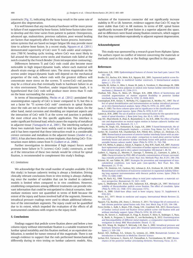
Fig. 1. L1–S1 segment on the testing apparatus after cobalt–chromium rods and trans-verse connector (CoCr-TC) implantation.
485J.J. Doulgeris et al. / Clinical Biomechanics 28 (2013) 483–489
(SD 1.0) axial pre-load, following previous in vitro biomechanical in-vestigations (Slucky et al., 2006), during all simulated motions. Dis-placements (accurate to within 0.1°) were optoelectronicallytracked via an Optotrak Certus System (Optotrak 3020, Northern Dig-ital, Inc., Waterloo, Canada), by sensors located on the superior andinferior frames. A 060S pressure transducer (Precision MeasurementCompany, Ann Arbor, MI, USA) was inserted in the nucleus of thedisc (Cannella et al., 2008; Tzermiadianos et al., 2008) between theL2 and L3 vertebral bodies to measure intradiscal pressure (accurateto within 0.01 MPa). The center of the intervertebral disc was target-ed by caliper measurements in the axial, coronal and sagittal planes. Acannulated needle was marked and inserted laterally to match themeasurements. The pressure transducer was inserted into the cannu-lated needle and then the needle was removed, following a similarinsertion protocol of Rao et al. (2002). Location of the pressure trans-ducer was verified by disc dissection after testing. Pressure transduc-er signals were amplified by a signal conditioner (System 2100,Vishay Micro Measurements, Wendell, NC, USA) and recorded bythe Optotrak data acquisition system (Optotrak 3020, NorthernDigital, Inc., Waterloo, Canada). Data acquisition rate was 10 Hz forall testing conditions.
Specimens were tested under 5 Nm of torque (Gay et al., 2008;Gerber et al., 2006) for flexion–extension (FE), lateral bending (LB)and dynamic axial rotation (AR). FE and LB loads were deliveredthrough a manual quasi-static procedure (±5.0 Nm, 4 cycles at0.10 Hz) by a series of pulleys and weights. Axial rotation loads weredelivered through an automated dynamic procedure (±5.0 Nm, 6 cy-cles at 0.125 Hz) by the servo hydraulic testing apparatus. The numberof cycles selected was based upon the deliverymethod andmotion di-rection. FE and LB both heavily rely on the disc space so a reducedrate and cycle procedure was used to minimize the creep throughoutthe study. Conversely, additional cycles were incorporated in AR toprovide a higher factor of safety on its repeatability. Moreover, re-producible data was obtained for the last two (2) cycles of eachtest hence an average of these two was considered for the analysisin FE, LB and AR.
The first testing cyclewas performed on the “intact” (control) condi-tion. Afterwards, the specimenswere removed from the testing appara-tus to simulate the following posterior and middle column injuries:laminectomy and a complete bilateral removal of the superior and infe-rior articular processes at L4 (to simulate L3–L4 and L4–L5 posteriorcolumn injury) and posterior half annulectomy and nucleus pulposusresection at L3–L4 and L4–L5 (to simulate a middle column injury).The L3 and L5 pedicles were then bored and tapped, using standardsurgical techniques and tools, and appropriate pedicle screw sizes (Zo-diac, Ti, 6.5 mm-diameter and 50–55 mm-length, Alphatec Spine,Carlsbad, CA, USA) were inserted bilaterally, which has been seen in aprevious publication (Goel et al., 2007). Secondly, Ti rods (Ti-6Al-4VELI per ASTM F136, 5.5 mm-diameter and 85–110 mm-length, AlphatecSpine, Carlsbad, CA, USA) were inserted and the test cycle wasperformed for the Ti condition. Thirdly, a rod-to-rod transverse con-nector (Ti, 40–60 mm-length, Alphatec Spine, Carlsbad, CA, USA)was used to augment the implant and the test cycle was performedfor the Ti-TC condition. Fourthly, the TC was removed and rodswere exchanged from Ti to CoCr (BioDur® CCM Plus® Alloy #2 perASTM F1537, 5.5 mm-diameter and 85–110 mm-length, AlphatecSpine, Carlsbad, CA, USA) and the test cycle for the CoCr conditionwas performed. Fifthly, the TC was included to the CoCr rods andthe test cycle for the CoCr-TC condition was performed (Fig. 1). Last-ly, all instrumentation was removed from the specimen to evaluatethe “injury condition”. This last test was conducted after completionof implanted tests, and not after intact condition, because the extentof the injury was classified as severely unstable and would havejeopardized the integrity of the specimen.
Including an “injury” condition in an in vitro biomechanical inves-tigation when comparing different conditions (i.e. intact vs several
constructs) is not required to establish direct comparison among con-structs, as it has been stated in previous publications (Brodke et al.,2001). All surgical procedures were performed by a skilled surgeon. In-termediatefixationwas purposely avoided to reproduce clinical scenar-ios of tumor removal and to expose the construct to more criticalconditions (i.e. greater loads). However, not being able to quantify theeffect of the injury, due to its extent, was not considered a limitationto establish comparisons among the constructs and to address the aimof this investigation.
The former testing cycle was randomized among all implantedtreatments (CoCr, CoCr-TC, Ti-TC, and Ti) to remove bias. The speci-men remained connected to the testing apparatus while exchangingrods (from Ti to CoCr or vice versa) and TCs (implantation or remov-al) to replicate the position of the previous tested condition and toalso maintain the position of the pressure transducer.
Range of motion (RoM [degree]) and intradiscal pressure (IDP[MPa]) were the dependent variables. Additionally, energy lost (EL[Nm ∗ rad]) in the system was analyzed for dynamic AR motion viatrapezoidal integration of the hysteresis loop. EL in FE and LB was ex-cluded because the step sizes between the applied torques weremuch larger in comparison to AR. All RoM measurements were com-piled together (i.e. flexion–extension, lateral bending and axial rota-tion), instead of treating them as separate motions (i.e. right andleft bending/rotation or flexion and extension).
2.3. Statistical analysis
A repeated measures ANOVA followed by a Bonferroni post hoctest was performed using a significance level of 0.05 to determine dif-ferences among treatments for RoM, intradiscal pressure and AR EL.Adjusted P-values were reported for multiple comparisons.

Fig. 2. Means of range of motion at 5.0 Nm for all conditions. CoCr = cobalt–chromium/Ti = titanium/TC = transverse connector. Error bars represent (+) standard deviation(n = 6). *Statistically different (P b 0.05) from intact condition.
486 J.J. Doulgeris et al. / Clinical Biomechanics 28 (2013) 483–489
3. Results
RoM of the different conditions is illustrated in Fig. 2. All im-planted conditions significantly reduced FE and LB RoM in compari-son to the intact condition, but there were no significant differencesamong the implanted groups for these motions (Table 2).
Mean AR RoM increased after all implantations, excluding theCoCr-TC condition, but differences were not statistically significantfrom intact (Table 2). Also, TC conditions showed a reduction in ARRoM with respect to no-TC conditions (Fig. 2); however, these differ-ences were not statistically significant (Table 2). There was notenough evidence to postulate that Ti (no-TC and TC) constructsperformed differently than CoCr (no-TC and TC) constructs for ARRoM. Means and standard deviations for RoM of all conditions arereferenced in Table 3.
In terms of intradiscal pressure (IDP), all implants significantly in-creased IDP for flexion (P b 0.01), extension (P b 0.01), LB (P b 0.01) and
Table 2Adjusted P-values from Bonferroni post-hoc test, after repeated measures ANOVA, forrange of motion at 5.0 Nm.
Comparisons Flexion–extension Lateral bending Axial rotation
Intact withCoCr b0.01⁎ b0.01⁎ 0.45CoCr-TC b0.01⁎ b0.01⁎ >0.99Ti b0.01⁎ b0.01⁎ 0.09Ti-TC b0.01⁎ b0.01⁎ >0.99
CoCr withCoCr-TC >0.99 >0.99 0.29Ti >0.99 >0.99 >0.99Ti-TC >0.99 >0.99 >0.99
CoCr-TC withTi >0.99 >0.99 0.06Ti-TC >0.99 >0.99 >0.99
Ti withTi-TC >0.99 >0.99 0.52
CoCr = cobalt–chromium.TC = transverse connector.Ti = titanium.⁎ Statistically significant (P b 0.05).
AR (P b 0.03) with respect to the intact condition (Fig. 3), except CoCr(P = 0.06) and CoCr-TC (P = 0.08) conditions for extension motion.There were no significant differences (P > 0.99) in any motion whencomparing IDP among all implanted treatments. All IDP means and stan-dard deviations are referenced in Table 4.
Energy loss (EL) of the system (L1–S1) significantly increased(P b 0.05) for dynamic AR motions in all implanted conditions with re-spect to intact (Table 3), excluding the CoCr-TC condition (P > 0.99).There was no significant difference (P = 0.20) between Ti and Ti-TC,but CoCr-TC significantly reduced AR EL with respect to CoCr(P = 0.05) and Ti (P b 0.01) conditions.
4. Discussion
The main advantage of CoCr over Ti is its higher modulus of elas-ticity (Table 1), so it is expected that CoCr would be stiffer than Tiin a pure mechanical testing environment. Transverse connectors(TC) are designed to add structure to the implant by mitigating theangular displacements between the rods (keeping the rods parallel).FE and LB motions do not significantly change the angle betweenthe rods which will result in a minimal stress on the TC. The FE andLB performance, in terms of RoM, between no-TC and TC conditions(Fig. 2) was not significantly affected, which is consistent with
Table 3Total range of motion and energy loss means (standard deviation) at 5.0 Nm.
Condition Flexion–extension[deg]
Lateral bending[deg]
Axial rotation[deg]
Axial rotationEnergy loss[Nm ∗ rad]
Intact 22.0 (4.2) 25.1 (8.1) 9.0 (2.3) 1.15 (0.38)CoCr 14.1 (5.3)⁎ 12.1 (4.5)⁎ 11.2 (0.9) 1.72 (0.08)⁎
CoCr-TC 14.0 (5.2)⁎ 11.6 (3.6)⁎ 8.8 (1.8) 1.32 (0.22)Ti 13.0 (5.8)⁎ 12.3 (3.5)⁎ 11.9 (1.9) 1.87 (0.10)⁎
Ti-TC 12.9 (5.7)⁎ 11.8 (4.3)⁎ 9.8 (0.6) 1.55 (0.10)⁎
CoCr = cobalt–chromium.TC = transverse connector.Ti = titanium.⁎ Statistically different (P b 0.05) from intact condition.

Fig. 3. Means of L2–3 intradiscal pressure at 5.0 Nm for all conditions. CoCr = cobalt–chromium/Ti = titanium/TC = transverse connector. Error bars represent (+) standarddeviation (n = 6). *Statistically different (P b 0.05) from intact condition.
487J.J. Doulgeris et al. / Clinical Biomechanics 28 (2013) 483–489
previous publications (Kuklo et al., 2008; Shaw et al., 2011). This sug-gests that the contribution of one TC in a two-level fusion constructwithout intermediate fixation may not be substantial for these mo-tions. Additionally, there were no significant differences between Tiand CoCr RoM or Ti-TC and CoCr-TC RoM, which suggests that, froma mechanical standpoint, either material can be used to stabilizethese motions.
AR RoM is affected by the presence of the facet joints and removal ofthe joints reduces AR stiffness because they constrain the vertebralbodies from shear stresses (Serhan et al., 2007; Zander et al., 2003).The differences in the means between Ti and CoCr, although not signifi-cant (Table 2),may be explained by the fact that AR applies a shear stresson the construct (Chutkan et al., 2008; Kuklo et al., 2008; Shaw et al.,2011) and that Ti is considered to have “poor shear strength”(Long andRack, 1998). Since the absence of TC makes the construct less stable(Lynn et al., 1997; Shaw et al., 2011; Zander et al., 2003), it is expectedthat Ti construct without TC would have been the weakest in AR RoM(Table 3), but differences were not significantly different than otherconstructs (Table 2). On the other hand, based on the RoM mean TCprovided the more stability in comparison to no-TC, but more evidenceis needed to confirm a statistical difference.
When performing the “injury test”, all six specimens deformedplastically to the extent of failure, and the injury was severe enough
Table 4Intradiscal pressure mean (standard deviation) for all conditions at 5.0 Nm.
Condition Extension[MPa]
Flexion[MPa]
Lateral bending[MPa]
Axial rotation[MPa]
Intact 0.16 (0.02) 0.31 (0.05) 0.19 (0.05) 0.27 (0.07)CoCr 0.23 (0.06) 0.43 (0.07)⁎ 0.30 (0.13)⁎ 0.40 (0.06)⁎
CoCr-TC 0.23 (0.05) 0.43 (0.06)⁎ 0.30 (0.12)⁎ 0.41 (0.07)⁎
Ti 0.25 (0.06)⁎ 0.45 (0.07)⁎ 0.30 (0.12)⁎ 0.38 (0.07)⁎
Ti-TC 0.25 (0.07)⁎ 0.42 (0.08)⁎ 0.30 (0.12)⁎ 0.40 (0.06)⁎
CoCr = cobalt–chromium.TC = transverse connector.Ti = titanium.⁎ Statistically different (P b 0.05) from intact condition.
to cause failure under the axial preload alone (before testing started),which was expected due to the significant effect of axial preloads inmiddle columns(Denis, 1983). These findings support that bilateralpedicle screw system provided the majority of the structure to thespecimen after injury.
Measuring EL by the spinal segment provides additional informa-tion, in terms of stability. EL is an indication of the plastic deforma-tion and is related to the disc contribution. The inclusion of EL isparticularly useful in fusion because it corresponds to rigidity.CoCr-TCwas expected to be themost rigid construct in AR due toma-terial and mechanical properties of CoCr with respect to Ti alloy(Table 1) and the additional rigidity applied from the contributionof TCs. However, more evidence is needed to show that CoCr-TC con-struct would provide significantlymore stability than others in termsof RoM. The lack of difference between the ELs is attributed to theoverlapping implants (same pedicle screws and TC) and limitedsample size.
The energy lost by the segment in AR under the CoCr-TC conditionwas not sufficient to be statistically different than the intact condition(Table 2), while all other conditions were. This evidence suggests thatCoCr-TC was the construct that promoted the least energy loss of allfixations, which translates to greater stability. Moreover, the Ti con-dition had the greatest average in AR EL (i.e. least stable) and eventhat the increase was not enough to be statistically different thanthe CoCr or Ti-TC conditions, it was sufficient to be significantly dif-ferent than the most rigid construct: the CoCr-TC condition.
When comparing TC and no-TC conditions in terms of AR EL, theinclusion of TC could increase AR stability when using CoCr rods,which goes in line with similar findings postulated by other authors(Chutkan et al., 2008; Lynn et al., 1997; Shaw et al., 2011).
In terms of intradiscal pressure, an increase at superior levels is acommon byproduct in fusion and has been validated in previousworks (Cunningham et al., 1997; Weinhoffer et al., 1995). It isthought that increased intradiscal pressure is the main factor leadingto accelerated adjacent level degeneration. Changing the stiffness ofthe construct may seriously change load distributions and divert theload from the disc space. Therefore, it is possible that different mate-rials may affect IDP differently. However, differences were not statis-tically significant in intradiscal pressures between the CoCr and Ti
488 J.J. Doulgeris et al. / Clinical Biomechanics 28 (2013) 483–489
constructs (Fig. 3), indicating that they may result in the same rate ofadjacent disc degeneration.
It is well known that any mechanical hardware will be more proneto failure if not supported by bone fusion. Solid bone fusion takes timeto develop and this time varies from patient to patient. Osteoporosis,advanced age, malnutrition, previous radiation, poor wound healingare factors that negatively affect the bone fusion process. Instrumen-tation systems that are based on longer fatigue life may provide moretime to achieve bone fusion. In a recent study, Nguyen et al. (2011)demonstrated superiority of CoCr over Ti rods under axial compres-sion (700 N) bending cyclic testing, where CoCr was most likely tofail around the titanium screw necks and titanium rods failed at thenotch created by the French Bender (from intraoperative contouring).
Differences between Ti and CoCr rods could also become morenoticeable in high impact/dynamic loads, where each material willabsorb strain energy at different rates. The stress magnitude on thescrews under impact/dynamic loads will depend on the mechanicalproperties of the rods, where rods with the greatest stiffness willconcentrate more stress on the screws. Ti screw/CoCr rod junctionmay be a critical area that eventually leads to hardware failure in anin vitro environment. Therefore, under impact/dynamic loads, it ishypothesized that CoCr rods will produce more stress than Ti rodson the bone surrounding the screws.
In terms of biocompatibility, Plecko et al. (2012) postulated thatosseointegration capacity of CoCr is lower compared to Ti, but this isnot a factor for “Ti screws–CoCr rods” constructs in spinal fixationsince the rods are not in direct contact with the bone surface. On theother hand, metal corrosion and shredding are important aspects andthe interaction of CoCr with Ti at the screw rod junction is probablythe most critical area for this specific application. This interface isunder significant frictional load, which causes themajority ofmetallosisin adjacent tissue (Yamaguchi et al., 2001). The interfering surfaces ofthese two materials have been widely used in total hip arthroplasty,and it has been reported that these interaction result in a considerablecrevice corrosion and metallosis in the adjacent tissues (Zander et al.,2003). It has also been shown, on knee implants, that during CoCr–Ti in-teraction more debris comes from Ti (Guibert et al., 2006).
Further investigation to determine if high impact forces wouldpromote bone failure in Ti (screws)–CoCr (rods) constructs, as wellas if the interaction of these two metals would be critical for spinalfixation, is recommended to complement this study's findings.
4.1. Limitations
We acknowledge that the small number of samples available (6 forthis study) in human cadaveric testing is always a limitation. Drivingclinically relevant conclusions from in vitro testing is always challeng-ing since the number of variables that can be studied in cadavericmodels is limited when compared to in vivo conditions. However,establishing comparisons among different treatments can provide rele-vant information that could be extrapolated to clinical scenarios. Inter-mediate motions were not quantified in terms of RoM because theextent of the injury and fusion involved half of the segments. However,intradiscal pressure readings were used to obtain additional informa-tion of the intermediate segments. The injury could not be quantifieddue to its extent, which impeded the evaluation of the effects of allinstrumented conditions with respect to the injury itself.
5. Conclusions
Findings suggest that pedicle screw fixation above and below a twocolumn injury without intermediate fixation is a suitable treatment forlumbar spinal instability and this fixation method, or an equivalent sta-bilization, is needed for tumor removal of this magnitude. There is notenough evidence to support that the rods performed biomechanicallydifferently during in vitro testing on lumbar cadaveric models. Also,
inclusion of the transverse connector did not significantly increasestability in FE or LB; however, evidence suggests that CoCr-TC may bemore stable than CoCr in AR motions. In terms of IDP, spinal fusionsignificantly increases IDP post fusion in a superior adjacent disc space,and no differences were found among fixation constructs, which suggestthat theymay contribute equivalently to adjacent segment degeneration.
Acknowledgment
This study was sponsored by a research grant from Alphatec Spine.The authors report no conflict of interest concerning the materials ormethods used in this study or the findings specified in this paper.
References
Andersson, G.B., 1999. Epidemiological features of chronic low-back pain. Lancet 354,581–585.
Brodke, D.S., Bachus, K.N., Mohr, R.A., Nguyen, B.K., 2001. Segmental pedicle screw fix-ation or cross-links in multilevel lumbar constructs. A biomechanical analysis.Spine J. 1, 373–379.
Cannella, M., Arthur, A., Allen, S., Keane, M., Joshi, A., Vresilovic, E., Marcolongo, M., 2008.The role of the nucleus pulposus in neutral zone human lumbar intervertebral discmechanics. J. Biomech. 41, 2104–2111.
Chutkan, N.B., Zhou, H., Akins, J.P., Wenger, K.H., 2008. Effects of facetectomy andcrosslink augmentation on motion segment flexibility in posterior lumbarinterbody fusion. Spine (Phila Pa 1976) 33, E828–E835.
Cunningham, B.W., Kotani, Y., McNulty, P.S., Cappuccino, A., McAfee, P.C., 1997. The ef-fect of spinal destabilization and instrumentation on lumbar intradiscal pressure:an in vitro biomechanical analysis. Spine (Phila Pa 1976) 22, 2655–2663.
Denis, F., 1983. The three column spine and its significance in the classification of acutethoracolumbar spinal injuries. Spine (Phila Pa 1976) 8, 817–831.
Gaines Jr., R.W., 2000. The use of pedicle-screw internal fixation for the operative treat-ment of spinal disorders. J. Bone Joint Surg. Am. 82-A, 1458–1476.
Gay, R.E., Ilharreborde, B., Zhao, K., Boumediene, E., An, K.N., 2008. The effect of loadingrate and degeneration on neutral region motion in human cadaveric lumbar mo-tion segments. Clin. Biomech. 23, 1–7.
Geetha, M., Singh, A.K., Asokamani, R., Gogia, A.K., 2009. Ti based biomaterials, the ul-timate choice for orthopaedic implants — a review. Prog. Mater. Sci. 54, 397–425.
Gerber, M., Crawford, N.R., Chamberlain, R.H., Fifield, M.S., LeHuec, J.C., Dickman, C.A.,2006. Biomechanical assessment of anterior lumbar interbody fusion with an ante-rior lumbosacral fixation screw-plate: comparison to stand-alone anterior lumbarinterbody fusion and anterior lumbar interbody fusion with pedicle screws in anunstable human cadaver model. Spine (Phila Pa 1976) 31, 762–768.
Goel, V.K., Mehta, A., Jangra, J., Faizan, A., Kiapour, A., Hoy, R.W., Fauth, A.R., 2007. Anatomicfacet replacement system (AFRS) restoration of lumbar segmentmechanics to intact: afinite element study and in vitro cadaver investigation. SAS Journal 1, 46–54.
Guibert, G., Irigaray, J.L., Moretto, P., Sauvage, T., Kemeny, J.L., Cazenave, A., Jallot, E.,2006. Characterisation by PIXE–RBS of metallic contamination of tissues surround-ing a metallic prosthesis on a knee. Nucl. Inst. Methods Phys. Res. B 251, 246–256.
Krismer, M., van Tulder, M., 2007. Strategies for prevention and management of mus-culoskeletal conditions. Low back pain (non-specific). Best Pract. Res. Clin.Rheumatol. 21, 77–91.
Kuklo, T.R., Dmitriev, A.E., Cardoso, M.J., Lehman Jr., R.A., Erickson, M., Gill, N.W., 2008.Biomechanical contribution of transverse connectors to segmental stability follow-ing long segment instrumentation with thoracic pedicle screws. Spine (Phila Pa1976) 33, E482–E487.
Long, M., Rack, H.J., 1998. Titanium alloys in total joint replacement—a materials sci-ence perspective. Biomaterials 19, 1621–1639.
Lynn, G., Mukherjee, D.P., Kruse, R.N., Sadasivan, K.K., Albright, J.A., 1997. Mechanicalstability of thoracolumbar pedicle screw fixation. The effect of crosslinks. Spine(Phila Pa 1976) 22, 1568–1572 (discussion 1573).
Marti, A., 2000. Cobalt-base alloys used in bone surgery. Injury 31. Supplement 4,D18–D21.
Mears, D.C., 1977. Metals in medicine and surgery. International Metals Reviews 22,119–155.
Nguyen, T.Q., Buckley, J.M., Ames, C., Deviren, V., 2011. The fatigue life of contoured co-balt chrome posterior spinal fusion rods. Proc. Inst. Mech. Eng. H 225, 194–198.
Perez-Orribo, L., Kalb, S., Reyes, P.M., Chang, S.W., Crawford, N.R., 2013. Biomechanicsof lumbar cortical screw-rod fixation versus pedicle screw–rod fixation with andwithout interbody support. Spine (Phila Pa 1976) 38, 635–641.
Plecko, M., Sievert, C., Andermatt, D., Frigg, R., Kronen, P., Klein, K., Stubinger, S., Nuss,K., Burki, A., Ferguson, S., Stoeckle, U., von Rechenberg, B., 2012. Osseointegrationand biocompatibility of different metal implants—a comparative experimental in-vestigation in sheep. BMC Musculoskelet. Disord. 13, 32.
Rao, R.D., Wang, M., Singhal, P., McGrady, L.M., Rao, S., 2002. Intradiscal pressure andkinematic behavior of lumbar spine after bilateral laminotomy and laminectomy.Spine J. 2, 320–326.
Ratner, B.D., Hoffman, A.S., Schoen, F.J., Lemons, J.E., 2004. Biomaterials Science: AnIntroduction to Materials in Medicine. Elsevier Science.
Serhan, H.A., Varnavas, G., Dooris, A.P., Patwadhan, A., Tzermiadianos, M., 2007. Biome-chanics of the posterior lumbar articulating elements. Neurosurg. Focus. 22, E1.

489J.J. Doulgeris et al. / Clinical Biomechanics 28 (2013) 483–489
Shaw, M.N., Morel, E.P., Utter, P.A., Gussous, Y.M., Ginoux, L., Berglund, L.J., Gay, R.E.,Krauss,W.E., 2011. Transverse connectors providing increased stability to the cervicalspine rod–screw construct: an in vitro human cadaveric study. J. Neurosurg. Spine 14,719–725.
Slucky, A.V., Brodke, D.S., Bachus, K.N., Droge, J.A., Braun, J.T., 2006. Less invasive posteriorfixation method following transforaminal lumbar interbody fusion: a biomechanicalanalysis. Spine J. 6, 78–85.
Tzermiadianos, M.N., Renner, S.M., Phillips, F.M., Hadjipavlou, A.G., Zindrick, M.R.,Havey, R.M., Voronov, M., Patwardhan, A.G., 2008. Altered disc pressure profileafter an osteoporotic vertebral fracture is a risk factor for adjacent vertebral bodyfracture. Eur. Spine J. 17, 1522–1530.
Weinhoffer, S.L., Guyer, R.D., Herbert, M., Griffith, S.L., 1995. Intradiscal pressuremeasure-ments above an instrumented fusion. A cadaveric study. Spine (Phila Pa 1976) 20,526–531.
Yamaguchi, K., Konishi, H., Hara, S., Motomura, Y., 2001. Biocompatibility studies of titanium-based alloy pedicle screw and rod system: histological aspects. Spine J. 1, 260–268.
Zander, T., Rohlmann, A., Klockner, C., Bergmann, G., 2003. Influence of graded facetectomyand laminectomy on spinal biomechanics. Eur. Spine J. 12, 427–434.