Embed Size (px)
Citation preview
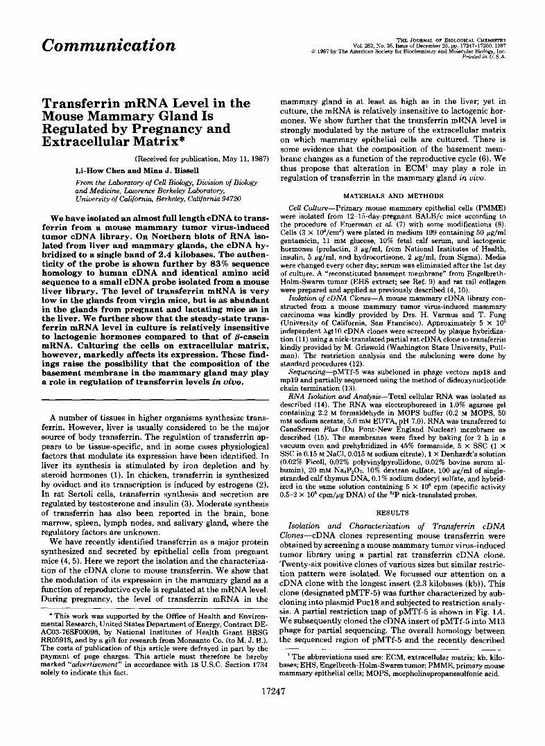
Communication Vol. 262, No. 36, Issue of December 25, pp. 17247-17250,198’7 0 1987 by The American Society for Biochemistry and Molecular Biology, Inc.
Printed in U.S.A.
THE JOURNAL OF BIOLOGICAL CHEMISTRY
Transferrin mRNA Level in the Mouse Mammary Gland Is Regulated by Pregnancy and Extracellular Matrix*
(Received for publication, May 11, 1987) Li-How Chen and Mina J. Bissell From the Laboratory of Cell Bwlogy, Division of Biology and Medicine, Lawrence Berkeley Laboratory, University of California, Berkeley, California 94720
We have isolated an almost full length cDNA to trans- ferrin from a mouse mammary tumor virus-induced tumor cDNA library. On Northern blots of RNA iso- lated from liver and mammary glands, the cDNA hy- bridized to a single band of 2.4 kilobases. The authen- ticity of the probe is shown further by 83% sequence homology to human cDNA and identical amino acid sequence to a small cDNA probe isolated from a mouse liver library. The level of transferrin mRNA is very low in the glands from virgin mice, but is as abundant in the glands from pregnant and lactating mice as in the liver. We further show that the steady-state trans- ferrin mRNA level in culture is relatively insensitive to lactogenic hormones compared to that of &casein mRNA. Culturing the cells on extracellular matrix, however, markedly affects its expression. These find- ings raise the possibility that the composition of the basement membrane in the mammary gland may play a role in regulation of transferrin levels in vivo.
A number of tissues in higher organisms synthesize trans- ferrin. However, liver is usually considered to be the major source of body transferrin. The regulation of transferrin ap- pears to be tissue-specific, and in some cases physiological factors that modulate its expression have been identified. In liver its synthesis is stimulated by iron depletion and by steroid hormones (1). In chicken, transferrin is synthesized by oviduct and its transcription is induced by estrogens (2). In rat Sertoli cells, transferrin synthesis and secretion are regulated by testosterone and insulin (3). Moderate synthesis of transferrin has also been reported in the brain, bone marrow, spleen, lymph nodes, and salivary gland, where the regulatory factors are unknown.
We have recently identified transferrin as a major protein synthesized and secreted by epithelial cells from pregnant mice (4,5). Here we report the isolation and the characteriza- tion of the cDNA clone to mouse transferrin. We show that the modulation of its expression in the mammary gland as a function of reproductive cycle is regulated at the mRNA level. During pregnancy, the level of transferrin mRNA in the
* This work was supported by the Office of Health and Environ- mental Research, United States Department of Energy, Contract DE- AC03-76SF00098, by National Institutes of Health Grant BRSG RR05918, and by a gift for research from Monsanto Co. (to M. J. E). The costs of publication of this article were defrayed in part by the payment of page charges. This article must therefore be hereby marked “advertisement” in accordance with 18 U.S.C. Section 1734 solely to indicate this fact.
mammary gland is at least as high as in the liver; yet in culture, the mRNA is relatively insensitive to lactogenic hor- mones. We show further that the transferrin mRNA level is strongly modulated by the nature of the extracellular matrix on which mammary epithelial cells are cultured. There is some evidence that the composition of the basement mem- brane changes as a function of the reproductive cycle (6). We thus propose that alteration in ECM’ may play a role in regulation of transferrin in the mammary gland i n uiuo.
MATERIALS AND METHODS
Cell Culture-Primary mouse mammary epithelial cells (PMME) were isolated from 12-15-day-pregnant BALB/c mice according to the procedure of Emerman et al. (7) with some modifications (8). Cells (3 X 10⁶/cm2) were plated in medium 199 containing 50 pg/ml gentamicin, 11 mM glucose, 10% fetal calf serum, and lactogenic hormones (prolactin, 3 pg/ml, from National Institutes of Health, insulin, 5 pg/ml, and hydrocortisone, 2 pg/ml, from Sigma). Media were changed every other day; serum was eliminated after the 1st day of culture. A “reconstituted basement membrane” from Engelbreth- Holm-Swarm tumor (EHS extract; see Ref. 9) and rat tail collagen were prepared and applied as previously described (4, 10).
Isolation of cDNA Clones-A mouse mammary cDNA library con- structed from a mouse mammary tumor virus-induced mammary carcinoma was kindly provided by Drs. H. Varmus and T. Fung (University of California, San Francisco). Approximately 5 X 10’ independent X g t l O cDNA clones were screened by plaque hybridiza- tion (11) using a nick-translatedpartial rat cDNA clone to transferrin kindly provided by M. Griswold (Washington State University, Pull- man). The restriction analysis and the subcloning were done by standard procedures (12).
Sequencing-pMTf-5 was subcloned in phage vectors mp18 and mp19 and partially sequenced using the method of dideoxynucleotide chain termination (13).
RNA Isolation and Analysis-Total cellular RNA was isolated as described (14). The RNA was electrophoresed in 1.0% agarose gel containing 2.2 M formaldehyde in MOPS buffer (0.2 M MOPS, 50 mM sodium acetate, 5.0 mM EDTA, pH 7.0). RNA was transferred to GeneScreen Plus (Du Pont-New England Nuclear) membrane as described (15). The membranes were fixed by baking for 2 h in a vacuum oven and prehybridized in 45% formamide, 5 X SSC (1 X SSC is 0.15 M NaC1,0.015 M sodium citrate), 1 X Denhardt’s solution (0.02% Ficoll, 0.02% polyvinylpyrollidone, 0.02% bovine serum al- bumin), 20 mM Na,PZ07, 10% dextran sulfate, 100 pg/ml of single- stranded calf thymus DNA, 0.1% sodium dodecyl sulfate, and hybrid- ized in the same solution containing 5 X 10‘ cpm (specific activity 0.5-2 X 10’ cpmlpg DNA) of the 32P nick-translated probes.
RESULTS
Isolation and Characterization of Transferrin cDNA Clones-cDNA clones representing mouse transferrin were obtained by screening a mouse mammary tumor virus-induced tumor library using a partial rat transferrin cDNA clone. Twenty-six positive clones of various sizes but similar restric- tion pattern were isolated. We focussed our attention on a cDNA clone with the longest insert (2.3 kilobases (kb)). This clone (designated pMTF-5) was further characterized by sub- cloning into plasmid PuclS and subjected to restriction analy- sis. A partial restriction map of pMTf-5 is shown in Fig. 1A. We subsequently cloned the cDNA insert of pMTf-5 into M13 phage for partial sequencing. The overall homology between the sequenced region of pMTf-5 and the recently described
The abbreviations used are: ECM, extracellular matrix; kb, kilo- bases; EHS, Engelbreth-Holm-Swarm tumor; PMME, primary mouse mammary epithelial cells; MOPS, morpholinepropanesulfonic acid.
17247

17248 Transferrin mRNA Modulatic
75
287
lW. 1. Partial restriction map of pMTf-5 ( A ) and the align- ment of human serum transferrin ( R n ) cDNA (18) and pMTf- 5 sequences and the deduced amino acid sequences (B) . The homology between pMTf-5 and HSTf is 83% at cDNA level and 81% at amino acid level. Boxes indicate amino acid residues that are
3n by the Extracellular Matrix
1 2 3 4 5 6
FIG. 2. The identity of mammary gland transferrin (n) mRNA and its modulation by pregnancy and lactation. Total cellular RNA (10 pg) extracted from mammary glands of virgin (lanes 1 and 2), pregnant (lones 3 and 4) , and lactating (lanes 5 and 6) mice, mammary glands (lanes 1,3, and 5), and liver (lanes 2,4, and 6) were separated on formaldehyde-agarose gel, blotted to Genescreen Plus membrane, and hybridized with nick-translated pMTf-5 as described under "Materials and Methods." A small 0.4-kb cDNA from a mouse liver library kindly provided by Pentacost and Teng (17) hybridized to the same bands (not shown).
human serum transferrin (16) was 83% at the DNA level and 81% at amino acid level (Fig. 1B). Most of the conserved amino acid residues for the transferrin family (including transferrin of human and the lactoferrin of human, mouse, mare, and chicken conalbumin) are preserved in this cDNA from mouse mammary gland (Fig. 1B). The deduced sequence of pMTf-5 is identical to that of a fragment of transferrin cDNA isolated from mouse liver (17) between amino acid 292 and 347, for which the sequences are available.
The pMTf-5 Recognizes a Single mRNA Species in Liver and Mammary Gland-To establish whether the mRNA spe- cies recognized by pMTf-5 are similar in the liver and the mammary gland, we isolated RNA from tissues of virgin, pregnant, and lactating mice and hybridized the 32P nick- translated pMTf-5 to the electrophoresed RNA on Northern blots (Fig. 2). pMTf-5 detected one RNA species of about 2.4 kb, present both in the liver and in the mammary gland (Fig. 2). The size of this mRNA agrees with the expected size for the transferrin message. The cDNA to a fragment of trans- ferrin isolated from a mouse liver library, mentioned above and kindly provided by B. Pentacost and T. Teng (17; Re- search Triangle Park, North Carolina), cross-hybridized to the same mRNA (not shown), indicating further that the mammary gland synthesizes authentic transferrin mRNA. A comparison of the mRNA levels in glands of virgin, pregnant, and early lactating mice indicated that the steady-state level of transferrin mRNA is increased by more than 40-fold in
conserved among all transferring protein families. The scale in A is in kb.

Transferrin mRNA Modulation by the Extracellular Matrix 17249
glands of pregnant as compared to that of virgin mice (Fig. 2); the level is decreased slightly in the lactating gland. This indicates that modulation of transferrin levels observed pre- viously (5) are at the level of transferrin mRNA.
Response to Lactogenic Hormones in Culture-In order to define the factors that regulate transferrin gene expression in the mammary gland during pregnancy, we cultured mammary epithelial cells from pregnant mice. It has been demonstrated in the same system that the expression of mRNA for another major milk protein, @-casein, as well as its synthesis and secretion, are dependent upon the presence of prolactin, hy- drocortisone, and insulin (8, 18). Transferrin mRNA level is partially affected by the addition and/or subtraction of these three hormones (Fig. 3A), yet its response is distinct from that of 8-casein. Elimination of either prolactin or insulin reduced the expression of transferrin mRNA but did not abolish it. Hydrocortisone, on the other hand, had little or no effect on transferrin expression while its removal decreased
A
B
1 2 3 4 5 6
- Tf
- p-casein
C 100 22 41 22 79 -Tf
100 1 6 1 14 -p-casein F ~ G . 3. Hormonal response of transferrin ( n ) and &casein
expression in cell culture. Epithelial cells were isolated from mammary glands of pregnant mice and cultured in the presence of different combinations of hormones; all lanes are on plastic except for lane 1. Lane I, lactating mammary gland; lane 2, PMME plus prolactin, insulin, and hydrocortisone; lune 3, minus all the three hormones; lane 4, plus insulin and hydrocortisone; lanes 1 and 5, plus prolactin and hydrocortisone; lane 6, plus prolactin and insulin. The blot was probed with pMTf-5 and @-casein cDNA simultaneously ( A ) . The blot was washed and reprobed with a genomic clone to 28 S rRNA ( B ) (courtesy of Dr. James Sylvesto, University of Pennsyl- vania, Philadelphia). The autoradiograms were scanned by a densi- tometer. The density was taken as peak height times peak width in scanning units and the levels of transferrin and &casein mRNA in lunes 3-6 are expressed as percentages of those in lane 2 (C).
1 2 3 4
I -T f
- p-casein
FIG. 4. Effect of culture substrata on transferrin (n) expression. Cells were isolated from mammary glands of pregnant mice and cultured in the presence of lactogenic hormones on plastic ( l a n e 2), EHS ( l a n e 3), and EHS on floating gel ( l u n e 4). Lane 1 was RNA isolated from lactating mammary gland. Northern blot was probed with nick-translated pMTf-5 and @casein cDNA simultane- ously. Transferrin-specific mRNA was detectable in lane 2 after longer autoradiographic exposure.
8-casein gene expression considerably (compare ratios of 8- casein mRNA to transferrin mRNA in lanes 1 and 2 as opposed to lanes 3-6 in Fig. 3, panel C ) .
Extracellular Matrix Influence on Transferrin Gene Expres- sion-PMME cells were seeded on either plastic, plastic- coated with EHS extracts, or on rat tail collagen gels coated with EHS extract and subsequently floated as described pre- viously (8). The -fold induction of transferrin on EHS matrix is dependent on the density of the culture and the quality of the EHS preparation as was found previously for 8-casein (8). The level of transferrin mRNA was highest when PMME were cultured on floating EHS-coated rat tail collagen (20- fold increase in comparison to plastic), followed by EHS matrix coated as a thin film on plastic (5-fold over plastic in this experiment) (Fig. 4; this ratio has been as high as 20 in recent experiments). The level of transferrin (and @-casein) mRNA on floating EHS/collagen gels was as high as the lactating gland (Fig. 4, compare lanes 1 and 4).
DISCUSSION
We have obtained an almost full length cDNA clone to mouse transferrin from the mammary gland. Since the major iron binding protein in the mammary gland until recently was thought to be lactoferrin, it was of interest to compare the sequence of pMTf-5 with a cDNA clone (pT267) to mouse lactoferrin isolated by Pentacost and Teng (17). Maximum alignment of pMTf-5 and PT267 showed 66% homology for the two cDNA sequences and 58% homology for the deduced amino acid sequences ( d a t a not shown). On the other hand, pMTf-5 has 100% homology at the amino acid level to a small cDNA to liver transferrin isolated by the same investigators (17). pMTf-5 shares 83% homology with human serum trans- ferrin and recognizes the same RNA species in the liver and mammary gland. It is therefore clear that the mouse mam- mary gland produces its own transferrin during pregnancy and lactation and that our isolated clone is indeed transferrin.
Two lines of evidence have suggested recently that trans- ferrin may be essential for cell proliferation: the requirement for transferrin in chemically defined culture medium to sup- port cell growth (19) and the widespread distribution of trans- ferrin receptor on many different cell types (20). Studies by Ekblom and Thesleff (21) have demonstrated that transferrin is necessary in metanephric mesenchyme differentiation. I t also has been shown that transferrin or transferrin-like sub- stances released from peripheral nerve promote the growth and the development of chicken embryo myoblasts in culture (22) and allow the regeneration of amphibian limb (23). Our data that mouse mammary gland expresses transferrin, and furthermore that its expression is regulated by pregnancy,

17250 Transferrin mRNA Modulation by the Extracellular Matrix
add to the growing evidence that transferrin synthesis is associated with growth and functional differentiation in tis- sues other than the liver. The simplest explanation for this association might be that transferrin stimulates growth and differentiation by transporting iron, a substance that partici- pates in reactions spanning all of biochemistry. Whether or not there is an additional role for transferrin in growth regulation and differentiation (distinct from an iron carrier) (21) is not clear at this time.
Mammary gland development and differentiation are influ- enced by the complex interplay of many factors including hormones and ECM. While the importance of cell-ECM in- teractions in modulation of mammary epithelial cell function has been addressed previously (24,25), transferrin appears to be unique among skim milk proteins as it is more sensitive to ECM than to lactogenic hormones. The finding that transfer- rin expression is greatly elevated on EHS matrix as compared to plastic surfaces leads us to propose that one important regulator of mammary transferrin could be the change in ECM structure and composition that occurs during pregnancy in vivo. Our preliminary data (26) indicate that mRNA levels for ECM components are greatly increased and altered during pregnancy and lactation. The relation of ECM to regulation of transferrin levels in vivo is under investigation.
Acknowledgments-We are grateful to Dr. M.-L. Li for helpful suggestions in the early part of this work and Dr. M. H. Barcellos- Hoff for critical reading of the manuscript.
1.
2.
3.
4.
5.
REFERENCES McNight, G. S., Lee, D. C., Hemmaplardh, D., Finch, C. A., and
Lee, D. C., Mcknight, G. S., and Palmiter, R. D. (1978) J. Biol.
Skinner, M. K., and Griswold, M. D. (1982) Biol. Reprod. 27,
Lee, E. Y.-H., Parry, G., and Bissell, M. J. (1984) J. Cell Biol.
Lee, E. Y.-H., Barcellos-Hoff, M. H., Chen, L.-H., Parry, G., and
Palmiter, R. D. (1980) J. Biol. Chem. 255,14&153
Chern. 263,3494-3503
211-221
98,146-155
Bissell, M. J. (1987) In Vitro Cell. Dev. Biol. 23,221-226
6.
7.
8.
9.
10.
11. 12.
13.
14.
15. 16.
17.
18.
19. 20. 21.
22.
23.
24.
25.
26.
Warburton, M. J., Mitchell, D., Ormerod, E. J., and Rudland, P. (1982) J. Histochem. Cytochem. 30,667-676
Emerman, J. T., Bartley, J. C., and Bissell, M. J. (1981) Ezp. Cell Res. 134, 241-250
Lee, E. Y.-H., Lee, W.-H., Kaetzel, C. S., Parry, G., and Bissell, M. J. (1985) Proc. Natl. Acad. Sci. U. S. A. 82,1419-1423
Kleinman, H. K., McGarvey, M. L., Hassell, J. R., Star, V. L., Cannon, F. B., Laurie, G. W., and Martin, G. R. (1986) Bio- chemistry 25,312-318
Li, M. L., Aggeler, J., Farson, D., Hatier, C., Hassell, J., and Bissell, M. J. (1987) Proc. Natl. Acad. Sci. U. S. A. 84, 136- 140
Benton, W. D. (1977) Science 196,180-182 Maniatis, T., Fritsch, E. F., and Sambrook, J. (1982) Molecular
Cloning: A Laboratory Manual, Cold Spring Harbor Laboratory, Cold Spring Harbor, NY
Sanger, F., Nicklen, S., and Coulsan, A. R. (1977) Proc. Natl. Acad. Sci. U. S. A. 74,5463-5467
Chirgwin, J. M., Przybylal, A. E., MacDonald, R. J., and Rutter, W. J. (1979) Biochemistry 18,5294-5299
Thomas, P. (1980) Proc. Natl. Acad. Sci. U. S. A. 77,5201-5205 Yang, F., Lum, J. B., Mcgill, J. R., Moore, C. M., Naylor, S. L.,
Van Brogt, P. H., Baldwin, W. D., and Brown, B. (1984) Proc. Natl. Acad. Sci. U. S. A. 81, 2752-2756
Pentacost, B. T., and Teng, C. T. (1987) J. Biol. Chem., 262,
Rosen, J. M., Jones, W. K., Campbell, S. M., Bisbee, C. A., and Lee, L.-Y. Y. (1985) in Proceedings of UCLA Symposium on Membrane Receptors and CeUular Reguktion (Kahu, C. R., and Czech, M., eds) pp. 385-396, Alan R. Liss, Inc., New York
10134-10139
Barnes, D., and Sato, G. (1980) Cell 22,649-655 Yong, S., and Bamford, A. (1984) Clin. Sci. (Land.) 67,273-278 Ekblom, P., and Thesleff, I. (1985) in Modern Cell Biology, (Satir,
B. H., ed) Vol. 4, pp. 85-127, Alan R. Liss, Inc., New York Beach. R. L.. PoDiela. H., and Festoff, B. W. (1983) FEES Lett. - . . 156,
. .
Mescher, A. L., and Munaim, S. I. (1984) J. Exp. Zool. 230,485- 490
Bissell, M. J., Hall, H. G., and Parry, G. (1982) J. Theor. Biol.
Bissell, M. J., and Hall, H. G. (1987) in The Mammary Gland: Development, Regulation and Function (Neville, M., and Daniel, C., eds) Plenum Publishing Corp., New York, in press
Park, C. S., and Bissell, M. J. (1986) J. Cell Bwl. 103, lOla (abstr.)
99,31-68