Embed Size (px)
Citation preview
RESEARCH PAPER
Combined effects of epileptic seizure andphenobarbital induced overexpression ofP-glycoprotein in brain of chemically kindled ratsbph_634 1511..1522
Xinyue Jing, Xiang Liu, Tao Wen, Shanshan Xie, Dan Yao, Xiaodong Liu, Guangji Wang andLin Xie
Key Laboratory of Drug Metabolism and Pharmacokinetics, China Pharmaceutical University, Nanjing, China
Background and purpose: The multidrug resistance of epilepsy may result from the overexpression of P-glycoprotein, but themechanisms are unclear. We investigated whether the overexpression of P-glycoprotein in the brains of subjects withpharmacoresistant epilepsy resulted from both drug effects and seizure activity.Experimental approach: Kindled rats were developed by injecting a subconvulsive dose of pentylenetetrazole(33 mg·kg-1·day-1, i.p.) for 28 days. Groups were then treated with an oral dose of phenobarbital (45 mg·kg-1·day-1) for 40days. In accord with behavioural observations, P-glycoprotein activity in brain was assessed using brain-to-plasma concentra-tion ratios of rhodamine 123. P-glycoprotein levels in the brain regions were further evaluated using RT-PCR and Western blotanalysis. The distribution of phenobarbital in the brain was assessed by measuring phenobarbital concentrations 1 h followingits oral administration.Key results: The kindling significantly increased P-glycoprotein activity and expression. Good associations were found amongP-glycoprotein activity, expression and phenobarbital concentration in the hippocampus. Short-term treatment with phe-nobarbital showed good anti-epileptic effect; the maximum effect occurred on day 14 when overexpression of P-glycoproteinwas reversed. Continuous treatment with phenobarbital had a gradually reduced anti-epileptic effect and on day 40,phenobarbital exhibited no anti-epileptic effect; this was accompanied by both a re-enhancement of P-glycoprotein expressionand decreased phenobarbital concentration in the hippocampus. P-glycoprotein function and expression were also increasedin age-matched normal rats treated with phenobarbital.Conclusions and implications: The overexpression of P-glycoprotein in the brain of subjects with pharmacoresistant epilepsyis due to a combination of drug effects and epileptic seizures.British Journal of Pharmacology (2010) 159, 1511–1522; doi:10.1111/j.1476-5381.2009.00634.x; published online 3March 2010
Keywords: P-glycoprotein; blood–brain barrier; pharmacoresistant epilepsy; phenobarbital; drug induction; anti-epilepticeffect; rhodamin 123
Abbreviations: AED, anti-epileptic drug; BBB, blood–brain barrier; CMC-Na, carboxymethylcellulose sodium; P-GP,P-glycoprotein; PB, phenobarbital; PTZ, pentylenetetrazole; Rho123, rhodamine 123
Introduction
Epilepsy is one of the most frequent neurological disorders,affecting approximately 1–2% of the world population(Sander and Shorvon, 1987; Kosopoulos et al., 2002).Accumulating evidence has shown that 20–30% of patientssuffering from epilepsy are pharmacoresistant (Cockerellet al., 1995; Lazarowski et al., 1999; Regesta et al., 1999;
Dombrowski et al., 2001; Sisodiya et al., 2002). These patientsdo not respond a specific anti-epileptic drug (AED) are oftenrefractory to other AEDs, even though these drugs act bydifferent mechanisms (Löscher and Potschka, 2002). Despitenumerous studies the real pathological mechanism of thisdrug resistance remains obscure (Wang et al., 2003; Bordet,2004; Kwan and Brodie, 2004; Löscher and Schmidt, 2004;Zimprich et al., 2004). Decreased accumulation of the AED inthe brain due to the overexpression of P-glycoprotein (P-GP)at the blood–brain barrier (BBB) is considered to be a cause ofpharmacoresistant epilepsy. This hypothesis is supported bydata showing that P-GP is overexpressed in endothelial cellsof the brain microvessels of patients with pharmacoresistantepilepsy (Dombrowski et al., 2001; Rogawski, 2002; Sisodiya
Correspondence: Xiaodong Liu, Key Laboratory of Drug Metabolism and Phar-macokinetics, China Pharmaceutical University, Nanjing 210009, China. E-mail:[email protected] 1 August 2009; revised 13 November 2009; accepted 25 November2009
British Journal of Pharmacology (2010), 159, 1511–1522© 2010 The AuthorsJournal compilation © 2010 The British Pharmacological Society All rights reserved 0007-1188/10www.brjpharmacol.org

et al., 2002) and in the brain of kindled rats (Rizzi et al., 2002;Volk et al., 2004; 2005; Brandt et al., 2006; Liu et al., 2007).
However, it is still not clear whether the overexpression ofP-GP in the epileptogenic brain tissue of patients withpharmacoresistant epilepsy is a consequence of epilepsy,uncontrolled seizures, chronic treatment with AEDs, orcombinations of these factors. Several studies have demon-strated that experimentally induced seizures may result in theoverexpression of P-GP in brain (Volk et al., 2004; 2005;Brandt et al., 2006; Liu et al., 2007). Results showing thatAEDs, including phenobarbital (PB), carbamazepine, pheny-toin, valproic acid and lamotrigine, increase P-GP expressionand function in human tumour cell lines and porcine braincapillary endothelial cell lines are limited (Weiss et al., 2003;Eyal et al., 2006). In previous studies we demonstrated thatchronic treatment with AEDs increased the expression ofP-GP in rat brain and rat brain microvascular endothelial cells(Wen et al., 2008; Yang et al., 2008). Taken together, theseresults may give a hypothesis that overexpression of P-GP inthe brains of patients with pharmacoresistant epilepsy mayinitially, at an early stage, result from the epileptic seizureand, at a later stage, be due to a combination of drug effectsand epileptic seizure.
PB is recommended by the World Health Organization as afirst-line approach for treating seizures in the developingworld, and remains a popular choice in many developedcountries (Brodie and Kwan, 2004). Several studies haveshown that the transport of PB at the BBB is mediated by P-GP(Potschka et al., 2002; Yang et al., 2008). Hence, PB was usedas a model drug in this study. The aim of this study was toverify our hypothesis using kindled rats induced by pentyle-netetrazole (PTZ) as an animal model chronically treated withPB. The function of P-GP was determined by measuring thedistribution of rhodamine 123 (Rho123). P-GP protein levelsand mRNA levels were measured by Western blot and RT-PCRanalysis respectively.
Methods
AnimalsMale Sprague-Dawley rats, weighing 180–220 g, were pur-chased from Sino-British Sippr/BK Laboratory Animal Ltd.(Shanghai, China). The rats were housed under controlledenvironmental conditions (temperature, 23 � 1°C; humidity,55 � 5%) and kept under a 12 h light/dark cycle; commercialfood and water were freely available. The studies wereapproved by the Animal Ethics Committee of China Pharma-ceutical University, and every effort was made to minimizestress to the animals.
Kindled model of epilepsyKindling was induced according to the method describedpreviously (Suzuki et al., 2001). A subconvulsive dose of PTZ(33 mg·kg-1) was injected i.p. to rats at 09 h 00 min every dayfor up to 28 days. The convulsive activity was monitored for30 min following this dose of PTZ. The intensity of the seizureresponse was scored according to the following scale (De Sarroet al., 1999): 0, no response; 1, mouth and facial jerks; 2,
nodding or myoclonic body jerks; 3, forelimb clonus; 4,rearing, falling down, hindlimb clonus and forelimb tonus;and 5, tonic extension of hindlimb, status epileptic and/ordeath. The maximum response was recorded for each animal.Incidence of seizure and latency of seizure were also recorded.Only animals showing at least five consecutive stage 2 sei-zures or three consecutive stage 4 or 5 seizures were consid-ered to be kindled rats and included in the study.
Behavioural observation of kindled rats treated with PBThe kindled rats were randomly divided into two groups.Group 1 served as the kindled control (PTZ-CT) and onlyreceived the vehicle. Group 2 (PTZ-PB) were given PB(45 mg·kg-1, p.o.) once a day for a given number of days. At40 min after the oral dose, each rat received PTZ (33 mg·kg-1,i.p.). Seizure stage, seizure times and latency of seizure wererecorded according to the scale described above.
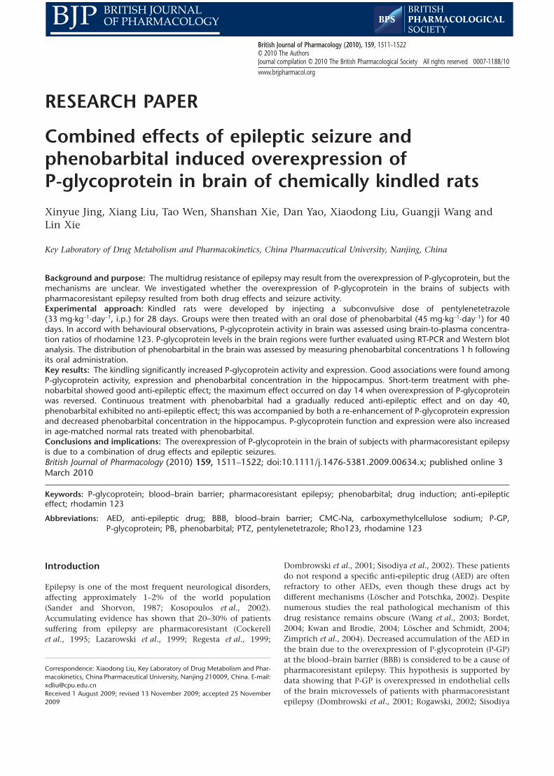
Another batch of experimental rats was used to determineP-GP function and expression in the brain. The experimentalprocedure used was the same as that described above. At thesame time, age-matched normal rats were also enrolled andrandomly divided into two groups, assigned to be groups 3and 4. Group 3 (PB-CT) was given PB (45 mg·kg-1, p.o.), oncea day for a given number of days and group 4 (CT) receivedonly the vehicle and served as an age-matched normalcontrol. At a designated time based on behavioural observa-tions during the PB treatment, some of the experimental ratswere selected for assaying PB concentration, P-GP functionand expression in their brains. A schematic illustration of theexperimental design and the different groups is shown inFigure 1.
Distribution of Rho123 and PB in brainTo evaluate the P-GP function or PB distribution in brain, ratsimmediately received Rho123 (0.2 mg·kg-1, i.v.) following oraladministration of PB or vehicle. At 1 h after the injection ofRho123, rats were killed under light ether anaesthesia, bloodwas collected and plasma samples were obtained. The hippoc-ampus and cerebral cortex were immediately removed andweighed. The plasma and brain samples were stored at -80°Cfor measuring PB and Rho123 concentration. The other braintissues were used for RT-PCR and Western blot analysisrespectively.
RT-PCR analysisTotal RNA was isolated from the hippocampus and cerebralcortex utilizing Trizol reagent according to the manufacturer’sinstructions. The purity of the RNA isolated was determinedusing UV absorption at 260 nm and 280 nm. The cDNA wassynthesized from 2 mg of total RNA using oligo(dT)15 andM-MLV reverse transcriptase. PCR was performed on GeneAmp PCR System 9600. Primers used for the amplificationof the cDNAs of interest were: for mdr1a gene (351 bp),forward 5′-GACGGAATTGATAATGTGGACA-3′ and reverse5′-AAGGATCAGGAACAATAAA-3′; for mdr1b gene(351 bp),forward 5′-GCCCATCCTGTTTGACTG-3′ and reverse5′-CGCTTCCTGGACGACCTT-3′; for glyceraldehydes
Epilepsy and P-glycoprotein overexpression in brain1512 X Jing et al
British Journal of Pharmacology (2010) 159 1511–1522
phosphate dehydrogenase (GAPDH) (365 bp): forward 5′-GGTGCTGAGTATGTCGTGGAG-3′ and reverse 5′-ATGCAGGGATGATGTTCTGG-3′ (Sheng-Xing Sci-Tech Co. NanjingChina). After denaturation of the samples at 95°C for 5 min,the amplification was obtained by 30 cycles of 94°C for 30 s,60°C for 30 s and 72°C for 1 min each. A final extension stepat 72°C for 5 min was employed. PCR products were subjectedto electrophoresis on 2% agarose gel and visualized byethidium bromide staining. Densitometric quantification wasrecorded using gel image analysis system 3.3 (Jiangsu JedaScience-Technology Co. Ltd, Nanjing, China). To normalizethe data, the ratio between the densitometric quantificationmdr1a/mdr1b gene and GAPDH gene was calculated by usingthe software Quantity One (Bio-Rad Laboratories, Richmond,CA, USA).
Western blotWestern blot analysis was used for assessing P-glycoproteinexpression in rat brain according to a method described pre-
viously (Liu et al., 2008). Briefly, the brain tissues were homo-genated and lysed in lysis buffer containing 10 mM Tris-HCl(pH 7.5), 1 mM EGTA, 1 mM MgCl2, 1 mM mercaptoethanol,1% glycerol, protease inhibitor cocktail (1 mM dithiothreitol,2 mM phenylmethylsulphonylfluoride. The lysate was incu-bated on ice for 30 min and centrifuged at 13 000¥ g for10 min at 4°C. The supernatant was obtained as membranefractions for Western blot. The protein concentration in thesolution was measured by the Bio-Rad Protein Assay. Analiquot of tissue sample was diluted with an volume of 4 ¥sodium dodecyl sulphate (SDS) sample buffer containing0.1 M Tris-HCl (pH 6.8), 4% SDS, 200 mM DTT, 20% glycerol,and 0.2% bromophenol blue. Proteins (25 mg per lane) wereseparated by electrophoresis on 8% SDS-polyacrylamide gel.After electrophoresis, the proteins were electrophoreticallytransferred to a nitrocellulose membrane. The membrane wasblocked in PBS containing 0.1%Tween-20, PBST and 5% driedskim milk for 60 min at room temperature and washed threetimes for 15 min in PBST. Then the membrane was incubatedwith the primary monoclonal antibody C219, diluted
Figure 1 Schematic illustration of the experimental protocol and group distribution of the study. PB, phenobarbital.
Epilepsy and P-glycoprotein overexpression in brainX Jing et al 1513
British Journal of Pharmacology (2010) 159 1511–1522

200-fold in PBST overnight at 4°C. After the membrane hadbeen washed with PBST, it was incubated in the appropriateHRP-conjugated goat anti-mouse secondary antibody at roomtemperature for another 1 h and washed again three times inPBST. The transferred proteins were incubated with ECL sub-strate solution for 5 min according to the manufacturer’sinstructions and visualized with autoradiography X-film. Therelative expressions were quantified densitometrically byusing the quantity one software (Bio-Rad Laboratories, Rich-mond, CA, USA) and calculated according to the referencebands of b-actin (Boshide Biotech Co., Wuhan, China).
Drug assayThe concentrations of Rho123 in plasma and brain were mea-sured by HPLC (Liu et al., 2007). The recoveries were higherthan 85%; the linear ranges of Rho123 in plasma and brainwere 3.12–200 ng·mL-1 and 0.32–10.4 ng·g-1 brain respec-tively. The relative standard deviations of the intra-day andinter-day concentrations in plasma and brain were both lessthan 10%. The concentrations of PB in plasma and braintissues were also measured according to our previous method(Liu et al., 2007). The lowest limits of quantification of PB inplasma and brain were 1.56 mg·mL-1 and 0.2 mg·g-1 brain tissuerespectively. The recoveries were higher than 80%; the linearranges of PB in plasma and brain were 1.56–100 mg·mL-1 and0.2–62.5 mg·g-1 respectively. The relative standard deviationsof the intra-day and inter-day levels were less than 10%.
Data analysisResults are expressed as mean � SD. The overall differencesbetween groups were determined by one-way ANOVA. If analy-sis was significant, the differences between groups were esti-mated using Student–Newman–Keuls multiple comparisonpost hoc test. A P-value of less than 0.05 indicated a significantdifference.
MaterialsPTZ, Rho123 and the lysis buffer were purchased from SigmaChemical Co. (St. Louis, MO, USA). PB was purchased from
New Asiatic Pharmaceutical Co., Ltd. (Shanghai, China). TheBio-Rad Protein Assay was obtained from Bio-Rad Laboratories(Richmond, CA, USA); ECL substrate solution from Cell Sig-naling (USA); Trizol reagent from Invitrogen Co. (USA). Allother reagents were commercially available and were of ana-lytical grade. PTZ was dissolved in physiological saline. PB wassuspended in solution of 0.25% carboxymethylcellulosesodium (CMC-Na). Rho123 was dissolved in 0.01 Mphosphate-buffered saline (PBS, pH 7.4) before use.
Results
Behavioural observationsThe development of PTZ-induced kindling was observed(Figure 2A). The animals treated with repeated subconvulsivedose of PTZ (33 mg·kg-1·day-1, i.p.) for 28 injections wentgradually through the stages. Almost all rats became kindledrats that showed at least five consecutive stage 2 seizures orthree consecutive stage 4 or 5 seizures. The kindled rats werehighly sensitive to sound. Even a mild applause could inducea seizure in them. Only fully kindled rats were chosen for thefollowing experiments.
The anti-epileptic effects of PB on PTZ-kindled rats wereobserved during PB treatment (Figure 2B). Severity, seizuretimes and latency of seizure were used as indicies of seizures.Significant inter-individual variations were observed for theseverity (seizure stage) and seizure times in the same group.The index of seizure was defined as seizure stage multipliedby seizure time. It was found that at an early stage, PB treat-ment gradually decreased the index of seizure and prolongedthe latency of seizure. On day 14 of PB treatment, a maximalanti-epileptic effect of PB was observed (P < 0.01 vs. PTZ-CTgroup). The intensity of the seizure was decreased to 0 or 1.However, continuous PB treatment had a gradually weak-ened anti-epileptic effect. On day 40 of PB treatment, therats again showed at least five consecutive stage 2 seizures orthree consecutive stage 4 or 5 seizures, and the index ofseizure was similar to that of the PTZ-CT rats (P > 0.05 vs.PTZ-CT group).
Figure 2 (A) Development of PTZ (33 mg·kg-1·day-1, i.p.)-induced seizure to the fully kindled state. Data are mean � SD (n = 10).(B) Anti-epileptic activity of PB during PB treatment. PTZ was given to kindled rats 40 min after oral administration of PB (45 mg·kg-1·day-1).Seizure stage and times were observed. Seizure indexes of PTZ-kindled model group (PTZ-CT rats) and PB treatment group (PTZ-PB rats) arepresented. Index of seizure was defined as seizure stages ¥ seizure times. Data are mean � SD (n = 5). *P < 0.05, **P < 0.01 versus PTZ-CT ratsusing Student’s t-test. PB, phenobarbital; PTZ, pentylenetetrazole.
Epilepsy and P-glycoprotein overexpression in brain1514 X Jing et al
British Journal of Pharmacology (2010) 159 1511–1522

Accordingly, experimental rats before and on day 14 and 40of PB treatment were selected for evaluating P-GP functionand expression in brain.
Function of P-GP in brainConcentrations of Rho123, a typical substrate of P-GP, inplasma and brain tissues of experimental rats were measured1 h after i.v. administration of Rho123 and the ratios of brain-to-plasma were calculated for evaluating P-GP function inbrain. It was found that kindling did not affect Rho123 con-centrations in plasma (Figure 3A), but significantly decreasedthe concentrations of Rho123 in both the hippocampus andcerebral cortex, resulting in a lower ratio of brain-to-plasma (P< 0.01, Figure 3B,C). PB treatment slightly altered Rho123concentration in plasma of treated rats. But 14 day PB treat-ment significantly reversed the decreased Rho123 concentra-tion in the hippocampus induced by kindling (2.45 �
0.69 ng·g-1 tissue in PTZ-PB rats vs. 1.60 � 0.29 ng·g-1 tissue inPTZ-CT rats, P < 0.01). However, continuous PB treatment
further decreased the concentration of Rho 123 in the hip-pocampus to that of PTZ-CT rats. In contrast, PB treatmentdid not modify the Rho123 concentration in the cerebralcortex of PTZ-kindled rats. In age-matched normal rats (PB-CTrats), both 14 day and 40 day PB treatments significantly (P <0.01) decreased the concentration of Rho123 in the hippoc-ampus and cerebral cortex as compared with age-matchedcontrol rats (CT), resulting in lower brain-to-plasma concen-tration ratios (Figure 3B,C).
Distribution of PB in the brainThe PB levels in plasma and brain were simultaneously mea-sured, 1 h after oral administration of PB on day 14 and day40 of PB treatment respectively (Table 1). It was found that PBconcentration in hippocampus was lower than that in cortex,which is in line with the Rho123 results. Compared with 14day PB treatment, although 40 day PB treatment showed atrend to increase plasma concentration of PB (P > 0.05), 40day PB treatment significantly decreased PB concentrations in
Figure 3 Effects of kindling and PB treatment on Rho123 distribution in plasma (A), cerebral cortex (B) and hippocampus (C). The treatedkindled rats (PTZ-PB rats) and age-matched treated rats (PB-CT rats) were given PB (45 mg·kg-1·day-1, p.o.) for 14 or 40 consecutive days.Age-matched control rats (CT rats) and untreated kindled rats (PTZ-CT rats) only received 0.25% CMC-Na. The concentrations of Rho123 inplasma, cerebral cortex and hippocampus were measured 1 h after injection of Rho123 (0.2 mg·kg-1, i.v.). Each value is presented as mean �SD (n = 8). *P < 0.05, **P < 0.01 versus CT rats and ##P < 0.01 versus PTZ-CT rats using one-way ANOVA following by Student–Newman–Keulsmultiple comparison post hoc test. PB, phenobarbital; PTZ, pentylenetetrazole; Rho123, rhodamine 123.
Epilepsy and P-glycoprotein overexpression in brainX Jing et al 1515
British Journal of Pharmacology (2010) 159 1511–1522
hippocampus (P < 0.01) and slightly decreased PB concentra-tion in cerebral cortex (P > 0.05). In age-matched normal rats,40 day PB treatment also decreased PB concentration in boththe hippocampus and cortex as compared with 14 day PBtreatment (P < 0.05).
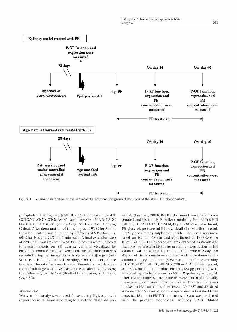
P-GP expression in brainWestern blot was used to investigate further the levels of P-GPin the brain regions of interest. The results revealed a band of
170 kDa, corresponding to P-GP. It was found that kindlinginduced by 28 injections of PTZ significantly (P < 0.01)increased the levels of P-GP in the hippocampus and cerebralcortex, to 147.2 � 23.9% and 144.1 � 6.07% of CT ratsrespectively (Figure 4). This effect of PB on P-GP levels in thebrain was dependent on the region and time. In the hippoc-ampus of PTZ-kindled rats, 14 day PB treatment reversed theincrease in P-GP levels induced by kindling, resulting in adecrease of 28.3% compared with PTZ-CT rats. However, 40day PB treatment further re-enhanced the P-GP level, which
Table 1 Effects of kindling and PB treatment on distribution of PB in plasma, cerebral cortex and hippocampus
PTZ-PB PB-CT
14 day treatment 40 day treatment 14 day treatment 40 day treatment
Plasma, mg·mL-1 72.19 � 6.98 77.04 � 5.16 64.84 � 11.04 66.50 � 16.52Cortex, mg·g-1 tissue 23.44 � 3.58 20.53 � 3.19 23.45 � 4.89 16.74 � 5.00*Cortex/plasma, mL·g-1 0.32 � 0.06 0.27 � 0.04* 0.36 � 0.04 0.25 � 0.04**Hippocampus, mg·g-1 tissue 52.26 � 9.57 35.93 � 5.56** 32.06 � 5.59 26.21 � 5.12*Hippocampus/plasma, mL·g-1 0.72 � 0.11 0.47 � 0.08** 0.50 � 0.06 0.39 � 0.10*
The treated kindled rats (PTZ-PB rats) and age-matched treated rats (PB-CT rats) were orally given PB (45 mg·kg-1·day-1, i.g.) for consecutive 14 or 40 days. At 1 hafter PB administration (i.g.), PB concentrations in plasma, cerebral cortex, and hippocampus were measured. Each value is presented as mean � SD (n = 8).*P < 0.05, **P < 0.01 versus the 14th day using Student’s t-test.PB, phenobarbital; PTZ, pentylenetetrazole.
Figure 4 Effects of kindling and PB treatment on protein levels of P-GP in hippocampus and cerebral cortex. The treated kindled rats (PTZ-PBrats) and age-matched treated rats (PB-CT rats) were given PB (45 mg·kg-1·day-1, p.o.) for 14 or 40 consecutive days. Age-matched controlrats (CT rats) and untreated kindled rats (PTZ-CT rats) only received 0.25% CMC-Na. Representative Western blot of P-GP in hippocampus (A)and cortex (C) and ratio of relative staining intensity for P-GP in hippocampus (B) and cortex (D) are shown. Each band corresponding to170 kDa was observed. Data are presented as mean � SD (n = 4). *P < 0.05, **P < 0.01 versus CT rats and ##P < 0.05 versus PTZ-CT rats usingANOVA statistics following by Student–Newman–Keuls multiple comparison post hoc test. P-GP, P-glycoprotein; PB, phenobarbital; PTZ,pentylenetetrazole.
Epilepsy and P-glycoprotein overexpression in brain1516 X Jing et al
British Journal of Pharmacology (2010) 159 1511–1522
was found to be similar to that of PTZ-CT rats (Figure 4B). Inthe cerebral cortex, neither the 14 day or 40 day PB treatmentmodified the increase in P-GP (Figure 4D). In age-matchednormal rats, both the 14 and 40 day PB treatments signifi-cantly increased the levels of P-GP in the hippocampus andcerebral cortex (Figure 4). The alterations of P-GP levels werein line with the Rho 123 concentrations in the brain.
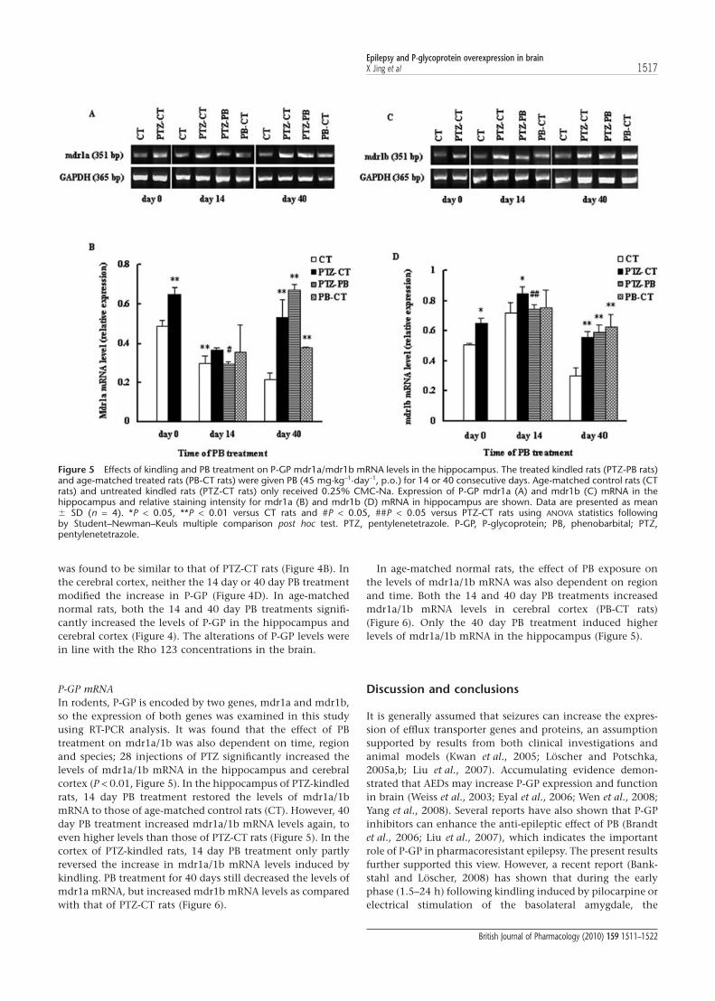
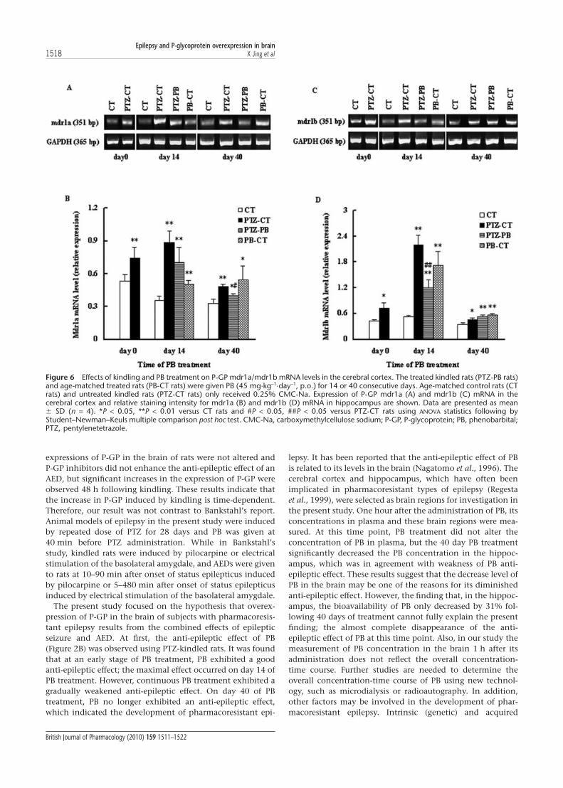
P-GP mRNAIn rodents, P-GP is encoded by two genes, mdr1a and mdr1b,so the expression of both genes was examined in this studyusing RT-PCR analysis. It was found that the effect of PBtreatment on mdr1a/1b was also dependent on time, regionand species; 28 injections of PTZ significantly increased thelevels of mdr1a/1b mRNA in the hippocampus and cerebralcortex (P < 0.01, Figure 5). In the hippocampus of PTZ-kindledrats, 14 day PB treatment restored the levels of mdr1a/1bmRNA to those of age-matched control rats (CT). However, 40day PB treatment increased mdr1a/1b mRNA levels again, toeven higher levels than those of PTZ-CT rats (Figure 5). In thecortex of PTZ-kindled rats, 14 day PB treatment only partlyreversed the increase in mdr1a/1b mRNA levels induced bykindling. PB treatment for 40 days still decreased the levels ofmdr1a mRNA, but increased mdr1b mRNA levels as comparedwith that of PTZ-CT rats (Figure 6).
In age-matched normal rats, the effect of PB exposure onthe levels of mdr1a/1b mRNA was also dependent on regionand time. Both the 14 and 40 day PB treatments increasedmdr1a/1b mRNA levels in cerebral cortex (PB-CT rats)(Figure 6). Only the 40 day PB treatment induced higherlevels of mdr1a/1b mRNA in the hippocampus (Figure 5).
Discussion and conclusions
It is generally assumed that seizures can increase the expres-sion of efflux transporter genes and proteins, an assumptionsupported by results from both clinical investigations andanimal models (Kwan et al., 2005; Löscher and Potschka,2005a,b; Liu et al., 2007). Accumulating evidence demon-strated that AEDs may increase P-GP expression and functionin brain (Weiss et al., 2003; Eyal et al., 2006; Wen et al., 2008;Yang et al., 2008). Several reports have also shown that P-GPinhibitors can enhance the anti-epileptic effect of PB (Brandtet al., 2006; Liu et al., 2007), which indicates the importantrole of P-GP in pharmacoresistant epilepsy. The present resultsfurther supported this view. However, a recent report (Bank-stahl and Löscher, 2008) has shown that during the earlyphase (1.5–24 h) following kindling induced by pilocarpine orelectrical stimulation of the basolateral amygdale, the
Figure 5 Effects of kindling and PB treatment on P-GP mdr1a/mdr1b mRNA levels in the hippocampus. The treated kindled rats (PTZ-PB rats)and age-matched treated rats (PB-CT rats) were given PB (45 mg·kg-1·day-1, p.o.) for 14 or 40 consecutive days. Age-matched control rats (CTrats) and untreated kindled rats (PTZ-CT rats) only received 0.25% CMC-Na. Expression of P-GP mdr1a (A) and mdr1b (C) mRNA in thehippocampus and relative staining intensity for mdr1a (B) and mdr1b (D) mRNA in hippocampus are shown. Data are presented as mean� SD (n = 4). *P < 0.05, **P < 0.01 versus CT rats and #P < 0.05, ##P < 0.05 versus PTZ-CT rats using ANOVA statistics followingby Student–Newman–Keuls multiple comparison post hoc test. PTZ, pentylenetetrazole. P-GP, P-glycoprotein; PB, phenobarbital; PTZ,pentylenetetrazole.
Epilepsy and P-glycoprotein overexpression in brainX Jing et al 1517
British Journal of Pharmacology (2010) 159 1511–1522
expressions of P-GP in the brain of rats were not altered andP-GP inhibitors did not enhance the anti-epileptic effect of anAED, but significant increases in the expression of P-GP wereobserved 48 h following kindling. These results indicate thatthe increase in P-GP induced by kindling is time-dependent.Therefore, our result was not contrast to Bankstahl’s report.Animal models of epilepsy in the present study were inducedby repeated dose of PTZ for 28 days and PB was given at40 min before PTZ administration. While in Bankstahl’sstudy, kindled rats were induced by pilocarpine or electricalstimulation of the basolateral amygdale, and AEDs were givento rats at 10–90 min after onset of status epilepticus inducedby pilocarpine or 5–480 min after onset of status epilepticusinduced by electrical stimulation of the basolateral amygdale.
The present study focused on the hypothesis that overex-pression of P-GP in the brain of subjects with pharmacoresis-tant epilepsy results from the combined effects of epilepticseizure and AED. At first, the anti-epileptic effect of PB(Figure 2B) was observed using PTZ-kindled rats. It was foundthat at an early stage of PB treatment, PB exhibited a goodanti-epileptic effect; the maximal effect occurred on day 14 ofPB treatment. However, continuous PB treatment exhibited agradually weakened anti-epileptic effect. On day 40 of PBtreatment, PB no longer exhibited an anti-epileptic effect,which indicated the development of pharmacoresistant epi-
lepsy. It has been reported that the anti-epileptic effect of PBis related to its levels in the brain (Nagatomo et al., 1996). Thecerebral cortex and hippocampus, which have often beenimplicated in pharmacoresistant types of epilepsy (Regestaet al., 1999), were selected as brain regions for investigation inthe present study. One hour after the administration of PB, itsconcentrations in plasma and these brain regions were mea-sured. At this time point, PB treatment did not alter theconcentration of PB in plasma, but the 40 day PB treatmentsignificantly decreased the PB concentration in the hippoc-ampus, which was in agreement with weakness of PB anti-epileptic effect. These results suggest that the decrease level ofPB in the brain may be one of the reasons for its diminishedanti-epileptic effect. However, the finding that, in the hippoc-ampus, the bioavailability of PB only decreased by 31% fol-lowing 40 days of treatment cannot fully explain the presentfinding; the almost complete disappearance of the anti-epileptic effect of PB at this time point. Also, in our study themeasurement of PB concentration in the brain 1 h after itsadministration does not reflect the overall concentration-time course. Further studies are needed to determine theoverall concentration-time course of PB using new technol-ogy, such as microdialysis or radioautography. In addition,other factors may be involved in the development of phar-macoresistant epilepsy. Intrinsic (genetic) and acquired
Figure 6 Effects of kindling and PB treatment on P-GP mdr1a/mdr1b mRNA levels in the cerebral cortex. The treated kindled rats (PTZ-PB rats)and age-matched treated rats (PB-CT rats) were given PB (45 mg·kg-1·day-1, p.o.) for 14 or 40 consecutive days. Age-matched control rats (CTrats) and untreated kindled rats (PTZ-CT rats) only received 0.25% CMC-Na. Expression of P-GP mdr1a (A) and mdr1b (C) mRNA in thecerebral cortex and relative staining intensity for mdr1a (B) and mdr1b (D) mRNA in hippocampus are shown. Data are presented as mean� SD (n = 4). *P < 0.05, **P < 0.01 versus CT rats and #P < 0.05, ##P < 0.05 versus PTZ-CT rats using ANOVA statistics following byStudent–Newman–Keuls multiple comparison post hoc test. CMC-Na, carboxymethylcellulose sodium; P-GP, P-glycoprotein; PB, phenobarbital;PTZ, pentylenetetrazole.
Epilepsy and P-glycoprotein overexpression in brain1518 X Jing et al
British Journal of Pharmacology (2010) 159 1511–1522
(disease-related) alterations to the structure and/or function-ality of AED targets in epileptogenic brain regions may lead toreduced drug effects (Schmidt and Löscher, 2009). It has beenshown that NKCC1 expression in the hippocampus ofpatients with refractory epilepsy increased, impairing theinhibitory capacity of GABA and resulting in hyperexcitabil-ity (Sen et al., 2007). It has also been suggested that a dys-functional adenosine kinase system is involved inpharmacoresistant epilepsy (Boison, 2006), and this is sup-ported by findings in experimental animals (Rebola et al.,2003; 2005) and patients (Glass et al., 1996). A decrease in theexpression of GABAA receptors in the hippocampus of chronicpharmacoresistant patients has also been observed (Wolfet al., 1994) and animal experiments have shown that thisalteration of GABAA receptors is associated with resistance tothe anti-epileptic effects of PB (Volk et al., 2006). Takentogether, these results demonstrate that the development ofpharmacoresistant epilepsy is a complex process.
Several studies have shown that the transport of manyAEDs including PB across the BBB is mediated by P-GP(Löscher and Potschka, 2005c; Liu et al., 2007; Yang et al.,2008). Our experiments were designed to investigate whetherthe decrease in PB concentration in brain tissues results froman increase in the expression and function of P-GP. P-GPfunction and protein levels as well as mdr1a/1b mRNA levelsin the specified brain regions were measured at three timepoints, the day before PB treatment (kindling development),day 14 of PB treatment (maximal anti-epileptic effectobserved) and day 40 of PB treatment (development of phar-macoresistant epilepsy).
Rho123 has been extensively used as an index of P-GPmediated transport in in vitro and in vivo studies (Kageyamaet al., 2006; Turncliff et al., 2006; Barta et al., 2008; Nishimuraet al., 2008; Pires et al., 2009; Tanaka et al., 2009) and wasselected as a marker for evaluating P-GP function in ourexperiments. In addition to P-GP, other ABC efflux transport-ers such as members of the multidrug resistance protein (MRP)family and breast cancer resistance protein (BCRP) have beenshown to contribute to BBB function (Löscher and Potschka,2005c) and may be involved in the transport of Rho123.However, other studies have indicated that MRP1 makes aminimal contribution to Rho123 efflux (Dogan et al., 2004)and Rho123 is not transported by BCRP (Alqawi et al., 2004);there is no evidence that MRP2 and MRP4 are involved in thetransport of Rho123 across the BBB. In the present study,compared with age-matched control rats, a lower concentra-tion of Rho123 was found in the specified brain regions ofkindled rats following 28 injections of PTZ, and this wasassociated with higher levels of P-GP protein and mdr1a/1bmRNA. Short-term exposure (14 day treatment) of PB reversedthe increase in P-GP activity (lower brain-to-plasma ratio ofRho123), levels of P-GP protein and mdr1a/1b mRNA inducedby kindling in the hippocampus; this was accompanied by analleviation of the symptoms of epilepsy. The finding thatshort-term exposure of PB prevented the overexpression ofP-GP induced by kindling indicates that it is unlikely thatchronic PTZ has a toxic action. Hence, P-GP overexpressioninduced during kindling is probably due to a direct effect ofthe presence of convulsions but not to the action of PTZ. It isreasonable therefore to deduce that at an early stage of epi-
lepsy the transporter-modifying factor is a consequence of theconvulsive state itself, but long-term exposure (40 day PBtreatment) to PB may result in an increase in P-GP activity,levels of P-GP protein and mdr1a/1b mRNA levels again, andthis is accompanied by lower levels of PB and an attenuationof the anti-epileptic effect of PB. These results further supportclinical findings demonstrating that an overexpression ofP-GP occurs in the brain of patients with pharmacoresistantepilepsy (Lazarowski et al., 1999; Dombrowski et al., 2001;Sisodiya et al., 2002). PB treatment also increased P-GP expres-sion and function in age-matched normal rats, which showsthat the effects of PB have an important role in the develop-ment of pharmacoresistant epilepsy. These results are in agree-ment with those from our previous report (Wen et al., 2008).
Seegers et al. (2002) found that an 11 or 7 day exposure ofAEDs did not affect P-GP expression, but it is possible that thisexposure time is too short to compare to clinical practice. Wefound that, after 40 days of PB treatment, the ratio ofhippocampus/plasma in the non-kindled, treated group washigher than that in the kindled, treated group, which indi-cates that P-GP function in the non-kindled, treated groupwas weaker than that in the kindled, treated group. All theseresults demonstrate that overexpression of P-GP in the brainat an early stage, is caused by the epileptic seizure and at thelater stage results from a combination of the effects of theseizures and the drug; the latter enhances the development ofpharmacoresistant epilepsy.
The present results also showed that concentrations ofRho123, as well as mdr1 mRNA in the brain were region-dependent. In age-matched control rats, the concentrations ofRho123 in the hippocampus were significantly higher thanthose in the cerebral cortex, indicating that the expression ofP-GP in the cortex is higher than that in the hippocampus. Thehigher level of mdr1a mRNA in the cortex supports this specu-lation. Kindling may increase the levels of P-GP protein andmdr1a/1b mRNA in specific brain regions. The effect of PBexposure on function and expression was dependent on treat-ment time, region and species. In the hippocampus, a 14 dayPB treatment restored the levels of mdr1a/1b mRNA inducedby kindling to levels of age-matched control rats. In contrast,after a 40 day PB treatment the overexpression of mdr1a/1bmRNA induced by kindling was further enhanced. The higherlevels of mdr1a/1b and P-GP protein were associated with anincrease in P-GP function (lower brain-to-plasma concentra-tion ratio of Rho123). In the cortex, the 14 day PB treatmentonly modified the levels of mdr1a/1b mRNA and the 40 day PBtreatment still did not reverse the decrease in mdr1a, butincreased the levels of mdr1b mRNA, as compared with PTZ-CTrats. This alteration of mdr1a/1b levels was not associated withP-GP protein levels, or P-GP function. We also found that theincrease in mdr1a/1b concentrations in the cortex induced bykindling following 28 injections of PTZ was larger than that inthe hippocampus, but alterations in P-GP protein levels in thecortex were similar to those in the hippocampus.
It was well known that under physiological conditions,P-GP is predominantly expressed at capillary endothelial cells,as well as at parenchymal and perivascular astrocytes at lowlevels (Aquilante et al., 2000). In addition to these three celltypes, seizures also induce the expression of P-GP in neurones(Volk et al., 2005). Using mdr1a-knock-out mice, Schinkel
Epilepsy and P-glycoprotein overexpression in brainX Jing et al 1519
British Journal of Pharmacology (2010) 159 1511–1522
et al. (1994) proposed that alterations in mdr1b mRNA mightcompensate for changes in mdr1a mRNA, but this would notexplain the present findings. In addition to their commonaction as a drug efflux transporter, individual mdr1 isoformsappear to have a specific function. Products of the mdr1a havebeen proposed to regulate cell volume by influencingswelling-activated chloride currents via a protein kinase Csensitive phosphorylation site on P-GP (Bond et al., 1998),whereas mdr1b has been shown to be involved in apoptoticmechanisms (Lecureur et al., 2001) and cellular stress (Zhouand Kuo, 1998; Ziemann et al., 1999). However, these findingsare still controversial. Other studies have suggested thatexpression of the mdr1 contributes to neuroprotectionbecause a link was found between loss of proapoptotic proteinp53 and expression of mdr1a/mdr1b (Bush and Li, 2002;Marroni et al., 2003). The exact location of each mdr1 isoformin the normal brain has not been fully elucidated, althoughmdr1a appears to be preferentially expressed in microvesselendothelium in the brain (Demeule et al., 2001) and themdr1b is mainly expressed in the cerebral cells (astrocytes orneurones) (Ballerin et al., 2002). Both kindling and long-termPB treatment may simultaneously increase the expression ofthese two genes, which indicates that overexpression of P-GPin the brain results from the concurrent effects of mdr1a andmdr1b, with mdr1a contributing more than mdr1b.
The RT-PCR results indicate that mdr1a/mdr1b changes inthe hippocampus were highly correlated with the changes inP-GP function and expression induced by epilepsy and PB. The40 day PB treatment again decreased the hippocampus-to-plasma concentration ratios of Rho123 and this was accompa-nied by an increase in mdr1a/mdr1b mRNA levels and adecrease in PB concentrations in the hippocampus. In contrast,mdr1a/mdr1b changes in the cortex were less associated withthe changes in P-GP function and expression under the sameconditions, which indicates that lesions of the hippocampusand the concentration of therapeutic drug in the hippocampusis closely associated with epilepsy. The hippocampus,amygdala, and parahippocampal region are components of themesial temporal lobe. The hippocampus is thought to be a keystructure for the facilitation of temporal lobe epilepsy, the mostprevalent of all epilepsies (Lowenstein, 1996; Wasterlain et al.,1996; Ben-Ari and Cossart, 2000). The surgical removal of thehippocampus and its surrounding structures has been found toresult in a decrease or complete cessation of seizures in humanpatients, which validates the epileptogenicity of the hippoc-ampus and its role in epileptogenesis (Falconer and Serafetin-ides, 1963; Rasmussen, 1983; King et al., 1986; Clusmann et al.,2002; Hardy et al., 2003; Wieser et al., 2003).
In conclusion, our data strongly suggest that the overex-pression of P-GP during the development of pharmacoresis-tant epilepsy is much more complex than previously thoughtand that the role of the hippocampus in the development ofpharmacoresistant epilepsy is more important than that ofthe cerebral cortex. Early PB treatment reversed the increasein P-GP function and expression induced by kindling, butlong-term PB treatment enhanced its overexpression. All theresults indicate that the overexpression of P-GP in the brain isinduced by the epileptic seizures during the early stages andby the combined effects of chronic drug treatment and epi-lepsy at the later stages.
Acknowledgement
The work was supported by the Project of ‘333’, Jiangsu Prov-ince (No. Y092009).
Conflict of interest
None.
References
Alqawi O, Bates S, Georges E (2004). Arginine482 to threonine muta-tion in the breast cancer resistance protein ABCG2 inhibitsrhodamine 123 transport while increasing binding. Biochem J 382:711–716.
Aquilante CL, Letrent SP, Pollack GM, Brouwer KL (2000). Increasedbrain P-glycoprotein in morphine tolerant rats. Life Sci 66: 47–51.
Ballerin P, Di IP, Ciccarelli R, Nargi E, D’Alimonte I, Traversa U et al.(2002). Glial cells express multiple ATP binding cassette proteins,which are involved in ATP release. Neuroreport 13: 1789–1792.
Bankstahl JP, Löscher W (2008). Resistance to antiepileptic drugs andexpression of P-glycoprotein in two rat models of status epilepticus.Epilepsy Res 82 (1): 70–85.
Barta CA, Sachs-Barrable K, Feng F, Wasan KM (2008). Effects ofmonoglycerides on P-glycoprotein: modulation of the activity andexpression in Caco-2 cell monolayers. Mol Pharm 5: 863–875.
Ben-Ari Y, Cossart R (2000). Kainate, a double agent that generatesseizures: two decades of progress. Trends Neurosci 23: 580–587.
Boison D (2006). Adenosine kinase, epilepsy and stroke: mechanismsand therapies. Trends Pharmacol Sci 27 (12): 652–658.
Bond TD, Valverde MA, Higgins CF (1998). Protein kinase C phospho-rylation disengages human and mouse-1a P-glycoproteins frominfluencing the rate of activation of swelling activated chloridecurrents. J Physiol 508: 333–340.
Bordet R (2004). Drug-resistant partial epilepsy: pharmacological cri-teria. Rev Neurol (Paris) 160: 36–42.
Brandt C, Bethmann K, Gastens AM, Löscher W (2006). The multidrugtransporter hypothesis of drug resistance in epilepsy: proof-of-principle in a rat model of temporal lobe epilepsy. Neurobiol Dis24: 202–211.
Brodie MJ, Kwan P (2004). Phenobarbital: a drug for the 21st century?Epilepsy Behav 5: 802–803.
Bush JA, Li G (2002). Regulation of the p53-deficient mouse model.Carcinogenesis 23: 1603–1607.
Clusmann H, Schramm J, Kral T, Helmstaedter C, Ostertun B,Fimmers R et al. (2002). Prognostic factors and outcome afterdifferent types of resection for temporal lobe epilepsy. J Neurosurg97: 1131–1141.
Cockerell OC, Johnson AL, Sander JW, Hart YM, Shorvon SD (1995).Remission of epilepsy: results from the National General PracticeStudy of Epilepsy. Lancet 346: 140–144.
De Sarro A, Naccari F, De Sarro G (1999). Enhanced susceptibility ofpentylenetetrazole kindled mice to quinolone effects. Int J Antimi-crob Agents 12: 239–244.
Demeule M, Labelle M, Régina A, Berthelet F, Béliveau R (2001).Isolation of endothelial cells from brain, lung, and kidney: expres-sion of the multidrug resistance P-glycoprotein isoforms. BiochemBiophys Res Commun 281: 827–834.
Dogan AL, Legrand O, Faussat AM, Perrot JY, Marie JP (2004). Evalu-ation and comparison of MRP1 activity with three fluorescent dyesand three modulators in leukemic cell lines. Leuk Res 28: 619–622.
Dombrowski SM, Desai SY, Marroni M, Cucullo L, Goodrich K, Bin-gaman W et al. (2001). Overexpression of multiple drug resistance
Epilepsy and P-glycoprotein overexpression in brain1520 X Jing et al
British Journal of Pharmacology (2010) 159 1511–1522
genes in endothelial cells from patients with refractory epilepsy.Epilepsia 42: 1501–1506.
Eyal S, Lamb JG, Smith-Yockman M, Yagen B, Fibach E, Altschuler Yet al. (2006). The antiepileptic and anticancer agent, valproic acid,induces P-glycoprotein in human tumour cell lines and in rat liver.Br J Pharmacol 149: 250–260.
Falconer MA, Serafetinides EA (1963). A follow-up study of surgery intemporal lobe epilepsy. J Neurosurg Psychiatry 26: 154–165.
Glass M, Faull RLM, Bullock JY, Jansen K, Mee EW, Walker EB et al.(1996). Loss of A1 adenosine receptors in human temporal lobeepilepsy. Brain Res 710: 56–68.
Hardy SG, Miller JW, Holmes MD, Born DE, Ojemann GA, Dodrill CBet al. (2003). Factors predicting outcome of surgery for intractableepilepsy with pathologically verified mesial temporal sclerosis.Epilepsia 44: 565–568.
Kageyama M, Fukushima K, Togawa T, Fujimoto K, Taki M,Nishimura A et al. (2006). Relationship between excretion clearanceof rhodamine 123 and P-glycoprotein (Pgp) expression induced byrepresentative Pgp inducers. Biol Pharm Bull 29: 779–784.
King DW, Flanigin HF, Gallagher BB, So EL, Murvin AJ, Smith DB et al.(1986). Temporal lobectomy for partial complex seizures: evalua-tion, results and 1-year follow-up. Neurology 36: 334–339.
Kosopoulos IA, van Merode T, Kessels FG, de Krom MC, Knottnerus JA(2002). Systematic review and meta-analysis of incidence studies ofepilepsy and unprovoked seizures. Epilepsia 43: 1402–1409.
Kwan P, Brodie MJ (2004). Drug treatment of epilepsy: when does itfail and how to optimize its use? CNS Spectr 9: 110–119.
Kwan P, Brodie MJ (2005). Potential role of drug transporters in thepathogenesis of medically intractable epilepsy. Epilepsia 46: 224–235.
Lazarowski A, Sevlever G, Taratuto A, Massaro M, Rabinowicz A(1999). Tuberous sclerosis associated with MDR1 gene expressionand drug-resistant epilepsy. Pediatr Neurol 21: 731–734.
Lecureur V, Thottassery JV, Sun D, Schuetz EG, Lahti J, Zambetti GPet al. (2001). Mdr1b facilitates p53-mediated cell death and p53 isrequired for mdr1b upregulation in vivo. Oncogene 20: 303–313.
Liu HY, Liu XD, Jia L, Liu YC, Yang HW, Wang GJ et al. (2008). Insulintherapy restores impaired function and expression ofP-glycoprotein barrier of experimental diabetes. Biochem Pharmacol75: 1649–1658.
Liu XD, Yang ZH, Yang JS, Yang HW (2007). Increased P-glycoproteinexpression and decreased phenobarbital distribution in the brain ofpentylenetetrazole-kindled rats. Neuropharmacology 53: 657–663.
Löscher W, Potschka H (2002). Role of multidrug transporters inpharmacoresistance to antiepileptic drugs. J Pharmacol Exp Ther301: 7–14.
Löscher W, Potschka H (2005a). Drug resistance in brain diseases andthe role of drug efflux transporters. Nat Rev Neurosci 6: 591–602.
Löscher W, Potschka H (2005b). Role of drug efflux transporters in thebrain for drug disposition and treatment of brain diseases. ProgNeurobiol 76: 22–76.
Löscher W, Potschka H (2005c). Blood-brain barrier active efflux trans-porters: ATP-binding cassette gene family. NeuroRX 2: 86–98.
Löscher W, Schmidt D (2004). New horizons in the development ofantiepileptic drugs: the search for new targets. Epilepsy Res 60:77–159.
Lowenstein DH (1996). Recent advances related to basic mechanismsof epileptogenesis. Epilepsy Res Suppl 11: 45–60.
Marroni M, Agrawal ML, Kight K, Hallene KL, Hossain M, Cucullo L(2003). Relationship between expression of multiple drug resistanceproteins and p53 tumor suppressor gene proteins in human brainastrocytes. Neuroscience 121: 605–617.
Nagatomo I, Akasaki Y, Nagase F, Nomaguchi M, Takigawa M (1996).Relationships between convulsive seizures and serum and brainconcentrations of phenobarbital and zonisamide in mutant inbredstrain EL mouse. Brain Res 731: 190–198.
Nishimura A, Honda N, Sugioka N, Takada K, Shibata N (2008). Evalu-
ation of carbamazepine pharmacokinetic profiles in mice withkainic acid-induced acute seizures. Biol Pharm Bull 31: 2302–2308.
Pires MM, Emmert D, Hrycyna CA, Chmielewski J (2009). Inhibitionof P-glycoprotein-mediated paclitaxel resistance by reversiblylinked quinine homodimers. Mol Pharmacol 75: 92–100.
Potschka H, Fedrowitz M, Löscher W (2002). P-glycoprotein-mediatedefflux of phenobarbital, lamotrigine and felbamate at theblood-brain barrier: evidence from microdialysis experiments inrats. Neurosci Lett 327: 173–176.
Rasmussen TB (1983). Surgical treatment of complex partial seizures:results, lessons and problems. Epilepsia 24 (Suppl. 1): S65–S76.
Rebola N, Coelho JE, Costenla AR, Lopes LV, Parada A, Oliveira CRet al. (2003). Decrease of adenosine A1 receptor density and ofadenosine neuromodulation in the hippocampus of kindled rats.Eur J Neurosci 18 (4): 820–828.
Rebola N, Porciúncula LO, Lopes LV, Oliveira CR, Soares-da-Silva P,Cunha RA (2005). Long-term effect of convulsive behavior on thedensity of adenosine A1 and A 2A receptors in the rat cerebralcortex. Epilepsia 46 (Suppl. 5): 159–165.
Regesta G, Tanganelli P (1999). Clinical aspects and biological bases ofdrug-resistant epilepsies. Epilepsy Res 34: 109–122.
Rizzi M, Caccia S, Giuso G, Richichi C, Gorter JA, Aronica E et al.(2002). Limbic seizures induce P-glycoprotein in rodent brain: func-tional implications for pharmacoresistance. J Neurosci 22: 5833–5839.
Rogawski MA (2002). Does P-glycoprotein play a role in pharmacore-sistance to antiepileptic drugs? Epilepsy Behav 3: 493–495.
Sander JW, Shorvon SD (1987). Incidence and prevalence studies inepilepsy and their methodological problems: a review. J NeurolNeurosurg Psychiatry 50: 829–839.
Schinkel AH, Smit JJ, van Tellingen O, Beijnen JH, Wagenaar E, vanDeemter L et al. (1994). Disruption of the mouse mdr1aP-glycoprotein gene leads to a deficiency in the blood-brain barrierand to increased sensitivity to drugs. Cell 77: 491–502.
Schmidt D, Löscher W (2009). New developments in antiepilepticdrug resistance: an integrative view. Epilepsy Curr 9 (2): 47–52.
Seegers U, Potschka H, Löscher W (2002). Lack of effects of prolongedtreatment with phenobarbital or phenytoin on the expression ofP-glycoprotein in various rat brain regions. Eur J Pharmacol 451:149–155.
Sen A, Martinian L, Nikolic M, Walker MC, Thom M, Sisodiya SM(2007). Increased NKCC1 expression in refractory human epilepsy.Epilepsy Res 74: 220–227.
Sisodiya SM, Lin WR, Harding BN, Squier MV, Thom M (2002). Drugresistance in epilepsy: expression of drug resistance proteins incommon causes of refractory epilepsy. Brain 125: 22–31.
Suzuki K, Omura S, Ohashi Y, Kawai M, Iwata Y, Tani K et al. (2001).FK506 facilitates chemical kindling induced by pentylenetetrazolein rats. Epilepsy Res 46: 279–282.
Tanaka S, Masuda M, Nakajima K, Ido N, Ohtsuka T, Nishida M et al.(2009). P-glycoprotein function in peripheral T lymphocyte subsetsof myasthenia gravis patients: clinical implications and influence ofglucocorticoid administration. Int Immunopharmacol 9: 284–290.
Turncliff RZ, Tian X, Brouwer KL (2006). Effect of culture conditionson the expression and function of Bsep, Mrp2, and Mdr1a/b insandwich-cultured rat hepatocytes. Biochem Pharmacol 71: 1520–1529.
Volk HA, Arabadzisz D, Fritschy JM, Brandt C, Bethmann K, Löscher W(2006). Antiepileptic drug resistant rats differ from drug responsiverats in hippocampal neurodegeneration and GABAA-receptorligand-binding in a model of temporal lobe epilepsy. Neurobiol Dis21: 633–646.
Volk HA, Potschka H, Löscher W (2004). Increase expression of themultidrug transporter P-glycoprotein in limbic brain regions afteramygdala-kindled seizures in rats. Epilepsy Res 58: 67–79.
Volk HA, Potschka H, Löscher W (2005). Immunohistochemical local-ization of P-glycoprotein in rat brain and detection of its increased
Epilepsy and P-glycoprotein overexpression in brainX Jing et al 1521
British Journal of Pharmacology (2010) 159 1511–1522
expression by seizure are sensitive to fixation and staining variables.J Histochem Cytochem 34: 517–531.
Wang Y, Zhou D, Wang B, Li H, Chai H, Zhou Q et al. (2003). Akindling model of pharmacoresistant temporal lobe epilepsy inSprague-Dawley rats induced by Coriaria lactone and its possiblemechanism. Epilepsia 44: 475–488.
Wasterlain CG, Shirasaka Y, Mazarati AM, Spigelman I (1996). Chronicepilepsy with damage restricted to the hippocampus: possiblemechanisms. Epilepsy Res 26: 255–265.
Weiss J, Kerpen CJ, Lindenmaier H, Dormann SM, Haefeli WE (2003).Interaction of antiepileptic drugs with human P-glycoprotein invitro. J Pharmacol Exp Ther 307: 262–267.
Wen T, Liu YC, Yang HW, Liu HY, Liu XD, Wang GJ et al. (2008). Effectof 21-day exposure of phenobarbital, carbamazepine and pheny-toin on P-glycoprotein expression and activity in the rat brain.J Neurol Sci 270: 99–106.
Wieser HG, Ortega M, Friedman A, Yonekawa Y (2003). Long-termseizure outcomes following amygdalohippocampectomy. J Neuro-surg 98: 751–763.
Wolf HK, Spänle M, Müller MB, Elger CE, Schramm J, Wiestler OD(1994). Hippocampal loss of the GABAA receptor a1 subunit inpatients with chronic pharmacoresistant epilepsies. Acta Neuro-pathol 88 (4): 313–319.
Yang HW, Liu HY, Liu X, Zhang DM, Liu XD, Wang GJ et al. (2008).Increased P-glycoprotein function and level after long-term expo-sure of four antiepileptic drugs to rat brain microvascular endothe-lial cells in vitro. Neurosci Lett 434: 299–303.
Zhou G, Kuo MT (1998). Wild-type p53-mediated induction of ratmdr1b expression by the anticancer drug daunorubicin. J Biol Chem273: 15387–15394.
Ziemann C, Burkle A, Kahl GF, Hirsch-Ernst KI (1999). Reactiveoxygen species participate in mdr1b mRNA and P-glycoproteinoverexpression in primary rat hepatocyte cultures. Carcinogenesis20: 407–414.
Zimprich F, Sunder-Plassmann R, Stogmann E, Gleiss A, Dal-Bianco A,Zimprich A et al. (2004). Association of an ABCB1 gene haplotypewith pharmacoresistance in temporal lobe epilepsy. Neurology 63:955–958.
Epilepsy and P-glycoprotein overexpression in brain1522 X Jing et al
British Journal of Pharmacology (2010) 159 1511–1522


![[A_R_EXE] Comparison of Fractal Dimension Estimation Algorithms for Epileptic Seizure Onset Detection](https://img.dokumen.tips/doc/110x75/5695d25e1a28ab9b029a269d/arexe-comparison-of-fractal-dimension-estimation-algorithms-for-epileptic.jpg)


![Epileptic Seizure Forecasting With Generative Adversarial ...clustering,Gaussianmixturemodels,HiddenMarkovModels and autoencoders [13], [14]. Most of these unsupervised learning techniques](https://img.dokumen.tips/doc/110x75/60b177a4482be642596be326/epileptic-seizure-forecasting-with-generative-adversarial-clusteringgaussianmixturemodelshiddenmarkovmodels.jpg)