Embed Size (px)
Citation preview

Combinatorial Administration of Molecules That SimultaneouslyInhibit Angiogenesis and Invasion Leads to IncreasedTherapeutic Efficacy in Mouse Models ofMalignant Glioma
Lorenzo Bello,1 Valeria Lucini,2 Francesco Costa,1
Mauro Pluderi,1 Carlo Giussani,1
Francesco Acerbi,1 Giorgio Carrabba,1
Marilou Pannacci,2 Dario Caronzolo,2
Silvia Grosso,2 Svetlana Shinkaruk,4
Federica Colleoni,2 Xavier Canron,3
Giustino Tomei,1 Gerard Deleris,4 andAndreas Bikfalvi31Neurosurgery, Department of Neurological Sciences, University ofMilano, Ospedale Maggiore di Milano, Istituto di Ricovero e Cura aCarattere Scientifico, Milan, Italy; 2Department of Pharmacology,University of Milano, Milan, Italy; 3INSERM Unit EMI 0113,Molecular Angiogenesis Laboratory, University of Bordeaux I,Talence, France; and 4Groupe de Chimie Bio-Organique (InstitutNational de la Sante et de la Recherche Medicale U 577), UniversiteVictor Segalen Bordeaux 2, Bordeaux Cedex, France
ABSTRACTPurpose: We investigated the ability of the combinato-
rial administration of different inhibitors with activities onglioma angiogenesis, migration, and proliferation to producea prolonged inhibition of glioma growth.
Experimental Design: We combined inhibitors affectingsolely tumor angiogenesis (PF-4/CTF, cyclo-VEGI) or inhib-itors affecting both angiogenesis and invasion together(PEX, PF-4/DLR).
Results: When administered in combination, thesedrugs produced a prolonged and increased inhibition ofglioma growth independently from the type of inhibitorused. The combinatory administration was more effectivethan the administration of a single inhibitor alone, and a
strong therapeutic response was reached with a significantlylower amount of protein. The strongest inhibition was ob-served when human PEX and PF-4/DLR, which affect bothglioma angiogenesis and invasion by separate mechanisms,were combined.
Conclusions: This supports the concept that prolongedglioma growth inhibition can be achieved by simultaneousdelivery of molecules that target both tumor and endothelialcells and acting by separate mechanisms.
INTRODUCTIONThe growth of malignant gliomas such as that of others
solid tumors depends on the balance between positive andnegative regulators of cell proliferation, migration, or angiogen-esis (1–4). Negative regulators are directly produced by thetumor itself or depend on the activity of the tumor cells on thehost microenvironment (1–6). Some of them are acting asspecific regulators of angiogenesis. Others have more complexfunctions and may also affect invasion and/or cell proliferation(5–7).
We and others (8–13) have shown that the systemic ad-ministration of negative regulators successfully inhibited thegrowth of malignant gliomas and glioma recurrence in variousanimal models. The treatment was always very well toleratedand not associated with the occurrence of any side effects.Inhibitors have been also administered in combination withmetronomic chemotherapy optimizing and prolonging the extentof the therapeutic response (14).
It has been observed that after a period of treatment withantiangiogenic agents, the treated tumor eventually relapses andmay acquire resistance to the treatment (15, 16). In the presence ofa redundancy of stimulating factors, treatments that target only aspecific factor may lead to variants that privilege alternative growthpathways (15). In addition, it is possible that during long-termantiangiogenic treatment, a selection of subpopulations of lessvascular-dependent tumor cells, characterized by an increased ca-pacity to survive in a nutrient- and oxygen-deprived condition, mayoccur (15, 16). Moreover, there is evidence that hypoxia providesa selection mechanism for cells with a diminished susceptibility toapoptosis (17). Furthermore, it has been recently showed thathypoxia activates a cell motility program that helps the cells toescape the hostile hypoxic environment by invading the adjacenttissues where oxygen and nutrients are not limited, facilitating thedissemination of tumor cells in the surrounding tissue (17). It hasalso been reported that blocking angiogenesis with antibodies di-rected against the vascular endothelial growth factor receptor-2 inexperimental glioma mouse models may paradoxically increaseinvasion, and thus, simultaneously blocking angiogenesis and in-vasion may contribute beneficially to tumor therapy (12, 17, 18).
Received 2/1/04; revised 3/18/04; accepted 3/26/04.Grant support: Associazione Italiana Ricerca sul Cancro, ProgettoOncologia, Compagnia di San Paolo, Torino, Italy, Fondazione ItaloMonzino, Milan, Italy, Progetto Galileo, CRUI, Italy (L. Bello); the“Ligue contre le Cancer” (“Equipe Labelisee”), the “Conseil RegionalD’Aquitaine” (A. Bikfalvi); and the “Ligue contre le Cancer” and the“Conseil Regional d’Aquitaine” (G. Deleris).The costs of publication of this article were defrayed in part by thepayment of page charges. This article must therefore be hereby markedadvertisement in accordance with 18 U.S.C. Section 1734 solely toindicate this fact.Note: L. Bello and V. Lucini contributed equally to this work.Requests for reprints: Lorenzo Bello, Neurosurgery, Department ofNeurological Sciences, University of Milano, Ospedale Maggiore Poli-clinico, Istituto di Ricovero e Cura a Carattere Scientifico, ViaFrancesco Sforza 35, 20122 Milan, Italy. Phone: 39-02-5503-5502; Fax:39-02-5990-2239; E-mail: [email protected].
4527Vol. 10, 4527–4537, July 1, 2004 Clinical Cancer Research
Cancer Research. on December 29, 2019. © 2004 American Association forclincancerres.aacrjournals.org Downloaded from
Cancer cells possess a broad spectrum of migration andinvasion mechanisms (19). They can also modify their migrationmechanisms in response to different conditions. This form ofadaptation occurs naturally during the course of tumor progres-sion and may also occur during treatment (19). Thus, an effi-cient anti-invasive therapy needs to target more than one mech-anism at the same time.
These data have important therapeutic implications andsuggest that combinatory strategies, which inhibit simulta-neously different mechanisms of tumor invasion and angiogen-esis, may significantly increase therapeutic efficacy.
We investigated herein whether the combinatorial admin-istration of inhibitor molecules significantly increased inhibitionof glioma growth. We provide evidence that maximum inhibi-tory activity is observed when molecules are associated thatboth exhibit antiangiogenesis and anti-invasive properties.
MATERIALS AND METHODSProduction of Recombinant Human PEX, PF-4/CTF, PF-4/DLR, and Cyclo-VEGI
PEX RNA was amplified from U87 glioblastoma cellsas described previously (14). The fragment was cloned intothe pRSET vector (Invitrogen, Carlsbad, CA) and transformedin BL21 bacteria. Transformed BL21 bacteria were grown inLuria-Bertani media followed by induction with 1 mM isopro-pylthiolgalactoside. PEX was purified under denaturing condi-tions followed by extraction by nickel-charged chelating aga-rose. Recombinant protein was refolded and dialyzed againstwater, and the protein concentration was determined. Purity ofthe preparation was confirmed by running the purified proteinon a 12% SDS-PAGE gel.
The COOH-terminal peptide of human PF-4-, PF-4/CTF-,and the PF-4/CTF-modified peptide PF-4/DLR was synthesizedusing standard solid-phase methodology and purified by high-performance liquid chromatography using a C18 column and a0–80% linear acetonitrile gradient in 0.1% trifluoroacetic acid.Lyophilized peptides were dissolved in sterile H2O and stored at�20°C before use (11, 20, 21).
Cyclo-VEGI was synthesized by Fmoc/tBu batch solid-phase synthesis on an Applied Biosystems 430°C automatedpeptide synthesizer as described previously (22).
Purity of the preparation was confirmed by running themolecules on a 12% SDS-PAGE gel followed by Western blotanalysis (10, 11, 20–22). The biological activity of human PEX,PF-4/CTF, PF-4/DLR, or cyclo-VEGI was tested in vitro usingangiogenic and proliferation assays as reported previously (10,11, 20–22).
Cell Cultures and Other ReagentsThe human glioma cell line U87-MG (American Type
Culture Collection, Manassas, VA) and the murine gliomaGL261 cell line (kindly provided by Dr. David Zagzag, Depart-ment of Pathology, New York University) were used in theanimal experiments. U87-MG cells were cultured in �-MEM.GL261 were grown in DMEM. Both media were supplementedwith 2 mM L-glutamine, 10% FBS, and 1000 units/ml penicillin/streptomycin solution.
Two endothelial cell lines were used. Porcine aortic endo-
thelial cells stably transfected with KDR (PAE/KDR) werecultured in Ham’s F-12 media with 10% nonheat-inactivatedFCS and 10 �g/ml Geneticin (G418 sulfate; Refs. 17, 18).Bovine capillary endothelial (American Type Culture Collec-tion) cells were cultured in DMEM plus L-glutamine and 10%FBS. All media were supplemented with 1000 units/ml penicil-lin/streptomycin solution, and the cells were cultured in a 5%CO2 incubator at 37°C. Endothelial cell lines were used in theangiogenic and proliferation assays.
Proliferation and Apoptosis AssaysThese assays were used to test the biological activity of
human PEX, PF-4/CTF, PF-4/DLR, or cyclo-VEGI. Bovinecapillary endothelial, PAE/KDR, U87, or GL261 cells wereplated on a 96-well plate (20,000 cells for each well) andcultured in the presence of increasing concentrations of humanPEX, PF-4/CTF, PF-4/DLR, or cyclo-VEGI (0.1–25 �g/ml),alone or in combination, in the presence of 10% serum for 24 h.The relative number of cells was calculated using the 3-(4,5-dimethylthiazol-2-yl)-2,5-diphenyltetrazolium bromide conver-sion assay (Promega, Madison, WI). An irrelevant substance(BSA) was used as a negative control. Each experiment was runsix times in triplicate.
Apoptotic cells were detected with ApopTag plus kit (In-tergen, New York, NY) with 1% methyl green as a counterstain.Apoptosis index was quantified by determining the percentageof positively stained cells for all nuclei in 20 randomly chosenfields/section at �200 magnification (8, 10, 11, 20, 21).
Tube Formation AssayIn vitro tube formation assays were performed to test the
antiangiogenic activity of the inhibitors, alone or in combination(10, 11, 23, 24). PAE/KDR cells were used (10, 11, 24). PAE/KDR cells were seeded in a 96-well plate coated with 0.5-mmthick type I collagen gel (4 � 10,000 cells/cm2) and allowed toattach and spread for 3 h. The seeded cells were subjected tothree different conditions: condition 1, cells grown in Ham’sF-12, 10% FCS; condition 2, cells incubated with U87 condi-tioned media; and condition 3, cells incubated with U87 condi-tioned media plus PEX, PF-4/CTF, PF-4/DLR, or cyclo-VEGIat different concentrations (1, 5, 10, and 20 �g/ml), alone or incombination. Microvessel counts were performed on micro-plates and scored as described previously (10, 11, 25).
Migration Studies: Matrigel and Wound AssaysTwo different types of migration assays were used: a
wound assay and a Matrigel invasion assay.The wound assay was performed according to a method
described earlier (11, 22). Endothelial or tumor cells wereseeded in 35-mm culture plates and were allowed to grow toconfluence. Complete medium was replaced with serum-freemedia, and incubation was continued overnight. One linear scarwas drawn in the monolayer and divided into five equal fields.A set of digital photos were taken of each scar, and the denudedarea was marked using digital image analysis software. Thedishes were washed, and fresh medium containing 0.1% BSA,10 ng/ml fibroblast growth factor (FGF)-2 (bovine capillaryendothelial and tumor cell migration), or 10 ng/ml VEGF165
4528 Combinatorial Therapy in Malignant Gliomas
Cancer Research. on December 29, 2019. © 2004 American Association forclincancerres.aacrjournals.org Downloaded from
(PAE/KDR migration) and peptides, at increasing concentra-tions (1, 5, 10, and 20 �g/ml), alone or in combination, wasadded. After 6, 12, and 24 h, a second and additional sets ofphotos were taken. Photos were superimposed, and cells thatmigrated across the line drawn at the border of the scar in thefirst photo set were counted. Each condition was tested induplicates in three independent experiments. Means for all fieldsof each group were calculated, background migration sub-tracted, and plotted as percentage of the mean of untreatedstimulated control.
For the Matrigel invasion assay, chamber Permanox slides(Miles Scientific, Naperville, IL) were coated with Matrigel andincubated with media containing 10% FBS (8). A sterile sedi-mentation cylinder was placed into the lumen of the cylinderand allowed to attach. The cylinder was removed, and theendothelial or glioma cells were allowed to spread. The areaoccupied by the attached cells was imaged with a camera(Olympus) placed on an inverted microscope. The migrationwas calculated as the increase of the radius beyond the initialradius and expressed as the mean � SE. Human PEX, PF-4/CTF, PF-4/DLR, or cyclo-VEGI were added alone or in com-bination to the cultured medium, at increasing concentrations (1,5, 10, and 20 �g/ml), and the medium was changed daily. Anirrelevant substance (BSA) was added at the same concentrationand used as a negative control. Migration was quantified incomparison to unstimulated controls.
Statistical analysis was performed by ANOVA followed byStudent’s Newman-Keul pairwise comparison (Statview forMacintosh).
Animal StudiesShort-Term Combinations Experiments. In this set of
experiments, groups of 10 6-week-old male nude or BALB/cmice were implanted with 50,000 U87 or GL261 glioblastomacells. Twelve days after tumor cell injection, animals wereimplanted with Alzet 2004 osmotic minipumps, which the res-ervoir was filled with human PEX, PF-4/CTF, PF-4/DLR, orcyclo-VEGI alone or in combination, at different concentra-tions, according to the following groups: group 1, 0.5 mg ofhuman PEX � 0.5 mg of PF-4/DLR; group 2, 0.5 mg of humanPEX � 0.5 mg of PF-4/CTF; group 3, 0.5 mg of PF-4/CTF �0.5 mg of cyclo-VEGI; group 4, 0.5 mg of human PEX; group5, 0.5 mg of PF-4/DLR; group 6, 0.5 mg of PF-4/CTF; group 7,0.5 mg of cyclo-VEGI; group 8, controls; group 1, 0.25 mgof human PEX � 0.25 mg of PF-4/DLR; group 2, 0.25 mg ofhuman PEX � 0.25 mg of PF-4/CTF; group 3, 0.25 mgof PF-4/CTF � 0.25 mg of cyclo-VEGI; group 4, 0.25 mg ofhuman PEX; group 5, 0.25 mg of PF-4/DLR; group 6, 0.25 mgof PF-4/CTF; group 7, 0.25 mg of cyclo-VEGI; group 8, con-trols; and group 1, 0.5 mg of human PEX � 0.5 mg of PF-4/DLR; group 2, 0.5 mg of human PEX � 0.5 mg of PF-4/CTF;group 3, 0.5 mg of PF-4/CTF � 0.5 mg of cyclo-VEGI; group4, 0.25 mg of human PEX � 0.25 mg of PF-4/DLR; group 5,0.25 mg of human PEX � 0.25 mg of PF-4/CTF; group 6, 0.25mg of PF-4/CTF � 0.25 mg of cyclo-VEGI; group 7: controls.
In each experiment, treatment was continued for 28 days,corresponding to the working period of the pumps. Animalswere sacrificed at the occurrence of any side effect or neuro-logical distress and, in any case, 29 days after pump implanta-
tion. At sacrifice, brains were removed, fixed in 5% paraform-aldehyde in PBS for 24 h at 4°C, dehydrated in 30% sucrose inPBS for 24 h at 4°C, embedded in ornithine carbamyl transfer-ase, and stored at �70°C. The brains were then sectioned, anda portion of them submitted to routine histological examinationwith H&E staining. Tumor volume was calculated and ex-pressed as a mean � SE. Tumor volume was estimated using theformula for ellipsoid [(width2 � length)/2]. The remainingslides were used for the immunohistochemistry analysis asdescribed below. Statistical analysis of tumor volumes wasperformed with a two-way ANOVA. Pairwise comparison be-tween treatment groups at each dose was performed by theNewman-Keuls posttest.
Long-Term Combinations Experiments. In this set ofexperiments, groups of 10 6-week-old male nude or BALB/cmice were implanted with 50,000 U87 or GL261 glioblastomacells. Twelve days after tumor cell injection, animals wereimplanted with Alzet 2004 osmotic minipumps, which the res-ervoir was filled with human PEX, PF-4/CTF, PF-4/DLR, orcyclo-VEGI, alone or in combination, at the following concen-trations: 0.25, 0.5, and 1 mg/kg/day. The pump reservoirs werechanged four times to afford a treatment period of 140 days.Animals were sacrificed at the occurrence of any side effects orneurological distress and, in any case, 152 days after tumor cellimplantation.
At sacrifice, the brains were removed and processed asdescribed previously. Kaplan-Meier survival curves were de-signed, and survival for treated and untreated mice were com-pared with log-rank test.
All animal experiments were repeated at least two times.
Immunohistochemistry and ImmunofluorescenceImmunohistochemistry was performed on 5- and 100-�m
sections. Immunohistochemistry on 5-�m sections was carriedusing the Vectastain Elite kit (Vector Laboratories, Burlingame,CA). Primary antibodies include anti-CD31 (1:100 dilution; BDPharMingen), anti-Ki-67 (1:100 dilution; Dako, Carpinteria,CA). Detection was carried out using 3,3�-diaminobenzidinechromogen. Sections were counterstained with hematoxylin.Negative control slides were obtained by omitting the primaryantibody. Ki-67 staining was quantified by counting the numberof positively stained cells of 100 nuclei in 20 randomly chosenfields (8–10, 14). Microvessel count and density were scored asreported previously (8–10, 14, 25). Apoptotic cells were de-tected with ApopTag plus kit (Intergen) with 1% methyl greenas a counterstain. Apoptosis and proliferative indices were quan-tified by determining the percentage of positively stained cellsfor all nuclei in 20 randomly chosen fields/section at �200magnification (8–10, 14, 22). One-hundred-�m sections wereused for immunofluorescence staining for CD31 to study thevascular network and vessel morphological changes. A donkeyantirat IgG FITC (AP189F; Chemicon, Temecula, CA) was usedas a secondary antibody. Analysis was performed by co focalmicroscope (Zeiss), followed by three-dimensional reconstruc-tion (10). Serial H&E slides were investigated for the pattern ofglioma cell invasion. The interface between tumor mass andnormal brain parenchyma was carefully examined for the pres-
4529Clinical Cancer Research
Cancer Research. on December 29, 2019. © 2004 American Association forclincancerres.aacrjournals.org Downloaded from
ence of signs of local infiltration consisting of trails of tumorcells invading the normal brain parenchyma and tumor satellitesdistant from the main tumor mass (9). Signs of tumor infiltrationwere scored as described previously (12).
RESULTSBiological Activities of Each Inhibitor. We combined
in this study four different inhibitors, which include PF-4/CTF,cyclo-VEGI, human PEX, and PF-4/DLR. We have chosen twocompounds that only inhibit angiogenesis and two compoundsthat simultaneously inhibit angiogenesis and invasion. PF-4/CTF inhibits only endothelial cell proliferation, migration, andangiogenesis (Fig. 1; Refs. 11, 21 and data not shown). Gliomacell migration in the wound and Matrigel assays is not inhibitedby PF-4/CTF (Fig. 2 and data not shown). Cyclo-VEGI onlyinhibits angiogenesis as well (Fig. 1; Ref. 22 and data notshown). Human PEX inhibits simultaneously glioma cell pro-liferation, migration, and angiogenesis (Figs. 1 and 2; Ref. 8).Similarly, PF-4/DLR also targets endothelial and tumor cells(Figs. 1 and 2; Ref. 11 and data not shown). PF-4/DLR not onlyinhibits angiogenesis but also impairs potently glioma cell mi-gration in the wound assay, invasion in the Matrigel assay,apoptosis in vitro, and glioma cell proliferation to a smallerextent (Figs. 1 and 3 and data not shown). Although human PEXand PF-4/DLR are both inhibiting angiogenesis and tumor cellinvasion, they have different mechanisms of action (see “Dis-cussion”).
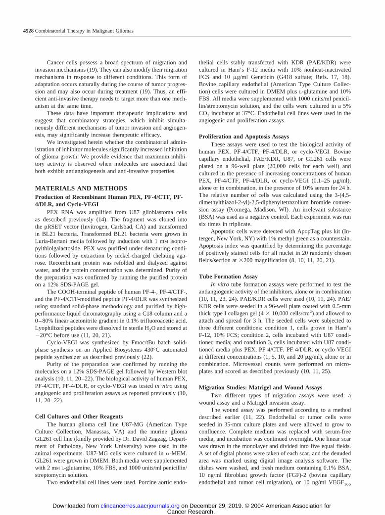
Effect of the Combinatorial Administration of HumanPEX, PF-4/DLR, PF-4/CTF, and Cyclo-VEGI on GliomaAngiogenesis in Vitro. The effect of the combinatorialadministration of human PEX, PF-4/DLR, PF-4/CTF, andcyclo-VEGI on tumor angiogenesis in vitro was investigatedby performing in vitro angiogenic assays. In these experi-ments, we evaluated the ability of endothelial cells to formtubes when grown in the presence of media from glioblas-toma cells and exposed to different concentrations of humanPEX, PF-4/DLR, PF-4/CTF, or cyclo-VEGI, administeredalone or in combination. The combinatorial administration ofendogenous inhibitors resulted in all of the cases in anincrease in inhibition of tube formation. This effect wasparticularly evident when the inhibitors were used at lowconcentration and when human PEX (1 �g/ml, 40 nM) andPF-4/DLR (1 �g/ml, 370 nM) were combined (Fig. 1).
Fig. 1 Effect of the combinatorial administration of inhibitors on an-giogenesis in vitro. The combinatorial administration of human PEX,PF-4/CTF, PF-4/DLR, and cyclo-VEGI produced an increase in theinhibition of angiogenesis in vitro. PAE/KDR endothelial cells seededon a collagen gel were grown in the presence of U87 glioblastoma cellcultured media and exposed to 1 �g/ml human PEX, PF-4/CTF, PF-4/DLR, or cyclo-VEGI, supplemented alone or in combination. Mediawere replaced every day for 4 days. When grown in the presence ofglioblastoma-cultured media, PAE/KDR cells formed tubes after 6 h.When the media were supplemented with the inhibitors, given alone orin combination, a decrease in the tube formation activity was observed.When given in combination, a significant increase in the inhibition oftube formation was observed in comparison when the inhibitors wereadministered alone (human PEX � PF-4/CTF versus human PEX orPF-4/CTF � P 0.01 or P 0.001; human PEX � PF-4/DLR versushuman PEX or PF-4/DLR � P 0.001 or P 0.05; PF-4/CTF �cyclo-VEGI versus PF-4/CTF or cyclo-VEGI � P 0.001 or P 0.05). The number of vessels in each condition was quantified aspreviously described (9, 24) and expressed as a percentage of thecontrols � SE. The data are representative for six experiments done intriplicates.
4530 Combinatorial Therapy in Malignant Gliomas
Cancer Research. on December 29, 2019. © 2004 American Association forclincancerres.aacrjournals.org Downloaded from

Effect of the Combinatorial Administration of HumanPEX, PF-4/DLR, PF-4/CTF, and Cyclo-VEGI on Gliomaand Endothelial Cell Invasion and Migration. The effect ofthe combinatorial administration of the inhibitors on gliomaand endothelial cells invasion and migration was investigatedby performing Matrigel invasion and monolayer wound as-says. In the Matrigel invasion assay, we evaluated the effectof different concentrations of human PEX, PF-4/DLR, PF-4/CTF, or cyclo-VEGI, used alone or in combination, on tumorand endothelial cells invasion inside a Matrigel gel. In thisexperiment, the combinatorial administration of differentconcentrations of human PEX, PF-4/DLR, PF-4/CTF, orcyclo-VEGI produced an increase in the inhibition of endo-thelial cell migration in comparison when the inhibitors wereused alone (Fig. 2, A and B, and data not shown). The effectwas more prominent when human PEX and PF-4/DLR werecombined and particularly evident when the inhibitors wereused at low concentration (40 nM for human PEX and 370 nM
for PF-4/DLR). The inhibitory effect on glioma cell migra-tion was dependent on the type of the inhibitor used. Thecombinatorial administration of human PEX and PF-4/CTFdid not significantly increase the antimigratory effect, whichwas similar to that observed when human PEX was admin-istered alone (Fig. 2C). On the contrary, when human PEXand PF-4/DLR were combined, a significant increase in theantimigratory activity was documented, particularly when the
inhibitors were used at low concentration (1 �g/ml; 40 nM forhuman PEX and 370 nM for PF-4/CTF; Fig. 2D). Similarresults were observed when a monolayer wound assay wasperformed (data not shown).
Effect of the Combinatorial Administration of HumanPEX, PF-4/DLR, PF-4/CTF, and Cyclo-VEGI on Endothe-lial and Glioma Cell Apoptosis and Proliferation in Vitro.To investigate the effect of the combinatorial administrationon endothelial and glioma cell apoptosis, endothelial andglioma cells were exposed to increasing concentrations of theinhibitors, alone or in combination, for 24 h. The presence ofapoptotic cells was then determined by terminal deoxynucle-otidyl transferase-mediated nick end labeling staining. Thecombinatorial administration of human PEX, PF-4/DLR, PF-4/CTF, or cyclo-VEGI was associated with a significantincrease in endothelial cell apoptosis. The effect was partic-ularly evident when the inhibitors were used at low concen-tration and human PEX and PF-4/DLR were combined (Fig.3A and data not shown). On the contrary, the combination ofPF-4/CTF and cyclo-VEGI did not exert any effect on gliomacell apoptosis (data not shown). Similarly, the combination ofhuman PEX and PF-4/CTF did not result in a significantincrease in the apoptotic rate of glioma cells, which wassimilar to that measured when human PEX was used alone(data not shown). On the contrary, the apoptotic rate of
Fig. 2 Effect of combinatorial administration of inhibitors on endothelial and glioma cell migration in the Matrigel invasion assay. PAE/KDR (Aand B) and U87 (C and D) cells were seeded in a Matrigel gel and grew in the presence of 1 �g/ml human PEX, PF-4/CTF, PF-4/DLR, or cyclo-VEGI,given alone or in combination. The migration of the cells from the initial radius was measured after 24, 48, and 72 h and compared with the controls.The combinatorial administration of the inhibitors increased the inhibition of endothelial cell migration (A and B; human PEX � PF-4/CTF, P � 0.05;human PEX � PF-4/CTF, P 0.001). The combinatorial administration of human PEX and PF-4/CTF did not increase the antimigratory effect onglioma cell, which was similar to that exerted by human PEX given alone (C). On the contrary, the combinatorial administration of human PEX andPF-4/DLR produced an increase in the inhibition of glioma cell migration (D; P 0.0001). The data are representative for six experiments done intriplicates and are expressed as a percentage of controls � SE.
4531Clinical Cancer Research
Cancer Research. on December 29, 2019. © 2004 American Association forclincancerres.aacrjournals.org Downloaded from

glioma cells was significantly increased when human PEXand PF-4/DLR were supplemented in combination (Fig. 3B).
We also performed proliferation assays with the differentinhibitors administered alone or in combination. In all of the
cases, a dose-dependent inhibition of endothelial cells prolifer-ation was observed (data not shown). Under similar experimen-tal conditions, an increase in the inhibition of glioma cell pro-liferation was only documented when human PEX and PF-4/DLR were combined. The combination of human PEX andPF-4/CTF did not significantly reduce glioma cell proliferation,and the inhibition was similar to that observed when humanPEX was used alone (data not shown).
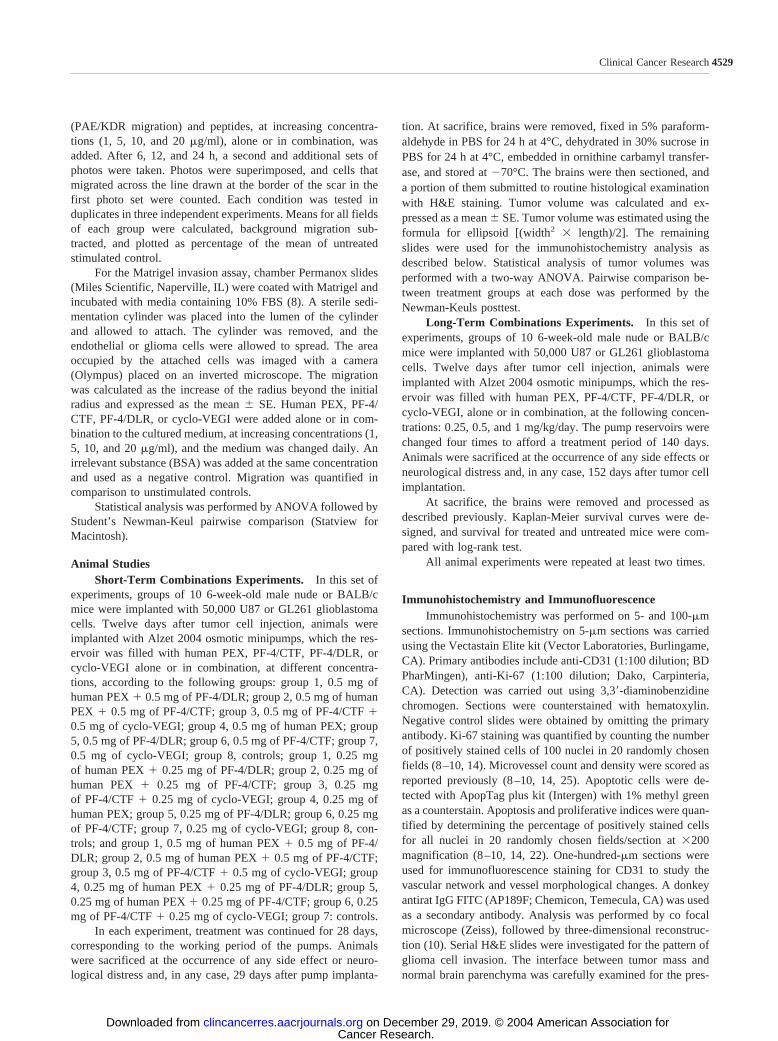
The Combinatorial Administration of Human PEX,PF-4/DLR, PF-4/CTF, and Cyclo-VEGI in Nude and Syn-geneic Mice Glioma Models Reduces Glioma Growth inVivo. The effect of the combinatorial administration of humanPEX, PF-4/DLR, PF-4/CTF, or cyclo-VEGI on glioma growthin vivo was initially investigated by performing short-term stud-ies. In these studies, the inhibitors were administered alone or incombination, continuously and systemically, by the use of os-motic minipumps implanted s.c. in the right flank of the animals.The pumps were implanted 12 days after tumor cell injection.Animals were sacrificed after 29 days from the implantation ofthe pumps, at the end of their working period. After the sacri-fice, the brains were removed, fixed, and sectioned, and thetumor volumes were calculated. The combinatorial administra-tion of human PEX, PF-4/DLR, PF-4/CTF, or cyclo-VEGIresulted in a potent inhibition of glioma growth. The highestinhibition was observed when human PEX and PF-4/DLR werecombined (98% tumor volume inhibition at 0.5 mg/kg/day) andthe lowest when PF-4/CTF and cyclo-VEGI were administeredtogether (93.5% tumor volume inhibition at 0.5 mg/kg/day; Fig.4A). The combination was more effective than the administra-tion of a single inhibitor alone. No significant difference inactivity was observed when the experiments were performed innude or immunocompetent BALB/c mice (Fig. 4B).
The Combinatorial Administration of Human PEX,PF-4/DLR, PF-4/CTF, and Cyclo-VEGI Sustains a Pro-longed Inhibition of Glioma Growth in Vivo. We next stud-ied the ability of the combinatorial administration of inhibitorsto sustain a prolonged inhibition of glioma growth in long-termin vivo experiments. In addition, we identified which combina-tion afforded the longest therapeutic response.
The inhibitors were administered at different concentra-tions (0.25, 0.5, and 1 mg/kg/day), alone or in combination, bys.c. osmotic minipumps, starting 12 days after tumor cells in-jection. The pump reservoir was replaced four times over a140-day period. Animals were sacrificed at the occurrence ofany signs of distress or neurological deficits.
The combinatorial administration of human PEX, PF-4/DLR, PF-4/CTF, or cyclo-VEGI resulted in a prolonged inhi-bition of glioma growth in all of the cases (Fig. 5A). The effectwas dose dependent and stronger than that observed when asingle inhibitor was administered alone, independently from thetype of inhibitor used (Fig. 5B–D). The longest survival wasobserved when human PEX and PF-4/DLR were used in com-bination (50% survival of 113, 87, and 68 days at 1, 0.5 and 0.25mg each, respectively), the shortest when PF-4/CTF and cyclo-VEGI were combined (50% survival of 78, 58, and 49 days at 1,0.5, and 0.25 mg each, respectively).
The treatment was always very well tolerated without theoccurrence of any side effects.
Fig. 3 Effect of the combinatorial administration of human PEX andPF-4/DLR on endothelial and glioma cell apoptosis. PAE/KDR (A) andU87 (B) cells were grown in the presence of 1 �g/ml of the inhibitors,given alone or in combination, for 24 h. The number of apoptotic cellswas measured by terminal deoxynucleotidyl transferase-mediated nickend labeling assay, counted as number of positive cells on 100 nuclei,and expressed as a percentage of controls � SE. The combinatorialadministration of human PEX and DLR increased both PAE/KDR andU87 cells apoptosis (P 0.001 and P 0.001). Data are representativefor six experiments done in triplicates.
4532 Combinatorial Therapy in Malignant Gliomas
Cancer Research. on December 29, 2019. © 2004 American Association forclincancerres.aacrjournals.org Downloaded from
Histological Analysis. Histological and immunohisto-chemical analysis of tumors from treated and untreated animalsshowed that the inhibition of glioma growth was associated witha decrease and change in tumor vasculature, an increase inapoptosis, and in some cases a decrease in cell proliferation anda change in the pattern of glioma cell invasion (Fig. 6). Thesignificance of effect was dependent on the type of inhibitorsused in the combination.
The most significant changes were observed when humanPEX and PF-4/DLR were used together. Tumors from animalstreated with human PEX and PF-4/DLR showed a markeddecrease in microvessel count (human PEX � PF-4/DLR versushuman PEX or PF-4/DLR � P 0.001) and change in vesselmorphology. Tumor vessels were composed mostly by capil-lary-like tubes, few large telangectatic vessels, and withoutglomeruloid structures. The same tumors showed the highestapoptosis index (P 0.001) and the lowest proliferation rate(P 0.001). Tumors from animals that received the combina-tion of PF-4/CTF and cyclo-VEGI showed a decrease in tumormicrovessel count (PF-4/CTF � cyclo-VEGI versus PF-4/CTFor cyclo-VEGI � P 0.001 or P 0.01) and a change in tumorvessel morphology. As in the tumors from animals treated withhuman PEX and DLR, tumors from animals treated with PF-4/CTF and cyclo-VEGI showed mostly capillary-like tubes, fewerlarge vessels, mainly in the tumor center and delimitated by aunilayer of endothelial cells, and no glomeruloid structures.Although the decrease in microvessel count was similar to thatobserved when other inhibitors were administered in combina-tion, the same tumors showed the lowest increase in apoptosisindex and no change in tumor cell proliferation. Tumors fromanimals treated with human PEX and PF-4/CTF were charac-terized by a similar decrease in microvessel count in comparisonto the other two groups of combinations and a similar change invessel morphology (human PEX � PF-4/CTF versus humanPEX or PF-4/CTF � P 0.05 or P 0.01). The changesobserved in apoptosis index and proliferation rate did not sig-nificantly differ from those observed in tumors from animalstreated with human PEX alone.
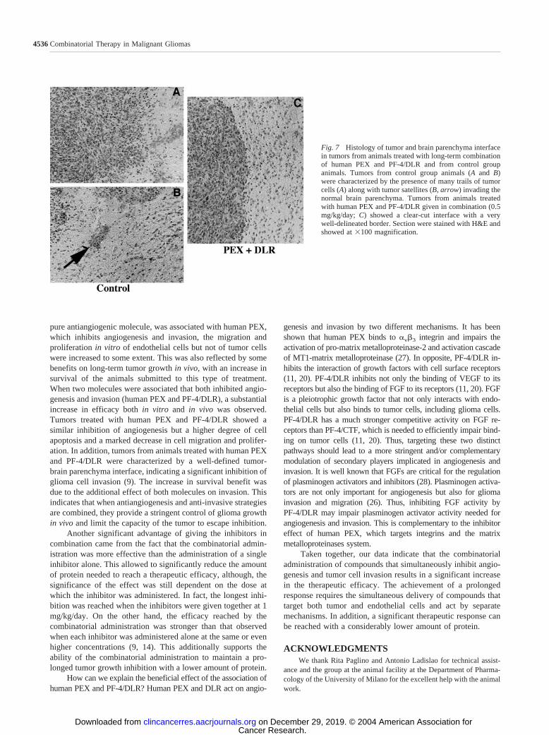
The most intriguing finding consisted in the modificationin the pattern of tumor cell invasion at the tumor-normal brainparenchyma interface (Figs. 6 and 7). Tumors from animalsbelonging to the control group and those submitted to PF-4/CTFand cyclo-VEGI showed a similar pattern of invasion, consisting
Fig. 4 Efficacy of the combinatorial administration of human PEX,PF-4/CTF, PF-4/DLR, and cyclo-VEGI on glioma growth in vivo. A,groups of male nude mice received injections of U87 cells intracranially.Twelve days later, mice were s.c. implanted with osmotic minipumpscontaining 0.5 mg/kg/day of the inhibitors, alone or in combination.Twenty-nine days later, corresponding to the working period of thepumps, mice were sacrificed, the brains removed, and the tumor volumemeasured. The combinatorial administration of human PEX, PF-4/CTF,PF-4/DLR, and cyclo-VEGI produced an increase in the tumor growthinhibition in comparison when the inhibitors were used alone (P 0.05). The highest inhibition was observed when human PEX and DLRwere given in combination (human PEX � PF-4/DLR versus humanPEX � PF-4/CTF or PF-4/CTF � cyclo-VEGI � P 0.001 or P 0.001). Data are expressed as percentages of untreated controls � SE(100%, not indicated in the figure) and representative for three animalexperiments (n � 30 mice in each group). B, the efficacy of thecombinatorial administration of the inhibitors produced a similar reduc-tion in tumor volumes in BALB/c mice models. BALB/c mice receivedinjections of GL261 cells. Twelve days later, mice received osmoticminipumps implanted s.c. in the right flank, which the reservoirs werefilled with 0.5 mg/kg/day human PEX, PF-4/CTF, PF-4/DLR, or cyclo-VEGI, administered alone or in combination. Twenty-nine later, micewere sacrificed, their brains removed, and the tumor volumes measured.Data are expressed as percentages of untreated controls � SE (100%,not indicated in the figure) and representative for three animal experi-ments (n � 30 mice in each group).
4533Clinical Cancer Research
Cancer Research. on December 29, 2019. © 2004 American Association forclincancerres.aacrjournals.org Downloaded from

of undefined interface, with trails of invading tumor cells anddistant tumor satellites. On the contrary, tumors from animalsthat were treated with human PEX and DLR showed a well-defined tumor-parenchyma interface, without trails of invadingcells or tumor satellites, and a clear-cut decrease in the periph-eral vessel recruitment. In addition, this pattern was particularlyprominent in tumors from animals submitted to the long-termtreatment, which were small, round, and with well-delineatedborders (Fig. 7).
DISCUSSIONIt has been shown that the administration of antiangiogen-
esis and anti-invasive molecules successfully inhibited thegrowth of human malignant gliomas, as well as that of other
solid tumors in various animal models (5–12, 14, 26). This formof therapy is usually free of side effects and may represent apromising therapeutic venue for the treatment of tumors (5–7).For maximum efficacy, molecules should be administered on along-term basis (5–7). The administration of these inhibitorsfaces several problems. First, the long-term treatment requires alarge amount of proteins (5, 10). Furthermore, selection mayfacilitate the outgrowth of cell populations, the growth of whichmay be less dependent on the vasculature. This may be due toselection mechanisms that lead to a diminished susceptibility toapoptosis or facilitate tumor cell dissemination in the surround-ing normal tissue through an increase in migration (15–18).Cancer cells can also modify their migration mechanisms duringthe course of tumor progression or in response to treatment (19).
Fig. 5 Long-term efficacy of the combinatorial administration of human PEX, PF-4/CTF, PF-4/DLR, and cyclo-VEGI on glioma growth in vivo. A,long-term efficacy of the combinatorial administration of human PEX, PF-4/CTF, PF-4/DLR, and cyclo-VEGI in the nude mice glioma model. Twelvedays after injection of U87 cells intracranially, nude mice were implanted s.c. with osmotic minipumps filled with 0.25 mg/kg/day of the inhibitors,alone or in combination. The pump reservoirs were changed three times to afford a period of treatment of 112 days. Animals were sacrificed at theonset of signs of distress or neurological deficits. Kaplan-Meier survival curves were designed. The longest survival was observed when human PEXand DLR were combined (50% survival of 68 days). B, the significance of the effect depends on the dose of the inhibitor administered. Mice thatreceived injections of U87 cells intracranially were implanted 12 days later with osmotic minipumps, which reservoirs were filled with 0.25, 0.5, and1 mg/kg/day of human PEX or PF-4/DLR, given in combination. The pumps were replaced four times to afford a period of treatment of 140 days.C and D, the combinatorial administration of human PEX, PF-4/CTF, or PF-4/DLR was more effective than the administration of a single inhibitoralone (P 0.001). The pump reservoirs were filled with 0.25 mg/kg/day (C) or 1 mg/kg/day (D) of the inhibitors, given alone or in combination.The pumps were replaced four times to afford a period of treatment of 140 days. All experiments have been repeated three times. Data arerepresentative of 30 mice for each group.
4534 Combinatorial Therapy in Malignant Gliomas
Cancer Research. on December 29, 2019. © 2004 American Association forclincancerres.aacrjournals.org Downloaded from

Thus, an efficient anti-invasive therapy needs to target morethan one mechanism at the same time.
The solution to overcome these problems is to associatemolecules that both inhibit angiogenesis and invasion by usingdifferent mechanisms. This will both limit potential escapemechanisms and may, in addition, decrease the amount ofprotein needed for long-term treatment.
We therefore investigated the efficacy of combinatorial ad-ministration by associating molecules with pure antiangiogenicactivity and molecules that have both antiangiogenic and anti-invasive properties. Different doses for these various associationswere used in this study. We hypothesized that the highest andlongest therapeutic effect would have been reached when inhibitorsdisplaying the largest activities were combined together, specifi-
cally when human PEX and PF-4/DLR would have been used incombination. Consequently, the lowest and more limited actionwould have been expected when two antiangiogenic drugs, namelyPF-4/CTF and cyclo-VEGI, were given in combination.
Association of pure antiangiogenesis molecules (PF-4/CTF, cyclo-VEGI) increased the inhibition of capillary tubeformation in vitro to some extent. This correlated with a slightincrease in survival when the combination was used in compar-ison to the single agent alone. Furthermore, the analysis oftumors from animals treated with PF-4/CTF and cyclo-VEGIshowed a significant decrease and changes in tumor vasculatureand an increase in apoptosis without modifications of cell pro-liferation. These findings are in agreement with an antiangio-genic effect of the molecule (13, 20, 21). When PF-4/CTF, a
Fig. 6 Microvessels counts,apoptotic index, proliferationindex, and tumor invasion in tu-mors from animals treated with0.5 mg/kg/day human PEX, PF-4/CTF, PF-4/DLR, and cyclo-VEGI, alone or in combination.Data are expressed as a percent-age of controls � SD.
4535Clinical Cancer Research
Cancer Research. on December 29, 2019. © 2004 American Association forclincancerres.aacrjournals.org Downloaded from
pure antiangiogenic molecule, was associated with human PEX,which inhibits angiogenesis and invasion, the migration andproliferation in vitro of endothelial cells but not of tumor cellswere increased to some extent. This was also reflected by somebenefits on long-term tumor growth in vivo, with an increase insurvival of the animals submitted to this type of treatment.When two molecules were associated that both inhibited angio-genesis and invasion (human PEX and PF-4/DLR), a substantialincrease in efficacy both in vitro and in vivo was observed.Tumors treated with human PEX and PF-4/DLR showed asimilar inhibition of angiogenesis but a higher degree of cellapoptosis and a marked decrease in cell migration and prolifer-ation. In addition, tumors from animals treated with human PEXand PF-4/DLR were characterized by a well-defined tumor-brain parenchyma interface, indicating a significant inhibition ofglioma cell invasion (9). The increase in survival benefit wasdue to the additional effect of both molecules on invasion. Thisindicates that when antiangiogenesis and anti-invasive strategiesare combined, they provide a stringent control of glioma growthin vivo and limit the capacity of the tumor to escape inhibition.
Another significant advantage of giving the inhibitors incombination came from the fact that the combinatorial admin-istration was more effective than the administration of a singleinhibitor alone. This allowed to significantly reduce the amountof protein needed to reach a therapeutic efficacy, although, thesignificance of the effect was still dependent on the dose atwhich the inhibitor was administered. In fact, the longest inhi-bition was reached when the inhibitors were given together at 1mg/kg/day. On the other hand, the efficacy reached by thecombinatorial administration was stronger than that observedwhen each inhibitor was administered alone at the same or evenhigher concentrations (9, 14). This additionally supports theability of the combinatorial administration to maintain a pro-longed tumor growth inhibition with a lower amount of protein.
How can we explain the beneficial effect of the association ofhuman PEX and PF-4/DLR? Human PEX and DLR act on angio-
genesis and invasion by two different mechanisms. It has beenshown that human PEX binds to �v3 integrin and impairs theactivation of pro-matrix metalloproteinase-2 and activation cascadeof MT1-matrix metalloproteinase (27). In opposite, PF-4/DLR in-hibits the interaction of growth factors with cell surface receptors(11, 20). PF-4/DLR inhibits not only the binding of VEGF to itsreceptors but also the binding of FGF to its receptors (11, 20). FGFis a pleiotrophic growth factor that not only interacts with endo-thelial cells but also binds to tumor cells, including glioma cells.PF-4/DLR has a much stronger competitive activity on FGF re-ceptors than PF-4/CTF, which is needed to efficiently impair bind-ing on tumor cells (11, 20). Thus, targeting these two distinctpathways should lead to a more stringent and/or complementarymodulation of secondary players implicated in angiogenesis andinvasion. It is well known that FGFs are critical for the regulationof plasminogen activators and inhibitors (28). Plasminogen activa-tors are not only important for angiogenesis but also for gliomainvasion and migration (26). Thus, inhibiting FGF activity byPF-4/DLR may impair plasminogen activator activity needed forangiogenesis and invasion. This is complementary to the inhibitoreffect of human PEX, which targets integrins and the matrixmetalloproteinases system.
Taken together, our data indicate that the combinatorialadministration of compounds that simultaneously inhibit angio-genesis and tumor cell invasion results in a significant increasein the therapeutic efficacy. The achievement of a prolongedresponse requires the simultaneous delivery of compounds thattarget both tumor and endothelial cells and act by separatemechanisms. In addition, a significant therapeutic response canbe reached with a considerably lower amount of protein.
ACKNOWLEDGMENTSWe thank Rita Paglino and Antonio Ladislao for technical assist-
ance and the group at the animal facility at the Department of Pharma-cology of the University of Milano for the excellent help with the animalwork.
Fig. 7 Histology of tumor and brain parenchyma interfacein tumors from animals treated with long-term combinationof human PEX and PF-4/DLR and from control groupanimals. Tumors from control group animals (A and B)were characterized by the presence of many trails of tumorcells (A) along with tumor satellites (B, arrow) invading thenormal brain parenchyma. Tumors from animals treatedwith human PEX and PF-4/DLR given in combination (0.5mg/kg/day; C) showed a clear-cut interface with a verywell-delineated border. Section were stained with H&E andshowed at �100 magnification.
4536 Combinatorial Therapy in Malignant Gliomas
Cancer Research. on December 29, 2019. © 2004 American Association forclincancerres.aacrjournals.org Downloaded from
REFERENCES1. Hahahan D, Folkman J. Patterns and emerging mechanisms of theangiogenic switch during tumorigenesis. Cell 1996;86:353–64.2. Liotta LA, Steeg PS, Stetler-Stevenson WG. Cancer metastasis andangiogenesis: an imbalance of positive and negative regulation. Cell1997;64:327–36.3. Carmeliet P, Jain RK. Angiogenesis in cancer and other diseases.Nature (Lond.) 2000;407:249–57.4. Bjerkvig R, Lund-Johansen M, Edvarsen K. Tumor cell invasion andangiogenesis in the central nervous system. Curr Opin Oncol 1997;9:223–9.5. Cao Y. Endogenous angiogenesis inhibitors and their therapeuticalimplications. Int J Biochem. Cell Biol 2001;33:357–69.6. Bikfalvi A, Bicknell R. Recent advances in angiogenesis, antiangio-genesis, and vascular targeting. Trends Pharmacol Sci 2002;23:576–82.7. Hagedorn M, Bikfalvi A. Target molecules for anti angiogenic ther-apy: from basic research to clinical trials. Crit Rev Oncol Hematol2000;34:89–110.8. Bello L, Lucini V, Carrabba G, et al. Simultaneous inhibition ofglioma angiogenesis, cell proliferation, and invasion by a naturallyoccurring fragment of human metalloproteinase-2. Cancer Res 2001;61:8730–6.9. Bello L, Giussani C, Carrabba G, et al. Suppression of malignantglioma recurrence in a newly developed animal model by endogenousinhibitors Clin Cancer Res 2002;8:3539–638.10. Giussani C, Carrabba G, Pluderi M, et al. Local intracerebraldelivery of endogenous inhibitors by osmotic minipumps effectivelysuppresses glioma growth in vivo. Cancer Res 2003;63:2499–505.11. Hagedorn M, Zilberberg L, Wilting J, et al. Domain swapping in aCOOH terminal fragment of platelet factor 4 generates potent angio-genesis inhibitors. Cancer Res 2002;62:6884–90.12. Kunkel P, Ulbricht U, Bohlen P. Inhibition of glioma angiogenesisand growth in vivo by systemic treatment with a monoclonal antibodyagainst vascular endothelial growth factor receptor 2. Cancer Res 2001;61:6624–8.13. Kirsch M, Strasser J, Allende R, Bello L, Zhang J, Black PM.Angiostatin suppresses malignant glioma growth in vivo. Cancer Res1998;58:4654–9.14. Bello L, Carrabba G, Giussani C, et al. Low-dose chemotherapycombined with an antiangiogenic drug reduces human glioma growth invivo. Cancer Res 2001;61:7501–6.
15. Yu JL, Rak JW, Carmeliet P, Nagy A, Kerbel RS, Coomber B.Heterogeneous vascular dependence of tumor cell populations. Am JPath 2001;158:1325–34.
16. Yu JL, Coomber BL, Kerbel RSA. paradigm for therapy inducedmicroenvironmental changes in solid tumors leading to drug resistance.Differentiation 2002;70:599–609.
17. Pennacchetti S, Michieli P, Galluzzo M, Mazzone M, Giordano S,Comoglio PM. Hypoxia promotes invasive growth by transcriptionalactivation of the met proto-oncogene. Cancer Cell 2003;3:347–61.
18. Steeg PS. Angiogenesis inhibitors: motivators of metastasis? NatMed 2003;9:822–3.
19. Friedl P, Wolf A. Tumor cell invasion and migration: diversity andescape mechanisms. Nat Rev Cancer 2003;3:362–74.
20. Hagedorn M, Zilberberg L, Lozano RM, et al. A short peptidedomain of platelet factor 4 blocks angiogenic key events induced byFGF-2. FASEB J 2001;15:550–2.
21. Jouan V, Canron X, Alemany M, et al. Inhibition of in vitroangiogenesis by platelet factor 4-derived peptides and mechanism ofaction. Blood 1999;94:984–93.
22. Zilberberg L, Shinkaruk S, Lequin O, et al. Structure and inhibitoryeffect on angiogenesis and tumor development of a new vascular endo-thelial growth inhibitor. J Biol Chem 2003;278:35564–73.
23. Deroanne CF, Hajitou D, Calberg CM, Nusgens BV, Lapiere CM.Angiogenesis by fibroblast growth factor 4 is mediated through anautocrine up-regulation of vascular endothelial growth factor expres-sion. Cancer Res 1997;47:5590–7.
24. Joki T, Heese O, Nikas D, et al. Expression of cyclooxygenase 2(COX2) in human glioma and in vitro inhibition by a specific COX-2inhibitor, NS398. Cancer Res 2000;60:4926–31.
25. Leon SP, Folkerth R, Black PM. Microvessel density is a prognosticindicator for patients with astroglial brain tumors. Cancer (Phila.) 1996;77:362–72.
26. Rao JS. Molecular mechanisms of glioma invasiveness: the role ofproteases. Nat Rev Cancer 2003;3:489–501.
27. Brooks PC, Silletti S, von Schalcha TL, Friedlander M, ChereschDA. Disruption of angiogenesis by PEX, a noncatalytic metalloprotein-ase fragment with integrin binding activity. Cell 1998;92:391–400.
28. Javerzat S, Auguste P, Bikfalvi A. The role of fibroblast growthfactors in vascular development. Trends Mol Med 2002;8:483–9.
4537Clinical Cancer Research
Cancer Research. on December 29, 2019. © 2004 American Association forclincancerres.aacrjournals.org Downloaded from

2004;10:4527-4537. Clin Cancer Res Lorenzo Bello, Valeria Lucini, Francesco Costa, et al. Malignant GliomaIncreased Therapeutic Efficacy in Mouse Models ofSimultaneously Inhibit Angiogenesis and Invasion Leads to Combinatorial Administration of Molecules That
Updated version
http://clincancerres.aacrjournals.org/content/10/13/4527
Access the most recent version of this article at:
Cited articles
http://clincancerres.aacrjournals.org/content/10/13/4527.full#ref-list-1
This article cites 28 articles, 10 of which you can access for free at:
Citing articles
http://clincancerres.aacrjournals.org/content/10/13/4527.full#related-urls
This article has been cited by 4 HighWire-hosted articles. Access the articles at:
E-mail alerts related to this article or journal.Sign up to receive free email-alerts
SubscriptionsReprints and
To order reprints of this article or to subscribe to the journal, contact the AACR Publications
Permissions
Rightslink site. (CCC)Click on "Request Permissions" which will take you to the Copyright Clearance Center's
.http://clincancerres.aacrjournals.org/content/10/13/4527To request permission to re-use all or part of this article, use this link
Cancer Research. on December 29, 2019. © 2004 American Association forclincancerres.aacrjournals.org Downloaded from


