Embed Size (px)
Citation preview

HAL Id: tel-01779930https://tel.archives-ouvertes.fr/tel-01779930
Submitted on 27 Apr 2018
HAL is a multi-disciplinary open accessarchive for the deposit and dissemination of sci-entific research documents, whether they are pub-lished or not. The documents may come fromteaching and research institutions in France orabroad, or from public or private research centers.
L’archive ouverte pluridisciplinaire HAL, estdestinée au dépôt et à la diffusion de documentsscientifiques de niveau recherche, publiés ou non,émanant des établissements d’enseignement et derecherche français ou étrangers, des laboratoirespublics ou privés.
Combination of nano and microcarriers for stem celltherapy of Huntington’s disease : new regenerative
medicine strategyEmilie Andre
To cite this version:Emilie Andre. Combination of nano and microcarriers for stem cell therapy of Huntington’s disease :new regenerative medicine strategy. Human health and pathology. Université d’Angers, 2015. English.�NNT : 2015ANGE0047�. �tel-01779930�
Emilie ANDRE
Thesis presented for the obtention of the Doctor degree from the University of Angers and from the University of Santiago de Compostela Under le label University of Nantes Angers Le Mans Doctoral school: Biology-health Disciplin: Biomolecules and therapeutic Pharmacology Speciality: Neuroscience Unit of research: INSERM U1066 and Department of Pharmacy and Pharmaceutical Technology Presented in public: 11.12.2015 Thesis N°: 78146
Combination of nano and microcarriers for stem
cell therapy of Huntington's disease: new
regenerative medicine strategy
JURY
Reviewers: Ana GRENHA, Associate Professor, University of Algarve, Faro, Portugal Isabelle LOUBINOUX, Research director, INSERM UMR-825, France Examiners: Eduardo FERNANDEZ-MEGIA, Professor, University of Santiago de Compostela, Spain
Marie MORILLE, Associate Professor, University of Montpellier, France Thesis Directors: Claudia MONTERO-MENEI, Associate Professor, University of Angers, France Alejandro SANCHEZ-BARREIRO, Professor, University of Santiago de Compostela, Spain Co-director : Catherine PASSIRANI, Professor, University of Angers, France


Firstly, I would like to thank Professor Jean-Pierre BENOIT , Director of
the INSERM U1066, and Professor Francisco OTERO ESPINAR , Director of the
department of Pharmacy and Pharmaceutical Technology , for welcoming me in
their laboratory during my thesis.
I would like to cordially and sincerely thank Associate Professor Claudia
MONTERO-MENEI who welcomed me for the thesis’ adventure . I thank you for
the trust that you gave me during these three years. I enjoyed working
autonomously and learning from you. While enjoying your availability you
provided me advices when needed, this was a clever mixture closed to the
perfection. I also would like thank you about your management: you g ave me a lot
of your time, your knowledge and your scientific background. These three years
with you have been very pleasant and educative, professionally and humanely
speaking. Thank you for this unique experience , which I will remember for long. I
am very delight to continue one more year with you.
I would like to sincerely thank Professor Alejandro SANCHEZ-BARREIRO
who welcomed me in Spain during one year . When I was in Spain, I learned so
much things such how to manage a project with a company , about patents and
nanoparticles’ process. I know that maybe I did not always live up to your
expectations, but nevertheless I hope you appreciate my work. From my side, these
three years with you have been very interesting and I learned so much. Thank you
for this great experience; I would like to continue to work with you and your team.
I would like honestly thanking Professor Catherine PASSIRANI for your
availabili ty and your humanity during my thesis. I really appreciated to work with
you on the formulation part. Thank you for your scientific convers ation, and
especially your advices and suggestions during some experiments.
I wish to thank you dear Associate Professor Begona SEIJO . I’m sincerely
sorry that you will not be present during my “viva voice”. I appreciated so much to
work with you. Thank you for all: for your confidence, for your management, for
your enthusiasm, your French conversation and memorizes that you have shared
ACKNOWLEDGMENT

with me. Thank you for your time, for your patience, your sympathy and especially
your kindness. I will remember you for long time.
I thank the Associate Professor Ana GRENHA , from the University of
Algarve and the Research Director Isabelle LOUBINOUX , from the University
of Toulouse, to make me the honor to evaluate this thesis as reviewers .
I thank the Professor Eduardo FERNANDEZ-MEGIA , from the University
of Santiago de Compostela, to make me the honor to evaluate this thesis as
examiner.
I also sincerely thank Associate Professor Marie MORILLE , from the
University of Montpellier for agreeing to be my thesis examiner. Thank you for
your help during my Master 2 with my C .V. and for giving me the contact of
Associate Professor Claudia MONTERO-MENEI. You kindly advised me when I
was lost. I would like to thank you again for giving me the honor of evaluating
this thesis.
My thanks also go out to the different work teams wh ich so kindly
participated in these researches and make this work possible : Doctor Luis BRAZ
and Doctor Ana ROSA DE COSTA from the University of Algarve, Faro
(Portugal), who gave me the pullulan. I also have special thanks for the Doctor
Andrea PENSADO BELEN and PhD student Ines FERNANDEZ-PINEIRO from
the University of Santiago de Compostela who teach me a lot about nanoparticles
(Spain). The Professor Paul SCHILLER and the Associate Professor Gaëtan
DELCROIX, from the University of Miami Miller School o f Medicine, for your
help during the last experiments (USA).
I would like to sincerely thank all the MONTERO -MENEI Team: The Doctor
Nicolas DAVIAUD, the technician Laurence SINDJI and the PhD student
Saikrishna KANDALAM from the unit Inserm U1066. Laurence , thank you so
much for your kindness!! Thank you for your time, for your patience, your
sympathy and especially your kindness. I owe you all the technical skills that I
have learned during all these years. Thank you for listening me during the last
month, and helping me with experiments, so thank you! Krishna, I loved working
with you! You are very professional. I also appreciated who you are, thank you for

dinners, thank you for magic tricks, and you concern; You will be the next one, so
good luck for the end of your thesis. I would like to visit India, let us go after
your thesis!
I would like to express my sincere appreciation to my Thesis Monitoring
Committee, the Professor Anselm PERRIER and the Associate Professor
Laurent DAVID , for their guidance and encouragement s.
I would also like to express my heartfelt gratitude for their time and expertise
generously offered to Jérôme CAYON from the PCR platform, Rodolphe
PERROT from the Service Commun d’Imageries et d’Analyses Microscopiques
(SCIAM), Pierre LEGRAS and Jérôme ROUX from the Service Commun
d’Animalerie Hospitalo -Universitaire (SCAHU), and Dr Laurent LEMAIRE , Dr
Guillaume BASTIAT, Pr Franck BOURY, Dr Jérémie RIOU, Dr Anne
CLAVREUL, Edith GRELEAU and all the persons from the laboratory in Angers
and in Santiago de Compostela .
Immeasurable appreciation for the moral support and the wonderful time
spent with:
- My office coworkers in France and in Spain: Drs UMERSKA Anita and
CORDONNIER Thomas, Chantal alias Chacha, Hélène, Rose-Monde
(ex co-workers), Jesus, Joaquin, Michaela, Eli , Diana from spain .
- My other lab mates: Drs Anne-Claire Groo, Fabienne Danhier, Pauline
Resnier, Emilie Roger and Fabien Violet, Marion Pittore, Nada
Matougi, Angélique Montagu, Thomas Briot and Aurélien Contini ;
- My NanoFar colleagues: Ana, Lu, Emma, Floja, Be, Zeynep , Krishna,
Subaash, and all NanoFar students.
Chacha, Emilie, Fabienne, Hélène, Marion, Nada and Thomas, I would not
just include you in this liste, I would like to sincery thank you, for supporting me
during this thesis! ChaCha you had share a flat with me, good and bad time!
Popcorns with bacon and maple syrup, beers in the JJ and our best memory, the
London trip!!! Please don’t sing again with Hélène! You know how much I h ate
the rain. You are sharing a “work space” with me; I know how much I was

difficult. You will finish very soon and I hope all the best for you! I hope that we
will subscribe to “La France a un incroyable talent”; presenting our choreography.
We should win! Marion , you did it!!! I’m so proud of you; I would like to thank
you for your support during these moments, also for al l nights in the Baroque café,
drinking Mojito! Fabienne , thank you to be here, I was very happy to share “the
day the most important o f your life!!” I very hope that finally everything will be
good and you enjoyed your honeymoon. Hélène , I would like to thank you to
welcome me in your flat when I get back from Spain. Please don’t sing again, lol!
You are sharing a “work space” with me; I know how much it was difficult . You
will finish very soon and I hope all the best for you! Emilie, please tell me the
name of your baby!!! You have the entire things that every girls desire, a lovely
house, an interesting job, but i t is nothing compare wi th the fact that you will
become a mother! I hope all the best for you and Fabien! I also hope continuing
the squash with you for long time! Thominette, thank you for all , thank you for
listening to me, to play squash with me, and your support when I was c omplaining
about a lot of things! I learnt with you r patience and to be more attentive and
mature in my comments. I hope that you will obtain your graduation.
Fabien, Noémie and Jules!!! I would like to address a special thank to
Fabien who helped me with this work. We learnt from each other during those 3
years, thank you for all the dinner with cook fish… I hope to share a lot of beers
with you, and eliminate with squash! Noémie, I don’t know you so much but I
really appreciate discussing with you. The pr egnancy suits you so well . Kiss kiss
to my Jules .
I would l ike to thank Mrs Marie PARIS , the Associate Professor Daniel
NOEL , and the Professor John DEVOS who gave me support throughout these
years.
I would like to state my deepest gratitude for my closest friends over the
years and the distance: Romain (Thank Roro for the windsurf, you are t he best
teacher!!), Ana (You really performed windsurf with pump?!) , Benjamin and
Aurianne (Now we are available for some poker dinner) , Laura, Laurenn,
Pauline Therond Alias Popo (Thank you so much to be on my side since all those
years! We were graduated at the same time, and look now! I will always remember
your lit tle kindly notes on my fridge and the O’bar night , Maelle Aguilar (out of

sight out of mind, I believed in this expression before you). After l iving with you,
sharing so much memories and laugh ing with you, I can clearly said how much this
sentence is wrong! I will come to visit you very soon, I promise, furthermore I
write it! I want to see your house, your life and maybe a li ttle Maelle! T’as promis
que je serais la tata) Delphine and Manu (I’m not drunk ahahaha you favorite
sentence my Delphine-dophin …You are my best memories from the Unit u646.
Manu thank you for your support during “the London Trip” and Delphine’s
move…) , RoRo and Alex (Squash?! We share a lot of good memories. Alex , you
were the first person to teach me cell culture! Thanks for that , and to be always on
my side! Roro, you always were here to play squash, drink beers and helping me
with the RT-qPCR. You finally moved with your Nounou in Bordeaux and pacsed
together! I ‘m so glad for you ) , Ma petite Aude and her family, Madenn and
Cyrille (I would like a suspended bed barrel… I hope that you will have your
house, I’m very exciting to visit you . )…Anais, Marion Peyressatre, Marie,
Benoit, Crapaud (Cycy and Iris) (I am so impatient to meet the last lit tle one on
your family my crapaud!) , Sylvianne, Alicia, Raphaël and Jenifer and Damien.
Because a deep and caring love provides an unconditional aid, thank you so
much, Kevin , for these last months and during those 2 years . You know following
a doctoral thesis implies difficult times, and you demonstrated selflessness and
kindness. I hope only one thing: our relationship keeps going on those
foundations. Dear Kevin, I will always be grateful (Yes, promised on Sunday
morning, you will have French toast!!)
Je souhaite finir ces remerciements en Francais (j’espère que vous
m’excuserez pour cela) à ma Famille, Maman, Papa, Mamie, Papi et Titou . Je
tenais à vous remercier pour être toujours à mes côtés, pour me supporter malgré
mon mauvais caractère ! On se chamaille, on s’époudraille et je vous l’ai
probablement jamais assez dit mais je vous aime ! Ce travail, je l’ai réalisé grâce à
vous, à votre confiance. Avec tout mon amour.
Finally, I am thankful to Angers Loire Métropole and La Fondation de
l’Avenir for their financial supports. And all partners who have contributed to this
project which is supported by the “Education Audiovisual” and the cultural
executive agency of the European Union through the NanoFar Erasmus Mundus
joint Doctoral program, and also by grants of the Ministry of Economy and

Competitiveness of Spain (MAT2013-47501-C2-2-R) and Xunta de Galicia
(Competitive Reference Groups, FEDER Fu nds, Ref. 2014/043), as well as
National Portuguese funding through FCT - Fundação para a Ciência e a
Tecnologia, project PEst -OE/QUI/UI4023/2011.

"La science a fait de nous des Dieux, avant même que nous soyons dignes d'être des
hommes."
Jean Rostand


INTRODUCTION 1
1. HUNTINGTON’S DISEASE 2
2. IN VIVO MODELS OF HUNTINGTON’S DISEASE 6
3. EX VIVO MODELS OF HUNTINGTON’S DISEASE 9
4. EXPERIMENTAL TREATMENT IN HUNTINGTON’S DISEASE 11
4.1 Tissue engineering and regenerative medicine 12
4.2 Potential therapeutic use of the neurotrophic factor BDNF 14
4.3 Potential therapeutic use of scaffolds 15
4.4 Potential therapeutic use of stem cells 17
4.5 Potential therapeutic of small interfering RNAs for MSC differentiation 19
5. NANOCARRIERS 21
5.1 Different types of nanocarriers 21
5.2 Lipid nanocapsules 22
5.3 Solid span nanoparticle 23
6. OBJECTIVES OF THIS THESIS 24
REVIEW 27
REVIEW: “NANO AND MICROCARRIERS FOR STEM CELL THERAPY OF NEURODEGENERATIVE
DISORDERS: APPLICATION TO HUNTINGTON’S DISEASE 27
CHAPITRE I: Modelization of Huntington’s disease 65
INTRODUCTION 66
PUBLICATION N°1: "MODELING MSN DEGENERATION IN ORGANOTYPIC CULTURES, EX
VIVO MODEL OF HD 69
DISCUSSION 91
CHAPITRE II: Innonative strategy to modify stem cells for regenerative
medicine 93
INTRODUCTION 94
PUBLICATION N°2: " CHARACTERIZATION AND COMPARISON OF TWO NOVEL
NANOSYSTEMS ASSOCIATED WITH SIRNA FOR CELLULAR THERAPY 97
DISCUSSION 134
SOMMAIRE

CHAPITRE III: Pharmacologically active microcarriers as innovative
strategy for commited MIAMI cells 136 INTRODUCTION 137
PUBLICATION N°3: " MIAMI NEURONAL COMMITTED CELLS COMBINED WITH
PHARMACOLOGICALLY ACTIVE MICROCARRIERS: AN INNOVATIVE THERAPEUTIC
STRATEGY IN HUNTINGTON'S DISEASE 130
DISCUSSION 175
GENERALE DISCUSSION AND CONCLUSION 177
REFERENCES 189
ANNEXES 203
PUBLICATION N°4: "EFFICIENT IN VITRO GENE THERAPY WITH PEG SIRNA
LIPID NANOCASPULES FOR PASSIVE TARGETING STRATEGY IN MELANOMA” 204
CURICULUM VITAE 219
A
AP: aminated pullulan
B
BDNF: brain-derived neurotrophic
factor
BSA: bovine serum albumin
C
cryo-TEM: cryo- transmission electron
microscopy
CNS: central nervous system
CNTF: ciliary neurotrophic factor
D
DARPP32: dopamine and cAMP regulated
phosphoprotein 32
DMEM: Dulbecco's modified eagle's
Medium
DMSO: dimethyl sulfoxide
DOPE: 1,2-Dioleoyl-sn-Glycero-3-
Phosphoethanolamine
DOTAP: 1,2-dioleoyl-3-
trimethylammonium-propane
E
EDTA: ethylenediaminetetraacetic acid
EGF: epthileium growth factor
ELISA: enzyme-linked immunosorbent
assay
ESC: embryonic stem cells
F
FDA: food and drug administration
bFGF: basic fibroblast growth factor
G
GABA: gamma-aminobutyric acid
GAD65: glutamic acid decarboxylase 67
GDNF: glial cell line-derived
neurotrophic factor
GF: growth factor
GRAS: generally recognized as safe
H
HD: Huntington's disease
HTT: Huntingtin gene
htt: protein huntingtin
hMSC: human mesenchymal stem cells
I
IA: ibotenic acid
iPS: induced pluripotent stem cells
iRNA: interferent RNA
K
KA: kainic acid
L
LGE: lateral ganglion emiscence
LNC: lipids nanocaspules
M
MIAMI: Marrow-isolated adult multilineage
inducible cells
mhtt: mutant of the protein huntingtin
MSC: mesenchymal stem cell
MSN: medium spiny neurons
N
NCs: nanocarriers
NMDA: N-methyl-D-aspartate
NMR: nuclear magnetic resonance
NP: Nanoparticles
P
P188: poloxamer 188
PA: poly-arginine
PAM: phamacologically active
microcarriers
PBS: phosphate buffer saline
ABREVIATIONS

PD: Parkinson's disease
PEG: polyethylene glycol
PFA: paraformaldehyde
PLGA: poly(lactic-co-glycolic acid)
Q
QA: quinoilinic acid
qPCR: polymerase chain reaction
R
REST/NRSF: RE1-silencing
transcription factor/neuron-restrictive silencer
factor
RNA: ribonucleic acid
RNAi: interference RNA
RT: reverse transcription
S
SEM: scanning electron microscopy
siRNA: small interfering RNA
Shh: Sonic hedgehog
SP: solid span nanoparticles
T
TEM: transmission electron
microscopy
V
VPA: valproic acid
W
W: water
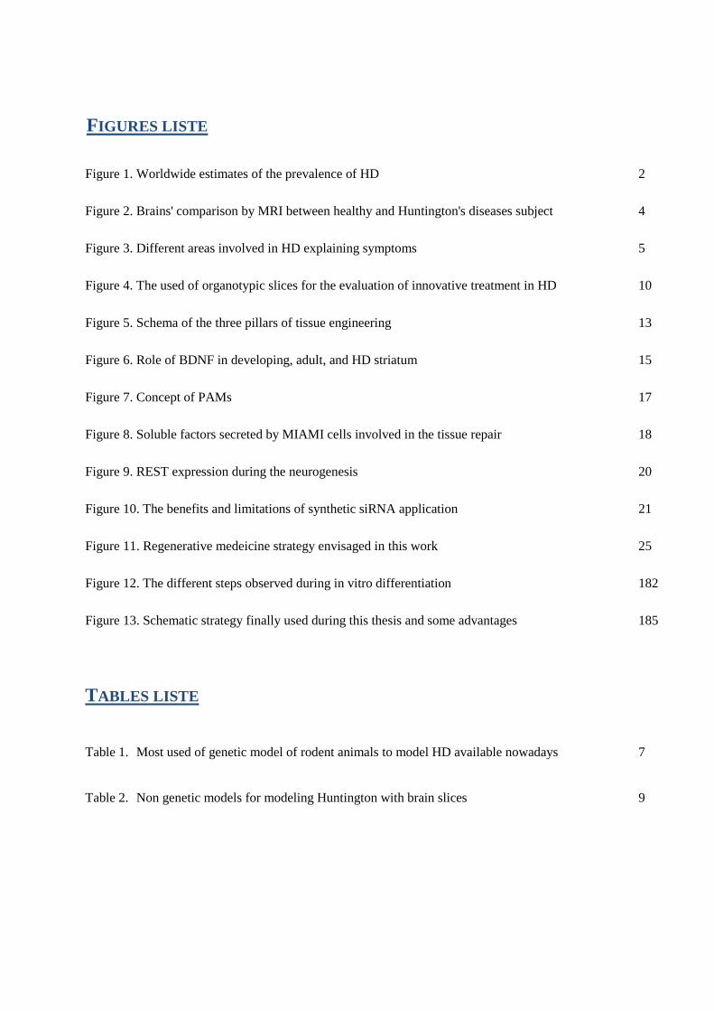
Figure 1. Worldwide estimates of the prevalence of HD 2
Figure 2. Brains' comparison by MRI between healthy and Huntington's diseases subject 4
Figure 3. Different areas involved in HD explaining symptoms 5
Figure 4. The used of organotypic slices for the evaluation of innovative treatment in HD 10
Figure 5. Schema of the three pillars of tissue engineering 13
Figure 6. Role of BDNF in developing, adult, and HD striatum 15
Figure 7. Concept of PAMs 17
Figure 8. Soluble factors secreted by MIAMI cells involved in the tissue repair 18
Figure 9. REST expression during the neurogenesis 20
Figure 10. The benefits and limitations of synthetic siRNA application 21
Figure 11. Regenerative medeicine strategy envisaged in this work 25
Figure 12. The different steps observed during in vitro differentiation 182
Figure 13. Schematic strategy finally used during this thesis and some advantages 185
Table 1. Most used of genetic model of rodent animals to model HD available nowadays 7
Table 2. Non genetic models for modeling Huntington with brain slices 9
FIGURES LISTE
TABLES LISTE

1

1
INTRODUCTION

INTRODUCTION
2
This PhD thesis has as main objective to offer an innovative tissue
engineering strategy for Huntington's disease by combining nanocarriers
delivering siRNA in mesenchymal stem cells and microcarriers releasing
therapeutic proteins . This project is part of a general strategy of the
laboratory INSERM U1066 "Biomimetic Micro and nanomedicine" from
Angers and the department "Pharmacy and Pharmaceutical Technology" from
Santiago de Compostela proposing an alternative and safe treatment in
neurodegenerative disorders .
Huntington’s disease (HD) is an inherited autosomal dominant
neurodegenerative disorder with prevalence in Europe of about 10 per
100,000 births (Figure 1.) [1, 2]. HD appears in mid-life leading to death 15-
20 years later and involves the triad signs and symptoms: involuntary
movement disorders called Huntington’s chorea, cognitive impair ment and
psychiatric manifestations.
Figure 1. Worldwide estimates of the prevalence of HD. Overall, the
prevalence of HD is much higher in European populations than in East Asia [3].
HD is one of the nine neurodegenerative disorders caused by the
expansion of cytosine-adenine-guanine (CAG) triplet repeat sequence [4].
1. HUNTINGTON'S DISEASE.

INTRODUCTION
3
This disorder is characterized by an unstable repetition of triplet cytosine -
adenine-guanine (CAG) of the Huntingtin (htt) gene in exon 1 of chromosome
4. It is translated at the protein level by a polyglutamine expansion at the
NH2-terminal part of the protein huntingtin ( HTT) [5]. The htt gene is
considered as normal when it contains less than 27 CAG repeats and generally
more than 40 repeats defines the adult -onset HD. The age of HD onset is
inversely correlated with the length of the expansion, with variable age -
dependent penetrance between 36 and 39 CAG repeats, but full penetrance at
40 or more repeats. In other words, people with 36-39 CAG repeats are at a
risk of developing all the HD symptoms [6] and conversely, a larger number
of repeats is usually associated with an earlier onset of signs and symptoms
[7].
The HTT protein has ubiquitous roles in apoptosis [8], regulating
microtubule-based transport [9] and scaffolding of cytoskeletal molecules at
synapses [10]. Therefore, mutant HTT (mHTT) primarily affects the central
nervous system (CNS). The translated wild-type huntingtin protein is a 350-
kDa protein containing a polymorphic stretch of between 6 and 35 glutamine
residues in its N-terminal domain [11]. For the length superior to 35 CAG
repeat in the htt gene, the accumulation of polyglutamine in the protein le ads
to its aggregation in specific areas in the brain such as: striatum, cortex,
thalamus, hypothalamus and the substancia nigra pars compacta. mHTT has a
toxic gain of function that causes cell death vi a very different mechanisms,
which still remain unclear . However, it is known to result in transcriptional
dysregulation as well as mitochondrial dysfunction and energy deficits (for
review see [1,12]). The accumulation of the mutant htt protein progressively
compromises survival and normal neuronal functioning, primarily in the
striatum (caudate/putamen). The mutant htt lead s also to proteosomal
dysfunction, induction of autophaghy, release of Calcium from intrace llular
stores and excitotoxicity at extrasynaptic NMDA receptors. It particularly
affects the GABAergic neurons, called medium spiny neurons (MSN) situated
in the striatum, which have axonal projections to the globus pallidus and
substantia nigra. They express Dopamine- and cAMP-Regulated neuronal
PhosphoProtein of 32kDa (DARPP32). The progressive loss of these neurons
is accompanied by a corresponding ventricular enlargement and gliosis

INTRODUCTION
4
(Figure 2) . The disease progresses with the degeneration of cortical
pyramidal neurons, mainly projecting to the caudate/putamen [13]. Many of
the symptoms of HD result from the loss of inhibitory connections from the
striatum to other structures such as the globus pallidus (Figure 3). The cause
of such specific regional and sub -population neuronal loss and the absence of
cell loss in other t issues remain uncertain . Brain pathological hallmarks
leading to 25% of brain weight loss in HD develop well before evident
symptoms appear.
Figure 2. Brains' comparison by MRI between healthy and Huntington's
disease subjects [14].
The most visible symptom of HD is the presence of involuntary jerky
movements named chorea. During the first stage of the disease, the chorea
dominates and when the disease progress, dystonia, rigidity and bradykinesia
are also observed. Cognitive impairments also progressively appear as well as
emotional disturbances marked in the most case by anxiety, memory loss,
dementia, depression and psychosis [15]. They frequently lead to considerable
distress and psychologic difficulty for patients , which have more prevalence
than the general population to commit suicide. The unequivocal presence of
chorea in a person with a family history or genetic confirmation of risk for
HD forms the basis for clinical diagnosis. Recently, the Ameri can Academy
of Neurology published guidelines to evaluate the motor and cognitive
function, behavioral symptoms and functional capacity based on the Unified
Huntington’s Disease Rating Scale -Total Motor Score [16].

INTRODUCTION
5
Figure 3. Different areas involved in HD explaining symptoms. The cortico-striatal
system is essential for the execution of movements (left). In patients with HD, progressive
degeneration of neurons in the striatum, and later also in cerebral cortex, disrupts function
in the cortico-striato-pallidal circuit and induces severe impairments in both motor and
cognitive functions. The striatal GABAergic projection neurons provide an inhibitory
control of two major striatal output structures, globus pallidus and pars reticulata of the
substantia nigra (not shown). Loss of these neurons, in animals with striatal lesions or in
HD patients, results in disinhibition of pallidal outflow (right) [17].
There is no effective treatment for the progressive neurodegenerative
process underlying HD, and management includes pharmacological
symptomatic control of the movement disorder and psychiatric features, as
well as non-pharmacological treatments, such as parenteral feeding and
therapy services [18]. When the physiotherapy is not enough, tetrabenazine is
the first choice of medication for uncomplicated chorea. Tetrabenazine is
acting to decrease dopamine levels and can be helpful to reduce movement
disorder but presents various side effects like the increase of depression or
psychiatric disorders. Neuroleptics or benzodiazepines can also be prescribed
and mood stabilizers such as anti-depressants and anti -anxiety reducing
psychological dysfunctions are also proposed [18]. Unfortunately, many of
these medications have adverse side effects that can worsen HD symptoms. In
order to help to define the best treatment The Unified Huntington’s Disease
Rating Scale-Total Motor Score classified the level of evidence for drugs to
reduce chorea based on a review of randomized clinical trials [19,20].

INTRODUCTION
6
Nowadays, two approaches are under pre-clinical evaluation: disease
modifying treatments, more particularly by reducing polyQ repeats and more
recently by the suppression of the mRNA of the HTT gene with interference
RNA (iRNA) [21]. Another approach consists in recent tissue engineering
strategies to replace lost neurons by new ones obtained in vitro from stem
cells. These methods need to be further improved and developed in order to
be validated in the in vivo models of HD [18].
Most animal models of HD fall into two broad categorie s, genetic and
non-genetic. Historically, non-genetic models have dominated the field of HD
research. Although George Huntington first described HD in 1872,
researchers did not identify the actual genetic mutation responsible for the
disease until 1993, which delayed the development of appropriate genetic
models until the last decade [22].
The emergence of genetic and molecular technology allow ed the
development of animal models expressing a truncated [23] or full length
[24,25] form of mutant htt (mhtt) (Table 1) . Animal models are divided in to
two genetic categories: transgenic or knock -in. Transgenic models result from
the random insertion of mutated human htt. The R6/1 and R6/2 transgenic
mouse models were the first characterized [26] (Table 1). These mice express
only mutant exon 1 of the human htt gene with di fferent length repeats. These
principal models containing only the truncated human mhtt gene are st ill the
most used nowadays, together with yeast artificial chromosome's (YAC)
models [24]. The latter consist on the cloning of an artificial yeast vector that
contains the entire human mhtt with different expanded CAG repeats (YAC
46, YAC 72, YAC 128) which are then integrated into the rodent genome [27]
(Table 1) . Alternatively, the knock-in models result by the insert ion in the htt
mouse genome of the CAG repeats of human mhtt, which are then within the
context of the rodent mhtt gene [28,29] (Table 1) . In all of these animal
models the pathophysiology of the disease appears in adult age as for the HD
patients.
2. IN VIVO MODELS OF HUNTINGTON’S DISEASE

INTRODUCTION
7
In any case none of these models exactl y reproduce the human
pathology. Moreover, the existence of various genetic animal models did not
allow predicting HD symptoms. Indeed, we can believe that the progression is
correlated with the number of CAG repeat length, but the mechanism of the
disease seems to be more complicated. Moreover, it has been shown that
therapeutic success in animal models is not always paralleled by clinical
success in patients .

INTRODUCTION
8
Animal
models
Transgene product CAG
repeat
length
Promoter and transgene
expression
Method of cell
death
Symptoms Ref
Truncated N-terminal fragment models
R6/1 mice 67 amino acids of
N-terminal fragment
(human HTT)
116 1kb human HTT
promoter.
Transgene expression ~
31%
Aggregation and
nuclear inclusion of
htt
Slow progression of
the symptoms, brain
atrophy, dystonic
movements, motor
performance, grip
strength and body
weight loss
[30]
R6/2 mice 67 amino acids of
N-terminal fragment
(human HTT)
144 1kb human HTT promoter
Transgene expression
~75%
Aggregation and
nuclear inclusion of
htt
High progression of
the symptoms, brain
atrophy, dystonic
movements, motor
performance, grip
strength
[30]
Full length HD models: knock-in models
HdhQ92
mice
Full length chimeric
human HTT exon 1:
mouse Htt
92 Endogenous mouse Htt
promoter
Transgene expression ~
100%
Nuclear inclusion
but not cytoplasm
inclusion,
aggregation of htt,
Not communicated [31]
HdhQ111
mice
Full length chimeric
human HTT exon 1:
mouse
111 Endogenous mouse Htt
promoter
Transgene expression ~
100%
Nuclear inclusion
but not cytoplasmic
inclusion,
aggregation of htt
Not communicated but
model to study
juvenile HD
[31]
zQ175mice Full length chimeric
human HTT exon 1:
mouse
188 Endogenous mouse Htt
promoter
Transgene expression ~
100%
Nuclear inclusion
aggregation of htt
Tremor, hypokinesia,
abnormal gait, poor
grooming, lost
coordination, deficit in
grip strength
[32]
Full length HD models: transgenic models
YAC46
mice
Full length human
HTT
48 Human HTT promoter
and regulatory elements
Tansgene expression
~40%
Increased
intracellular calcium
concentration
Any obvious abnormal
behavior,
electrophysiological
abnormalities
[24]
YAC72
mice
Full length human
HTT
72 Human HTT promoter
and regulatory elements
Tansgene expression
~40%
Very few nuclear
inclusion,
aggregation of htt
Symptoms YAC line
dependent, tremor,
ataxia, brain reduction
[24]
YAC128
mice
Full length human
HTT
128 Human HTT promoter
and regulatory elements
Tansgene expression
~100%
Nuclear inclusion,
aggregation of htt
Slow progression of
the symptoms, brain
atrophy, hypokinesia,
[33]
BACHD
mice
Full length human
HTT
97 Human HTT promoter
and regulatory elements
Tansgene expression
~100%
Progressive nuclear
inclusion,
aggregation of htt
Very slow progression
of the symptoms, brain
atrophy, dystonic
movements, not body
weight loss
[34]
Table 1. Most used of genetic model of rodent animals to model HD available nowadays.
Following early ideas that lesions of the striatum were responsible for
HD, non genetic animal models used intrastriatal injections of neurotoxins

INTRODUCTION
9
with glutamatergic targets such as kainic acid (KA) [35], ibotenic acid (IA),
quinolinic acid (QA) [36] [37] and N-methyl-d-aspartate (NMDA) [38].
Indeed, glutamatergic excitotoxicity is involved in the pathophysiology of the
disease. These lesions induced death of striatal medium spiny neurons similar
to the neuropathology present in HD patients . The symptoms most commonly
associated with the diseased state induced by the neurotoxins are loss of
weight, possible tremor or seizures, eventual paralysis, recumbence, and often
death representing the later stages of the disease. But these mode ls do not
allow investigation of disease progression or the mechanism of
neuropathology because the htt protein does not present the mutation. In
addition, genetic and non-genetic models need proper care and animals must
be daily monitored. Immediately after toxin administration or in later stages
in genetic models, euthanasia may be necessary for moribund animals. To
reduce the number of animal experiments ex vivo models represents an
excellent compromise between single cell cultures and animal studies.
Another way for modelling HD consists in using organotypic brain
slices, which have been also reported for modelling Parkinson's disease and
cerebral ischemia. Brain slices are kept alive during several weeks in culture
and represent a simple method to model the neurodegeneration and evaluate
potential treatments before in vivo studies.
Brain slice models offer unique advantages over other in vitro platforms
in that they can replicate many aspects of the in vivo context. Slices preserve
largely the tissue architecture of the brain regions that they originated from
and maintain neuronal activities with intact functional local synaptic circuitry
[39]. Nevertheless, brain slices remain fragile, they can be easily distorted,
and often flatten during the culture. In other word, organotypic brain sl ices
are delicate and frequently become damaged during the preparative stages, so
brain slice preparation and culture need experience. Functional outcome of a
therapeutic strategy cannot be evaluated with this technique , but as brain
slices can be maintained for a few weeks in culture they offer the possibility
to screen a large quantity of therapeutic strategies. Pharmacologicals [40],
3. EX VIVO MODELS OF HUNTINGTON'S DISEASE

INTRODUCTION
10
gene manipulation strategies with the goal to reduce the CAG repeat on HD
[41] or injection of growth factors or cells can be evaluated for their
neuroprotective or neurorestorative abil ity of damaged structures (Figure 4).
Furthermore, organotypic slides represent a powerful tool for understanding
the interaction between grafted cells and resident cellular matrix, or for
comprehending the mechanisms of experimental treatments in HD [42].
Figure 4. The use of organotypic slices for the evaluation of innovative
treatment in HD [43].
As in vivo models, organotypic brain slices of HD are divided in 2
categories: genetic and non-genetic models. Genetic models can be derived
from adult animals already described above, presenting the MSN
degeneration, or by transfecting the mutated htt within the organotypic
culture. However, these organotypic cultures are difficult to generate due to
the diminished neuronal plasticity and the fragility of these brains . Non-
genetic models can be generated from pups or young animals by neurotoxin
administration directly in the organotypic slice culture. The first HD
organotypic model was developed in 1986 by injecting KA in the striatal
organotypic slices [44]. Over the years, brain slice cultures have been
successfully established with QA and/o r 3-NPA, and KA after addition in the
media of striatal organotypic slices to mimic the disease. In order to study
later stage of onset disease all these neurotoxins have been injected in the
frontal cortex, hippocampus or caudate nucleus for modeling HD in rat [45–
47] (Table 2) . But the use of neurotoxins leads to a heterogeneity in the
results obtained, which must be taken in consideration. Furthermore, with that
type of model, only the cellular aspect of HD can be studied, they cannot take

INTRODUCTION
11
into account the genetic component of the pathology. Advantages are the
speed and low-cost associated to this model when normal rodents are used
[42].
Neurotoxins Ex vivo models Cells modification Ref
Kainic acid (KA) Striatal organotypic
model
Enlargement of mitochondria,
dilation of rough endoplasmic
reticulum, presence of
numerous vacuoles, glial
fibrillary changes
[44]
Quinoleic acid (QA) Corticostriatal
organotypic model
Excitotoxic damages, presence
of numerous vacuoles
[48,49]
α-Amino-3-hydroxy-
5-methylisoxazole-4-
propionic acid
hydrate
Corticostriatal
organotypic model
Excitotoxic damages, presence
of numerous vacuoles
[50]
3-nitropropionic
acid (3-NPA) Striatal and
corticostriatal
organotypic model
No described but reductions of
complex II–III Activity,
mitochondrial function is
impaired
[46]
QA + 3-NPA Striatal organotypic
model
No described. [51,52]
Table 2. Non genetic models for modeling Huntington with brain slices.
During the past years, many preclinical studies initially reported the
efficacy of human fetal striatal tissue to replace and provide functional
recovery in a variety of rodent and non-human primate models of HD. Some
teams demonstrated the feasibility and the safety of this therapeutic strategy
[53–57] and functional improvements were obtained in the study led by
Bachoud-Lévi. They reported graft survival, which contained striatal
projection neurons and interneurons, and received host -derived afferents
[53,54]. But the survival is sti ll very poor and the comparisons of these
clinical trials are very difficult because of the heterogeneity in their design
and the lack of controls . Nevertheless, the reported improvements in these
trials appear to be modest and transient (for review see [58]). Moreover, the
use of human fetal brain for striatal transplantation derived from elective
abortions is limited by the lack of standardization inevitably correlated with
the use of such a source as well as ethical, practical , and regulatory concerns
and is dependent upon availability of donor tissue [59]. Stem cell -based
4. EXPERIMENTAL TREATMENT IN HUNTINGTON’S DISEASE

INTRODUCTION
12
therapies can provide a limitless source of cells due to their self -renewal
capacity and their neuronal differentiation potential, but good cell
engraftment remains a drawback. On this basis, the development of cell
replacement strategies for regenerative medicine and more particularly tissue
engineering has been under light the last decade for HD.
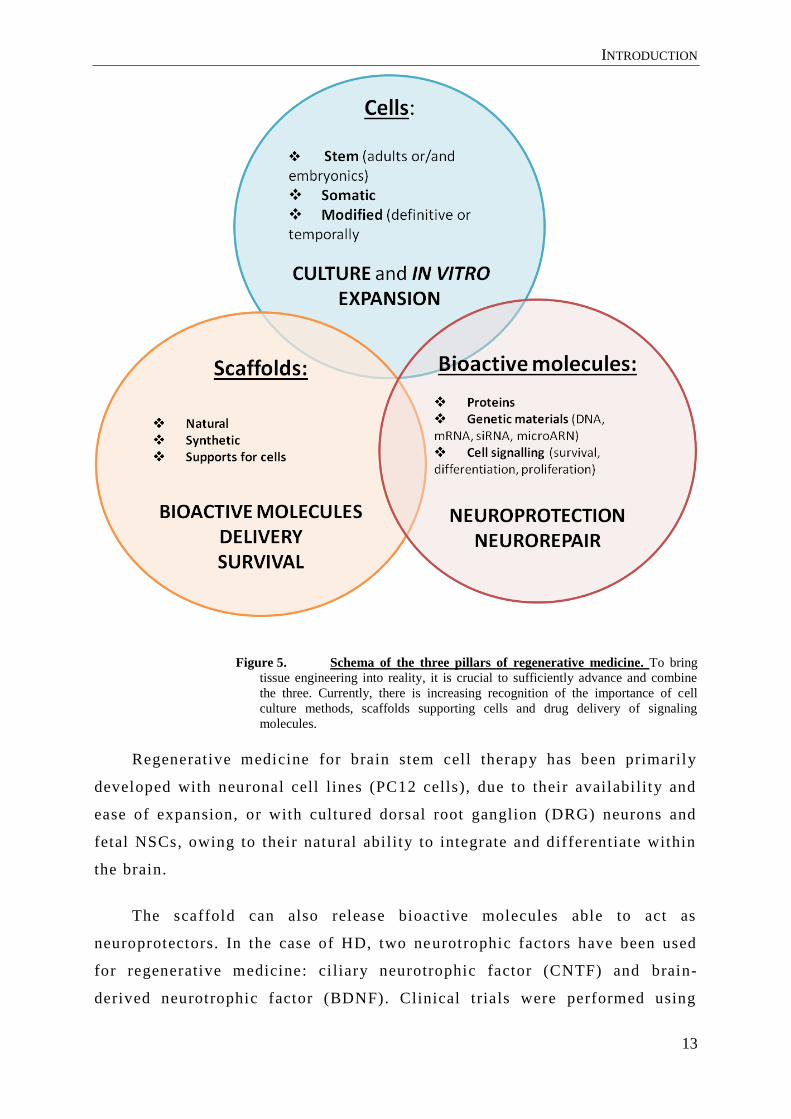
4.1 TISSUE ENGINEERING AND REGENERATIVE MEDICINE
Tissue engineering and regenerative medicine are fields which have a
unique tactic to solve clinical problems aforementioned by combini ng the
principles of engineering, clinical medicine, biology and materials science
[60]. Tissue engineering, according to the National Institute of Health (NIH),
is a broad field which involves “ biomaterials development and refers to the
practice of combining scaffolds, cells, and biologically active molecules into
functional t issues”[61] and regenerative medicine as the development of “
therapies to restore lost, damaged, or aging cells and tissues in the human
body”. Those approaches may include, but is not limited to, th e use of soluble
molecules [62], gene therapy, stem cell s transplantation [63], reprogramming
of cells. The strategic introduction of these bioactive and soluble molecules,
as well as stem cells into the human body is directed not only to replacing
tissue but also at inducing regeneration and revascularization by host tissue .
Various cells can be considered in t issue engineering , stem or modified cells
to replace lost neurons and somatic cells for t heir neuroprotective properties
(Figure 4). Biomaterial scaffolding is often employed to provide a supporting
spatial and biomolecular environment for transplanted cells. This approach
named “top-down” in which cells are seeded onto a scaffold with
biocompatible and biodegradable propertie s is the most used (Figure 4).
INTRODUCTION
13
Figure 5. Schema of the three pillars of regenerative medicine. To bring
tissue engineering into reality, it is crucial to sufficiently advance and combine
the three. Currently, there is increasing recognition of the importance of cell
culture methods, scaffolds supporting cells and drug delivery of signaling
molecules.
Regenerative medicine for brain stem cell therapy has been primarily
developed with neuronal cell lines (PC12 cells), due to their availability and
ease of expansion, or with cultured dorsal root ganglion (DRG) neurons and
fetal NSCs, owing to their natural ability to integrate and differentiate within
the brain.
The scaffold can also release bioactive molecules able to act as
neuroprotectors. In the case of HD, two neurotrophic factors have been used
for regenerative medicine: ci liary neurotrophic factor (CNTF) and brain-
derived neurotrophic factor (BDNF). Clinical trials were performed using

INTRODUCTION
14
CNTF-producing cells with stage 1 and 2 HD pat ients [64,65]. During this
phase 1 study, subjects received one capsule implanted into the right lateral
ventricle, and the capsule was exchanged every 6 months during 2 years.
While the CNTF-induced sparing of s triatal neurons and maintenance of
intrinsic circuitry in animal models was impressive, the effect in human was
less than that seen in rodents. Finally, human clinical trials did not present
relevant positive effects, and progressively the supplementation of CNFT in
human has been given up. To our knowledge, no clinical trial has been
conducted with BDNF.
4.2 POTENTIAL THERAPEUTIC USE OF THE NEUROTROPHIC FACTOR BDNF
Neurotrophic factors a re essential for the survival of the central nervous
system neurons and demonstration of their reduced availability in HD
indicates that they may play an important role in this disorder. Indeed, the
reduction of BDNF in HD contributes to the disease onset and or progression
[66,67]. BDNF is essential in sustaining the physiological processes of
normal intact adult brain [68] and more particularly for GABAergic striatal
neurons (Figure 5) . Indeed, although widely expressed in the adult
mammalian central nervous system, BDNF is particularly abundant in the
hippocampus and cerebral cortex where it is anterogradely transported to i ts
striatal targets via the corticostriatal afferents [69]. Several evidences
demonstrate the role of BDNF in the maturation of striatal neurons and how
BDNF promotes the survival of DARPP-32 positive neurons [70,71].
INTRODUCTION
15
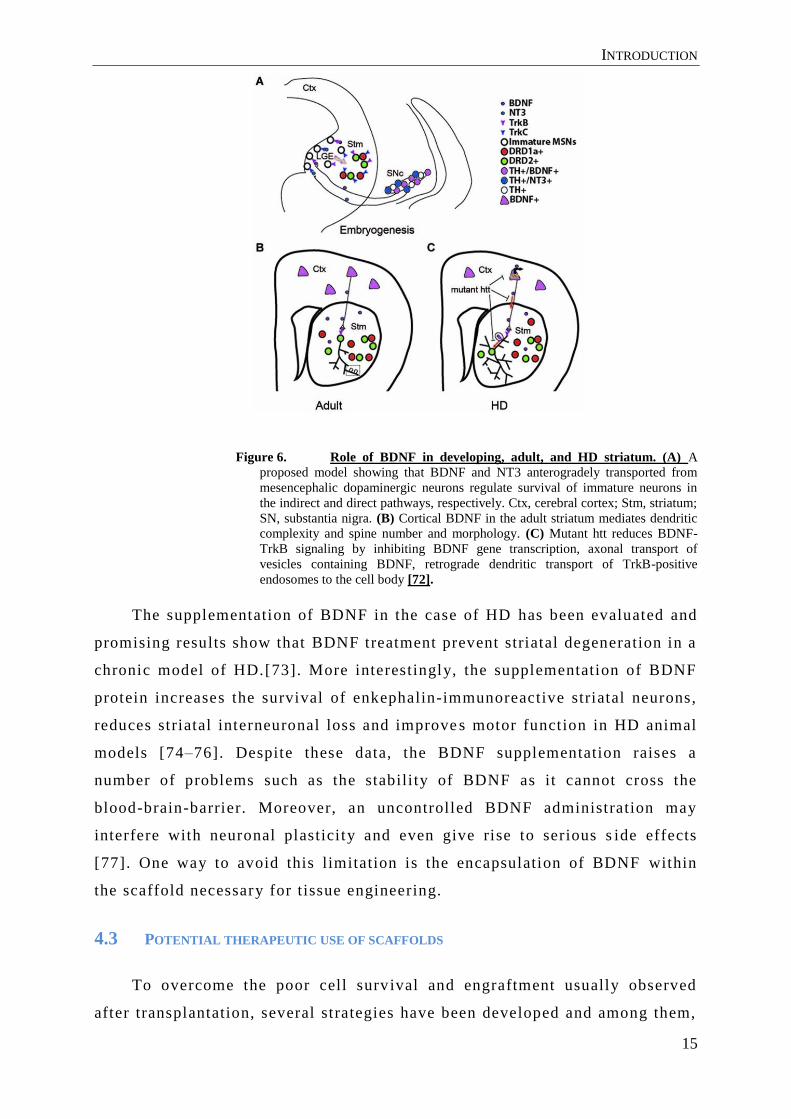
Figure 6. Role of BDNF in developing, adult, and HD striatum. (A) A
proposed model showing that BDNF and NT3 anterogradely transported from
mesencephalic dopaminergic neurons regulate survival of immature neurons in
the indirect and direct pathways, respectively. Ctx, cerebral cortex; Stm, striatum;
SN, substantia nigra. (B) Cortical BDNF in the adult striatum mediates dendritic
complexity and spine number and morphology. (C) Mutant htt reduces BDNF-
TrkB signaling by inhibiting BDNF gene transcription, axonal transport of
vesicles containing BDNF, retrograde dendritic transport of TrkB-positive
endosomes to the cell body [72].
The supplementation of BDNF in the case of HD has been evaluated and
promising results show that BDNF treatment prevent striatal degeneration in a
chronic model of HD.[73]. More interestingly, the supplementation of BDNF
protein increases the survival of enkephalin-immunoreactive striatal neurons,
reduces striatal interneuronal loss and improve s motor function in HD animal
models [74–76]. Despite these data, the BDNF supplementation raises a
number of problems such as the stabili ty of BDNF as it cannot cross the
blood-brain-barrier. Moreover, an uncontrolled BDNF administration may
interfere with neuronal plastici ty and even give rise to serious s ide effects
[77]. One way to avoid this limitation is the encapsulation of BDNF within
the scaffold necessary for tissue engineering.
4.3 POTENTIAL THERAPEUTIC USE OF SCAFFOLDS
To overcome the poor cell surv ival and engraftment usually observed
after transplantation, several strategies have been developed and among them,

INTRODUCTION
16
two methods seem particularly promising: in situ controlled drug delivery and
implantation of cells adhered on biomaterial -based scaffolds. Such scaffolds
should provide an adequate 3D support for transplanted cells, thereby
increasing cell survival and even guiding cell differentiation and fate in vivo
[78,79]. However, delivery of cells with scaffolds to the damaged brain still
remains challenging due to practical l imitations of delive ry [80]. Ideal
properties of a scaffold for brain tissue engineering are biocompatibility, very
small size, controlled biodegradability with non -toxic derivative products,
and three-dimensional (3D) matrices with appropriate mechanical p roperties
to mimic the extracellular matrix [81,82].
An innovative scaffold for tissue engineering combining the se ideal
properties with a biomimetic 3D approach and the release of bioactive
molecules has been developed in our laboratory. This scaffold or
microcarriers named Pharmacologically activ e microcarriers (PAMs) are
constituted of a synthetic polymer based on poly(lactic-co-glycolic acid)
(PLGA) [78]. They are obtained by a non-denaturing solid-in-oil-in-water
(s/o/w) emulsion evaporation/extraction t echnique. The protein is first
nanoprecipitated with poloxamer 188 a biocompatible hydrophilic polymer
that protects the protein against irreversible denaturation [83]. PAMs are
biodegradable and biocompatible with a mean size of 60 µm, covered by a
biomimetic surface providing a 3D support for the cells and delivering a
therapeutic protein in a prolonged manner . All these combined properties
stimulate the survival and differentiation of the transported cells [84]. Their
small size allows their implantation into the brain through a needle or
catheter and after the complete degradation of the polymer the cells may
integrate the parenchyma. These PAMs have been successfully employed for
different tissue engineering strategies, in neurodegenerative disorders,
cerebral ischemia, myocardial infarction, and cartilage re pair (Figure 6)
[85,78,86,83,87] . For these strategies the appropriate growth factor and
extracellular matrix protein have been combined to either progenitors or stem
cells. In this work we will use stem cells combined with PAMs presenting a
biomimetic surface of laminin , which stimulates neuronal differentiation [88]
and delivering BDNF as a therapeutic strategy for HD.

INTRODUCTION
17
Figure 7. Concept of PAMs: the biomimetic surface of PAMs is obtained by
coating their surface with extracellular matrix proteins that can favor cell
adhesion. During their formulation, the encapsulation of neurotrophic factor is
performed.
4.4 . POTENTIAL THERAPEUTIC USE OF STEM CELLS
During the last decade, several preclinical experiments have used cell
replacement strategies in order to restore MSN using HD animal models.
Different human stem cell sources are being actively explored for potential
cell replacement therapy including embryonic stem cells (ESCs), induced
pluripotent or neural stem cells (iPSC or NSC), fetal and adult neural
precursors and mesenchymal stem cells (MSCs). Both ESC, iPSC have been
successfully committed into MSN in vitro and then grafted into rodent models
of HD [37,89,90]. But after human ESC transplantation into rat brains , tumor
formation has been reported, which was not the case for iPSC -MSN-derived
cells, which were further committed with in this lineage. In addition, the
ethical issues related to the use of ESCs and the lack of availability of fetal
neural precursors drive us to focus in other cell sources. To find the best way
to obtain the most important benefits with less ethical and pr actical
constraints, mesenchymal stem cells have been investigated.
Human MSCs, as Friedenstein reported, are capable of differentiating
into cells deriving from the mesodermal layer, such as osteoblasts,
chondrocytes and adipocytes. Along with their self -renewal property, MSCs
secrete tissue repair factors, such as growth factors, which affect the

INTRODUCTION
18
surrounding microenvironment to promote angiogenesis, decrease
inflammation, and enhance tissue repair [91]. In this way, MSCs are being
widely evaluated in many clinical trials for cell therapy showing the
feasibility of this approach. Recent advances in s tem cell biology hold great
promise in the development of MSCs-based therapy for tissue engineering . It
was also demonstrated that these cells could differentiate to an ectodermal
neural/neuronal phenotype, particularly under the influence of specific fact ors
[85] [79,92] enabling their use for cell therapy for neurodegenerative disease
including HD. The principal limitation is that MSCs are a heterogeneous
population with cells presenting different differentiation properties. To avoid
these limitations a homogenous subpopulation of MSCs, named “Marrow-
Isolated Adult Multilineage Inducible” (MIAMI), which present an unique
genetic profile expressing several pluripotency markers (Oct4, Sox2, Nanog,
SSEA4) and secrete many varied cytokines (Figure 7) are interesting for the
treatment of neurodegenerative disorders [93,94].
Figure 8. Soluble factors secreted by MIAMI cells involved in the tissue
repair. [93,94]
They are able to generate cells derived from all three embryonic germ
layers and cultured on fibronectin; they are capable of differentiating into
neuron-like cells under treatment with various factors. After treatment, the
cells show neurites, express neuronal factors and present some
electrophysiological characteristics similar to those observed in mature
neurons [95]. Recently, in a rat model of Parkinson's disease (PD), striatal
implantation of MIAMI cells pre -committed towards the dopaminergic
phenotype adhered to microsphere releasing bio active molecules improved

INTRODUCTION
19
stem cell survival and showed dopaminergic differentiation. This led to the
protection/repair of the nigro -striatal pathway and to functional recovery of
the PD rats. Furthermore, implantation of pre -treated MIAMI cells also
induced functional recovery in PD rats, probably due to the release of glial
cell line derived neurotrophic factor (GDNF) [88]. But MIAMI cells ' abil ity
to differentiate into neurons, although better than the simple MSC needs to be
improved.
4.5 POTENTIAL THERAPEUTIC OF SMALL INTERFERING RNAS FOR MSC
DIFFERENTIATION
The conventional methods of generating neurons from MSC s, through
bolus supplementation of small molecules or neurotrophic factors (growth
factors: GF), still lack in efficiency in neural conversion and lineage
selection. One possible reason may be the inadequacy of GFs to control gene
expression. Cell differentiation may be achieved by RNA interference (RNAi)
strategies and more particularly by small interfering RNA (siRNA), which
selectively knock-down the expression of only a few pivotal genes. Indeed,
siRNAs are synthetic duplex of 21–23 nucleotides, approximately 7.5 nm long
and 2 nm in diameter , which are capable of specifically target ing one gene
and silencing it in a post-transcriptional way. SiRNA are rapidly taken up
into an enzyme complex, RNA induced silencing complex (RISC), that
degrades the mRNA through guidance to a specific target mRNA resulting in
specific gene si lencing. At RISC one siRNA strand is t aken into the effector
complex, the catalytic subunit Argonaute2, and then serves as a template,
guiding the hydrolysis of complementary or near complementary mRNA
sequences [96]. Initially siRNA emerged as a potential therapeutic treatment
for cancer. Although current applications in stem cells remain largely
restricted to studies on molecular pathways and s ignalling, RNAi can be used
as a biomedical strategy to direct li neage-specific differentiation of stem cells
for therapeutic purposes [97]. One key factor that can possibly be adapted
into the siRNA strategy for directing neuronal differentiation of neural stem
cells, is the repressor element 1 (RE-1) silencing transcription factor (REST)
[98]. In most differentiated non-neuronal cells and uncommitted neural stem
cells, REST functions as a transcriptional repressor fo r a myriad of neuronal

INTRODUCTION
20
specific genes such as ion channels, synaptic vesicles proteins, and
neurotransmitter receptors by binding to a highly conserved DNA sequence
known as RE-1. During neurogenesis, REST is rapidly down regulated in
embryonic stem cells (ESCs) and neural stem cells upon differentiation into
neurons [99] (Figure 8) .
Figure 9. REST expression during the neurogenesis [100].
Conversely, induced down regulation of REST has been shown to
promote neuronal commitment in mouse ESC and mouse MSCs. Specifically,
the knockdown of REST in ESCs induced neural progenitors formation [101]
(Figure 8) and when applied to MSCs, cross -lineage differentiation to
neurons was observed [102,103]. Although the knockdown of REST holds
great potential , its therapeutic applications in neuronal differentiation is
hindered by poor cellular uptake of siRNA molecules and their rapid
enzymatic degradation [104]. siRNAs' molecular weight (∼13 kDa) and strong
anionic charge due to the presence of aphospho-diester backbone (∼40
negative phosphate charges), make them incapable of freely crossing the cell
membrane. The electrostatic repulsion from the anionic cell membrane
surface results in the failure of siRNA to passively diffuse through the cell
membrane (Figure 9). Moreover, the synthetic siRNA molecules show low
stability in physiological fluids, poor tissue/cell specificity, and rapid
clearance [105]. Therefore, successful siRNA therapeutics requires effective
and safe carrier systems to overcome the inherent limitations of siRNA and
achieve maximum gene silencing effect . In the last decade, two different
approaches for siRNA delivery have been developed: viral and non -viral
vectors. In particular, the advantages of non -viral vectors are their low
immunogenicity, their relatively low production cost and reproducibility

INTRODUCTION
21
potentially. These reasons make them promising carriers for siRNA delivery
[106].
Figure 10. The benefits and limitations of synthetic siRNA application. The
representation of the limitations involved in the siRNA delivery. [107]
5.1 DIFFERENT TYPES OF NANOCARRIERS
Nanocarriers (NCs), including nanoparticles and nanocapsules were first
developed for the potential delivery of therapeutic factors such as
5. NANOCARRIERS

INTRODUCTION
22
chemotherapeutic agents to tumors or, when combined to stem cells, mostly
for stem cell imaging [108]. First , the use of nanocarriers aims to protect an
active ingredient against a potential degradation , and secondly to modify the
natural distribution of the active substance in the body and in cells. It is
theoretically possible to accumulate the active ingredient to the desired si te
of action and away from undesirable sites to limit side effects. NCs ranging
from 1 to 1000 nanometer sizes are divided into 2 main categories:
- Organic NCs which include liposomes, lipid nanoparticles, solid
nanoparticles and dendrimers .
- Inorganic NCs with quantum dots, carbon nanotubes, iro n or gold
nanoparticles [109] .
The organic NCs and more particularly lipid based NCs are interesting to
transfect cells because lipid based nanoparticles can contain lipids present in
the biological membranes which help the entry of nanoparticles. Cationic
charges contained in some lipids are able to int eract with nucleic acid. In
addition, the risk of undesirable immunogenic reactions to lipids is also
relatively lower than most of the polymeric materials which generally have
higher molecular weights [110]. Furthermore, some clinical trials have been
conducted with siRNA and lipids based nanoparticles [111,112]. To our
knowledge, clinical trials with nanoparticles and MSCs have not been
performed in HD. When compared with l iposomes, l ipid -based nanoparticles
such as solid lipid nanoparticles generally have solid, lipophilic core regions
so it is inherently difficult to truly encapsulate the hydrophilic, poly -anionic
RNA molecules. As a result , there are relatively few lipid nanoparticles for
RNA delivery [113,114].
5.2 LIPID NANOCAPSULES
Lipid nanocapsules (LNC) are nanocarriers developed and recently
patented. These nanocapsules are constituted by oily core of tryglicerides and
a shell made of surfactants particularly polyethylene glycol hydroxystearate .
They are obtained by a phase inversion temperature dependent process. This

INTRODUCTION
23
solvent-free process requires little energy and allows easy large-scale
transposition [115].
The formulation is based on a simple process, named emulsion's phase
inversion, developed and patented in 2002. This is realized by oil in water
emulsion (O / W) using the various consti tuents described above. This
emulsion is subjected to an increase of temperature which induces a change in
the hydrophilic / lipophilic balance. Several temperature cycles (between 50
and 90°C) are produced and the addition of cool water final stablizes and
solidifies LNCs.
Previous studies demonstrated the possibility to encapsulate plasmid
DNA within the LNCs to develop a gene therapy strategy [116]. For this, the
DNA is complexed with cationic lipids by electrostatic interactions leading to
formation of complexes called lipoplexes which are added to other
components of the LNC. Moreover, the phase inversion temperature was
reduced to avoid degradation of the plasmid [116]. This strategy has
demonstrated the capacity of LNCs to transfect in the in vivo models of
gliobastoma [117–119].
5.3 SOLID SPAN NANOPARTICLES
Solid span nanoparticles have been recently developed and patented
[120]. These nanocarriers are based on sorbitan esters, which are components
widely used in the pharmaceutical industry due to its non -ionic surfactant
properties at low concentrations . These nanoparticles can be prepared using a
simple, one-step and easily scalable procedure
(https://www.youtube.com/watch?v=fda9CtJ5zF0 ) and they can associate
different components and/or bioactive molecules . The internal structure of
this nanocarrier is not an aqueous inner space surrounded by a lipid bilayer
nor i t is based on nanoemulsions, but rather it is a homogenous
nanoparticulate solid structure. It is also possible to incorporate various
additional components. These addit ional components allow to modulate the
nanosystem features conferring a great versatility in terms of physical -
chemical characteristics and interaction with other components, and facilitate
INTRODUCTION
24
the incorporation of active ingredients : hydrophilic and l ipophilic nature
[121].
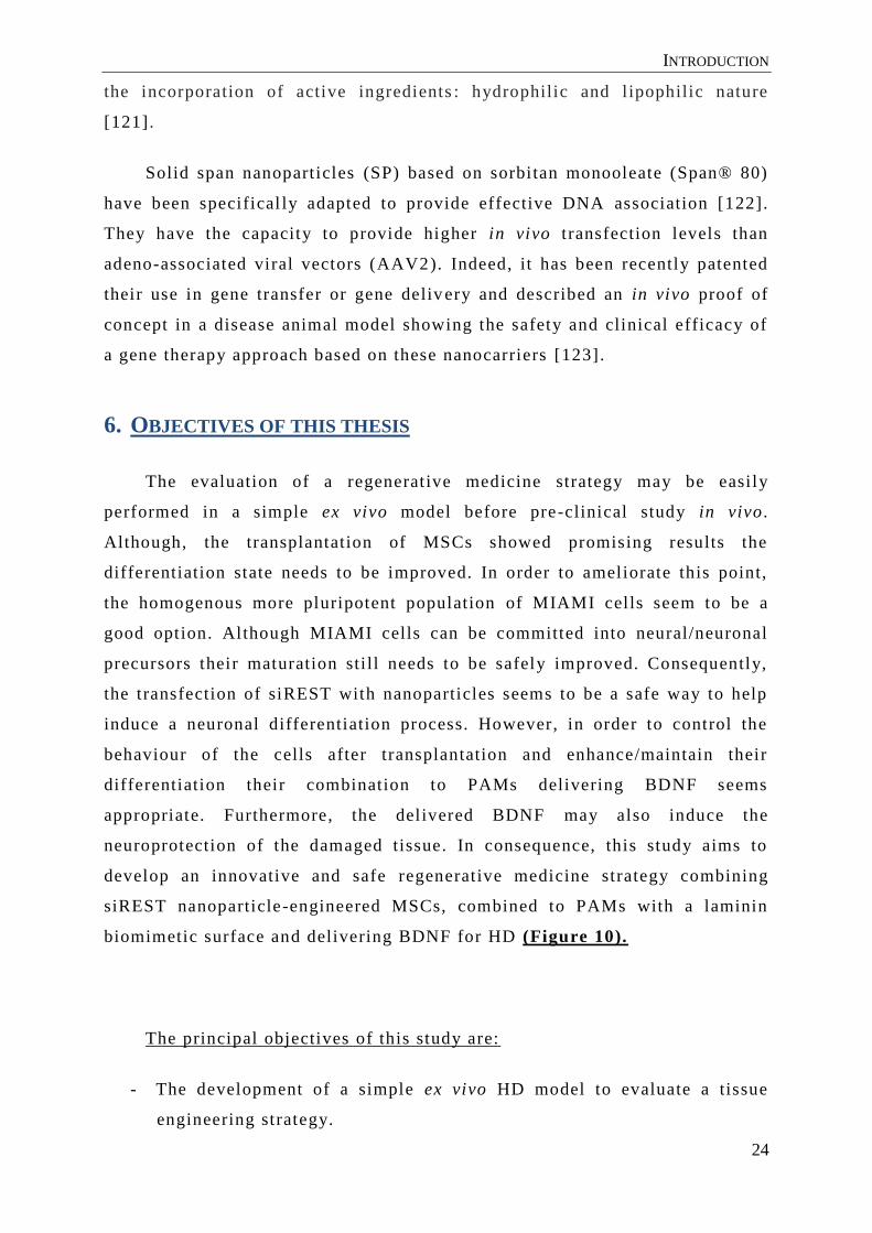
Solid span nanoparticles (SP) based on sorbitan monooleate (Span® 80)
have been specifical ly adapted to provide effective DNA association [122].
They have the capacity to provide higher in vivo t ransfection levels than
adeno-associated viral vectors (AAV2). Indeed, it has been recently patented
their use in gene transfer or gene deliv ery and described an in vivo proof of
concept in a disease animal model showing the safety and clinical efficacy of
a gene therapy approach based on these nanocarriers [123].
The evaluation of a regenerative medicine strategy may be easily
performed in a simple ex vivo model before pre-clinical study in vivo .
Although, the transplantation of MSCs showed promising results the
differentiation state needs to be improved. In order to ameliorate this point,
the homogenous more pluripotent population of MIAMI cells seem to be a
good option. Although MIAMI cells can be committed into neural/neuronal
precursors their maturation sti ll needs to be safely improved. Consequently,
the transfection of siREST with nanoparticles seems to be a safe way to help
induce a neuronal differentiation process. However, in order to control the
behaviour of the cells after transplantation and enhance/maintain their
differentiation their combination to PAMs delivering BDNF seems
appropriate. Furthermore, the delivered BDNF may also induce the
neuroprotection of the damaged tissue. In consequence, this study aims to
develop an innovative and safe regenerative medicine strategy combining
siREST nanoparticle-engineered MSCs, combined to PAMs with a laminin
biomimetic surface and delivering BDNF for HD (Figure 10).
The principal objectives of this study are:
- The development of a simple ex vivo HD model to evaluate a tissue
engineering strategy.
6. OBJECTIVES OF THIS THESIS
INTRODUCTION
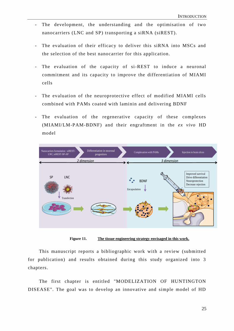
25
- The development, the understanding and the optimisation of two
nanocarriers (LNC and SP) transporting a siRNA (siREST).
- The evaluation of their efficacy to deliver this siRNA into MSCs and
the selection of the best nanocarrier for this application.
- The evaluation of the capacity of si-REST to induce a neuronal
commitment and i ts capacity to improve the differentiation of MIAMI
cells
- The evaluation of the neuroprotective effect of modified MIAMI cells
combined with PAMs coated with laminin and delivering BDNF
- The evaluation of the regenerative capacity of these complexes
(MIAMI/LM-PAM-BDNF) and their engraftment in the ex vivo HD
model
Nanocarriers formulation : siREST-
LNC; siREST-SP-AP
Differentiation in neuronal
progenitorsComplexation with PAMs Injection in brain slices
2 dimension 3 dimension
SP LNC
Transfection
Improved survival
Drive differentiation
Neuroprotection
Decrease rejectionBDNF
Encapsulation
Figure 11. The tissue engineering strategy envisaged in this work.
This manuscript reports a bibliographic work with a review (submitted
for publication) and results obtained during this study organized into 3
chapters.
The first chapter is enti tled "MODELIZATION OF HUNTINGTON
DISEASE". The goal was to develop an innovative and simple model of HD

INTRODUCTION
26
without the addition of neurotoxins into the media and to model the
neurodegeneration of medium spiny neurons (Publication n°1).
Then the second chapter "INNONATIVE STRATEGY TO MODIFY
STEM CELLS FOR TISSUE ENGINEERING" shows by the development, the
characterization and the optimisation of two nanocarriers capable of
transfecting mesenchymal stem cells and the evaluation of the effect of the
siRNA against REST (named siREST) (Publication n°2).
Finally, the third chapter named "P HARMACOLOGICALLY ACTIVE
MICROCARRIERS AS INNOVATIVE STRATEGY FOR COMMITED MIAMI
CELLS" describes different protocols used for the differentiation and th e
interaction between committed MIAMI with siREST and PAMs and a
preliminary evaluation of their engraftment (Publication n°3).
A general discussion comparing the existing strategies will close these
studies and open new prospects.

INTRODUCTION
27
With the aim to propose an innovative cell -based regenerative medicine
strategy for the neurodegenerative disorder HD, stem cells or neuronal
progenitors derived from these cells can be considered. The progress in cell
engineering by reprogramming/programming cells to obtain induced pluripotent
stem cells or induced neuron cells have revolutionized this field. It is however
crucial to better monitor their proliferation, improve their survival and
differentiation and hence ameliorate their engraftment after transplantat ion.
To direct stem cell fate, a delicate control of gene expression through RNA
interference (RNAi) is emerging as a safe epigenetic approach. RNAi allows
selecting specific knock-down the expression of mRNAs by degrading them.
This epigenetic modification is quite simple, does not need genetic
manipulation, is transitory and is now quite well understood. Nonetheless,
nucleic acids need to be vectorized to be protected and to be able to cross
biological membranes. Thus, the RNAi used for gene suppression strategies in
many cell models are conventionally mixed with cationic lipids . Their toxicity
limits their use and thus many nanocarriers have been designed to carry RNA
inside cells.
A bibliographic research work has been undertaken here to identify new
tissue engineering strategies currently under evaluation for HD. The first part of
this study is focused on the possible source of cell for tissue engineering,
presenting thei r advantages and disadvantages. A detailed review of the
different formulations available for RNAi transport within stem cells, their
mode of action and some examples of their use to control cell behavior follow.
In the last part, innovative tissue engineering strategies using stem cells,
biomaterials and epigenetic cell regulation are r eported and discussed.
This work is submitted for publication in Biomaterials
Revisions demanded
REVIEW

INTRODUCTION
28
André Emilie 1,2 ; Passirani Catherine 1,2 ; Seijo Begona 3,4; Sanchez Alejandro 3,4;
and Montero-Menei Claudia1,2
1 PRES LUNAM – University of Angers, F-49933 Angers, France
2 INSERM U1066 – Micro et Nanomédecines Biomimétiques, 4 rue larrey, F-49933
Angers, France
3 Department of Pharmacy and Pharmaceutical Technology, Faculty of Pharmacy,
University of Santiago de Compostela (USC), Campus Vida, 15782, Santiago de Compostela,
Spain
4. Molecular Image Group. Health Research Institute-University Clinical Hospital of
Santiago de Compostela (IDIS), A Choupana, 15706 Santiago de Compostela, Spain.
* Corresponding author
C. MONTERO-MENEI
INSERM U1066, IBS-IRIS, 4 rue Larrey, 49 933 Angers Cedex 9, France
Tel : +33 244 688 536, Fax : +33 244 688 546 : E-mail : claudia.montero-menei@univ-
angers.fr
NANO AND MICROCARRIERS FOR STEM CELL
THERAPY OF NEURODEGENERATIVE DISORDERS:
APPLICATION TO HUNTINGTON’S DISEASE

INTRODUCTION
29
The potential treatments for neurodegenerative disorders will be
revolutionized by the transplantation of stem cells or neuronal progenitors
derived from these cells. It is however crucia l to better monitor their
proliferation, improve their survival and differentiation and hence ameliorate
their engraftment after transplantation. To direct stem cell fate, a delicate
control of gene expression through RNA interference (RNAi) is emerging as a
safe epigenetic approach. The development of novel biomaterials (nano and
microcarriers) capable of delivering proteins, nucleic acids and cells, open the
possibility to regulate cell fate while achieving neuroprotection and neurorepair.
This review first provides an overview of stem cell therapy for the
neurodegenerative disorder Huntington ’s disease . Within that context, an
integrative discussion follows of the control of stem cell behaviour by RNAi
delivered by different nanocarriers in vitro prior to their transplantation.
Finally, combined in vivo strategies using stem cells, biomaterials and
epigenetic cell regulation are reported.
Stem cells; nanoparticles; microcarriers; t issue engineering; RNAi
HD, Huntington’s disease; MSN , medium spiny neurons; ESCs, embryonic
stem cells; iPS, induced pluripotent stem cells; NSC, neural stem cells;
MSC, mesenchymal stem cells; RNAi, interference RNA; siRNA, short
interfering RNA; miRNA, micro-RNA; NPs, nanoparticles; REST, repressor
element-1 silencing transcription factor ; PLGA, polylactide-co-glycolide;
BDNF, brain-derived neurotrophic factor; ECM, extracellular matrix.
ABSTRACT:
KEYWORDS
ABBREVIATIONS

INTRODUCTION
30
1.1 HUNTINGTON’S DISEASE
Huntington’s disease (HD) is an inherited autosomal dominant
neurodegenerative disorder with a general prevalence of about 10 per 100,000
births [1–3]. HD appears in middle life leading to death 15 -20 years later and
involves the triad signs and symptoms: involuntary movement disorders called
Huntington’s chorea, cognitive impairment and ps ychiatric manifestations. This
disorder is characterized by an unstable repetition of triplet cytosine -adenine-
guanine (CAG) of the Huntingtin gene, translated at the protein level by the
polyglutamine expansion at the NH2 -terminal part of the protein hunt ingtin
(HTT)[4]. The gene is considered as normal when it contains less than 27 CAG
repeats and generally more than 40 repeats defines the adult -onset HD, with
people developing the disease at 30-40 years of age. However, people with 36 -
39 CAG repeats are at a risk of developing all the HD symptoms [5].
Conversely, a larger number of repeats is usually associated with an earlier
onset of signs and symptoms [6]. Aggregation of the mutated htt results in
transcriptional dysregulation as well as mitochondrial dysfunction and energy
deficits (for review see [1,7]). The accumulation of the mutant htt protein is
excitotoxic, therefore it progressive ly compromises survival and normal
neuronal functioning, primarily in the striatum (caudate/putamen). It
particularly affects the GABAergic neurons, called medium spiny neurons
(MSN), which have axonal projections to the globus pallidus and substantia
nigra. They express Dopamine- and cAMP-Regulated neuronal PhosphoProtein
of 32kDa (DARPP32). The progressive loss of these neurons is accompanied by
a corresponding ventricular enlargement and gliosis. The disease progresses
with the degeneration of cortical p yramidal neurons, mainly projecting to the
caudate/putamen [8].
Currently, no treatment can prevent the disease or stop the progression of
HD. Recently, the American Academy of Neurology published guidelin es for the
pharmacological symptomatic treatment of HD [9]. It classifies the level of
1. INTRODUCTION

INTRODUCTION
31
evidence for drugs to reduce chorea based on a review of randomized clinical
trials using the Unified Huntington’s Disease Rating Scale -Total Motor Score
(UHDRS-TMS) to choose the best treatment. Tetrabenazine, acting to decrease
dopamine levels, is the most prescribed treatment but in some cases,
antipsychotics can help to reduce chore a. Anti-depressants and anxiolytics can
be prescribed to reduce psychological dysfunctions. Unfortunately, many of
these medications have adverse side effects that can worsen HD symptoms.
During the past years, many preclinical studies initially reported t he
efficacy of human fetal striatal tissue to provide functional recovery in a variety
of rodent and non-human primate models of striatal neuronal loss. On this basis,
some clinical trials then assessed the potential of fetal neural transplants for the
treatment of HD. In this review, we will briefly outline the emergence of fetal
neural therapy replacement and i ts l imitations. We will continue by describing
the pre-clinical studies performed with different stem cells, which represent an
alternative cell source, and we will comment on their limitations, the most
important one being their limited engraftment. We will further provide an
integrative description and discussion of nanoparticles transporting interference
RNA therapeupic (RNAi) to initiate cell dif ferentiation and increase survival in
order to avoid some of the limitations described above. Finally, in order to
improve their engraftment within the brain parenchyma, increase
neuroprotection and neuro-repair, we will present combining approaches with
cell modified with RNAi therapeutics nanoparticles and drug delivery devices.
1.2 CELL TRANSPLANTATION
To replace the degenerating neurons in HD patients, some teams explored
the transplantation of fetal human brain tissue. They demonstrated the
feasibility and the safety of this technique [10–14]. Functional improvements
were obtained in the study led by Bachoud-Lévi. They reported graft survival,
which contained striatal projection neurons and interneurons, and receive d host-
derived afferents [10,11]. Comparisons of these clinical trials are very difficult
because of the heterogeneity in their design, lack of controls, unblended nature,
and different methods used to assess clinical and motor outcome in each.
Nevertheless, the reported improvements in these trials appear to be modest and
transient (for review see [15]). Indeed, limitation of cell therapies resulted from

INTRODUCTION
32
the extent of damage affecting HD patients. Moreover, the use of human fetal
brain for striatal transplantation derived from elective abortions is limited by
ethical, practical , and regulatory concerns and is dependent upon availabili ty of
donor t issue [16]. Besides the limited supply of human fresh fetal t issue, the
strategy of striatal transplantation is further complicated by the lack of
standardization inevitably correlated with the use of such a source.
One of the challenges is to ident ify an alternative cell source able to
differentiate into MSN such as pluripotent stem cells (PSC), which are currently
under investigation, including embryonic stem cells (ESCs) and induced
pluripotent stem cells (iPSC). Adult neural stem cells (NSCs) and mesenchymal
stem cells (MSCs) are also alternative candidates for regenerative medicine
(Figure 1).
1.3 PLURIPOTENT STEM CELLS FOR CELLULAR THERAPY
PSC are defined by their capacity of self -renewal thus offering the
possibility of an unlimited supply of cel ls and by their pluripotency that is their
abili ty to differentiate into all three germ layers. In vitro differentiation of
pluripotent stem cells into GABAergic MSNs generally follows a multi -step
process: i) induction of neural lineages; i i) regional pat terning and the
differentiation of neural progenitor cells; and iii) specialization of a mature
neuronal subtype. GABAergic MSNs can be generated using several culture
methods, including co-culture on feeder cells and suspension culture as
embryoid bodies. Additionally, in vitro differentiation of pluripotent stem cells
into MSNs requires cytokine signalling, controlled timing, duration, and
concentration of exposure to developmental factors and reliable markers to
identify mature MSNs capable to acquire el ectrophysiology properties.
1.3.1 Embryonic stem cells
Human ESCs (hESCs) derive from the inner cell mass of blastocyst stage
embryos created by in vitro fertilization. Aubry et al ., were the first ones to
describe the differentiation of hESC into MSNs [17]. The authors proposed a
novel 3 step-protocol to obtain striatal progenitors using 2 major proteins
involved in the striatum ontogenesis: Sonic Hedgehog (SHH) and Dickkopf -
INTRODUCTION
33
related protein 1 (DKK1). The caudate/putamen or striatum derives principally
from the lateral ganglionic eminence (LGE) [18], with DLX2-positive cells
differentiating into MSNs, the most important population in the striatum. The
injection into an adult rat model of HD (quinolinate -lesioned adult rats) of an
early striatal differentiation stage of these hESCs presented massively over -
proliferating neural progenitor cells. However, later stage striatal progenitors
engrafted into the quinolinate -lesioned striatum and proved the feasibili ty of
this cellular therapy [17]. The optimal time and dosage of SHH pathways in
these cells was further determined as well as a major improvement to obtain the
optimal commitment stage of the cells for transplantation [19]. When these
LGE-like ESC-derived cells were induced to terminal GABAergic differentiation
in vitro , the majority of cells expressed GABA (90%) and DARPP32 (81%) and
presented appropriate neuronal characteristics as determined by HPLC and
whole-cell patch-clamp. In addition, there was no sign of massive overgrowth or
tumor formation up to 16 weeks after transplantation. The graft -derived GABA-
ergic projection neurons were integrated into the host ne ural circuitry, receiving
dopaminergic inputs from the midbrain and glutamatergic inputs from the cortex
while projecting fibres to the substantia nigra [20].However, ESC research is
laden with ethical concerns, particularly regarding the ideas of personhoo d and
justice toward humankind that arise from dealing with human life in one of its
earliest forms, the embryo. It is a controversial international issue, and many
governing bodies have either banned the research altogether or placed
restrictions on what may be done with embryos and ESCs, limiting their use for
cell therapy.
1.3.2 Induced pluripotent stem cells
Initially, iPSCs are human somatic cells that are reprogrammed with four
retroviral -incorporated specific transcription factors to a pluripotent stem -like
state. IPSCs gained immediate international attention for their apparent
similarity to embryonic stem cells after their successful creation in 2006 by the
group of Yamanaka and in 2007 by Thomson and collaborators [21,22]. Since,
many methods have been reported to obta in these cells including high
reprogramming efficiency, introducing non-viral and non-integrating methods of
cell reprogramming, and using novel gene editing techniques for generating

INTRODUCTION
34
genetically corrected lines from patient -derived iPSCs, or for generating
mutations in control cell lines [23,24]. Moreover, striatal neurons derived from
iPSCs obtained from HD patient s, provide an in vitro disease model of HD.
IPSC grafts avoid the ethical issues intrinsic to hESC work whilst also allowing
autografts to be performed (for review see [25]). This latter strategy is
intuitively more attractive, but does bring with it concerns, particularly in the
case of HD patients, that the disease being treated may develop in the grafted
tissue given it is derived from the patient themselves. iPSCs may appear to
solve the controversy over the destruction of embryos in ESC research, by
involving only the genetic reprogramming of somatic cells. However, further
analysis of this approach and its subsequent technical and ethical issues such as
low reprogramming efficacy, genetic instability, oncogeni c potential,
conservation of the somatic cell’s epigenetic origin, reprogramming process
creating totipotent stem cells, could potentially lessen some advantages iPSCs
seemingly hold over ESC [26].
Concerning the use of iPSCs in vitro for striatal differentiation, the most
recent protocol coupled neural induction via Bone Morphogenic Protein (BMP)
and Transforming growth factor beta (TGF-β), and ventral telencephalic
specification with exposure to the developmental factors SHH and DKK -1,
followed by terminal GABAergic differentiation. They obtained a neuronal
population monitored by the appearance of progenitors of the LGE and able to
mature into GABA- and DARPP-32-positive cells mimicking the striatum
ontogenesis in 80 days. These cells presented electrophysiological properties
expected for fully functional MSNs. The authors did not explore the iPSC
transplantation but studied the outcome of hESC transplantation with their
protocol [27]. Results indicated cell survival and extensive axonal projections,
suggesting integration of the donor cells into the neuronal network of the host
brain with the reduction of motor asymmetry.
For both ESCs and iPSCs, a bonafide differentiation protocol, relies on the
identification of region and stage specific marker gene determinants of brain
development. From a practical standpoint, the general aim is to recapitulate in
vitro the human foetal development up to the specific and committed neuron
precursors of the desired region. The transplanted cell population for HD will

INTRODUCTION
35
most probably consist of committed LGE progenitors as it has been shown that
the highest proportion of s triatal-like cells in humans is obtained from grafts
comprising the LGE expressing DLX2 under SHH pathways. Further challenges
consist in defining the proportion of specific phenotypes that will give the best
therapeutic outcome after transplantation, and whether it will consist or not of a
population of pure MSN progenitors [28].
Figure 1. The origin, isolation, & specialization of stem cell to produce neurons in vitro. The
induction of neuronal commitment can be realized by external and internal factors. Recently the
combinations of both methods obtain best results.
1.4 ADULT STEM CELLS
1.4.1 Neural stem cells
An alternative source of cell transplantation in HD would be neural stem
cells (NSCs) that participate i n normal central nervous system (CNS)

INTRODUCTION
36
development and differentiate into regionally appropriate cell types in response
to environmental factors [29]. In this way, previous studies have shown that
NSCs isolated from fetal or adult mammalian CNS can be propagated in vitro
[30] and subsequently intracerebrally implanted i n animal models of human
neurological disorders including HD [31,32]. In the latter case, some cells
differentiated in vivo into DARPP32+ neurons replacing neurons primarily
targeted in this disorder [31]. Also, genetically modified NSCs producing
neurotrophic factors have been used to protect striatal neurons against
excitotoxic insults [33]. NSCs derived from human brain exhibited ext ensive
migration in the rat brain [34,35] and adult rats receiving intrastriatal
transplantation of human NSCs prior to striatal damage induced by 3 -NP toxin
exhibited significantly improved motor performan ce and increased resistance to
striatal neuron damage compared with control sham injections [36]. The
neuroprotection provided by the proactive transplantation of human NSCs in the
rat model of HD appears to be mediated by brain -derived neurotrophic factor
(BDNF) secreted by the transplanted human NSCs. Many studies have also
demonstrated that BDNF could block neuronal injury under pathological
conditions in animal models of HD [37,38]. Recently, human striatal NSCs were
treated with a hedgehog agonist to generate DARPP -32 cells and transplanted in
R6/2 HD transgenic mouse brain. The results were disappointing as the outcome
was the same as a vehicle control injection. This is the only study using human
NSCs for cell therapy in a HD genetic animal model [39].
1.4.2 Mesenchymal stem cells
Since the discovery by Friedenstein, of colony forming unit fibroblast cells
(CFU-F) [40], MSCs never ceased to amaze by the many advantages they have
in terms of regenerative medicine. The mechanisms through which MSCs exert
their therapeutic potential in tissue repair, although not fully defined, might rely
on some fundamental key properties of these cel ls: i) their abili ty to secrete
soluble factors capable to stimulate survival and functional recovery of injured
cells; ii) the ability to home to sites of damage; iii) the ability to modulate
immune responses and iv) their easy accessibility and potential for autologous
transplantation. Although these cells differentiate to mesodermal l ineage

INTRODUCTION
37
(chondrocytes, adipocytes, osteoblasts) differentiation of MSCs toward non -
mesodermal cells has also been reported (for review see [41]).
MSCs may be expanded in vitro with varying degrees of additional
differentiation towards neuronal l ineages [41,42] enabling their use for cell
therapy for neurodegenerative disease including HD. MSCs are a heterogeneous
population with cells presenting different differentiation properties and capable
of expressing specific embryonic markers. A homogenous subpopulation of
MSCs, named “Marrow -Isolated Adult Multilineage Inducible” (MIAMI ), which
express several pluripotency markers (Oct4, Sox2, Nanog, SSEA4) are able to
generate cells derived from all three embryonic germ layers [43]. They are
capable of different iating into neuronal-like cells with electrophysiological
properties of immature neurons under the influence of a sequential addition of
specific growth factors and respond to dopaminergic inducers acquiring this
neuronal phenotype [44,45]. Moreover, they also show some degree of in vivo
neuronal differentiation and they can secrete high amounts of tissue repair
factors, some of which are involved in the protection/induction of blood vessels
ex vivo [46]. Recently, adult human somatic cells, such as MSCs and dermal
fibroblasts were shown to contain a small number of se vere stress-tolerant
pluripotent stem cells, named ‘‘multilineage differentiating stress enduring’’
(Muse) cells [47]. These cells expressing pluripotency markers such as Nanog,
Oct3/4, and Sox, can be isolated from MSCs or from fibroblasts after severe
stress as stage-specific embryonic antigen-3-positive cells (a marker for human
ES cells). Most importantly, they possess specific properties like self -renewal,
and pluripotency as they are able to generate cells representative of all three
germ layers from a single cell. One limitation to the use of Muse cells is l inked
to their low yield (with the range between 1% – 5%), but their stress-resistance
makes of them an interesting cell therapy candidate [47].
These cells are implanted either non-differentiated or after a pre-
differentiation stage, as p recursors, relying on the host environment to drive
selective functional differentiation for cell replacement. However, the lack of
consistent neuronal differentiation of transplanted MSCs has limited their
therapeutic efficacy in animal models of HD (Table 1). In vitro and ex vivo
characterizations of MSCs have revealed the presence/secretion of many growth

INTRODUCTION
38
repair factors, including BDNF, Glial cell -derived neurotrophic factor (GDNF),
vascular endothelial growth factor (VEGF) and nerve growth factor (NGF) [48–
50]. Researchers have recently explored the secretome of MSCs, with the aim of
identifying all secreted molecules, which, in turn, m ay provide insight into the
mechanisms of MSC benefits (for review see [51]). Indeed, intra striatal
transplanted MSCs integrated in the host brain and exerted neurotrophic effects
that correlated with increased levels of laminin, von Willebrand factor (VWF),
stromal cell -derived factor-1 (SDF-1) α, and its receptor in the damage d
striatum [52]. Moreover, after MSCs transplantation into the QA model of HD,
rats presented a significant improvement in apomorphine -induced rotation tests,
which correlated with a reduced lesion volume and a lower number of apoptotic
striatal cells compared to control animals [53]. In the R6/2 mouse HD model
MSCs transplanted mice showed improved motor functions compared to
untreated R6/2 controls, although the overall performance continued to decline.
Interestingly, mice, which received MSCs transplantation, displayed a
significant longer survival time than untreated R6/2 mice [54,55]. The
improvement may be imputed to BDNF secreted by MSCs into the striatum.
Moreover, the secretion of BDNF has been associated with recruitment of
forebrain neural progenitors [56][57] . MSCs pre-differentiated towards a
neuronal phenotype, in order to better engraft within the brain parenchyma, may
also be able to release the required repair factors involved in neuroprotection.
One study showed that the specification of MIAMI cells towards a neural/early
neuronal phenotype still enabled the secretion of neurotrophic factors by these
cells [45], but the appropriate degree of commitment before transplantation
needs to be investigated.

INTRODUCTION
39
Table 1. Human mesenchymal stem cells based treatment of Huntington’s disease in
preclinical animal’s model.
1.4.3 Induced neuron cells
It has recently been shown that mouse dermal fibroblasts can be directly
programmed to functional neurons by forced expression of a set of neural
lineage transcript ional factors, named induced Neurons cells (iNs) (for review
see [61]). The authors initially tested a pool of 19 transcription factors from
which three (Ascl1, Brn2 and Myt1l, abbreviated hereafter as BAM factors)
were found sufficient to generate functional neuronal phenotype from
fibroblasts [62]. Several different groups, after reprogramming fibroblast from
Transplanted cells Animal
models
Mechanism Functional outcome References
Adipose MSC Rat, QA Increase CREB, PGC1alpha ,
reduced apoptosis
Slowed striatal
Degeneration and decreased
lesion volume
[55]
Bone marrow
MSC
Mice,
QA
SDF1 and VWF secretion,
reduced apoptosis
Improved survival
Decrease lesion volume
Improved rotared
performance
[58]
Bone marrow
MSC
R6/2-
mice
SDF1 secretion,Von Willebrand factor
(VWF) secretion,
reduced apoptosis
Improved survival [58]
Bone marrow
MSC
Rat, QA Expressed BDNF, NGF, GDNF, CNTF Decreased lesion volume
Improved rotared
performance
[59]
Adipose MSC R6/2-
mice
Reduction HTT aggregates
Increase PGC1alpha, Akt/cAMP
expression
Improved rotared
performance
Improve striatal volume
[55]
Adipose MSC YAC
128 mice
Increased BDNF, HGF, IGF, LIF, and
VEGF expressions
Improved rotared
performance
Motor function
Improved striatal volume
[54]
Bone marrow
MSC producing
BDNF
YAC
128 mice
Both BDNF-modified and non-
modified MSCs had significant effects
in reducing the behavioral defects in an
HD mouse model.
Improved rotared
performance
Reduced hindlimb clasping
[60]
INTRODUCTION
40
mouse, reported the generation of human iN cells (hiNs) [63–65]. None of the
groups were successful in producing functionally mature hiN cells using the
same three transcription factors (BAM) that worked in mouse cells [65]. A
major limitation of direct conversion to a te rminally differentiated mature cell
type is the inabil ity to expand the programmed cells in sufficient quantity for
various applications [66]. Therefore, direct conversion to expandable neural
stem/progenitor cells (NSCs) is desirable in practical applications that demand
large amount of cells.
As described, cells may be “manipulated” to control their proliferation, to
improve their survival and differentiation and therefore ameliorate their
engraftment after transplantation. This can be achieved by a previous in vitro
genetic or epigenetic manipulation of the transplanted cells or by co -
transplanting them with transcriptiona l and/or trophic factors that can be
delivered by vectors and biomaterials. The latter being moreover able to provide
a 3D template of extracellular matrix (ECM) contributing to shape the cells into
a functional tissue and increase their engraftment [17,45,19,27] . The real
challenge consists in controll ing stem cell survival and differentiation by
monitoring intracellular levels of relevant biomolecules while replacing
lesioned tissues with these cells. Moreover, if possible, they should maintain
their neuroprotective effects, when present. Typically, cell programming
(differentiation within the same lineage) or cell reprogramming (reversal to
pluripotency) consists in overexpression or knocked -down expression of genes
involved in stem cell differentiation or transdifferentiation (p assage to a
different lineage) in a specified manner. The control of gene expression may be
achieved by RNAi, by selectively knocking down the expression of only a few
pivotal genes. This epigenetic modification is quite simple, does not need
genetic manipulation, is transitory and is now quite well understood.
RNAi were first used in 2003 to promote differentiation to the myogenic
lineage from pluripotent P19 teratocarcinoma [67]. Differentiation to the
neuronal lineage is one of the most popular areas in cell therapy due to the poor
regenerative potential of the nervous system. In fact, a large number of studies
2. EPIGENETIC MANIPULATION

INTRODUCTION
41
have been dedicated to identifying potential knockdown targets to facilitate
neurogenesis in ESCs, NSCs and MSCs. For example, the use of a lentiviral -
mediated RNAi vector that down-regulates the expression of REST (repressor
element-1 silencing transcription factor) was shown to promote MSC
differentiation into neuronal cells [68]. The unrestricted potential of RNAi has
encouraged strategies for large scale si lencing of protein encoding genes in the
human genome. RNAi can be triggered by three different pathways: 1) a RNA -
based approach where the effectors siRNAs are delivered to target cells as
preformed 21 base duplexes; or 2) a DNA-based strategy in which the siRNA
effectors are produced by intracellular processing of longer RNA hairpin
transcripts; 3) a RNA-based approach, similar to siRNAs, produced
endogenously by the cells, the pre-miRNAs stem-loops which are then spliced to
release the mature miRNA duplex (Figure 2).
Figure 2. Inspired from Cullen, nature genetics 2005, we represent the intracellular trafficking
of siRNA delivery systems [69]. Internalization of delivery systems can be performed by several
mechanisms as clathrin, caveole pathways or lipid rafts. Two modalities are observed in this
phenomenon, a receptor mediated endocytosis and a non-receptor mediated endocytosis. RNAi is
delivered into the cytoplasm and produces its inhibitor effect.
In vitro, RNAi does not readily bind to or cross the cell membrane, so
RNAi must be introduced in cells by innovate delivery systems. In the case of

INTRODUCTION
42
RNAi, the siRNA or miRNA must be delivered to the RNA -induced silencing
complex (RISC) in the cytoplasm (Figure 2). The most common delivery system
relies on recombinant virus, but although very effective, viral methods for RNAi
delivery have been associated with immunogenicity and tumorigenicity [70].
Non-viral delivery systems are traditionally less effective but can be designed to
avoid issues typical of viruses. They have considerable advantages over viral -
based vectors due to the control of their molecular composition, their simplified
manufacturing, modification and analysis, and their tolerance for cargo sizes,
while displaying a relatively low immunogenicity [71]. Non-viral gene delivery
utilizes chemical reagents including lipids, cationic polymers and nanoparticles
or physical means such as electroporation. More interestingly, some
nanocarriers can secure the control of cellular behaviour. Indeed, materials with
unique nanotopographical characteristics and size offer properties producing
similar effects than growth factors [72]. They can be used to induce specific
biological responses. However, before RNAi reaches its target in vitro and in
vivo when co-transplanted with delivery vectors , it faces a number of significant
barriers. More importantly, cellular and local delivery strategies have to deal
with the need for internalization, release, and distribution in the proper
subcellular compartment (Figure 2). Many early efforts at RNAi delivery used
materials that were already well studied for DNA delivery and can be
applicable, with some limitations, to the RNAi delivery. Development of
different strategies to encapsulate and deliver RNAi has been described in
literature [73] (Figure. 3) . We will first discuss the properties of these
nanocarriers, which make them suitable for nucleic acid delivery in general and
then describe their utility for overcoming barriers spe cific to RNAi delivery and
their use in stem cells for controlling neuronal differentiation or survival .
[Attirez l’attention du lecteur avec une citation du document ou utilisez cet espace pour
mettre en valeur un point clé. Pour placer cette zone de texte n’importe où sur la page, faites-la
simplement glisser.]

INTRODUCTION
43
Figure 3. Methods of RNAi complexation with delivery systems. The RNAi localization into
nanocarriers differs according to structure and organization of the delivery system. RNAi can be
incorporated into a matrix structure and allow a global repartition in the entire volume of the
sphere (cluster), with polymers components. Other strategies consist in either associating by
electrostatic or Van der Walls interactions the RNAi on the surface of delivery systems. The
chemical cross-link is also a possibility. Last organization concerns shell/core structure observed
for liposomes and some nanoparticles. [74].
2.1 NANOCARRIERS TO CONTROL STEM CELL FATE
The use of nanocarriers, also named nanoparticles (NPs) ranging in size
from 1nm to 1000nm to cont rol stem cell fate after intracellular incorporation is
a novel approach. NPs were first developed for the potential delivery of
therapeutic factors such as chemotherapeutic agents to tumours or, when
combined to stem cells, mostly for stem cell imaging [71] . The nanocarriers
most commonly used in stem cell research are organic (liposomes, l ipid
nanoparticles , solid nanoparticles and dendrimers) and inorganic nanocarriers
(quantum dots, carbon nanotubes, iron or gold nanoparticles) (Figure 4) . These
last materials with unique characteristics such as carbon nanotubes, gold
nanoparticles, and gold nanorods have attracted attention as innovative carriers
for RNAi (for review see [75]) but have not yet been used for cell programming
in vitro combined with RNAi. However, inorganic nanoparticles seem to possess
certain properties stimulating neuronal differentiation. Iron oxide nanoparticles
incorporated within rat PC12 cells induced extensive changes in genes related to
the cytoskeleton, signalling molecules, receptors for growth hormones and ion
channels, all required and expected to occur during neuronal differentiation
[76]. One explanation for the effect on gene expression is that several inorganic
particles, including iron oxide nanoparticles, have the potential to release
inorganic ions. In the case of manganese, the ions play an important role in
neuronal differentiation by activating cell adhesion molecules, which interact
Chemical cross-link
or absorption

INTRODUCTION
44
with the extracellular matrix and direct cell binding and signalling. Neuronal
differentiation is known to be influenced by the amount and subcellular
distribution of integrin clusters, i .e. cell adhesion [77]. However, the
mechanism by which metal ions affect gene expression is still unclear.
The encapsulation of hydrophilic drugs such as nucleic acids and also
proteins has been developed from different formulations. A large variety of
synthetic materials or polymers, such as polylactide -co-glycolide (PLGA), poly-
caprolactone, and natural materials, such as chitosan, collagen, pullulan, and
poly-arginine can be used to formulate nanopart icles for medical applications.
Figure 4. Different types of nanocarriers able to control the stem cell fate. Nanoparticles are
commonly defined as objects with a rank of dimensions of 1-1000nm, which includes micelles,
reversed micelles, nanoparticles, nanogels, and nanofibers. We represent here the morphology of
the most commonly particles used ones for siRNA complexation and regenerative medicine.
2.2 LIPID-BASED NANOCARRIERS
Lipid-based nanocarriers are the most widely used biomaterials for
nanoparticulate RNAi delivery. Of over 20 siRNA phase I clinical trials, nearly

INTRODUCTION
45
half use NPs as the delivery vehicle, and almost all of these are lipid -based
[78]. Although lipid-based nanoparticles have been historically designed for
lipophilic drug delivery, the idea of using cationic l ipids with their positive
charged head group to efficiently bind negatively cha rged RNAi became rapidly
evident.
2.2.1 Liposomes
Transfection typically involves the use of packaging particles called
liposomes to facilitate the cellular uptake of RNAi. Cationic l ipids in the
liposomes mimic the physical characteristics of natural phospholi pids that
represent the individual components of a cellular membrane. Liposomes possess
long hydrophobic chains and a positively charged head group, allowing the
formation of nano-sized complexes with negatively charged RNAi (lipoplexes)
that is encapsulated by a lipid bilayer. Since first being used for gene therapy in
1990, numerous commercial cationic lipids (also called cytofectins, lipofection,
oligofection reagents) have been synthesized and used for delivery in cell
culture [79]. Lipoplexes offer protection to RNAi from enzymatic degradation
and efficient endocytosis of RNAi by the cell. Lipid complexation with the
payload (i.e. RNAi) simply involves mixing and incubation [71]. Neutral lipids
such as dioleylphosphatidylethanolamine (DOPE) and cholesterol are usually
incorporated in the liposomal formulations, where they serve as helper lipids:
increasing the transfection efficiency of the gene or molecule -containing
liposome by facil itating membrane fusion and aid in the destabil i zation of the
plasma membrane [80]. Liposomes can escape from the endosome through a so
called ‘proton sponge’ effect where positively charged cationic l ipids cause
influx of protons and water leading to endosome swelling and eventually
disruption to release the lipoplexes to cytoplasm. Safe and e fficacious delivery
in vivo is however rarely achieved due to toxicity, nonspecific uptake, and
unwanted immune response.
Very few studies however transfect human iPS or ESC cells with RNAi to
drive neuronal differentiation because limited transfection ef ficiencies have
been reported in this type of cells and transitional action reported with these
materials is not sufficient on their own to have an effect . Indeed, in vitro
transfection efficiency is affected by the type of cell, proliferative activity of
INTRODUCTION
46
the cell, the type of culture media and cell plating density. It appears that
lipoplexe transfection performed well in human MSCs [81,82], but produced
more inferior outcomes in NSCs [83] and ESCs [84,85]. These results suggest
that conventional lipoplexe transfection is an efficient RNAi delivery means for
human MSCs but not for human ESCs. In order to determine guidelines to
transfect human ESCs while inducing their differentiation, two groups have
attempted to knock down Oct4, a pluripotent stem cell gene, in ESCs with
transfection efficiencies of only ∼60% [84] and 69.8% [85] compared to Lin28,
another pluripotent marker. This low efficiency for Oct -4 suggests that the type
of target molecule will also affect the transfection outcome. Interestingly, the
transfection efficiencies and the resulting silencing efficiencies were not strictly
proportional to each other; with a more efficient Oct4 knock down being
obtained by the less effective transfection. This is probably attributed to auto -
regulation mechanisms of the levels of Oct4, which were moreover quickly
restored after 1 day, indicating the need for additional boosters to further
increase the si lencing efficiency to >90%.
Cationic lipids were the first non -viral vectors demonstrating their
capacity to easily transfect mouse embryonic stem cells in vitro with
microRNAs (miRNA) in order to induce neuronal commitment [86]. In order to
determine the role of miRNA-125b mimic in human MSC during in vitro
neuronal differentiation, the cells were transfected or lentiviral infected with
miRNA-125b resulting in the increased percentage of TH -positive neurons. With
the two methods used, similar results were obtained confirming its role in
dopaminergic differentiation. The miRNA -181* also increases the number of
TH-positive neurons, intriguingly, just one of the strands allows this
differentiation [87]. Furthermore, the role between miRNA 9 and REST to
promote neuronal commitment was determined. A mutual down -regulation
between REST and miRNA-9 was observed, which may contribute to the
maintenance of a neuronal differentiation program [88]. More interestingly,
MSCs after liposomal transfection with miRNA -124 and miRNA-145, are able to
deliver exogenous miRNAs to human NSC in co -culture with astrocytes via
exosomes. Moreover, the delivered miRNAs altered gene expression in th e
recipient neural cells and impacted their phenotype and function. The delivered
miRNA124 increased the expression of the glutamate transporters,

INTRODUCTION
47
EAAT1/EAAT2 in NSC and astrocytes and down-regulated Sox9 expression
which increased the expression of β3-tubulin, a marker of neuronal precursors
[89]. Similar results were obtained with MSCs transfected with pre -miRNA-124.
MSCs can therefore be easily transfected with exogenous miRNA to further
induce neuroprotection or the differentiation of neuronal progenitors after
transplantation.
The transfection efficiency with these delivery vehicles in vitro is, in
part, determined by their stability and particle size. For example, serum proteins
can decrease transfection efficiency by neutralization of the positive zeta
potential [90] of the complexes. Toxicity may, in part, result from the large size
of the complexes, and the high positive zeta potential required for their uptake
[91]. The toxicity is normally closely associated with the charge ratio between
the cationic lipid species and the nucleic acids, as wel l as the dose of lipoplexes
administered. So although cationic lipid formulations have been used to deliver
RNAi in vitro and in vivo , cell toxicity caused by cationic lipid is stil l a major
concern. To bypass problematic toxicity, particle instability and to maximize
siRNA delivery, fragments of liposomes or lipoplexes can be encapsulated into
nanoparticles.
2.2.2 Lipid based nanoparticles
Solid lipid nanoparticles (SLNs) and nanostructured lipid carriers (NLCs)
consisting of spherical solid lipid particles in t he nanometer range are an
interesting alternative to l iposomal toxicity. They are comparatively stable
colloidal carrier systems in which melted lipids are dispersed in an aqueous
surfactant by high-pressure homogenization, solvent injection, solvent
emulsification or micro-emulsification [92]. The lipids which are used in their
production are solid at room temperature, and most of them have an approved
status, e.g. GRAS (Generally Recognized As Safe), due to their low toxicity
[93]. Although cationic lipids are an inherent part of SLN formulations, it is
unclear to which extent their presence bears relevance to the mechanism by
which SLNs deliver their cargo. Whether their p olymorphic properties play a
role in destabilizing cellular membranes, as discussed above for cationic lipid -
based nanocarriers, remains to be determined. In vitro , the comparison of cell
time retention of siRNA between SLN, lipofectamine® and liposomes showed
INTRODUCTION
48
that the siRNA is present during five to nine days for SLN and three days for
commercial reagents or liposomal formulations. This property is quite
interesting for maintaining a differentiated cellular state over time for cell
therapy applications. However, the percentage of siRNA loading is quite low
around 35% and stil l needs to be improved [94]. Nanostructured lipid carriers
possess the advantage of offering increas ed hydrophilic drug loading and
decreased drug leakage from the NPs.
Lipid nanocapsules (LNCs) came into the spotlight as a strategy to
overcome positively charged lipoplexe toxicity encapsulated within the carriers,
while enhancing cell uptake and protec ting the nucleic acid from lysosome
degradation. Our research group has reported innovative LNCs, consisting of a
lipid liquid core of triglycerides and a rigid shell of lecithin and polyethylene -
glycol. The simple solvent -free formulation process based on the phase
inversion of an emulsion makes them an ideal nanocarrier for translational
studies. They are highly stable (more than 3 months) [74] in comparison to
SLNs [95] and, in function of their composition and size, can escape the endo -
lysosomal compartment [96]. These LNCs can mediate highly efficient gene
transfer not only in vitro but also in vivo [97,98]. They were recently modified
to encapsulate siRNA, complexed with DOTAP/DOPE lipids forming lipoplexes
(Fig. 3), and showed efficient encapsulation and protein inhibition in a glioma
cell-l ine [98]. Interestingly, the cellular uptake mechanism with siRNA -LNCs
seems to be membrane fusion. The study of this mechanism by Fluorescence
confocal spectral imaging demonstrated the degradation of this nanocarrier and
the release of siRNA in the cytoplasm explained beside by the presence of
DOPE lipids in the formulation [99]. LNC-siRNA transfection for neuronal
commitment and resulting protein inhibition capacity is currently being
evaluated in MSCs.
Recently, our research group has reported a novel lipid nanocarrier based
on sorbitan monooleate, more commonly known by the commercial name of
Span 80 (SP80); a surfactant widely used in the pharmaceutical industry and
generally recognized as safe (GRAS). However, instead of using span as a
surfactant, this component was used as a lipophilic m ain ingredient of these
nanosystems which have demonstrated different component loading capacit ies

INTRODUCTION
49
(Sanchez et al. , WO2013068625A1) [100]. This new system can incorporate
cationic components such as oleylamine or poly-arginine to complex RNAi by
electrostatic interaction at the surface (Fig. 3). The capacity to load a variety of
polymers into the shell offers an adaptable system for all the types of cells and
the possible incorporation of na tural polymers can reduce the cytotoxicity.
2.3 POLYMER BASED NANOCARRIERS
Polymer based nanocarriers are a very promising class of biomaterials for
the delivery of nucleic acids and are an active subject of ongoing research.
Because many of the polymers used for RNAi delivery were originally
investigated as DNA delivery materials, generalities about DNA delivery can be
extended to RNAi delivery. Polymers used for RNAi delivery can be divided
into two categories: (i) those with synthetic components, such as den drimers,
polyethylenimines (PEI), etc (ii) those with biodegradable natural components,
such as collagen and pullulan. We will briefly describe most synthetic polymers
used for RNAi delivery for stem cell neuronal differentiation and then we will
present novel nanoparticles with natural polymers.
2.3.1 Synthetic components
PEIs, presented in branched or linear forms, available in a broad range of
molecular weights are widely used for gene delivery. In the physiological
medium, PEI is positively charged due to pro tonation of the amine groups and
thus can be used to condense nucleic acids. [101]. Cationic polyplexes, formed
by PEI and nucleic acids, typically retain a net posit ive charge promoting
interactions with negatively charged polysaccharides on the cell surface
followed by endocytosis . The polyplexe escape from the endosome by the same
‘proton sponge’ effect described for cationic l iposomes. PEI is known to
mediate highly efficient gene transfer, but also exhibits significant toxicity
during the transfection process [102]. Therefore, many studies have reduced the
cytotoxicity caused by PEI-mediated delivery by conjugating PEI to synthetic or
natural polymers, such as hyaluronic acid [103]. In this way, several studies
have shown that PEI-modified polymers can be used to deliver genes to regulate
stem cell differentiation usually with DNA but also in combination with siRNA
[104]. In this regard, combined transfection of a Sox 9-bearing plasmid and

INTRODUCTION
50
Cbfa-1 siRNA complexed with PEI–PLGA nanoparticles were found to enhance
hMSC chondrogenic differentiation [105]. To our knowledge, no neuronal stem
cell differentiation study has yet been performed with these polymers.
Nevertheless, the inhibition of ROCK (with PEG–PEI/siROCK2) increased the
neuroprotection against an external agent of primary cortical neurons [106].
Despite these advances in PEI-based gene delivery, it remains a challenge to
balance the transfection efficiency and cytotoxicity of PEI -based delivery
systems.
Dendrimers have attracted a great deal of interest in different areas
including nucleic acid delivery applications for cell differentiation due to their
uniform, well-defined, three dimensional structures [107]. The name
"dendrimer" originated from the greek words which describe a structure
consisting of a central core molecule that acts as a root, from which a number of
highly branched, tree-like arms originate in a well -ordered and symmetrical
manner [107]. The commercial dendrimers named PAMAM transfection reagents
(Superfect and Polyfect) are mainly indicated for plasmid DNA and RNAi
transfection. Amine-terminated PAMAM dendrimers develop highly positive
charged densities at their surfaces when they are at physiological pH or
dissolved in water. However, double stranded RNA including siRNA and
miRNA are less flexible than pDNA, which can lead to the incomplete
encapsulation or the formation of undesirably large complexes. Highly branched
dendrimers have thus been developed for siRNA efficient delivery [108,109].
Different strategies have been further developed to reduce the toxicity of
dendrimers, such as the neutralization o f the surface charge with PEG, the
attachment of peptides at the surface [110], or the association with natural
components such as collagen [111]. PAMAM, with a reported abili ty to trigger a
proton sponge endosomal escape, showed an efficient delivery of RNAi in
neurons in vitro and in vivo (intracranial injection in rabbits) with very low
toxicity levels [112]. Interestingly, the silencing of high mobility group box -1
(HMGB1, a novel cytokine-like molecule) in primary cortical cultures
successfully reduced both basal and H 2O2- or NMDA-induced neuronal cell
death [113]. A similar approach could be tested with neur onal committed stem
cells to increase their survival after transplantation in HD paradigm.

INTRODUCTION
51
Nowadays, the combination between siRNA and morphogens are effective
for stem cell differentiation. Shah et al. (2013) developed cyclodextrin-modified
dendritic polyamine construct (termed DexAM) for effective stem cell
differentiation [114]. DexAM present the ability to s imultaneously deliver
nucleic acids (siRNA against Sox9) and hydrophobic small molecules (Retinoic
acid) to achieve a synergistic enhancement in stem cell differentiation. When rat
NSCs were transfected with siSox9 using these nanoparticles, they
differentiated into neurons, wherein more than 70% expressing the neuronal
precursor marker B3-tubulin [114].
2.3.2 Natural components:
Recently, siRNA-grafted natural polymers have been proposed as a
promising strategy for siRNA delivery. They can be divided into two main
categories: (i) polymers obtained strict ly from natural sources, (ii) and semi -
synthetic polymers obtained from natural sources but which are rationally
modified with the aim of adapting their properties according to the delivery
requirements of the therapeutic molecules.
Chitosan has been widely described in the formulation with RNAi because
of its cationic nature, low toxicity, biocompatibility, and degradability in the
human body and has been designated as safe (GRAS) by the FDA [115]. This
linear polysaccharide of randomly distributed N-acetylglucosamine and
glucosamine units is protonated in slightly acidic conditions, which permits an
efficient complexation of nucleic acids into NPs. Recently, a commercial DNA
transfection reagent based on a chitosan oligomer has been developed
(Novafect, Novamatrix). Regarding siRNA delivery, various studies describe the
importance of chitosan characteristics to achieve good efficiency in vitro,
including the degree of deacetylation and molecular weight (MW) of the
chitosan, charge ratio of amine (chitos an) to phosphate (RNA), serum
concentration, pH and cell type [116]. However, this system has a significant
limitation, owing to its low transfection efficiency [117].
Spermine had been recently shown to enhance transfection when grafted in
the anionically modified pullulan exhibit ing excellent blood compatibility and
in vitro t ransfection. More interestingly a complexation to pullulan–spermine

INTRODUCTION
52
achieved successful in vitro neuronal gene delivery irrespective of the
negatively charged complexes with no measurable cytotoxicity at up to 20 g/ml
DNA [118]. Actually, the tendency to improve transfection efficiency and
reduce the cytotoxicity is mixing naturals components with lipids, or inorganic
components to obtain hybrid systems [119].
Although many efforts have been made to drive cells into specific lineages,
maturity and functionality remains a problem for regenerative medicine. Most
probably, two dimensions (2D) culture do not authorize a complete and
functional differentiation. Number of publications reporting benefits on three
dimensions (3D) culture is increasing, and 3D supports have been demonstrated
as improving cell survival after grafting. Micro - and nano-scale chemical and
physical cues from the ECM environment control and direct various key cell
behaviours including their adhesion, proliferation, migration and differentiation.
Strategies to achieve brain protection in HD, repair and recovery include
the delivery of neuroprotective compounds to prevent cellular degeneration, cell
transplantation to replace lost cells, approaches using tissue engineering, and
methods to enhance plasticity by promoting the intrinsic capacity of the brain to
regenerate and reorganize. The latter strategy still has poor control of cellular
growth processes, differentiation and migration to the appropriate location in
vivo . Cell-based therapies have encountered poor cell survival and integration in
the host. Moreover, it is necessary to recapitulate the sophisticated and precise
architecture and functional wiring present in the cellular and molecular
environment of the brain. Therefore, combined in vivo strategies using stem
cells, biomaterials, growth factors and epigenetic control of gene expression
with different vectors are nowadays being investigated (Figure. 5).
3.1 CELL AND TROPHIC FACTOR RELEASING MICROCARRIERS
Transplanted s tem and progenitor cells can promote the survival of host
cells by releasing neuroprotective trophic factors. In addition, many studies
have demonstrated the preclinical feasibility of encapsulation as a means of
3. COMBINED STRATEGIES FOR CELL TRANSPLANTATION

INTRODUCTION
53
delivering factors to the CNS, and more par ticularly, genetically engineered
cells secreting trophic factors for HD [120]. In general , these studies
demonstrate that encapsulated cells can be protected and remain viable for
extended periods of time to produce significant neuroprotective and behavioral
benefits . Clinical trials have been conducted on evaluating the benefits of
encapsulated cells deliver ing ciliary neurotrophic factor for HD [121]. In this
case, the implanted cells were safely tolerated without serious adverse events,
justifying further clinical evaluation. However, the relatively modest cell
survival and the heterogeneity of the results in these studies need to be
improved.

INTRODUCTION
54
Figure 5. Schematic illustrating technologies to spatially control stem cell fate. During
microcarriers’ process, nanocarriers containing RNAi or neurotrophic factors can be integrated
directly into these biomaterials. All possibilties for combined strategies for stem cells
transplantation have been presented. The first consideration for microcarriers tailoring in brain is
size: microcarriers should necessarily be small enough to be easily implanted into the striatum,
either via stereotactic implantation or under neuro-navigation. Moreover, small-sized scaffolds
render repeated implantations possible, with no need for open-surgery [122].

INTRODUCTION
55
Another way to deliver a growth factor is after enc apsulation within
polymeric microcarriers that in their turn may transport cells on their surface
(for review [123]). In this sense, our group has developed the pharmacologically
active microcarriers (PAMs) combining these two approaches. These PAMs are
biodegradable and biocompatible PLGA microspheres conveying cells on their
biomimetic surface, therefore providing an adequate 3D microenvironment in
vivo . Moreover, the controlled delivery of a trophi c factor in combination with a
biomimetic surface acts synergistically to st imulate the survival and/or
differentiation of the grafted cells toward a specific phenotype, therefore
enhancing their engraftment after their complete degradation [124]. Finally, it
should be noted that the delivered molecule might also affect the host
microenvironment allowing the integration of the grafted cells and/or
stimulating the lesioned brain repair capacities. The efficacy of PAMs for cell
therapy of Parkison's disease in a clinical paradi gm was demonstrated using
GDNF-releasing PAMs, conveying a small number of embryonic ventral
mesencephalon dopaminergic cells [44]. Similarly, PAMs with a biomimetic
laminin surface delivering Neurotrophin factor -3 and conveying adult stem cells
showed a neuroprotective and repair effect leadin g to an ameliorated behavior in
an animal model of Parkinson's disease [125]. This technology can be adapted
for HD, delivering stem cells and neurotrophic factors able to drive GABAergic
commitment such as BDNF. However, as described, external factors (media and
growth factors) are insufficient to significantly control stem cell fate on their
own; the ideal strategy relies on their combination with the modification of gene
expression by RNAi (Fig. 5) .
Another study demonstrated enhancement of neuronal differentiation of
NPCs by nanofibrous carriers -mediated release of BDNF and retinoic acid (RA).
In that study, nanofibrous were constructed with the copolymers of -
caprolactone and ethyl ethylene phosphate. BDNF and RA were then
encapsulated in the carrier. Neuronal differentiation of NSCs was enhanced due
to the synergistic effects of nanofibrous topography and sustained delivery of
BDNF and RA [126]. These cell -loaded capsules can be implanted into the
damaged brain area favoring the local , targeted and long-term release of drugs
or proteins.

INTRODUCTION
56
3.2 RNAI LOADED NANOCARRIERS AND MICROCARRIERS FOR REGENERATIVE MEDICINE
Conventional approaches using cationic polymers or l ipids usually only
permit transient gene expression in stem cells af ter transfection [127]. To
overcome the limitation of gene delivery using only cationic substances, porous
3D carriers made of biodegradable polymers, such as sponges, particles, o r
hydrogels, can be employed as gene carriers and depots for prolonged gene
delivery. Immobilization or incorporation of gene (DNA, RNA) complexes with
cationic polymers or lipids in the carriers may facilitate sustained delivery to
stem cells cultured on the scaffolds, leading to prolonged transgene expression
in the stem cells and extended control of stem cell differentiation [128].
MSCs osteogenic differentiation was enhanced by decorated nanostructured
scaffold, composed by poly-ε-caprolactone capable of retaining and delivering
siRNA, with broad applications for controlling stem cell differentiation in vitro
and in vivo . [129]. Recently, a simple and efficient siRNA delivery system
based on nanotopography-mediated reverse transfection was developed. The
authors deposited a self -assembled si lica nanoparticle monolayer on a glass
cover slip and then coated extracellular matrix proteins and siRNA on top of the
nanoparticle monolayer [128]. This platform allowed highly efficient
transfection of Sox9 siRNA into rat NSCs due to nanotopographical cues for
stimulating endocytosis and intracellular gene transfer, which enhanced
neuronal differentiation of NSCs while inhibit ing glial differe ntiation [128].
Low et al. (2010) used nanofibrous carriers -mediated siRNA delivery to enhance
neuronal differentiation of stem cells. By using a mussel - inspired coating with
strong adhesive properties, siRNA complexed with cationic transfection reagent
(Lipofectamine RNAimax) was immobilized on the electrospun PCL nanofibrous
scaffolds. Application of this system resulted in significant enhancement of
neuronal differentiation of mouse neural progenitor cells due to the synergistic
effects of 3D nanofibrous topographical cues and carriers -mediated knockdown
of the REST [130].

INTRODUCTION
57
In the treatment of HD, so far there is a limited success rate in modifying
disease symptoms by tradit ional pharmacological agents. Nowadays advances in
drug delivery and cell/ tissue engineering open the possibilities to achieve,
beyond symptomatic relief in HD, neuroprotection and neurorepair. RNAi
engineered nanotherapeutics has emerged during these years as an innovate
strategy to control stem cell fate in vitro and in vivo . Nano and microcarriers for
nucleotide delivery offer numerous benefits over lipoplexe transfection,
electroporation and lentiviral transduction. Nano and microcarriers based
delivery of cells, growth factors and RNAi may be among the best means for
controlling the differentiation and survival of the delivered cells while
stimulating neuroprotection and repair.
This project is supported by the Education Audiovisual and cultural
excetutive agency of the European Union throught the NanoFar Erasmus Mundus
join Doctoral program and financed by the Angers Loire Métropol e.
[1] E.R. Fisher, M.R. Hayden, Multisource ascertainment of Huntington disease in Canada: Prevalence and population at risk: Multisource Ascertainment of BC HD Patients, Mov. Disord. 29 (2014) 105–114. doi:10.1002/mds.25717.
[2] A. Lekoubou, J.B. Echouffo-Tcheugui, A.P. Kengne, Epidemiology of neurodegenerative diseases in sub-Saharan Africa: a systematic review, BMC Public Health. 14 (2014) 653.
[3] F. Squitieri, A. Griguoli, G. Capelli, A. Porcellini, B. D’Alessio, Epidemiology of Huntington disease: first post- HTT gene analysis of prevalence in Italy: Prevalence of Huntington disease in Italy, Clin. Genet. (2015) n/a–n/a. doi:10.1111/cge.12574.
[4] S.E. Andrew, Y.P. Goldberg, B. Kremer, H. Telenius, J. Theilmann, S. Adam, et al., The relationship between trinucleotide (CAG) repeat length and clinical features of Huntington’s disease, Nat. Genet. 4 (1993) 398–403. doi:10.1038/ng0893-398.
[5] C. Kenney, S. Powell, J. Jankovic, Autopsy-proven Huntington’s disease with 29 trinucleotide repeats, Mov. Disord. 22 (2007) 127–130. doi:10.1002/mds.21195.
[6] M.A. Nance, W. Seltzer, T. Ashizawa, R. Bennett, N. McIntosh, R.H. Myers, et al., Laboratory guidelines for Huntington disease genetic testing, Am. J. Hum. Genet. 62 (1998) 1243ñ1247.
[7] E. Roze, Huntington’s disease and striatal signaling, Front. Neuroanat. 5 (2011). doi:10.3389/fnana.2011.00055.
[8] C. Zuccato, M. Valenza, E. Cattaneo, Molecular Mechanisms and Potential Therapeutical Targets in Huntington’s Disease, Physiol. Rev. 90 (2010) 905–981. doi:10.1152/physrev.00041.2009.
CONCLUSION
ACKNOWLEDGEMENT
BIBLIOGRAPHY

INTRODUCTION
58
[9] M.J. Armstrong, J.M. Miyasaki, Evidence-based guideline: Pharmacologic treatment of chorea in Huntington disease Report of the Guideline Development Subcommittee of the American Academy of Neurology, Neurology. 79 (2012) 597–603.
[10] A.-C. Bachoud-Lévi, P. Rémy, J.-P. Nǵuyen, P. Brugières, J.-P. Lefaucheur, C. Bourdet, et al., Motor and cognitive improvements in patients with Huntington’s disease after neural transplantation, The Lancet. 356 (2000) 1975–1979.
[11] A.-C. Bachoud-Lévi, V. Gaura, P. Brugières, J.-P. Lefaucheur, M.-F. Boissé, P. Maison, et al., Effect of fetal neural transplants in patients with Huntington’s disease 6 years after surgery: a long-term follow-up study, Lancet Neurol. 5 (2006) 303–309.
[12] S.B. Dunnett, A.E. Rosser, Stem cell transplantation for Huntington’s disease, Exp. Neurol. 203 (2007) 279–292. doi:10.1016/j.expneurol.2006.11.007.
[13] P. Gallina, M. Paganini, L. Lombardini, R. Saccardi, M. Marini, M.T. De Cristofaro, et al., Development of human striatal anlagen after transplantation in a patient with Huntington’s disease, Exp. Neurol. 213 (2008) 241–244. doi:10.1016/j.expneurol.2008.06.003.
[14] I. Reuter, Y.F. Tai, N. Pavese, K.R. Chaudhuri, S. Mason, C.E. Polkey, et al., Long-term clinical and positron emission tomography outcome of fetal striatal transplantation in Huntington’s disease, J. Neurol. Neurosurg. Psychiatry. 79 (2008) 948–951. doi:10.1136/jnnp.2007.142380.
[15] A. Benraiss, S.A. Goldman, Cellular Therapy and Induced Neuronal Replacement for Huntington’s Disease, Neurotherapeutics. 8 (2011) 577–590. doi:10.1007/s13311-011-0075-8.
[16] M.A. Melone, F.P. Jori, G. Peluso, Huntington’s disease: new frontiers for molecular and cell therapy, Curr. Drug Targets. 6 (2005) 43–56.
[17] L. Aubry, A. Bugi, N. Lefort, F. Rousseau, M. Peschanski, A.L. Perrier, Striatal progenitors derived from human ES cells mature into DARPP32 neurons in vitro and in quinolinic acid-lesioned rats, Proc. Natl. Acad. Sci. 105 (2008) 16707–16712.
[18] Z. Pei, B. Wang, G. Chen, M. Nagao, M. Nakafuku, K. Campbell, Homeobox genes Gsx1 and Gsx2 differentially regulate telencephalic progenitor maturation, Proc. Natl. Acad. Sci. 108 (2011) 1675–1680. doi:10.1073/pnas.1008824108.
[19] L. Ma, B. Hu, Y. Liu, S.C. Vermilyea, H. Liu, L. Gao, et al., Human Embryonic Stem Cell-Derived GABA Neurons Correct Locomotion Deficits in Quinolinic Acid-Lesioned Mice, Cell Stem Cell. 10 (2012) 455–464. doi:10.1016/j.stem.2012.01.021.
[20] M. Parmar, A. Björklund, Generation of Transplantable Striatal Projection Neurons from Human ESCs, Cell Stem Cell. 10 (2012) 349–350. doi:10.1016/j.stem.2012.03.004.
[21] K. Takahashi, K. Tanabe, M. Ohnuki, M. Narita, T. Ichisaka, K. Tomoda, et al., Induction of Pluripotent Stem Cells from Adult Human Fibroblasts by Defined Factors, Cell. 131 (2007) 861–872. doi:10.1016/j.cell.2007.11.019.
[22] J. Yu, M.A. Vodyanik, K. Smuga-Otto, J. Antosiewicz-Bourget, J.L. Frane, S. Tian, et al., Induced pluripotent stem cell lines derived from human somatic cells, Science. 318 (2007) 1917–1920.
[23] M. Stadtfeld, M. Nagaya, J. Utikal, G. Weir, K. Hochedlinger, Induced Pluripotent Stem Cells Generated Without Viral Integration, Science. 322 (2008) 945–949. doi:10.1126/science.1162494.
[24] N. Fusaki, H. Ban, A. Nishiyama, K. Saeki, M. Hasegawa, Efficient induction of transgene-free human pluripotent stem cells using a vector based on Sendai virus, an RNA virus that does not integrate into the host genome, Proc. Jpn. Acad. Ser. B. 85 (2009) 348–362. doi:10.2183/pjab.85.348.
[25] T. Zhao, Z.-N. Zhang, Z. Rong, Y. Xu, Immunogenicity of induced pluripotent stem cells, Nature. 474 (2011) 212–215. doi:10.1038/nature10135.
[26] V.K. Singh, M. Kalsan, N. Kumar, A. Saini, R. Chandra, Induced pluripotent stem cells: applications in regenerative medicine, disease modeling, and drug discovery, Front. Cell Dev. Biol. 3 (2015). doi:10.3389/fcell.2015.00002.
[27] A. Delli Carri, M. Onorati, V. Castiglioni, A. Faedo, S. Camnasio, M. Toselli, et al., Human Pluripotent Stem Cell Differentiation into Authentic Striatal Projection Neurons, Stem Cell Rev. Rep. 9 (2013) 461–474. doi:10.1007/s12015-013-9441-8.
[28] C. Nicoleau, C. Varela, C. Bonnefond, Y. Maury, A. Bugi, L. Aubry, et al., Embryonic stem cells neural differentiation qualifies the role of Wnt/β-Catenin signals in human telencephalic

INTRODUCTION
59
specification and regionalization: Human ESC Telencephalic Differentiation, STEM CELLS. 31 (2013) 1763–1774. doi:10.1002/stem.1462.
[29] E.D. Mariano, Adult stem cells in neural repair: Current options, limitations and perspectives, World J. Stem Cells. 7 (2015) 477. doi:10.4252/wjsc.v7.i2.477.
[30] F.H. Gage, Mammalian neural stem cells, Science. 287 (2000) 1433–1438. [31] R.J. Armstrong, C. Watts, C. Svendsen, S.B. Dunnett, A.E. Rosser, Survival, neuronal
differentiation, and fiber outgrowth of propagated human neural precursor grafts in an animal model of Huntington’s disease. - PubMed - NCBI, Cell Transplant. (2000) 55–64.
[32] A.J. Mothe, T. Zahir, C. Santaguida, D. Cook, C.H. Tator, Neural Stem/Progenitor Cells from the Adult Human Spinal Cord Are Multipotent and Self-Renewing and Differentiate after Transplantation, PLoS ONE. 6 (2011) e27079. doi:10.1371/journal.pone.0027079.
[33] A. Martınez-Serrano, A. Björklund, Protection of the neostriatum against excitotoxic damage by neurotrophin-producing, genetically modified neural stem cells, J. Neurosci. 16 (1996) 4604–4616.
[34] S.B. Dunnett, R.J. Carter, E.M. Torres, A. Mahal, L. Mangiarini, G. Bates, et al., Striatal Transplantation in a Transgenic Mouse Model of Huntington’s Disease, Exp. Neurol. (1998) 31–40.
[35] R.A. Fricker, M.K. Carpenter, C. Winkler, C. Greco, M.A. Gates, A. Björklund, Site-specific migration and neuronal differentiation of human neural progenitor cells after transplantation in the adult rat brain, J. Neurosci. 19 (1999) 5990–6005.
[36] J.K. Ryu, J. Kim, S.J. Cho, K. Hatori, A. Nagai, H.B. Choi, et al., Proactive transplantation of human neural stem cells prevents degeneration of striatal neurons in a rat model of Huntington disease, Neurobiol. Dis. 16 (2004) 68–77. doi:10.1016/j.nbd.2004.01.016.
[37] T.J. Roberts, J. Price, S.C.R. Williams, M. Modo, Preservation of striatal tissue and behavioral function after neural stem cell transplantation in a rat model of Huntington’s disease, Neuroscience. 139 (2006) 1187–1199.
[38] C. Giampà, E. Montagna, C. Dato, M.A.B. Melone, G. Bernardi, F.R. Fusco, Systemic Delivery of Recombinant Brain Derived Neurotrophic Factor (BDNF) in the R6/2 Mouse Model of Huntington’s Disease, PLoS ONE. 8 (2013) e64037. doi:10.1371/journal.pone.0064037.
[39] G. El-Akabawy, I. Rattray, S.M. Johansson, R. Gale, G. Bates, M. Modo, Implantation of undifferentiated and pre-differentiated human neural stem cells in the R6/2 transgenic mouse model of Huntington’s disease, BMC Neurosci. 13 (2012) 97.
[40] A. Friedenstein, R. chailakhjan, K. Lalykina, The development of fibroblast colonies in monolayer cultures of guinea-pig bone marrow and spleen cells. - PubMed - NCBI, Cell Tissue Kinet. (1970) 393–403.
[41] G.J.-R. Delcroix, P.C. Schiller, J.-P. Benoit, C.N. Montero-Menei, Adult cell therapy for brain neuronal damages and the role of tissue engineering, Biomaterials. 31 (2010) 2105–2120. doi:10.1016/j.biomaterials.2009.11.084.
[42] A. Crane, J. Rossignol, G. Dunbar, Use of Genetically Altered Stem Cells for the Treatment of Huntington’s Disease, Brain Sci. 4 (2014) 202–219. doi:10.3390/brainsci4010202.
[43] G. D’Ippolito, Marrow-isolated adult multilineage inducible (MIAMI) cells, a unique population of postnatal young and old human cells with extensive expansion and differentiation potential, J. Cell Sci. 117 (2004) 2971–2981. doi:10.1242/jcs.01103.
[44] V.M. Tatard, L. Sindji, J. (Godbee) Branton, A. Aubert-Pouëssel, J. Colleau, J.-P. Benoit, et al., Pharmacologically active microcarriers releasing glial cell line – derived neurotrophic factor: Survival and differentiation of embryonic dopaminergic neurons after grafting in hemiparkinsonian rats, Biomaterials. 28 (2007) 1978–1988. doi:10.1016/j.biomaterials.2006.12.021.
[45] G.J.-R. Delcroix, K.M. Curtis, P.C. Schiller, C.N. Montero-Menei, EGF and bFGF pre-treatment enhances neural specification and the response to neuronal commitment of MIAMI cells, Differentiation. 80 (2010) 213–227. doi:10.1016/j.diff.2010.07.001.

INTRODUCTION
60
[46] N. Daviaud, E. Garbayo, P.C. Schiller, M. Perez-Pinzon, C.N. Montero-Menei, Organotypic cultures as tools for optimizing central nervous system cell therapies, Exp. Neurol. 248C (2013) 429–440. doi:10.1016/j.expneurol.2013.07.012.
[47] S. Wakao, M. Kitada, Y. Kuroda, T. Shigemoto, D. Matsuse, H. Akashi, et al., Multilineage-differentiating stress-enduring (Muse) cells are a primary source of induced pluripotent stem cells in human fibroblasts, Proc. Natl. Acad. Sci. 108 (2011) 9875–9880. doi:10.1073/pnas.1100816108.
[48] L. Crigler, R.C. Robey, A. Asawachaicharn, D. Gaupp, D.G. Phinney, Human mesenchymal stem cell subpopulations express a variety of neuro-regulatory molecules and promote neuronal cell survival and neuritogenesis, Exp. Neurol. 198 (2006) 54–64. doi:10.1016/j.expneurol.2005.10.029.
[49] S. Roche, G. D’Ippolito, L.A. Gomez, T. Bouckenooghe, S. Lehmann, C.N. Montero-Menei, et al., Comparative analysis of protein expression of three stem cell populations: Models of cytokine delivery system in vivo, Int. J. Pharm. 440 (2013) 72–82. doi:10.1016/j.ijpharm.2011.12.041.
[50] N. Daviaud, E. Garbayo, N. Lautram, F. Franconi, L. Lemaire, M. Perez-Pinzon, et al., Modeling nigrostriatal degeneration in organotypic cultures, a new ex vivo model of Parkinson’s disease, Neuroscience. 256 (2014) 10–22. doi:10.1016/j.neuroscience.2013.10.021.
[51] D. Drago, C. Cossetti, N. Iraci, E. Gaude, G. Musco, A. Bachi, et al., The stem cell secretome and its role in brain repair, Biochimie. 95 (2013) 2271–2285. doi:10.1016/j.biochi.2013.06.020.
[52] W. Im, Lee, M. Kim, Transplantation of patient-derived adipose stem cells in YAC128 Huntington’s disease transgenic mice, PLoS ONE. (n.d.). http://www.ncbi.nlm.nih.gov.gate2.inist.fr/pmc/articles/PMC2946829/?report=reader (accessed April 1, 2015).
[53] S.-T. Lee, K. Chu, K.-H. Jung, W.-S. Im, J.-E. Park, H.-C. Lim, et al., Slowed progression in models of huntington disease by adipose stem cell transplantation, Ann. Neurol. 66 (2009) 671–681. doi:10.1002/ana.21788.
[54] W.-S. Im, J. Ban, J.Y. Lim, M. Lee, S. Lee, K. Chu, et al., Extracts of adipose derived stem cells slows progression in the R6/2 model of Huntington’s disease. - PubMed - NCBI, PLoS ONE. (2013). http://www.ncbi.nlm.nih.gov.gate2.inist.fr/pubmed/23565152 (accessed April 1, 2015).
[55] S.-T. Lee, K. Chu, K.-H. Jung, W.-S. Im, J.-E. Park, H.-C. Lim, et al., Slowed progression in models of huntington disease by adipose stem cell transplantation, Ann. Neurol. 66 (2009) 671–681. doi:10.1002/ana.21788.
[56] C.H. Jeong, S.M. Kim, J.Y. Lim, C.H. Ryu, J.A. Jun, S.-S. Jeun, Mesenchymal Stem Cells Expressing Brain-Derived Neurotrophic Factor Enhance Endogenous Neurogenesis in an Ischemic Stroke Model, BioMed Res. Int. 2014 (2014) 1–10. doi:10.1155/2014/129145.
[57] P. Samadi, A. Boutet, V.V. Rymar, K. Rawal, J. Maheux, J.-C. Kvann, et al., Relationship between BDNF expression in major striatal afferents, striatum morphology and motor behavior in the R6/2 mouse model of Huntington’s disease: BDNF expression, behavior and striatum morphology, Genes Brain Behav. 12 (2013) 108–124. doi:10.1111/j.1601-183X.2012.00858.x.
[58] Y.-T. Lin, Y. Chern, C.-K.J. Shen, H.-L. Wen, Y.-C. Chang, H. Li, et al., Human Mesenchymal Stem Cells Prolong Survival and Ameliorate Motor Deficit through Trophic Support in Huntington’s Disease Mouse Models, PLoS ONE. 6 (2011) e22924. doi:10.1371/journal.pone.0022924.
[59] Y. Jiang, L. Hailong, H. Shanshan, H. Tan, Y. Zhang, H. Li, Bone marrow mesenchymal stem cells can improve the motor function of a Huntington’s disease rat model, Neurol. Res. (2011) 331–337.
[60] N.D. Dey, M.C. Bombard, B.P. Roland, S. Davidson, M. Lu, J. Rossignol, et al., Genetically engineered mesenchymal stem cells reduce behavioral deficits in the YAC 128 mouse model of Huntington’s disease, Behav. Brain Res. 214 (2010) 193–200. doi:10.1016/j.bbr.2010.05.023.
[61] J. Kim, R. Ambasudhan, S. Ding, Direct lineage reprogramming to neural cells, Curr. Opin. Neurobiol. 22 (2012) 778–784. doi:10.1016/j.conb.2012.05.001.
[62] T. Vierbuchen, A. Ostermeier, Z.P. Pang, Y. Kokubu, T.C. Südhof, M. Wernig, Direct conversion of fibroblasts to functional neurons by defined factors, Nature. 463 (2010) 1035–1041. doi:10.1038/nature08797.

INTRODUCTION
61
[63] R. Ambasudhan, M. Talantova, R. Coleman, X. Yuan, S. Zhu, S.A. Lipton, et al., Direct Reprogramming of Adult Human Fibroblasts to Functional Neurons under Defined Conditions, Cell Stem Cell. 9 (2011) 113–118. doi:10.1016/j.stem.2011.07.002.
[64] U. Pfisterer, A. Kirkeby, O. Torper, J. Wood, J. Nelander, A. Dufour, et al., Direct conversion of human fibroblasts to dopaminergic neurons, Proc. Natl. Acad. Sci. 108 (2011) 10343–10348. doi:10.1073/pnas.1105135108.
[65] A.S. Yoo, A.X. Sun, L. Li, A. Shcheglovitov, T. Portmann, Y. Li, et al., MicroRNA-mediated conversion of human fibroblasts to neurons, Nature. 476 (2011) 228–231. doi:10.1038/nature10323.
[66] M. Caiazzo, M.T. Dell’Anno, E. Dvoretskova, D. Lazarevic, S. Taverna, D. Leo, et al., Direct generation of functional dopaminergic neurons from mouse and human fibroblasts, Nature. 476 (2011) 224–227. doi:10.1038/nature10284.
[67] M.L. Hribal, Regulation of insulin-like growth factor-dependent myoblast differentiation by Foxo forkhead transcription factors, J. Cell Biol. 162 (2003) 535–541. doi:10.1083/jcb.200212107.
[68] Y. Yang, Y. Li, Y. Lv, S. Zhang, L. Chen, C. Bai, et al., NRSF silencing induces neuronal differentiation of human mesenchymal stem cells, Exp. Cell Res. 314 (2008) 2257–2265. doi:10.1016/j.yexcr.2008.04.008.
[69] L.M. Cullen, G.M. Arndt, Genome-wide screening for gene function using RNAi in mammalian cells, Immunol. Cell Biol. 83 (2005) 217–223. doi:10.1111/j.1440-1711.2005.01332.x.
[70] C.E. Thomas, A. Ehrhardt, M.A. Kay, Progress and problems with the use of viral vectors for gene therapy, Nat. Rev. Genet. 4 (2003) 346–358. doi:10.1038/nrg1066.
[71] M. Morille, C. Passirani, A. Vonarbourg, A. Clavreul, J.-P. Benoit, Progress in developing cationic vectors for non-viral systemic gene therapy against cancer, Biomaterials. 29 (2008) 3477–3496. doi:10.1016/j.biomaterials.2008.04.036.
[72] J.-P. Karam, C. Muscari, C.N. Montero-Menei, Combining adult stem cells and polymeric devices for tissue engineering in infarcted myocardium, Biomaterials. 33 (2012) 5683–5695. doi:10.1016/j.biomaterials.2012.04.028.
[73] J.-M. Williford, J. Wu, Y. Ren, M.M. Archang, K.W. Leong, H.-Q. Mao, Recent Advances in Nanoparticle-Mediated siRNA Delivery, Annu. Rev. Biomed. Eng. 16 (2014) 347–370. doi:10.1146/annurev-bioeng-071813-105119.
[74] P. Resnier, P. LeQuinio, N. Lautram, E. André, C. Gaillard, G. Bastiat, et al., Efficient in vitro gene therapy with PEG siRNA lipid nanocapsules for passive targeting strategy in melanoma, Biotechnol. J. (2014) 1389–1401.
[75] Y. Higuchi, S. Kawakami, M. Hashida, Strategies for In Vivo Delivery of siRNAs - Springer, BioDrugs. (2010) 195–205.
[76] J.A. Kim, N. Lee, B.H. Kim, W.J. Rhee, S. Yoon, T. Hyeon, et al., Enhancement of neurite outgrowth in PC12 cells by iron oxide nanoparticles, Biomaterials. 32 (2011) 2871–2877. doi:10.1016/j.biomaterials.2011.01.019.
[77] K. Kolkova, V. Novitskaya, N. Pedersen, V. Berezin, E. Bock, Neural cell adhesion molecule-stimulated neurite outgrowth depends on activation of protein kinase C and the Ras–mitogen-activated protein kinase pathway, J. Neurosci. 20 (2000) 2238–2246.
[78] K.L. Kozielski, S.Y. Tzeng, J.J. Green, Bioengineered nanoparticles for siRNA delivery: Bioengineered nanoparticles for siRNA delivery, Wiley Interdiscip. Rev. Nanomed. Nanobiotechnol. (2013) n/a–n/a. doi:10.1002/wnan.1233.
[79] D.A. Eppstein, P.L. Felgner, T.R. Gadek, G.H. Jones, R.B. Roman, Double coated liposome complex made up of polyanion and a lipid, Google Patents, 1990. http://www.google.com/patents/US4946787 (accessed April 2, 2015).
[80] J.E. Parraga, M. Ravina, B. Seijo, A. Sanchez, Nanocarriers: siRNA Delivery, in: Encycl. Biomed. Polym. Polym. Biomater., 2014.
[81] F.N. Kwong, S.M. Richardson, C.H. Evans, Chordin knockdown enhances the osteogenic differentiation of human mesenchymal stem cells, Arthritis Res. Ther. 10 (2008) R65.
INTRODUCTION
62
[82] O. Levy, E. Ruvinov, T. Reem, Y. Granot, S. Cohen, Highly efficient osteogenic differentiation of human mesenchymal stem cells by eradication of STAT3 signaling, Int. J. Biochem. Cell Biol. 42 (2010) 1823–1830. doi:10.1016/j.biocel.2010.07.017.
[83] K. Tsujimura, M. Abematsu, J. Kohyama, M. Namihira, K. Nakashima, Neuronal differentiation of neural precursor cells is promoted by the methyl-CpG-binding protein MeCP2, Exp. Neurol. 219 (2009) 104–111. doi:10.1016/j.expneurol.2009.05.001.
[84] D.C. Hay, L. Sutherland, J. Clark, T. Burdon, Oct-4 knockdown induces similar patterns of endoderm and trophoblast differentiation markers in human and mouse embryonic stem cells, Stem Cells. 22 (2004) 225–235.
[85] Y. Ma, J. Jin, C. Dong, E.-C. Cheng, H. Lin, Y. Huang, et al., High-efficiency siRNA-based gene knockdown in human embryonic stem cells, RNA. 16 (2010) 2564–2569. doi:10.1261/rna.2350710.
[86] J.O. Brett, V.M. Renault, V.A. Rafalski, A.E. Webb, A. Brunet, The microRNA cluster miR-106b∼ 25 regulates adult neural stem/progenitor cell proliferation and neuronal differentiation, Aging. 3 (2011) 108.
[87] L. Stappert, L. Borghese, B. Roese-Koerner, S. Weinhold, P. Koch, S. Terstegge, et al., MicroRNA-Based Promotion of Human Neuronal Differentiation and Subtype Specification, PLoS ONE. 8 (2013) e59011. doi:10.1371/journal.pone.0059011.
[88] P. Laneve, U. Gioia, A. Andriotto, F. Moretti, I. Bozzoni, E. Caffarelli, A minicircuitry involving REST and CREB controls miR-9-2 expression during human neuronal differentiation, Nucleic Acids Res. 38 (2010) 6895–6905. doi:10.1093/nar/gkq604.
[89] H.K. Lee, S. Finniss, S. Cazacu, C. Xiang, C. Brodie, Mesenchymal Stem Cells Deliver Exogenous miRNAs to Neural Cells and Induce Their Differentiation and Glutamate Transporter Expression, Stem Cells Dev. 23 (2014) 2851–2861. doi:10.1089/scd.2014.0146.
[90] H. Eliyahu, A. Joseph, J.P. Schillemans, T. Azzam, A.J. Domb, Y. Barenholz, Characterization and in vivo performance of dextran–spermine polyplexes and DOTAP/cholesterol lipoplexes administered locally and systemically, Biomaterials. 28 (2007) 2339–2349. doi:10.1016/j.biomaterials.2006.09.001.
[91] D. Friends, D. Papahadjopoulos, R. Debs, Endocytosis and intracellular processing accompanying transfection mediated by cationic liposomes., Biochim. Biophys. Acta BBA - Mol. Basis Dis. (1996) 41–50.
[92] M.A. Iqbal, S. Md, J.K. Sahni, S. Baboota, S. Dang, J. Ali, Nanostructured lipid carriers system: Recent advances in drug delivery, J. Drug Target. 20 (2012) 813–830. doi:10.3109/1061186X.2012.716845.
[93] L. Battaglia, M. Gallarate, Lipid nanoparticles: state of the art, new preparation methods and challenges in drug delivery, Expert Opin. Drug Deliv. 9 (2012) 497–508. doi:10.1517/17425247.2012.673278.
[94] H.Y. Xue, H.L. Wong, Tailoring nanostructured solid-lipid carriers for time-controlled intracellular siRNA kinetics to sustain RNAi-mediated chemosensitization, Biomaterials. 32 (2011) 2662–2672. doi:10.1016/j.biomaterials.2010.12.029.
[95] M.B. de Jesus, I.S. Zuhorn, Solid lipid nanoparticles as nucleic acid delivery system: Properties and molecular mechanisms, J. Controlled Release. 201 (2015) 1–13. doi:10.1016/j.jconrel.2015.01.010.
[96] A. Paillard, F. Hindré, C. Vignes-Colombeix, J.-P. Benoit, E. Garcion, The importance of endo-lysosomal escape with lipid nanocapsules for drug subcellular bioavailability, Biomaterials. 31 (2010) 7542–7554. doi:10.1016/j.biomaterials.2010.06.024.
[97] S. David, B. Pitard, J.-P. Benoît, C. Passirani, Non-viral nanosystems for systemic siRNA delivery, Pharmacol. Res. 62 (2010) 100–114. doi:10.1016/j.phrs.2009.11.013.
[98] S. David, P. Resnier, A. Guillot, B. Pitard, J.-P. Benoit, C. Passirani, siRNA LNCs – A novel platform of lipid nanocapsules for systemic siRNA administration, Eur. J. Pharm. Biopharm. 81 (2012) 448–452. doi:10.1016/j.ejpb.2012.02.010.

INTRODUCTION
63
[99] P. Resnier, A.L. Emina, N. Galopin, J. Bejaud, S. David, C. Ballet, et al., Innovative Affitin and PEG modifications onto siRNA lipid nanocapsules influence cell uptake, in vivo biodistribution and tumor targeting, Biomaterials. (2015).
[100] Pensado, A., Fernandez-Piñeiro, I., Seijo, B., Sanchez, A., 2014. Anionic nanoparticles based on Span 80 as low-cost, simple and efficient non-viral gene-transfection systems. International Journal of Pharmaceutics, 476, 23-30. doi: http://dx.doi.org/10.1016/j.ijpharm.2014.09.032.
[101] O. Boussif, M. Zanta, J. Behr, Optimized galenics improve in vitro gene transfer with cationic molecules up to 1000-fold., Gene Ther. (1996) 1074–80.
[102] D. Fischer, T. Bieber, H. Elsässer, A novel non-viral vector for DNA delivery based on low molecular weight, branched polyethylenimine: effect of molecular weight on transfection efficiency and cytotoxicity., Pharm. Res. (1999) Aug;16(8):1273–9.
[103] C. Roques, A. Salmon, M. Fiszman, E. Fattal, Y. Fromes, Intrapericardial administration of novel DNA formulations based on thermosensitive Poloxamer 407 gel, Int. J. Pharm. 331 (2007) 220–223. doi:10.1016/j.ijpharm.2006.11.056.
[104] C.H. Lee, J.-H. Kim, H.J. Lee, K. Jeon, H. Lim, H. yeon Choi, et al., The generation of iPS cells using non-viral magnetic nanoparticlebased transfection, Biomaterials. 32 (2011) 6683–6691. doi:10.1016/j.biomaterials.2011.05.070.
[105] S.Y. Jeon, J.S. Park, H.N. Yang, D.G. Woo, K.-H. Park, Co-delivery of SOX9 genes and anti-Cbfa-1 siRNA coated onto PLGA nanoparticles for chondrogenesis of human MSCs, Biomaterials. 33 (2012) 4413–4423. doi:10.1016/j.biomaterials.2012.02.051.
[106] Y. Liu, X. Yang, Q. Lei, Z. Li, J. Hu, X. Wen, et al., PEG–PEI/siROCK2 Protects Against Aβ42-Induced Neurotoxicity in Primary Neuron Cells for Alzheimer Disease, Cell. Mol. Neurobiol. (2015). doi:10.1007/s10571-015-0178-6.
[107] A. Carlmark, C. Hawker, A. Hult, M. Malkoch, New methodologies in the construction of dendritic materials, Chem Soc Rev. 38 (2009) 352–362. doi:10.1039/B711745K.
[108] O.M. Merkel, M.A. Mintzer, D. Librizzi, O. Samsonova, T. Dicke, B. Sproat, et al., Triazine Dendrimers as Nonviral Vectors for in Vitro and in Vivo RNAi: The Effects of Peripheral Groups and Core Structure on Biological Activity, Mol. Pharm. 7 (2010) 969–983. doi:10.1021/mp100101s.
[109] M. Ravina, P. Paolicelli, B. Seijo, A. Sanchez, Knocking Down Gene Expression with Dendritic Vectors | BenthamScience, Med. Chem. (2010) 73–86.
[110] T. Minko, M.L. Patil, M. Zhang, J.J. Khandare, M. Saad, P. Chandna, et al., LHRH-Targeted Nanoparticles for Cancer Therapeutics - Springer, Cancer Nanotechnol. (2010) 281–294.
[111] C. Kojima, E. Nishisaka, T. Suehiro, K. Watanabe, A. Harada, T. Goto, et al., The synthesis and evaluation of polymer prodrug/collagen hybrid gels for delivery into metastatic cancer cells, Nanomedicine Nanotechnol. Biol. Med. 9 (2013) 767–775. doi:10.1016/j.nano.2013.01.004.
[112] J. Zhou, J. Wu, N. Hafdi, J.-P. Behr, P. Erbacher, L. Peng, PAMAM dendrimers for efficient siRNA delivery and potent gene silencing, Chem. Commun. (2006) 2362. doi:10.1039/b601381c.
[113] I.-D. Kim, C.-M. Lim, J.-B. Kim, H.Y. Nam, K. Nam, S.-W. Kim, et al., Neuroprotection by biodegradable PAMAM ester (e-PAM-R)-mediated HMGB1 siRNA delivery in primary cortical cultures and in the postischemic brain, J. Controlled Release. 142 (2010) 422–430. doi:10.1016/j.jconrel.2009.11.011.
[114] S. Shah, A. Solanki, P.K. Sasmal, K.-B. Lee, Single Vehicular Delivery of siRNA and Small Molecules to Control Stem Cell Differentiation, J. Am. Chem. Soc. 135 (2013) 15682–15685. doi:10.1021/ja4071738.
[115] M. Garcia-Fuentes, M.J. Alonso, Chitosan-based drug nanocarriers: Where do we stand?, J. Controlled Release. 161 (2012) 496–504. doi:10.1016/j.jconrel.2012.03.017.
[116] H. Katas, H.O. Alpar, Development and characterisation of chitosan nanoparticles for siRNA delivery, J. Controlled Release. 115 (2006) 216–225. doi:10.1016/j.jconrel.2006.07.021.
[117] S.T. Kim, C.-K. Kim, Water-soluble chitosan-based antisense oligodeoxynucleotide of interleukin-5 for treatment of allergic rhinitis, Biomaterials. 28 (2007) 3360–3368. doi:10.1016/j.biomaterials.2007.03.029.

INTRODUCTION
64
[118] D.K. Thakor, Y.D. Teng, H. Obata, K. Nagane, S. Saito, Y. Tabata, Nontoxic Genetic Engineering of Mesenchymal Stem Cells Using Serum-Compatible Pullulan-Spermine/DNA Anioplexes, Tissue Eng. Part C Methods. 17 (2011) 131–144. doi:10.1089/ten.tec.2010.0120.
[119] S. Colombo, D. Cun, K. Remaut, M. Bunker, J. Zhang, B. Martin-Bertelsen, et al., Mechanistic profiling of the siRNA delivery dynamics of lipid–polymer hybrid nanoparticles, J. Controlled Release. 201 (2015) 22–31. doi:10.1016/j.jconrel.2014.12.026.
[120] D. Emerich, C. Cain, C. Greco, J. Saydoff, Z. HU, H. Liu, et al., Cellular delivery of human CNTF prevents motor and cognitive dysfunction in a rodent model of Huntington’s disease. - PubMed - NCBI, Cell Transplant. (1997) 249–266.
[121] J. Bloch, A.C. Bachoud-Levi, N. Deglon, J.P. Lefaucheur, L. Winkel, S. Palfi, et al., Neuroprotective gene therapy for Huntington’s disease, using polymer-encapsulated cells engineered to secrete human ciliary neurotrophic factor: results of a phase I study, Hum. Gene Ther. 15 (2004) 968–975.
[122] P. Menei, C.N. Montero-Menei, M.-C. Venier-Julienne, J.P. Benoit, Drug delivery into the brain using poly(lactide-co-glycolide) microspheres, Expert Opin. Drug Deliv. (2005) 363–376.
[123] V.M. Tatard, P. Menei, J.P. Benoit, C.N. Montero-Menei, Combining Polymeric Devices and Stem Cells for the Treatment of Neurological Disorders: A Promising Therapeutic Approach., Curr. Drug Targets. (2005) 81–96.
[124] V.M. Tatard, M.C. Venier-Julienne, P. Saulnier, E. Prechter, J.P. Benoit, P. Menei, et al., Pharmacologically active microcarriers: a tool for cell therapy, Biomaterials. 26 (2005) 3727–3737. doi:10.1016/j.biomaterials.2004.09.042.
[125] G.J.-R. Delcroix, E. Garbayo, L. Sindji, O. Thomas, C. Vanpouille-Box, P.C. Schiller, et al., The therapeutic potential of human multipotent mesenchymal stromal cells combined with pharmacologically active microcarriers transplanted in hemi-parkinsonian rats, Biomaterials. 32 (2011) 1560–1573. doi:10.1016/j.biomaterials.2010.10.041.
[126] W.C. Low, P.-O. Rujitanaroj, F. Wang, J. Wang, S.Y. Chew, Nanofiber-mediated release of retinoic acid and brain-derived neurotrophic factor for enhanced neuronal differentiation of neural progenitor cells, Drug Deliv. Transl. Res. 5 (2015) 89–100. doi:10.1007/s13346-013-0131-5.
[127] S.-W. Kang, J.-H. Bae, S.-A. Park, W.-D. Kim, M.-S. Park, Y.-J. Ko, et al., Combination therapy with BMP-2 and BMSCs enhances bone healing efficacy of PCL scaffold fabricated using the 3D plotting system in a large segmental defect model, Biotechnol. Lett. 34 (2012) 1375–1384. doi:10.1007/s10529-012-0900-0.
[128] A. Solanki, S. Shah, P.T. Yin, K.-B. Lee, Nanotopography-mediated Reverse Uptake for siRNA Delivery into Neural Stem Cells to Enhance Neuronal Differentiation, Sci. Rep. 3 (2013). doi:10.1038/srep01553.
[129] M.Ø. Andersen, J.V. Nygaard, J.S. Burns, M.K. Raarup, J.R. Nyengaard, C. Bünger, et al., siRNA Nanoparticle Functionalization of Nanostructured Scaffolds Enables Controlled Multilineage Differentiation of Stem Cells, Mol. Ther. 18 (2010) 2018–2027. doi:10.1038/mt.2010.166.
[130] W.C. Low, P.-O. Rujitanaroj, D.-K. Lee, P.B. Messersmith, L.W. Stanton, E. Goh, et al., Nanofibrous scaffold-mediated REST knockdown to enhance neuronal differentiation of stem cells, Biomaterials. 34 (2013) 3581–3590. doi:10.1016/j.biomaterials.2013.01.093.

CHAPTER I

CHAPTER I
66
In vitro cell culture models are of particular importance in Neuroscience
Research. While single cell cultures are widely used, the organotypic brain slice
culture model is the closest to an in vivo situation. Indeed, brain slices contain
all the cells present in the brain and maintain its three dimensional architecture
[39]. Brain sl ice cultures allow maintaining the survival of all cell types, the
functionality of synapses and neuronal properties. In other words, the basic
cellular and connective organization of the donor brain regions are well
preserved, thus the s lice cultures provide an easily accessible platform to study:
neurodegeneration, mechanisms of disease (Alzheimer, Parkinson's disease,
Huntington's disease), and test different treatments such as pharmacological
treatments, cells replacement or tissue eng ineering treatments [42,124].
The first description of organotypic brain slices have been described for
the culture of dorsal cerebellum in 1970 [125,126]. Then the culture of brain
slice model was perfectly developed in 1982 using spinal cord -dorsal root
ganglia [127]. The characterization and the improvement of this unique ex vivo
model was realized by Gähwiler and Hefti (1984) with roller tube cultures
[128].The technique was modified as a permanent culture model on membrane
inserts [129,130] and is nowadays used by several research groups [131–133].
Slices can be cultured as single slices from a respective brain area or as a co-
culture where two functional related brain sl ices are connected. Since then ex
vivo models of Parkinson's disease, Alzheimer, cerebral ischemia, HD have been
developed (For a review see [42])
In the case of HD, organotypic model development dates back to 1986
with the study of the effects of KA administration on glial cells , both in the
animal model of HD (which uses this drug to damage GABAergic neurons) and
in striatal organotypic cultures [44]. Since then other genetic and non-genetic
models have been developed to model HD.
Genetic models of HD derive principally from animal models expressing a
truncated [23] or full length [24,25] form of mutant htt (mhtt). This model
mimics the pathology including the genetic aspect of HD and as a consequence
INTRODUCTION

CHAPTER I
67
leads to the MSN death. The model acquirement does not demand specific
knowledge but may be more expensive in a long term as genetic mice need to be
purchased. The R6/2 and knock-in Q175 mouse are mostly used, from mice of12
weeks old until 1 year or more [134–136]. So usually, organotypic sl ices
deriving from genetic models are made with adult brain. These brain slices are
more difficult to prepare as the cranium is unbending and the culture of adult
brain is more complicated. Indeed, most brain slice culture systems have used
the perinatal rodent brain, taking advantages of its higher viabili ty relative to
the older one. The survival of adult brain slice s did not exceed 1 week in
cultures [137] with serum and 3-4 weeks without serum [138].
Another way to obtain genetic models of HD brain sl ices is the
transfection of HD-polyQ-GFP plasmids directly onto the slice. Three different
non-viral transfection methods were tested on mouse cerebellum organotypic
slices [139]. And then authors proposed an innovate model obtained in 72h with
a transfected mutant huntingtin gene which when expos ed to malonate produced
HD-like lesions and provided a new model of HD, conserving the correlation
between CAG repetitions and aggregation length [140]. This new model is
powerful because in 72h they obtained a genetic model of HD, but even with the
best transfection methods the maximum of cells containing the plasmid were
around 35%. Slice transfection requires high technology equipments and a
skilled operator, which make them difficult to use in routine.
Non genetic ex vivo models of HD have been performed using neurotoxins
(KA [44], IA, QA [48], NMDA [52], 3-NPA [46]) added into the media, which
then mainly affect the striatum, the cortex and the hippocampus, inducing
neuronal loss [36,49,141] . Striatal degeneration of HD can also be caused in
vivo by 3-NPA, a mitochondrial inhibitor. This model allows understanding the
neurotoxicity and the excitotoxicity mechanisms [46]. Although only the
cellular aspect of HD can be studied, they present many advantages: they are
easy and rapid to develop and do not involve high economic impact as normal
rodents are used. For these models, usually newborn or one day old rodent are
sacrificed and cultured during 3-4 weeks and then brain slice cultures are
exposed for 48h to neurotoxins. In thi s way, a model is obtained in 1 month,

CHAPTER I
68
which is quite some time. Moreover, the heterogeneity of the results after
treatment evaluation prompts to focus on another system.
In this work, we propose a novel and simple approach to model HD. We
developed a coronal organotypic culture model obtained just by vibrosectioning
of the brain. This brain slice model allows selecting the area that we want to
study. The main areas involved in HD are obtained in a unique slice without
neurotoxins to induce the GABAergic MSNs depopulation. The aim was to
induce progressive striatal MSNs degeneration in a single step while preparing
the slices, in order to obtain a simple reproducible HD ex vivo model. To our
knowledge, this is the first time that vibrosection was investiga ted for modeling
HD. We have already demonstrated the interest of vibrosectioning the brain for
developing a fully characterized ex vivo PD model. Interestingly, we obtained
all the areas involved in PD: cortex, striatum, ventral pallidum the visualization
of the fibers present in the medial forebrain bundle and the substantia
nigra[133]. Based on the same method, we elaborated this unique model of HD.
Publication in process for Neuroscience methods
69
PUBLICATION N°1

CHAPTER I
70
André E. 1,2 ; Daviaud N.1,2 ; Sindji L.1,2 and Montero-Menei CN.1,2 *
1 PRES LUNAM – University of Angers, F-49933 Angers, France
2 INSERM U1066 – Micro et Nanomédecines Biomimétiques, 4 rue larrey, F-49933
Angers, France
* Corresponding author
C. MONTERO-MENEI
INSERM U1066, IBS-IRIS, 4 rue Larrey, 49 933 Angers Cedex 9, France
Tel : +33 244 688 536, Fax : +33 244 688 546 : E-mail : claudia.montero-menei@univ-
angers.fr
A METHOD FOR A SIMPLE IN VITRO
HUNTINGTON'S MODEL: SLICE CULTURE FOR
MODELING THE DISEASE AND STUDY THE REPAIR OF
THE GABAERGIC NEURONS

CHAPTER I
71
BACKGROUND: Organotypic brain slice cultures represent an excellent
compromise between single cell cultures and animal. They preserve brain three-
dimensional architecture, synapti c connectivity and brain cells
microenvironment. This model has allowed researchers to observe cellular
interactions and mechanisms through a simple and rapid method. Moreover,
slice culture model systems provide a unique opportunity to monitor the
circuits’ repair in a dish after cells transplantation without any concern about
the possible interaction between neurotoxins and cells grafted.
NEW METHODS: We aim to develop a novel model of Huntington’s
disease. We have generated a coronal slice from rats ' br ains that includes all the
areas involved in HD in a single slice preparation, without using neurotoxins to
induce the lesion.
RESULTS: After investigated different axis to cut the rats ' brains, we
determined that coronal brain slices can be cultured durin g approximatively 3
weeks with a preservation of normal cyto -architecture except in the striatum.
After 1 week, we observed a reduced volume of the striatum associated with the
decrease of GABAergic medium spiny neurons markers mimicking HD cellular
hallmarks.
COMPARISON WITH EXISTING METHODS: This organotypic model
involves GABAergic MSNs degeneration simply by cutting the tissue with a
specific axis. Inversely, the use of neurotoxins such as quinoleic acid to model
Huntington’s disease lead to heterogeneity in the results obtained, which must
be taken into consideration.
CONCLUSIONS: This unique model presents a new approach for modeling
Huntington's disease in vitro , and provides a useful innovative method for
screening new potential therapies for neurodegenerative diseases .
Organotypic slices, Huntington's model, neurodegenerescence,
methodology, DARPP32, GAD67
DARPP32: Dopamine- and cAMP-Regulated neuronal PhosphoProtein of
32kDa; GAD67: Glutamate decarboxylase 67; GSBS: Grey’s Salt Balanced
Solution; HD: Huntington's disease; HTT: Huntington gene; htt: protein
huntingtin; MEM: minimum essential medium eagle, MSN: medium spiny
neurons; NeuN: neuronal nuclei; PBS: phosphate buffered saline; PFA:
paraformaldehyde.
ABSTRACT:
Keywords:
Abbreviations:

CHAPTER I
72
Huntington’s disease (HD) is an inherited autosomal dominant
neurodegenerative disorder with a general prevalence of about 10 per 100.000
births [1,2]. This high CAG triplet repetit ion is localized in the IT-15 gene of the
chromosome 4: the huntingtin gene (HTT). This repeti tion will lead, at the
protein level, to a polyglutamine repetition at the NH 2-terminal part of the
huntingtin protein [3]. The accumulation of the mutant htt protein is excitotoxic,
therefore it progressively compromises survival and normal neuronal
functioning, primarily in the striatum or caudate/putamen and pr ogresses with
the degeneration of cortical pyramidal neurons, mainly projecting to the
striatum [4]. Striatal GABAergic medium spiny neurons (MSN), which constitute
the majority of cells in this region are affected by this pathology. They extend
axonal projections to the external segment of the globus pallidus (GP), and to
the substantia nigra pars reticulata . The progressive loss of these neurons is
accompanied by a corresponding ventricular enlargement and gliosis [5].
Clinically, this neurodegenerative disorder lead s to involuntary movements ,
cognitive impairment and psychiatric manifestations [6]. Medications are
available to help managing the symptoms of Huntington's disease, but
treatments can't prevent the physical, mental and behavioral progressive decline
associated with the disease.
Novel drug and cell therapy approaches in development require extensive
evaluation before use in humans [7]. Considering the role of mHtt in the
pathogenesis of HD, this protein has been transfected in different cell lines for
the study of HD in vitro . [8] Recent studies have explored the use of embryonic
stem cells and induced pluripotent stem cells as cellular models for disease
research and the development of biomedical applications. Significant work
being done in the field is the establishment of human iPSC lines from patients
with HD mutations [9]. Although these models are relevant to study important
hallmarks of HD, allowing the investigation of key intrace llular mechanism
involved in the disease, as well as the identification of novel pharmacological
targets, in vitro models do not reflect the complexity of the disease. The effect
of the microenvironnement, and the functional aspects of the disease can only be
1. INTRODUCTION

CHAPTER I
73
provided by in vivo models.These models can be broadly divided into genetic
models presenting the mutated htt gene in various forms or into neurotoxic
models showing a quite specific GABAergic MSN degeneration [10]. These
models have been designed to elucidate the pathogenesis, cell death mechanisms
and to evaluate therapeutic strategies for HD [11]. But in vivo studies require
high technical and financial resources and they do not allow to simultaneously
test several conditions in the same animal [12–14]. For innovative cell therapy or
tissue engineering studies it is very complicated and time consuming t o
understand and elucidate all the interactions between grafted cells and host
brain. Even more, generally human cells are grafted into rats or mice models
involving an immunological component that needs to be taken into account in
these models.
Organotypic brain slices, which can be maintained in culture for several
weeks, are commonly used in brain disease research as they provide unique
advantages over in vivo and in vitro platforms [15,16]. They largely preserve
tissue structures, maintain neuronal activities and synapse circuitry, and
replicate many aspects of the in vivo context [17]. Further advantages of these
brain cultures reside in their low-cost, rapid preparation and the use of many
brain slices/animal allowing the study of an important number of conditions.
Thus, gene functions and pathways can be easily studied as in an in vitro
system.
Recently, organotypic brain slice cultures have been used for HD
modeling. Two major techniques can lead to this model. The first one uses
organotypic slices made directly from transgenic mice expressing HD patterns,
such as R6/2 transgenic mice [18,19]. But these models require the use of adult
tissue, as mice develop the disease over t ime. This adult t issue is very difficult
to manipulate in ex vivo conditions, the cranium is unbending and the
organotypic slices are not viable over time. The survival of adult brain slices
did not exceed 1 week in culture with serum [20] and around 3 weeks without
serum [21]. Indeed, most brain slice culture systems have used the perinatal
rodent brain, taking advantages of its higher viability relative to the older one.
In the second technique, organotypic slices were prepared from normal rodent s
and GABAergic neuron loss was obtained by injecting neurotoxic agents such as

CHAPTER I
74
quinolic acid or 3-NPA, a mitochondrial inhibitor, added in the media. However,
the use of neurotoxins leads to heterogeneity in the results, which must be taken
into consideration. Furthermore only the cellular aspect of HD can be studied, in
detriment of the genetic component of the pathology [22–24]. Another solution is
to transfect the sl ices with HD-polyQ plasmids or with DNA constructs derived
from the human pathological HTT gene [25–27]. However, slice transfection
involves high technology equipment and a skilled operator as org anotypic brain
slices are delicate and frequently become damaged during the preparative stages
[16]. Notwithstanding these obstacles, the introduced exogenous proteins into
both neuronal cells have been consistently difficult to achieve.
In this study, we developed a novel approach to model HD. We developed
a coronal organotypic culture model that includes the main areas involved in HD
in a unique slice that does not need neurotoxins to i nduce the GABAergic MSN
depopulation. The aim was to induce progressive striatal MSNs degeneration in
a single step while preparing the slices, in order to obtain a simple reproducible
HD ex vivo model. We first studied and characterized different axis to cut the
whole brain to determine the one that induces the degeneration of MSN. Then
we explored the viability of this unique sl ice containing all the areas affected by
the disease and characterized the decrease of striatal GABAergic neurons
visualized with DARPP32 and GAD67. Our study is the first one describing a
cheap, simple and reproducible model of HD without neurotoxin.
2.1 PREPARATION OF ORGANOTYPIC SLIDES
Animal care and use were in strict accordance with the regulations of the
French ministry of agriculture and all animal procedures were approved by the
animal experimentation ethic committee of “Pays de la Loire”. Every effort was
made to minimize animal suffering and the number of animals used.
Timed pregnant Sprague-Dawley rats were purchased from Janvier (St
Berthevin, France), or from SCAHU (Service commun d'animalerie hospitalo -
universitaire, University of Angers, France). Postnatal 6 to 8 days old pups were
2. MATERIALS AND METHODS
CHAPTER I
75
used to prepare organotypic slices according to the Stoppini metho d [28]
recently modified [29] by our team. Before starting the experiment, solution 1
(Table 1) needs to be prepared and kept at 4°C and the membrane has to be
hydrated with solution 1 for at least 30 min. Animals were rapidly sacrificed
after intraperitoneal injection of 80 mg/kg of ketamine ( Clorketam 1000,
Vetoquirol, Lure, France) and 10 mg/kg of xylazine (Rompum 2 %, Bayer
Health Care, Kiel Germany) (Figure 1A). Brain are removed and rapidly
dissected (Figure 1B) before being glued onto the chuck of a water -cooled
vibratome. This gesture must be quick in order to preserve the brain, and
immediately immersed in the buffer solution (Table 1) at 4°C (Figure 1C) .
Under aseptic conditions, 400 µm slices were cut in different configurations in
order to obtain a progressive degeneration of the GABA ergic MSNs. Finally,
cerebellum and olfactory bulbs/prefrontal cortex were cut off and brains were
glued, onto the chuck of a water cooled vibratome (Motorized Advance
Vibroslice MA752, Campdem instruments). Coronal sections were collected and
placed in sterile ice-cold Grey’s Salt Balanced Solution (GSBS) (Sigma Aldrich,
St Louis, USA) supplemented with 6.5 mg/L of glucose and antibiotics (100
U/mL penicillin, 0.1 mg/mL streptomycin, 0.25 μg/mL amphotericin B) (Sigma
Aldrich, St Louis, USA) for one hour (Table 1).
Slices were cultured in two different media. From days 0 to 3, a serum
containing medium was used: 50 % MEM (Minimum Essential Medium Eagle,
Sigma Aldrich), 25 % Hank’s (Hank’s Balanced Salt Solution, Sigma Aldrich),
25 % of horse serum (decomplemented horse serum, Gibco), 6.5 mg/mL of
glucose, 1 mM of L-glutamine (Sigma Aldrich, St -Louis, USA) and 1 % of
antibiotics (Sigma Aldrich, St -Louis, USA) (Table 2). From days 3 to 18, a
serum free medium was used: Neurobasal medium (Gibco, Life Technolog ies,
Paisley, UK) supplemented with 6.5 mg/L of glucose, 1 mM of L -glutamine, 1X
B27 supplements (Gibco, Life Technologies, Paisley, UK) and 1% of antibiotics
(Table 2). The media was changed the first day after of culture and was then
renewed every two days during the entire culture period.
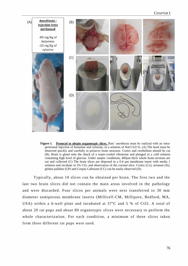
CHAPTER I
76
Figure 1. Protocol to obtain organotypic slices. Rats’ anesthesia must be realized with an intra-
peritoneal injection of ketamine and xylazine, in a solution of NaCl 0,9 %. (A) The head must be
dissected quickly and carefully to preserve brain structure. Cortex and cerebellum should be cut
(B). Brain is glued onto the chuck of a water-cooled vibratome and plunged in a cold solution
containing high level of glucose. Under aseptic conditions, 400µm thick whole brain sections are
cut and collected (C) The brain slices are disposed in a 0.4 µm membrane insert with media 1
solution and incubate in 5% CO2 and observation of the coronal slice. Cortex (Cx), striatum (St),
globus pallidus (GP) and Corpus Callosum (CC) can be easily observed (D).
Typically, about 10 slices can be obtained per brain. The first two and the
last two brain slices did not contain the main areas involved in the pathology
and were discarded. Four slices per animals were next transferred to 30 mm
diameter semiporous membrane inserts (Millicell -CM, Millipore, Bedford, MA,
USA) within a 6-well plate and incubated at 37ºC and 5 % of CO2. A total of
about 20 rat pups and about 80 organotypic slices were necessary to perform the
whole characterization. For each conditi on, a minimum of three slices taken
from three different rat pups were used.

CHAPTER I
77
Products Quantity Special instructions
Gey's balanced salt
solution
50 mL Must be at 4°C
D-glucose 6.5 mg/mL Must be filtrated at 0.22
µm
Antibiotics 1 % Streptomycine and
penicilline
Table 2. Description of the buffer solution needed during the first step of organotypic slice
preparation. This solution must be prepared under aseptic conditions.
Solution Products Quantity Special
instructions
Media 1
(From day 0 to
day 3)
Minimum Essential
Medium Eagle
(MEM) Alpha
Modifications
50 % sterile
Hanks balance
salt solution
25 % sterile
Horse serum 25 % Must be
inactivated
D-glucose 6.5 mg/mL Must be filtrated
at 0.22µm
L-glutamine 1 mM Must be filtrated
at 0.22µm
Antibiotics 1 % Streptomycine
and penicilline
Media 2
(From day 3 to
the end of the
experiment)
Neurobasal
media
sterile
D glucose 3.25 mg/mL Must be filtrated
at 0.22µm
L-glutamine 1 mM Must be filtrated
at 0.22µm
B27 2 X
Antibiotics 1 % Streptomycine
and penicilline
Table 1. Description of the media needed during all the organotypic slices culture. These
media should be prepared under aseptic conditions. Media should be renewed every 2 or 3 days
during culture.
2.2 SLICE ANGLE SELECTION TO OBTAIN A HUNTINGTON’S DISEASE MODEL.
Brains were cut with different angles to obtain the most complete
DARPP32 and GAD67-posit ive cell degeneration over t ime. For this analysis,

CHAPTER I
78
three angles have been chosen: sagittal , coronal and transversal . For sagittal
slices, brain hemispheres were separated and glued onto the chuck of a water -
cooled vibratome and slices were cut alongside of the midline. Concerning
coronal slices, cerebellum and olfactive bulbs/prefrontal cortex were cut off and
brains were glued, on their dorsal side, onto t he vibratome chuck. At last, to
perform transversal slices, the underside of the brain was glued on the
vibratome chuck. For each condition, 400 µm slices were performed with razor
blade angle of 14°.
2.3 HISTOLOGICAL STUDIES
At different times, ranging from 0 to 19 days, organotypic slices were
washed with phosphate-buffered saline (PBS) (Lonza, Verviers, Belgium), fixed
with 4 % paraformaldehyde (PFA) (Sigma–Aldrich, St Louis, MO, USA) in PBS
pH 7.4 for 2 h and then washed three times with PBS. Finally, sli ces were
removed from membrane inserts and stored at 4°C in PBS until use.
2.3.1 DARPP32 and GAD 67 Immunohistochemistry
A mouse anti GAD67 antibody (5 µg/mL, clone 1G10.2, Millipore SA,
Guyancourt , France) and mouse anti -DARPP32 (250 µg/mL, clone 15, DB
science, Le Pont de Claix, France) were used to observe striatal -globus pallidus
GABAergic pathway. Slices were incubated 48 h with the primary antibody
diluted into PBS, BSA 4 % at 4°C. After washes, sl ices were incubated with the
biotinylated anti -mouse secondary antibody (7.5 µg/mL, Vector Laboratories,
Burlingame, USA). Then sl ices were washed and quenching of peroxidase was
performed with 0.3 % H 2O2 (Sigma, St Louis, USA) in PBS , at RT for 20 min.
After PBS washes sl ices were incubated with Vectastain ABC r eagent (Vector
Laboratories, Burlingame, USA) in PBS at RT for 1 h. Sections were then
washed and revealed with 0.03 % H 2O2 , 0.4 mg/mL diaminobenzidine (DAB,
Sigma, St Louis, USA) in PBS, 2.5 % nickel chloride (Sigma, St Louis, USA)
and dehydrated before mounting.
2.3.2 Quantification of DARPP32 and GAD 67 positive fibers and cells
GAD67-positive fibers and DARPP32-positive neurons were quantified in
the striatum at different time-points, from 0 until 19 days post -lesion, using the

CHAPTER I
79
Metamorph® software from Molecular Devices. Results were presented as mean
differences +/ - average deviation and were ca lculated from 6 independent
pictures taken from 4 different rats for each group.
2.3.3 NeuN Immunohistochemistry
An antibody against neuronal nuclei (NeuN) (clone A60, Merck Mill ipore,
Billerica, MA, USA) was used to observe the viability of cells within the bra in
organotypic sl ices. Slices were incubated 48 h with the primary antibody diluted
into PBS BSA 4 % (1/50) at 4°C. After washes, slices were incubated with the
biotinylated anti -mouse secondary antibody (7.5 µg/mL, Vector Laboratories,
Burlingame, USA). Then sl ices were washed and quenching of peroxidase was
made with 0.3 % H 2O2 (Sigma, St Louis, USA) in PBS-T, at RT for 20 min.
After PBS washes, incubation with Vectastain ABC reagent (Vector
Laboratories, Burlingame, USA) in PBS was performed at RT for 2 h. Sections
were washed and revealed with 0.03 % H 2O2 , 0.4 mg/mL diaminobenzidine
(DAB, Sigma, St Louis, USA) in PBS, 2.5 % nickel chloride (Sigma, St Louis,
USA) and dehydrated before mounting.
2.3.4 Quantification of NeuN-positive neurons
The survival of total neurons in certain organotypic slice regions was
estimated by immunohistochemestry against NeuN. NeuN -positive neuron was
calculated, from days 0 to 19 using the Metamorph® software. At each time -
point, six pictures taken from three different slices showing cortex, striatum and
lateral septum were used. NeuN-positive neuron in these areas was expressed as
a percentage of NeuN staining at day 0 considered as positive control. Results
were presented as mean differences ± average deviation.
2.3.5 Statistical analysis
Data are presented as the mean value of three independent experiments ±
standard deviation (SD), unless otherwise stated. Significant differences
between samples were determined using an analysis of variance (ANOVA) test,
followed by a Scheffe post hoc test which indicates if conditions were
significantly different. P-value was set to 0.05, unless otherwise stated.

CHAPTER I
80
3.1 SLICE ANGLE SELECTION TO OBTAIN A HUNTINGTON’S DISEASE MODEL.
To determine the best model of GABAergic MSN neurodegeneration,
mimicking one of the pathological hallmarks of HD, the neonatal brain was cut
following three different axis to obtain the organotypic slices (Figure 2A and
2B) . With the three types of sectioning planes the striatum, cortex and globus -
pallidus, the brain areas mainly affected in HD were visible. The progressive
degeneration of MSN was evaluated by DARPP32, expression. We observed that
DARPP32 staining was present and observable in the whole striatum
independently of the slice plane chosen (Figure 2C) . Moreover, in horizontal
and sagittal slices, DARPP32 positive cells were still highly present in the
striatum after 4 days and even after 7 days of culture (Figure 2C) . In coronal
sections (Figure 2C), the number of DARPP32-positive cells was reduced by
around 30% after 4 days and up to 70% after 7 days. As a quick degeneration of
DARPP32-positive cells was desired, coronal sections were used for the rest of
this study. Moreover, the sections can be kept as a whole therefore containing
the striatum from the two hemispheres, providing a control in the same slice. If
desired, and in order to increase the viability of the sl ices they can also be cut
along the midline.
3. RESULTS

CHAPTER I
81
C
S
HGP
SN
C
S
H
GP
Hp
Cb
(A)
(B)
(C)
A
B
C
25µm
Saggital HorizontalCoronal
CS
Hp
GP
SN
Cb
Day
0D
ay 4
Day
7
Figure 2. Slice angle selection to model HD. Rat's brain was cut in order to obtain saggital,
coronal or horizontal brain slices (A). Which each different angles, we can vizualized all the areas
involved in the HD (B) DARPP32 was detected in brain slices obtained by immunofluorescency at
day 1, 4 and 7 after sagittal, coronal and horizontal (C) sections.
3.2 ORGANOTYPIC SLICES MORPHOLOGY AND VIABILITY OF CULTURED NEONATAL BRAIN
SLICES.
Culture conditions remarkably affect the organotypic culture viability. We
already described a protocol using 2 specific media with and without serum [29].
Using these conditions, the whole coronal HD organotypic slices can be kept
viable during at least 3 weeks in culture (Figure 3). We observed a decrease of
striatum volume from 4 days onwards and a good conservation of organotypic

CHAPTER I
82
slice's morphology during 3 weeks (Figure 3A). No important distortion or
flattening of the slices was detected. However, an important thinning of the
striatum within 6 days was observed by bright field microscopy, predicting a
reduction of viability of the striatal neurons (Figure 3A).
To confirm the viabili ty of organotypic brain slices, neurons were
visualized with an antibody against NeuN and quantified at day 0, 7, 11 and 19.
During the 3 weeks of culture the number of NeuN -positive cells decreased less
than 20 % +/- 4 % in the cortex and lateral septum compared to striatum, (Figure
3C and 3D), underscoring the viability of the neuronal cells in these areas.
However, only 20 % +/- 5 % of NeuN-positive cells were counted in the
striatum, explaining the decrease of volume observed during the culture and
confirming the neurodegeneration (Figure 3).

CHAPTER I
83
Figure 3. Morphology and viability of organotypic slices. Observation of coronal brain slices was
realized by bright field microscopy (A). Immunohistochemistry against neuronal nuclei (NeuN)
showing three brain regions: cortex, striatum and lateral septum at day 0, 11 and at day 19 (B) at
the bottom. Immunohistochemistry against neuronal nuclei (NeuN) showing the striatum (C)
Quantification of NeuN-positive cells in cortex, striatum and SN shows no important loss of
staining in the cortex after 19 days in culture (D). While NeuN expression decreased by 80 % and
27 % in the striatum. ∗: Significantly different results with n = 3 and P-value = 0.05. Pictures
about organotypic slides until day 19.
3.3 ORGANOTYPIC SLIDES CULTURES DISPLAY PROGRESSIVE DEGENERATION OF
GABAERGIC PATHWAY
In order to demonstrate the degeneration of medium spiny n eurons in the
striatum, we followed by immunohistochemistry the progressive decrease of
DARPP32 (Figure 4A) and GAD67 (Figure 4B), which are markers of

CHAPTER I
84
GABAergic MSNs. These markers were perfectly specific because we didn't find
any positive cells in the cortex (Figure 4A and 4B). The number of DARPP32-
positive neurons and GAD67-positive neurons decreased over time with around
a 50% decrease during the first week when compared to day 0 (Figure 4A and
4B). A quantification of the GABAergic marker density GAD 67 shows a
progressive loss of 30 % at day 4, 45 % at day 7 and 70 % at day 11 when
compared to day 0. It was almost complete by day 19 with an average of only 1
% staining left. However, the staining intensity varied between sl ices due to the
tissue damage (Figure 4D). Indeed, a more dramatic loss of DARPP32 staining
was detected over time with an average of 25 % decrease at day 2, 50 % at day 4
and 75% at day 7 compared to day 0, and was complete by day 19 (Figure 4C).
Even though the average loss of DARP P32 staining was a bit more rapid the
profile of these 2 graphs are similar, confirming the progressive loss of
GABAergic neurons in the organotypic sl ices.

CHAPTER I
85
Figure 4. Modelisation of specific MSN markers. Immunohistochemistry against striatal A)
DARPP32 positive neurons and B) GAD67 positive neurons at day 0, 7, 11 and 19. C) Striatal
DARPP32 positive neurons decreased progressively until day 11 and became total at day 19 in
comparison with day 0 which represents 100% represented with SD in doped lines. D) Striatal
GAD67 positive neurons quantification with SD in doped lines. A 10% decreased was detected at
day 2 which reached 30 % at day 4, 45 % at day 7 and reached 100 % at day 19, holding the fact
that day 0 represent 100 %. N=4
Organotypic slices have been widely used as model of neurological
pathologies including Parkinson's disease, brain stroke and cerebral ischemia
[16]. In the present work an innovative ex vivo model of HD in which
progressive striatal MSN degeneration was obtained in a single step while
preparing the slices, was developed. The MSN degeneration occurred in around
4. DISCUSSION

CHAPTER I
86
three weeks and was specific to the striatum as the viability of the other brain
regions was mainta ined during this time. We didn't observe morphological
modification except for the volume reduction in the striatum confirming what is
observed in HD. This simple model thus allows evaluating different
neuroprotective and neuroreparative therapeutic strate gies. Organotypic slices
were obtained from 6 to 8 day old pups It is well known and established that
tissue from embryonic or post natal donors survive better [30,31]. Slices post-
natal donors is recommended because at this age, brain slices will conserved a
better morphology, an increase survival and we will obtain a more stable models
regarding to the lesions. Animals were sacrificed between days 6 to 8 which
provides a perfect time window for establishing brain slice cultures on
membranes. More particularly, for Parkinson's disease the slices are principally
harvested between 4 and 9 weeks after birth [32,33].
Hypothermia has been shown to improve the preservation of
hippocampus brain slices [34–36]. In our study, in order to preserve brain slices
we insured that the brain was kept at 4°C in solution buffer containing high
percentage of glucose during the whole slice preparation process. We chose not
to place the head in ethanol 70 % bath as some teams have reported [31] in order
to maintain physiological conditions of organotypic brain slices. Within the
same line, the media containing horse serum was only used during the first 3
days even though horse serum diminishes tissue flattening, and promotes
survival of neurons, astroglia or microglia in organotypic brain slices [37]. In
order to diminish the variability between slices due to changes in serum batches
we then used a serum-free media containing glucose, glutamine, antibiotics and
B-27® . Indeed, serum increased the degeneration of neurons in brain slices when
the media contain serum for a long-time period [21]. In a previous study, we
confirmed the benefice of using horse serum during a short period followed by
“synthetic” media and accord ing to these results, we applied this method to
culture organotypic brain slices modeling HD. In this way, we are able to
maintain the viabili ty of brain ex vivo for at least 3 weeks, and obtained the HD
model in 5 days. Other studies demonstrated the ben efit to culture organotypic
slices without serum such as our study, and brain sl ices from transgenic mouse
or rat brain slice with injection of neurotoxins were principally cultured with

CHAPTER I
87
art ificial cerebrospinal fluid or specific media containing HEPES (( 4-(2-
hydroxyethyl)-1-piperazineethanesulfonic acid ) [38–40].
This organotypic model involves GABAergic MSNs degeneration due to
mechanical cutting of t issue between the striatum and substantia nigra, which
leads to retrograde and anterograde neuronal degeneration in the substantia
nigra and in the striatum, respectively. In this report , a complete
characterization of the behavior of MSNs and GABAergic pathway was assessed
by immunostaining through the visualization of the decrease of DARPP32 and
GAD67, two specific markers of MSNs [5]. It revealed that a rapid loss of
DARPP32-positive neurons occurred in the striatum followed by a decrease in
GAD67 staining. A decrease of 50% and 30% of the DARPP32 and the GAD67
positive cells respectively was detected 4 days after slice preparation compared
to day 0. This allows the development of an early model of th e disease, in which
a window of time of at least 15 days was obtained to study new therapeutic
approaches. One important reason to culture brain slices is to study and
characterize the interaction between neuronal populations. The coronal section
allows studying those parameters, thanks to the observation of the globus
pallidus, the striatum and all the structures involved in the HD in one slice. We
can easily imagine testing different molecules with therapeutic effect such as
growth factors directly onto s lices or in the media and observe their effect in all
the MSNs afferents and projections [41,42]. [43]. Indeed, with the same method,
we developed an innovative model of PD and we were able to evaluate new
therapeutic approaches combining stem cells and biomaterials releasing growth
factors in order to treat the disease or to reduce the related symptoms. [29]
In summary, this model represents a promising tool to quickly and
efficiently test innovating treatment in the HD such as regenerative medicine
with stem cells and biomaterials. The development of simple an d non expensive
model with precise characterization of neurodegeneration could allow to easily
study the therapeutic effect of treatment but also the role of microenvironment
in the response of the treatment.

CHAPTER I
88
In this study we developed and cha racterized a relevant ex vivo model of
HD using membrane techniques culture and specific media. This new model can
be use to study the early stage of the pathology because we observed the
reduction of striatum such as in the HD in 4 days. This represents a perfect
model to study the relevance of new and innovative treatment.
The authors thank the SCAHU and more particularly Pierre LEGRAS and
Jérôme Roux for animal care. We are also grateful to Jeremy RIOU for his
expertise in statistical analysis. This project is supported by the “Education
Audiovisual” and the cultural executive agency of the European Union through
the NanoFar Erasmus Mundus joint Doctoral program, by Angers Loire
Métropole.
[1] E.R. Fisher, M.R. Hayden, Multisource ascertainment of Huntington disease in Canada: Prevalence and population at risk: Multisource Ascertainment of BC HD Patients, Mov. Disord. 29 (2014) 105–114. doi:10.1002/mds.25717.
[2] A. Lekoubou, J.B. Echouffo-Tcheugui, A.P. Kengne, Epidemiology of neurodegenerative diseases in sub-Saharan Africa: a systematic review, BMC Public Health. 14 (2014) 653.
[3] F. Squitieri, A. Griguoli, G. Capelli, A. Porcellini, B. D’Alessio, Epidemiology of Huntington disease: first post- HTT gene analysis of prevalence in Italy: Prevalence of Huntington disease in Italy, Clin. Genet. (2015) n/a–n/a. doi:10.1111/cge.12574.
[4] C. Zuccato, M. Valenza, E. Cattaneo, Molecular Mechanisms and Potential Therapeutical Targets in Huntington’s Disease, Physiol. Rev. 90 (2010) 905–981. doi:10.1152/physrev.00041.2009.
[5] R.L. Albin, D.A. Tagle, Genetics and molecular biology of Huntington’s disease, Trends Neurosci. 18 (1995) 11–14.
[6] E.B. Clabough, huntington’s Disease: The Past, Present, and future search for Disease Modifiers, Yale J. Biol. Med. 86 (2013) 217.
[7] K.M. Shannon, A. Fraint, Therapeutic advances in Huntington’s Disease, Mov. Disord. Off. J. Mov. Disord. Soc. (2015). doi:10.1002/mds.26331.
[8] G. Cisbani, F. Cicchetti, An in vitro perspective on the molecular mechanisms underlying mutant huntingtin protein toxicity, Cell Death Dis. 3 (2012) e382. doi:10.1038/cddis.2012.121.
[9] I.-H. Park, N. Arora, H. Huo, N. Maherali, T. Ahfeldt, A. Shimamura, et al., Disease-specific induced pluripotent stem cells, Cell. 134 (2008) 877–886. doi:10.1016/j.cell.2008.07.041.
[10] T. Philips, J.D. Rothstein, M.A. Pouladi, Preclinical models: needed in translation? A Pro/Con debate, Mov. Disord. Off. J. Mov. Disord. Soc. 29 (2014) 1391–1396. doi:10.1002/mds.26010.
CONCLUSION
ACKNOWLEDGEMENTS
BIBLIOGRAPHY

CHAPTER I
89
[11] L. Wang, Z. Qin, Animal models of Huntington’s disease: implications in uncovering pathogenic mechanisms and developing therapies, Acta Pharmacol. Sin. 27 (2006) 1287–1302. doi:10.1111/j.1745-7254.2006.00410.x.
[12] N.D. Dey, M.C. Bombard, B.P. Roland, S. Davidson, M. Lu, J. Rossignol, et al., Genetically engineered mesenchymal stem cells reduce behavioral deficits in the YAC 128 mouse model of Huntington’s disease, Behav. Brain Res. 214 (2010) 193–200. doi:10.1016/j.bbr.2010.05.023.
[13] J. Rossignol, C. Boyer, X. Lévèque, K.D. Fink, R. Thinard, F. Blanchard, et al., Mesenchymal stem cell transplantation and DMEM administration in a 3NP rat model of Huntington’s disease: Morphological and behavioral outcomes, Behav. Brain Res. 217 (2011) 369–378. doi:10.1016/j.bbr.2010.11.006.
[14] K.D. Fink, J. Rossignol, A.T. Crane, K.K. Davis, M.C. Bombard, A.M. Bavar, et al., Transplantation of umbilical cord-derived mesenchymal stem cells into the striata of R6/2 mice: behavioral and neuropathological analysis, Stem Cell Res. Ther. 4 (2013) 130. doi:10.1186/scrt341.
[15] B.H. Gähwiler, M. Capogna, D. Debanne, R.A. McKinney, S.M. Thompson, Organotypic slice cultures: a technique has come of age, Trends Neurosci. 20 (1997) 471–477. doi:10.1016/S0166-2236(97)01122-3.
[16] N. Daviaud, E. Garbayo, P.C. Schiller, M. Perez-Pinzon, C.N. Montero-Menei, Organotypic cultures as tools for optimizing central nervous system cell therapies, Exp. Neurol. 248C (2013) 429–440. doi:10.1016/j.expneurol.2013.07.012.
[17] S. Cho, A. Wood, M.R. Bowlby, Brain Slices as Models for Neurodegenerative Disease and Screening Platforms to Identify Novel Therapeutics, Curr. Neuropharmacol. 5 (2007) 19–33.
[18] M.A. Johnson, V. Rajan, C.E. Miller, R.M. Wightman, Dopamine release is severely compromised in the R6/2 mouse model of Huntington’s disease, J. Neurochem. 97 (2006) 737–746. doi:10.1111/j.1471-4159.2006.03762.x.
[19] A.N. Ortiz, B.J. Kurth, G.L. Osterhaus, M.A. Johnson, Impaired dopamine release and uptake in R6/1 Huntington’s disease model mice, Neurosci. Lett. 492 (2011) 11–14. doi:10.1016/j.neulet.2011.01.036.
[20] E. Wilhelmi, U.H. Schöder, A. Benabdallah, F. Sieg, J. Breder, K.G. Reymann, Organotypic brain-slice cultures from adult rats: approaches for a prolonged culture time, Altern. Lab. Anim. ATLA. 30 (2002) 275–283.
[21] H. Kim, E. Kim, M. Park, E. Lee, K. Namkoong, Organotypic hippocampal slice culture from the adult mouse brain: a versatile tool for translational neuropsychopharmacology, Prog. Neuropsychopharmacol. Biol. Psychiatry. 41 (2013) 36–43. doi:10.1016/j.pnpbp.2012.11.004.
[22] J.B. Schulz, R.T. Matthews, T. Klockgether, J. Dichgans, M.F. Beal, The role of mitochondrial dysfunction and neuronal nitric oxide in animal models of neurodegenerative diseases, Mol. Cell. Biochem. 174 (1997) 193–197.
[23] C.T. Sheline, J. Zhu, W. Zhang, C. Shi, A.-L. Cai, Mitochondrial inhibitor models of Huntington’s disease and Parkinson’s disease induce zinc accumulation and are attenuated by inhibition of zinc neurotoxicity in vitro or in vivo, Neurodegener. Dis. 11 (2013) 49–58. doi:10.1159/000336558.
[24] P. Pierozan, C.G. Fernandes, M.F. Dutra, P. Pandolfo, F. Ferreira, B.O. de Lima, et al., Biochemical, histopathological and behavioral alterations caused by intrastriatal administration of quinolic acid to young rats, FEBS J. 281 (2014) 2061–2073. doi:10.1111/febs.12762.
[25] L.B. Menalled, Knock-in mouse models of Huntington’s disease, NeuroRx J. Am. Soc. Exp. Neurother. 2 (2005) 465–470. doi:10.1602/neurorx.2.3.465.
[26] L.B. Menalled, A.E. Kudwa, S. Miller, J. Fitzpatrick, J. Watson-Johnson, N. Keating, et al., Comprehensive behavioral and molecular characterization of a new knock-in mouse model of Huntington’s disease: zQ175, PloS One. 7 (2012) e49838. doi:10.1371/journal.pone.0049838.
[27] L. Menalled, D. Brunner, Animal models of Huntington’s disease for translation to the clinic: Best practices, Mov. Disord. 29 (2014) 1375–1390. doi:10.1002/mds.26006.
[28] L. Stoppini, P.A. Buchs, D. Muller, A simple method for organotypic cultures of nervous tissue, J. Neurosci. Methods. 37 (1991) 173–182.

CHAPTER I
90
[29] N. Daviaud, E. Garbayo, N. Lautram, F. Franconi, L. Lemaire, M. Perez-Pinzon, et al., Modeling nigrostriatal degeneration in organotypic cultures, a new ex vivo model of Parkinson’s disease, Neuroscience. 256 (2014) 10–22. doi:10.1016/j.neuroscience.2013.10.021.
[30] K. Ostergaard, S.A. Jones, C. Hyman, J. Zimmer, Effects of donor age and brain-derived neurotrophic factor on the survival of dopaminergic neurons and axonal growth in postnatal rat nigrostriatal cocultures, Exp. Neurol. 142 (1996) 340–350. doi:10.1006/exnr.1996.0203.
[31] C. Humpel, Organotypic brain slice cultures: A review, Neuroscience. (n.d.). doi:10.1016/j.neuroscience.2015.07.086.
[32] D. Plenz, S.T. Kitai, Organotypic cortex-striatum-mesencephalon cultures: the nigrostriatal pathway, Neurosci. Lett. 209 (1996) 177–180.
[33] D. Moses, J. Drago, Y. Teper, I. Gantois, D.I. Finkelstein, M.K. Horne, Fetal striatum- and ventral mesencephalon-derived expanded neurospheres rescue dopaminergic neurons in vitro and the nigro-striatal system in vivo, Neuroscience. 154 (2008) 606–620. doi:10.1016/j.neuroscience.2008.03.058.
[34] H. Wassmann, C. Greiner, S. Hülsmann, D. Moskopp, E.J. Speckmann, J. Meyer, et al., Hypothermia as cerebroprotective measure. Experimental hypoxic exposure of brain slices and clinical application in critically reduced cerebral perfusion pressure, Neurol. Res. 20 Suppl 1 (1998) S61–65.
[35] R. Ramani, Hypothermia for brain protection and resuscitation, Curr. Opin. Anaesthesiol. 19 (2006) 487–491. doi:10.1097/01.aco.0000245272.84539.97.
[36] M. Hernández-Guillamon, L. Ortega, C. Merino-Zamorano, M. Campos-Martorell, A. Rosell, J. Montaner, Mild hypothermia protects against oxygen glucose deprivation (OGD)-induced cell death in brain slices from adult mice, J. Neural Transm. Vienna Austria 1996. 121 (2014) 113–117. doi:10.1007/s00702-013-1090-1.
[37] C. Ullrich, N. Daschil, C. Humpel, Organotypic vibrosections: novel whole sagittal brain cultures, J. Neurosci. Methods. 201 (2011) 131–141. doi:10.1016/j.jneumeth.2011.07.021.
[38] R.J. Ferrante, Mouse models of Huntington’s disease and methodological considerations for therapeutic trials, Biochim. Biophys. Acta. 1792 (2009) 506–520. doi:10.1016/j.bbadis.2009.04.001.
[39] D. Colle, J.M. Hartwig, F.A. Antunes Soares, M. Farina, Probucol modulates oxidative stress and excitotoxicity in Huntington’s disease models in vitro, Brain Res. Bull. 87 (2012) 397–405. doi:10.1016/j.brainresbull.2012.01.003.
[40] A. Ferrante, A. Martire, R. Pepponi, K. Varani, F. Vincenzi, L. Ferraro, et al., Expression, pharmacology and functional activity of adenosine A1 receptors in genetic models of Huntington’s disease, Neurobiol. Dis. 71 (2014) 193–204. doi:10.1016/j.nbd.2014.08.013.
[41] C. Riley, B. Hutter-Paier, M. Windisch, E. Doppler, H. Moessler, R. Wronski, A peptide preparation protects cells in organotypic brain slices against cell death after glutamate intoxication, J. Neural Transm. Vienna Austria 1996. 113 (2006) 103–110. doi:10.1007/s00702-005-0302-8.
[42] G. Lynch, E.A. Kramar, C.S. Rex, Y. Jia, D. Chappas, C.M. Gall, et al., Brain-derived neurotrophic factor restores synaptic plasticity in a knock-in mouse model of Huntington’s disease, J. Neurosci. Off. J. Soc. Neurosci. 27 (2007) 4424–4434. doi:10.1523/JNEUROSCI.5113-06.2007.
[43] N. Daviaud, E. Garbayo, L. Sindji, A. Martinez-Serrano, P.C. Schiller, C.N. Montero-Menei, Survival, Differentiation, and Neuroprotective Mechanisms of Human Stem Cells Complexed With Neurotrophin-3-Releasing Pharmacologically Active Microcarriers in an Ex Vivo Model of Parkinson’s Disease, Stem Cells Transl. Med. 4 (2015) 670–684. doi:10.5966/sctm.2014-0139.

CHAPTER I
91
The use of organotypic slices allowed the development of a new simple
model of Huntington's disease. Indeed, GABAergic MSNs degeneration is
observed after mechanical cutting necessary to obtain brain slices. In this way,
no other supplementation with neurotoxins, quinoleic acid or 3 -NPA is needed
for the progressive loss of striatal neurons, which reduces the risk of
heterogeneous results. Usually, only some areas such as the striatum, the
hippocampus or cortex and striatum are used in ex vivo models of HD[42]
[44,52] Here we have all the regions involved in HD, the cortico -striatal-globus
pallidus area of one hemisphere or of both hemisphere s allowing to use one side
as control.
Nonetheless, the full characterization of this model is required. Indeed, we
followed NeuN and GABAergic markers for only 19 days. We should know how
much time brain slices survive and understand exactly the mechanism of the
decrease of DARPP32 and GAD67. DARPP-32 is a fundamental component of
the dopamine-signaling cascade, and its expression is essential to the ability of
dopamine to regulate the physiology of striatal neurons . In our study, the
decrease of GAD67 is progressive and comparable to NeuN while DARPP32
decreased drastically from day 5. The results presented here suggest a selective
impairment in several aspects of dopaminergic signaling. Indeed, dopaminergic
signaling has been already described in the li t erature to be involved in
presymptomatic defects and contribute to HD pathology [142] , and DARPP32 is
the first marker decreasing. DARPP32 is also a mediator of signaling cascades
in dopaminoceptive neurons which corroborate this hypothesis. So, our model
seems to be severely impaired in GABAergic neurons' abil ity to regulate the
physiological state of striatal neurons via dopamine. To confirm our hypothesis,
we can imagine checking the dopamine-regulated ion channels by
electrophysiological studies and determine if a reduction in the striat al-specific
dopamine targets, PKA, DARPP-32, ARPP-16, and ARPP-21 is associated, with
RT-qPCR or immunofluorescence.
DISCUSSION

CHAPTER I
92
The aim of this study has been to develop a simple model to screen
potential treatment including cell replacement with MIAMI cells combined with
PAMs. This model allows the development of an early model of the disease , in
which a window of time of at least 7 days was obtained if we considered
DARPP32 and 14 days if we considered GAD67 . The timing differences are
evidenced when comparing this model with those in which organotypic brain
slices are cultured during 3 weeks followed by the addit ion of neurotoxins in the
media in order to mimic HD [44–47]. Thus, stem cell transplants coupled or not
with MPA in this model enable the evaluation of the survival and differentiation
of grafted cells and the study of the behavior of the host t issue in particular
related to the evaluation of the secret ion of neurotrophic factors by stem cells
that can be made during 14 days. We can also easily imagine testing different
molecules with therapeutic effect such as growth factors directly onto slices or
in the media and observe their effect in all the MSNs a fferents and projections
[143,144]. In summarize, our model is so much faster, simpler and less
expensive than the in vivo model .

93
CHAPTER II

CHAPTER II
94
Transplantation of stem cells for the treatment of HD garnered much
attention prior to the turn of the century. Several studies using MSCs have
indicated that these cells have enormous therapeutic potentia l in this disease
[145,146]. In the case of HD, the beneficial effect of stem cel l transplantation
should be provided by the generation of new graft -derived neurons and by the
secretome of these cells [147]. MSCs are not only able to differentiate into
neuron-like cells but they also exert paracrine effects by modulati ng the
plasticity of damaged host tissues.
The trans-differentiation of MSCs into neural lineage has been achieved in
vitro by culturing them with specific media. This media is often composed of
growth factors or small molecules involved in neuronal linea ge differentiation.
The degree to which the cells will differentiate varies depending on the protocol
and cells used [148,79,149] . In all cases, although the MSCs were committed to
neuronal linage, the functionali ty at the end of the process was still lacking.
Attempting to understand how to improve the di fferentiation, REST also known
as neural-restrict ive silencing factor (NRSF) has been proposed as it functions
as a master negative regulator of neurogenesis [150]. REST is a zinc finger
transcriptional repressor able to bound the RE -1 sequence in his target gene
promoter which allows recruiting histone deacetylases and methyltransferase to
modulate the chromatin structure [151,152]. In non-neural cells , REST is
abundant and represses neuronal genes. Importantly, REST itself is
differentially regulated throughout neural development. In MSCs, the role of
REST was first investigated by Trzaska et al. (2008) with the goal to obtain
mature dopaminergic neurons. Indeed, the down -regulation of REST with
lentivirus has been demonstrated to promote neuronal commitment [102].
Knock-down of gene expression can be obtained by different ways and in
this study, we retained the siRNA epigenetic strategy to silence REST (siREST)
in MSCs as it specifically targets the expression of the gene of interest as
presented above (Introduction and aims) . To introduce siREST, we chose two
nanocarriers which present all the advantages that we discussed above
INTRODUCTION

CHAPTER II
95
(Introduction and aims). We focused on two innovative systems: lipid
nanocapsules and solid span nanoparticles.
Concerning LNCs, previous studies demonstrated the possibility to
encapsulate plasmid DNA to develop a gene therapy strategy [116]. For this,
theDNA is complexed with cationic lipids by electrostatic interactions leading
to formation of complexes called lipoplexes , which are added to other
components of the LNC. This strategy has demonstrated the capacity of LNCs to
transfect in vivo models of gliobastoma [117–119]. Strong of these results, the
encapsulation of siRNA in LNCs was successfully performed [153] and this
system was able to transfect melanoma and glioma cells in vitro [154,155].
Nonetheless, the instabili ty over-time of this system needed to be improved. In
this way, the optimization of LNC -siRNA had to be performed with two goals:
improving the stability over time of LNCs and protecting the siRNA from
denaturation. In any case this system provides a p ositive surface, which can
easily interact with negatively charged cell membranes. Although it is well
known that nanoparticles with a positive surface charge transfect very well , the
risk of toxicity is higher with those systems. However, recent evidence proves
that anionic nanocarriers can also be effectively internalized [156], and that the
transfection efficiency is not only dependent on the surface charge but also on
the particle size and composition [157,158].
So, to determine the optimal nanocarrier to transfect MSCs, we chose a
second system: Solid span nanoparticles (SP) to compare with LNCs. SP based
on sorbitan monooleate (Span® 80) have been specifically adapted to provide
effective siRNA association . Previous studies demonstrated the efficiency of SP
to associate DNA by incorporating to their composit ion positively charged
molecules like oleylamine (OA) [159]. This fatty amine is employed in the
creation of emulsions and other delivery systems [160]. Poly-L-arginine (PA), a
cationic moiety broadly used in gene therapy approaches [161] was also
evaluated. OA and PA were used to provide a positive surface charge for the SP
nanoparticles, thus enabling the association with the negatively charged, nucleic
acids thus resulting in Span® 80-oleylamine nanosystems (SP-OA) or Span® 80-
poly-L-arginine nanosystems (SP-PA), with a characteristic net negative surface
charge [122].Their capacity to provide higher in vivo t ransfection levels than

CHAPTER II
96
adeno-associated viral vectors (AAV2) has been recently reported [123]. In this
work, we conserved SP-PA to transfect MSCs and further modified the SP
nanoparticles in order to coat them with cationized pullulan (AP), a natural
polysaccharide obtaining SP-AP, as a strategy to easily associate siRNA by
electrostatic interaction [162].
In this chapter, to improve human stem cell transfection and efficiency
control of the stem cell fate, we have designed, optimized, characterized and
evaluated two different siRNA nanocarriers transpo rting siRNA.
Accepted in International journal of Pharmaceutics

97
PUBLICATION N°2

CHAPTER II
98
André E. 1,2 ; Pensado A3, Resnier P.1,2 , Braz L.4,5,6, Rosa da Costa A.M.4,7, Passirani
C.1,2 ; Sanchez A.3,8 and Montero-Menei C.N.1,2 *
1 PRES LUNAM – University of Angers, F-49933 Angers, France
2 INSERM U1066 – Micro et Nanomédecines Biomimétiques, 4 rue larrey, F-49933
Angers, France
3 Department of Pharmacy and Pharmaceutical Technology, Faculty of Pharmacy,
University of Santiago de Compostela, Campus Vida, 15782 Santiago de Compostela, Spain.
4 CIQA – Algarve Chemistry Research Centre, University of Algarve, 8005-139 Faro,
Portugal
5 School of Health – University of Algarve, 8000-510 Faro, Portugal
6 Department of Pharmaceutical Technology, Faculty of Pharmacy, University of Porto,
4050-313 Porto, Portugal
7 Department of Chemistry and Pharmacy, Faculty of Science and Technology, University
of Algarve, Campus de Gambelas, 8005-139 Faro, Portugal
8 Molecular Image Group. Health Research Institute-University Clinical Hospital of
Santiago de Compostela (IDIS), A Choupana, 15706 Santiago de Compostela, Spain.
* Corresponding author
C. MONTERO-MENEI
INSERM U1066, IBS-IRIS, 4 rue Larrey, 49 933 Angers Cedex 9, France
Tel : +33 244 688 536, Fax : +33 244 688 546 : E-mail : claudia.montero-menei@univ-
angers.fr
CHARACTERIZATION AND COMPARISON OF
TWO NOVEL NANOSYSTEMS ASSOCIATED WITH
SIRNA FOR CELLULAR THERAPY

CHAPTER II
99
ABSTRACT:
To direct stem cell fate, a delicate control of gene expression through
small interference RNA (siRNA) is emerging as a new and safe promising
strategy. In this way, the expression of proteins hindering neuronal commitment
may be transiently inhibited thus driving differentiation. Mesenchymal stem
cells (MSC), which secrete tissue repair factors, possess immunomodulatory
properties and may differentiate towards the neuronal lineage, are a promising
cell source for cell therapy studies in the central nervous system. To better drive
their neuronal commitment the repressor element -1 silencing transcription
(REST) factor, may be inhibited by siRNA technology. The design of novel
nanoparticles (NP) capable of safely delivering nucleic acids is crucial in order
to successfully develop this strategy. In this study we developed and
characterized two different siRNA NP. On one hand, sorbitan monooleate
(Span®80) based NP incorporating the cationic components poly -L-arginine or
cationized pullulan, thus allowing the association of siRNA were designed.
These NP presented a small size (205 nm) and a negative surface charge ( -38
mV). On the other hand, lipid nanocapsules (LNC) associating polymers with
lipids and allowing encapsulation of siRNA complexed with lipoplexes were
also developed. Their size was of 82 nm with a positive surface charge of +7
mV. Both NP could be frozen with appropriate cryoprotectors. Cytotoxicity and
transfection efficiency at different siRNA doses were monitored by evaluating
REST expression. An inhibition of around 60% of REST expression was
observed with both NP when associating 250 ng/mL of siRNA -REST, as
recommended for commercial reagents. Span NP were less toxic for human
MSCs than LNCs, but although both NP showed a similar inhibition of REST
over time and the induction of neuronal commitment, LNC-siREST induced a
higher expression of neuronal markers. Therefore, two different tailored siRNA
NP offering great potential for human stem cell differentiation have been
developed, encouraging the pursuit of further in vitro and in vivo in s tudies.
GRAPHICAL ABSTRACT:

CHAPTER II
100
Nanocarriers, siRNA, mesenchymal stem cells, neuronal differentiation,
REST.
MSC: mesenchymal stem cells; siRNA: small interfering RNA; SP: Span
80®; PA: poly-Arginine; AP: ammonium pullulan / cation ized pullulan; NP:
nanoparticles; DOPE: 1,2 -dioleoyl-sn-glycero-3-phosphoethanolamine; DOTAP:
1,2-dioleoyl-3- trimethylammoniumpropane; LNCs: lipid nanocapsules;
REST/NRSF: (Repressor Element -1 silencing Transcription /Neuron -Restrictive
Silencing Factor-1)
Mesenchymal stem cells (MSC) are multipotent stromal cells, capable of
self-renewal and able to differentiate into a diverse set of cells within the
mesoderm lineage [1]. They can also be an interesting source of cells for brain
regenerative medicine because they secrete tissue repair factors, possess
immunomodulatory properties and, in some conditions, may differentiate
towards the neuronal lineage [2–4]. In order to safely implant them in the brain
parenchyma they should be pre-committed to a neuronal phenotype. However,
protocols only using external growth factors to drive MSCs into specific
lineages seem to be insufficient to obtain high percentages of differentiated
cells [5,6]. A lentiviral-mediated RNA interference vector that down -regulates
the expression of REST/NRSF (Repressor Element -1 silencing Transcription
/Neuron-Restrict ive Silencing Factor-1) has been recently used to promote MSC
differentiation into neuronal cells, which exhibited neuron -like morphology and
function [7]. Indeed, REST/NRSF is a repressor transcription factor functioning
as a master negative regulator of neurogenesis by binding to a specific DNA
domain named RE1 motif [8,9]. In non-neural cells and in pluripotent stem
cells, REST is abundant and represses neuronal genes [10] Consequently, the
loss of REST in neural progenitors resulted in an acceleration of neuronal and
oligodendrocyte differentiation [11].
KEYWORDS
ABBREVIATIONS
1. INTRODUCTION

CHAPTER II
101
Nowadays, a transitory inhibit ion of targeted gene expression in cells may
be obtained by small interference RNAs (siRNA) without affecting the genome.
These siRNA are able to produce a partial or total extinction of targeted
messenger RNA (mRNA) and protein levels by degrading the mRNA. This
epigenetic modification is quite simple, does no t need genetic manipulation, is
transitory and is now quite well understood. Although current applications in
stem cells remain largely restricted to studies on molecular pathways and
signaling, siRNA can be also used as a biomedical strategy to improve su rvival,
direct lineage-specific differentiation of stem cells or help maintain a desired
phenotype [12,13]. Within this line, the inhibition of Noggin with siRNA was
capable to induce new bone formation in vivo [14]. But the mode of delivery of
these novel therapeutic agents is of crucial importance for an efficient ex vivo
or in vivo therapeutic strategy.
In the last decade, two different approaches for siRNA delivery have been
developed: vi ral and non-viral vectors. Non-viral vectors (also named
nanoparticles (NP) or nanocarriers) have been formulated to associate and to
efficiently and safely deliver nucleic acids both in vitro and in vivo , opening
many possible applications. In addit ion, t hey have many advantages and
potentialities, including large -scale manufacture, low toxicity and low
immunogenicity, and the possibility to customize them to target specific cell
types [7,8]. Initially, NPs were designed to have a pos itive surface charge, in
order to improve the interaction with the cell membrane that has a negative
surface, and to avoid electrostatic repulsion [9]. However, recent evidence
proves that anionic NPs can also be effectively internali zed [15], and that the
transfection efficiency is not only dependent on the surface charge but also on
the particle size and composition [16,17] . In this frame, our research group has
reported a novel NP based on sorbitan monooleate (Span ® 80) (SP) that can be
prepared using a simple, one-step and easily scalable procedure [18]. SP is a
component widely used in the pharmaceutical industry (generally recognized as
safe (GRAS)) due to its non-ionic surfactant properties at low concentrations
[19]. These NPs are able to transfect in vivo the retinal pigment epithelial cells
with a similar efficiency as adeno-associated viral vectors [20]

CHAPTER II
102
Transfection techniques currently use packaging particles such as
liposomes to facilitate the cellular uptake of RNAi. They have s hown high
biocompatibility and are available in the market such as l ipofectamine ®,
oligofectamine® , ready to be used. However, they exhibit some disadvantages
such as high positive zeta potentials destabilising cell membrane, low
transfection efficiencies in human stem cells, and instabili ty immediately after
complexation [21]. Moreover, transitional action reported with these liposomal
carriers is not sufficient on their own to have an ef fect over-t ime . In vitro
liposome transfection efficiency is also affected by the type of culture media
and cell plating density, and does not provide an efficient protection against
lysosomal nucleic acid degradation [22]. Lipid nanocapsules (LNCs) consisting
of a lipid liquid core of triglycerides and a rigid shell of lecithin and
polyethylene glycol hydroxystearate, are formulated by a simple and easily
industrialized solvent-free process based on the phase inversion of an emulsion
[23,24]. They allow entrapping the liposomes within their core and were thus
recently modified to encapsulate the siRNA complexed with DOTAP (1,2 -
dioleyl-3- trimethylammoniumpropane) and DOPE (1,2 -dioleyl-sn-glycero-3-
phosphoethanolamine), protecting the siRNA from degradation [25]. DOTAP
lipids presenting a cationic charge, allow the fixation of siRNA by electrostatic
force and the interaction with the cell membrane improving the cellular uptake
[26–28]. Moreover, DOTAP and DOPE are able to destabilize lysosome’s
membranes by a proton sponge effect [29]. However, the stability of these LNCs
should be improved.
The transfection efficiency of the siRNA delivered by NP is often affected
by the proliferative activity of human stem cells, and varies widely depending
on the type of target cells [30]. To ameliorate the transfection of human stem
cells and efficiently induce a neuronal -l ike differentiation, we have designed a
novel SP-based NP conveying REST and further developed LNCs incorporating
siREST with a good stability over time. [20]. First , we have modified SP NPs
with cationized pullulan (AP) as a strategy to easily associate siRNA by
electrostatic interaction thus obtaining a novel NP. Pullulan is a non -toxic
natural polysaccharide presenting non-immunogenic properties [31]. This FDA

CHAPTER II
103
approved component possesses a wide range of industrial applications including
health care and pharmacy [31]. Secondly, we further developed LNCs, and
evaluated different formulations of LNCs incorporating siREST in order to
obtain a better stability. The siREST -NPs were characterized by dynamic light
scattering, UV spectrophotometry and electron microscopic methods in order to
improve the understanding of their organization and structure. After evaluation
of the long-term storage and stability of siRNA -NPs over time, we tested the
cytotoxicity of both systems on human (h)MSCs and, finally, we evaluated their
abili ty to transfect hMSCs and drive the commitment of hMSCs towards a
neuronal phenotype determined by expression of neuronal genes.
2.1 FORMULATION
2.1.1 Synthesis of pullulan derivate :
Pullulan was chemically modified in order to obtain a posit iv ely charged
derivative (ammonium pullulan, AP) by alkylation of the original polymer.
Synthesis was performed based on a previously described methodology [32]: an
aqueous solution (5 mL) of KOH (0.504g, 9 eq), was prepared in a round bottom
flask, under st irring, at 60 ºC, to which pullulan (500 mg) and 3.6 2 mL (4.09 g,
27 eq) of GTMAC were added. After 5 h, an equal amount of GTMAC was
added to the mixture, which was allowed to react until the completion of 24 h. It
was then diluted with 10 mL of miliQ water, allowed to cool down to room
temperature, and neutralized with HCl (2M). The resulting solution was dialyzed
for 3 days, the water being replaced every 24 h, and the modified polymer
recovered by freeze drying.
2.1.2 SP-PA and SP-AP nanoparticles associated with siRNA
To produce the SP nanoparticles, a solut ion of 6.6 mg/ml of sorbitan
monooleate (Span ® 80) (Sigma, Saint-Quentin Fallavier, France) in ethanol
(organic phase) was prepared and it was subsequently added under magnetic
stirring to an aqueous phase, leading to the spontaneous nanoparticle formation
2. MATERIALS AND METHODS

CHAPTER II
104
[18]. To produce SP-PA and SP-AP nanoparticles, Poly-L-arginine (PA) or
ammonium pullulan (AP) were dissolved in Mili -Q water at 0.16 mg/ml. Finally,
ethanol was removed under reduced pressure on a rotary evaporator and the
nanoparticles were concentrated to a final volume of 10 ml. Nanopar ticles were
isolated by filtrat ion-centrifugation (Amicon Ultra-0.5 centrifugal filter
Devices, Merck Millipore, Ireland). The genetic materials (siRNA REST and
Scramble) were associated to the nanoparticles ' surface at different
concentrations (from 0.1mg/ml to 0.5mg/ml) by incubation with nanoparticles at
a 1:1 (v:v) ratio (100-100 µl) under magnetic starring at room temperature
during 2 hours in order to obtain siRNA -SP-AP and siRNA-SP-PA
nanoparticles.
2.1.3 Liposomes and lipoplexes
For liposome preparation, a cationic lipid DOTAP (1,2-dioleyl-3-
trimethylammoniumpropane) (Avanti® Polar Lipids Inc., Alabaster, AL, USA),
solubilized in chloroform, was weighted at a 1/1 molar ratio with the neutral
lipid DOPE (1,2-dioleyl-sn-glycero-3-phosphoethanolamine) (Avanti® Polar
Lipids Inc. , Alabaster, AL, USA) to obtain a final concentration of 30 mM of
cationic l ipid, (considering the number of charges per lipid molecule), i .e 1 for
DOTAP. After chloroform evaporation under vacuum, deionized water was
added to rehydrate the lipid film overnight at 4 °C which was sonicated the day
after during 30 min.
Lipoplexes were formulated by simple equivolumar mix of siRNA and
liposomes. This complex is characterized by the charge ratio [27] corresponding
to the ratio between positive charge of lipids and negative charge of nucleic
acids at 5.
2.1.4 siRNA-LNCs
LNCs were formulated, as described previously [23] by mixing 20 % w/w
Labrafac® WL 1349 (caprylic-capric acid triglycerides, Gatefossé S.A. Saint -
Priest, France), 1.5 % w/w Lipoid S75 -3® (Lecithin, Ludwigshafen, Germany),
17 % w/w Kolliphor® HS 15 ( Polyethylene glycol -15-Hydroxystearate HSPEG
BASF, Ludwigshafen, Germany), 1.8 % w/w NaCl (Prolabo, Fontenay -sous-
CHAPTER II
105
Bois, France) and 59.8 % w/w water (obtained from a Milli -Q system, Millipore,
Paris, France) together under magnetic stirring. Briefly, three temperature
cycles between 60 and 95 °C were performed to obtain phase inversions of the
emulsion. A subsequent rapid cooling and dilution with ice cooled water (1:1.4)
at the last phase inversion temperature (PIT) le d to blank LNC formation.
To obtain siRNA LNCs, the water introduced at the last PIT was replaced
by lipoplexes, i .e. REST siRNA: (sense sequence: 5 ′ - CAG-AGU-UCA-CAG-
UGC-UAA-GAA -3′; Eurogentec, Seraing, Belgium) and control (scrambled)
siRNA (sense sequence: 5 ′- UCUACGAGGCACGAGACUU-3′; Eurogentec,
Seraing, Belgium) complexed with cationic liposomes in a defined charge ratio
as described above.
To obtain siRNA LNCs, the water introduced at the last PIT was replaced
by lipoplexes, i .e. REST siRNA: (sense sequence: 5 ′- CAG-AGU-UCA-CAG-
UGC-UAA-GAA -3′; Eurogentec, Seraing, Belgium) and control (scrambled)
siRNA (sense sequence: 5 ′- UCUACGAGGCACGAGACUU-3′; Eurogentec,
Seraing, Belgium) complexed with cationic liposomes in a defined charge ratio
as described above.
To avoid the possible denaturation of siRNA by the high temperatures, the
addition of lipoplexes at 75°C or 40°C was tested within the classical
formulation. Two other formulations were performed, one with reduce PIT by
increasing the NaCl concentration to 8% in the formulation and the last one
without l ipoid®.
2.2 CHARACTERIZATION OF BOTH NANOPARTICLES
2.2.1 Size, zeta potential, transmission electron microscopy and cryo-transmission electron
microscopy
Size and zeta potential of LNCs and SP were measured by D ynamic Light
Scattering (DLS) method using a Malvern Zetasizer ® apparatus (Nano Series ZS,
Malvern Instruments S.A., Worcestershire, UK) at 25 °C, in triplicate, after
dilution in a ratio of 1:100 with deionized water. These parameters were
followed daily during one month.

CHAPTER II
106
The morphological examination of siRNA -SP-AP was conducted by
transmission electron microscopy (TEM) (CM 12 Philips, Eindhoven, The
Netherlands). Samples were placed on copper grids with carbon films (400
mesh) (Ted Pella, USA) and stained with 2% (w/v) phosphotungstic acid
solution for TEM observation.
The morphological examination of siRNA -LNC for cryo-TEM imaging were
prepared using a cryo-plunge cryo-fixation device (Gatan, Pleasanton USA) in
which a drop of the aqueous suspension was deposited on a carbon-coated grid
(Ted Pella Inc., Redding, USA). The TEM grid was then prepared by blott ing
the drop containing the specimen so that a thin liquid layer remained across the
holes of the carbon support film. The liquid film was vitrified by rapidly
plunging the grid into liquid ethane cooled by liquid nitrogen. The vitrified
specimens were mounted in a Gatan 910 specimen holder (Gatan, Pleasanton,
USA), which was inserted into the microscope using a CT -3500-cryotransfer
system (Gatan, Pleasanton, USA), and cooled with liquid nitrogen. TEM images
were obtained from specimens preserved in vitreous ice and suspended across a
hole in the supporting carbon substrate
2.2.2 Encapsulation efficiency (EE)
Qualitative BET electrophoresis detection - The EE and the integrity of
siRNA molecules after the process of nanoparticle formulation were evaluated
by agarose gel electrophoresis. A volume of nanoparticle suspension equivalent
to 2.5 μg of siRNA before and after treatment with Triton® X100 (Sigma, Saint -
Quentin Fallavier, France) was mixed with gel -loading solution (Sigma, Saint -
Quentin Fallavier, France) and disposed in each well of 1% agarose gel
containing ethidium bromide (Sigma, Saint -Quentin Fallavier, France) [12].
Free siRNA in solution corresponding to 2.5 μg constituted the controls.
Samples were let to migrate 40 min at 50 V and 10 min at 90V in a Tris –EDTA
buffer (Sigma, Saint -Quentin Fallavier, France) .
Quantitative UV detection – A spectrophotometric method based on the
study recently reported by David et al ., (2012), was used to evaluate the EE for
LNC. Briefly, siRNA LNCs were mixed with chloroform and water to separate
respectively hydrophilic and lipophilic components, sodium hydroxide was

CHAPTER II
107
added to destabilize lipoplexes and finally absolute ethanol to destroy LNCs
[28]. After multiple centrifugations, four compartments were obtained: free
siRNA, free lipoplexes (i.e. siRNA associated with liposomes), encapsulated
siRNA and encapsulated lipoplexes within LNCs.
siRNA and SP-AP were separated from supernatant using Nanosep® Omega
30 kD microcentrifuge filters (Pall Corporation, Ann Arbor, USA) and siRNA
was measured in the supernatant. To determine the concentration of siRNA,
optical density of each sample was determined at 260 nm by UV
spectrophotometer (UV-2600, Shimadzu, Noisiel , France) in triplicate
conditions. The EE was determined by dividing the experimental drug loading
by the theoretical drug loading. EE was followed every week during one month
by these two methods.
2.2.3 Encapsulated siRNA stability
In order to determine the stabil ity of the encapsulated siRNA during the
transfection, a spectrophotometric method based on the study recently reported
by David et al ., (2012), was used to evaluate the encapsulated si -RNA of LNCs.
Briefly, siRNA LNCs were dissolved at a ratio of 1:10 in Opti -MEM® media
(Life technologies, France) to mimic the transfection condition at two
temperatures: 4°C (to mimic the storage condition) and 3 7°C (to mimic
transfection condition in the incubator). Methods used to separate the different
phases were described earl ier. The encapsulated siRNA was determined by
dividing the experimental drug loading by the theoretical drug loading and
taking into account the dilution. It was followed at different times: 0, 4, 8, and
12 h after formulation. To confirm results, the integrity of siRNA was evaluated
by agarose gel electrophoresis in the same conditions.
2.2.4 Freezing
The long-term storage assessment of siRNA-LNCs and siRNA-SP-AP was
carried out by freezing at -20°C. The siRNA-LNCs and siRNA-SP-AP were
frozen with phosphate buffered saline (PBS) (control solution) or with an
aqueous solution of trehalose or of glucose as cryoprotectants in a 1:1 (v/v)
ratio nanoparticles:cryoprotectants with a range from 1% to 15%. They were

CHAPTER II
108
then analyzed after 1-month storage at -20°C by evaluating size and zeta
potential measures by Dynamic Light Scattering (DLS) method using a Malvern
Zetasizer® apparatus (Nano Series ZS, Malvern Instruments S.A.,
Worcestershire, UK) at 25 °C, in triplicate, after dilution in a ratio of 1:100
with deionized water. The integrity of siRNA molecules after storage at -20°C
of nanoparticle formulations was evaluated by agarose gel electrophoresis.
2.3 MENSENCHYMAL STEM CELL (MSC) ISOLATION AND EXPANSION:
Whole bone marrow was obtained from the il iac crest of 15 -yr-old (#34984)
living male donor (Lonza, Walkersville, MD). Cells were seeded with -MEM :
BE 12169F (LONZA) supplemented with 10% FBS, L -Glutamine 2mM (Sigma-
aldrich, France, G7513) FGF 1 ng/ml, Heparin 5 µg/ml, 1% penicillin-
streptomycin (Sigma, Saint -Quentin Fallavier, France). The cells were
maintained at 37°C with 5% CO 2 humidified atmosphere. Passage numbers
between 5-7 were used for the following experiments.
2.4 MSC TRANSFECTION
Experiments were performed in 1mL of MSC Opti -MEM® media (Life
technologies, France). Oligofectamine ® (Life technologies, France), which
served as a positive control, was used with 100 ng or 250 ng of siRNA. SiRNA -
LNCs were filtered with a 0.2 μm filter (Acrodisc PALL GHP, VWR, Radnor,
USA). siRNA-LNCs and siRNA-SP-AP were incubated with cells at 37 °C in a
humidified atmosphere with 5 % CO 2 for 4h before serum addition, following
Oligofectamine–siRNA protocol. Cells were harvested after appropr iate time
and assayed for mRNA expression levels by RT -qPCR and protein expression
levels by western blot.
2.5 IN VITRO VIABILITY:
Cytotoxicity assays were performed using MTS (3 -(4,5-dimethylthiazol -2-
yl)-5-(3-carboxymethoxyphenyl) -2-(4-sulfophenyl)-2H tetrazolium) (Promega,
Madison, USA). and AlamarBlueR (resazurin) (Invitrogen). Twenty-five
thousand cells , cells were transfected in a 24 -well plate with the siRNA-REST
and siRNA scramble with increasing concentrations of siRNA (0.05, 0.1, 0.25,

CHAPTER II
109
0.5 and 1 mg/mL). To determine the IC 5 0 , cells were subsequently cultured
during two days and 20 µL of MTS/well was added and plates incubated 2.5 h at
37 °C in a humidified atmosphere with 5 % CO 2 . The OD was evaluated by
Mutliskan Ascent (Labsystems, Fisher Scientific , Wilmington, USA) at 492 nm.
For the AlamarBlue assay, 10% AlamarBlue assay was added in RPMI media
without serum and phenol red. After 4h of incubation, the fluorescence was read
using a SpectraMax fluorescence multi -well plate reader (Molecular Devices,
Sunnyvale, CA) at 530/590 excitation/emission wavelengths. Three replicates
were used for each treatment.
Furthermore, cell numbers were also counted after each transfection
performed. Forty-eight hours after transfection 200.000 cells were resuspended
by trypsin (0.5 g porcin trypsin, 0.2 g EDTA; Sigma, Saint Louis, USA), washed
twice with PBS and trypan blue staining was used to count the number of cells
per well.
2.6 RNA EXTRACTION, RT-QPCR :
These experiments were performed following the guidelines of th e PACEM
core facility ("Plate-forme d'Analyse Cellulaire et Moléculaire”, Angers,
France). Sense and antisense desalted primer pairs (Eurofins MWG Operon,
Ebersberg, Germany) were mixed in RNAse free water at a final concentration
of 5 µM (Table 1). Total RNA of cells were extracted and purified using
RNeasyMicrokit (Qiagen, Courtaboeuf, France), and treated with DNase (10 U
DNase I/µg total RNA). RNA concentrations were determined using a ND -2000
NanoDrop (Thermo Fisher Scientific, Wilmington, Delaware USA ) and used for
normalization of the input RNA in the RT. First strand complementary DNA
(cDNA) synthesis was performed with a SuperScriptTM II Reverse Transcriptase
(Invitrogen), in combination with random hexamers, according to the
manufacturer’s instruct ions. Following first -strand cDNA synthesis, cDNAs
were purified (Qiaquick PCR purification kit, Qiagen, Courtaboeuf, France) and
eluted in 40 µL RNAse free water (Gibco). 2,5ng of cDNA was mixed with
MaximaTM SYBR Green qPCR Master Mix (Fermentas) and pri mer mix (0.3
µM) in a final volume of 10µL. Amplification was carried out on a Chromo4
thermocycler (Biorad) or LightCycler 480 (Roche) with a first denaturation step
at 95°C for 10 min and 40 cycles of 95°C for 15 s, 60°C for 30 s. After

CHAPTER II
110
amplification, a melting curve of the products determined the specificity of the
primers for the targeted genes. Several housekeeping genes, Glyceraldehyde -3-
phosphate dehydrogenase (Gapdh), Beta -2-microglobulin (B2M), Beta actin
(Actb), and Heat shock 90 kDa protein 1 be ta (Hspcb) were tested for
normalization. The GeNormTM freeware (http://medgen.ugent.be/ -
jvdesomp/genorm/) was used to determine that GAPDH and ACTB were the two
most stable housekeeping genes. The relative transcript quantity (Q) was
determined by the del ta Cqmethod Q=E(Cq min in all the samples tested - Cq of
the sample), where E is related to the primer efficiency (E=2 if the primer
efficiency=100%). Relative quantities (Q) were normalized using the multiple
normalization method described in Vandesompele et al [33]. Q
normalized=Q/(geometric mean of the three most stable housekeeping genes Q).
The 2(-ΔΔCt) method was retained, using the housekeeping genes and gene of
interest tested on control sample and treated sample [34].

CHAPTER II
111
Gene Full name NM accession number
sequences
GAPDH glyceraldehyde-3-
phosphate
dehydrogenase
NM_001289745.1 Fwd:CAAAAGGGTCATCATCTCTGC
Rev:AGTTGTCATGGATGACCTTGG
ACTB Actin NM_001101.3 Fwd: CCAGATCATGTTTGAGACCT
Rev: GGCATACCCCTCGTAGAT
Β3-TUB Tubulin beta 3 class
III NM_006086 Fwd: CCAGTATGAGGGAGATCG
Rev: CACGTACTTGTGAGAAGAGG
REST RE1-silencing
transcription factor NM_001193508.1 Fwd: ACTCATACAGGAGAACGCC
Rev: GTGAACCTGTCTTGCATGG
SCG10 stathmin 2 NM_001199214.1 Fwd: TGTCACTGATCTGCTCTTGC
Rev: AGAAGCTAAAGTTCGTGGGG
NFM Neurofilament,
medium polypeptide
(Variant 1)
NM_005382.2 Fwd: GACCTCAGCAGCTACCAG
Rev: TAGTCTCTTCACCCTCCAG
Table 2. Sequence of primers validated in RT-qPCR
2.7 WESTERN BLOT:
Total proteins were isolated by sonication of cells in lysis buffer composed
of 50 mM HEPES, pH 7.5, 150 mM NaCl, 1 mM EDTA, pH 8, 2.5 mM EGTA,
pH 7.4, 0.1% Tween 20, 10% glycerol, 0.1 mM sodium orthovanadate, 1 mM
sodium fluoride, 10 mM β -glycerophosphate, 0.1 mM phenylmethylsulfonyl
fluoride (PMSF), and complete protease inhibitor cocktail (Roche). The
quantification of protein used was performed according to Bradford method
(26).
Twenty μg of protein extracts were separated by SDS -10% PAGE and
transferred to PVDF membranes (GE Healthcare, VWR, Milan, Italy). The
membranes were incubated overnight with rabbit anti -REST antibody (1/200)
(Abcam, Cambridge, UK), washed with Tris -buffered Saline and incubated with
anti-rabbit (1/5000) (32460, Thermo scientific) for 6 hours. Immunostaining was
revealed by the Immuno-Star HRP substrate (BioRad, Segrate (MI), Italy)

CHAPTER II
112
according to manufacturer’s instructions and quantified by Kodak Ima ge Station
440CF. The image analysis was performed using the Kodak 1D 3.5 software
(27). To confirm that equal amounts of protein were lo aded membranes were
incubated with anti -β actin (Sigma-Aldrich, Italy) and revealed in the same way.
2.8 STATISTICAL ANALYSIS:
Three independent biological replicates were performed for all experiments
described in this paper. Comparisons between all groups, supposed with normal
distribution, were performed using a classical analysis of variance (one -way
ANOVA) followed by a Tukey’s post-hoc analysis. The encapsulation
efficiencies for modified LNCs and non -modified LNC were compared using a t -
test . Statistical significance was ascribed to a threshold p -value of 0.05 (*p≤
0.05; **p≤ 0.01; ***p≤ 0.001).
3.1 SP NANOPARTICLES: SELECTION OF SURFACE COMPOSITION AND SIRNA DOSE.
Using SP we can spontaneously form negatively charged nanoparticles
with a size of about 140 nm and a zeta-potential of - 17 mV, being these
nanoparticles stable during at least 1 month (Figures 1A and 1B). However, the
negative surface charge of this nanocarrier renders a stable interaction with the
negatively charged siRNA difficult to accomplish. In order to modify the
surface charge of these SP nanoparticles and to easily associate the siRNA to
the nanoparticle surface we evaluated the effect of incorporating on their
composition two different cationic molecules, poly-L-ariginine (PA) and
ammonium pullulan (AP) to the SP formulation. The developed SP -PA and SP-
AP blank nanoparticles (no siRNA associated) showed a nanoparticle size of 180
nm and 140 nm, respectively, which did not vary much from the SP
nanoparticles. They also presented appropriate positive surface charges of
3. RESULTS

CHAPTER II
113
+40mV and +38mV, respectively (Figure 1B). No change in terms of size or
zeta potential were observed when NPs were conserved for 1 month at 4°C
(Figures 1A and 1B) . Once developed, we complexed each system with the
siRNA-scramble and the siRNA REST by simply mixing them together. Similar
sizes and zeta-potentials were observed with both siRNAs. At day zero, the
increase in size and the decrease on surface charge for siRNA -SP-PA NPs (280
nm and -37 mV) and siRNA-SP-AP NPs (205 nm and -38mV) (Figures 1A and
1B) indicated an effective electrostatic interaction between siRNA and the
nanoparticles. On the contrary, when combining AP and siRNA to form AP -
siRNA complexes as a control formulation a broad -range particle size
distribution was observed, being characterized by the presence of aggregates
rather than nanosystems. We also performed a stability study with siRNA -SP-PA
and siRNA-SP-AP during 30 days. siRNA-SP-AP and siRNA-SP-PA were both
perfectly stable in terms of size and in surface charge (Figures 1A and 1B).
We selected SP-AP for the rest of experiments because pullulan is a non -toxic
FDA approved natural polysaccharide presenting non -immunogenic properties
with very interesting characteristics. Indeed, pullulan may be chemically
modified in order to afford either hydrophobized or cationized derivatives, the
former with the abil ity to carry hydrophobic molecules, and the latter showing
high affinity towards DNA and RNA [35].
To determine the most favorable siRNA co ncentration capable of
interacting with these NPs, we have tested different concentrations of siRNA
ranging from 0.1 mg/ml to 0.5 mg/ml and checked the size and zeta -potential for
each one. Similar particle sizes were obtained in this concentration range
(around 190 nm). However, the surface charge decreased when the siRNA
concentration increased, demonstrating the high capacity of AP to complex

CHAPTER II
114
siRNA on the surface. In this case, we therefore speak of association efficiency.
Above 0.3mg/ml of siRNA, we observed a stagnation of the surface charge ( -45
mV) (Figure 1C) revealing a saturation of the system. This observation was
confirmed by electrophoresis experiments performed to further assess the siRNA
association ability of the developed SP-AP NPs. The absence of the typical
bands of free siRNA in the formulations of NPs incubated with 0.1 and 0.3
mg/ml siRNA corroborated the effective association to the nanosystems in this
concentration range (Figure 1D). At 0.3 mg/ml of siRNA the efficiency of
association with the SP-AP was 33 % +/- 6 and we selected this dose for the
subsequent
studies.

CHAPTER II
115
Figure 1. Characterization of SP nanoparticles with different surface composition and siRNA
dose: (A ) Size of blank nanoparticles based on SP, SP and poly-arginine (SP-PA) or SP and
cationized pullulan (SP-AP), siRNA-SP-PA and siRNA-SP-AP at days 0 and 30 after storage (D0
and D30): (B) zeta-potentials at days 0 and 30 after storage; (C) Size and zeta-potentials of
nanoparticles associating siRNA doses ranging from 0.1 to 0.5 mg/ml; (D) Electrophoresis
experiments performed with free siRNA (Line 1) and siRNA-SP-AP associating different siRNA
at different concentrations, such as (2): 0.1 mg/mL of siRNA; (3): 0.3 mg/mL; (4): 0.4 mg/mL and
(5): 0.5 mg/mL. Free siRNA can be observed in the lines 4 and 5 illustrating the saturation on the
association efficiency of the nanocarrier. Results are expressed as mean ± standard deviation
(n=3). No difference was demonstrated on stability of SIRNA-SP-AP using ANOVA 1W, post-
hoc Tukey.

CHAPTER II
116
3.2 OPTIMISATION OF SIRNA-LNC
Initially, during siRNA-LNC formulation lipoplexes were added at the
beginning of the last phase inversion temperature (PIT) (75°C), but an
instability of the system has been observed [33]. To improve the stability and to
avoid the possible denaturation of siRNA by the high temperatures this initial
formulation was further modified. First, we increased the NaCl concentration to
8% thus obtaining a PIT at 60°C. In a second experime nt, a novel formulation
(modification of some components recently patented -ref: FR 4185991, 24 Sept
2014) was developed and the lipoplexes were also introduced at lower
temperatures (40°C). Blank LNCs were used as control because their size and
zeta potential were perfectly stable for 2 months ( Figures 2A and 2B ). We first
noticed a difference in size (increased size) and zeta potential (increased
surface charge) when comparing blank -LNC and the siRNA-LNCs formulations
developed under the different experim ental conditions (75°C, 60°C and 40°C),
which suggest an effective encapsulation of lipoplexes in LNCs. The initial size
(D0) of the siRNA-LNCs prepared at 75°C progressively decreased during a 2
months storage period from an initial value of 102 nm, where as an increase of
zeta-potential can be observed. These results suggest and instability and a
rearrangement of the system (Figures 2A and 2B ). The same behavior was
observed with the formulation having a PIT at 60°C. There was an increase in
size followed by a sharp decrease at D60, as well as a slight decrease in zeta
potential. The last formulation at 40°C had a size of 85 nm with a positive
surface charge of +7 mV which was perfectly stable over -time (Figures 2A and
2B). To evaluate the encapsulation eff iciency of siRNA in LNCs electrophoresis
experiments were performed. A low fluorescence band may be visible,
indicating an incomplete encapsulation. The encapsulation yield determined by
UV spectrophotometry was 43% +/ - 7%, confirming that approximately ha lf of
the siRNA is stably incorporated within the LNCs. As in the case of SP -AP
nanoparticles, generally no siRNA was visible probably due to free liposomes
outside LNC able to complex and protect the siRNA.
The encapsulation efficiency was also evaluated after storage at 4°C under
the conditions subsequently used in the transfection studies (37°C, 4h of
incubation in Opti -MEM), in order to determine the stability of the encapsulated

CHAPTER II
117
siRNA. No differences were observed in both conditions at 0h and at 4h an d no
significant release was observed at 37°C in the 4h and 12h period.
Figure 2. Optimization of different siRNA-LNCs. Size (A) and zeta potential (B) at different
storage times (D0, D15, D30 and D60) showing a long-term stability for siRNA LNCs 40°C. EE
evaluation by UV spectrophotometry of siRNA in LNCs in the Opti-MEM® at 4 and 37°C,
showing no release during the first 4h (C), further confirmed in the electrophoresis experiments
(D). Results (n=3) are expressed as mean measure ± standard deviation and the encapsulation
efficiencies were compared using a t-test.
3.3 MORPHOLOGICAL COMPARISON BETWEEN BOTH NANOCARRIERS
The morphology of SP-AP and LNCs was observed by transmission
electronic microscopy (TEM) and cryo -TEM, respectively. The images illustrate
the homogeneity of each nanocarrier population ( Figure 3) and are in
accordance with the sizes obtained by the light scattering technique for both
siRNA-SP-AP (200 nm) and siRNA-LNC (80 nm) (Figures 3B and 3D).

CHAPTER II
118
Figure 3. Morphological comparison between SP-AP and LNC observed by transmission
electronic microscopy (TEM) for Blank-SP-AP (A) and siRNA-SP-AP (B). and by cryo-
transmission electronic microscopy (cryo-TEM) for Blank-LNC (C) and siRNA-LNC (D) showing
homogeneous populations of spherical nanocarriers of around 200 nm (SP-AP based nanosystems)
or 80 nm (LNC nanosystems).
3.4 SIRNA-NANOPARTICLE FROZEN STORAGE.
Table 2 summarizes the NPs size, zeta potential and polydispersity of the
LNC and SP-AP formulations after 1 month storage at -20°C. The frozen NPs
can be easily resuspended by gentle shaking. However, in order to standardize
the redispersion conditions, the particle size was determined after redispersing
the frozen samples by vortex stirring. siRNA -LNCs were perfectly conserved
with 1% of glucose without size or zeta -potential modification and si -RNA-AP
could be frozen independently with 5% of glucose or trehalose, allowing
integrity conservation of siRNA. The EE decreased of around 10% (from 43% to
33%) but the integrity of the siRNAs was still conserved for each nano carrier as
the siRNAs were st ill visible by electrophoresis after freezing and both NPs
were able to transfect efficiently (data not show).

CHAPTER II
119
Simples Size (nm) before
freezing
Zeta-Potential
(mV) before
freezing
Size (nm)
after
freezing
Zeta-Potential
(mV) after
freezing
siRNA-LNC PBS 81,7 +/- 2,45 4,929 +/- 0,82 6000 ND
siRNA-LNC 1%
trehalose
83,9 +/- 3,15 5,35 +/- 1,56 95 +/- 4,72 17,2 +/- 6,18
siRNA-LNC 1%
glucose
82,34 +/- 1,87 7,23 +/- 2,27 80,6 +/- 0,59 6 +/- 0,25
siRNA-SP-AP PBS 195,64 +/- 3,90 -36,3 +/- 1,22 275 +/- 7,40 -14,56 +/- 5,3
siRNA-SP-AP 5%
trehalose
202,65 +/- 2,39 -41,2 +/- 0,74 199 +/- 1,98 -27,1 +/- 1,04
siRNA-SP-AP 5%
glucose
202,12 +/- 2,83 -36,7 +/- 3,2 179 +/- 2,57 -27,4 +/- 0,88
Table 3. Physicochemical characterization of siRNA-SP-AP and siRNA-LNC after storage at
-20°C. Formulations were stored at -20°C during 1 month with PBS (control), trehalose or glucose
(cryoprotectors). Size and zeta-potential were determined using DLS (Zetasizer, Malvern) before
and after storage. Most relevant results were presented here. Results (n=3) are expressed as mean
measure ± standard deviation.
3.5 EVALUATION OF NANOCARRIERS TOXICITY AND SELECTION OF THE SIRNA DOSE FOR
MSC TRANSFECTION:
In order to study the toxicity of LNCs and SP -AP we evaluated the cell
viability after 48 h exposure to different NPs concentrations expressed in terms
of siRNA concentrations, ranging from 1µg/mL to 50ng/mL, that corresponds to
a range of 60 µg/mL to 1000 µg/mL in the case of LNC nanosystems or 1 µg/mL
to 20 µg/mL in the case of the SP-AP ones. A decrease in cell viabil ity
correlated to an increase in concentration of NPs was observed for both
nanocarriers. No difference between both formulations was observed for 100
ng/ml of siRNA. However, such differences can be app reciated at higher
concentrations. Thus, in the case of LNCs, the most prominent decrease in cell
viability was observed at 500 μg/mL (corresponding to 500 ng/mL si -RNA). At
250 ng/mL and 500 ng/mL of siRNA, SP-AP are significantly less toxic than
LNCs (Figure 4A). On the basis of these results we selected siRNA doses of

CHAPTER II
120
100 and 250 ng for the initial transfection studies in human MSCs with siCtl and
siREST (Figure 4B ). The RT-qPCR analysis revealed that the down -regulation
of REST seems to be dose-dependent for SP-AP in contrast to the LNCs, which
showed an important effect at the lower concentrations assayed. Concretely, for
LNC NPs a 77 % decrease of REST expression was observed using a siREST
dose of only 100 ng, providing higher REST inhibition levels t han the
Oligofectamine® reagent that served as a positive control . A similar inhibition
of REST expression of around 60% was observed with all the nanosystems when
a concentration of 250 ng/mL of siRNA was evaluated. So, we selected this
siRNA dose for further experiments. These experiments include the
quantification of the SCG10 or, in other words, the expression of one direct
target of REST. As we can appreciate in Figure 4C , a slight increase of SCG10
expression can be observed at 48 hrs after transfecti on when using both
nanosystems at 250ng. The comparison of cell number 48 h after transfection
with SP-AP, LNC and the Oligofectamine ® reagent revealed the same profile
with the LNCs and Oligofectamine (53 % of cell death) ( Figure 4 D), while with
SP-AP we observed only a 25 % of cell death. These results confirmed that SP -
AP NPs are less toxic for hMSC.
To confirm the down regulation of gene expression provided by the
different NPs we determined the protein knock -down by western blot analysis 36
h after t ransfection at a siRNA dose of 250 ng. In Figure 4E we can appreciate
that REST protein was strongly inhibited with Oligofectamine ® and LNC
confirming the RT-qPCR data.

CHAPTER II
121
Figure 4. Nanosystems cytotoxicity and siRNA dose selection for transfection studies:
cell survival for the SP-AP and LNC formulations assayed in human mesenchymal stem cells at
increasing nanosystem concentrations (100% survival corresponded to 200 000 cells) (A), showing
that SP-AP nanoparticles are the formulation with a lower toxicity at 250 ng/ml of siRNA.
Quantification of REST inhibition by RT-qPCR 48 h after transfection with SP-AP and LNC,
using the Oligofectamine reagent as positive control and si-Control (siCtrl) as negative control
with each nanocarriers (REST expression was normalized to REST expression in MSC with siCtrl)
(B), showing that REST was equally inhibited by both nanocarriers at a siREST dose of 250
ng/ml. Quantification of the overexpression of a direct target of REST (SCG10) (The SCG10
expression was normalized to SCG10 expression in MSC with siCtrl) (C). Cell counting 48h after
transfection with the different formulations at a siRNA dose of 250ng/ml (D). Quantification of
REST protein by western blot 56 h after transfection with the different formulations (250 ng/mL of
siRNAs) to confirm the efficacy of REST knock-down (siCtrl was used as negative control and
actin served as protein loading control) (mean REST expression ± standard error of mean SEM
(n=3)) (E). Statistical analysis performed with ANOVA 1W, post-hoc Tukey, * p=0.05 **p=0.005.

CHAPTER II
122
3.6 MESENCHYMAL STEM CELL DIFFERENTIATION
In order to determine the efficacy of siREST associated to the different
formulations to drive neuronal differentiation over time, we quantified the
mRNA expression for REST, his direct target (SCG10 ) and two neuronal
markers: β3-Tubulin (β3 -TUB) an early neuronal marker and a more mature
neuronal marker, neurofilament medium polypeptide (NFM). MSCs were
maintained in expansion media and not in differentiation media to exclusively
evaluate the effect of REST inhibition.
The inhibition of REST was maximal at 48 h increasing progressively
until day 9 for all nanocarriers (Figure 5). siREST-LNC was slightly more
efficient compared to SP-AP over time and quite comparable to
Oligofectamine® reagent used as positive control (Figure 5A), even when no
statistically significant differences can be found.
On the other hand, although no real change was observed in SCG10
expression over-time (data not shown), the expression of β3 -TUB, which was
already slightly expressed without treatment increased with both nanocarriers
conveying siREST, particularly at D9 a fter transfection (until 160 % with
siREST-LNC vs siCtrl-LNC), demonstrating the neuronal commitment induced
by siREST. More interestingly, NFM expression appeared at day 4 and was four
times more expressed with siREST-LNC than with siCtl -LNC. Its expression
was also higher in comparison with siREST SP-AP and oligofectamine at D9
(Figure 5C).

CHAPTER II
123
Figure 5. Mesenchymal stem cells differentiation: Transfection of hMSC was performed with
250 ng/ml of siControl and siREST. The expression of genes REST (A), β3 Tubulin, (B) NFM (C)
was quantified at D0, D2, D4 and D9 after transfection with SP-AP, LNC and Oligofectamine®.
Results were normalized with the expression of each gene with siControl. Results (n=3) are
expressed as mean measure ± standard deviation. Statistical analysis performed with ANOVA 1W,
post-hoc Tukey, * p=0.05 **p=0.005.

CHAPTER II
124
For brain regenerative medicine, embryonic stem cells (ESC) and induced
pluripotent stem cells (iPS) have been proposed for transplantation therapy in
human neuronal diseases. But the use of ESC or iPS present the risk of over -
proliferation in the brain, in addition to the numerous ethical problems [34,35].
Research studies need to focus on resolving the choice of cell type. In order to
preserve brain function in neuronal disorders, the graft of MSC is considered as
an alternative therapeutic strategy. In this study we used. MSCs as they are
easily accessible, al low autologous grafting and present neuroprotective and
tissue repair properties due to their paracrine activity [36]. Moreover, many
clinical trials have been engaged worldwide in the last few years d emonstrating
their safety [37]. The versati le differentiation potentialities of MSCs have been
demonstrated not only for various cells of mesodermal ori gin, but also for
ectodermal origin such as neural/neuronal cells under defined culture conditions
[36,3,38]. For brain protection and repair it is not necessary to obtain a mature
neuronal phenotype establishing functional synapses with the surrounding cells,
as the grafted cells will mainly function via a paracrine effect. However, they
should present a neuronal -like phenotype to avoid any potential proliferation or
non-desired mesodermal phenotype. In this study we demonstrated that a
transitory inhibition of siREST, without altering t heir genome, is enough to
induce neuronal commitment in vitro of MSCs. These pre-committed cells may
thus be safely transplanted in the brain parenchyma and exert their tissue repair
function [39].
Classically, growth factors and cytokines are added in the media to drive
the differentiation of stem cells to neuronal -like progenitors [36,40]. The
incorporation of a RNA interference approach to existing bio -chemical based
differentiation protocols may provide an alternative synergistic approach to
enhance the efficiency of directing stem cell fate. REST, a major negative
regulator of several neuronal genes, thus repressing neuronal differentiation, is
a target of choice for this approach [10]. Moreover, REST is differentially
regulated throughout neuronal differentiation: highly expressed in embryonic
stem cells, reduced in neural stem cells and neural progenitors [41] and largely
4. DISCUSSION

CHAPTER II
125
absent in mature neurons [8]. The inhibition of REST to improve neuronal
differentiation has been previously evaluated by a permanent expression of
siREST in mouse MSCs using a lentivirus carrying siREST [7], or by combining
its inhibit ion with a cocktail of growth factors inducing neuronal differentiation
in MSCs [42,43]. Our results are in agreement with these studies showing that
the knock-down of REST induced a neuronal differentiation of MSCs [8].
However, in our hands, the sole inhibition of REST, without any other
differentiation media, induced the neuronal commitment of hMSCs. Moreover,
these NPs effectively delivered siREST to human stem cells which are not easily
transfected and which generally show a high mortali ty rate [30,44]. The
development of these biocompatible, large - scale manufactured NP (SP-AP and
LNC) delivering siREST to hMSCs and inducing their neuronal commitment
allow us to envisage this approach in human neuroregenerati ve medicine.
In this work, we have selected SP based nanocarriers on the basis of their
demonstrated high in vivo t ransfection efficacy, being even higher than that
provided by adeno associated viruses (AAV2) vectors [20]. For effective
association between SP NP and siRNA we incorporated to these nanocarriers’
two different cationic moietes: poly- l-arginine (PA) – a synthetic polyamino
acid - and ammonium pullulan (AP) - a cationised polysaccharide-. Although PA
was initially selected for its extensive use as a tool for gene delivery and due to
its capacity for binding siRNA, we rejected PA based SP NP due to the result ing
large particle size. We chose pullulan, due to its non-immunogenic, non-toxic,
non-carcinogenic and non-mutagenic nature. Moreover, pullulan has been
proposed for regenerative medicine and as a nanocarrier component, on the basis
of its potential in the fields of immunization, gene del ivery, and also in the
design of imaging tools [45]. Although, pullulan is a promising polysaccharide
for biomedical applications, the inherent neutral charge of this sugar does not
allow its association with genetic materials [45]. To avoid this limitation we
modified this polysaccharide and cationised pullulan (AP) was used as a
component to be incorporated in SP nanocarriers [32] to efficiently transfect
hMSC. Our results show that using AP we can develop homogeneous
populations of SP-AP, which can be easily chemically modified in order to
afford either hydrophobized or cationized derivatives, the former with the
abili ty to carry hydrophobic molecules, and the latter showing high affi nity

CHAPTER II
126
towards DNA and RNA. SP-AP NP effectively associated with the siRNA
showing a spherical morphology that seemed more compact than that
corresponding to LNCs. We have now managed to ameliorate the EE by
optimizing the formulation of SP -AP thus obtaining 70% of siRNA
complexation.
Concerning LNCs, these NPs were previously used for the encapsulation of
lipophilic compounds, such as pacli taxel [46] and with MSCs as vehicles to
deliver drugs into the brain [47]. LNCs were more recently adapted to the
encapsulation of hydrophilic compounds (DNA, siRNA) [27,24,28] . However,
stability over time for siRNA-LNC was not satisfying, especially in terms of
size. In addit ion, due to their preparation requirements (phase inversion process
at 75°C) the siRNA could be denatured during the as sociation to these
nanocarriers. However, recent experiments confirm the possibility to optimize
these parameters. The optimization of the process (patent ref: FR 4185991, 24
sept 2014) and the diminution of the temperature down to 40°C led to LNCs
with a long stable profile. Furthermore, the addition of siRNA after the phase
inversion at 40°C avoids their denaturation, while preserving the characteristic
positive charge on the surface of these nanocarriers. Recently, a new method of
detection has been developed allowing the measure of the EE of siRNA -LNC
which is now of 75%.
In this work we decided to evaluate the potential of transfection of the
above described two types of nanosystems, which differ in the main properties
affecting interaction with the bio logical media in general and with cells in
particular (composition, part icle size and surface charge). S urface charges play
an important role in the internalisation within the cell [48]. Classically, NPs
have been designed with a positive surface charge to interact favourably with
the negatively charged phospholipid components of the cell membrane, but this
interaction causes membrane cell damages and can be toxic [49]. This has been
confirmed by this study, as the negatively charged span nanocarriers were only
slightly toxic, whereas the higher toxicity of LNCs can be explained by a better
cell interaction, internalisation and low cell density. Previous studies using
LNCs and cancer cells did not show a high toxicity [28,33] because cell lines
are more resistant than hMSCs and the density of cancer cells was ten times

CHAPTER II
127
more concentrated than in this study. Moreover, in a previous publication, we
showed that MSCs can be internalized with higher doses of LNCs without any
toxicity in 1h [50] and not in 4h as recommended in Oligofectamine’s protocol.
Indeed, in order to compare these NPs with a commercial reagent, we used the
same protocol recommended in the Oligofectamine ® guidelines. In addition, it is
noticeable that the negatively charged SP -AP nanocarriers were able to transfect
hMSCs with the same efficacy than the positiv ely charged commercial reagent
but avoiding cytotoxic events. Until recently, the internalisation of negatively
charged NPs was controversial. Indeed, the internalisation of negatively charged
NPs is believed to occur through nonspecific binding and cluste ring of the
particles on cationic sites on the plasma membrane [51]. We here confirm the
transfection ability of the negatively charged nanocarriers previously reported
and discussed by our group [52]. Based on the literature we can suppose that
SP-AP have the ability to undergo in ternalization via caveolae pathways,
whereas cationic NPs such as LNC, commonly use the clathrin pathways [53–
55].
The ability of a nanoparticulate system to stabilize various cargos from
degradation and aggregation during storage represents a major advantage for its
application. It is quite well known that aqueous suspension of NPs have a
tendency to aggregate during long term storage [56]. In order to avoid this, we
showed that the proposed NPs (LNC and SP-AP) could be frozen and stored
while preserving the integrity of the associated siRNA. Nonetheless the NPs
stability and genetic material association can be negatively influ enced during
freezing due to the ice crystals formed [57]. A suitable stabil izer, usually sugars
(glucose, trehalose,mannitol…) can be used in the formulation protecting the
physico-chemical properties of NPs and genetic materials [58]. On this respect,
we initially thought that the pullulan -based nanocarrier might be frozen without
cryoprotectant, due to its natural polysaccharidic nature, but we found that the
cationization treatment seems to modify the cryoprotecter capacity of pullulan.
In any case, the siRNA-SP-AP were easily frozen with different sugars (glucose
and trehalose), conserving intact the associated siRNA. On the contrary, siRNA -
LNCs conserved their size and zeta -potential only with glucose. Moreover, the
loading efficiency after freezing and storage in glucose decreased less than 10%
allowing using practically the same concentration of NPs. The observed increase

CHAPTER II
128
of the zeta-potential with trehalose was probably due to the loss of a fraction of
the siRNA. Such features represent an advantage of LNC and SP -AP as
compared to commercial transfection reagents. Indeed, several studies have
demonstrated that lipoplexes (oligofectamine ®) are not stable in l iquid
suspension for long-term storage [59,57], requiring their preparation
immediately before use [60].
The two NPs effectively delivered siREST in a transient manner in hMSCs
inducing a marked inhibit ion of REST resulting in a significant increase of
neuronal markers (NFM and B3-TUB), which was maximal at day 9. This
induced commitment of hMSCs to a neuronal phenotype in expansion media by
an epigenetic approach using only synthetic Np is very encouraging. One other
study reported the transfection of mouse neural progenitors with siREST and
showed comparable levels of B3TUB expression at day 5 which was further
increased at 2 weeks when a second transfection was performed to maintain
REST inhibition [43]. However, in the cited study they added retinoic acid to
better induce a neural differentia tion. In our study we also observed with LNC -
siREST a high increase of neurofilament levels, a mature neuronal marker,
which was previously described for MSCs, but with a stable inhibition of REST
using viral vectors [7]. The cell differentiation process occurs throughout time
and requires a number of sequential events that lead from one cell
differentiation state to another. Each new step is facilitated by the previous one,
and our results show that only a transient inhibition of REST is necessary to
engage the cel ls towards a neuronal -like phenotype. However, we didn’t observe
any significant change of SCG10 expression which is a direct target of REST
and correlates with neurite out -growth [61]. Studies showing neurite out -growth
and morphological changes use a stable inh ibit ion of REST with lentivirus or
shREST [7,42] which is not our case. The high level of NFM and B3 -TUB
observed in this work with LNCs, could be explained by a slow release of
siRNA from these NPs. Indeed, LNCs remain at least 7 days in MSCs [50]
compared to commercial reagents which are able to release siRNA during only
3-5 days [62]. A very fast siRNA delivery could saturate the endogenous
microRNA (miRs) processing mechanisms or enter in competit ion with them.
For example for the incorporation and retention in RNA-Induced Silencing
Complex (RISC), which is essential for the silencing mechanism of both

CHAPTER II
129
interfering RNAs [63]. An over-expression of miR-124 increases neuronal (early
and mature) markers in MSCs [64]. Furthermore, several miRs, such as miR -
124, miR-132, miR-9, are essential for the neuronal differentiation and are
direct targets of REST [65]. In our study, we can suppose that the slow release
of siREST allows the cooperation with miRs to enhance the neuronal
commitment. However, more experiments are necessarily to confirm this
hypothesis.
For regenerative medicine s tudies, we can envisage the possibility to elicit
a synergistic effect on neuronal commitment of stem cells by the combined
delivery of a morphogen with a REST knock -down. This approach may be
applied to SP-AP where siRNA is in the surface letting the core free. It could
also be envisaged with LNCs, which have the capacity to associate different
morphogens, such as retinoic acid [66]. It could be a useful strategy for
generating functional neurons for therapeutic purposes and drive differen tiation
directly into the brain in vivo .
In this study we designed and optimized two novel nanocarriers capable of
safely associating and delivering nucleic acids: LNC and SP -AP. These systems
were perfectly reproducible and could stand long t ime storage. We have
demonstrated the capacity of both systems to knock -down REST and
differentiate human stem cells towards a neuronal phenotype at least with the
same efficacy of a commercial reagent. Therefore, these nanocarriers can be
considered as promising platforms for the development of effective and safe
gene based regenerative approaches.
The authors thank the SFR ICAT and PACEM core facility for RT -qPCR
and especially Jérôme CAYON for his expertise in this field and Cedric
GAILLARD for the cryo-TEM images. We are also grateful to Guillaume
BASTIAT and Laurent LEMAIRE for their expertise in statistical analysis and
in RMN, respectively. This project is supported by the “Education Audiovisual”
and the cultural executive agency of the European Union through the NanoFar
CONCLUSION
ACKNOWLEDGEMENTS
CHAPTER II
130
Erasmus Mundus joint Doctoral program, by Angers Loire Métropole, and also
by grants of the Ministry of Economy and Competitiveness of Spain (MAT2013 -
47501-C2-2-R) and Xunta de Galicia (Competitive Reference Groups, FEDER
Funds, Ref. 2014/043), as well as National Portuguese funding through FCT -
Fundação para a Ciência e a Tecnologia, project PEst -OE/QUI/UI4023/2011.
[1] H.D. Zomer, A.S. Vidane, N.N. Gonçalves, C.E. Ambrósio, Mesenchymal and induced pluripotent stem cells: general insights and clinical perspectives, Stem Cells Cloning Adv. Appl. 8 (2015) 125–134. doi:10.2147/SCCAA.S88036.
[2] A. Augello, C. De Bari, The Regulation of Differentiation in Mesenchymal Stem Cells, Hum. Gene Ther. 21 (2010) 1226–1238. doi:10.1089/hum.2010.173.
[3] G.J.-R. Delcroix, E. Garbayo, L. Sindji, O. Thomas, C. Vanpouille-Box, P.C. Schiller, et al., The therapeutic potential of human multipotent mesenchymal stromal cells combined with pharmacologically active microcarriers transplanted in hemi-parkinsonian rats, Biomaterials. 32 (2011) 1560–1573. doi:10.1016/j.biomaterials.2010.10.041.
[4] S. Roche, G. D’Ippolito, L.A. Gomez, T. Bouckenooghe, S. Lehmann, C.N. Montero-Menei, et al., Comparative analysis of protein expression of three stem cell populations: Models of cytokine delivery system in vivo, Int. J. Pharm. 440 (2013) 72–82. doi:10.1016/j.ijpharm.2011.12.041.
[5] H.-T. Zhang, Z.-L. Liu, X.-Q. Yao, Z.-J. Yang, R.-X. Xu, Neural differentiation ability of mesenchymal stromal cells from bone marrow and adipose tissue: a comparative study, Cytotherapy. 14 (2012) 1203–1214. doi:10.3109/14653249.2012.711470.
[6] C. Tantrawatpan, S. Manochantr, P. Kheolamai, Y. U-Pratya, A. Supokawej, S. Issaragrisil, Pluripotent gene expression in mesenchymal stem cells from human umbilical cord Wharton’s jelly and their differentiation potential to neural-like cells, J. Med. Assoc. Thail. Chotmaihet Thangphaet. 96 (2013) 1208–1217.
[7] Y. Yang, Y. Li, Y. Lv, S. Zhang, L. Chen, C. Bai, et al., NRSF silencing induces neuronal differentiation of human mesenchymal stem cells, Exp. Cell Res. 314 (2008) 2257–2265. doi:10.1016/j.yexcr.2008.04.008.
[8] N. Ballas, C. Grunseich, D.D. Lu, J.C. Speh, G. Mandel, REST and Its Corepressors Mediate Plasticity of Neuronal Gene Chromatin throughout Neurogenesis, Cell. 121 (2005) 645–657. doi:10.1016/j.cell.2005.03.013.
[9] N. Ballas, G. Mandel, The many faces of REST oversee epigenetic programming of neuronal genes, Curr. Opin. Neurobiol. 15 (2005) 500–506. doi:10.1016/j.conb.2005.08.015.
[10] N. Ballas, E. Battaglioli, F. Atouf, M.E. Andres, J. Chenoweth, M.E. Anderson, et al., Regulation of neuronal traits by a novel transcriptional complex, Neuron. 31 (2001) 353–365.
[11] M.V. Covey, J.W. Streb, R. Spektor, N. Ballas, REST regulates the pool size of the different neural lineages by restricting the generation of neurons and oligodendrocytes from neural stem/progenitor cells, Development. 139 (2012) 2878–2890. doi:10.1242/dev.074765.
[12] S.K. Cheema, E. Chen, L.D. Shea, A.B. Mathur, Regulation and guidance of cell behavior for tissue regeneration via the siRNA mechanism, Wound Repair Regen. 15 (2007) 286–295. doi:10.1111/j.1524-475X.2007.00228.x.
[13] E. Chen, M.K. Tang, Y. Yao, W.W.Y. Yau, L.M. Lo, X. Yang, et al., Silencing BRE expression in human umbilical cord perivascular (HUCPV) progenitor cells accelerates osteogenic and chondrogenic differentiation, PloS One. 8 (2013) e67896. doi:10.1371/journal.pone.0067896.
BIBLIOGRAPHY

CHAPTER II
131
[14] T. Manaka, A. Suzuki, K. Takayama, Y. Imai, H. Nakamura, K. Takaoka, Local delivery of siRNA using a biodegradable polymer application to enhance BMP-induced bone formation, Biomaterials. 32 (2011) 9642–9648. doi:10.1016/j.biomaterials.2011.08.026.
[15] S. Resina, P. Prevot, A.R. Thierry, Physico-Chemical Characteristics of Lipoplexes Influence Cell Uptake Mechanisms and Transfection Efficacy, PLoS ONE. 4 (2009) e6058. doi:10.1371/journal.pone.0006058.
[16] A.R. Thierry, P. Rabinovich, B. Peng, L.C. Mahan, J.L. Bryant, R.C. Gallo, Characterization of liposome-mediated gene delivery: expression, stability and pharmacokinetics of plasmid DNA, Gene Ther. 4 (1997) 226–237.
[17] L. Migliore, C. Uboldi, S. Di Bucchianico, F. Coppedè, Nanomaterials and neurodegeneration: Nanomaterials and Neurodegeneration, Environ. Mol. Mutagen. 56 (2015) 149–170. doi:10.1002/em.21931.
[18] A. Sanchez, B. Seijo, G.K. Zorzi, E.. Calvalho, A. Pensado, Nanoparticulate systems prepared from Sorbitan esters., 2013, WO2013068625 A1,.16 May
[19] S. Zhan, Z. Zhou, W. Wang, Q. Zhao, W. Hou, Effect of nonionic compound emulsifiers Tween80 and Span80 on the properties of microencapsulated phase change materials, J. Microencapsul. 31 (2013) 317–322. doi:10.3109/02652048.2013.843728.
[20] A. Pensado, B. Seijo, B. De La Cerda, L. Valdés-Sánchez, SS. Bhattacharya, A. Sánchez, FJ. Diaz-Corrales. Span nanoparticles as more efficient vectors for in vivo transfection of photoreceptors and RPE cells than adeno-associated viral vectors. 41st Annual Meeting & Exposition of the Controlled Release Society. Chicago, USA.13-16 July 2014)., (n.d.).
[21] J.L. Santos, D. Pandita, J. Rodrigues, A.P. Pêgo, P.L. Granja, H. Tomás, Non-viral gene delivery to mesenchymal stem cells: methods, strategies and application in bone tissue engineering and regeneration, Curr. Gene Ther. 11 (2011) 46–57.
[22] M. Sioud, On the delivery of small interfering RNAs into mammalian cells, Expert Opin. Drug Deliv. 2 (2005) 639–651. doi:10.1517/17425247.2.4.639.
[23] Heurtault B, Saulnier P, Pech B, Proust JE, Benoit JP, A novel phase inversion-based process for the preparation of lipid nanocarriers., Pharm Res. (2002) 19:875–80.
[24] M. Morille, T. Montier, P. Legras, N. Carmoy, P. Brodin, B. Pitard, et al., Long-circulating DNA lipid nanocapsules as new vector for passive tumor targeting, Biomaterials. 31 (2010) 321–329. doi:10.1016/j.biomaterials.2009.09.044.
[25] Z. ur Rehman, I.S. Zuhorn, D. Hoekstra, How cationic lipids transfer nucleic acids into cells and across cellular membranes: recent advances, J. Control. Release Off. J. Control. Release Soc. 166 (2013) 46–56. doi:10.1016/j.jconrel.2012.12.014.
[26] M. Morille, C. Passirani, A. Vonarbourg, A. Clavreul, J.-P. Benoit, Progress in developing cationic vectors for non-viral systemic gene therapy against cancer, Biomaterials. 29 (2008) 3477–3496. doi:10.1016/j.biomaterials.2008.04.036.
[27] S. David, B. Pitard, J.-P. Benoît, C. Passirani, Non-viral nanosystems for systemic siRNA delivery, Pharmacol. Res. 62 (2010) 100–114. doi:10.1016/j.phrs.2009.11.013.
[28] S. David, P. Resnier, A. Guillot, B. Pitard, J.-P. Benoit, C. Passirani, siRNA LNCs – A novel platform of lipid nanocapsules for systemic siRNA administration, Eur. J. Pharm. Biopharm. 81 (2012) 448–452. doi:10.1016/j.ejpb.2012.02.010.
[29] R. Wattiaux, M. Jadot, M.-T. Warnier-Pirotte, S. Wattiaux-De Coninck, Cationic lipids destabilize lysosomal membrane in vitro, FEBS Lett. 417 (1997) 199–202. doi:10.1016/S0014-5793(97)01283-0.
[30] W. Wang, X. Xu, Z. Li, A. Lendlein, N. Ma, Genetic engineering of mesenchymal stem cells by non-viral gene delivery, Clin. Hemorheol. Microcirc. 58 (2014) 19–48. doi:10.3233/CH-141883.
[31] R.S. Singh, G.K. Saini, J.F. Kennedy, Pullulan: Microbial sources, production and applications, Carbohydr. Polym. 73 (2008) 515–531. doi:10.1016/j.carbpol.2008.01.003.
[32] M. Dionísio, C. Cordeiro, C. Remuñán-López, B. Seijo, A.M. Rosa da Costa, A. Grenha, Pullulan-based nanoparticles as carriers for transmucosal protein delivery, Eur. J. Pharm. Sci. 50 (2013) 102–113. doi:10.1016/j.ejps.2013.04.018.

CHAPTER II
132
[33] P. Resnier, P. LeQuinio, N. Lautram, E. André, C. Gaillard, G. Bastiat, et al., Efficient in vitro gene therapy with PEG siRNA lipid nanocapsules for passive targeting strategy in melanoma, Biotechnol. J. 9 (2014) 1389–1401. doi:10.1002/biot.201400162.
[34] L. Aubry, A. Bugi, N. Lefort, F. Rousseau, M. Peschanski, A.L. Perrier, Striatal progenitors derived from human ES cells mature into DARPP32 neurons in vitro and in quinolinic acid-lesioned rats, Proc. Natl. Acad. Sci. 105 (2008) 16707–16712.
[35] A. Delli Carri, M. Onorati, V. Castiglioni, A. Faedo, S. Camnasio, M. Toselli, et al., Human Pluripotent Stem Cell Differentiation into Authentic Striatal Projection Neurons, Stem Cell Rev. Rep. 9 (2013) 461–474. doi:10.1007/s12015-013-9441-8.
[36] G.J.-R. Delcroix, K.M. Curtis, P.C. Schiller, C.N. Montero-Menei, EGF and bFGF pre-treatment enhances neural specification and the response to neuronal commitment of MIAMI cells, Differentiation. 80 (2010) 213–227. doi:10.1016/j.diff.2010.07.001.
[37] A. Trounson, R.G. Thakar, G. Lomax, D. Gibbons, Clinical trials for stem cell therapies, BMC Med. 9 (2011) 52. doi:10.1186/1741-7015-9-52.
[38] Y. Chen, F.Y.H. Teng, B.L. Tang, Coaxing bone marrow stromal mesenchymal stem cells towards neuronal differentiation: progress and uncertainties, Cell. Mol. Life Sci. CMLS. 63 (2006) 1649–1657. doi:10.1007/s00018-006-6019-5.
[39] J.S. Heo, S.-M. Choi, H.O. Kim, E.H. Kim, J. You, T. Park, et al., Neural transdifferentiation of human bone marrow mesenchymal stem cells on hydrophobic polymer-modified surface and therapeutic effects in an animal model of ischemic stroke, Neuroscience. 238 (2013) 305–318. doi:10.1016/j.neuroscience.2013.02.011.
[40] S.B. Nandy, S. Mohanty, M. Singh, M. Behari, B. Airan, Fibroblast Growth Factor-2 alone as an efficient inducer for differentiation of human bone marrow mesenchymal stem cells into dopaminergic neurons, J. Biomed. Sci. 21 (2014) 83. doi:10.1186/s12929-014-0083-1.
[41] Y.-M. Sun, D.J. Greenway, R. Johnson, M. Street, N.D. Belyaev, J. Deuchars, et al., Distinct profiles of REST interactions with its target genes at different stages of neuronal development, Mol. Biol. Cell. 16 (2005) 5630–5638.
[42] K.A. Trzaska, B.Y. Reddy, J.L. Munoz, K.-Y. Li, J.-H. Ye, P. Rameshwar, Loss of RE-1 silencing factor in mesenchymal stem cell-derived dopamine progenitors induces functional maturity, Mol. Cell. Neurosci. 39 (2008) 285–290. doi:10.1016/j.mcn.2008.07.006.
[43] W.C. Low, W.W.Y. Yau, L.W. Stanton, G. Marcy, E. Goh, S.Y. Chew, Directing Neuronal Differentiation of Primary Neural Progenitor Cells by Gene Knockdown Approach, DNA Cell Biol. 31 (2012) 1148–1160. doi:10.1089/dna.2011.1557.
[44] J.S. Park, S. Suryaprakash, Y.-H. Lao, K.W. Leong, Engineering mesenchymal stem cells for regenerative medicine and drug delivery, Methods. (2015). doi:10.1016/j.ymeth.2015.03.002.
[45] T. Nochi, Y. Yuki, H. Takahashi, S. Sawada, M. Mejima, T. Kohda, et al., Nanogel antigenic protein-delivery system for adjuvant-free intranasal vaccines, Nat. Mater. 9 (2010) 572–578. doi:10.1038/nmat2784.
[46] E. Roger, F. Lagarce, E. Garcion, J.-P. Benoit, Reciprocal competition between lipid nanocapsules and P-gp for paclitaxel transport across Caco-2 cells, Eur. J. Pharm. Sci. 40 (2010) 422–429. doi:10.1016/j.ejps.2010.04.015.
[47] M. Roger, A. Clavreul, L. Sindji, A. Chassevent, P.C. Schiller, C.N. Montero-Menei, et al., In vitro and in vivo interactions between glioma and marrow-isolated adult multilineage inducible (MIAMI) cells, Brain Res. 1473 (2012) 193–203. doi:10.1016/j.brainres.2012.07.030.
[48] M.H. Kafshgari, F.J. Harding, N.H. Voelcker, Insights into cellular uptake of nanoparticles, Curr. Drug Deliv. 12 (2015) 63–77.
[49] J. Liu, S. Legros, G. Ma, J.G.C. Veinot, F. von der Kammer, T. Hofmann, Influence of surface functionalization and particle size on the aggregation kinetics of engineered nanoparticles, Chemosphere. 87 (2012) 918–924. doi:10.1016/j.chemosphere.2012.01.045.
[50] M. Roger, A. Clavreul, M.-C. Venier-Julienne, C. Passirani, L. Sindji, P. Schiller, et al., Mesenchymal stem cells as cellular vehicles for delivery of nanoparticles to brain tumors, Biomaterials. 31 (2010) 8393–8401. doi:10.1016/j.biomaterials.2010.07.048.
CHAPTER II
133
[51] A. Verma, F. Stellacci, Effect of surface properties on nanoparticle-cell interactions, Small Weinh. Bergstr. Ger. 6 (2010) 12–21. doi:10.1002/smll.200901158.
[52] A. Pensado, I. Fernandez-Piñeiro, B. Seijo, A. Sanchez, Anionic nanoparticles based on Span 80 as low-cost, simple and efficient non-viral gene-transfection systems, Int. J. Pharm. 476 (2014) 23–30. doi:10.1016/j.ijpharm.2014.09.032.
[53] O. Harush-Frenkel, E. Rozentur, S. Benita, Y. Altschuler, Surface Charge of Nanoparticles Determines Their Endocytic and Transcytotic Pathway in Polarized MDCK Cells, Biomacromolecules. 9 (2008) 435–443. doi:10.1021/bm700535p.
[54] L.W. Zhang, N.A. Monteiro-Riviere, Mechanisms of Quantum Dot Nanoparticle Cellular Uptake, Toxicol. Sci. 110 (2009) 138–155. doi:10.1093/toxsci/kfp087.
[55] K. Murugan, Y.E. Choonara, P. Kumar, D. Bijukumar, L.C. du Toit, V. Pillay, Parameters and characteristics governing cellular internalization and trans-barrier trafficking of nanostructures, Int. J. Nanomedicine. 10 (2015) 2191–2206. doi:10.2147/IJN.S75615.
[56] J.C. Kasper, C. Troiber, S. Küchler, E. Wagner, W. Friess, Formulation development of lyophilized, long-term stable siRNA/oligoaminoamide polyplexes, Eur. J. Pharm. Biopharm. 85 (2013) 294–305. doi:10.1016/j.ejpb.2013.05.010.
[57] T.J. Anchordoquy, J.F. Carpenter, D.J. Kroll, Maintenance of Transfection Rates and Physical Characterization of Lipid/DNA Complexes after Freeze-Drying and Rehydration, Arch. Biochem. Biophys. 348 (1997) 199–206. doi:10.1006/abbi.1997.0385.
[58] W. Abdelwahed, G. Degobert, S. Stainmesse, H. Fessi, Freeze-drying of nanoparticles:
Formulation, process and storage considerations☆, Adv. Drug Deliv. Rev. 58 (2006) 1688–1713. doi:10.1016/j.addr.2006.09.017.
[59] P.L. Felgner, Y.J. Tsai, L. Sukhu, C.J. Wheeler, M. Manthorpe, J. Marshall, et al., Improved cationic lipid formulations for in vivo gene therapy, Ann. N. Y. Acad. Sci. 772 (1995) 126–139.
[60] N.J. Caplen, E.W. Alton, P.G. Middleton, J.R. Dorin, B.J. Stevenson, X. Gao, et al., Liposome-mediated CFTR gene transfer to the nasal epithelium of patients with cystic fibrosis, Nat. Med. 1 (1995) 39–46.
[61] G. Grenningloh, S. Soehrman, P. Bondallaz, E. Ruchti, H. Cadas, Role of the microtubule destabilizing proteins SCG10 and stathmin in neuronal growth, J. Neurobiol. 58 (2004) 60–69. doi:10.1002/neu.10279.
[62] S. Bochicchio, A. Dalmoro, A. Barba, G. Grassi, G. Lamberti, Liposomes as siRNA Delivery Vectors, Curr. Drug Metab. 15 (2015) 882–892. doi:10.2174/1389200216666150206124913.
[63] P. Svoboda, Off-targeting and other non-specific effects of RNAi experiments in mammalian cells, Curr. Opin. Mol. Ther. 9 (2007) 248–257.
[64] D. Zou, Y. Chen, Y. Han, C. Lv, G. Tu, Overexpression of microRNA-124 promotes the neuronal differentiation of bone marrow-derived mesenchymal stem cells, Neural Regen. Res. 9 (2014) 1241–1248. doi:10.4103/1673-5374.135333.
[65] F.-B. Gao, Context-dependent functions of specific microRNAs in neuronal development, Neural Develop. 5 (2010) 25. doi:10.1186/1749-8104-5-25.
[66] M. Gonnet, Evaluation in vitro et in vivo de la stabilité et de la biodisponibilité de molécules liposolubles encapsulées et incluses dans une matrice modele, Université d’Angers, 2010. http://www.hal.inserm.fr/tel-00538930/ (accessed July 30, 2015).

CHAPTER II
134
DISCUSSION
The use of non-viral siRNA delivery seems to be a promising strategy to
induce a neuronal MSC differentiation prior to cell transplantation for a
regenerative and safe therapeutic strategy in HD. In this study we showed that
MSCs were only induced to neuronal progenitors with the inhibition of R EST
which have already described in the l iterature to play a role in the neurogenesis
[163]. Indeed, REST is highly expressed in ES cells but it is down-regulated to
a minimal level in neural stem cells and neural progenitors [99,152]. After the
differentiation of these progenitors, REST remains present in oligodendrocytes
[164] and largely absent in neurons [152]. REST is crucial for maintaining the
self-renewal of neural progenitors and the ratio of neurons. In deed the loss of
REST in neural progenitors resulted in an acceleration of neuronal and
oligodendrocyte differentiation [165]. We demonstrated the neuronal
commitment with the significant apparit ion of β3-tubulin and NFM two days
after transfection with siREST. Nonetheless, siRNA cannot cross cell
membranes without modifications or carriers. So we have designed two
nanocarriers LNC and SP-AP. In the design of such nanocarriers significant
factors have been taking into account to overcome limitations associated with
insufficient siRNA delivery, such as nanocarrier size, surface charge, shape,
chemical composition, and stabil ity of f nanocarriers [166].
In order to compare these nanosystems, we chose to trans fect the same
quantity of siRNA, which corresponded to different quantities of nanoparticles .
As siRNA-SP-AP were non toxic we can easily imagine to transfect MSC s for a
longer time, repeat the transfection or increase the dose to further induce the
neuronal differentiation of these cells. These data are interesting because the
major problem nowadays in transfection is the toxicity [167]. Indeed, MSCs are
difficult to transfect without affecting their viability, resulting in very low
efficiencies of transfection with range from 2 to 35% [168].
While siRNA-LNC had a better capacity to transfect MSC, siRNA -SP-AP
had the same efficacy as a commercial reagent without any toxicity. The
difference in terms of size (less than 100 nm for LNC, more than 100 nm for SP -
AP), the composit ion (both are lipid based nanocarriers but their components

CHAPTER II
135
are different), the surface charge (posit ive for siRNA-LNC, and negative for
siRNA-SP-AP), as well as the localization of siRNA (entrapped in liposomes for
siRNA-LNC and on the surface for siRNA-SP-AP) could explain the results
observed.
Indeed, we can imagine that the positively charged siRNA-LNCs favorably
interacted with the negatively charged phospholipid components o f the cell
membrane, and that this interaction caused membrane cell damages probably
explaining their toxicity [169]. On the other hand, the negatively charged span
nanocarriers were only slightly toxic . However, the better neuronal commitment
with siREST-LNC can be explained by a better cell interaction and
internalization demonstrated . Long-term conservation of those nanocarriers
represents another advantage of LNC and SP -AP as compared to commercial
transfection reagents or viral vectors [170,171]. Therefore, these nanocarriers
can be considered as promising platforms for the development of effective and
safe gene based regenerative approaches.

136
CHAPTER III

CHAPTER III
137
Cell therapy strategies using mesenchymal stem cells in HD have been
investigated for the last 20 years because MSCs secrete many tissue repair
factors favorable for neuroprotection. The paracrine effect of MSCs on HD was
investigated by Lee et al. (2009) in cell culture experiments and in vivo in two
different rodent HD models [145]. Transplantation of MSCs into QA model of
HD led to improvement in behavior and reduced the lesion volume. These
beneficial effect could be explain ed by the secretion of neurotrophic factors
including BDNF, CNTF, nerve growth factor, insulin -l ike growth factor 1 and
epidermal growth factor [147]. Then similar results were obtained in genetic
models of HD, YAC128, N171-Q82Q and BACHD, demonstrat ing the interest of
the use of mesenchymal stem cells in this disease [145,146,172] . More
importantly, these studies reveal the stimulation of neuronal differentiation of
endogenous neural stem cells by MSCs due to the paracrine effect [146].
However, the lacks of consistent neuronal differentiation of transplanted MSCs
have limited their therapeutic efficacy in slowing the progression of HD -like
symptoms in animal models of HD.
MIAMI cells , which are a primitive and unique subpopulation of MSCs
secreting more tissue repair factors than MSCs [94] seem to be a good
alternative. MIAMI cells, have also been shown to have a neuroprotective effect
after implantation in Parkinson’s disease and we have already demonstrated
their neuronal commitment using a pre -treatment with EGF (epithelial growth
factor) and bFGF (Basic fibroblast growth factor) which i nduced a decrease in
the expression of stem cells markers such as Oct4A, Notch1 and Hes5 and
increased markers of neural precursors such as Nestin and β3 -tubuline.
Recently, we showed in a rat model of Parkinson's disease (PD), that striatal
implantation of EGF/bFGF pre-treated MIAMI cells had a neuroprotective effect
leading to functional recovery. This effect was probably due to their secretion
of GDNF, but also of VEGF as observed in an ex vivo model of PD [173]. A
similar neuroprotective effect was also observed for MIAMI cells and
EGF/bFGF pre-treated MIAMI cells in an ex vivo and in an in vivo model of
cerebral ischemia [174]. PAMs, which can stimulate the survival and
INTRODUCTION

CHAPTER III
138
differentiation of cells, were combined to MIAMI cells to evaluate their effect
in these models. In both of the cerebral ischemia models as well as in the ex
vivo model of PD, if the cells were adhered onto PAMs, an increased survival of
the transported cells was observed after transplantation leading to an
enhancement of their neuroprotective effects [84,174].
In addit ion, MIAMI cells can be induced towards a neuronal phenotype in a
NT-3 dependant manner and into a dopaminergic phenotype using specific
inductors (SHH and FGF8) in vitro . Moreover, a laminin substrate favours the
neuronal differentiation of EGF/bFGF pre -treated MIAMI cells. The
implantation of the E/F MIAMI pre-committed towards the dopaminergic
phenotype and adhered onto laminin-coated PAMs (LM-PAMs) releasing NT-3
(LM-PAMs-NT-3) led to the protection/repair of the nigro -striatal pathway and
to functional recovery of the PD rats. Moreover, the evaluation of these cells in
the Parkinson organotypic slices, demonstrated the partial capacity of the E/F
MIAMI cells adhered onto LM-PAM-NT-3 to differentiate into dopaminergic
neurons [84]. Based on these promising results, we wanted to investigate the
effect of PAMs delivering BDNF as neuroprote ctive strategy in HD. Indeed,
BDNF has neuroprotective properties and sustain the physiological processes of
normal intact adult brain [68] and more particularly for GABAergic striatal
neurons.
In this Chapter, we described a new strategy to induce neuronal
commitment using siREST, media, growth factors to improve the differentiation.
Inspired from the literature, we tested different protocols to obtain GABAergic
neurons. The implantation of cells adhered to PAMs releasing BDNF then has
been realized into organotypic brain slices modeling HD.
Publication in process

CHAPTER III
139
PUBLICATION N°3

CHAPTER III
140
André E. 1,2 ; Kandalam S.1,2 ; Sindji L.1,2 , Daviaud N1,2., Passirani C1,2. and
Montero-Menei CN.1,2 *
1 PRES LUNAM – University of Angers, F-49933 Angers, France
2 INSERM U1066 – Micro et Nanomédecines Biomimétiques, 4 rue larrey, F-49933
Angers, France
* Corresponding author
C. MONTERO-MENEI
INSERM U1066, IBS-IRIS, 4 rue Larrey, 49 933 Angers Cedex 9, France
Tel : +33 244 688 536, Fax : +33 244 688 546 : E-mail : claudia.montero-menei@univ-
angers.fr
MIAMI NEURONAL COMMITTED CELLS
COMBINED WITH PHARMACOLOGICALLY ACTIVE
MICROCARRIERS: AN INNOVATIVE THERAPEUTIC
STRATEGY IN HUNTINGTON'S DISEASE

CHAPTER III
141
Clinical trials using mesenchymal stem cells in the central nervous syst em
are now underway, and are focused on the safety of these cells. Marrow -isolated
adult multi lineage inducible (MIAMI cells), a subpopulation of mesenchymal
stem cells raises great interest for cell therapy in Huntington's disease (HD)
because they possess immunomodulatory properties and t issue repair capacities.
Moreover, they can be differentiated into neurons -like cells under specific
conditions. In this study, we investigated the role of REST inhibition in the
GABAergic differentiation of MIAMI cells. We designed an in vitro protocol
combining siRNA-nanocapsules, substrates, media, and cytokines to push
MIAMI to a GABAergic neuronal lineage. We further combined these cells to
pharmacologically active microcarriers with a biomimetic coating of laminine
and releasing brain derived neurotrophic factor (BDNF) to improve their
survival and differentiation. These results show that siREST combined with
media containing growth factors allowed differentiating MIAMI cells toward the
neural lineage in vitro . These data are supported by the slow delivery during 6
days of siRNA on MIAMI cells due to the nanocapsules. The monitoring of gene
expression of neuronal and GABAergic markers during the differentiation period
suggested that GABAergic precursors and finally GAB Aergic neuronal-l ike cells
were obtained. Importantly, those cells did not lose their paracrine effect.
Indeed, the secretion of VEGFa and BDNF are interesting for the
neuroprotection in HD. Finally, preliminary data shows that the combination of
PAMs releasing BDNF improved the survival of cells and drives their
differentiation. Ours results open the possibility toward cell based therapy for
HD.
Tissue engineering, Huntington’s disease, siRNA, lipid nanocaspules,
microcarriers.
MSCs: Mesenchymal stem cells, siRNA: Small interfering RNA; NPs:
Nanoparticles, DOPE: 1,2 -dioleoyl-sn-glycero-3-phosphoethanolamine; DOTAP:
1,2-dioleoyl-3- trimethylammoniumpropane; LNCs: Lipid nanocapsules;
REST/NRSF: (Repressor Element -1 silencing Transcription /Neuron-Restrictive
Silencing Factor-1); DARPP32: Dopamine- and cAMP-Regulated neuronal
PhosphoProtein of 32kDa; GAD67: Glutamate decarboxylase 67; GSBS: Grey’s
Salt Balanced Solution; HD: Huntington's disease; HTT: Huntingtin gene; htt:
protein huntingtin; MEM: minimum essential medium eagle, MSN: medium
ABSTRACT:
Keywords:
Abbreviations:

CHAPTER III
142
spiny neurons; NeuN: neuronal nuclei; PBS: phosphate buffered saline; PFA:
paraformaldehyde; PAMs: pharmacologically active microcarriers.
Huntington's disease (HD) is a genetic disorder caused by the abnormal
repetition of CAG in the Huntingtin gene which results in a pathological
expansion of a polyglutamine (polyQ) tract in the huntingtin (htt) protein [1,2].
This neurodegenerative disorder is characterized by aggregation of htt in the
brain, and more specifically in the striatum [3]. The neuropathological changes
in HD are selective, and progressive degeneration of striatal GABAergic
medium spiny projection neurons is observed [4]. Clinically, this results in
involuntary movements , cognitive impairment and psychiatric manifestations [5].
Typically, HD eventually culminates in death around 15 –20 years after the onset
of motor symptoms [6]. Currently, there is no proven medical therapy to
alleviate the onset or progression of Huntington's disease [7].
Cell replacement therapies in neurodegenerative diseases have been
investigated for the last 30 years. Although the procedures are theoretically
feasible, some limitations of the therapy sti ll give cause for concern. At the
beginning, fetal striatal tissues were transplanted to modify HD progression in
humans [8,9]. Nonetheless, some favorable effects have been demonstrated but
the difficulties in tissue availability and viability remain the major concern.
Moreover, fetal tissue grafts do not alter the toxic effects of mutant huntingtin
and has a high risk of rejection. There are also ethical arguments and concerns
about contamination and heterogeneity of the tissues [10]. Recently, embryonic
and induced pluripotent stem cells (ESC and iPS) were shown to successfully
differentiate into medium spiny neurons [11–13]. But after human ESC
transplantation into rat brains, tumor formation was reported. Mesenchymal
stem cells (MSCs), have emerged for clinical transplantation studies due to their
easy availability and their capacity to release neurotrophic factors and create a
neuroprotective microenvironment [14]. Clinical trials using MSCs in the central
nervous system (CNS) are now also underway for many neurological disorders
and have shown the feasibility of this approach [8,10]. Pre-clinical studies with
HD models have shown improvement in behaviour and reduced the lesion
1. INTRODUCTION

CHAPTER III
143
volume. These beneficial effects could be explained by the secretion of
neurotrophic factors including BDNF, CNTF, nerve growth factor, insulin -like
growth factor 1 and epidermal growth factor [15,16].
The trans-differentiation of MSCs into a neural/neuronal lineage is
possible but their functional maturity is still insufficient. The MSC must commit
to a neuronal cell and maintain this phenotype and if possible try to replace the
damaged neurons. In order to induce a neural/neuronal differentiation of MSCs
the si lencing of a master gene involved in neural specification named repressor
element 1 (RE-1) silencing transcription factor (REST) [17,18] was obtained by a
recombinant lentivirus. Nanoparticles have been formulated to associate and to
efficiently and safely deliver siRNA both in vitro and in vivo . In particular,
lipid nanocapsules (LNC) were recently modified to encapsulate the siRNA
complexed to lipids into their core, thus protecting the siRNA from degradation.
LNCs consisting of a lipid liquid core of triglycerides and a rigid shell of
lecithin and PEG are formulated by a simple and easily industrialized solvent -
free process based on the phase inversion of an emulsion [19,20]. They have a
high stability and are able to destabilize lysosome’s membranes by a proton
sponge effect [21]. Recently, we demonstrated that LNCs associated with siREST
in MSCs were able to induce their neuronal commitment with a better efficiency
than a commercial reagent (Publication submitted). MIAMI cells, which are a
primitive and unique subpopulation of MSCs secreting more t issue repair factors
than MSCs [22] seem to be a good alternative because they can be induced
differentiated in all three germ layers [23]. The demonstration of the capacity to
MIAMI cells to differentiate into dopaminergic neurons presenting appropriate
electrophysiological properties after commitment with EGF (epithelial growth
factor) and bFGF (Basic fibroblast growth factor) have already done [24].
The major problem concerning cell therapy studies is the survival and
engraftment of transplanted cells. Brain-derived neurotrophic factor (BDNF) is
a neurotrophic factor that is widely used in brain regeneration applications due
to its abil ity to support the survival of neurons [25,26] and promote neuronal
differentiation [27]. Moreover, in the case of HD, several studies demonstrated
that the expression of BDNF is reduced in the patient 's brains. Promising results
show that the BDNF supplementation increases the survival of en kephalin-

CHAPTER III
144
immunoreactive striatal neurons, reduces striatal interneuronal loss and
improves motor function in HD animal models [28–30]. Despite these promising
results, the therapeutic delivery of human BDNF has raised a number of
problems related to its, short in vivo half-l ife, poor availabil ity resulting from
its degradation after injection, and an uncertain passage through the blood -brain
barrier (BBB) when administered in the periphery [31]. The delivery of
therapeutic proteins requires microcarriers able to protect them and control their
release over-t ime. In this way, pharmacologically active microcarriers (PAMs)
developed by our laboratory are carriers constituted of PLGA a “Food and Drug
Administration” approved polymer [32] . PAMs are biodegradable and
biocompatible PLGA microspheres covered with ECM molecules, t hus
conveying cells on their biomimetic surface providing an adequate 3D
microenvironment for the transplanted cells in vitro and in vivo. Recently,
laminine substrate favours the neuronal differentiation of EGF/bFGF pre -treated
MIAMI cells, and can integrate to the PAMs surface. Moreover, the
encapsulation of protein during the formulation allows a controlled release of
bioactive molecules. In this way, BDNF which is also involved in neuronal
GABAergic differentiation and may maintain the differentiated p henotype of the
transported cells could be encapsulated in PAMs. So PAMs represented an ideal
microcarrier able to release active proteins [33–35] and providing a microcarrier
for MIAMI cells due to their biomimetic surface [36].
In this study, we propose an innovative strategy for cell replacement in
HD. Inspired by recent studies; a simple protocol will be tested to differentiate
MIAMI cells into GABAergic neurons -like. In order, to improve the
commitment, the transfection of siREST will be performed with LNC. Finally,
we plan to combine PAMs releasing BDNF with pre -committed MIAMI cells and
graft these complexes in an ex vivo model of HD to evaluate their impact on the
regeneration of the lesioned striatal GABAergic cells.

CHAPTER III
145
2.1 LNC FORMULATION
2.1.1 Liposomes and lipoplexes
For liposome preparation, a cationic lipid DOTAP (1,2 -dioleyl-3-
trimethylammoniumpropane) (Avanti® Polar Lipids Inc., Alabaster, AL, USA),
solubilized in chloroform, was weighted at a 1/1 molar ratio with the neutral
lipid DOPE (1,2-dioleyl-sn-glycero-3-phosphoethanolamine) (Avanti® Polar
Lipids Inc. , Alabaster, AL, USA) to obtain a final concentration of 30 mM of
cationic lipid. After chloroform evaporation under vacuum, deionized water was
added to rehydrate the lipid film overnight at 4 °C. It was then was sonicated
the day after during 30 min.
Lipoplexes were formulated by simple equivolumar mix of siRNA and
liposomes. This complex is characterized by the charge ratio [37], corresponding
to a ratio of 5 between positive charge of lipids and negative charge of nucleic
acids. .
2.1.2 siRNA-LNC
LNCs were formulated, as described previously [19] by mixing 20 % w/w
Labrafac® WL 1349 (caprylic -capric acid triglycerides, Gatefossé S.A. Saint-
Priest, France), 1.5 % w/w Lipoid S75 -3® (Lecithin, Ludwigshafen, Germany),
17 % w/w Kolliphor® HS 15 ( Polyethylene glycol -15-Hydroxystearate HSPEG
BASF, Ludwigshafen, Germany), 1.8 % w/w NaCl (Prolabo, Fontenay -sous-
Bois, France) and 59.8 % w/w water (obtained from a Milli -Q system, Millipore,
Paris, France) together under magnetic stirring. Briefly, three temperature
cycles between 60 and 95°C were performed to obtain phase inversions (PI) of
the emulsion. A subsequent rapid cooling and diluti on with ice cooled water
(1:1.4) at the last phase inversion temperature (PIT) led to blank LNC
formation.
To obtain siRNA LNCs, the water introduced at the last PIT was replaced
by lipoplexes, i .e. REST siRNA: (sense sequence: 5 ′ - CAG-AGU-UCA-CAG-
UGC-UAA-GAA -3′; Eurogentec, Seraing, Belgium) and control (scrambled)
2. MATERIALS AND METHODS

CHAPTER III
146
siRNA (sense sequence: 5 ′- UCUACGAGGCACGAGACUU-3′; Eurogentec,
Seraing, Belgium) complexed with cationic liposomes in a defined charge ratio
as described above.
2.1.3 Fluorescent siRNA-LNC-DID
To formulate fluorescent siRNA LNCs, a solution of DiD (1,1′ -
dioctadecyl-3,3,3′,3′-tetramethylindodicarbocyanine perchlorate; em. = 644 nm;
exc. = 665 nm) (Invitrogen, Cergy-Pontoise, France) solubilized in acetone at 25
mg/mL was prepared.
DiD concentration was fixed at 200 µg/mL of LNC suspension or
corresponding to 1,36 mg of DiD per grams of Labrafac ® [38]. The adequate
volume of DiD I solubilized in acetone was incorporated in Labrafac ® and
acetone was evaporated at room temperature. The formulat ion process was
unchanged and formulation was stored at 4°C, protected from light. For siRNA
fluorescent LNCs, a fluorescent Alexa488 siRNA (em. = 488 nm; exc. = 524 nm,
Eurogentec) was used.
2.2 CHARACTERIZATION OF SIRNA LNCS
2.2.1 Size and Zeta potential
The size and zeta potential of LNCs were measured by using the Dynamic
Light Scattering (DLS) method using a Malvern Zetasizer® apparatus (Nano
Series ZS, Malvern Instruments S.A., Worcestershire, UK) at 25 °C, in
triplicate, after dilution at a ratio of 1:200 wit h deionized water.
2.2.2 Encapsulation efficiency
A spectrophotometric method was used to evaluate the encapsulation
efficiency (EE %) as recently described [9]. Briefly, siRNA LNCs were mixed
with chloroform and water to separate hydrophilic and lipophilic components,
respectively. Sodium hydroxide was added to destabilize lipoplexes, and finally
absolute ethanol was added to destroy the LNCs. After two cent rifugations, four
fractions were obtained: free siRNA, free lipoplexes, encapsulated siRNA and
encapsulated lipoplexes into LNCs. To determine the concentration of siRNA,
the optical density of each sample was read at 260 nm (UV -2600, Shimadzu,

CHAPTER III
147
Noisiel , France) in triplicate conditions and compared to a range curve to
determine the ratio of the encapsulated siRNA per total siRNA detected in the
formulation (EE%).
2.3 CELL CULTURE
2.3.1 MIAMI E/F
MIAMI cells were expanded in vitro from passage 4 -5 on fibronectin
(Sigma Aldrich, St Louis, USA) coated flasks at 125 cells/cm² in low oxygen
tension in Dulbecco's Modified Eagle Medium -low glucose (DMEM, Gibco, Life
Technologies, Paisley, UK), supplemented with 3% of serum, 30µg/ml ascorbic
acid and a mixture of lipids (working concentration of 510nM lipoic, 70nM
linolenic and 150nM linoleic acid, al l from Sigma). Then a 10 day treatment
with an addition of 20ng/mL of EGF and 20ng/mL of bFGF (both from R&D
systems, Lil le, France) and 5µg/ml of Heparin (Sigma Aldrich, St -Louis, USA)
is conducted to enhance neuronal specification. Cells were fed every 3 days by
changing half of the medium, and split every 5 days.
2.4 MIAMI CELL DIFFERENTIATION
2.4.1 MIAMI cell transfection
MIAMI E/F cells were seeded at 3000 cells per cm² coated with laminine
(2µg/cm², Sigma Aldrich, St -Louis, USA). Experiments were performed in
MIAMI Opti-MEM® media (Life technologies, France). SiRNA -LNCs were
incubated with cells at 37 °C in a humidified atmosphere with 5 % CO 2 for 4h
before serum addition. Cells were harvested at appropriate times after culture
and assayed for mRNA expression levels by RT -qPCR or protein expression by
immunofluorescence.
2.4.2 LNC cell time retention
MIAMI cells were seeded on glass at 3000 cells per cm² coated with
laminine (2µg/cm², Sigma Aldrich, St-Louis, USA). Experiments were
performed in MIAMI Opti -MEM® media (Life technologies, France). SiRNA
fluorescent LNC and 100 n LysoTracker Red (Molecular Probes, Eugene, OR,
U.S.A.) were incubated with cells at 37 °C in a humidified atmosphe re with 5 %

CHAPTER III
148
CO2 for 4h before phosphate buffered saline (PBS) washing and
paraformaldehyde fixation or serum addition. After washing, cells were
visualized from day 0 to day 6 post transfection using a fluorescence confocal
multispectral imaging, FCSI (Lei ca TCS SP8, France).
2.4.3 MIAMI cell neuronal differentiation
MIAMI cells were seeded at 3000 cells per cm² coated with laminine
(2µg/cm², Sigma Aldrich, St -Louis, USA). Then, different conditions were
tested to obtain the best GABAergic differentiation proto col (Table 1). Briefly,
the differentiation was performed in two steps. In the first step, allow to obtain
LGE: DMEM/F12 (Glutamax, Gibco, Life Technologies, Paisley, UK) was
supplemented with N2 (both from Gibco, Life Technologies, Paisley, UK), and
200 ng/ml of Sonic hedgehog (SHH, Peprotech, Rocky Hill, USA) during
fourteen days. In the second step, for GABAergic differentiation: Neurobasal
media (Neurobasal, Gibco, Life Technologies, Paisley, UK) was supplemented
with 10µM of valproic acid (Sigma Aldrich, St-Louis, USA) and 30ng/ml of
BDNF (Peprotech, Rocky Hill, USA) during fourteen days or followed by BDNF
treatment during 7 days. Length and surface area were quantified using MetaVue
software®. 6 pictures from each condition (24 in total) were performe d with X10
objective and used to determine total area and length. Only cells responding to
the treatment (with neurite like structures) were evaluated in this experiment.

CHAPTER III
149
Media DMEM-
F12
Neurobasal N2 B27 SHH VPA BDNF
STEP 1:
Medium 1 X 15% 200ng/ml
STEP 2:
Medium 1:
VPA + BDNF
X 2% 10µM 30ng/mL
Medium 2:
VPA
X 2% 10 µM
Medium3:
BDNF
X 2% 30ng/mL
Table 1. Different media tested for the differentiation of MIAMI cells
2.5 REVERSE TRANSCRIPTION AND REAL TIME QUANTITATIVE PCR
The following experiments were performed following the guidelines of the
PACEM core facil i ty ("Plate -forme d'Analyse Cellulaire et Moléculaire”,
Angers, France). Sense and antisense primer pairs (Eurofins MWG Operon,
Ebersberg, Germany) were mixed in water at a final concentrat ion of 5 µM
(Table 2). Total RNA of cells was extracted, purified using RNeasyMicrokit
(Qiagen, Courtaboeuf, France), treated with DNase (10 U DNase I/µg total
RNA) and its integrity verified on Experion RNA StdSens chip (Bio -Rad). RNA
concentrations determined with a ND-2000 NanoDrop (Thermo Fisher Scientific,
Wilmington, Delaware USA) were used for normalization of the Reverse
Transcription (RT). First strand complementary DNA (cDNA) synthesis was
performed with a SuperScriptTM II Reverse Transcriptase (I nvitrogen), in
combination with random hexamers, according to the manufacturer‟s
instructions. cDNAs were then purified (Qiaquick PCR purification kit, Qiagen,
Courtaboeuf, France) and eluted in 40 µL water (Gibco). 3ng of cDNA was
mixed with MaximaTM SYBR Green qPCR Master Mix (Fermentas) and primer

CHAPTER III
150
mix (0.3 µM) in a final volume of 10µL. Amplification was carried out on
LightCycler 480 (Roche) with a first denaturation step at 95°C for 10 min and
40 cycles of 95°C for 15 s, 60°C for 30 s. After amplificat ion, a melting curve
of the products determined the specificity of the primers for the targeted genes.
Two housekeeping genes, Glyceraldehyde-3- phosphate dehydrogenase (Gapdh),
Beta actin (Actb), were tested for normalization. The GeNormTM freeware
(http://medgen.ugent.be/-jvdesomp/genorm/) was used to determine that GAPDH
and ACTB were the two most stable housekeeping genes. The relative transcript
quantity (Q) was determined by the delta Cq method Q=E(Cq min in all the
samples tested - Cq of the sample), where E=2 if the primer efficiency=100%. It
was normalized using the multiple normalization method described in
Vandesompele et al (Vandesompele et al. , 2002). Q normalized=Q/(geometric
mean of the three most stable housekeeping genes Q). The 2( -Ct) method was
retained, using housekeeping genes and gene of interest (Livak and Schmittgen,
2001) tested on control sample and treated sample.
Gene Full name NM accession
number
sequences
GAPDH glyceraldehyde-3-
phosphate dehydrogenase
NM_001289745.1 Fwd: CAAAAGGGTCATCATCTCTGC
Rev: AGTTGTCATGGATGACCTTGG
ACTB Actin NM_001101.3 Fwd: CCAGATCATGTTTGAGACCT
Rev: GGCATACCCCTCGTAGAT
β3-TUB Tubulin beta 3 class III NM_006086 Fwd: CCAGTATGAGGGAGATCG
Rev: CACGTACTTGTGAGAAGAGG
REST RE1-silencing transcription
factor
NM_001193508.1 Fwd: ACTCATACAGGAGAACGCC
Rev: GTGAACCTGTCTTGCATGG
SCG10 stathmin 2 NM_001199214.1 Fwd: TGTCACTGATCTGCTCTTGC
Rev: AGAAGCTAAAGTTCGTGGGG
NFM neurofilament, medium
polypeptide (Variant 1)
NM_005382.2 Fwd: GACCTCAGCAGCTACCAG
Rev: TAGTCTCTTCACCCTCCAG
BDNF Brain-derived neurotrophic
factor
NM_001143816 Fwd: CAAACATCCGAGGACAAGG
Rev: TACTGAGCATCACCCTGG
TrkB Tropomyosin receptor
kinase B
NM_006180 Fwd:TTGTCTGAACTGATCCTGGTGGGC
Rev: AGGTTAGGTGCGGCCAGATTTGC
GAD67 Glutamic Acid
Decarboxylase 67
NM_000817 Fwd: GGTGGCTCCAAAAATCAAAGC
Rev: CAATGTCAGACTGGGTAGCG
DARPP32 Dopamine- and cAMP-
regulated phosphoprotein,
NM_181505 Fwd: GAGAGCCTCAGGAGAGGG
Rev:CTCATTCAAATTGCTGATAGACTGC

CHAPTER III
151
Mr 32 kDa
Pax6 Paired box 6 NM_000280 Fwd: TTTCAGCACCAGTGTCTACC
Rev: TAGGTATCATAACTCCGCCC
Oct3/4 Octamer-binding
transcription factor 4
NM_203289 Fwd: GGAAGGTATTCAGCCAAACG
Rev: GTTCGCTTTCTCTTTCGGG
Nanog Homeobox Transcription
Factor Nanog
NM_024865 Fwd: GATCCAGCTTGTCCCCAAAG
Rev: GCTGTCCTGAATAAGCAGATCC
Col2A Collagen type II, alpha 1 NM_001844 Fwd: GAGGGGATCGTGGTGACAAAGG
Rev: TTGCATTACTCCCAACTGGGCG
FABP4 Fatty acid binding protein
4, adipocyte
NM_001442 Fwd: ACAGCACCCTCCTGAAAACTGC
Rev: TGTTAGGTTTGGCCATGCCAGC
Runx2 Runt related transcription
factor2
NM_001015051 Fwd: ACAAATCCTCCCCAAGTAGC
Rev: GACACCTACTCTCATACTGGG
Dlx2 Distal-less homeobox 2 NM_004405 Fwd: GACCTTGAGCCTGAAATTCG
Rev: ACCTGAGTCTGGGTGAGG
VEGFA Vascular endothelial
growth factor A
NM_001204384 Fwd: CAGCGCAGCTACTGCCATCCA
Rev: CAGTGGGCACACACTCCAGGC
NGF Nerve growth factor NM_002506 Qiagen, ref #QT00043330
GDNF Glial cell line-derived
neurotrophic factor
NM_01167.2 Qiagen, ref #QT00001589
Table 2. Sequence of primers validated in RT-qPCR
2.6 IMMUNOFLUORESCENCE
After treatment, cells were fixed by addition of 1ml of ice cold 4%
paraformaldehide (PFA, Sigma, St Louis, USA) in PBS (Lonza, Verviers,
Belgium) pH 7.4 during 15min. Then cells were washed in DPBS three ti mes.
Non-specific sites were blocked with DPBS, Triton 0.1% (PBS -T, Triton X-100,
Sigma, St Louis, USA), bovine serum albumin 4% (BSA, Fraction V, PAA Lab,
Austria), normal goat serum 10% (NGS, Sigma, St Louis, USA) during 45 min at
RT. A mouse anti human β3-tubulin (2ng/ml, clone SDL.3D10, Sigma, St Louis,
USA), a mouse anti human neurofilament medium (NFM, 1:50, clone NN18,
Sigma Aldrich, USA), a monoclonal rabbit anti human dopamine - and cAMP-
regulated neuronal phosphoprotein (DARPP32, 1:400, clone EP721 Y, Abcam,
Paris, France), A mouse anti glutamic acid decarboxylase -67 antibody (GAD67,
5µg/ml, clone 1G10.2, Millipore SA, Guyancourt , France), were used to
characterize cell differentiation. Cells were incubated overnight with the
primary antibody diluted in PBS-T, BSA 4% at 4°C. After washes, slices were

CHAPTER III
152
incubated with the biotinylated mouse or rabbit secondary antibody (7,5 µg/ml,
Vector Laboratories, Burlingame, USA) for 1 hour at RT. Then slices were
washed and incubated with Streptavidin Fluoroprobes R488 or R547H
(Interchim, Montluçon, France) diluted 1:200 in PBS for 1 hour before mounting
with a fluorescent mounting medium. They were observed observed with a
fluorescence microscope (Axioscop, Carl Zeiss, LePecq, France).
2.7 PHARMACOLOGICALLY ACTIVE MICROSPHERES
2.7.1 BDNF microspheres preparation
PLGA-P188-PLGA, which was synthesized by IBMM -CRBA CNRS UMR
5247 (Montpell ier, France). BDNF Microspheres were prepared using a
solid/oil/water (s/o/w) emulsion solvent evaporation –extraction process
previously described [39–41] . Protein loading was 1 μg of protein and 5 μg of
human serum albumin (HSA)/mg of MS. BDNF and HSA were nano -precipitated
separately using a process previously described [39] but adapted to lyophilized
BDNF. Briefly the protein powder was first dissolved in a non -buffered aqueous
solution of sodium chloride containing poloxamer and this solution was
introduced into glycofurol. After 30 min at 4 °C, the nanoprecipitated proteins
were recovered by centrifugation and dispersed in the organic phase (670 μL of
50 mg PLGA–P188–PLGA dissolved in a 3:1 methylene chloride:acetone
solution). The suspension was then emulsified in a poly(vinyl alcohol) aqueous
solution (30 mL, 6% w/v at 1 °C) and mechanically stirred at 995 rpm for 1 min.
After addition of 33 mL of deionized water and stirring for 10 min, the emulsion
was added to 167 mL deionized water and stirred for 20 min to extract the
organic solvent. Finally, the MS were filtered on a 5 μm High Volume Low
Pressure (HVLP) type filter, washed and freeze -dried. MS without protein were
prepared following the same process, and called blank -MS or blank-PAMs when
covered with laminin.
2.7.2 LM-PAM preparation & characterization
PLGA-P188-PLGA MS were coated with LM and poly-D-Lysine (PDL).
Coating solutions were prepared in Dulbecco's Phosphate-Buffered Saline
DPBS. The concentration of the coating molecules was 6 μg/mL of LM and 9
μg/mL of PDL (corresponding to a 40:60 ratio of LM:PDL). 5 mg of MS was

CHAPTER III
153
suspended in DPBS and sonicated until full dispersion of the MS. The solution
containing PDL and LM molecules was mixed to the MS suspension (final
volume: 10 mL) and placed under rotation at 15 rpm at 37 °C during 1h30min.
After coating, LM-PAMs were washed 3 times in steri le distilled water
containing 1% antibiotic, lyophilized and kept at−20 °C. Each tube was covered
with sigmacote® to prevent product loss on the tu be walls. The laminine surface
was characterized by confocal microscopy after LM immunostaining.
Lyophilized PAMs (1 mg) were suspended in DPBS containing 4% bovine serum
albumin (BSA), 0.2% Tween 20 and incubated for 30 min at room temperature
(Rt) under 15 rpm stirring. Samples were then washed three times with DPBS
and centrifuged (9000 g, 5 min). Anti -LM mouse monoclonal antibody (100
μg/mL in DPBSBT) was incubated at 37 °C for 1.5 h under rotation. Samples
were then washed 4 times before incubation with biotinylated anti -mouse IgG
antibody (2.5 μg/mL in DPBS) for 1 h, at RT, under rotation. After three
washes, samples were incubated wi th streptavidin–fluoroprobe 547 (1:500 in
DPBS) at RT, for 40 min, under rotation. Samples were observed under confocal
microscopy (Olympus FluoviewTM TU 300, Rungis, France). Three independent
experiments were performed and every condition was observed in triplicate.
2.7.3 Formation of PAM-cell complexes
MIAMI differentiated cells were detached and pelleted at 295g for 10 min.
Pellets were resuspended in culture medium supplemented with 3% FBS (Lonza,
Verviers, Belgium). Lyophilized microspheres (0.50mg) were r esuspended in
coated eppendorf tubes (Sigmacote, Sigma, St Louis, USA) containing DMEM -
F12 (Gibco, Life Technologies, Paisley, UK), for 15 min. PAM suspension was
mixed with 0.5mL of cell suspension (2.5x10⁵ cells/0.50 mg PAMs). The
mixture was then gently flushed and plated in 1.9 cm2 Costar ultra -low
adherence plate (Corning, Avon, France). Plates were incubated at 37°C during
4h for to allow cell attachment on PAM surface. PAMs/cell aggregates were
pelleted by centrifugation at 200g for 2 min. Cell adhes ion to PAM surface was
assessed by microscopic observation and cells adhered to PAMs were quantified
using the Cyquant cell proliferation assay (CyQuant Cell proliferation Assay
kit, Invitrogen). Complexes were further studied using light and fluorescence

CHAPTER III
154
microscopy and scanning electron microscopy. Samples were prepared for
scanning electron microscopy analysis as previously described [34].
2.8 INJECTION OF PAMS-CELLS IN ORGANOTYPIC SLICES
2.8.1 Organotypic slices preparation
Six to eight days Sprague Dawley rat pups were sacrificed after
anaesthetic and brains were removed. Cerebral hemispheres were separated and
glued on the vibratome plate on their central side. 400µm coronal slices were
obtained using a vibratome (motorized vibroslice, Campben instruments,
Loughborough, England) °. Cortico -striatal slices were then dived into Grey‟s
Salt Balanced Solution (Sigma Aldrich) supplemented with 6,5mg/L of glucose
(Sigma Aldrich, St -Louis, USA) and antibiotics (Sigma Aldrich, St Louis, USA).
Three to four slices per hemisphere were next transferred to 30 mm diameter
semi-porous membrane inserts (Mill icell -CM, Millipore, Bedford, MA, USA)
within a 6-well plate and put in culture at 37ºC, 5% CO2. From day 0 to day 3, a
serum containing medium was used: 50% MEM (Minimum Ess ential Medium
Eagle, Sigma Aldrich), 25% Hank‟s (Hank‟s Balanced Salt Solution, Sigma
Aldrich), 25% of horse serum (decomplemented horse serum, Gibco), 6.5mg/ml
of glucose, 1 mM of L-glutamine (Sigma Aldrich, St -Louis, USA) and
antibiotics (Sigma Aldrich, St-Louis, USA). From day 3 to day 16, a serum free
medium was used: neurobasal medium (Gibco) supplemented with 6.5mg/L of
glucose, 1mM of -glutamine (Sigma Aldrich, St Louis, USA), B27 supplements
(50x, B27 supplements, Gibco) and antibiotics (Sigma Aldri ch, St-Louis, USA)
this media was changed every two days.
2.8.2 Injection of stem cell/ PAM complexes into organotypic slices
Five days after organotypic sl ice preparation, treatments were injected
into the striatum using a 22-gauge needle (Hamilton, Bonaduz, Switzerland)
connected to a micromanipulator. Three experimental groups were tested: (1)
MIAMI siREST SHH cells, (2) MIAMI siREST SHH LM -PAM, and (3) MIAMI
siREST SHH LM-PAM-BDNF. Total injection volume consisted of 4µl of
culture media containing approxim ately 100.000 cells alone or adhered to 0.2
mg of PAMs. Injections were done at 0.5µl/minute infusion rate. The needle was

CHAPTER III
155
left in place for 5 min to avoid the cells being expelled from the organotypic
slices.
2.8.3 Histological study
Seven days after injection , organotypic slices were fixed by addition of
5ml of ice cold 4% PFA (Sigma, St Louis, USA) in PBS (Lonza, Verviers,
Belgium) pH 7.4 during 2 hours. Then slices were washed in PBS three t imes.
Nonspecific sites were blocked with PBS, Triton 1% (PBS -T, Triton X-100,
Sigma, St Louis, USA), BSA 4% (Fraction V, PAA Lab, Austria), NGS 10%
(Sigma, St Louis , USA) during 4 hours at RT under agitation (expect for CD31
study, in this case, 0.05% Triton is employed).
MIAMI cells were detected using a mouse anti -human mitochondria
antibody (10ng/ml, clone MTCO2, Abcam, Paris, France). A monoclonal rabbit
anti human dopamine- and cAMP-regulated neuronal phosphoprotein (DARPP32,
1:400, clone EP721Y, Abcam, Paris, France), antibody was used to characterize
grafted cells differentiation. Slices were incubated 48h with the primary
antibody diluted in PBS-T, BSA 4% at 4°C. After washes, slices were incubated
with the corresponding biotinylated mouse or biotinylated rabbit secondary
antibody (7.5 µg/ml, Vector Laboratories, B urlingame, USA). Then slices were
washed and incubated with Streptavidin Fluoroprobes R488 or R547H
(Interchim, Montluçon, France) diluted 1:200 in PBS for 2 hours before
mounting with a fluorescent mounting medium. Isotypic controls and/or
omission of the primary antibody were performed to assess the specificity of the
immunostainings. Slices were observed with a fluorescence microscope
(Axioscop, Carl Zeiss, LePecq, France).
3.1 CELLULAR UPTAKE AND LNC TIME RETENTION IN MIAMI CELLS
SiRNA delivery and cell time retention of LNCs was studied on MIAMI
E/F cells by fluorescence confocal multispectral imaging, FCSI (Figure 1A). In
order to follow both the LNC and the siRNA distribution, double -labelled LNCs
were generated by loading them with the DiD and w ith the siRNA-Alexa488.
3. RESULTS

CHAPTER III
156
Lysosomes were observed with the lyzotracker. Directly, after transfection,
named Day 0, we observed a varying number of LNCs in the majority of MIAMI
E/F cells (Figure 1A and 1B). The heterogeneity of LNC distribution in each
cell disappeared at Day 2. (Figure 1A) . The number of positive cells for the
siRNA decreased progressively but we still observed 12% of positive cells at
day 6 (Figure 1B) . In general , there are more DiD tagged LNCs in the cells than
fluorescent siRNA as indeed the encapsulation efficiency of siRNA -REST in
LNC’s is around 50%. Although many LNCs co -localized with lysosomes, we
clearly observed that the siRNA did not co -localize, suggesting that it was
released.

CHAPTER III
157
Figure 1. Cellular uptake and retention time of siRNA-LNC in MIAMI E/F cells. After 4
hours’ incubation, confocal microscopy was performed on MIAMI cells with siRNA LNCs. Cells
werefixed on glass slide and nucleus staining was performed with DAPI (blue). Double fluorescent
probes were used to follow siRNA LNCs: lipophilic DiD (yellow) and Alexa488 siRNA (green).
Analysis confirmed the internalization of siRNA LNCs and it presence until day 6 (A) Positif
siRNA cells were counting using imageJ. 6 images per conditions in X10 objective were selected
(B) Scale bar represents 10µm.

CHAPTER III
158
3.2 MIAMI CELL NEURONAL COMMITMENT
We previously described MIAMI E/F as neural precursors [24] . In order to
obtain neuronal precursors of the lateral ganglionic eminence like (LGE-like)
that later give rise to the striatal GABAergic neurons, we used SHH as an
inductor (Figure 2A). To investigate the role of REST silencing to better induce
the neuronal commitment of MIAMI E/F, we transfected siREST and a siRNA
control, named siCtl. Fourteen days after treatment, most of the MIAMI-siREST
SHH cells exhibited a neuron-like morphological change, with long neurite -like
structures (Figure 2B). The density of cells dur ing the first step is very
important and if the confluence is superior to 30%, over -proliferation is
observed (data not shown). No obvious morphological changes were observed in
MIAMI-siCtl-SHH under these same culture conditions. There was a tendency to
increase the cell surface (Figure 2C), and the majority of cells presented a total
length of around 600µm (Figure 2C) due to the long neurite -like structures.
Thirty percent inhibition of mRNA of REST was still observed fourteen days
after transfection compared with siCtl expression (Figure 2D).
LGE progenitors express Gsx2, Dlx2, and Meis2 and to a lesser degree
Pax6. To further characterize these cells, we performed RT -PCR analysis to
detect the decrease of embryonic markers and the increase of neural/neu ronal
differentiation markers. The expression of pluripotent markers has been
quantified and some variations on results are observed (supplementary data).
The expression of Pax6 involved during the neurogenesis decreased slightly
fourteen days after the treatment as described in the literature (Figure 2D).
Dlx2, a major gene, characteristic from LGE progenitors is strongly expressed
in MIAMI E/F cells and decreased at the end of the commitment (Figure 2D).
β3-tubulin protein increased during the differentia tion with siREST further
suggesting obtaining LGE-like progenitors (Figure 2E). Furthermore, the
immunofluorescence of neurofilament (NFM) protein revealed a high expression
in MIAMI-siREST-SHH, which was not detected in MIAMI E/F cells (Figure
2E).

CHAPTER III
159
Figure 2. Specification of MIAMI siREST committed into LGE-like progenitors. For neuronal
commitment, a simple protocol has been designed using sonic Hedgehog protein. Transfection of
MIAMI E/F was performed with 250 ng/ml of siControl and siREST-LNCs (A). During the
culture period of 14 days, MIAMI-siCtl-SHH cells show very few morphological changes. In
contrast, MIAMI-siREST-SHH stretched out short neurite at 7 days, while at 14 days later, almost
all of the MIAMI-siREST-SHH exhibited a neuron-like morphology (B). The quantification of
surface area and the measure of cell lenght was performed fourteen days after transfection for
MIAMI E/F, MIAMI-siCtl-SHH and MIAMI-siREST-SHH (C) the characterization of the
differentiation was realized by RT-qPCR and immunofluorescence. The expression of genes

CHAPTER III
160
REST, Pax6, Dlx2 (D), was quantified fourteen days after the commitment. Results were
expressed in Fold increase. Results (n=3) are expressed as mean measure ± standard deviation. In
vitro immunofluorescence against β3-tubulin and NFM on MIAMI E/F, MIAMI-siCtl-SHH and
MIAMI-siREST-SHH (F) Scale bar = 50 μm.
3.3 MIAMI CELL DIFFERENTIATION TO A GABAERGIC LIKE PHENOTYPE
The LGE-like progenitors were further exposed to Valproic acid (VPA)
for 7 days and then BDNF until day 28 (named VPA/ BDNF) or to a mix of VPA
and BDNF for 15 days (named VPA+BDNF) (Figure 3A). At this step, no
further morphological change was observed. However, siREST increased the
number of cells responding to the second protocol using VPA (10µM) and BDNF
(30ng/mL) in the same time treatment (Figure 3B) . In other words, 45,09% +/ -
3,4 of cells presented neurite-l ike structures with siCtl and 59,63% +/- 10,4 with
siREST. At the end of the differentiation, only a slight expression of Dlx2 or
REST was detected by RT-qPCR (Supplementary data). A high expression of
β3-tubulin was observed in all conditions. Importantly, the majority of the
GABA-like cells were positive for dopamine- and cAMP-regulated phos-
phoprotein of 32 kDa (DARPP32) and glutamate decarboxylase (67kDA), a
feature of MSNs. These two proteins appeared when VPA and BDNF were in
combination (Figure 3C). With siREST and VPA/BDNF; β3-tubulin, GAD67,
DARPP32 and GAT1 protein were detected (Figure 3D) suggesting a
GABAergic like differentiation enhanced by siREST.
MIAMI E/F cells are very interesting because they secrete many t issue
repair factors [24]. The expression of mRNA in vitro allowed detecting which
factors may be secreted by cells alone. In all cases, VEGFa, BDNF and NGF
were expressed demonstrating the potential neuroprotective effect of MIAMI
differentiated cells in the HD (Figure 3C). Nonetheless, the mRNA of those
factors were highly present in MIAMI-siREST-VPA+BDNF when compared with
the other conditions; two-fold higher than MIAMI-siREST-VPA/BDNF. These
results need to be confirmed.

CHAPTER III
161
Figure 3. Differentiation and Characterization of GABA neurons. Schematic procedure of
GABAergic differentiation (A). The quantification of cell length superior to 600µm considered as
positive response to the treatment, was performed fourteen days after the differentiation for
MIAMI-siCtl-VPA/BDNF, MIAMI-siRest-VPA/BDNF, MIAMI-siCtl-VPA+BDNF, and MIAMI-
siREST-VPA+BDNF (B). The characterization of the differentiation was realized by RT-qPCR
and immunofluorescence. The expression of genes Dlx2, REST, β3 Tubulin, DARPP32 and
GAD67 (C) was quantified at the end of the differentiation Results (n=1). Cytokines expression
profile of differentiated cells was also determined by RTqPCR of cells alone at the end of the
differentiation (C). For both cells VEGFA mRNA is the most expressed followed by BDNF
mRNA and NGF. Results were in 2^-Delta Ct. Results (n=1). In vitro immunofluorescence against
β3-tubulin, DARPP32, GAD67, GAT1 on MIAMI-siREST-VPA+BDNF (D) was performed.
Scale bar = 50 μm.
3.4 CARACTERIZATION OF PAM/CELLS COMPLEXES
The particle size of PAMs measured using a Multisizer Coulter Counter
was around 30µm (data not shown). Observation of the microspheres and PAMs
by bright field microscopy and with scanning electron microscope was

CHAPTER III
162
performed to ensure the quality of the formulation (Figure 4A and 4C) .
Microspheres were perfectly spherical with a smooth surface, and no pores on
their surface (Figure 4C). During the commitment and the differentiation, no
serum and no ant ibiotics were used. In order to respect these conditions
different media for cell adherence were tested with MIAMI SHH cells (Figure
4A). Unfortunately, we observed a high proportion of cells adhered to the
plastic or cells alone with B27 and N2 media, whi ch is not the case for 3% of
serum (Figure 4A). PAMs with LM surface allowed MIAMI SHH cells
adherence on their surface in 4hours (Figure 4B and 4D). The percentage of
cells adhered onto PAMs' surface at the end of the cell attachment protocol was
about 95% (Figure 4E). The optimal attachment could be explained by the
homogenous coating of laminin observed with confocal microscopy (Figure 4F).

CHAPTER III
163
Figure 4. Adherence of MIAMI siRNA-SHH on PAMs. Different media for adherence were
tested (A). We selected the media with 3% of serum for the rest of experiments. In this condition,
we observe the adherence onto PAMs by brightfield microscopy at time 0 and 4H after cells and
PAMs contact (B). Observation of blank PAMs (C) and cells/PAMs complexed (D) by scanning
electronic microscopy. Cells number after 4h adherence onto PAMs was quantified by Cyquant®
Reagent (E). Immunofluorescence of laminin on PAM was observed by confoncal microscopy (F).

CHAPTER III
164
3.5 INJECTION OF PAM/CELLS IN ORGANOTYPIC SLICES HD
Cells (MIAMI siREST-SHH) alone or complexed to Blank-PAMs or
BDNF-PAMS were grafted in organotypic slices at day 5 which represent 30%
of GABAergic striatal cell degeneration. Immunofluorescence against human
Mitochondria was used to visualize MIAMI cells in rat brain, 7 days after
graft ing (Figure 5). PAMs clearly improved survival of cells. Indeed,
immunofluorescent staining was faint with cells alone, suggesting that some
cells died. Blank-PAMs improved the survival compared to cells alone but the
intensity of fluorescence was higher with B DNF-PAMs (Figure 5).
Immunofluorescence against DARPP32 was used to determine MSN -like
differentiated MIAMI cells 7 days after graft ing in the HD organotypic slices.
Very few cells were positive for DARPP32 and only with BDNF -PAMs was
there some immunofluorescence clearly different from the background staining,
suggesting the expression by MIAMI cells of DARPP32 with this condition
(Figure 5). Nonetheless, these observations have been realized only seven days
after grafting, and the mRNA of DARPP32 appea red fourteen days after
treatment in vitro.

CHAPTER III
165
Figure 5. Ex vivo GABAergic MSN differentiation of grafted cells. Immunofluorescence against
Human mitochondria and human DARPP32 for MIAMI cells alone, seven days after grafting of
cells or complexes with blank or BDNF PAMs. Scale bar is 150µm.

CHAPTER III
166
The combination of mesenchymal stem cells and polymeric bioactive
scaffold to enhance cell survival and differentiation needs to be developed to
improve therapeutic approaches for HD. In this study we showed that a
subpopulation of MSCs, the MIAMI cells were able to differentiate towards the
neuronal GABAergic lineage by an epigenetic ARN interfering approach
inhibit ing REST expression combined to GABAergic inducers. Moreover, these
cells committed towards this phenotype cells could be transplanted with PAMs
delivering BDNF in an ex vivo model of HD, survive and maintain this
GABAergic neuronal precursor phenotype, part icularly in response to BDNF.
It has been shown that transplantation of MSCs into QA model of HD le d
to improvement in behavior and reduced the lesion volume. These beneficial
effects could be explained by the secretion of neurotrophic factors including
BDNF, CNTF, nerve growth factor, insulin -like growth factor 1 and epidermal
growth factor [42]. Within this line, previous studies have shown that MIAMI
cells release more tissue repair factors than MSCs [22]. Moreover, in a PD model
and after stroke their tissue repair capacity allowed neuroprotection of these
damaged cells [14,34,35]. Indeed, in this study MIAMI cells conserved their
paracrine effect even at the end of the differentiation with high level of VEGFA,
BDNF and NGF mRNA. Although, it was previously re ported that the addition
of GDNF on organotypic slices cultures media enhanced the expression of
DARPP32 in striatal neurons [30], the low level of GDNF should not increase
by itself DARPP32 in our model. However, the paracrine effect may be indirect
and increase the survival not only of the transplanted cells but also of the issue
parenchyma by an angiogenic effect. Indeed, i t has been previously shown in an
ex vivo model of PD that release of VEGF by MIAMI cells increased the
vascularization around the graft si te probably participating to the
neuroprotective effects observed in this paradigm [35]. Secretion of neurotrophic
factors have not been reported for ESC and iPS cells, which show the advantage
of MIAMI cells for a neuroprotective treatment of HD. The release of these
factors by the MIAMI cells alone or combined to PAMs needs to be further
investigated. However, the lacks of consistent neuronal differentiation of
transplanted MSCs have limited their therapeutic efficacy in slowing the
4. DISCUSSION

CHAPTER III
167
progression of HD-like symptoms in animal models of HD. Indeed, their
capacity to differentiate into neuronal like cells is st ill i nsufficient.
MIAMI cells have already demonstrated their capacity to differentiate into
dopaminergic neurons, with electrophysiological properties of immature neurons
[14,24]. Based on the literature, we developed and tested a simple protocol with
different steps in order to obtain GABA -like neurons [11–13]. During the first
step we decided to inhibit REST and drive the commitment with sonic hedgehog.
Indeed, we already demonstrated that REST inhibition induced s pontaneously
neural differentiation, increasing the mRNA of neurofilament (NFM). In
particular, LNC siREST had a better effect on the differentiation than a
commercial reagent ( lipofectamine®) with siREST (paper in press). In order to
better comprehend this effect, we followed LNC and siRNA retention time in
MIAMI E/F cells during 6 days. The internalization of LNC seems to be by
endocytosis because we observed concentrated spots exactly as those observed
after endocytosis phenomenon such as with calcium ph osphate particles recently
developed [43]. More interestingly, LNC properties proved their capacity to
destabilized the lysosomal compartment [21]. In this study, LNCs co-localized
with lysosomes while this was not the case for th e siRNA. The observation in
confocal microscopy showed a high proportion of siRNA - positive cells during
two days, and a progressive decrease of positive cells until day 6 where we
found 15% of cells still presenting siRNA. Conversely, in vitro cell time
retention of siRNA with lipofectamine and liposomes showed that the siRNA
was present during only three days [44]. The progressive siRNA delivery in our
study suggests that this LNC property is quite interesting for maintaining a
differentiated cellular state for cell therapy applications.
The effect of siREST on the neuronal commitment to obtain lateral
ganglionic eminence was promising. Through 14 days of differentiation of
MIAMI E/F-siREST under commitment culture conditions [12], these cells
gradually stretched out long neurite -like structures. The morphological
similarity of these cells with neurons suggests that REST knock -down allows
MIAMI cells to engage into the neuronal lineage. Furthermore, the cells
presented some similarities to LGE precursor cells with the decrease of Pax6
(neural stem cell marker), the sl ight increase of β3-tubulin (neuronal precursor
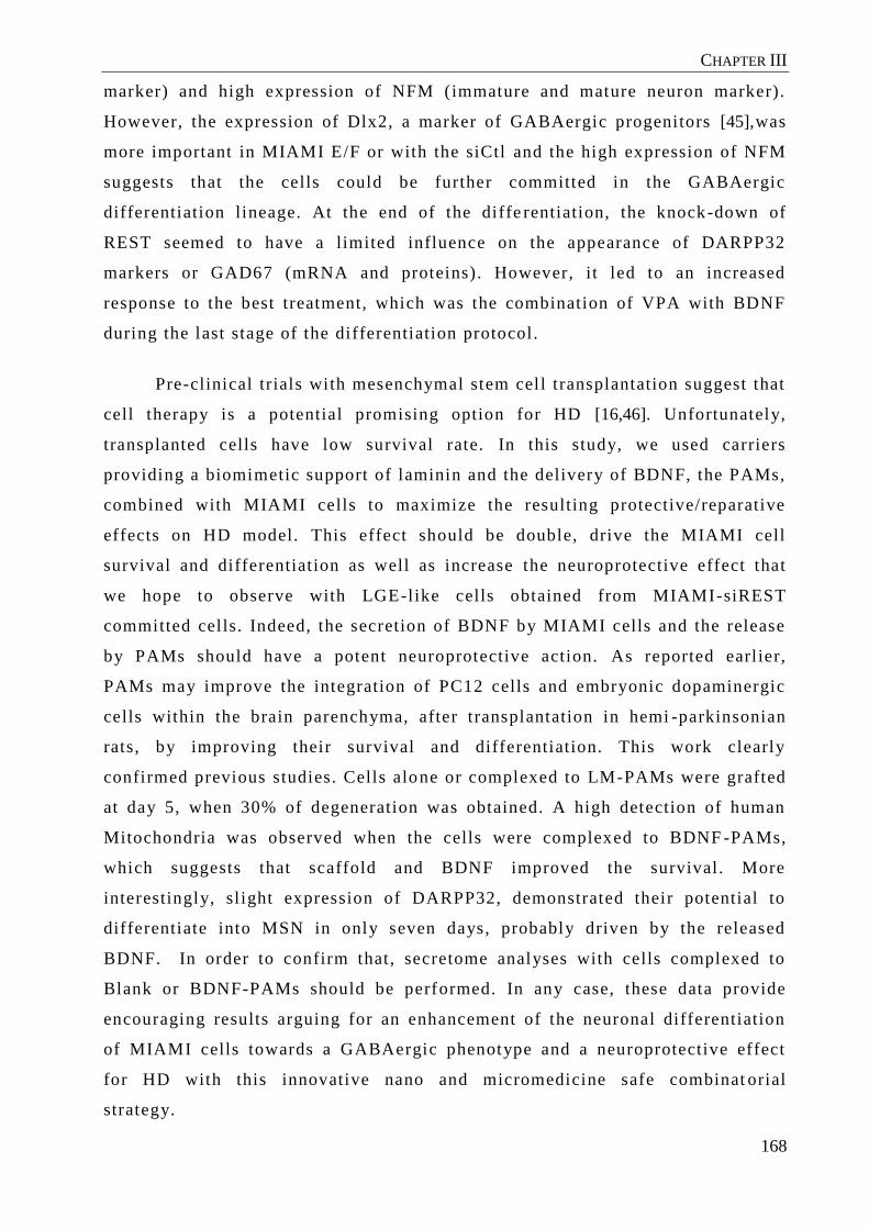
CHAPTER III
168
marker) and high expression of NFM (immature and mature neuron marker).
However, the expression of Dlx2, a marker of GABAergic progenitors [45],was
more important in MIAMI E/F or with the siCtl and the high expression of NFM
suggests that the cells could be further committed in the GABAergic
differentiation lineage. At the end of the diffe rentiation, the knock-down of
REST seemed to have a limited influence on the appearance of DARPP32
markers or GAD67 (mRNA and proteins). However, it led to an increased
response to the best treatment, which was the combination of VPA with BDNF
during the last stage of the differentiation protocol.
Pre-clinical trials with mesenchymal stem cell transplantation suggest that
cell therapy is a potential promising option for HD [16,46]. Unfortunately,
transplanted cells have low survival rate. In this study, we used carriers
providing a biomimetic support of laminin and the delivery of BDNF, the PAMs,
combined with MIAMI cells to maximize the resulting protective/reparative
effects on HD model. This effect should be double, drive the MIAMI cell
survival and differentiation as well as increase the neuroprotective effect that
we hope to observe with LGE-like cells obtained from MIAMI-siREST
committed cells. Indeed, the secretion of BDNF by MIAMI cells and the release
by PAMs should have a potent neuroprotective action. As reported earl ier,
PAMs may improve the integration of PC12 cells and embryonic dopaminergic
cells within the brain parenchyma, after transplantation in hemi -parkinsonian
rats, by improving their survival and differentiation. This work clearly
confirmed previous studies. Cells alone or complexed to LM-PAMs were grafted
at day 5, when 30% of degeneration was obtained. A high detection of human
Mitochondria was observed when the cells were complexed to BDNF -PAMs,
which suggests that scaffold and BDNF improved the survival. More
interestingly, sl ight expression of DARPP32, demonstrated their potential to
differentiate into MSN in only seven days, probably driven by the released
BDNF. In order to confirm that , secretome analyses with cells complexed to
Blank or BDNF-PAMs should be performed. In any case, these data provide
encouraging results arguing for an enhancement of the neuronal differentiation
of MIAMI cells towards a GABAergic phenotype and a neuroprotective effect
for HD with this innovative nano and micromedicine safe combinat orial
strategy.

CHAPTER III
169
In this study, we tested an innovate strategy combining stem cells and
biomaterials in ex vivo model of HD. We demonstrated in this way, the capacity
to our model to screen new therapy. We proved, in vitro , the capacity to MIAMI
cells to differentiate into GABAergic -l ike neurons by REST inhibition and
appropriate media. After graft , the survival of MIAMI cells combined with LM -
BDNF-PAMs seems to be increased, probably due to the synergic effect between
the 3D support and the release of BDNF. Nonetheless, the biological effect of
committed MIAMI-LM-BDNF-PAMs on the HD environment needs to be
characterized and understood.
We thank the SCIAM (“Service Commun d' Imagerie et d 'Analyse
Microscopique”) of Angers for confocal microscopy images , more particularly
Rodolphe PERROT and MEB images as well as the PACEM core facility
("Plateforme d'Analyse Cellulaire et Moléculaire”, Angers, France) of Angers
for the use of PCR facilities. We also wanted to thanks Professor Pau l C.
Schiller and Gaëtan Delcroix for helpful cri tiques and experiments. This project
is supported by the “Education Audiovisual” and the cultural executive agency
of the European Union through the NanoFar Erasmus Mundus Joint Doctoral
program, by Angers Loire Métropole,
[1] M.E. MacDonald, C.M. Ambrose, M.P. Duyao, R.H. Myers, C. Lin, L. Srinidhi, et al., A novel gene containing a trinucleotide repeat that is expanded and unstable on Huntington’s disease chromosomes, Cell. 72 (1993) 971–983. doi:10.1016/0092-8674(93)90585-E.
[2] E. Cattaneo, D. Rigamonti, D. Goffredo, C. Zuccato, F. Squitieri, S. Sipione, Loss of normal huntingtin function: new developments in Huntington’s disease research, Trends Neurosci. 24 (2001) 182–188.
[3] T.C. Hadzi, A.E. Hendricks, J.C. Latourelle, K.L. Lunetta, L.A. Cupples, T. Gillis, et al., Assessment of cortical and striatal involvement in 523 Huntington disease brains, Neurology. 79 (2012) 1708–1715.
CONCLUSION
ACKNOWLEDGMENTS
BIBLIOGRAPHY

CHAPTER III
170
[4] J. Klempíř, O. Klempířová, J. Štochl, N. Špačková, J. Roth, The relationship between impairment of voluntary movements and cognitive impairment in Huntington’s disease, J. Neurol. 256 (2009) 1629–1633. doi:10.1007/s00415-009-5164-9.
[5] E.B. Clabough, huntington’s Disease: The Past, Present, and future search for Disease Modifiers, Yale J. Biol. Med. 86 (2013) 217.
[6] K.D. Fink, P. Deng, A. Torrest, H. Stewart, K. Pollock, W. Gruenloh, et al., Developing stem cell therapies for juvenile and adult-onset Huntington’s disease, Regen. Med. 10 (2015) 623–646. doi:10.2217/rme.15.25.
[7] S. Ramaswamy, J.H. Kordower, Gene therapy for Huntington’s disease, Neurobiol. Dis. 48 (2012) 243–254. doi:10.1016/j.nbd.2011.12.030.
[8] A.-C. Bachoud-Lévi, P. Rémy, J.-P. Nǵuyen, P. Brugières, J.-P. Lefaucheur, C. Bourdet, et al., Motor and cognitive improvements in patients with Huntington’s disease after neural transplantation, The Lancet. 356 (2000) 1975–1979.
[9] C.D. Keene, J.A. Sonnen, P.D. Swanson, O. Kopyov, J.B. Leverenz, T.D. Bird, et al., Neural transplantation in Huntington disease: long-term grafts in two patients, Neurology. 68 (2007) 2093–2098. doi:10.1212/01.wnl.0000264504.14301.f5.
[10] A.-C. Bachoud-Lévi, V. Gaura, P. Brugières, J.-P. Lefaucheur, M.-F. Boissé, P. Maison, et al., Effect of fetal neural transplants in patients with Huntington’s disease 6 years after surgery: a long-term follow-up study, Lancet Neurol. 5 (2006) 303–309.
[11] L. Aubry, A. Bugi, N. Lefort, F. Rousseau, M. Peschanski, A.L. Perrier, Striatal progenitors derived from human ES cells mature into DARPP32 neurons in vitro and in quinolinic acid-lesioned rats, Proc. Natl. Acad. Sci. 105 (2008) 16707–16712.
[12] L. Ma, B. Hu, Y. Liu, S.C. Vermilyea, H. Liu, L. Gao, et al., Human Embryonic Stem Cell-Derived GABA Neurons Correct Locomotion Deficits in Quinolinic Acid-Lesioned Mice, Cell Stem Cell. 10 (2012) 455–464. doi:10.1016/j.stem.2012.01.021.
[13] A. Delli Carri, M. Onorati, V. Castiglioni, A. Faedo, S. Camnasio, M. Toselli, et al., Human Pluripotent Stem Cell Differentiation into Authentic Striatal Projection Neurons, Stem Cell Rev. Rep. 9 (2013) 461–474. doi:10.1007/s12015-013-9441-8.
[14] G.J.-R. Delcroix, E. Garbayo, L. Sindji, O. Thomas, C. Vanpouille-Box, P.C. Schiller, et al., The therapeutic potential of human multipotent mesenchymal stromal cells combined with pharmacologically active microcarriers transplanted in hemi-parkinsonian rats, Biomaterials. 32 (2011) 1560–1573. doi:10.1016/j.biomaterials.2010.10.041.
[15] D. Emerich, C. Cain, C. Greco, J. Saydoff, Z. HU, H. Liu, et al., Cellular delivery of human CNTF prevents motor and cognitive dysfunction in a rodent model of Huntington’s disease. - PubMed - NCBI, Cell Transplant. (1997) 249–266.
[16] Y. Jiang, L. Hailong, H. Shanshan, H. Tan, Y. Zhang, H. Li, Bone marrow mesenchymal stem cells can improve the motor function of a Huntington’s disease rat model, Neurol. Res. (2011) 331–337.
[17] N. Ballas, G. Mandel, The many faces of REST oversee epigenetic programming of neuronal genes, Curr. Opin. Neurobiol. 15 (2005) 500–506. doi:10.1016/j.conb.2005.08.015.
[18] N. Ballas, C. Grunseich, D.D. Lu, J.C. Speh, G. Mandel, REST and Its Corepressors Mediate Plasticity of Neuronal Gene Chromatin throughout Neurogenesis, Cell. 121 (2005) 645–657. doi:10.1016/j.cell.2005.03.013.
[19] Heurtault B, Saulnier P, Pech B, Proust JE, Benoit JP, A novel phase inversion-based process for the preparation of lipid nanocarriers., Pharm Res. (2002) 19:875–80.
[20] M. Morille, T. Montier, P. Legras, N. Carmoy, P. Brodin, B. Pitard, et al., Long-circulating DNA lipid nanocapsules as new vector for passive tumor targeting, Biomaterials. 31 (2010) 321–329. doi:10.1016/j.biomaterials.2009.09.044.
[21] A. Paillard, F. Hindré, C. Vignes-Colombeix, J.-P. Benoit, E. Garcion, The importance of endo-lysosomal escape with lipid nanocapsules for drug subcellular bioavailability, Biomaterials. 31 (2010) 7542–7554. doi:10.1016/j.biomaterials.2010.06.024.

CHAPTER III
171
[22] S. Roche, G. D’Ippolito, L.A. Gomez, T. Bouckenooghe, S. Lehmann, C.N. Montero-Menei, et al., Comparative analysis of protein expression of three stem cell populations: Models of cytokine delivery system in vivo, Int. J. Pharm. 440 (2013) 72–82. doi:10.1016/j.ijpharm.2011.12.041.
[23] G. D’Ippolito, Marrow-isolated adult multilineage inducible (MIAMI) cells, a unique population of postnatal young and old human cells with extensive expansion and differentiation potential, J. Cell Sci. 117 (2004) 2971–2981. doi:10.1242/jcs.01103.
[24] G.J.-R. Delcroix, K.M. Curtis, P.C. Schiller, C.N. Montero-Menei, EGF and bFGF pre-treatment enhances neural specification and the response to neuronal commitment of MIAMI cells, Differentiation. 80 (2010) 213–227. doi:10.1016/j.diff.2010.07.001.
[25] E.J. Huang, L.F. Reichardt, Neurotrophins: roles in neuronal development and function, Annu. Rev. Neurosci. 24 (2001) 677–736. doi:10.1146/annurev.neuro.24.1.677.
[26] S.D. Skaper, The neurotrophin family of neurotrophic factors: an overview, Methods Mol. Biol. Clifton NJ. 846 (2012) 1–12. doi:10.1007/978-1-61779-536-7_1.
[27] S. He, L. Shen, Y. Wu, L. Li, W. Chen, C. Hou, et al., Effect of Brain-Derived Neurotrophic Factor on Mesenchymal Stem Cell-Seeded Electrospinning Biomaterial for Treating Ischemic Diabetic Ulcers via Milieu-Dependent Differentiation Mechanism, Tissue Eng. Part A. 21 (2014) 928–938. doi:10.1089/ten.tea.2014.0113.
[28] J.M. Canals, J.R. Pineda, J.F. Torres-Peraza, M. Bosch, R. Martín-Ibañez, M.T. Muñoz, et al., Brain-derived neurotrophic factor regulates the onset and severity of motor dysfunction associated with enkephalinergic neuronal degeneration in Huntington’s disease, J. Neurosci. Off. J. Soc. Neurosci. 24 (2004) 7727–7739. doi:10.1523/JNEUROSCI.1197-04.2004.
[29] A.P. Kells, R.A. Henry, B. Connor, AAV–BDNF mediated attenuation of quinolinic acid-induced neuropathology and motor function impairment, Gene Ther. 15 (2008) 966–977. doi:10.1038/gt.2008.23.
[30] D.A. Simmons, C.S. Rex, L. Palmer, V. Pandyarajan, V. Fedulov, C.M. Gall, et al., Up-regulating BDNF with an ampakine rescues synaptic plasticity and memory in Huntington’s disease knockin mice, Proc. Natl. Acad. Sci. U. S. A. 106 (2009) 4906–4911. doi:10.1073/pnas.0811228106.
[31] C. Zuccato, E. Cattaneo, Brain-derived neurotrophic factor in neurodegenerative diseases, Nat. Rev. Neurol. 5 (2009) 311–322. doi:10.1038/nrneurol.2009.54.
[32] V.M. Tatard, M.C. Venier-Julienne, P. Saulnier, E. Prechter, J.P. Benoit, P. Menei, et al., Pharmacologically active microcarriers: a tool for cell therapy, Biomaterials. 26 (2005) 3727–3737. doi:10.1016/j.biomaterials.2004.09.042.
[33] P. Menei, C.N. Montero-Menei, M.-C. Venier-Julienne, J.P. Benoit, Drug delivery into the brain using poly(lactide-co-glycolide) microspheres, Expert Opin. Drug Deliv. (2005) 363–376.
[34] E. Garbayo, A.P. Raval, K.M. Curtis, D. Della-Morte, L.A. Gomez, G. D’Ippolito, et al., Neuroprotective properties of marrow-isolated adult multilineage-inducible cells in rat hippocampus following global cerebral ischemia are enhanced when complexed to biomimetic microcarriers: BMMs enhance MIAMI cell protection in cerebral ischemia, J. Neurochem. 119 (2011) 972–988. doi:10.1111/j.1471-4159.2011.07272.x.
[35] N. Daviaud, E. Garbayo, L. Sindji, A. Martinez-Serrano, P.C. Schiller, C.N. Montero-Menei, Survival, Differentiation, and Neuroprotective Mechanisms of Human Stem Cells Complexed With Neurotrophin-3-Releasing Pharmacologically Active Microcarriers in an Ex Vivo Model of Parkinson’s Disease, Stem Cells Transl. Med. 4 (2015) 670–684. doi:10.5966/sctm.2014-0139.
[36] J.-P. Karam, F. Bonafè, L. Sindji, C. Muscari, C.N. Montero-Menei, Adipose-derived stem cell adhesion on laminin-coated microcarriers improves commitment toward the cardiomyogenic lineage, J. Biomed. Mater. Res. A. 103 (2015) 1828–1839. doi:10.1002/jbm.a.35304.
[37] S. David, B. Pitard, J.-P. Benoît, C. Passirani, Non-viral nanosystems for systemic siRNA delivery, Pharmacol. Res. 62 (2010) 100–114. doi:10.1016/j.phrs.2009.11.013.
[38] G. Bastiat, C.O. Pritz, C. Roider, F. Fouchet, E. Lignières, A. Jesacher, et al., A new tool to ensure the fluorescent dye labeling stability of nanocarriers: a real challenge for fluorescence imaging, J. Control. Release Off. J. Control. Release Soc. 170 (2013) 334–342. doi:10.1016/j.jconrel.2013.06.014.
CHAPTER III
172
[39] A. Giteau, M.-C. Venier-Julienne, S. Marchal, J.-L. Courthaudon, M. Sergent, C. Montero-Menei, et al., Reversible protein precipitation to ensure stability during encapsulation within PLGA microspheres., Eur. J. Pharm. Biopharm. Off. J. Arbeitsgemeinschaft Für Pharm. Verfahrenstechnik EV. 70 (2008) 127–36. doi:10.1016/j.ejpb.2008.03.006.
[40] A. Paillard-Giteau, V.T. Tran, O. Thomas, X. Garric, J. Coudane, S. Marchal, et al., Effect of various additives and polymers on lysozyme release from PLGA microspheres prepared by an s/o/w emulsion technique, Eur. J. Pharm. Biopharm. Off. J. Arbeitsgemeinschaft Für Pharm. Verfahrenstechnik EV. 75 (2010) 128–136. doi:10.1016/j.ejpb.2010.03.005.
[41] M. Amiji, K. Park, Prevention of protein adsorption and platelet adhesion on surfaces by PEO/PPO/PEO triblock copolymers, Biomaterials. 13 (1992) 682–692.
[42] D. Drago, C. Cossetti, N. Iraci, E. Gaude, G. Musco, A. Bachi, et al., The stem cell secretome and its role in brain repair, Biochimie. 95 (2013) 2271–2285. doi:10.1016/j.biochi.2013.06.020.
[43] Y. Xie, H. Qiao, Z. Su, M. Chen, Q. Ping, M. Sun, PEGylated carboxymethyl chitosan/calcium phosphate hybrid anionic nanoparticles mediated hTERT siRNA delivery for anticancer therapy, Biomaterials. 35 (2014) 7978–7991. doi:10.1016/j.biomaterials.2014.05.068.
[44] H.Y. Xue, H.L. Wong, Tailoring nanostructured solid-lipid carriers for time-controlled intracellular siRNA kinetics to sustain RNAi-mediated chemosensitization, Biomaterials. 32 (2011) 2662–2672. doi:10.1016/j.biomaterials.2010.12.029.
[45] P. Viegas, C. Nicoleau, A.L. Perrier, Derivation of striatal neurons from human stem cells, in: Prog. Brain Res., Elsevier, 2012: pp. 373–404. http://linkinghub.elsevier.com/retrieve/pii/B978044459575100017X (accessed June 16, 2014).
[46] S.-T. Lee, K. Chu, K.-H. Jung, W.-S. Im, J.-E. Park, H.-C. Lim, et al., Slowed progression in models of huntington disease by adipose stem cell transplantation, Ann. Neurol. 66 (2009) 671–681. doi:10.1002/ana.21788.

CHAPTER III
173
Supplementary data
Table 3. Gene expression determined during the differentiation. Results are axpressed
in Fold increase and normalized with E/F MIAMI gene expression

CHAPTER III
174
In this study we showed that MIAMI cells could be induced towards the
GABAergic phenotype in vitro and that when transplanted as GABAergic
precursors in an ex vivo model of HD they survived better when attached to
PAMs delivering or not BDNF compared to cells alone. Through this study, we
also showed that the ex vivo model developed for modelling HD, can be used for
evaluating different strategies in the treatment of this disease.
We first determined that LNCs were able to release the siRNA during 6
days explaining the persistent effect of siREST to induce neuronal commitment
already observed in normal MSCs. MIA MI cells were first treated with EGF and
bFGF as these factors induced their specification towards the neural/neuronal
phenotype. Based on Ma et al., 2013 discovery, we decided to further treat
MIAMI cells with sonic hedgehog [89] after transfection to obtain LGE-like
progenitors. Indeed, the inhibit ion of REST before the differentiation allowed
obtaining functional neurons [2]. In our case, the inhibition of REST during the
first step of the differentiation had a favourable effect during al l our protocol.
Even if siREST is not able to silence REST during fourteen days during the LGE
commitment, we clearly observed an increase of cells responding to the
treatment during the GABAergic differentiation. Moreover, some evidences
suggested that we overpassed the LGE stage with the inhibition of REST. We
observed the protein expression of NFM at the end of the commitment, which
was not reported previously with SHH treatment. A better characterization
should be performed with for example the evaluati on of the CTIP2 expression in
the LGE-like progenitors obtained. Indeed, in a recent study, CTIP2 was
reported as the only transcription factor tested that was able to directly
reprogram dermal fibroblasts into DARPP32 + neurons, suggesting that it a key
regulator for GABAergic differentiation [176].
As part of the therapeutic strategy relies on the neuroprotective effect of
LM-PAMs-MIAMI-committed cells, we also checked the neuroprotective effect
by RT-qPCR in vitro . The high expression of VEGFa at the end of the treatment
permits to think that these GABAergic -like neurons still retain some
characteristics from MIAMI cells, and will be able to have a beneficial effect on
DISCUSSION

CHAPTER III
175
brain slices. It has already been demonstrated that VEGFa possesses angiogenic
capacit ies, and maintained the blood vessels in the in brain slices [84].
Furthermore, during the differentiation VEGFa enhances the gamma-
aminobutyric acid (GABA) synaptic activity in embryonic spinal motoneurons
[177]. We can easily imagine that VEGFa will induce an improvement of the
differentiation of MIAMI cells by stimulating GABAergic synapses. But VEGFa
was not the only one secreted by MIAMI derived GABA -like cells, because
BDNF and GDNF were also detected in those cells. MSCs secreting BDNF and
GDNF have been already described to improve the behavior of rats with HD
[145,178,179]. In this way, we can suppose that LM -PAM-LGE-like progenitors
will reduce the degeneration. But the major advantage of MIAMI cells is their
capacity to express DARPP32, a marker of GABAergic neurons, when grafted
attached to LM-PAMs-BDNF. These results are very surprising because when
ES cells differentiated into striatal progenitors were grafted in quinolinc acid
treated rats, they exhibited a 15% of DARPP32 positive neurons after 13 weeks
in situ [37], while a LGE-like progenitors grated reveal a 50% of DARPP32
expression [89]. The advantage of the prolonged release of BDNF by the PAMs
should be further investigated. With MIAMI cells we have not quantified yet the
pourcentage of DARPP32 expression which could be very interesting in future
studies

CHAPTER III
176
.

177
GENERAL DISCUSSION
AND CONCLUSION

GENERAL DISCUSSION AND CONCLUSION
178
Since the discovery of Huntington' disease in 1872, pharmacological
treatment or other therapeutic approaches are sti ll insufficient to cure the
disease [16]. Progressively, cell therapy became one of the most promising
treatments [58]. However, despite many pre-clinical and clinical trials showing
functional benefits with this strategy for CNS disorders, i t is still not used
routinely. In the last twenty years, the implantation of fetal cells for the
treatment of HD has demonstrated the possibility of this approach, but shows an
important diversity of results in clinical trials due to the h eterogeneity in
protocols [180,55]. Moreover, the use of ESC or iPS present the risk of over -
proliferation in the brain, in addition to the numerous ethical problems
previously mentioned [37,90]. Research studies need to focus on resolving the
choice of cell type, the number of injected cells and the time of injection.
Moreover, the optimization of their survival and differentiation need to be
improved before imagining using them in patients. In consequence, in this work,
we evaluated an innovative and safe strategy based on regenerative medicine for
HD with modified MSCs and appropriate biomimetic scaffold.
In regenerative medicine, an innovative approach is the association of
modified cells with therapeutic molecules, morphogens, materials or
biomaterials with different physicochemical properties to enhance their
biological effects and optimize cell therapy. Howe ver, the use of adult stem
cells showed numerous limits with a significant death of the implanted cells
quickly detected. In addition, relatively low rates of implanted cells
differentiate into mature neurons. The capacity of MSCs to undergo functional
differentiation into neurons has been questioned over the years and remains
controversial [181]. Despite the initial skepticism regarding the capacity of
MSCs to differentiate into neurons or glial cells , it was discovered that MSCs
express a considerable repertoire of neural genes, which l ikely contributes to
the contested neurogenic predisposition of these cells [182,183]. In addition,
bone marrow MSCs express higher neuronal markers [184] compared to MSCs
derived from other origins.
In this work, we decided to use a homogenous subpopulation of bone
marrow mesenchymal stem cells, MIAMI cells, which have already

GENERAL DISCUSSION AND CONCLUSION
179
demonstrated their capacity to differentiate into dopaminergic neurons with
appropriate electrophysiological properties of immature neurons [95,185,88].
Nonetheless, in order to improve the differentiation and their integration in the
parenchyma, we chose to knock-down REST. The approach used here is slightly
different from strategies generally used for reprogramm ing MSCs into neurons.
Indeed, recent studies have shown that over -expression of transcription factors
can serve to promote neuronal differentiation and maintain a neuronal
phenotype. Based on the same model to obtain induced Neurons cells (iNs) by
forcing the expression of a set of neural lineage transcriptional factors [186],
the differentiation of MSCs towards a neuronal lineage has been achieved with
Neurogenin1, LMX1 and/or PDX1 (For a review see [187]). Very few studies
using the opposite strategy to knock -down and not over-express transcription
factors have been performed to improve neuronal commitment [102,175]. In
those studies, knock-down of REST was effective with lentivirus and plasmids
allowing a stable inhibition of REST.
In this study, we preferred to safely modify the MSCs and focused on
transitory inhibition with siREST carried by non -viral vectors to boost the
differentiation. We thus designed and characterized two different nanocarriers
LNC and SP-AP to efficiently deliver siRNA. Normally, epigenetic
manipulation of stem cells is a delicate and complex task. Their remarkable
capacity of proliferation makes them poor candidates for non -viral vectors,
electroporation and nucleofection, and most stem cells are sensitive to antibiotic
selection; hence, attempts to stably establish transf ected stem cells are rarely
successful [188]. But, in our hands both nanocarriers were perfectly able to
delivery the siRNA with a varying degree of toxicity. We also tried to reduce
the off-target effect usually described in the literature with cationic lipids for
siRNA delivery[189] . These effects are obtained due to si lencing of genes other
than the intended one and thus compromise the use of a siRNA to study gene
function and can even cause cell death. Furthermore, i t has been observed that
even scrambled sequences, which theoretically do not target any mRNA, can
have a moderate to high impact on the cell viabil ity, depending on the cell l ine
and siRNA concentration [190,191]. In order to avoid this, particular care was
provided, in this study and in previous ones, to design siRNA nanocarriers with
liposomes representing only 20% of the system for the LNCs or without any
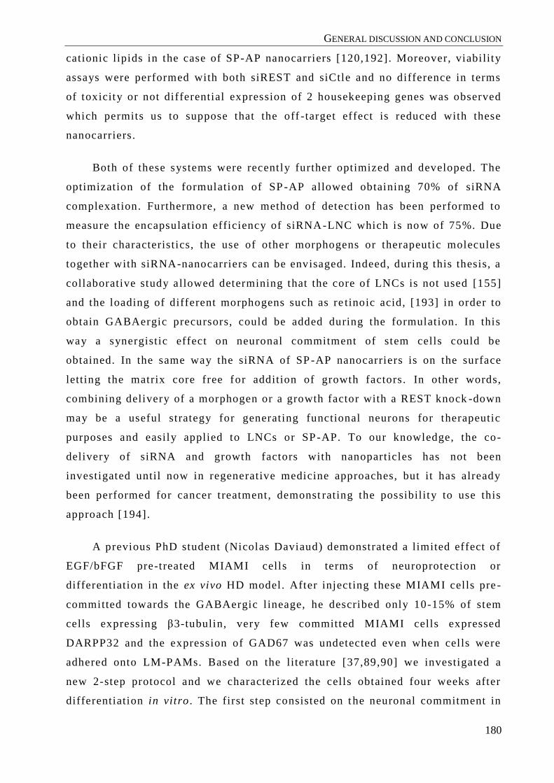
GENERAL DISCUSSION AND CONCLUSION
180
cationic lipids in the case of SP-AP nanocarriers [120,192]. Moreover, viability
assays were performed with both siREST and siCtle and no difference in terms
of toxicity or not differential expression of 2 housekeeping genes was observed
which permits us to suppose that the off -target effect is reduced with these
nanocarriers.
Both of these systems were recently further optimized and developed. The
optimization of the formulation of SP -AP allowed obtaining 70% of siRNA
complexation. Furthermore, a new method of detection has been performed to
measure the encapsulation efficiency of siRNA -LNC which is now of 75%. Due
to their characteristics, the use of other morphogens or therapeutic molecules
together with siRNA-nanocarriers can be envisaged. Indeed, during this thesis, a
collaborative study allowed determining that the core of LNCs is not used [155]
and the loading of different morphogens such as re tinoic acid, [193] in order to
obtain GABAergic precursors, could be added during the formulation. In this
way a synergistic effect on neuronal commitment of stem cells could be
obtained. In the same way the siRNA of SP -AP nanocarriers is on the surface
letting the matrix core free for addition of growth factors. In other words,
combining delivery of a morphogen or a growth factor with a REST knock -down
may be a useful strategy for generating functional neurons for therapeutic
purposes and easily applied to LNCs or SP-AP. To our knowledge, the co-
delivery of siRNA and growth factors with nanoparticles has not been
investigated until now in regenerative medicine approaches, but it has already
been performed for cancer treatment, demonst rating the possibility to use this
approach [194].
A previous PhD student (Nicolas Daviaud) demonstrated a limited effect of
EGF/bFGF pre-treated MIAMI cells in terms of neuroprotection or
differentiation in the ex vivo HD model. After injecting these MIAMI cells pre -
committed towards the GABAergic l ineage, he described only 10-15% of stem
cells expressing β3-tubulin, very few committed MIAMI cells expressed
DARPP32 and the expression of GAD67 was undetected even when cells were
adhered onto LM-PAMs. Based on the literature [37,89,90] we investigated a
new 2-step protocol and we characterized the cells obtained four weeks after
differentiation in vitro . The first step consisted on t he neuronal commitment in

GENERAL DISCUSSION AND CONCLUSION
181
order to obtain LGE progenitors, from which MSN derive, and the second one
on the final differentiation to obtain these GABAergic -like neurons. Specific
media with Sonic Hedgehog was used during fourteen days after siREST
transfection. SHH is a pleiotropic factor for the development of CNS driving
proliferation, specification, and axonal targeting in multiple sites within the
forebrain, hindbrain, and spinal cord. Studies in embryonic CNS have shown
how gradients of this morphogen are translated by neuroepithelial precursors to
determine the types of neurons and glial cells they produced [195]. Using these
conditions LGE progenitors were obtained [89] (Figure 12) . Nonetheless, in our
work, we concluded that we over -passed this stage with the high expression of
NFM and β3-tubulin as well as the decrease of DLX2 at the end of the
commitment. In order to better understand those results the mechani sm and
signaling forced by the inhibition of REST must be elucidated.
REST expression is very high in ESCs compared to most other cell
types[152], and is thought to be involved in their self -renewal. REST is
regulated directly by the pluripotent factors Oct4/Sox2 and Nanog, maintaining
a high expression of REST, but its function in the self -renewal transduction
network is unclear [196–198]. In this work, the inhibition of REST without any
other differentiated media was enough to induce neuronal commitment of MSCs,
which is not surprising due to the functionality of REST [151,151]. But, at the
end of this period we still observed a slight diminution of REST. This inhibition
is not only explained by the prolonged release of REST with LNCs, because we
did not observe any siRNA after 6 days (data not shown). We thus assume that
siREST helped and accelerated the commitment as described in mouse
previously [198,199] and during neurogenesis in vivo[200] However, various
other explanations can be advanced. One explanation relates to the levels of
pluripotent factors such as Nanog. Indeed, the level of Oct4 and Nanog is lower
in EGF/bFGF pre-treated MIAMI cells compared to non pre -treated MIAMI
cells[185] and they should continue to decrease during the differentiation. In
this way, they are unable to maintain or re -activate high levels of REST within
the cell. The microenvironnement is also very important for REST regulatio n. In
a recent publication, Shing et al ., investigated the role of laminin on
pluripotency and demonstrated the reduction of self -renewal of ESCs on laminin
coated surfaces [201]. In this way, we chose to transfect MIAMI cells after

GENERAL DISCUSSION AND CONCLUSION
182
laminin adherence because laminin coated surfaces also play a modest role in
the loss of pluripotency [201]. Moreover, in vivo reports describe the role of
laminin in axonal regeneration of the CNS [202]. In our study, proliferation
seems to diminish after transfection with siREST and some neurites appeared
during the commitment step with SHH but in order to confirm those data the
possible interaction between REST and kinase or cyclin proteins involved in the
cell cycle needs to be explored. Finally, REST activity represses the expression
of GluR2 in neurons destined to die, indicating that REST might play an
important role in insult -induced death of neurons[203] [204].
The final differentiation step allowed obtaining GABAergic -like neurons in
vitro . The best condition was a 4 weeks treatment with the supplementation of
Valproic acid and BDNF at the same time (Figure 12). Those results will be
confirmed with repeated experiments, but they established the choice of BDNF
as the growth factor to be delivered by the PAMs for the graft ing of committed
MIAMI cells and PAMs. Accordingly, laminine was confirmed as the best
molecule for functionalizing the surface of the PAMs. In this way we chose to
graft the EGF/bFGF MIAMI cells after commitment towards GABAergic
precursors with siREST LNCs and SHH with or without laminine PAMs
delivering or not BDNF. We already successfully showed the regenerative effect
of EGF/bFGF MIAMI cells and PAMs releasing NT -3 in a PD model
[84,88,174]. In addition, the transduction pathway allowing NT -3 to induce
differentiation into a neuronal cell type was studied in vitro and it was
demonstrated that the fixation of NT-3 to its receptor stimulated the kinase
pathway MEK / ERK. Moreover, the action of Rac1b on this pathway to block
proliferation and st imulate the development of neurites was also elucidated
[205]. So, strong of those results, we could intend a similar strategy for HD.

GENERAL DISCUSSION AND CONCLUSION
183
Figure 12. The different steps observed during in vitro differentiation
The preliminary evaluation of this combined regenerative medicine strategy
has been performed with innovate organotypic brain slices modeling the
GABAergic degeneration observed in HD. Indeed, taking into account the
numerous advantages of organot ypic brain slices, we developed a simple model
of HD, where GABAergic MSNs degeneration is due to mechanical transection
of the axonal pathway between the striatum and substantia nigra. We wanted to
develop a simple, inexpensive and mono -parametric model of HD. Based on the
same principle that we have already developed to model Parkinson’s disease
[206], we demonstrated that the axis of sectioning is very important as only
coronal sections allowed modeling the progressive degeneration of MSNs. The
slices were viable at least until day 19 as deter mined by Neu N expression as
well as the MSN degeneration determined by the decrease of DARPP32 and
GAD67 expression. The mechanism of degeneration is still poorly understood i n
this model, and we hypothesize that the regulation of the physiological state of
these striatal neurons via dopamine is impaired. Nonetheless, the determination
of the dopamine-regulated ion channels could be performed by
electrophysiological studies. In addition, a possible reduction in the striatal -
specific dopamine targets, PKA, DARPP-32, ARPP-16, and ARPP-21 could be
performed, with RT-qPCR or immunofluorescence.

GENERAL DISCUSSION AND CONCLUSION
184
This ex vivo model will allow screening the best strategy for HD and
further comprehending the mechanisms involved in the regeneration of the
GABAergic neurons as performed with an ex vivo model of PD by our group
[206]. For example, we can interrogate the amount of cells to be injected and if
they should be injected as a bolus or repeated injections. Furthermore, the
evaluation of the behavior of stem cells after grafting must be performed at
long-term. Therefore, we are going to follow NeuN and GAD67 over -time until
30 and 60 days to evaluate the long-term viability of the sl ices and follow the
degeneration of the GABAergic neurons. A recent study allowed observing few
axonal projections (0.017%) after 9 wee ks injection of differentiated ESC and
more axonal projections after in utero implantation, suggesting integration of
the donor cells into the neuronal network of the host brain [207]. But the poor
quantity of axonal projection demonstrated the diff iculty to hope replace
GABAergic neurons. Although transplanted cells integrated and were able to
reduce the motor asymmetry in the QA-lesioned model, long-term studies are
needed to assess circuit reconstruction and behavioral recovery. With our
model, we can obtain cortico -striatal brain slices or the whole coronal section.
In this way, we can screen the different regenerative medicine strategies and
select the best approach showing benefits in terms of neuroprotection, not only
in the striatum but also in the cortex and the globus pallidu s, which are involved
in HD.
In adult PD rat brain, we have first shown that the 60 µm size and PLGA -
PAMS could be stereotactically implant ed in precise areas of the brain without
causing damage to the surrounding tissue. Nonetheless in organotypic slices , we
thought that 30 µm could be better. So, in this study, the triblock copolymer
poloxamer P188 ((poly (ethylene oxide) (PEO)–poly(propylene oxide) (PPO)–
PEO) linked to PLGA to form this PLGA-P188-PLGA co-polymer was used to
formulate PAMs with 30 µm size. Indeed, PLGA based implantable devices are
FDA approved [208] but the efficient and sustained release of proteins from
PLGA-PAMs remains a challenge, mainly due to protein instabili ty (adsorpti on,
aggregation, and denaturation) during the formulation process or the release
period. Therefore this polymer was used to formulate the PAMs in this study.
Interestingly, PLGA-P188-PLGA 60 µm microsphere releasing TGFb3,
developed for cartilage tissue engineering, resulted in a prolonged and almost

GENERAL DISCUSSION AND CONCLUSION
185
complete TGFb3 release [209]. They offered the advantage to limit protein
adsorption but also to stabilize the pH inside the microsphere thus preventing
protein unfolding and denaturation. M oreover, an increase in MSC number was
observed after 7 days in culture on fibronectin -covered PLGA-P188-PLGA
PAMs compared to fibronectin-covered PLGA PAMs.
In our study the 3D support offered by the scaffold allowed cell retention
within the striatum and, probably cell survival as seen by the increased number
of human cells with the PAMs compared to cells implanted alone. However,
BDNF-PAMs also seemed to drive cell differentiation of pre -committed MIAMI
cells, demonstrated by the increase of DARPP32 mark ers compared to Blank-
PAMs. These results are in accordance with previous reports showing that
BDNF increases DARPP32 expression in neurons of the striatum [210,211].
Moreover, it appears to induce significant neuroprotection of NMS neurons in in
vivo models of Huntington's disease [212,213]. A collaborative work allowed
determining the release of BDNF from PAMs, which corresponded perfectly to
the amount (15 ng/day) needed by the cells for their differentiation [37,89,90]
Nonetheless, we don’t know if the protective effect on MIAMI cells is due to
the BDNF released from PAMs or BDNF secreted by MIAMI cells. The
neuroprotective effect of MIAMI on the GABAergic neurons is also another
important part of this project. In the present study, in preliminary results, we
observed in vitro that MIAMI cells expressed BDNF and GDNF as well as a
very high level of hVEGFA. BDNF and GDNF have both been reported as
neuroprotective factors whose effects were, in part, mediated by autophag y
regulation[214] and oxidative stress diminution[215,216], respectively. The
neuroprotective effect of BDNF and GDNF has been evaluated in pre -clinical
studies in HD [217–219]. In this way, pre-committed MIAMI cells are good
candidates for neuroprotection. hVEGFA is playing a dual role, indeed,
hVEGFA can increase grafted cells survival by creating new blood vessels
around the graft enhancing oxygen and nutrients supply and a neuroprotective
effect. Indeed, in a more recent study, the delivery of hVEGFA via injectable
hydrogels, to rats receiving striatal injection of quinolinic acid to mimic
neuronal loss and behavioral deficits characteristic of HD, was shown to be
neuroprotective at 3 weeks [220]. As hVEGFA is well -known to have pro-
angiogenic effect , a quantification of blood vess els in the brain slices should be

GENERAL DISCUSSION AND CONCLUSION
186
performed. Complete analyses of the secretome of MIAMI cells are going on in
order to better characterize proteins secreted by LGE -like progenitors.
Figure 13. Schematic strategy finally used during this thesis and some advantages.
However, a final assessment of this strategy includes the behavioral
evaluation in an appropriate in vivo model. In this work, intracranial injection
of MIAMI cells is envisaged and we think it is the best option as the cells and
PAMs need to be close to their site of action to avoid undesired side-effects. We
can also consider delivery of siREST by implantation of the si -REST
nanocarriers together with the cell graft. Indeed, REST expression is increased
in HD [221,222] and its association with HTT in the cytoplasm is disrupted in
HD due to the mutated HTT. This leads to increased nuclear REST and
concomitant repression of several neuronal -specific genes, including BDNF
[67]. The finding described indicates that inhibition of REST expression may
offer a new therapeutic avenue for treatment of HD. Nowadays, innovative
strategies including the use of RNAi to reduce REST expression [223] or decoy
oligonucleotides or synthetic peptide nucleic acid (PNA) oligomers to seque ster
REST away from its genomic sites [224,225] are underway. All of these
approaches lead to reduce interactions of REST with its target genes; however,

GENERAL DISCUSSION AND CONCLUSION
187
translation of this into a useful therapeutic strategy requires safe and efficient
delivery of these tools to the brain of HD patients. This is no easy task and
LNCs or SP-AP could be used for that, but this strategy needs to be care fully
analyzed. More particularly, a full characterization of this combined strategy
will be evaluated during a post -doctoral contract.

GENERAL DISCUSSION AND CONCLUSION
188

REFERENCES
189
REFERENCES

REFERENCES
190
[1] E.R. Fisher, M.R. Hayden, Multisource ascertainment of Huntington disease in Canada: Prevalence
and population at risk: Multisource Ascertainment of BC HD Patients, Mov. Disord. 29 (2014) 105–114. doi:10.1002/mds.25717.
[2] A. Lekoubou, J.B. Echouffo-Tcheugui, A.P. Kengne, Epidemiology of neurodegenerative diseases in sub-Saharan Africa: a systematic review, BMC Public Health. 14 (2014) 653.
[3] S.C. Warby, H. Visscher, J.A. Collins, C.N. Doty, C. Carter, S.L. Butland, et al., HTT haplotypes contribute to differences in Huntington disease prevalence between Europe and East Asia, Eur. J. Hum. Genet. EJHG. 19 (2011) 561–566. doi:10.1038/ejhg.2010.229.
[4] H.-C. Fan, L.-I. Ho, C.-S. Chi, S.-J. Chen, G.-S. Peng, T.-M. Chan, et al., Polyglutamine (PolyQ) diseases: genetics to treatments, Cell Transplant. 23 (2014) 441–458. doi:10.3727/096368914X678454.
[5] F. Squitieri, A. Griguoli, G. Capelli, A. Porcellini, B. D’Alessio, Epidemiology of Huntington disease: first post- HTT gene analysis of prevalence in Italy: Prevalence of Huntington disease in Italy, Clin. Genet. (2015) n/a–n/a. doi:10.1111/cge.12574.
[6] C. Kenney, S. Powell, J. Jankovic, Autopsy-proven Huntington’s disease with 29 trinucleotide repeats, Mov. Disord. 22 (2007) 127–130. doi:10.1002/mds.21195.
[7] M.A. Nance, W. Seltzer, T. Ashizawa, R. Bennett, N. McIntosh, R.H. Myers, et al., Laboratory guidelines for Huntington disease genetic testing, Am. J. Hum. Genet. 62 (1998) 1243ñ1247.
[8] S. Ghavami, S. Shojaei, B. Yeganeh, S.R. Ande, J.R. Jangamreddy, M. Mehrpour, et al., Autophagy and apoptosis dysfunction in neurodegenerative disorders, Prog. Neurobiol. 112 (2014) 24–49. doi:10.1016/j.pneurobio.2013.10.004.
[9] J.A. White, E. Anderson, K. Zimmerman, K.H. Zheng, R. Rouhani, S. Gunawardena, Huntingtin differentially regulates the axonal transport of a sub-set of Rab-containing vesicles in vivo, Hum. Mol. Genet. (2015). doi:10.1093/hmg/ddv415.
[10] J. Yao, S.-E. Ong, S. Bajjalieh, Huntingtin is associated with cytomatrix proteins at the presynaptic terminal, Mol. Cell. Neurosci. 63 (2014) 96–100. doi:10.1016/j.mcn.2014.10.003.
[11] M.E. MacDonald, C.M. Ambrose, M.P. Duyao, R.H. Myers, C. Lin, L. Srinidhi, et al., A novel gene containing a trinucleotide repeat that is expanded and unstable on Huntington’s disease chromosomes, Cell. 72 (1993) 971–983. doi:10.1016/0092-8674(93)90585-E.
[12] E. Roze, Huntington’s disease and striatal signaling, Front. Neuroanat. 5 (2011). doi:10.3389/fnana.2011.00055.
[13] C. Zuccato, M. Valenza, E. Cattaneo, Molecular Mechanisms and Potential Therapeutical Targets in Huntington’s Disease, Physiol. Rev. 90 (2010) 905–981. doi:10.1152/physrev.00041.2009.
[14] The Whole Brain Atlas, (n.d.). http://www.med.harvard.edu/aanlib/ (accessed October 11, 2015). [15] M. Papoutsi, I. Labuschagne, S.J. Tabrizi, J.C. Stout, The cognitive burden in Huntington’s disease:
Pathology, phenotype, and mechanisms of compensation, Mov. Disord. 29 (2014) 673–683. doi:10.1002/mds.25864.
[16] M.J. Armstrong, J.M. Miyasaki, American Academy of Neurology, Evidence-based guideline: pharmacologic treatment of chorea in Huntington disease: report of the guideline development subcommittee of the American Academy of Neurology, Neurology. 79 (2012) 597–603. doi:10.1212/WNL.0b013e318263c443.
[17] A. Björklund, O. Lindvall, Cell replacement therapies for central nervous system disorders., Nat. Neurosci. 3 (2000).
[18] K.M. Shannon, A. Fraint, Therapeutic advances in Huntington’s Disease, Mov. Disord. Off. J. Mov. Disord. Soc. (2015). doi:10.1002/mds.26331.
[19] K. Youssov, G. Dolbeau, P. Maison, M.-F. Boissé, L. Cleret de Langavant, R.A.C. Roos, et al., Unified Huntington’s disease rating scale for advanced patients: validation and follow-up study, Mov. Disord. Off. J. Mov. Disord. Soc. 28 (2013) 1717–1723. doi:10.1002/mds.25654.
REFERENCES

REFERENCES
191
[20] K. Youssov, G. Dolbeau, P. Maison, M.-F. Boissé, L. Cleret de Langavant, R.A.C. Roos, et al., The unified Huntington’s Disease Rating Scale for advanced patients: validation and follow-up study, Mov. Disord. Off. J. Mov. Disord. Soc. 28 (2013) 1995–2001. doi:10.1002/mds.25678.
[21] L.M. Stanek, S.P. Sardi, B. Mastis, A.R. Richards, C.M. Treleaven, T. Taksir, et al., Silencing Mutant Huntingtin by Adeno-Associated Virus-Mediated RNA Interference Ameliorates Disease Manifestations in the YAC128 Mouse Model of Huntington’s Disease, Hum. Gene Ther. 25 (2014) 461–474. doi:10.1089/hum.2013.200.
[22] D.M. Danks, Defining the location of the Huntington disease gene., Am. J. Hum. Genet. 52 (1993) 214.
[23] S. von Hörsten, I. Schmitt, H.P. Nguyen, C. Holzmann, T. Schmidt, T. Walther, et al., Transgenic rat model of Huntington’s disease, Hum. Mol. Genet. 12 (2003) 617–624. doi:10.1093/hmg/ddg075.
[24] J.G. Hodgson, N. Agopyan, C.-A. Gutekunst, B.R. Leavitt, F. LePiane, R. Singaraja, et al., A YAC Mouse Model for Huntington’s Disease with Full-Length Mutant Huntingtin, Cytoplasmic Toxicity, and Selective Striatal Neurodegeneration, Neuron. 23 (1999) 181–192. doi:10.1016/S0896-6273(00)80764-3.
[25] P.H. Reddy, V. Charles, M. Williams, G. Miller, W.O. Whetsell, D.A. Tagle, Transgenic mice expressing mutated full-length HD cDNA: a paradigm for locomotor changes and selective neuronal loss in Huntington’s disease., Philos. Trans. R. Soc. B Biol. Sci. 354 (1999) 1035–1045.
[26] L. Mangiarini, K. Sathasivam, M. Seller, B. Cozens, A. Harper, C. Hetherington, et al., Exon 1 of the HD Gene with an Expanded CAG Repeat Is Sufficient to Cause a Progressive Neurological Phenotype in Transgenic Mice, Cell. 87 (1996) 493–506. doi:10.1016/S0092-8674(00)81369-0.
[27] G. Schilling, M.W. Becher, A.H. Sharp, H.A. Jinnah, K. Duan, J.A. Kotzuk, et al., Intranuclear inclusions and neuritic aggregates in transgenic mice expressing a mutant N-terminal fragment of huntingtin, Hum. Mol. Genet. 8 (1999) 397–407.
[28] V.C. Wheeler, W. Auerbach, J.K. White, J. Srinidhi, A. Auerbach, A. Ryan, et al., Length-dependent gametic CAG repeat instability in the Huntington’s disease knock-in mouse, Hum. Mol. Genet. 8 (1999) 115–122.
[29] L. Menalled, D. Brunner, Animal models of Huntington’s disease for translation to the clinic: Best practices, Mov. Disord. 29 (2014) 1375–1390. doi:10.1002/mds.26006.
[30] J.Y. Li, N. Popovic, P. Brundin, The use of the R6 transgenic mouse models of Huntington’s disease in attempts to develop novel therapeutic strategies, NeuroRx J. Am. Soc. Exp. Neurother. 2 (2005) 447–464. doi:10.1602/neurorx.2.3.447.
[31] V.C. Wheeler, J.K. White, C.A. Gutekunst, V. Vrbanac, M. Weaver, X.J. Li, et al., Long glutamine tracts cause nuclear localization of a novel form of huntingtin in medium spiny striatal neurons in HdhQ92 and HdhQ111 knock-in mice, Hum. Mol. Genet. 9 (2000) 503–513.
[32] L.B. Menalled, A.E. Kudwa, S. Miller, J. Fitzpatrick, J. Watson-Johnson, N. Keating, et al., Comprehensive behavioral and molecular characterization of a new knock-in mouse model of Huntington’s disease: zQ175, PloS One. 7 (2012) e49838. doi:10.1371/journal.pone.0049838.
[33] E.J. Slow, J. van Raamsdonk, D. Rogers, S.H. Coleman, R.K. Graham, Y. Deng, et al., Selective striatal neuronal loss in a YAC128 mouse model of Huntington disease, Hum. Mol. Genet. 12 (2003) 1555–1567. doi:10.1093/hmg/ddg169.
[34] M. Gray, D.I. Shirasaki, C. Cepeda, V.M. André, B. Wilburn, X.-H. Lu, et al., Full-Length Human Mutant Huntingtin with a Stable Polyglutamine Repeat Can Elicit Progressive and Selective Neuropathogenesis in BACHD Mice, J. Neurosci. 28 (2008) 6182–6195. doi:10.1523/JNEUROSCI.0857-08.2008.
[35] R. Schwarcz, J.P. Bennett, J.T. Coyle, Loss of striatal serotonin synaptic receptor binding induced by kainic acid lesions: correlations with Huntington’s Disease, J. Neurochem. 28 (1977) 867–869.
[36] M.F. Beal, N.W. Kowall, D.W. Ellison, M.F. Mazurek, K.J. Swartz, J.B. Martin, Replication of the neurochemical characteristics of Huntington’s disease by quinolinic acid, Nature. 321 (1986) 168–171. doi:10.1038/321168a0.
[37] L. Aubry, A. Bugi, N. Lefort, F. Rousseau, M. Peschanski, A.L. Perrier, Striatal progenitors derived from human ES cells mature into DARPP32 neurons in vitro and in quinolinic acid-lesioned rats, Proc. Natl. Acad. Sci. 105 (2008) 16707–16712.

REFERENCES
192
[38] S. Ramaswamy, J.L. McBride, J.H. Kordower, Animal Models of Huntington’s Disease, ILAR J. 48 (2007) 356–373. doi:10.1093/ilar.48.4.356.
[39] C. Humpel, Organotypic brain slice cultures: A review, Neuroscience. (n.d.). doi:10.1016/j.neuroscience.2015.07.086.
[40] S. Beraki, L. Litrus, L. Soriano, M. Monbureau, L.K. To, S.P. Braithwaite, et al., A pharmacological screening approach for discovery of neuroprotective compounds in ischemic stroke, PloS One. 8 (2013) e69233. doi:10.1371/journal.pone.0069233.
[41] A. Martire, R. Pepponi, M.R. Domenici, A. Ferrante, V. Chiodi, P. Popoli, BDNF prevents NMDA-induced toxicity in models of Huntington’s disease: the effects are genotype specific and adenosine A2A receptor is involved, J. Neurochem. 125 (2013) 225–235. doi:10.1111/jnc.12177.
[42] N. Daviaud, E. Garbayo, P.C. Schiller, M. Perez-Pinzon, C.N. Montero-Menei, Organotypic cultures as tools for optimizing central nervous system cell therapies, Exp. Neurol. 248C (2013) 429–440. doi:10.1016/j.expneurol.2013.07.012.
[43] C.D. Clelland, R.A. Barker, C. Watts, Cell therapy in Huntington disease, Neurosurg. Focus. 24 (2008) E9. doi:10.3171/FOC/2008/24/3-4/E8.
[44] E. Matyja, Ultrastructural evaluation of the damage of postsynaptic elements after kainic acid injection into the rat neostriatum, J. Neurosci. Res. 15 (1986) 405–413. doi:10.1002/jnr.490150311.
[45] M.I. Behrens, J. Koh, L.M. Canzoniero, S.L. Sensi, C.A. Csernansky, D.W. Choi, 3-Nitropropionic acid induces apoptosis in cultured striatal and cortical neurons, Neuroreport. 6 (1995) 545–548.
[46] J. Storgaard, B.T. Kornblit, J. Zimmer, J.B.P. Gramsbergen, 3-Nitropropionic Acid Neurotoxicity in Organotypic Striatal and Corticostriatal Slice Cultures Is Dependent on Glucose and Glutamate, Exp. Neurol. 164 (2000) 227–235. doi:10.1006/exnr.2000.7428.
[47] J.C. Vis, R.T. de Boer-van Huizen, M.M. Verbeek, R.M.W. de Waal, H.J. ten Donkelaar, B. Kremer, 3-Nitropropionic acid induces cell death and mitochondrial dysfunction in rat corticostriatal slice cultures, Neurosci. Lett. 329 (2002) 86–90. doi:10.1016/S0304-3940(02)00581-5.
[48] W.O. Whetsell, R. Schwarcz, Prolonged exposure to submicromolar concentrations of quinolinic acid causes excitotoxic damage in organotypic cultures of rat corticostriatal system, Neurosci. Lett. 97 (1989) 271–275.
[49] M.F. Beal, R.J. Ferrante, K.J. Swartz, N.W. Kowall, Chronic quinolinic acid lesions in rats closely resemble Huntington’s disease, J. Neurosci. Off. J. Soc. Neurosci. 11 (1991) 1649–1659.
[50] P. Schmitt, F. Souliere, C. Dugast, G. Chouvet, Regulation of substantia nigra pars reticulata neuronal activity by excitatory amino acids, Naunyn. Schmiedebergs Arch. Pharmacol. 360 (1999) 402–412.
[51] V.P.-D.L. Cruz, D. Elinos-Calderón, P. Carrillo-Mora, D. Silva-Adaya, M. Konigsberg, J. Morán, et al., Time-course correlation of early toxic events in three models of striatal damage: Modulation by proteases inhibition, Neurochem. Int. 56 (2010) 834–842. doi:10.1016/j.neuint.2010.03.008.
[52] D. Colle, J.M. Hartwig, F.A. Antunes Soares, M. Farina, Probucol modulates oxidative stress and excitotoxicity in Huntington’s disease models in vitro, Brain Res. Bull. 87 (2012) 397–405. doi:10.1016/j.brainresbull.2012.01.003.
[53] A.-C. Bachoud-Lévi, P. Rémy, J.-P. Nǵuyen, P. Brugières, J.-P. Lefaucheur, C. Bourdet, et al., Motor and cognitive improvements in patients with Huntington’s disease after neural transplantation, The Lancet. 356 (2000) 1975–1979.
[54] A.-C. Bachoud-Lévi, V. Gaura, P. Brugières, J.-P. Lefaucheur, M.-F. Boissé, P. Maison, et al., Effect of fetal neural transplants in patients with Huntington’s disease 6 years after surgery: a long-term follow-up study, Lancet Neurol. 5 (2006) 303–309.
[55] S.B. Dunnett, A.E. Rosser, Stem cell transplantation for Huntington’s disease, Exp. Neurol. 203 (2007) 279–292. doi:10.1016/j.expneurol.2006.11.007.
[56] P. Gallina, M. Paganini, L. Lombardini, R. Saccardi, M. Marini, M.T. De Cristofaro, et al., Development of human striatal anlagen after transplantation in a patient with Huntington’s disease, Exp. Neurol. 213 (2008) 241–244. doi:10.1016/j.expneurol.2008.06.003.
[57] I. Reuter, Y.F. Tai, N. Pavese, K.R. Chaudhuri, S. Mason, C.E. Polkey, et al., Long-term clinical and positron emission tomography outcome of fetal striatal transplantation in Huntington’s disease, J. Neurol. Neurosurg. Psychiatry. 79 (2008) 948–951. doi:10.1136/jnnp.2007.142380.

REFERENCES
193
[58] A. Benraiss, S.A. Goldman, Cellular Therapy and Induced Neuronal Replacement for Huntington’s Disease, Neurotherapeutics. 8 (2011) 577–590. doi:10.1007/s13311-011-0075-8.
[59] M.A. Melone, F.P. Jori, G. Peluso, Huntington’s disease: new frontiers for molecular and cell therapy, Curr. Drug Targets. 6 (2005) 43–56.
[60] A.M. Hopkins, E. DeSimone, K. Chwalek, D.L. Kaplan, 3D in vitro modeling of the central nervous system, Prog. Neurobiol. 125 (2015) 1–25. doi:10.1016/j.pneurobio.2014.11.003.
[61] Tissue Engineering and Regenerative Medicine | National Institute of Biomedical Imaging and Bioengineering, (n.d.). http://www.nibib.nih.gov/science-education/science-topics/tissue-engineering-and-regenerative-medicine (accessed August 31, 2015).
[62] J.F. Cherry, N.K. Bennett, M. Schachner, P.V. Moghe, Engineered N-cadherin and L1 biomimetic substrates concertedly promote neuronal differentiation, neurite extension and neuroprotection of human neural stem cells, Acta Biomater. 10 (2014) 4113–4126. doi:10.1016/j.actbio.2014.06.001.
[63] Y.-T. Kim, R. Hitchcock, K.W. Broadhead, D.J. Messina, P.A. Tresco, A cell encapsulation device for studying soluble factor release from cells transplanted in the rat brain, J. Controlled Release. 102 (2005) 101–111. doi:10.1016/j.jconrel.2004.10.003.
[64] A.-C. Bachoud-Lévi, N. Deglon, J.-P. Nguyen, J. Bloch, C. Bourdet, L. Winkel, et al., Neuroprotective gene therapy for Huntington’s disease using a polymer encapsulated BHK cell line engineered to secrete human CNTF, Hum. Gene Ther. 11 (2000) 1723–1729.
[65] J. Bloch, A.C. Bachoud-Levi, N. Deglon, J.P. Lefaucheur, L. Winkel, S. Palfi, et al., Neuroprotective gene therapy for Huntington’s disease, using polymer-encapsulated cells engineered to secrete human ciliary neurotrophic factor: results of a phase I study, Hum. Gene Ther. 15 (2004) 968–975.
[66] C. Zuccato, Loss of Huntingtin-Mediated BDNF Gene Transcription in Huntington’s Disease, Science. 293 (2001) 493–498. doi:10.1126/science.1059581.
[67] C. Zuccato, E. Cattaneo, Brain-derived neurotrophic factor in neurodegenerative diseases, Nat. Rev. Neurol. 5 (2009) 311–322. doi:10.1038/nrneurol.2009.54.
[68] H.W. Horch, L.C. Katz, BDNF release from single cells elicits local dendritic growth in nearby neurons, Nat. Neurosci. 5 (2002) 1177–1184. doi:10.1038/nn927.
[69] Z.C. Baquet, J.A. Gorski, K.R. Jones, Early Striatal Dendrite Deficits followed by Neuron Loss with Advanced Age in the Absence of Anterograde Cortical Brain-Derived Neurotrophic Factor, J. Neurosci. 24 (2004) 4250–4258. doi:10.1523/JNEUROSCI.3920-03.2004.
[70] S. Ivkovic, O. Polonskaia, I. Fariñas, M.E. Ehrlich, Brain-derived neurotrophic factor regulates maturation of the DARPP-32 phenotype in striatal medium spiny neurons: studies in vivo and in vitro, Neuroscience. 79 (1997) 509–516.
[71] S. Ivkovic, M.E. Ehrlich, Expression of the striatal DARPP-32/ARPP-21 phenotype in GABAergic neurons requires neurotrophins in vivo and in vitro, J. Neurosci. Off. J. Soc. Neurosci. 19 (1999) 5409–5419.
[72] M. Baydyuk, B. Xu, BDNF signaling and survival of striatal neurons, Front. Cell. Neurosci. 8 (2014) 254. doi:10.3389/fncel.2014.00254.
[73] D. Zala, A. Benchoua, E. Brouillet, V. Perrin, M.-C. Gaillard, A.D. Zurn, et al., Progressive and selective striatal degeneration in primary neuronal cultures using lentiviral vector coding for a mutant huntingtin fragment, Neurobiol. Dis. 20 (2005) 785–798. doi:10.1016/j.nbd.2005.05.017.
[74] J.M. Canals, J.R. Pineda, J.F. Torres-Peraza, M. Bosch, R. Martín-Ibañez, M.T. Muñoz, et al., Brain-derived neurotrophic factor regulates the onset and severity of motor dysfunction associated with enkephalinergic neuronal degeneration in Huntington’s disease, J. Neurosci. Off. J. Soc. Neurosci. 24 (2004) 7727–7739. doi:10.1523/JNEUROSCI.1197-04.2004.
[75] A.P. Kells, R.A. Henry, B. Connor, AAV–BDNF mediated attenuation of quinolinic acid-induced neuropathology and motor function impairment, Gene Ther. 15 (2008) 966–977. doi:10.1038/gt.2008.23.
[76] D.A. Simmons, C.S. Rex, L. Palmer, V. Pandyarajan, V. Fedulov, C.M. Gall, et al., Up-regulating BDNF with an ampakine rescues synaptic plasticity and memory in Huntington’s disease knockin mice, Proc. Natl. Acad. Sci. U. S. A. 106 (2009) 4906–4911. doi:10.1073/pnas.0811228106.
[77] D.K. Binder, S.D. Croll, C.M. Gall, H.E. Scharfman, BDNF and epilepsy: too much of a good thing?, Trends Neurosci. 24 (2001) 47–53.

REFERENCES
194
[78] P. Menei, C.N. Montero-Menei, M.-C. Venier-Julienne, J.P. Benoit, Drug delivery into the brain using poly(lactide-co-glycolide) microspheres, Expert Opin. Drug Deliv. (2005) 363–376.
[79] G.J.-R. Delcroix, P.C. Schiller, J.-P. Benoit, C.N. Montero-Menei, Adult cell therapy for brain neuronal damages and the role of tissue engineering, Biomaterials. 31 (2010) 2105–2120. doi:10.1016/j.biomaterials.2009.11.084.
[80] A. Subramanian, U.M. Krishnan, S. Sethuraman, Development of biomaterial scaffold for nerve tissue engineering: Biomaterial mediated neural regeneration, J. Biomed. Sci. 16 (2009) 108. doi:10.1186/1423-0127-16-108.
[81] S. Amado, M.J. Simões, P.A.S. Armada da Silva, A.L. Luís, Y. Shirosaki, M.A. Lopes, et al., Use of hybrid chitosan membranes and N1E-115 cells for promoting nerve regeneration in an axonotmesis rat model, Biomaterials. 29 (2008) 4409–4419. doi:10.1016/j.biomaterials.2008.07.043.
[82] N.B. Skop, F. Calderon, C.H. Cho, C.D. Gandhi, S.W. Levison, Improvements in biomaterial matrices for neural precursor cell transplantation, Mol. Cell. Ther. 2 (2014) 19. doi:10.1186/2052-8426-2-19.
[83] M. Morille, T. Van-Thanh, X. Garric, J. Cayon, J. Coudane, D. Noël, et al., New PLGA-P188-PLGA matrix enhances TGF-β3 release from pharmacologically active microcarriers and promotes chondrogenesis of mesenchymal stem cells, J. Control. Release Off. J. Control. Release Soc. 170 (2013) 99–110. doi:10.1016/j.jconrel.2013.04.017.
[84] N. Daviaud, E. Garbayo, L. Sindji, A. Martinez-Serrano, P.C. Schiller, C.N. Montero-Menei, Survival, Differentiation, and Neuroprotective Mechanisms of Human Stem Cells Complexed With Neurotrophin-3-Releasing Pharmacologically Active Microcarriers in an Ex Vivo Model of Parkinson’s Disease, Stem Cells Transl. Med. 4 (2015) 670–684. doi:10.5966/sctm.2014-0139.
[85] V.-J.M. Tatard VM, M.P. Benoit JP, Montero-Menei CN, In vivo evaluation of pharmacologically active microcarriers releasing nerve growth factor and conveying PC12 cells. Cell Transplantation. 2004;13:573-83., Cell Transplant. 13 (2004) 573–83.
[86] J.-P. Karam, C. Muscari, C.N. Montero-Menei, Combining adult stem cells and polymeric devices for tissue engineering in infarcted myocardium, Biomaterials. 33 (2012) 5683–5695. doi:10.1016/j.biomaterials.2012.04.028.
[87] J.-P. Karam, F. Bonafè, L. Sindji, C. Muscari, C.N. Montero-Menei, Adipose-derived stem cell adhesion on laminin-coated microcarriers improves commitment toward the cardiomyogenic lineage, J. Biomed. Mater. Res. A. 103 (2015) 1828–1839. doi:10.1002/jbm.a.35304.
[88] G.J.-R. Delcroix, E. Garbayo, L. Sindji, O. Thomas, C. Vanpouille-Box, P.C. Schiller, et al., The therapeutic potential of human multipotent mesenchymal stromal cells combined with pharmacologically active microcarriers transplanted in hemi-parkinsonian rats, Biomaterials. 32 (2011) 1560–1573. doi:10.1016/j.biomaterials.2010.10.041.
[89] L. Ma, B. Hu, Y. Liu, S.C. Vermilyea, H. Liu, L. Gao, et al., Human Embryonic Stem Cell-Derived GABA Neurons Correct Locomotion Deficits in Quinolinic Acid-Lesioned Mice, Cell Stem Cell. 10 (2012) 455–464. doi:10.1016/j.stem.2012.01.021.
[90] A. Delli Carri, M. Onorati, V. Castiglioni, A. Faedo, S. Camnasio, M. Toselli, et al., Human Pluripotent Stem Cell Differentiation into Authentic Striatal Projection Neurons, Stem Cell Rev. Rep. 9 (2013) 461–474. doi:10.1007/s12015-013-9441-8.
[91] J.S. Park, S. Suryaprakash, Y.-H. Lao, K.W. Leong, Engineering mesenchymal stem cells for regenerative medicine and drug delivery, Methods. (2015). doi:10.1016/j.ymeth.2015.03.002.
[92] A. Crane, J. Rossignol, G. Dunbar, Use of Genetically Altered Stem Cells for the Treatment of Huntington’s Disease, Brain Sci. 4 (2014) 202–219. doi:10.3390/brainsci4010202.
[93] G. D’Ippolito, Marrow-isolated adult multilineage inducible (MIAMI) cells, a unique population of postnatal young and old human cells with extensive expansion and differentiation potential, J. Cell Sci. 117 (2004) 2971–2981. doi:10.1242/jcs.01103.
[94] S. Roche, G. D’Ippolito, L.A. Gomez, T. Bouckenooghe, S. Lehmann, C.N. Montero-Menei, et al., Comparative analysis of protein expression of three stem cell populations: Models of cytokine delivery system in vivo, Int. J. Pharm. 440 (2013) 72–82. doi:10.1016/j.ijpharm.2011.12.041.
[95] V.M. Tatard, G. D’Ippolito, S. Diabira, A. Valeyev, J. Hackman, M. McCarthy, et al., Neurotrophin-directed differentiation of human adult marrow stromal cells to dopaminergic-like neurons, Bone. 40 (2007) 360–373. doi:10.1016/j.bone.2006.09.013.

REFERENCES
195
[96] T.M. Fountaine, M.J.A. Wood, R. Wade-Martins, Delivering RNA interference to the mammalian brain, Curr. Gene Ther. 5 (2005) 399–410.
[97] P. Lingor, U. Michel, M. Bähr, The Long Processes of Short Interfering RNAs – RNA Interference and Its Implications in Neuronal Cells, Neurodegener. Dis. 1 (2004) 3–8. doi:10.1159/000076664.
[98] Z. Gao, K. Ure, P. Ding, M. Nashaat, L. Yuan, J. Ma, et al., The Master Negative Regulator REST/NRSF Controls Adult Neurogenesis by Restraining the Neurogenic Program in Quiescent Stem Cells, J. Neurosci. 31 (2011) 9772–9786. doi:10.1523/JNEUROSCI.1604-11.2011.
[99] Y.-M. Sun, D.J. Greenway, R. Johnson, M. Street, N.D. Belyaev, J. Deuchars, et al., Distinct profiles of REST interactions with its target genes at different stages of neuronal development, Mol. Biol. Cell. 16 (2005) 5630–5638.
[100] J.M. Coulson, Transcriptional Regulation: Cancer, Neurons and the REST, Curr. Biol. 15 (2005) R665–R668. doi:10.1016/j.cub.2005.08.032.
[101] S. Kumar Gupta, P. Gressens, S. Mani, NRSF downregulation induces neuronal differentiation in mouse embryonic stem cells, Differentiation. 77 (2009) 19–28. doi:10.1016/j.diff.2008.09.001.
[102] Y. Yang, Y. Li, Y. Lv, S. Zhang, L. Chen, C. Bai, et al., NRSF silencing induces neuronal differentiation of human mesenchymal stem cells, Exp. Cell Res. 314 (2008) 2257–2265. doi:10.1016/j.yexcr.2008.04.008.
[103] Y.J. Yang, A.E. Baltus, R.S. Mathew, E.A. Murphy, G.D. Evrony, D.M. Gonzalez, et al., Microcephaly Gene Links Trithorax and REST/NRSF to Control Neural Stem Cell Proliferation and Differentiation, Cell. 151 (2012) 1097–1112. doi:10.1016/j.cell.2012.10.043.
[104] S. Zhang, B. Zhao, H. Jiang, B. Wang, B. Ma, Cationic lipids and polymers mediated vectors for delivery of siRNA, J. Controlled Release. 123 (2007) 1–10. doi:10.1016/j.jconrel.2007.07.016.
[105] K.A. Whitehead, R. Langer, D.G. Anderson, Knocking down barriers: advances in siRNA delivery, Nat. Rev. Drug Discov. 8 (2009) 129–138. doi:10.1038/nrd2742.
[106] M.K. Joo, J.Y. Yhee, S.H. Kim, K. Kim, The potential and advances in RNAi therapy: Chemical and structural modifications of siRNA molecules and use of biocompatible nanocarriers, J. Controlled Release. 193 (2014) 113–121. doi:10.1016/j.jconrel.2014.05.030.
[107] J.B. Bramsen, J. Kjems, Development of Therapeutic-Grade Small Interfering RNAs by Chemical Engineering, Front. Genet. 3 (2012) 154. doi:10.3389/fgene.2012.00154.
[108] M. Morille, C. Passirani, A. Vonarbourg, A. Clavreul, J.-P. Benoit, Progress in developing cationic vectors for non-viral systemic gene therapy against cancer, Biomaterials. 29 (2008) 3477–3496. doi:10.1016/j.biomaterials.2008.04.036.
[109] Y. Higuchi, S. Kawakami, M. Hashida, Strategies for In Vivo Delivery of siRNAs - Springer, BioDrugs. (2010) 195–205.
[110] H. Xue, P. Guo, W.-C. Wen, H. Wong, Lipid-Based Nanocarriers for RNA Delivery, Curr. Pharm. Des. 21 (2015) 3140–3147. doi:10.2174/1381612821666150531164540.
[111] NCT00927459 - A Placebo-Controlled, Single-Blind, Single-Ascending Dose Study to Evaluate the Safety, Tolerability, Pharmacokinetics, and Pharmacodynamics of PRO-040201 in Male and Female Subjects With Hypercholesterolemia, (n.d.). https://covalentdata.com/clinical-trial/NCT00927459 (accessed September 15, 2015).
[112] Safety, Tolerability and Pharmacokinetic First in Human (FIH) Study of Intravenous (IV) TKM-100201 Infusion (NCT01518881) | FindClinicalTrials.us, (n.d.). http://findclinicaltrials.us/trials/NCT01518881.html (accessed September 15, 2015).
[113] H.-K. Kim, E. Davaa, C.-S. Myung, J.-S. Park, Enhanced siRNA delivery using cationic liposomes with new polyarginine-conjugated PEG-lipid, Int. J. Pharm. 392 (2010) 141–147. doi:10.1016/j.ijpharm.2010.03.047.
[114] H.Y. Xue, H.L. Wong, Tailoring nanostructured solid-lipid carriers for time-controlled intracellular siRNA kinetics to sustain RNAi-mediated chemosensitization, Biomaterials. 32 (2011) 2662–2672. doi:10.1016/j.biomaterials.2010.12.029.
[115] Heurtault B, Saulnier P, Pech B, Proust JE, Benoit JP, A novel phase inversion-based process for the preparation of lipid nanocarriers., Pharm Res. (2002) 19:875–80.

REFERENCES
196
[116] M. Morille, C. Passirani, E. Letrou-Bonneval, J.-P. Benoit, B. Pitard, Galactosylated DNA lipid nanocapsules for efficient hepatocyte targeting, Int. J. Pharm. 379 (2009) 293–300. doi:10.1016/j.ijpharm.2009.05.065.
[117] M. Morille, T. Montier, P. Legras, N. Carmoy, P. Brodin, B. Pitard, et al., Long-circulating DNA lipid nanocapsules as new vector for passive tumor targeting, Biomaterials. 31 (2010) 321–329. doi:10.1016/j.biomaterials.2009.09.044.
[118] S. David, T. Montier, N. Carmoy, P. Resnier, A. Clavreul, M. Mével, et al., Treatment efficacy of DNA lipid nanocapsules and DNA multimodular systems after systemic administration in a human glioma model, J. Gene Med. 14 (2012) 769–775. doi:10.1002/jgm.2683.
[119] S. David, C. Passirani, N. Carmoy, M. Morille, M. Mevel, B. Chatin, et al., DNA nanocarriers for systemic administration: characterization and in vivo bioimaging in healthy mice, Mol. Ther. Nucleic Acids. 2 (2013) e64. doi:10.1038/mtna.2012.56.
[120] A. Sanchez, B. Seijo, G.K. Zorzi, E.. Calvalho, A. Pensado, Nanoparticulate systems prepared from Sorbitan esters., 2013, WO2013068625 A1,.16 May.
[121] A. Pensado, B. Seijo, A. Sanchez, Current strategies for DNA therapy based on lipid nanocarriers, Expert Opin. Drug Deliv. 11 (2014) 1721–1731. doi:10.1517/17425247.2014.935337.
[122] A. Pensado, I. Fernandez-Piñeiro, B. Seijo, A. Sanchez, Anionic nanoparticles based on Span 80 as low-cost, simple and efficient non-viral gene-transfection systems, Int. J. Pharm. 476 (2014) 23–30. doi:10.1016/j.ijpharm.2014.09.032.
[123] A. Pensado, B. Seijo, B. De La Cerda, L. Valdés-Sánchez, SS. Bhattacharya, A. Sánchez, FJ. Diaz-Corrales. Span nanoparticles as more efficient vectors for in vivo transfection of photoreceptors and RPE cells than adeno-associated viral vectors. 41st Annual Meeting & Exposition of the Controlled Release Society. Chicago, USA.13-16 July 2014)., (n.d.).
[124] B.H. Gähwiler, M. Capogna, D. Debanne, R.A. McKinney, S.M. Thompson, Organotypic slice cultures: a technique has come of age, Trends Neurosci. 20 (1997) 471–477. doi:10.1016/S0166-2236(97)01122-3.
[125] M.K. Wolf, M. Dubois-Dalcq, Anatomy of cultured mouse cerebellum. I. Golgi and electron microscopic demonstrations of granule cells, their afferent and efferent synapses, J. Comp. Neurol. 140 (1970) 261–280. doi:10.1002/cne.901400303.
[126] M.K. Wolf, Anatomy of cultured mouse cerebellum. II. Organotypic migration of granule cells demonstrated by silver impregnantion of normal and mutant cultures, J. Comp. Neurol. 140 (1970) 281–298. doi:10.1002/cne.901400304.
[127] S.M. Crain, B. Crain, E.R. Peterson, Development of cross-tolerance to 5-hydroxytryptamine in organotypic cultures of mouse spinal cord-ganglia during chronic exposure to morphine, Life Sci. 31 (1982) 241–247.
[128] B.H. Gähwiler, Organotypic monolayer cultures of nervous tissue, J. Neurosci. Methods. 4 (1981) 329–342.
[129] L. Stoppini, P.A. Buchs, D. Muller, A simple method for organotypic cultures of nervous tissue, J. Neurosci. Methods. 37 (1991) 173–182.
[130] P.A. Buchs, L. Stoppini, D. Muller, Structural modifications associated with synaptic development in area CA1 of rat hippocampal organotypic cultures, Brain Res. Dev. Brain Res. 71 (1993) 81–91.
[131] K. Ostergaard, J.P. Schou, J. Zimmer, Rat ventral mesencephalon grown as organotypic slice cultures and co-cultured with striatum, hippocampus, and cerebellum, Exp. Brain Res. 82 (1990) 547–565.
[132] B.H. Gähwiler, M. Capogna, D. Debanne, R.A. McKinney, S.M. Thompson, Organotypic slice cultures: a technique has come of age, Trends Neurosci. 20 (1997) 471–477. doi:10.1016/S0166-2236(97)01122-3.
[133] N. Daviaud, E. Garbayo, N. Lautram, F. Franconi, L. Lemaire, M. Perez-Pinzon, et al., Modeling nigrostriatal degeneration in organotypic cultures, a new ex vivo model of Parkinson’s disease, Neuroscience. 256 (2014) 10–22. doi:10.1016/j.neuroscience.2013.10.021.
[134] R.J. Ferrante, Mouse models of Huntington’s disease and methodological considerations for therapeutic trials, Biochim. Biophys. Acta. 1792 (2009) 506–520. doi:10.1016/j.bbadis.2009.04.001.

REFERENCES
197
[135] A.M. Wójtowicz, A. Dvorzhak, M. Semtner, R. Grantyn, Reduced tonic inhibition in striatal output neurons from Huntington mice due to loss of astrocytic GABA release through GAT-3, Front. Neural Circuits. 7 (2013) 188. doi:10.3389/fncir.2013.00188.
[136] T. Indersmitten, C.H. Tran, C. Cepeda, M.S. Levine, Altered excitatory and inhibitory inputs to striatal medium-sized spiny neurons and cortical pyramidal neurons in the Q175 mouse model of Huntington’s disease, J. Neurophysiol. 113 (2015) 2953–2966. doi:10.1152/jn.01056.2014.
[137] E. Wilhelmi, U.H. Schöder, A. Benabdallah, F. Sieg, J. Breder, K.G. Reymann, Organotypic brain-slice cultures from adult rats: approaches for a prolonged culture time, Altern. Lab. Anim. ATLA. 30 (2002) 275–283.
[138] H. Kim, E. Kim, M. Park, E. Lee, K. Namkoong, Organotypic hippocampal slice culture from the adult mouse brain: a versatile tool for translational neuropsychopharmacology, Prog. Neuropsychopharmacol. Biol. Psychiatry. 41 (2013) 36–43. doi:10.1016/j.pnpbp.2012.11.004.
[139] R.C. Murphy, A. Messer, Gene Transfer Methods for CNS Organotypic Cultures: A Comparison of Three Nonviral Methods, Mol. Ther. 3 (2001) 113–121. doi:10.1006/mthe.2000.0235.
[140] R.C. Murphy, A. Messer, A single-chain Fv intrabody provides functional protection against the effects of mutant protein in an organotypic slice culture model of Huntington’s disease, Mol. Brain Res. 121 (2004) 141–145. doi:10.1016/j.molbrainres.2003.11.011.
[141] M.P. Heyes, K.J. Swartz, S.P. Markey, M.F. Beal, Regional brain and cerebrospinal fluid quinolinic acid concentrations in Huntington’s disease, Neurosci. Lett. 122 (1991) 265–269.
[142] J.A. Bibb, Z. Yan, P. Svenningsson, G.L. Snyder, V.A. Pieribone, A. Horiuchi, et al., Severe deficiencies in dopamine signaling in presymptomatic Huntington’s disease mice, Proc. Natl. Acad. Sci. 97 (2000) 6809–6814. doi:10.1073/pnas.120166397.
[143] C. Riley, B. Hutter-Paier, M. Windisch, E. Doppler, H. Moessler, R. Wronski, A peptide preparation protects cells in organotypic brain slices against cell death after glutamate intoxication, J. Neural Transm. Vienna Austria 1996. 113 (2006) 103–110. doi:10.1007/s00702-005-0302-8.
[144] G. Lynch, E.A. Kramar, C.S. Rex, Y. Jia, D. Chappas, C.M. Gall, et al., Brain-derived neurotrophic factor restores synaptic plasticity in a knock-in mouse model of Huntington’s disease, J. Neurosci. Off. J. Soc. Neurosci. 27 (2007) 4424–4434. doi:10.1523/JNEUROSCI.5113-06.2007.
[145] S.-T. Lee, K. Chu, K.-H. Jung, W.-S. Im, J.-E. Park, H.-C. Lim, et al., Slowed progression in models of huntington disease by adipose stem cell transplantation, Ann. Neurol. 66 (2009) 671–681. doi:10.1002/ana.21788.
[146] B.R. Snyder, A.M. Chiu, D.J. Prockop, A.W.S. Chan, Human multipotent stromal cells (MSCs) increase neurogenesis and decrease atrophy of the striatum in a transgenic mouse model for Huntington’s disease, PloS One. 5 (2010) e9347. doi:10.1371/journal.pone.0009347.
[147] D. Drago, C. Cossetti, N. Iraci, E. Gaude, G. Musco, A. Bachi, et al., The stem cell secretome and its role in brain repair, Biochimie. 95 (2013) 2271–2285. doi:10.1016/j.biochi.2013.06.020.
[148] A.P. Croft, S.A. Przyborski, Mesenchymal stem cells expressing neural antigens instruct a neurogenic cell fate on neural stem cells, Exp. Neurol. 216 (2009) 329–341. doi:10.1016/j.expneurol.2008.12.010.
[149] T.J. Huat, A.A. Khan, S. Pati, Z. Mustafa, J.M. Abdullah, H. Jaafar, IGF-1 enhances cell proliferation and survival during early differentiation of mesenchymal stem cells to neural progenitor-like cells, BMC Neurosci. 15 (2014) 91. doi:10.1186/1471-2202-15-91.
[150] C.J. Schoenherr, D.J. Anderson, The neuron-restrictive silencer factor (NRSF): a coordinate repressor of multiple neuron-specific genes, Science. 267 (1995) 1360–1363.
[151] N. Ballas, G. Mandel, The many faces of REST oversee epigenetic programming of neuronal genes, Curr. Opin. Neurobiol. 15 (2005) 500–506. doi:10.1016/j.conb.2005.08.015.
[152] N. Ballas, C. Grunseich, D.D. Lu, J.C. Speh, G. Mandel, REST and Its Corepressors Mediate Plasticity of Neuronal Gene Chromatin throughout Neurogenesis, Cell. 121 (2005) 645–657. doi:10.1016/j.cell.2005.03.013.
[153] S. David, P. Resnier, A. Guillot, B. Pitard, J.-P. Benoit, C. Passirani, siRNA LNCs – A novel platform of lipid nanocapsules for systemic siRNA administration, Eur. J. Pharm. Biopharm. 81 (2012) 448–452. doi:10.1016/j.ejpb.2012.02.010.

REFERENCES
198
[154] P. Resnier, S. David, N. Lautram, G.J.-R. Delcroix, A. Clavreul, J.-P. Benoit, et al., EGFR siRNA lipid nanocapsules efficiently transfect glioma cells in vitro, Int. J. Pharm. 454 (2013) 748–755. doi:10.1016/j.ijpharm.2013.04.001.
[155] P. Resnier, P. LeQuinio, N. Lautram, E. André, C. Gaillard, G. Bastiat, et al., Efficient in vitro gene therapy with PEG siRNA lipid nanocapsules for passive targeting strategy in melanoma, Biotechnol. J. 9 (2014) 1389–1401. doi:10.1002/biot.201400162.
[156] S. Resina, P. Prevot, A.R. Thierry, Physico-Chemical Characteristics of Lipoplexes Influence Cell Uptake Mechanisms and Transfection Efficacy, PLoS ONE. 4 (2009) e6058. doi:10.1371/journal.pone.0006058.
[157] A.R. Thierry, P. Rabinovich, B. Peng, L.C. Mahan, J.L. Bryant, R.C. Gallo, Characterization of liposome-mediated gene delivery: expression, stability and pharmacokinetics of plasmid DNA, Gene Ther. 4 (1997) 226–237.
[158] L. Migliore, C. Uboldi, S. Di Bucchianico, F. Coppedè, Nanomaterials and neurodegeneration: Nanomaterials and Neurodegeneration, Environ. Mol. Mutagen. 56 (2015) 149–170. doi:10.1002/em.21931.
[159] E. Martini, E. Fattal, M.C. de Oliveira, H. Teixeira, Effect of cationic lipid composition on properties of oligonucleotide/emulsion complexes: Physico-chemical and release studies, Int. J. Pharm. 352 (2008) 280–286. doi:10.1016/j.ijpharm.2007.10.032.
[160] L.N. Borgheti-Cardoso, L.V. Depieri, H. Diniz, R.A.J. Calzzani, M.C. de Abreu Fantini, M.M. Iyomasa, et al., Self-assembling gelling formulation based on a crystalline-phase liquid as a non-viral vector for siRNA delivery, Eur. J. Pharm. Sci. Off. J. Eur. Fed. Pharm. Sci. 58 (2014) 72–82. doi:10.1016/j.ejps.2014.04.001.
[161] Y.F. Tan, R.C. Mundargi, M.H.A. Chen, J. Lessig, B. Neu, S.S. Venkatraman, et al., Layer-by-layer nanoparticles as an efficient siRNA delivery vehicle for SPARC silencing, Small Weinh. Bergstr. Ger. 10 (2014) 1790–1798. doi:10.1002/smll.201303201.
[162] K.-C. Cheng, A. Demirci, J.M. Catchmark, Pullulan: biosynthesis, production, and applications, Appl. Microbiol. Biotechnol. 92 (2011) 29–44. doi:10.1007/s00253-011-3477-y.
[163] N. Ballas, E. Battaglioli, F. Atouf, M.E. Andres, J. Chenoweth, M.E. Anderson, et al., Regulation of neuronal traits by a novel transcriptional complex, Neuron. 31 (2001) 353–365.
[164] L.E. Dewald, J.P. Rodriguez, J.M. Levine, The RE1 binding protein REST regulates oligodendrocyte differentiation, J. Neurosci. Off. J. Soc. Neurosci. 31 (2011) 3470–3483. doi:10.1523/JNEUROSCI.2768-10.2011.
[165] M.V. Covey, J.W. Streb, R. Spektor, N. Ballas, REST regulates the pool size of the different neural lineages by restricting the generation of neurons and oligodendrocytes from neural stem/progenitor cells, Development. 139 (2012) 2878–2890. doi:10.1242/dev.074765.
[166] K. Murugan, Y.E. Choonara, P. Kumar, D. Bijukumar, L.C. du Toit, V. Pillay, Parameters and characteristics governing cellular internalization and trans-barrier trafficking of nanostructures, Int. J. Nanomedicine. 10 (2015) 2191–2206. doi:10.2147/IJN.S75615.
[167] W. Wang, X. Xu, Z. Li, A. Lendlein, N. Ma, Genetic engineering of mesenchymal stem cells by non-viral gene delivery, Clin. Hemorheol. Microcirc. 58 (2014) 19–48. doi:10.3233/CH-141883.
[168] J. L. Santos, D. Pandita, J. Rodrigues, A. P. Pego, P. L. Granja, H. Tomas, Non-Viral Gene Delivery to Mesenchymal Stem Cells: Methods, Strategies and Application in Bone Tissue Engineering and Regeneration, Curr. Gene Ther. 11 (2011) 46–57. doi:10.2174/156652311794520102.
[169] J. Liu, S. Legros, G. Ma, J.G.C. Veinot, F. von der Kammer, T. Hofmann, Influence of surface functionalization and particle size on the aggregation kinetics of engineered nanoparticles, Chemosphere. 87 (2012) 918–924. doi:10.1016/j.chemosphere.2012.01.045.
[170] P.L. Felgner, Y.J. Tsai, L. Sukhu, C.J. Wheeler, M. Manthorpe, J. Marshall, et al., Improved cationic lipid formulations for in vivo gene therapy, Ann. N. Y. Acad. Sci. 772 (1995) 126–139.
[171] T.J. Anchordoquy, J.F. Carpenter, D.J. Kroll, Maintenance of Transfection Rates and Physical Characterization of Lipid/DNA Complexes after Freeze-Drying and Rehydration, Arch. Biochem. Biophys. 348 (1997) 199–206. doi:10.1006/abbi.1997.0385.
REFERENCES
199
[172] W. Kwan, A. Magnusson, A. Chou, A. Adame, M.J. Carson, S. Kohsaka, et al., Bone marrow transplantation confers modest benefits in mouse models of Huntington’s disease, J. Neurosci. Off. J. Soc. Neurosci. 32 (2012) 133–142. doi:10.1523/JNEUROSCI.4846-11.2012.
[173] N. Daviaud, E. Garbayo, L. Sindji, A. Martínez-Serrano, P.C. Schiller, C.N. Montero-Menei, Survival, differentiation, and neuroprotective mechanisms of human stem cells complexed with neurotrophin-3-releasing pharmacologically active microcarriers in an ex vivo model of Parkinson’s disease, Stem Cells Transl. Med. 4 (2015) 670–684. doi:10.5966/sctm.2014-0139.
[174] E. Garbayo, A.P. Raval, K.M. Curtis, D. Della-Morte, L.A. Gomez, G. D’Ippolito, et al., Neuroprotective properties of marrow-isolated adult multilineage-inducible cells in rat hippocampus following global cerebral ischemia are enhanced when complexed to biomimetic microcarriers: BMMs enhance MIAMI cell protection in cerebral ischemia, J. Neurochem. 119 (2011) 972–988. doi:10.1111/j.1471-4159.2011.07272.x.
[175] K.A. Trzaska, B.Y. Reddy, J.L. Munoz, K.-Y. Li, J.-H. Ye, P. Rameshwar, Loss of RE-1 silencing factor in mesenchymal stem cell-derived dopamine progenitors induces functional maturity, Mol. Cell. Neurosci. 39 (2008) 285–290. doi:10.1016/j.mcn.2008.07.006.
[176] C. Arber, S.V. Precious, S. Cambray, J.R. Risner-Janiczek, C. Kelly, Z. Noakes, et al., Activin A directs striatal projection neuron differentiation of human pluripotent stem cells, Dev. Camb. Engl. 142 (2015) 1375–1386. doi:10.1242/dev.117093.
[177] S. Guérit, A.-E. Allain, C. Léon, W. Cazenave, N. Ferrara, P. Branchereau, et al., VEGF modulates synaptic activity in the developing spinal cord, Dev. Neurobiol. 74 (2014) 1110–1122. doi:10.1002/dneu.22187.
[178] J. Rossignol, C. Boyer, X. Lévèque, K.D. Fink, R. Thinard, F. Blanchard, et al., Mesenchymal stem cell transplantation and DMEM administration in a 3NP rat model of Huntington’s disease: Morphological and behavioral outcomes, Behav. Brain Res. 217 (2011) 369–378. doi:10.1016/j.bbr.2010.11.006.
[179] Y.-T. Lin, Y. Chern, C.-K.J. Shen, H.-L. Wen, Y.-C. Chang, H. Li, et al., Human Mesenchymal Stem Cells Prolong Survival and Ameliorate Motor Deficit through Trophic Support in Huntington’s Disease Mouse Models, PLoS ONE. 6 (2011) e22924. doi:10.1371/journal.pone.0022924.
[180] S.B. Dunnett, R.J. Carter, E.M. Torres, A. Mahal, L. Mangiarini, G. Bates, et al., Striatal Transplantation in a Transgenic Mouse Model of Huntington’s Disease, Exp. Neurol. (1998) 31–40.
[181] Y. Chen, F.Y.H. Teng, B.L. Tang, Coaxing bone marrow stromal mesenchymal stem cells towards neuronal differentiation: progress and uncertainties, Cell. Mol. Life Sci. CMLS. 63 (2006) 1649–1657. doi:10.1007/s00018-006-6019-5.
[182] N.R. Blondheim, Y.S. Levy, T. Ben-Zur, A. Burshtein, T. Cherlow, I. Kan, et al., Human Mesenchymal Stem Cells Express Neural Genes, Suggesting a Neural Predisposition, Stem Cells Dev. 15 (2006) 141–164. doi:10.1089/scd.2006.15.141.
[183] J. Deng, B.E. Petersen, D.A. Steindler, M.L. Jorgensen, E.D. Laywell, Mesenchymal stem cells spontaneously express neural proteins in culture and are neurogenic after transplantation, Stem Cells Dayt. Ohio. 24 (2006) 1054–1064. doi:10.1634/stemcells.2005-0370.
[184] T. Tondreau, L. Lagneaux, M. Dejeneffe, M. Massy, C. Mortier, A. Delforge, et al., Bone marrow-derived mesenchymal stem cells already express specific neural proteins before any differentiation, Differ. Res. Biol. Divers. 72 (2004) 319–326. doi:10.1111/j.1432-0436.2004.07207003.x.
[185] G.J.-R. Delcroix, K.M. Curtis, P.C. Schiller, C.N. Montero-Menei, EGF and bFGF pre-treatment enhances neural specification and the response to neuronal commitment of MIAMI cells, Differentiation. 80 (2010) 213–227. doi:10.1016/j.diff.2010.07.001.
[186] J. Kim, R. Ambasudhan, S. Ding, Direct lineage reprogramming to neural cells, Curr. Opin. Neurobiol. 22 (2012) 778–784. doi:10.1016/j.conb.2012.05.001.
[187] R. Barzilay, E. Melamed, D. Offen, Introducing transcription factors to multipotent mesenchymal stem cells: making transdifferentiation possible, Stem Cells Dayt. Ohio. 27 (2009) 2509–2515. doi:10.1002/stem.172.
[188] C. Conrad, R. Gupta, H. Mohan, H. Niess, C.J. Bruns, R. Kopp, et al., Genetically engineered stem cells for therapeutic gene delivery, Curr. Gene Ther. 7 (2007) 249–260.

REFERENCES
200
[189] A.L. Jackson, P.S. Linsley, Recognizing and avoiding siRNA off-target effects for target identification and therapeutic application, Nat. Rev. Drug Discov. 9 (2010) 57–67. doi:10.1038/nrd3010.
[190] Y. Fedorov, E.M. Anderson, A. Birmingham, A. Reynolds, J. Karpilow, K. Robinson, et al., Off-target effects by siRNA can induce toxic phenotype, RNA N. Y. N. 12 (2006) 1188–1196. doi:10.1261/rna.28106.
[191] L.S. Mendonça, F. Firmino, J.N. Moreira, M.C. Pedroso de Lima, S. Simões, Transferrin receptor-targeted liposomes encapsulating anti-BCR-ABL siRNA or asODN for chronic myeloid leukemia treatment, Bioconjug. Chem. 21 (2010) 157–168. doi:10.1021/bc9004365.
[192] S. David, P. Resnier, A. Guillot, B. Pitard, J.-P. Benoit, C. Passirani, siRNA LNCs--a novel platform of lipid nanocapsules for systemic siRNA administration, Eur. J. Pharm. Biopharm. Off. J. Arbeitsgemeinschaft Für Pharm. Verfahrenstechnik EV. 81 (2012) 448–452. doi:10.1016/j.ejpb.2012.02.010.
[193] M. Gonnet, Evaluation in vitro et in vivo de la stabilité et de la biodisponibilité de molécules liposolubles encapsulées et incluses dans une matrice modele, Université d’Angers, 2010. http://www.hal.inserm.fr/tel-00538930/ (accessed July 30, 2015).
[194] N.S. Gandhi, R.K. Tekade, M.B. Chougule, Nanocarrier mediated delivery of siRNA/miRNA in combination with chemotherapeutic agents for cancer therapy: current progress and advances, J. Control. Release Off. J. Control. Release Soc. 194 (2014) 238–256. doi:10.1016/j.jconrel.2014.09.001.
[195] A. Álvarez-Buylla, R.A. Ihrie, Sonic hedgehog signaling in the postnatal brain, Semin. Cell Dev. Biol. 33 (2014) 105–111. doi:10.1016/j.semcdb.2014.05.008.
[196] S.K. Singh, M.N. Kagalwala, J. Parker-Thornburg, H. Adams, S. Majumder, REST maintains self-renewal and pluripotency of embryonic stem cells, Nature. 453 (2008) 223–227. doi:10.1038/nature06863.
[197] H.F. Jørgensen, Z.-F. Chen, M. Merkenschlager, A.G. Fisher, Is REST required for ESC pluripotency?, Nature. 457 (2009) E4–5; discussion E7. doi:10.1038/nature07783.
[198] Y. Yamada, H. Aoki, T. Kunisada, A. Hara, Rest promotes the early differentiation of mouse ESCs but is not required for their maintenance, Cell Stem Cell. 6 (2010) 10–15. doi:10.1016/j.stem.2009.12.003.
[199] J.J. Abrajano, I.A. Qureshi, S. Gokhan, D. Zheng, A. Bergman, M.F. Mehler, Differential deployment of REST and CoREST promotes glial subtype specification and oligodendrocyte lineage maturation, PloS One. 4 (2009) e7665. doi:10.1371/journal.pone.0007665.
[200] Z.F. Chen, A.J. Paquette, D.J. Anderson, NRSF/REST is required in vivo for repression of multiple neuronal target genes during embryogenesis, Nat. Genet. 20 (1998) 136–142. doi:10.1038/2431.
[201] S.K. Singh, B.L. Veo, M.N. Kagalwala, W. Shi, S. Liang, S. Majumder, Dynamic status of REST in the mouse ESC pluripotency network, PloS One. 7 (2012) e43659. doi:10.1371/journal.pone.0043659.
[202] B. Grimpe, S. Dong, C. Doller, K. Temple, A.T. Malouf, J. Silver, The critical role of basement membrane-independent laminin gamma 1 chain during axon regeneration in the CNS, J. Neurosci. Off. J. Soc. Neurosci. 22 (2002) 3144–3160. doi:20026284.
[203] A. Calderone, T. Jover, K. Noh, H. Tanaka, H. Yokota, Y. Lin, et al., Ischemic Insults Derepress the Gene Silencer REST in Neurons Destined to Die, J. Neurosci. 23 (2003) 2112–2121.
[204] T.F. Westbrook, E.S. Martin, M.R. Schlabach, Y. Leng, A.C. Liang, B. Feng, et al., A Genetic Screen for Candidate Tumor Suppressors Identifies REST, Cell. 121 (2005) 837–848. doi:10.1016/j.cell.2005.03.033.
[205] K.M. Curtis, L.A. Gomez, P.C. Schiller, Rac1b regulates NT3-stimulated Mek-Erk signaling, directing marrow-isolated adult multilineage inducible (MIAMI) cells toward an early neuronal phenotype, Mol. Cell. Neurosci. 49 (2012) 138–148. doi:10.1016/j.mcn.2011.10.008.
[206] N. Daviaud, E. Garbayo, N. Lautram, F. Franconi, L. Lemaire, M. Perez-Pinzon, et al., Modeling nigrostriatal degeneration in organotypic cultures, a new ex vivo model of Parkinson’s disease, Neuroscience. 256 (2014) 10–22. doi:10.1016/j.neuroscience.2013.10.021.
[207] A. Delli Carri, M. Onorati, M.J. Lelos, V. Castiglioni, A. Faedo, R. Menon, et al., Developmentally coordinated extrinsic signals drive human pluripotent stem cell differentiation toward authentic DARPP-32+ medium-sized spiny neurons, Dev. Camb. Engl. 140 (2013) 301–312. doi:10.1242/dev.084608.

REFERENCES
201
[208] V.M. Tatard, P. Menei, J.P. Benoit, C.N. Montero-Menei, Combining Polymeric Devices and Stem Cells for the Treatment of Neurological Disorders: A Promising Therapeutic Approach., Curr. Drug Targets. (2005) 81–96.
[209] M. Morille, T. Van-Thanh, X. Garric, J. Cayon, J. Coudane, D. Noël, et al., New PLGA-P188-PLGA matrix enhances TGF-β3 release from pharmacologically active microcarriers and promotes chondrogenesis of mesenchymal stem cells, J. Control. Release Off. J. Control. Release Soc. 170 (2013) 99–110. doi:10.1016/j.jconrel.2013.04.017.
[210] B.R. Snyder, A.M. Chiu, D.J. Prockop, A.W.S. Chan, Human multipotent stromal cells (MSCs) increase neurogenesis and decrease atrophy of the striatum in a transgenic mouse model for Huntington’s disease, PloS One. 5 (2010) e9347. doi:10.1371/journal.pone.0009347.
[211] T. Hathorn, A. Snyder-Keller, A. Messer, Nicotinamide improves motor deficits and upregulates PGC-1α and BDNF gene expression in a mouse model of Huntington’s disease, Neurobiol. Dis. 41 (2011) 43–50. doi:10.1016/j.nbd.2010.08.017.
[212] A. Reiner, H.B. Wang, N. Del Mar, K. Sakata, W. Yoo, Y.P. Deng, BDNF may play a differential role in the protective effect of the mGluR2/3 agonist LY379268 on striatal projection neurons in R6/2 Huntington’s disease mice, Brain Res. 1473 (2012) 161–172. doi:10.1016/j.brainres.2012.07.026.
[213] P. Samadi, A. Boutet, V.V. Rymar, K. Rawal, J. Maheux, J.-C. Kvann, et al., Relationship between BDNF expression in major striatal afferents, striatum morphology and motor behavior in the R6/2 mouse model of Huntington’s disease: BDNF expression, behavior and striatum morphology, Genes Brain Behav. 12 (2013) 108–124. doi:10.1111/j.1601-183X.2012.00858.x.
[214] A. Chen, L.-J. Xiong, Y. Tong, M. Mao, Neuroprotective effect of brain-derived neurotrophic factor mediated by autophagy through the PI3K/Akt/mTOR pathway, Mol. Med. Rep. 8 (2013) 1011–1016. doi:10.3892/mmr.2013.1628.
[215] A.D. Ebert, A.E. Barber, B.M. Heins, C.N. Svendsen, Ex vivo delivery of GDNF maintains motor function and prevents neuronal loss in a transgenic mouse model of Huntington’s disease, Exp. Neurol. 224 (2010) 155–162. doi:10.1016/j.expneurol.2010.03.005.
[216] I. Tasset, F. Sánchez, I. Túnez, [The molecular bases of Huntington’s disease: the role played by oxidative stress], Rev. Neurol. 49 (2009) 424–429.
[217] E. Pérez-Navarro, E. Arenas, S. Marco, J. Alberch, Intrastriatal grafting of a GDNF-producing cell line protects striatonigral neurons from quinolinic acid excitotoxicity in vivo, Eur. J. Neurosci. 11 (1999) 241–249.
[218] J. Alberch, E. Pérez-Navarro, J.M. Canals, Neuroprotection by neurotrophins and GDNF family members in the excitotoxic model of Huntington’s disease, Brain Res. Bull. 57 (2002) 817–822.
[219] A. Silva, L. Naia, A. Dominguez, M. Ribeiro, J. Rodrigues, O.V. Vieira, et al., Overexpression of BDNF and Full-Length TrkB Receptor Ameliorate Striatal Neural Survival in Huntington’s Disease, Neurodegener. Dis. 15 (2015) 207–218. doi:10.1159/000375447.
[220] D.F. Emerich, D.J. Mooney, H. Storrie, R.S. Babu, J.H. Kordower, Injectable hydrogels providing sustained delivery of vascular endothelial growth factor are neuroprotective in a rat model of Huntington’s disease, Neurotox. Res. 17 (2010) 66–74. doi:10.1007/s12640-009-9079-0.
[221] A. Bithell, R. Johnson, N.J. Buckley, Transcriptional dysregulation of coding and non-coding genes in cellular models of Huntington’s disease, Biochem. Soc. Trans. 37 (2009) 1270–1275. doi:10.1042/BST0371270.
[222] C. Soldati, A. Bithell, C. Johnston, K.-Y. Wong, L.W. Stanton, N.J. Buckley, Dysregulation of REST-regulated coding and non-coding RNAs in a cellular model of Huntington’s disease, J. Neurochem. 124 (2013) 418–430. doi:10.1111/jnc.12090.
[223] P. Conforti, A.M. Monteys, C. Zuccato, N.J. Buckley, B. Davidson, E. Cattaneo, In vivo delivery of DN: REST improves transcriptional changes of REST-regulated genes in HD mice, Gene Ther. (2012). http://www.nature.com/gt/journal/vaop/ncurrent/full/gt201284a.html (accessed June 14, 2013).
[224] C. Zuccato, N. Belyaev, P. Conforti, L. Ooi, M. Tartari, E. Papadimou, et al., Widespread Disruption of Repressor Element-1 Silencing Transcription Factor/Neuron-Restrictive Silencer Factor Occupancy at Its Target Genes in Huntington’s Disease, J. Neurosci. 27 (2007) 6972–6983. doi:10.1523/JNEUROSCI.4278-06.2007.

REFERENCES
202
[225] H. Fang, K. Zhang, G. Shen, K.L. Wooley, J.-S.A. Taylor, Cationic shell-cross-linked knedel-like (cSCK) nanoparticles for highly efficient PNA delivery, Mol. Pharm. 6 (2009) 615–626. doi:10.1021/mp800199w.

ANNEXES
203
ANNEXES

ANNEXES
204
PUBLICATION

ANNEXES
205

ANNEXES
206

ANNEXES
207

ANNEXES
208
ANNEXES
209

ANNEXES
210

ANNEXES
211

ANNEXES
212

ANNEXES
213

ANNEXES
214

ANNEXES
215

ANNEXES
216

ANNEXES
217

ANNEXES
218

ANNEXES
219
Curriculum Vitae

ANNEXES
220

ANNEXES
221

ANNEXES
222

ANNEXES
223
224
Emilie ANDRE
Title Combination of nano and microcarriers for stem cell therapy of Huntington's disease: new regenerative medicine strategy
Résumé
La combinaison de biomatériaux et cellules souches, a pour but de protéger des cellules endommagées et de ralentir la progression des maladies neurodégénératives, comme la maladie de Huntington (MH). Les cellules souches mésenchymateuses et particulièrement une sous-population, les cellules MIAMI, ont déjà démontré leur efficacité dans la maladie de Parkinson. Il est cependant essentiel d’améliorer leur différenciation neuronale, leur survie et évaluer leur sécrétome. L’objectif principal de ce travail fut de proposer une stratégie innovante de médecine régénératrice pour la MH associant cellules souches, nano et micro médecines. Pour l’évaluer, un nouveau modèle animale ex vivo de la MH a été mis en place. Nous avons ensuite développé et optimisé deux nano-vecteurs, des nanocapsules lipidiques et des nanoparticules solides de SPAN, et les avons associés à un inhibiteur de REST qui est un facteur de transcription qui empêche la différenciation neuronale. La transfection de ce siREST a montré une amélioration du phénotype neuronal. Ces cellules ainsi modifiées furent ensuite induites vers un phénotype GABAergic grâce à des facteurs de croissance. Puis elles ont été associées à un support 3D, les microcarriers pharmacologiquement actif (MPA) permettant une meilleure intégration des cellules après greffe. Les MPA sont des microsphères ayant une surface biomimétique de laminine et libérant de façon contrôlée un facteur trophique le « brain derived neurotrophic factor » (inducteur d’un phénotype neuronal et neuro-protecteur). Des résultats prometteurs ont été obtenus, encourageant à continuer l’évaluation de cette stratégie in vivo dans des modèles génétiques de la MH.
Mots clés Médecine régénérative, cellules cellules souches mesenchymateuse, Maladie de Huntington, siREST, nanoparticules, microcarriers pharmacologiquement
actif
Abstract
The combination of biomaterials and stem cells aims to protect damaged cells and slow the progression of neurodegenerative diseases such as Huntington's disease (HD). Mesenchymal stem cells, particularly a sub-population known as MIAMI cells, have already demonstrated their effectiveness in Parkinson's disease. However, it is essential to improve their neuronal differentiation, survival, and to assess their secretome. The main objective of this work was to propose an innovative regenerative medicine strategy for HD by combining stem cells, micro and nano medicines. To perform this assessment, a new ex vivo animal model of HD has been set up. We then developed and optimized two nano-vectors, lipid nanocapsules and solid SPAN nanoparticles, carrying an inhibitor of REST a transcription factor, which prevents neuronal differentiation. The transfection of this siREST showed an improvement in the neuronal phenotype. These modified cells were then induced into a GABAergic phenotype through growth factors. They were then associated with a 3D support, the pharmacologically active microcarriers (PAM) allowing a high rate of engraftment. The PAM are microspheres which have a biomimetic surface of laminin and release a trophic factor BDNF, brain derived neurotrophic factor (inducer of a neural phenotype and neuroprotective) in a controlled manner. Promising results were obtained, further encouraging continuing the evaluation of this strategy in vivo in genetic models of HD.
Key Words
Regenerative medicine; mesenchymal stem cells, Huntington's disease, siREST, nanoparticles,
pharmacologically active microcarriers.
L4u L’Université Nantes Angers Le Mans