Embed Size (px)
Citation preview

Coma & Brain Death

Brain Death
• Defined by documentation of irreversible coma and irreversible loss of brainstem reflex responses and f(x) of respiratory centre
• OR by the demonstration of the cessation of intracranial flow

Brain Death
• 2 Clinical examinations must be performed by 2 medical practitioners 6 hours apart;
• Response to painful stimulation within cranial nerve distribution• Pupillary responses to light• Corneal reflexes• Gag reflex• Cough reflex• Vestibulo-ocular reflexes• Respiratory function
Apnoea test – Preoxygenation with 100% O2 followed by cessation of ventilation While mechanical ventilation is stopped O2 is supplied through tracheal catheter AT the end of the period w/o O2 apnoea must persist in the presence of adequate
stimulus to spontaneous ventilation PCO2 > 60mmHg and arterial pH < 7.30

Brain Death
• If this assessment is not possible then radiocontract angiography can be used to look at intracranial blood flow
• Flow should be absent in the supratentorial & vertebro-basilar circulation to show brain death
• NB: All reversible causes should be ruled out
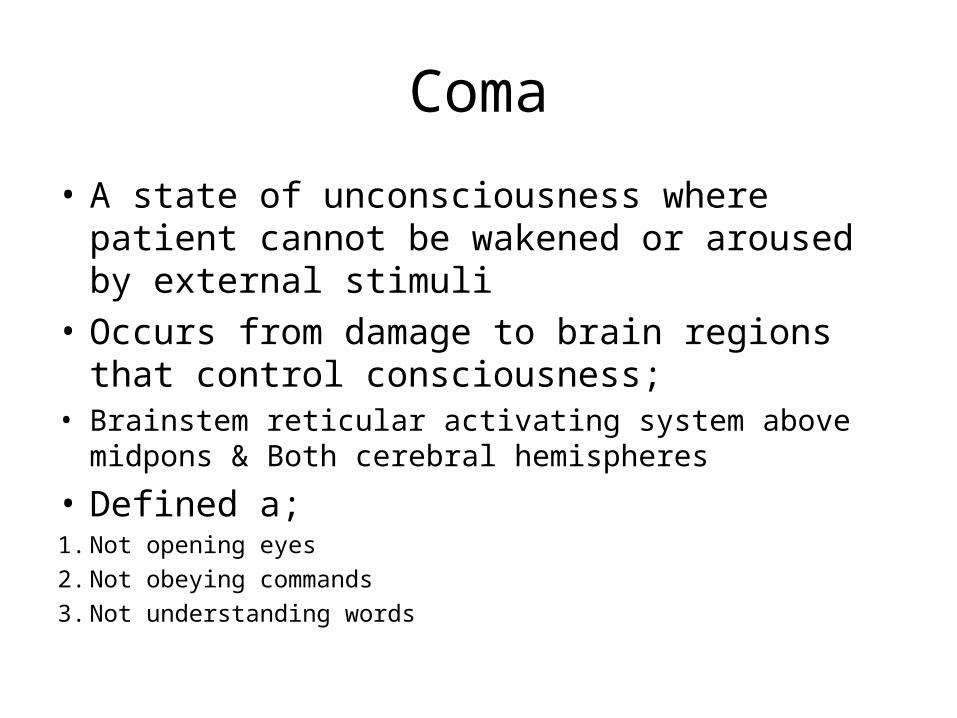
Coma
• A state of unconsciousness where patient cannot be wakened or aroused by external stimuli
• Occurs from damage to brain regions that control consciousness;
• Brainstem reticular activating system above midpons & Both cerebral hemispheres
• Defined a;1. Not opening eyes2. Not obeying commands3. Not understanding words
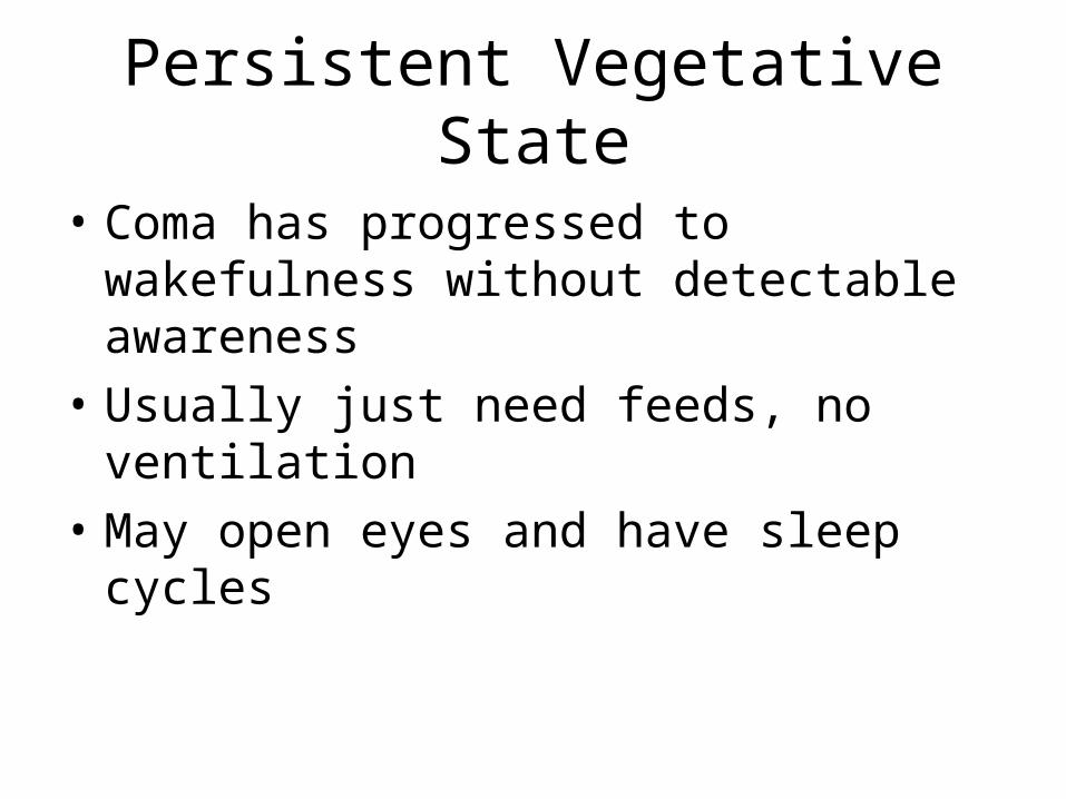
Persistent Vegetative State
• Coma has progressed to wakefulness without detectable awareness
• Usually just need feeds, no ventilation• May open eyes and have sleep cycles
Causes of Coma
• Diffuse Brain Dysfunction (generalised metabolic toxic injury/widespread inflammation)
• Hyperglycaemia (DKA)/Hypoglycaemia• ETOH • Drug intoxication• Hypoxic/IBI• Electrolyte imbalances• Acidosis (resp or metabolic)• SAH• Encephalitis• Cerebral Malaria• Endocrine (hypothroidism/hypoadrenalism)

Causes of Coma
• Direct Effect within Brain Stem• Trauma• Brainstem haemorrhage/infarction• Neoplasm
• Pressure effect of the Brain Stem• Hemisphere tumour or abscess• Cerebellar mass lesion• Trauma (SDH/EDH)• Encephalitis

Assessment
• Signs of Trauma• Swelling of soft tissues• Racoon eyes – periorbital eccymoses• Blood behind the tympanic membrane (haemotypanum)• Battle’s sign – discoloured swelling over the mastoid bone behind
the ear• CSF Rhinorrhoea/Otorrho

AssessmentVital Signs- BPo HT may indicate intracerebral haemorrhage or strokeo May also give clue to the cause of the coma (SAH?)- Tempo Hypothermia – ETOH, sedatives, hypoglycaemiao Hyperthermia – heat stroke, infection, hypothalamic lesions- Respirationo Cheyne- Stokes (periodic respiration with hyperpnoea & apnoea due to delay in
medullary chemoreceptor response – LVF, brain damage, altitude)o Kussmaul (acidotic) – deep sighing hyperventilation due to stimulation of
inspiratory centres – DKA, uraemia, metabolic acidosis o Ataxic – shallow, halting irregular respiration in response to medullary
respiratory centre damage

AssessmentPupilsNormal- 3-4mm in diameter, equal bilaterally- Constrict briskly+ symmetrically to light- Metabolic acidosis & CNS depressant drugs (not opiates)
Pin-point- 1-1.5mm in diameter- Opioid overdose- Pontine lesions, organophosphate poisoning

PupilsFixed Dilated- 7mm or more and fixed (not reactive to light- Results from compression of CN III - Common in herniation of the medial temporal lobe
- Fixed Mid-size- 5mm in diameter & fixed- Commonly from brainstem lesion at midbrain level
Anisocoria (Assymetrical)- Less than 1mm difference in normal people (20% cases)- Pupil that has reduced constriction – lesion affecting midbrain or CNIII

AssessmentOptic Fundi- Papilloedema/retinal haemorrhages – HT or raised ICP- Subhyaloid (superficial retinal) haemorrhages – SAHOcular Movements- Ocular Axes
o Usually slightly divergent in comao Slow, roving, side to side eye movements in light coma
- Doll’s Eye Reflex (Vestibulo-ocular reflex)o Passive head turning produces ocular deviation away from the direction of head
rotationo Lost in very deep coma and brainstem lesions
- Calorics Testingso Ice water is irrigated into the tympanic membraneo Slow tonic ocular deviation towards irrigated ear (intact brainstem)o Commonly used to Dx brainstem death

GCSEye Opening -None
-To Pain-To speech/verbal command-Spontaneous
1234
Verbal Response
-NoneIncomprehensible soundInappropriate wordsConfused/disorientedTalking & orientated
12345
Motor Response
NoneExtensionFlexionWithdrawalLocalised painObeys commands
123456

CGS
• 8 is the critical score• 90% less than 8 – coma• After 6 hours at 8 50% death rate

Investigations
• FBE• Biochemistry – U&E’s, Glucose, Ca, LFt’s• Drugs screen – salicylates, benzodiazepines,
narcotics, amphetamines• TFT’s• Blood cultures• CT or MRI – mass leson or intracranial haemorrhage• CSF• EEG – metabolic coma, encephalitis, brain death

Management (ED)
• DRABC• IV catheter and Bloods• IV infusion (routine)
o Thiamine 100mg + dextrose 25go Thiamine always precedes dextrose as dextrose along can worsen
Wernicke’s encephalophatyo Naloxone 0.4-1.2mg (routine)

Management (LT)• Fluids & Feeding (NGT or paraenteral)• Skin Care (Pressure sores)• Oral Hygiene (mouth washes + suction)• Eye care – tape eye lids• Physiotherapy – muscles + joints• TED stockingss/heparin – DVT risk• Sphincter Control – Catherisation, rectal evacuation• Family Wishes
References: Acknowledgment J Koh & D Cheng

Case• Mr GF, 75 y/o retired cook• Wife found him slumped over table
– Unable to speak, but could undertand– Right side of face drooping, weak right arm
• Presented to ED 2 hours later• O/E: unable to speak, obeys simple commands
– Right facial droop (pronounced in lower part of face)– Vision & Visual fields intact– Power reduced in right arm, only weak elevation of shoulder and
shoulder adduction– Mild weakness of right hip flexion– Soft bruit on auscultation of left carotid artery– BP 165/100

• PHx:– HT (on ACE inhibitors)– Ex-smoker (40 pack/years)
• Quit 10 years ago• FHx
– Father, also smoker, died of stroke at 65 • Tests:
– Haem/biochemical screens normal – Coag screen normal– CT brain reported as normal– MRI: shows area of diffuse restriction in anterior portion of
left middle cerebral artery– Repeated CT scan done 48 hours later shows area of low
attenuation in same region
• Speech therapy shows dysphagia w/ uncoordinated swallowing
• Made ‘nil by mouth’ + nasogastric tube• Given aspirin• Physio commenced next day• Becomes febrile– Right lower lobe consolidation detected clinically, seen on
CXR– Antibiotic & chest physiotherapy
• Over next week, return of speech & right arm weakness improves
• Carotid doppler shows 80-90% occlusion left carotid– Vascular surgery consulted– Left carotid endarterectomy planned in 6 weeks


![[PPT]PowerPoint Presentation - Vanderbilt University · Web viewBrain death – irreversible loss of all functions of the entire brain. Not Coma Paralysis Locked-in state – voluntary](https://img.dokumen.tips/doc/110x75/5aa6bf2c7f8b9ac8748eaf2e/pptpowerpoint-presentation-vanderbilt-university-viewbrain-death-irreversible.jpg)


![Pathology of non-thermal irreversible electroporation (N ... · values causes cell death in the treated volume via irreversible electroporation (IR E) [6]. It has been reported that](https://img.dokumen.tips/doc/110x75/6076fa2afafbc1083a0e49cb/pathology-of-non-thermal-irreversible-electroporation-n-values-causes-cell.jpg)