Embed Size (px)
Citation preview

8/2/2019 Com Minuted Patellar Fracture
http://slidepdf.com/reader/full/com-minuted-patellar-fracture 1/3
40
Malaysian Orthopaedic Journal 2008 Vol 2 No 2 T K Ong, et al
ABSTRACT
In comminuted patellar fractures, a combination of cerclage
wiring and tension band fixation is said to provide good
mechanical stability. This is a retrospective review of four
patients treated with this method. All fractures describedherein were classified as 45-C3 (based on Orthopaedic
Trauma Association classification) and were fixed with a
1.25mm cerclage wire and tension band wire proximally
looped through the quadriceps tendon and distally through
the patellar ligament in a figure-of-eight configuration. The
average follow-up period was 10 months. The Activity of
Daily Living Scale (ADLS) of the Knee Outcome Survey
was used to assess symptoms and functional capability of the
knee. In all the cases, fracture union was achieved at an
average of 11 weeks. The average ADLS score was good
(92.5 %). Full range of knee motion was achieved by end of
the third postoperative month. None of the patients hadcomplications, such as infection and implant failure.
Key Words:
Patella fracture, Cerclage wiring, Tension band wiring
INTRODUCTION
Patella fractures account for 1% of all skeletal fractures 1.
Comminuted patellar fractures usually occur with direct
trauma. About one third of patella fractures require surgery,
which is indicated if there is damage to the extensor
mechanism or in fractures associated with 2 mm step-off incongruity 2. The objectives of surgical treatment include
precise anatomic reduction of the articular surface by stable
fixation, and restoration of the knee-extensor mechanism,
thus allowing early mobilization.
Currently, several fixation methods of patellar fractures are
in use, including tension band wiring, cerclage wiring, and
screw fixation. Curtis, in a cadaveric study comparing the
AO method to the Pyrford technique using a cerclage wire
and an anterior tension band wire looping through the
quadriceps tendon, found that the latter gave greater strength
of fixation 3.
We reviewed a series of four patients with comminuted
fracture of patella, treated with a combination of cerclage
and tension band wire in figure-of-eight configuration
(Figure 1).
Case Series
Four patients with a mean age of 28 years (range 18 to 46)
were treated with the combined cerclage and tension band
wiring method (Table I). All patients were male and injuries
occurred due to involvement in a motor vehicle accident.
Fractures occurred on the right knee in three patients, on the
left side in one patient. One patient also had a closed fracture
of the ipsilateral femur, requiring internal fixation with an
interlocking nail. According to Orthopaedic Trauma
Association (OTA) classification, all fractures were coded as
45-C3 - characterized by comminuted and complete ar ticular
involvement with loss of the extensor mechanism (Figure 2).
Three fractures were closed and one was opened requiringwound debridement, arthrotomy washout and osteosynthesis
of the patella within twelve hours of the injury.
Surgical technique
Surgery was performed with patients in the supine position
with the injured knee extended. A longitudinal midline skin
incision was made over the patella. After incision of the
superficial fascia, the extensor apparatus was exposed and
any tear in the auxiliary extensor was identified. This was
followed by a medial parapatellar arthrotomy. Using a 14G
cannula, a 1.25mm cerclage wire was passed around the
equator of the patella, as close as possible to the bone.Another wire was passed proximally through the quadriceps
tendon and distally through the patellar ligament, posterior to
the cerclage wire and in the form of a figure-of-eight (figure
1). The cerclage wire is tightened to prevent further
displacement of bone fragments. While tightening the
second wire, congruity of the articular surface was checked
by palpating the retropatellar surface. The affected knees
were protected with a cylinder backslab for 4 weeks,
followed by a course of physiotherapy that included knee
bending, quadriceps and hamstring exercises. Full weight
bearing was allowed in all cases 8 weeks after the surgery.
Fixation of Comminuted Patellar Fracture with CombinedCerclage and Tension Band Wiring Technique
T K Ong, MBBS, E K Chee, MS (Ortho), C L Wong, FRCS (Ortho), K Thevarajan, MS (Ortho)
Department of Orthopaedics and Traumatology, Hospital Seberang Jaya, Seberang Jaya, Malaysia
Corresponding Author: Ong Teng Khiam, Department of Orthopaedics and Traumatology, Hospital Seberang Jaya, Jalan Tun HusseinOnn, 13700 Seberang Jaya, Pulau Pinang , Malaysia Email: tengkhiam@hot mail.com
8/2/2019 Com Minuted Patellar Fracture
http://slidepdf.com/reader/full/com-minuted-patellar-fracture 2/3
Comminut ed Patellar Fixation
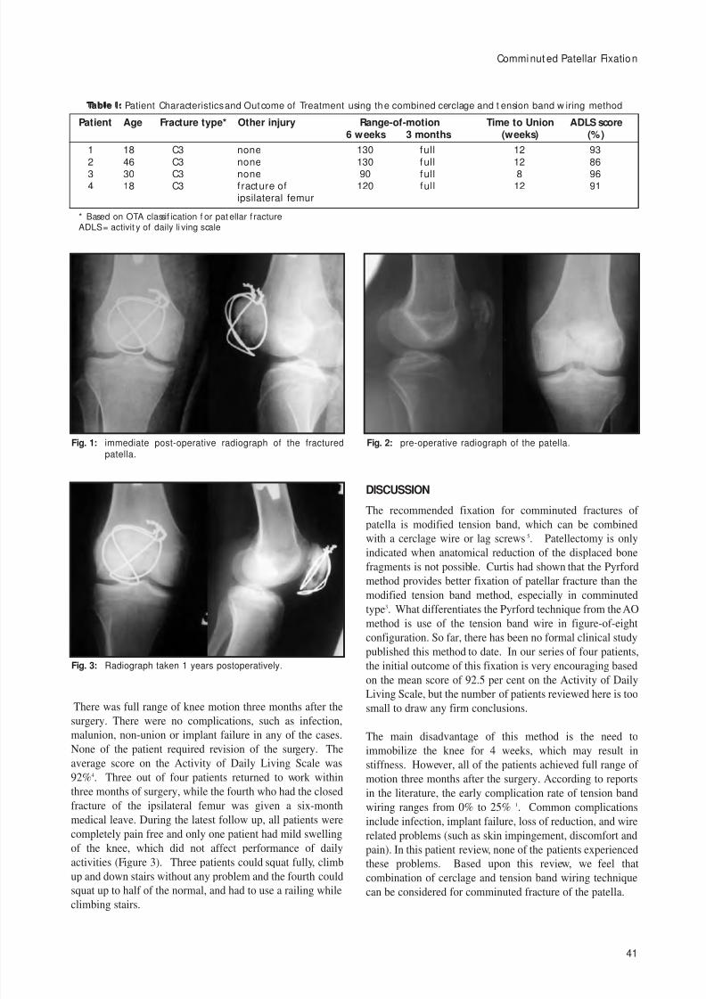
There was full range of knee motion three months after the
surgery. There were no complications, such as infection,
malunion, non-union or implant failure in any of the cases.
None of the patient required revision of the surgery. The
average score on the Activity of Daily Living Scale was
92%4. Three out of four patients returned to work within
three months of surgery, while the fourth who had the closed
fracture of the ipsilateral femur was given a six-month
medical leave. During the latest follow up, all patients were
completely pain free and only one patient had mild swelling
of the knee, which did not affect performance of daily
activities (Figure 3). Three patients could squat fully, climb
up and down stairs without any problem and the fourth couldsquat up to half of the normal, and had to use a railing while
climbing stairs.
DISCUSSION
The recommended fixation for comminuted fractures of
patella is modified tension band, which can be combinedwith a cerclage wire or lag screws 5. Patellectomy is only
indicated when anatomical reduction of the displaced bone
fragments is not possible. Curtis had shown that the Pyrford
method provides better fixation of patellar fracture than the
modified tension band method, especially in comminuted
type3. What differentiates the Pyrford technique from the AO
method is use of the tension band wire in figure-of-eight
configuration. So far, there has been no formal clinical study
published this method to date. In our series of four patients,
the initial outcome of this fixation is very encouraging based
on the mean score of 92.5 per cent on the Activity of Daily
Living Scale, but the number of patients reviewed here is too
small to draw any firm conclusions.
The main disadvantage of this method is the need to
immobilize the knee for 4 weeks, which may result in
stiffness. However, all of the patients achieved full range of
motion three months after the surgery. According to reports
in the literature, the early complication rate of tension band
wiring ranges from 0% to 25% 1. Common complications
include infection, implant failure, loss of reduction, and wire
related problems (such as skin impingement, discomfort and
pain). In this patient review, none of the patients experienced
these problems. Based upon this review, we feel that
combination of cerclage and tension band wiring techniquecan be considered for comminuted fracture of the patella.
Fig. 1: immediate post-operative radiograph of the fracturedpatella.
Fig. 2: pre-operative radiograph of the patella.
Fig. 3: Radiograph taken 1 years postoperatively.
41
Patient Age Fracture type* Other injury Range-of-motion Time to Union ADLS score6 weeks 3 months (weeks) (%)
1 18 C3 none 130 full 12 93
2 46 C3 none 130 full 12 86
3 30 C3 none 90 full 8 964 18 C3 fracture of 120 full 12 91
ipsilateral femur
* Based on OTA classif ication f or pat ellar f ractureADLS = activit y of daily li ving scale
Table I: Patient Characteristics and Outcome of Treatment using the combined cerclage and t ension band w iring method

8/2/2019 Com Minuted Patellar Fracture
http://slidepdf.com/reader/full/com-minuted-patellar-fracture 3/3
Malaysian Orthopaedic Journal 2008 Vol 2 No 2 T K Ong, et al
42
REFERENCES
1. Lotke PA, Ecker ML. Transverse fractures of the patella. Clin Orthop. 1981; 158: 180–4.
2. Carpenter JE, Kasman R, Matthews LS. Fracture of the patella: Instr Course Lect. 1994; 43: 97-108.
3. Curtis MJ. Internal fixation for fractures of the patella. A comparison of two methods. J Bone Joint Surg Br. 1990; 72: 280-2.
4. James J Irrgang, Lynn Snyder-Mackler, Robert S Wainner Fu FH, Harner CD. Development of patient-reported measure of
function of the knee. J Bone Joint Surg Br. 1998; 80: 1132-45.
5. Reudi TP, Murphy WM. AO Principles of Fracture Management. New York, NY: Thieme-Stuttgart, 2000: 483-91.

![Management of comminuted patellar fracture fixation using ... … · effect on simple transverse patellar fracture [13], and the curative effect on the comminuted patella remains](https://img.dokumen.tips/doc/110x75/60a273c826934d09c56642c1/management-of-comminuted-patellar-fracture-fixation-using-effect-on-simple.jpg)