Embed Size (px)
Citation preview
Biotechnology Advances xxx (2015) xxx–xxx
JBA-06912; No of Pages 15
Contents lists available at ScienceDirect
Biotechnology Advances
j ourna l homepage: www.e lsev ie r .com/ locate /b iotechadv
Research review paper
Colorimetric biosensing of pathogens using gold nanoparticles
Mohit S. Verma a,b, Jacob L. Rogowski a,b, Lyndon Jones a,c, Frank X. Gu a,b,⁎a Department of Chemical Engineering, University of Waterloo, 200 University Avenue W, Waterloo, Ontario N2L 3G1, Canadab Waterloo Institute for Nanotechnology, University of Waterloo, 200 University Avenue W, Waterloo, Ontario N2L 3G1, Canadac Center for Contact Lens Research, University of Waterloo, 200 University Avenue W, Waterloo, Ontario N2L 3G1, Canada
⁎ Corresponding author at: Department of Chemical Engfax: +1 519 888 4347.
E-mail address: [email protected] (F.X. Gu).
http://dx.doi.org/10.1016/j.biotechadv.2015.03.0030734-9750/© 2015 Elsevier Inc. All rights reserved.
Please cite this article as: Verma MS, et al,dx.doi.org/10.1016/j.biotechadv.2015.03.003
a b s t r a c t
a r t i c l e i n f oArticle history:Received 25 September 2014Received in revised form 8 February 2015Accepted 2 March 2015Available online xxxx
Keywords:BacteriaVirusFungusPoint-of-careDNARNAProteinSmall moleculePCRColor
Rapid detection of pathogens is crucial tominimize adverse health impacts of nosocomial, foodborne, andwater-borne diseases. Gold nanoparticles are extremely successful at detecting pathogens due to their ability to providea simple and rapid color change when their environment is altered. Here, we review general strategies ofimplementing gold nanoparticles in colorimetric biosensors. First, we highlight how gold nanoparticles haveimproved conventional genomic analysis methods by lowering detection limits while reducing assay times.Then, we focus on emerging point-of-care technologies that aim at pathogen detection using simpler assays.These advanceswill facilitate the implementation of gold nanoparticle-based biosensors in diverse environmentsthroughout the world and help prevent the spread of infectious diseases.
© 2015 Elsevier Inc. All rights reserved.
Contents
1. Introduction . . . . . . . . . . . . . . . . . . . . . . . . . . . . . . . . . . . . . . . . . . . . . . . . . . . . . . . . . . . . . . . 02. Conventional methods for pathogen detection . . . . . . . . . . . . . . . . . . . . . . . . . . . . . . . . . . . . . . . . . . . . . . . 03. Principles of gold nanoparticle sensing . . . . . . . . . . . . . . . . . . . . . . . . . . . . . . . . . . . . . . . . . . . . . . . . . . . 04. Gold nanoparticles for detecting nucleic acids . . . . . . . . . . . . . . . . . . . . . . . . . . . . . . . . . . . . . . . . . . . . . . . . 0
4.1. Gold nanoparticles for amplified nucleic acids . . . . . . . . . . . . . . . . . . . . . . . . . . . . . . . . . . . . . . . . . . . . 04.1.1. Non-functionalized gold nanoparticles . . . . . . . . . . . . . . . . . . . . . . . . . . . . . . . . . . . . . . . . . . . 04.1.2. Functionalized gold nanoparticles . . . . . . . . . . . . . . . . . . . . . . . . . . . . . . . . . . . . . . . . . . . . . 0
4.2. Gold nanoparticles for unamplified nucleic acids . . . . . . . . . . . . . . . . . . . . . . . . . . . . . . . . . . . . . . . . . . . 04.2.1. Non-functionalized gold nanoparticles . . . . . . . . . . . . . . . . . . . . . . . . . . . . . . . . . . . . . . . . . . . 04.2.2. Functionalized gold nanoparticles . . . . . . . . . . . . . . . . . . . . . . . . . . . . . . . . . . . . . . . . . . . . . 0
5. Emerging biosensors for detecting non-nucleic acid analytes . . . . . . . . . . . . . . . . . . . . . . . . . . . . . . . . . . . . . . . . . 05.1. Non-functionalized gold nanoparticles . . . . . . . . . . . . . . . . . . . . . . . . . . . . . . . . . . . . . . . . . . . . . . . 05.2. Gold nanoparticles functionalized with proteins . . . . . . . . . . . . . . . . . . . . . . . . . . . . . . . . . . . . . . . . . . . 05.3. Gold nanoparticles functionalized with small molecules . . . . . . . . . . . . . . . . . . . . . . . . . . . . . . . . . . . . . . . . 0
6. Comparison of gold nanoparticles to conventional methods . . . . . . . . . . . . . . . . . . . . . . . . . . . . . . . . . . . . . . . . . 06.1. Analysis time . . . . . . . . . . . . . . . . . . . . . . . . . . . . . . . . . . . . . . . . . . . . . . . . . . . . . . . . . . . 06.2. Limit of detection . . . . . . . . . . . . . . . . . . . . . . . . . . . . . . . . . . . . . . . . . . . . . . . . . . . . . . . . . 06.3. Specificity . . . . . . . . . . . . . . . . . . . . . . . . . . . . . . . . . . . . . . . . . . . . . . . . . . . . . . . . . . . . 06.4. Technical requirements . . . . . . . . . . . . . . . . . . . . . . . . . . . . . . . . . . . . . . . . . . . . . . . . . . . . . . 0
ineering, University ofWaterloo, 200 University AvenueW,Waterloo, Ontario N2L 3G1, Canada. Tel.:+1 519 888 4567x38605;
Colorimetric biosensing of pathogens using gold nanoparticles, Biotechnol Adv (2015), http://
2 M.S. Verma et al. / Biotechnology Advances xxx (2015) xxx–xxx
7. Conclusions . . . . . . . . . . . . . . . . . . . . . . . . . . . . . . . . . . . . . . . . . . . . . . . . . . . . . . . . . . . . . . . 0Abbreviations . . . . . . . . . . . . . . . . . . . . . . . . . . . . . . . . . . . . . . . . . . . . . . . . . . . . . . . . . . . . . . . . . 0Acknowledgments . . . . . . . . . . . . . . . . . . . . . . . . . . . . . . . . . . . . . . . . . . . . . . . . . . . . . . . . . . . . . . . 0References . . . . . . . . . . . . . . . . . . . . . . . . . . . . . . . . . . . . . . . . . . . . . . . . . . . . . . . . . . . . . . . . . . 0
1. Introduction
Mankind has been fascinated by gold nanoparticles for centuries andthe Lycurgus cup is a prime example of their unique optical properties.In the 21st century, research involving gold nanoparticles haswitnessedsignificant growth with applications in drug delivery (Boisselier andAstruc, 2009; Ghosh et al., 2008; Paciotti et al., 2004), photothermaltherapy (Gobin et al., 2007; Huang et al., 2006; Jain et al., 2007), diag-nostic imaging (Eghtedari et al., 2007; Murphy et al., 2008; Popovtzeret al., 2008), and biosensors (Hutter and Fendler, 2004; Liu and Lu,2003; Mayer and Hafner, 2011). Along with being the most stable me-tallic nanoparticles (Daniel and Astruc, 2004), gold nanoparticles flauntseveral outstanding features, including facile reactivity with biomole-cules, high surface area to volume ratios, and environment dependentoptical properties, which make them the ideal candidate for use incolorimetric biosensors (Upadhyayula, 2012).
Pathogens—including bacteria, viruses, fungi, and protozoa—are aleading cause for loss of lives in the developing world, as well as ruralareas of developed countries, due to lack of infrastructure and resources(Tallury et al., 2010). Since pathogens can be transmitted via plants,animals, and humans, infectious diseases can spread exponentiallyand lead to a pandemic if left unchecked (Kaittanis et al., 2010). Themost effective method for preventing the spread of infectious diseasesis early diagnosis, which is challenging using conventional methodsbecause of expensive equipment, specialized sample preparation, andslow data output (Tallury et al., 2010). Modern biosensors haveovercome these obstacles by miniaturizing devices and providingsimple rapid output that can be analyzed at the point-of-care withoutspecialized training (Lazcka et al., 2007; Mao et al., 2009; Skottrupet al., 2008).
In addition to point-of-care diagnostics and early treatment of infec-tious diseases in humans, microbial pathogens are also a concern at var-ious levels of the food industry. Many bacterial genera are associatedwith food-borne illness such as Salmonella, Listeria, and Escherichia.Infections are typically caused by consumption of food or drink contam-inated with these pathogens, and may lead to various inflammatoryconditions including gastroenteritis, meningitis, and sepsis. Seriousinfections may require hospitalization and can be fatal for more vulner-able segments of the population (e.g., immunocompromised patients)(Farber and Peterkin, 1991). While low levels of bacteria and othermicrobial life are sometimes tolerable, high concentrations are fre-quently associated with food-borne illnesses (Velusamy et al., 2010).Various agencies have implemented guidelines for food production,preparation, and distribution, which aim to keep pathogen loads atacceptable levels. Often these guidelines have stringent concentrationrequirements and hence, screening assays require excellent detectionlimits.
Gold nanoparticles have been implemented for the detection ofpathogens, which contaminate food, water, and hospital surfaces(Agasti et al., 2010; Azzazy et al., 2012; Bunz and Rotello, 2010;Khanna, 2008; Saha et al., 2012; Tallury et al., 2010; Upadhyayula,2012). A major focus of research is to improve conventional genomicanalysis methods using gold nanoparticles such that the assays havelower detection limits and faster response times (Fig. 1). Concurrently,novel methods of detection have been developed independent of geneamplification and the most popular strategy is based on the surfacemodification of gold nanoparticles with antibodies, which has led toseveral commercially available products for easy and timely testing ofpathogens in complex samples such as plant extracts, foods, and bodily
Please cite this article as: Verma MS, et al, Colorimetric biosensing of pdx.doi.org/10.1016/j.biotechadv.2015.03.003
fluids. An emerging strategy is to exploit the intrinsic surface propertiesof gold nanoparticles and pathogenswhich lead to electrostatic interac-tions and a color change.
2. Conventional methods for pathogen detection
The importance of pathogen detection in several sectors has ledto continuous improvement in detection technologies. Currently,conventional methods for pathogen detection can be roughly divid-ed into three categories: culture and colony counting, immunologi-cal assays, and polymerase chain reaction (PCR)-based methods(Velusamy et al., 2010). These methods offer high sensitivity andspecificity, providing both quantitative and qualitative information,which is often a necessity. However, some key drawbacks, chief ofwhich being required processing times, clearly indicate a need forbetter solutions.
Colony counting is widely considered to be the gold standard forpathogen detection in settings ranging from clinical diagnosis to foodpathogen measurement (Lazcka et al., 2007; Peters et al., 2004;Velusamy et al., 2010). This process involves isolation and growth of asuspect pathogen, followed by visual inspection. Due to the inherentamplification during colony growth, this method is good for identifyingvery low levels of organisms (i.e., single cells). Unfortunately, turn-around times for results are very slow using this technique due tolong incubation periods and the need for intensive labor. Dependingon the pathogen, initial results often require at least 2 days,with confor-mation after 7–10 days (Peters et al., 2004; Velusamy et al., 2010).Furthermore, the colony counting method requires a pathogen to beculturable, which may not always be the case given stringent environ-mental or nutritional requirements.
Immunological assays are very common for pathogen detectiondue to their adaptability for a wide variety of pathogens includingbacteria and viruses. The enzyme-linked immunosorbent assay(ELISA) method is an example of a well-known immunologicalassay. These assays rely on antibody recognition of antigens andother biomolecules specific to the target. Once antibodies are identi-fied and available, the primary advantage of immunological assaysover colony counting is reduced assay time while maintaining highspecificity. ELISA is capable of generating an optical response andhence is widely deployed in clinical laboratories using commerciallyavailable ELISA kits. The technique still suffers from the drawbacks ofrequiring multiple steps, specialized training, and several hours ofruntime (Ahmed et al., 2014; Lazcka et al., 2007). Antibody-labeledgold nanoparticles have overcome these challenges by using animmunochromatographic strip (ICS) format and unique productsare available for testing of foods and clinical samples. The testing offood products is facilitated by Merck Millipore's Singlepath® andDuopath® products (Billerica, MA, USA), but these products requireselective enrichment of bacteria before the sample is analyzed,which is necessary because of low sensitivity and the need to detectlow concentration of pathogens in food. Thus, the assay requires sev-eral hours for completion even though the ICS can respond within20 min. In a clinical diagnostic setting, ICS-based assays have beendeveloped by Coris Bioconcept (Gembloux, Belgium) for the detec-tion of viruses and bacteria in stool, urine, and blood samples(Renuart et al., 2011). Current challenges faced by ICS-based assaysinclude the variability caused by user sample preparation andcross-reactivity of analytes, but ICS has been the biggest commercial-ly available success of colloidal gold nanoparticles because of their
athogens using gold nanoparticles, Biotechnol Adv (2015), http://
3M.S. Verma et al. / Biotechnology Advances xxx (2015) xxx–xxx
ability to analyze samples in a complex media withminimal purifica-tion. We will highlight how emerging technologies have adopted thesuccess of antibody-labeled gold nanoparticles in later sections ofthis review paper.
PCR-based methods constitute a wide variety of detection schemesrelying on nucleic acid amplification to increase the concentration ofthe detection target. Amplification of target deoxyribonucleic acid(DNA) sequences lends PCR-based conventional methods a high degreeof sensitivity, even capable of detecting single gene copies. It is impor-tant to note that unlike colony counting, this sensitivity is achievedwithout a prolonged incubation time since bacteria do not need to begrown (Ahmed et al., 2014). Specificity is achieved through the designof primers and probes that target sequences unique to the pathogen ofinterest. However, interference from non-pathogenic genetic materialmay lead to misleading results due to mismatch or non-specific ampli-fication (Ahmed et al., 2014; Velusamy et al., 2010). Precise geneticinformation is therefore required for confidence in results. Followingtarget amplification, samples are traditionally separated by gel electro-phoresis but complex sample preparations and manipulations increaselabor cost and processing times (Lazcka et al., 2007). Newer technologiessuch as real-time PCR and fluorescent molecular probes aim to reducethese factors. Perhaps the main drawback of traditional PCR-basedmethods for pathogen detection is the inability to distinguish viable andnon-viable cells, since both contain the amplification target (Velusamyet al., 2010). To address this issue, assays have been developed that em-ploy reverse transcription PCR (RT-PCR) to target rapidly degradingmes-senger ribonucleic acid (mRNA) strands present during the cell's growthcycle (Velusamy et al., 2010; Yaron and Matthews, 2002).
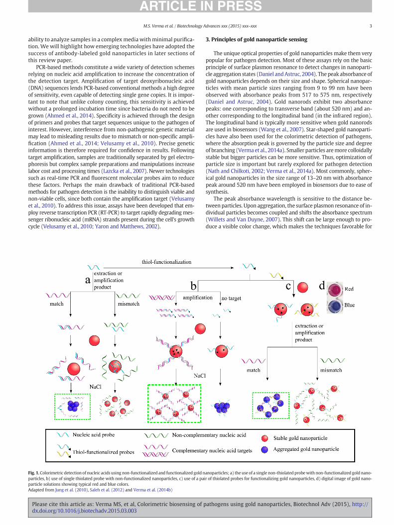
Fig. 1.Colorimetric detection of nucleic acids using non-functionalized and functionalized goldnparticles, b) use of single thiolated probe with non-functionalized nanoparticles, c) use of a paiparticle solutions showing typical red and blue colors.Adapted from Jung et al. (2010), Saleh et al. (2012) and Verma et al. (2014b)
Please cite this article as: Verma MS, et al, Colorimetric biosensing of pdx.doi.org/10.1016/j.biotechadv.2015.03.003
3. Principles of gold nanoparticle sensing
The unique optical properties of gold nanoparticles make them verypopular for pathogen detection. Most of these assays rely on the basicprinciple of surface plasmon resonance to detect changes in nanoparti-cle aggregation states (Daniel andAstruc, 2004). The peak absorbance ofgold nanoparticles depends on their size and shape. Spherical nanopar-ticles with mean particle sizes ranging from 9 to 99 nm have beenobserved with absorbance peaks from 517 to 575 nm, respectively(Daniel and Astruc, 2004). Gold nanorods exhibit two absorbancepeaks: one corresponding to transverse band (about 520 nm) and an-other corresponding to the longitudinal band (in the infrared region).The longitudinal band is typically more sensitive when gold nanorodsare used in biosensors (Wang et al., 2007). Star-shaped gold nanoparti-cles have also been used for the colorimetric detection of pathogens,where the absorption peak is governed by the particle size and degreeof branching (Vermaet al., 2014a). Smaller particles aremore colloidallystable but bigger particles can be more sensitive. Thus, optimization ofparticle size is important but rarely explored for pathogen detection(Nath and Chilkoti, 2002; Verma et al., 2014a). Most commonly, spher-ical gold nanoparticles in the size range of 13–20 nm with absorbancepeak around 520 nm have been employed in biosensors due to ease ofsynthesis.
The peak absorbance wavelength is sensitive to the distance be-tween particles. Upon aggregation, the surface plasmon resonance of in-dividual particles becomes coupled and shifts the absorbance spectrum(Willets and Van Duyne, 2007). This shift can be large enough to pro-duce a visible color change, which makes the techniques favorable for
anoparticles; a) theuse of a single non-thiolated probewith non-functionalized goldnano-r of thiolated probes for functionalizing gold nanoparticles, d) digital image of gold nano-
athogens using gold nanoparticles, Biotechnol Adv (2015), http://
4 M.S. Verma et al. / Biotechnology Advances xxx (2015) xxx–xxx
rapid point-of-care diagnostics. Peak absorbance wavelengths exhibit ared-shift with increases in size, typically giving stable (non-aggregated)nanoparticles a red color, while aggregated nanoparticles appear blue(Fig. 1d) (Kim and Lee, 2009). Use of an ultraviolet–visible spectropho-tometer can help quantify the shift in the surface plasmon resonancepeak.
Gold nanoparticles are typically stabilized electrostatically,where citrate-capped nanoparticles are negatively charged andcetyltrimetylammonium bromide (CTAB)-coated nanoparticlesare positively charged. The electrostatic repulsion between nanoparti-cles can be shielded by the addition of salts (most commonly sodiumchloride), which then leads to the aggregation of nanoparticles andhence, a color change (Zhao et al., 2008).
Optical effects of surface plasmon resonance have been implement-ed for pathogen detection by either inducing particle aggregation or sta-bilization. These effects are governed by target ligands, nanoparticlefunctionalization, competitive binding sites, or salts. The specific combi-nations of these factors make up the wide variety of applications inves-tigated in this review.
4. Gold nanoparticles for detecting nucleic acids
4.1. Gold nanoparticles for amplified nucleic acids
Amplification is a common strategy in molecular diagnostics for in-creasing signal strength. Various methods for the amplification of bothDNA and ribonucleic acid (RNA) have been highlighted inmolecular di-agnostic reviews (Cenciarini-Borde et al., 2009; Lauri andMariani, 2009;Monis and Giglio, 2006). Some of the amplification methods that havebeen used in conjunction with gold nanoparticles are: PCR, real-time(or quantitative) PCR, RT-PCR, asymmetric PCR, rolling circle amplifica-tion (RCA), and nucleic acid sequence-based amplification (NASBA).
4.1.1. Non-functionalized gold nanoparticlesNon-functionalized gold nanoparticles are usually used for the de-
tection of amplified products by the addition of salt. In the presence ofsalt, typically gold nanoparticles will aggregate and change color fromred to blue unless they can be stabilized by nucleic acids. Two primarystrategies can be utilized for stabilizing the gold nanoparticles: adsorp-tion of nucleic acids on the surface or reaction with thiol probe that hasbeen hybridized with the target nucleic acids (Fig. 1a, b). Another ap-proach involves the use of cationic gold nanoparticles, where the inter-actions between nucleic acids and the surface of gold nanoparticles leadto aggregation of the nanoparticles. This approach is similar to the oneexplained in Fig. 1c, except the gold nanoparticles are not functionalizedwith a thiol-probe but rather coated with the probe using electrostaticinteractions.
DNA from bacteria and viruses has been used for detection by ad-sorption on the surface of gold nanoparticles. Salmonella spp. are trou-blesome for causing foodborne illnesses. Regulatory levels publishedby the United States Food and Drug Administration (FDA) and UnitedStates Environmental Protection Agency (EPA) for food safety requirecomplete absence of Salmonella spp. in a 25 gram sample (U.S. Foodand Drug Administration, 2011) It is therefore important for detectionassays to have high sensitivity to very low (individual) pathogen levels.Salmonella spp. has been detected by targeting the stn gene where anoligonucleotide probe was designed to be complementary to the PCRproduct (Prasad et al., 2011). Here, 23 nm gold nanoparticles wereable to produce a detection limit 10× more sensitive than gel electro-phoresis. Also, a sensitivity (true positive rate) of 89.15% and specificity(true negative rate) of 99.04% was obtained for various food samples ascompared to conventional culture methods. Detection of Bacillusanthracis, the causative agent of anthrax, is possible by using a similarstrategy. Here, it was demonstrated that when the DNA is longer thanabout 100 nt (single-strandedDNA, ssDNA) or 100 bp (double-strandedDNA, dsDNA), it can prevent salt-induced aggregation of 15 nm gold
Please cite this article as: Verma MS, et al, Colorimetric biosensing of pdx.doi.org/10.1016/j.biotechadv.2015.03.003
nanoparticles (Deng et al., 2013) and the colorimetric response is visibleby the naked eye. When considering viruses, Hepatitis B virus (HBV) isnotorious for causing acute and chronic liver diseases worldwide. HBVhas been detected by designing a probe targeting the rtM204M wildtype gene (Liu et al., 2011). A colorimetric response from 13 nm goldnanoparticles was able to distinguish between target DNA and singlebase pair mismatched DNA. On the other hand, RCA has been used forthe detection of H1N1 viral DNA, where long ssDNA curled into ballsand could not stabilize 13 nm gold nanoparticles (Xing et al., 2013).
Thiol-modified probes coupled with non-functionalized nanoparti-cles have primarily been used for detection of bacterial DNA. Chlamydiatrachomatis is responsible for most of the bacterial sexually transmitteddiseasesworldwide. The gene encoding virulence proteinswas targetedwith thiolated probes and detected in human urine samples using13 nm gold nanoparticles (Jung et al., 2010). Listeria monocytogenesand Salmonella enterica are notorious for contaminating foods and caus-ing fatalities. FDA regulations have a “zero-tolerance” policy of no de-tectable L. monocytogenes in two 25 g samples of food or beverage(Hitchins and Jinneman, 1998). The detection of these food-borne bac-teria has been possible by designing thiolated probes to target the hlyand hut genes for L. monocytogenes and S. enterica respectively. Thisassay was able to detect bacteria in contaminated milk samples using13 nm gold nanoparticles and the specificity was confirmed by a lackof response from Escherichia coli (Fu et al., 2013).
The detection of DNA from human immunodeficiency virus type 1(HIV-1) has been possible using cationic gold nanorods. The probe is de-signed to target sections of the HB-hp3-LTR1.8 DNA and in the presenceof the target, aggregation is induced (He et al., 2008). The specificity ofthis assay was confirmed by comparing results against genes from My-cobacterium tuberculosis and genes encoding for Bacillus glucanase. Itwas possible to perform detection under physiological conditions be-cause the assay is tolerant to high salt concentrations. Another use ofgold nanorods is for the detection of Leishmania major, a protozoan par-asite that has led to 1.5million cases of cutaneous leishmaniasis annual-ly worldwide. The disease can lead to disabilities and even death. Thedetection of the parasite using culture-based methods is extremelyslow and insensitive. Thus, molecular diagnostics can offer an improvedmethod for detection. NASBA has been employed for the detection of18S ribosomal RNA (rRNA) of L. major by designing the appropriateprimer (Niazi et al., 2013). After amplification, the NASBA ampliconsare incubated with gold nanorods leading to aggregation. Clinical skinbiopsies were tested using this method and a sensitivity of 100% andspecificity of 80% was obtained as compared to RT-PCR and gelelectrophoresis.
Non-functionalized gold nanoparticles have the advantage of pro-viding rapid response as compared to gel electrophoresis. Additionally,the equipment necessary for gel electrophoresis is not needed since asimple colorimetric response is obtained, which can be visually ob-served with minimal training. The synthesis of non-functionalizednanoparticles can often be executed in a single step, which simplifiesthe assay. The main limitation to this approach is that the conditionsfor detection often need to be optimized such that the appropriate con-centrations of salts and reagents are used to avoid unnecessary aggrega-tion of gold nanoparticles. The optimization of assay conditionsdemands extra efforts for each target in question. The studies usingnon-functionalized gold nanoparticles for amplified nucleic acids havebeen summarized in Table 1. They are divided by pathogen type: bacte-ria, viruses, and protozoa, and then sorted chronologically.
4.1.2. Functionalized gold nanoparticlesGold nanoparticles can be easily functionalized with nucleic acid
probes by using thiol-gold chemistry. There are two primary ap-proaches to detection, which are governed by the number of probesused. In one scenario, salt is used to induce aggregation of probe-conjugated gold nanoparticles. Only one type of probe is used for bind-ing to the target sequence. This approach is similar to the illustration in
athogens using gold nanoparticles, Biotechnol Adv (2015), http://
Table 1Non-functionalized gold nanoparticles for detecting amplified nucleic acids.
Microorganisms of interest Sample type Analysis time Detection limit (copies/μL DNA) Working range (copies/μL DNA) References
Chlamydia trachomatis Urine 1 h post-amplification 20 20–20,000 Jung et al. (2010)Salmonella spp. Culture b8 h 2 × 10⁹ 2 × 10⁹–2 × 10¹¹ Prasad et al. (2011)Listeria monocytogenes andSalmonella enterica
Food 3–4 h 2.1 × 10⁴ (L. monocytogenes)2.6 × 10⁴ (S. enterica)
2.1 × 10⁴–2.1 × 10¹¹ (L. monocytogenes)2.6 × 10⁴–2.6 × 10¹¹ (S. enterica)
Fu et al. (2013)
Bacillus anthracis Nucleic acids – ~3.9 × 10⁶a ~3.9 × 10⁶–3.9 × 10⁸a Deng et al. (2013)HIV-1 Nucleic acids b5 min post-amplification 4.8 × 10⁷ 1.0 × 10⁸–7.0 × 10⁹ He et al. (2008)Hepatitis B virus Serum – 3 × 10⁹ 3 × 10⁹–3 × 10¹¹ Liu et al. (2011)H1N1 virus Nucleic acids 3 h 6.02 × 10⁵ 6.02 × 10⁵–6.02 × 10¹⁰ Xing et al. (2013)Leishmania major Skin biopsy – – – Niazi et al. (2013)
a A mixture of ssDNA and dsDNA was used, molecular weight of ssDNA was used for calculations. Sample type ‘culture’ refers to the bacteria being grown in the lab before testing.
5M.S. Verma et al. / Biotechnology Advances xxx (2015) xxx–xxx
Fig. 1b, except the gold nanoparticles are conjugated to the thiolatedprobe before hybridization. In this situation, binding of the target tothe probe results in double helix formation and particles can remain sta-ble under higher salt conditions. Consequently, absence of the targetwould lead to particle aggregation at similar salinity. In another scenar-io, two probes are used such that each probe can bind to the samenucleic acid strand. There are two main methods within the two-probe approach. In one method, gold nanoparticles are functionalizedwith each of the two probes separately and then mixed together. Thepresence of the target causes particle aggregation by cross-linkinggold nanoparticles together. In the absence of target sequence or thepresence of a mismatched sequence, aggregation does not occur andparticles remain stable in suspension (Fig. 1c). Another method usingtwo probes is called gold label silver stain. Here, one probe isimmobilized on a glass slide and another on the gold nanoparticles.The target nucleic acid binds to the glass slide first, followed by the ad-dition of the gold nanoparticles and then silver for signal enhancement(Taton et al., 2000). In recent studies, the probe immobilized on goldnanoparticle has been replaced by streptavidin and the PCR producthas been functionalized with biotin for facilitating binding viastreptavidin–biotin interactions instead of hybridization.
Using the one-probe approach,M. tuberculosis has been detected bydesigning probes targeting the rpoB gene and immobilizing them on14 nm gold nanoparticles (Veigas et al., 2010). The design is able todiscriminate against the non-tuberculosis causing Mycobacteriumkansasii. This design has also been implemented in a paper format, byusing wax-based ink for making a 384-well paper microplate (Veigaset al., 2012). The assay has been adapted for differentiating betweenMycobacterium bovis and M. tuberculosis by targeting the gyrB gene(Costa et al., 2010). Three probes were designed to target specific seg-ments of the gyrB gene and immobilized on gold nanoparticles. Eachstrain of Mycobacterium interacted differently with the probes andhence allowed accurate identification. Another notorious pathogenmethicillin-resistant Staphylococcus aureus (MRSA) has been responsi-ble for numerous persistent infections. It has been possible to detectMRSA by using probes towards 23S rRNA and mecA genes (Chan et al.,2014). In this study, the sensitivity and specificity were comparable toreal-time PCR assays but with a lower cost per reaction. RNA has alsobeen targeted using the one-probe approach. One example is the detec-tion of dnaK messenger RNA of S. enterica serovar Typhimurium afteramplification by NASBA (Mollasalehi and Yazdanparast, 2012). Theprobe was immobilized on 17 to 23 nm gold nanoparticles and theassay was able to distinguish between RNA from S. Typhimurium andBacillus firmus.
The two-probe approach has also gained popularity for a variety ofbacterial and viral targets. Helicobacter pylori is responsible for severalgastric conditions such as chronic gastritis, gastric adenocarcinomaand gastric ulcers. Detection of H. pylori is possible by designing probestowards the ureC gene and immobilizing them on gold nanoparticles.Target DNA was amplified using thermophilic helicase-dependent iso-thermal amplification and the assay was able to distinguish betweenH. pylori, E. coli, and human DNA (Gill et al., 2008). While some strains
Please cite this article as: Verma MS, et al, Colorimetric biosensing of pdx.doi.org/10.1016/j.biotechadv.2015.03.003
of E. coli can be harmless, Shiga toxin producing E. coli O157:H7 cancause disease outbreaks when it gets transmitted via food or water.FDA and EPA regulations for clams, mussels, oysters, and scallopsrequire E. coli levels to be below 330/100 g as determined by theMost Probable Number method, which translates to approximately3.3 colony forming unit (CFU)/g (U.S. Food and Drug Administration,2011). The detection of E. coli O157:H7 has been achieved by designinga pair of probes targeting the stx2 gene and immobilizing them on goldnanoparticles for a visible color change (Jyoti et al., 2010). Another food-borne pathogen S. Typhimurium can be detected by targeting the inva-sion (inv A) gene (Majdinasab et al., 2013). The specificity of this assaywas confirmed by comparing response to PCR products of other non-Salmonella spp. bacteria. The assay can provide better sensitivity com-pared to gel electrophoresis (Majdinasab et al., 2013). While moststudies have focused on detection of a single species of bacteria, itis also possible to design gold nanoparticles for the detection of mul-tiple bacteria, including the non-pathogenic ones. This is especiallyimportant in blood components because of the zero-tolerance policy.The 16S rDNA sequence is present in most bacteria and hence can beused as a target for detection (Wang et al., 2012b). A pair of 12-merprobes have been designed to target the 16S rDNA sequence andimmobilized on gold nanorods. This method was tested for detection ofthe following species of bacteria in platelet concentrates: Pseudomonasaeruginosa, Staphylococcus aureus, Staphylococcus epidermidis, Klebsiellapneumoniae, Serratia marcescens and Bacillus cereus. It was found thatthe assay was most sensitive for the detection of S. marcescens. Theassay provides a simple method for giving a yes/no result in contamina-tion of blood components, but it does not identify the species ofcontamination.
A slightly different two-probe approach has been adapted for thedetection of human papillomavirus (HPV) type 16 (HPV-16) and type18 (HPV-18). These viruses are responsible for over 70% of cervical can-cer cases and hence fall under the “high risk” category. Two pairs ofthiolated oligonucleotide probes have been designed to target the L1gene of HPV-16 and HPV-18. These probes were immobilized on13 nmgold nanoparticles, which aggregate in the presence of asymmet-ric PCR products. In the presence of the target, gold nanoparticlesremain stable under high salt conditions because they are spacedapart by the target DNA (Chen et al., 2009).
Modifications of gold label silver stain method have been imple-mented for detection of viruses and bacteria. HIV-1 and Treponemapallidum are prominent causes of sexually transmitted diseases andtheir prevalence has been rising. Amino-terminated oligonucleotideprobes have been designed to target the gag gene for HIV-1 and 47kAg gene for T. pallidum and immobilized on glass surfaces. The targetgenes were amplified and biotinylated by multiplex asymmetric PCRand then detected (Tang et al., 2009). A similar approach has been de-ployed for the detection of Acinobacter baumannii, which is responsiblefor a high incidence of bacteremia in hospitals. The specificity of theassay has been determined by comparing the response to other strainswithin the species (positive control), other species within the samegenus (negative control), and bacteria from other genera (negative
athogens using gold nanoparticles, Biotechnol Adv (2015), http://
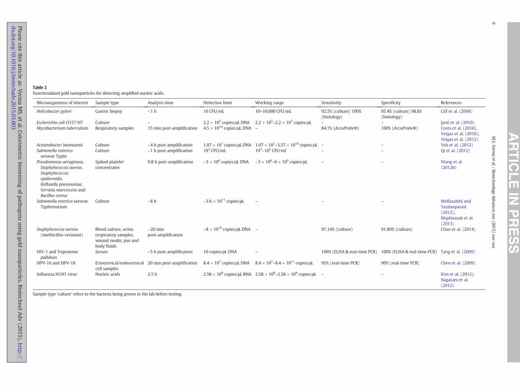
Table 2Functionalized gold nanoparticles for detecting amplified nucleic acids.
Microorganisms of interest Sample type Analysis time Detection limit Working range Sensitivity Specificity References
Helicobacter pylori Gastric biopsy b1 h 10 CFU/mL 10–10,000 CFU/mL 92.5% (culture) 100%(histology)
95.4% (culture) 98.8%(histology)
Gill et al. (2008)
Escherichia coli O157:H7 Culture – 2.2 × 10⁵ copies/μL DNA 2.2 × 10⁵–2.2 × 10⁷ copies/μL – – Jyoti et al. (2010)Mycobacterium tuberculosis Respiratory samples 15 min post-amplification 4.5 × 10¹⁰ copies/μL DNA – 84.7% (AccuProbe®) 100% (AccuProbe®) Costa et al. (2010),
Veigas et al. (2010),Veigas et al. (2012)
Acinetobacter baumannii Culture ~4 h post-amplification 1.07 × 10⁷ copies/μL DNA 1.07 × 10⁷–3.57 × 10¹⁰ copies/μL – – Yeh et al. (2012)Salmonella entericaserovar Typhi
Culture ~1 h post-amplification 103 CFU/mL 103–105 CFU/ml – – Qi et al. (2012)
Pseudomonas aeruginosa,Staphylococcus aureus,Staphylococcusepidermidis,Kelbsiella pneumoniae,Serratia marcescens andBacillus cereus
Spiked plateletconcentrates
0.8 h post-amplification ~3 × 10⁶ copies/μL DNA ~3 × 10⁶–6 × 10⁹ copies/μL – – Wang et al.(2012b)
Salmonella enterica serovarTyphimurium
Culture ~8 h ~3.6 × 10¹¹ copies/μL – – – Mollasalehi andYazdanparast(2012),Majdinasab et al.(2013)
Staphylococcus aureus(methicillin-resistant)
Blood culture, urine,respiratory samples,wound swabs, pus andbody fluids
~20 minpost-amplification
~8 × 10¹⁰ copies/μL DNA – 97.14% (culture) 91.89% (culture) Chan et al. (2014)
HIV-1 and Treponemapallidum
Serum ~5 h post-amplification 10 copies/μL DNA – 100% (ELISA & real-time PCR) 100% (ELISA & real-time PCR) Tang et al. (2009)
HPV-16 and HPV-18 Ectocervical/endocervicalcell samples
20 min post-amplification 8.4 × 10⁷ copies/μL DNA 8.4 × 10⁷–8.4 × 10¹¹ copies/μL 95% (real-time PCR) 90% (real-time PCR) Chen et al. (2009)
Influenza H1N1 virus Nucleic acids 2.5 h 2.58 × 10⁸ copies/μL RNA 2.58 × 10⁸–2.58 × 10⁹ copies/μL – – Kim et al. (2012),Nagatani et al.(2012)
Sample type ‘culture’ refers to the bacteria being grown in the lab before testing.
6M.S.V
ermaetal./Biotechnology
Advances
xxx(2015)
xxx–xxx
Pleasecite
this
articleas:
Verm
aMS,et
al,Colorimetric
biosensing
ofpath
ogensusin
ggold
nanoparticles,BiotechnolAdv
(2015),http://dx.doi.org/10.1016/j.biotechadv.2015.03.003

7M.S. Verma et al. / Biotechnology Advances xxx (2015) xxx–xxx
control) (Yeh et al., 2012). In order to test a large number of samples si-multaneously, the assay has been incorporated in microarrays. Typicalbiotin-tyraminemicroarray designs do not provide sufficient accumula-tion of gold nanoparticles and hence 1.4 nm gold nanoparticles havebeen modified with 3,3′-diaminobenzidine (DAB), which is a substratefor horseradish peroxidase (HRP) (Qi et al., 2012). Here, theHRP ismod-ified with streptavidin, which binds to biotinylated PCR products thatare immobilized on the glass surface via a probe. The presence of DABpromotes the accumulation of gold nanoparticles and simplifies theassay by reducing an incubation step compared to biotin-tyraminebased microarrays. This approach was deployed for the detection ofS. enterica serovar Typhi, which is responsible for causing typhoidfever (a life-threatening infection, especially in developing countries)(Qi et al., 2012).
Finally, biotin–streptavidin interactions have also been exploited forimplementing gold nanoparticles in an ICS format for the detection ofinfluenza H1N1 virus. An ICS format is ideal for detection because ofits portability and easy readout. In this design, gold nanoparticleswere functionalized with anti-hapten antibodies and added to the con-jugate pad. RT-PCR products labeled with biotin and Texas Red (a hap-ten) are added to the conjugate pad, where they attach to the goldnanoparticles. The test line contains streptavidin while the control linecontains anti-mouse IgG and thus, the gold nanoparticles attach to testline only if the biotin labeled RT-PCR products are present (Kim et al.,2012).
Functionalized gold nanoparticles share the advantage of elimi-nating the need for gel electrophoresis as was the case with non-functionalized nanoparticles. Additionally, functionalization widensthe scope of formats in which the assays are implemented rangingfrom solution-based methods to strip-based methods. The majorlimitation of functionalization is that the gold nanoparticles needto be modified for each analyte of interest and then purified beforeuse. These additional processing steps can require additional timeand technical expertise and also lead to loss of nanoparticle yield.The studies employing functionalized gold nanoparticles for ampli-fied nucleic acids have been summarized in Table 2.
4.2. Gold nanoparticles for unamplified nucleic acids
4.2.1. Non-functionalized gold nanoparticlesAs-synthesized gold nanoparticles can exert surface charges and
hence be used directly for detection without specific functionalization.Most of the studies that incorporate this strategy depend on the colorchange of gold nanoparticles from red to blue due to their electrostaticaggregation behavior. Two common coatings are present on as-synthesized nanoparticles: citrate for providing a net negative chargeand CTAB for providing a net positive charge.
Citrate capped nanoparticles have been used for the detection ofnucleic acids in a manner similar to the illustration in Fig. 1a, wherenanoparticles aggregate when the target is present. This approach hasbeen implemented in the detection of hepatitis C virus RNA by design-ing probes targeting the 5′UTR region and using them with 15 nmgold nanoparticles (Shawky et al., 2010).
CTAB-coated gold nanoparticles have been used for detecting DNAwhere the idea is similar to Fig. 1c, except instead of using thiolatedprobes, the probes are electrostatically adsorbed. Detection of HIV-1and B. anthracis has been possible by designing probes to target theU5 long terminal repeat sequence of HIV-1 and cryptic protein and pro-tective antigen precursor genes of B. anthracis. The probes wereadsorbed on 16–30 nm gold nanoparticles for obtaining a color changefrom red to purple (Pylaev et al., 2011).
Eliminating nucleic acid amplification provides major advantages inthe required analysis time and equipment. Specifically, the use of non-functionalized nanoparticles simplifies the synthesis of gold nanoparti-cles and thus the entire assay.
Please cite this article as: Verma MS, et al, Colorimetric biosensing of pdx.doi.org/10.1016/j.biotechadv.2015.03.003
4.2.2. Functionalized gold nanoparticlesUnamplified nucleic acids can also be detected by functionalizing
gold nanoparticles with specific thiolated probes. Threemain strategieshave been employed for implementing this method: functionalizingwith a single probe (Fig. 1b), functionalizing with two probes(Fig. 1c), and the use of DNA enzymes (DNAzymes). As compared toamplification-based methods, these assays are simpler and faster.
A thiolated nucleic acid probe has been designed for the detection ofMycobacterium spp. by targeting the 16s–23s DNA region ofmycobacte-rial species. The probe was immobilized on 15–20 nm gold nanoparti-cles and the presence of target DNA stabilized the nanoparticles uponaddition of HCl (Fig. 1a). Specificity of the assay was confirmed by com-paring the response from non-mycobacterial species (Liandris et al.,2009). Detection of E. coli genomic DNA has been possible by targetingthe malB gene and immobilizing the obtained probe on 20 nm goldnanoparticles. In this assay, the enzymatic degradation of DNA beforehybridization improved thedetection limit of the assay by 5 times. Spec-ificity was confirmed by comparing the response to other pathogenicbacteria (Bakthavathsalam et al., 2012).
Aggregation of nanoparticles by target DNA can also be used for thecolorimetric detection if a pair of appropriate probes is designed(Fig. 1c). One example of this approach is the detection of Kaposi'ssarcoma-associated herpesvirus (KSHV). KSHV is responsible forKaposi's sarcoma, an infectious cancer most commonly occurring inHIV positive patients. The detection of KSHV is challenging becauseseveral other diseases present similar symptoms and histopathologicalfeatures. One such confounding disease is bacillary angiomatosis,which can be caused by Bartonella quintana and Bartonella henselae.Thus, distinction between these pathogens is necessary and has beenachieved by designing pairs of thiolated oligonucleotide probestargeting the DNA that codes for vCyclin in KSHV and conserved regionsof Bartonella strains. The probes for KSHV and Bartonella were thenimmobilized on 15 nm gold and 20 nm silver nanoparticles respectivelyto obtain different color changes (Mancuso et al., 2013). Another studyhas demonstrated the detection of genomic DNAof S. enterica by the useof probes targeting the invA gene. Here, themechanismof detectionwasunclear because detection of genomicDNAwas possible using both one-probe and two-probe approaches. Additionally, the thiolated probeswere first incubated with the genomic DNA and then incubated with15 nm gold nanoparticles. In this study, the absence of target DNA al-lows gold nanoparticles to maintain stability, which is most likely be-cause of high coverage of the probe molecules on the surface of thenanoparticles. In the presence of the target, the probes hybridize withthe target DNA and hence, are probably unable to cover the gold nano-particles sufficiently to stabilize them. This leads to the aggregation ofgold nanoparticles and hence detection of the target DNA. This assayallowed the detection of dsDNA at room temperature (Kalidasan et al.,2013).
DNAzymes are nucleic acids that can catalyze the cleavage of othernucleic acids with multiple turnovers and hence are capable of provid-ing amplification in an assay. Multicomponent nucleic acid enzyme(MNAzyme) is a type of DNAzyme that can be designed to perform ca-talysis specifically in the presence of the target DNA. Gold nanoparticlecross-linkers can be used asMNAzyme substrates such that aggregationof gold nanoparticles can be modulated by the presence of target DNA.This approach has been applied for the detection of AF-1 and genetic se-quences from Neisseria gonorrhoeae, Treponema pallidum, Plasmodiumfalciparum, and HBV. In the absence of target DNA, the cross-linkerremained intact and led to aggregation of 13 nm gold nanoparticles.Designing the appropriate MNAzymes allows this method to detectmultiple targets, which is useful for diagnosing co-infections(Zagorovsky and Chan, 2013). Another example of DNAzymes is thedetection of dengue viruses. Dengue viruses cause periodic explosiveepidemics and can lead to 50–100 million infections annually. Theseviruses are typically carried by mosquitoes and can lead to denguefever or potentially fatal dengue hemorrhagic fever. DNAzymes have
athogens using gold nanoparticles, Biotechnol Adv (2015), http://

Table 3Gold nanoparticles for detecting unamplified nucleic acids.
Microorganisms of interest Sample type Analysis time Detection limit Working range References
Hepatitis C virusa,b Serum 30 min 2.5 copies/μL RNA ~2.5–100 copies/μL Shawky et al. (2010)HIV-1 and Bacillus anthracisa Nucleic acids ~30 min 6 × 10⁷ copies/μL DNA 6 × 10⁷–3 × 10⁹ copies/μL Pylaev et al. (2011)Mycobacterium spp.c Goat feces ~15 min post-extraction 18.8 ng/μL mycobacterial DNA 18.8–1,200 ng/μL Liandris et al. (2009)Escherichia colid Spiked urine b30 min post-extraction 5.4 ng/μL genomic DNA 5.4–43 ng/μL Bakthavathsalam et al. (2012)Kaposi's sarcoma-associatedherpes virus and Bartonella
Nucleic acids 2 h post-extraction 1 × 10⁹ copies/μL DNA 1–10 × 109 copies/μL Mancuso et al. (2013)
Neisseria gonorrhoeae,Treponema pallidum,Plasmodium falciparumand hepatitis B virus
Nucleic acids ~1.5 h post-extraction 3 × 10⁷ copies/μL model DNA 3 × 10⁷–6 × 10⁸ copies/μL Zagorovsky and Chan (2013)
Salmonella enterica Nucleic acids ~15 min post-extraction 2.2 × 10⁴ copies/μL genomic DNA 2.2 × 10⁴–3.8 × 10⁵ copies/μL Kalidasan et al. (2013)Dengue virus Culture 5 min post-extraction 4 × 10⁷ copies/μL RNA 4 × 10⁷–4 × 10¹² copies/μL Carter et al. (2013)
a Non-functionalized gold nanoparticles.b Sensitivity 92%, specificity 88.9% (RT-PCR).c Sensitivity 87.5%, specificity 100% (real-time PCR).d Specificity 100% (PCR); most studies used functionalized gold nanoparticles, except where indicated otherwise. Sample type ‘culture’ refers to themicroorganism being grown in the
lab before testing.
8 M.S. Verma et al. / Biotechnology Advances xxx (2015) xxx–xxx
been designed and immobilized on 15 nm gold nanoparticles to cleavedengue virus RNA in the presence of magnesium ions. The cleavedRNA leads to aggregation of gold nanoparticles in the presence of saltand heat (Carter et al., 2013).
Functionalizing gold nanoparticles with DNAzymes has allowed theincorporation of signal amplification during detection and hence pro-vided excellent detection limits. The major limitation of this approachhas been the requirement of nucleic acid extraction, since it can increasethe assay time by several hours. The studies employing gold nanoparti-cles for detection of unamplified nucleic acids are summarized inTable 3.
5. Emerging biosensors for detecting non-nucleic acid analytes
While several strategies have been presented for the detection ofnucleic acids, it is possible to detect pathogens by targeting otheranalytes of interest. Non-functionalized gold nanoparticles can use thenative surface charges of nanoparticles and bacteria for producinga color change. Functionalizing gold nanoparticles with proteins orsmall molecules can facilitate the detection of proteins, lipopolysaccha-rides, or even whole cells.
5.1. Non-functionalized gold nanoparticles
One approach for using citrate-capped nanoparticles is to designaptamers for specific targets and allow them to adsorb on the surfaceof gold nanoparticles. In the presence of the target, the aptamers getstripped from the surface of gold nanoparticles and bind to the target,which destabilizes the gold nanoparticles in high salt conditions. Thisstrategy has been applied for the detection of E. coli O157:H7 and S.Typhimurium, where aptamers were selected against these bacteriaand adsorbed on 15 nm gold nanoparticles. The specificity of the assaywas confirmed by testing the interaction with seven other species ofbacteria and a significant response was observed only when the desiredtarget was present (Wu et al., 2012).
In addition to whole cells, citrate-capped nanoparticles have alsobeen used to detect proteins. β-Lactamases are bacterial enzymes thatcleave β-lactam antibiotics and hence render them ineffective towardsbacterial infections. The detection of β-lactamase activity can assist indesigning better antibiotics. Enterobacter cloacae is a pathogen responsi-ble for producing class C P99β-lactamase,which can cleave cephalospo-rin derivatives and produce products with free thiols and positivelycharged amino groups. These products can replace some citrate ionson the surface of gold nanoparticles and then lead to their aggregationdue to electrostatic interactions. With the help of 16 nm citrate-capped gold nanoparticles, P99 β-lactamase could be detected (Liuet al., 2010). The same method has also been used for detection of
Please cite this article as: Verma MS, et al, Colorimetric biosensing of pdx.doi.org/10.1016/j.biotechadv.2015.03.003
class A β-lactamases, which are produced by E. coli, B. cereus andK. pneumoniae (Jiang et al., 2009). Another notorious enzyme is theimmunoglobulin A1 protease (IgA1P) produced by Streptococcuspneumoniae, which allows the bacterium to infect the lower respiratorytract, ear, or bloodstream and lead to diseases such as pneumonia, otitismedia, sepsis, and meningitis. The protease cleaves human IgA1 andcoats the bacteriumwith Fab fragments to act as a shield against the im-mune response and also to assist invasion into epithelial cells. Thus,IgA1P serves as a promising antibacterial target to curb the infection.The detection of IgA1P has been achieved using IgA1 and 20 nmcitrate-adsorbed gold nanoparticles. In the presence of IgA1P, the IgA1is cleaved to produce positively charged Fab regions, which is detectedby the aggregation of the anionic nanoparticles. The specificity of theassay was confirmed by the lack of response in the presence of IgA2,which is not cleaved by IgA1P (Garner et al., 2013).
Whole cell detection has also been achieved using CTAB-coated goldnanostars with a size range of 31 nm to 113 nm (Verma et al., 2014a).Here, the positive charges on gold nanostars interact with the negativecharges on bacterial cell walls presented by teichoic acids, lipopolysac-charides, and phospholipids. This strategy produced a unique degreeof color change for different species of bacteria when testing the ocularpathogens: S. aureus, Achromobacter xylosoxidans, Delftia acidovorans,and Stenotrophomonas maltophilia. An accuracy of 99% was obtainedfor identifying randomized samples of the four bacteria (Verma et al.,2014b).
ELISA has been used in a variety of applications for highly specificand sensitive detection of target molecules. Typically, a color change isobtained at the end of the assay because of enzymatic conversion ofthe substrate into a coloredmolecule, which is then detected by a spec-trophotometer. The color change could also be obtained using growth ofgold nanoparticles such that it would be visually detectable. In the ab-sence of target molecules, a high concentration of hydrogen peroxideis present, which rapidly reduces gold ions and forms spherical non-aggregated nanoparticles, producing a red color. In the presence of tar-get molecules, hydrogen peroxide is consumed by the enzyme andhence growth of the gold nanoparticles is slower, which results inaggregated particles with a blue color. This approach has been usedfor detection of HIV-1 capsid antigen p24 with the naked eye. Thismethod presents an extremely sensitive assay, which performs betterthan existing established methods based on nucleic acid detection (dela Rica and Stevens, 2012).
As compared to conventional methods for pathogen detection, thenon-functionalized gold nanoparticles provide a dramatic colorimetricoutput, which can often be visualized by the naked eye. The most im-portant limitation of this strategy is that various interferents from theenvironment can cause aggregation of nanoparticles and hence a falsepositive response, since the target analyte is often very general. The
athogens using gold nanoparticles, Biotechnol Adv (2015), http://

Table 4Non-functionalized gold nanoparticles for detection without nucleic acid amplification.
Microorganisms of interest Sample type Analysis time Detection limit Working range References
Escherichia coli, Bacillus cereus, Klebsiella pneumoniae Culture ~1 h ~108 CFU/mL – Jiang et al. (2009)Enterobacter cloacae β-Lactamase ~35 min 16 fmol/mL of P99 β-lactamase 15–80 fmol/mL Liu et al. (2010)Escherichia coli O157:H7 and Salmonella entericaserovar Typhimurium
Culture 20 min 105 CFU/mL 105–108 CFU/mL Wu et al. (2012)
Streptococcus pneumoniae Culture ~20 h – – Garner et al. (2013)Staphylococcus aureus, Achromobacter xylosoxidans,Delftia acidovorans, Stenotrophomonas maltophilia
Culture ~5 min ~1.5 × 10⁶ CFU/mL – Verma et al. (2014a,b)
HIV-1 Serum ~21 h 10−15 g/μL capsid antigen p24 10−15–10−18 g/μL de la Rica and Stevens (2012)
Sample type ‘culture’ refers to the microorganism being grown in the lab before testing.
9M.S. Verma et al. / Biotechnology Advances xxx (2015) xxx–xxx
studies using non-functionalized gold nanoparticles for detection havebeen summarized in Table 4.
5.2. Gold nanoparticles functionalized with proteins
Gold nanoparticles are often functionalized with antibodies that cantarget specific sites on the surface of pathogens. This antibody–antigenassociation leads to aggregation of gold nanoparticles around thepathogen of interest and can thus generate a colorimetric response(Fig. 2). Another common approach is to use aggregation of antibody-functionalized gold nanoparticles as a labeling method followed byamplification of the signal using the growth of silver or gold aroundthe initial seeds. Finally, these nanoparticles have been widely imple-mented in an ICS format as a replacement for ELISA.
The aggregation of gold nanoparticles around bacteria has been usedfor the detection of multi-drug resistant S. Typhimurium DT104. Thebacteriumpresents a great challenge in health care because of its persis-tent survival. The detection was possible by functionalizing 30 nmpopcorn-shaped gold nanoparticles with monoclonal M3038 antibodyagainst S. Typhimurium DT104. The response was specific to the drugresistant S. Typhimurium as compared to other Salmonella or E. colistrains (Khan et al., 2011).
Colorimetric response from the aggregation of nanoparticles canoften have insufficient sensitivity. Thus, the growth of gold or silver isused for signal amplification. This strategy has beendeployed for thede-tection of protozoa and bacteria. The detection of intestinal protozoanGiardia lamblia is possible by first separating it from solution usingcentrifugefiltration (0.45 μmpore size) and then incubating itwith a so-lution of anti-G. lamblia antibody-coated 15 nm gold nanoparticles. Theunbound gold nanoparticles are removed by centrifugefiltration follow-ed by the addition of a gold growth solution, which changes color de-pending on the concentration of gold nanoparticles. Since the assayuses centrifugation for concentration, it is possible to implement thisassay in large sample volumes (Li et al., 2009). The filtration approachcan be combined with magnetic nanoparticles to allow detection incomplex media. This approach has been used for the detection ofS. aureus in milk. Magnetic nanoparticles were first coated with bovineserum albumin (BSA) and then with 10 nm gold nanoparticles. Anti-S. aureus antibodies were then adsorbed on the surface of gold
Fig. 2. Gold nanoparticle functionalized with antibodiesAdapted from Wang et al. (2010).
Please cite this article as: Verma MS, et al, Colorimetric biosensing of pdx.doi.org/10.1016/j.biotechadv.2015.03.003
nanoparticles. This hybrid system of nanoparticles was incubated withthe sample contaminated with bacteria, magnetically separated, andthen filtered through a 0.8 μmcellulose acetatemembrane. Themagnet-ic separation retained all the nanoparticles and bacteria that wereattached to the nanoparticles. The filter retained bacteria and attachednanoparticles while allowing free nanoparticles to pass through. Finally,the color of nanoparticles on the filter was enhanced by a gold growthsolution. The specificity of the assay was confirmed by comparing theresponse to samples contaminated with other pathogenic bacteria(Sung et al., 2013).
In addition to nucleic acid detection, gold label silver staining hasalso been implemented for antibody-functionalized nanoparticles. Thismethod has been used for the detection of Campylobacter jejuni byusing monoclonal antibodies against the bacterium and coating themon 18 nm gold nanoparticles. In order to implement this method, aglass slide functionalized with streptavidin is first conjugated with bio-tinylated polyclonal antibodies against C. jejuni. This is followed by theaddition of the bacteria and then the functionalized gold nanoparticles.Then, the gold growth solution is added followed by silver enhance-ment. The silver enhancement is stopped by immersing the slide indeionized water. Using this method, specificity was confirmed bycomparing the response obtained from C. jejuni to that of Salmonellaenteritidis and E. coli (Cao et al., 2011).
Immobilization of antibodies has also been extended to nitrocellu-lose paper, which is followed by the addition of the target and thenthe protein-functionalized gold nanoparticles. This has been used forthe detection of Vi antigen of S. Typhi by adsorbing anti-Salmonellaantibodies on 30 nmgold nanoparticles. This assay has a potential of de-tecting typhoid early because it can not only detect the whole bacterialcell, but also just the Vi antigen (Pandey et al., 2012). Similarly, ICS-based assays have been developed for the detection of P. aeruginosaand S. aureus by using polyclonal antibodies against the bacteria andconjugating them to ~20 nm gold nanoparticles. The test line in theseassays had monoclonal antibodies against the bacteria and produced ared color in the presence of the target bacteria (Li et al., 2011). Anotherexample of ICS is the detection of toxic metabolites produced by themicroscopic fungi Aspergillus. These metabolites, such as ochratoxin A,can lead to nephrotoxicity, hepatotoxicity, and carcinogenicity inhumans. In this scenario, a competitive assay was developed by
aggregate around bacteria and lead to color change.
athogens using gold nanoparticles, Biotechnol Adv (2015), http://
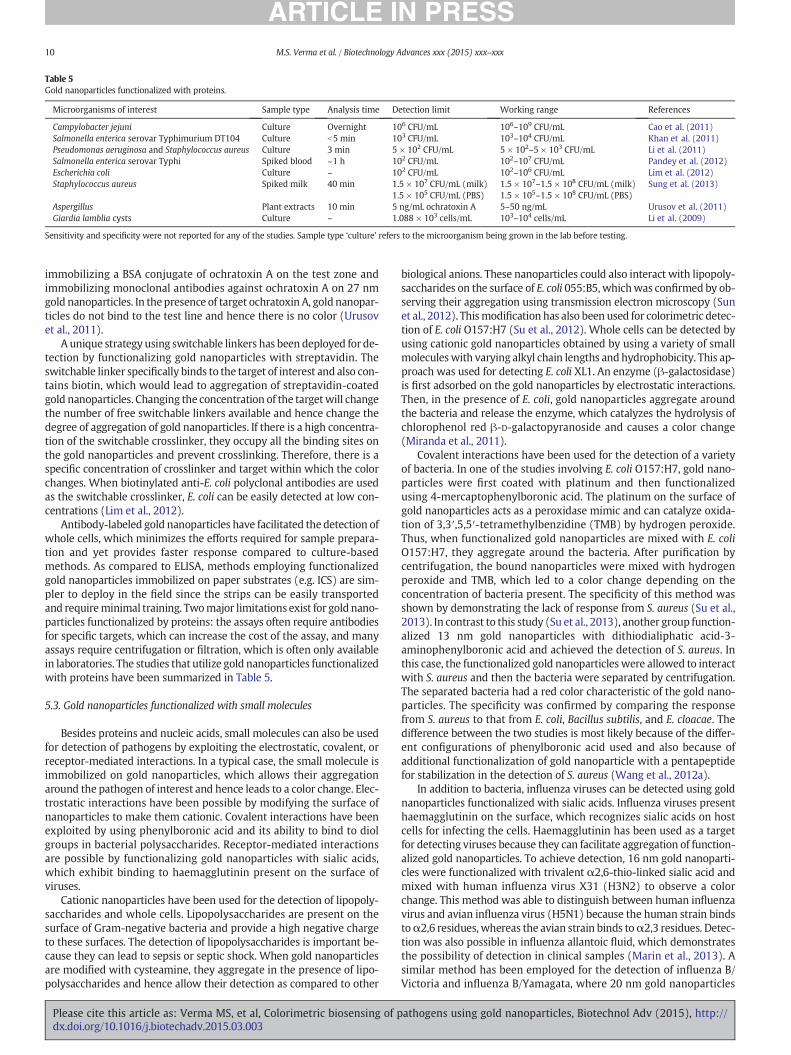
Table 5Gold nanoparticles functionalized with proteins.
Microorganisms of interest Sample type Analysis time Detection limit Working range References
Campylobacter jejuni Culture Overnight 106 CFU/mL 106–109 CFU/mL Cao et al. (2011)Salmonella enterica serovar Typhimurium DT104 Culture b5 min 103 CFU/mL 103–104 CFU/mL Khan et al. (2011)Pseudomonas aeruginosa and Staphylococcus aureus Culture 3 min 5 × 10² CFU/mL 5 × 10²–5 × 10³ CFU/mL Li et al. (2011)Salmonella enterica serovar Typhi Spiked blood ~1 h 102 CFU/mL 102–107 CFU/mL Pandey et al. (2012)Escherichia coli Culture – 102 CFU/mL 102–106 CFU/mL Lim et al. (2012)Staphylococcus aureus Spiked milk 40 min 1.5 × 10⁷ CFU/mL (milk)
1.5 × 10⁵ CFU/mL (PBS)1.5 × 10⁷–1.5 × 10⁸ CFU/mL (milk)1.5 × 10⁵–1.5 × 10⁸ CFU/mL (PBS)
Sung et al. (2013)
Aspergillus Plant extracts 10 min 5 ng/mL ochratoxin A 5–50 ng/mL Urusov et al. (2011)Giardia lamblia cysts Culture – 1.088 × 10³ cells/mL 103–104 cells/mL Li et al. (2009)
Sensitivity and specificity were not reported for any of the studies. Sample type ‘culture’ refers to the microorganism being grown in the lab before testing.
10 M.S. Verma et al. / Biotechnology Advances xxx (2015) xxx–xxx
immobilizing a BSA conjugate of ochratoxin A on the test zone andimmobilizing monoclonal antibodies against ochratoxin A on 27 nmgold nanoparticles. In thepresence of target ochratoxin A, gold nanopar-ticles do not bind to the test line and hence there is no color (Urusovet al., 2011).
A unique strategy using switchable linkers has been deployed for de-tection by functionalizing gold nanoparticles with streptavidin. Theswitchable linker specifically binds to the target of interest and also con-tains biotin, which would lead to aggregation of streptavidin-coatedgold nanoparticles. Changing the concentration of the targetwill changethe number of free switchable linkers available and hence change thedegree of aggregation of gold nanoparticles. If there is a high concentra-tion of the switchable crosslinker, they occupy all the binding sites onthe gold nanoparticles and prevent crosslinking. Therefore, there is aspecific concentration of crosslinker and target within which the colorchanges. When biotinylated anti-E. coli polyclonal antibodies are usedas the switchable crosslinker, E. coli can be easily detected at low con-centrations (Lim et al., 2012).
Antibody-labeled gold nanoparticles have facilitated the detection ofwhole cells, which minimizes the efforts required for sample prepara-tion and yet provides faster response compared to culture-basedmethods. As compared to ELISA, methods employing functionalizedgold nanoparticles immobilized on paper substrates (e.g. ICS) are sim-pler to deploy in the field since the strips can be easily transportedand requireminimal training. Twomajor limitations exist for gold nano-particles functionalized by proteins: the assays often require antibodiesfor specific targets, which can increase the cost of the assay, and manyassays require centrifugation or filtration, which is often only availablein laboratories. The studies that utilize gold nanoparticles functionalizedwith proteins have been summarized in Table 5.
5.3. Gold nanoparticles functionalized with small molecules
Besides proteins and nucleic acids, small molecules can also be usedfor detection of pathogens by exploiting the electrostatic, covalent, orreceptor-mediated interactions. In a typical case, the small molecule isimmobilized on gold nanoparticles, which allows their aggregationaround the pathogen of interest and hence leads to a color change. Elec-trostatic interactions have been possible by modifying the surface ofnanoparticles to make them cationic. Covalent interactions have beenexploited by using phenylboronic acid and its ability to bind to diolgroups in bacterial polysaccharides. Receptor-mediated interactionsare possible by functionalizing gold nanoparticles with sialic acids,which exhibit binding to haemagglutinin present on the surface ofviruses.
Cationic nanoparticles have been used for the detection of lipopoly-saccharides and whole cells. Lipopolysaccharides are present on thesurface of Gram-negative bacteria and provide a high negative chargeto these surfaces. The detection of lipopolysaccharides is important be-cause they can lead to sepsis or septic shock. When gold nanoparticlesare modified with cysteamine, they aggregate in the presence of lipo-polysaccharides and hence allow their detection as compared to other
Please cite this article as: Verma MS, et al, Colorimetric biosensing of pdx.doi.org/10.1016/j.biotechadv.2015.03.003
biological anions. These nanoparticles could also interact with lipopoly-saccharides on the surface of E. coli 055:B5, whichwas confirmed by ob-serving their aggregation using transmission electron microscopy (Sunet al., 2012). Thismodification has also been used for colorimetric detec-tion of E. coli O157:H7 (Su et al., 2012). Whole cells can be detected byusing cationic gold nanoparticles obtained by using a variety of smallmoleculeswith varying alkyl chain lengths andhydrophobicity. This ap-proach was used for detecting E. coli XL1. An enzyme (β-galactosidase)is first adsorbed on the gold nanoparticles by electrostatic interactions.Then, in the presence of E. coli, gold nanoparticles aggregate aroundthe bacteria and release the enzyme, which catalyzes the hydrolysis ofchlorophenol red β-D-galactopyranoside and causes a color change(Miranda et al., 2011).
Covalent interactions have been used for the detection of a varietyof bacteria. In one of the studies involving E. coli O157:H7, gold nano-particles were first coated with platinum and then functionalizedusing 4-mercaptophenylboronic acid. The platinum on the surface ofgold nanoparticles acts as a peroxidase mimic and can catalyze oxida-tion of 3,3′,5,5′-tetramethylbenzidine (TMB) by hydrogen peroxide.Thus, when functionalized gold nanoparticles are mixed with E. coliO157:H7, they aggregate around the bacteria. After purification bycentrifugation, the bound nanoparticles were mixed with hydrogenperoxide and TMB, which led to a color change depending on theconcentration of bacteria present. The specificity of this method wasshown by demonstrating the lack of response from S. aureus (Su et al.,2013). In contrast to this study (Su et al., 2013), another group function-alized 13 nm gold nanoparticles with dithiodialiphatic acid-3-aminophenylboronic acid and achieved the detection of S. aureus. Inthis case, the functionalized gold nanoparticles were allowed to interactwith S. aureus and then the bacteria were separated by centrifugation.The separated bacteria had a red color characteristic of the gold nano-particles. The specificity was confirmed by comparing the responsefrom S. aureus to that from E. coli, Bacillus subtilis, and E. cloacae. Thedifference between the two studies is most likely because of the differ-ent configurations of phenylboronic acid used and also because ofadditional functionalization of gold nanoparticle with a pentapeptidefor stabilization in the detection of S. aureus (Wang et al., 2012a).
In addition to bacteria, influenza viruses can be detected using goldnanoparticles functionalized with sialic acids. Influenza viruses presenthaemagglutinin on the surface, which recognizes sialic acids on hostcells for infecting the cells. Haemagglutinin has been used as a targetfor detecting viruses because they can facilitate aggregation of function-alized gold nanoparticles. To achieve detection, 16 nm gold nanoparti-cles were functionalized with trivalent α2,6-thio-linked sialic acid andmixed with human influenza virus X31 (H3N2) to observe a colorchange. This method was able to distinguish between human influenzavirus and avian influenza virus (H5N1) because the human strain bindstoα2,6 residues, whereas the avian strain binds toα2,3 residues. Detec-tion was also possible in influenza allantoic fluid, which demonstratesthe possibility of detection in clinical samples (Marin et al., 2013). Asimilar method has been employed for the detection of influenza B/Victoria and influenza B/Yamagata, where 20 nm gold nanoparticles
athogens using gold nanoparticles, Biotechnol Adv (2015), http://
Table6
Goldna
nopa
rticlesfunc
tion
alized
withsm
allm
olecules.
Microorga
nism
sof
interest
Sampletype
Ana
lysis
time
Detection
limit
Working
rang
eSm
allm
oleculeus
edRe
ferenc
es
Esch
erichiacoliXL1
Culture
~10
min
102CF
U/m
L(solution)
104CF
U/m
L(teststrip)
102–10
7CF
U/m
L(solution)
104–10
8CF
U/m
L(teststrip)
Seve
rald
ifferen
tcation
icmolecules
Miran
daet
al.(20
11)
Stap
hylococcus
aureus
Spiked
milk
,urine
,lun
gfluid
~2h
50CF
U/m
L5×10
2–5×10
6CF
U/m
LDithiod
ialip
haticacid-3-aminop
heny
lboron
icacid
Wan
get
al.(20
12a)
Esch
erichiacoliO15
7:H7
Culture
b40
min
7CF
U/m
L7–
6×10
6CF
U/m
L4-Mercaptop
heny
lboron
icacid
Suet
al.(20
12,2
013)
Esch
erichiacoli05
5:B5
Lipo
polysaccha
ride
s~5min
330fm
ol/m
Llip
opolysacch
arides
5–90
pmol/m
LCy
stea
mine
Sunet
al.(20
12)
Hum
aninflue
nzavirusX31
(H3N
2)Alla
ntoicfluid
~30
min
~1μg
/mLvirus
~1–
2μg
/mL
Trivalen
tα2,6-thio-linke
dsialic
acid
Marin
etal.(20
13)
Influe
nzaB/Victoriaan
dInflue
nza
B/Ya
mag
ata
Culture
~10
min
0.15
6vo
l.%dilution
ofhe
mag
glutinationassay
titer51
2virus
0.15
6–1.25
vol.%
Sialic
acid
(N-acetylneu
raminic
acid)
Leeet
al.(20
13)
Sens
itivityan
dsp
ecificity
wereno
trepo
rted
foran
yof
thestud
ies.Sampletype
‘culture’refersto
themicroorga
nism
beinggrow
nin
thelabbe
fore
testing.
11M.S. Verma et al. / Biotechnology Advances xxx (2015) xxx–xxx
Please cite this article as: Verma MS, et al, Colorimetric biosensing of pdx.doi.org/10.1016/j.biotechadv.2015.03.003
were synthesized and stabilized using sialic acid using a one-pot meth-od (Lee et al., 2013).
Gold nanoparticles modified with small molecules have typicallyprovided some of the fastest response times while maintaining excel-lent detection limits. Small molecules are typically cheaper than pro-teins or nucleic acids and hence the overall cost of the assay is lower.Themajor limitation of this approach is that small molecules target gen-eral components of the pathogens and hence cross-reactivity is likely.Thus, the assaymight provide a false positive response if a closely relat-ed pathogen was present instead of the targeted one. All the studiesusing gold nanoparticles functionalized with small molecules havebeen summarized in Table 6.
6. Comparison of gold nanoparticles to conventional methods
Conventional and gold nanoparticle-based pathogen detection as-says can be compared using a variety of metrics reflecting assay perfor-mance. Themain criteria bywhichwewill be evaluating the advantagesand disadvantages of the previously mentioned assays are time, limit ofdetection, specificity, technical complexity, and specific limitations.These parameters have been grouped by detection principle, and aresummarized in Table 7.
6.1. Analysis time
Analysis times were generally much longer for conventionalmethods than those using gold nanoparticles. Colony counting was byfar the most time-consuming method, due to the need for colonies tobe grown on selective media prior to visual identification (de Boer andBeumer, 1999). Of the organisms presented, the longest culture timewas reported for Campylobacter, where culture methods require 4–9 days for negative results and 14–16 days for positive confirmation(Brooks et al., 2004; Velusamy et al., 2010). In contrast, protein-functionalized gold nanoparticles have been used to detect Campylobac-ter following overnight incubation (Cao et al., 2011).
The fastest conventional methods are typically PCR-based assays,which can deliver results in 5–24 h, depending on the mode of analysisand pathogen of interest (Lazcka et al., 2007). This processing time isheavily dependent on the time required for sample enrichment andnucleic acid amplification, and is related to the detection limit (Cheet al., 2001; Ng et al., 1997). Amplification-based techniques with goldnanoparticles can improve upon conventional PCR-based methods bygenerating rapid color changes in response to pathogens, thereby sim-plifying the detection of target amplicon, and reducing the time re-quired to obtain a result. Furthermore, emerging biosensors that donot involve the time-consuming step of nucleic acid amplificationreported the shortest processing timeswith several groups reporting re-sults within an hour (Tables 4, 5, and 6).
6.2. Limit of detection
Despite advances in analysis time, reducing detection limits remainsa key challenge for gold nanoparticle-based assays. Conventionalmethods of colony counting and PCR are typically capable of detectingpathogens at concentrations in the range of 1 CFU/mL or 10 copies/μLDNA (Lazcka et al., 2007; Velusamy et al., 2010). Nanoparticle-basedmethods reported a wide variety of detection limits, ranging from 7 to108 CFU/ml or 101 to 3 × 10¹¹ copies/μL DNA depending on the target(Majdinasab et al., 2013; Mollasalehi and Yazdanparast, 2012; Tanget al., 2009). While some groups reported detection limits much higherthan those for conventional methods, particularly those assays withouttarget amplification, other nanoparticle-based assays were comparablein terms of detection limit.
athogens using gold nanoparticles, Biotechnol Adv (2015), http://
Table 7Comparing conventional and nanoparticle-based assays.
Category Detection principle Analysis time Detectionlimit
Specificity Technical requirements Limitations References
Conventional Colony counting 1–16 d 100–101 CFU/mL Good Basic microbiology lab equipmentand training
Only culturable strains are detected de Boer and Beumer (1999),Brooks et al. (2004)
Immunological assay 1–5 d 103–106 CFU/mL Good Specific antibodies for pathogen Sample enrichment is oftennecessary for high sensitivity
de Boer and Beumer (1999),Lazcka et al. (2007)
PCR 5–48 h b10 copies/μL Excellent Thermal cycling or isothermalamplification, gelelectrophoresisequipment
Distinguishing live and dead cells,presence of inhibitors incomplex media
Che et al. (2001), Drosten et al.(2002), Lazcka et al. (2007)
Gold nanoparticle assaysfor amplified nucleic acids
Non-functionalized 3–8 h 2 × 101–3 × 10⁹ copies/μL DNA Excellent Thermal cycling or isothermalamplification equipment
Need to design specific probesfor every pathogen of interest
Jung et al. (2010), Liu et al.(2011), Prasad et al. (2011)
Functionalized(nucleic acid)
1.5–8 h 101–3 × 10¹¹ copies/μL DNA Excellent Thermal cycling or isothermalamplification equipment
Functionalization requirespurification, can affectstability and yieldof nanoparticles
Tang et al. (2009), Mollasalehi andYazdanparast (2012), Majdinasabet al. (2013)
Gold nanoparticle assaysfor unamplifiednucleic acids
Non-functionalized ~30 min 2.5–6 × 10⁷ copies/μL RNA/DNA Good Minimal equipment Need to design specific probesfor targets of interest
Shawky et al. (2010),Pylaev et al. (2011)
Functionalized(nucleic acid)
5 min–2 h 2.2 × 10⁴–1 × 10⁹ copies/μL DNA Excellent Basic lab equpiment for nucleicacid extraction
Nucleic acid extraction canconsume considerable timecompared to assay time
Carter et al. (2013), Kalidasanet al. (2013), Mancuso et al. (2013)
Gold nanoparticle assaysfor non-nucleic acidtargets
Non-functionalized 5 min–21 h 105–108 CFU/mL Good Minimal equipment Nanoparticle stability in detectionmedia can be limited
Jiang et al. (2009), Wu et al. (2012)
Functionalized (protein) 3 min–overnight 102–1.5 × 10⁷ CFU/mL Good Often need filtration orcentrifugation equipment
Throughput limited byfiltration/centrifugation
Cao et al. (2011), Li et al. (2011),Pandey et al. (2012),Sung et al. (2013)
Functionalized(small molecule)
5 min–2 h 7–102 CFU/mL Poor Minimal equipment Cross-reactivity is often present Miranda et al. (2011),Su et al. (2013)
12M.S.V
ermaetal./Biotechnology
Advances
xxx(2015)
xxx–xxx
Pleasecite
this
articleas:
Verm
aMS,et
al,Colorimetric
biosensing
ofpath
ogensusin
ggold
nanoparticles,BiotechnolAdv
(2015),http://dx.doi.org/10.1016/j.biotechadv.2015.03.003

13M.S. Verma et al. / Biotechnology Advances xxx (2015) xxx–xxx
6.3. Specificity
Specificity of colony countingmethods is dependent on the ability toselectively isolate and culture particular pathogen strains. Due to theuse of morphological and physiological characteristics for pathogenidentification, specificity may be lower for closely related strainswhich are less distinguishable based on phenotypic traits. Similarly, im-munological assays may suffer from low specificity if antibodies are se-lected for target analytes that are present on more than one pathogenvariety. However, with proper antibody selection and species enrich-ment, immunological assays have good specificity. PCR-based methodsachieve specificity by targeting nucleic acid sequences with selectedprimers and/or probes. Excellent assay specificity can be achievedwhen the sequences targeted by PCR are unique to the strain of interestsince single base pair mismatches can often be discriminated.
Specificity of gold nanoparticle-based assays is determined by eithernucleic acids or antibodies in most cases. Thus, the specificity of theseassays is comparable to the methods based on PCR and immunologicalassays. In the case of small molecule modified nanoparticles and non-functionalized nanoparticles, the assays detect general targets andhence, the specificity suffers. One method for overcoming this specific-ity challenge is to adopt a “chemical nose” type system, where each an-alyte presents a unique set of responses and hence can be distinguished(Miranda et al., 2011; Verma et al., 2014b). The limitation of a “chemicalnose” approach is that the systemneeds to be trained for each analyte ofinterest before attempting the detection.
6.4. Technical requirements
Procedures for bacterial plating, colony counting, and species identi-fication vary according to the target organism. Generally, the first stepinvolves serial dilution of a sample or automatic plating (de Boer andBeumer, 1999) onto agar plates with selective media. Plates must thenbe incubated to allow for colony growth to a visually detectable level.This incubation period is dependent on the bacterial species and growthconditions. The number of resulting colonies is counted to infer patho-gen concentration in the original sample. This is a time consumingstep which can be done by hand or using automated systems (de Boerand Beumer, 1999). Pathogen identity is determined using variousmor-phological and biochemical tests. The colony counting method is goodfor workers in microbiology laboratories due to its reliability and useof common laboratory equipment and reagents, however the laboriousprocess is not adequate for rapid diagnostics and requires specializedtraining.
Immunological assays rely primarily on specificity of antibodies toantigens from the target pathogen. Awide variety of characterized anti-bodies and kits are available for most pathogens and complexity is de-pendent on the particular detection strategy (de Boer and Beumer,1999). While common immunological methods (e.g., ELISA) do not re-quire specialized lab equipment, they typically require some form ofsample enrichment due to decreased sensitivity (Brooks et al., 2004).
PCR-basedmethods are typically less laborious and time-consumingthan previously mentioned conventional methods (Lazcka et al., 2007).Specific DNA or RNA sequences amplified using PCR can be subsequent-ly visualized using a number of ways, depending on the type ofPCR. The most common methods are sample separation using gel-electrophoresis and fluorescence observation during real-time PCR withprobes. Primer and probe selection is dependent on the target pathogenbeing investigated. While traditional PCR-based methods require accessto a thermal cycler, advances in lab-on-a-chip and isothermal amplifica-tion techniques are reducing this barrier to out-of-laboratory fieldapplications.
Some of the main aims of nanoparticle assays are to simplify assayprocedure, reduce the need for complex lab equipment, and minimizelabor. Nucleic acid amplification-based techniques require either ther-mal cycling or isothermal amplification equipmentwhich is a significant
Please cite this article as: Verma MS, et al, Colorimetric biosensing of pdx.doi.org/10.1016/j.biotechadv.2015.03.003
issue for point-of-care or field applications. However, emergingamplification-free techniques require only basic laboratory equipment.In these cases, the primary technical requirement remains the abilityto extract and purify the target analyte (i.e., nucleic acids, proteins, orwhole cells) from the sample.
7. Conclusions
Gold nanoparticles with a variety of surface features have been usedfor the colorimetric detection of pathogens either by detecting nucleicacids, surface proteins, or whole cells. While the majority of the litera-ture has focused on the use of gold nanoparticles as a replacement forgel electrophoresis after nucleic acid amplification, there is a growingbody of work in detecting unamplified targets. There is a growingdrive towards developing methods or devices that could be used atthe point-of-care or in the field by providing a simple visual output.Overall, although gold nanoparticles have facilitated the developmentof simple and sensitive assays that are replacing conventional methodsof pathogen detection, current technologies are not yet ready to betranslated directly to the point-of-care or field use because the currentmethods require extensive sample processing before analysis. Addition-ally, current biosensors with gold nanoparticles suffer from lower sensi-tivity when complex media are involved because of non-specificadsorption, which can be mitigated in the future by modifying the sur-face of gold nanoparticles with non-fouling coatings.
In order to bring gold nanoparticles to the point-of-care or field use,it will be necessary to simplify the detection process and make it userfriendly. A few possibilities exist for achieving this goal: the use ofstrip-based detection, cellphone-based detectors, and disposablemicrofluidic chips. Strip-based sensors can be cheap and easy to use,where the color of the strip is compared to a reference. Some of thesestrategies have already been highlighted in this review, but in order tobroaden the scope of detection, we recommend the use of multipleshapes and sizes of gold nanoparticles such that a unique responsecould be obtained for each pathogen of interest. This color outputcould be combinedwith the use of powerful cellphones for colorimetricanalysis. Cellphone spectrometers are currently under development(Smith et al., 2011) and could be used in the field for sensitive assays.Since cellphones are widely used, such spectrometers could be easilyadapted by the general public. Another possibility of portable detectorsincludes the use of disposable microfluidic devices such that all compo-nents of detection are includedwithin the chip. This is possible by incor-porating gold nanoparticles in paper-based microfluidic devices (Lianaet al., 2012) such that their sensitivity and accuracy is improved. Reduc-ing assay time and simplifying the biosensors for direct use in complexmedia is the most promising avenue for future research. The optimalbiosensor will be designed to be simple such that the user can obtain re-sults with the push of a single button.
Abbreviations
BSA bovine serum albuminCTAB cetyltrimethylammonium bromideDAB 3,3′-diaminobenzidineDNA deoxyribonucleic acidDNAzymes deoxyribonucleic acid enzymesdsDNA double-stranded deoxyribonucleic acidELISA enzyme linked immunosorbent assayEPA United States Environmental Protection AgencyFDA United States Food and Drug AdministrationHBV hepatitis B virusHIV-1 human immunodeficiency virus type-1HPV human papillomavirusHPV-16 human papillomavirus type 16HPV-18 Human papillomavirus type 18HRP horseradish peroxidase
athogens using gold nanoparticles, Biotechnol Adv (2015), http://
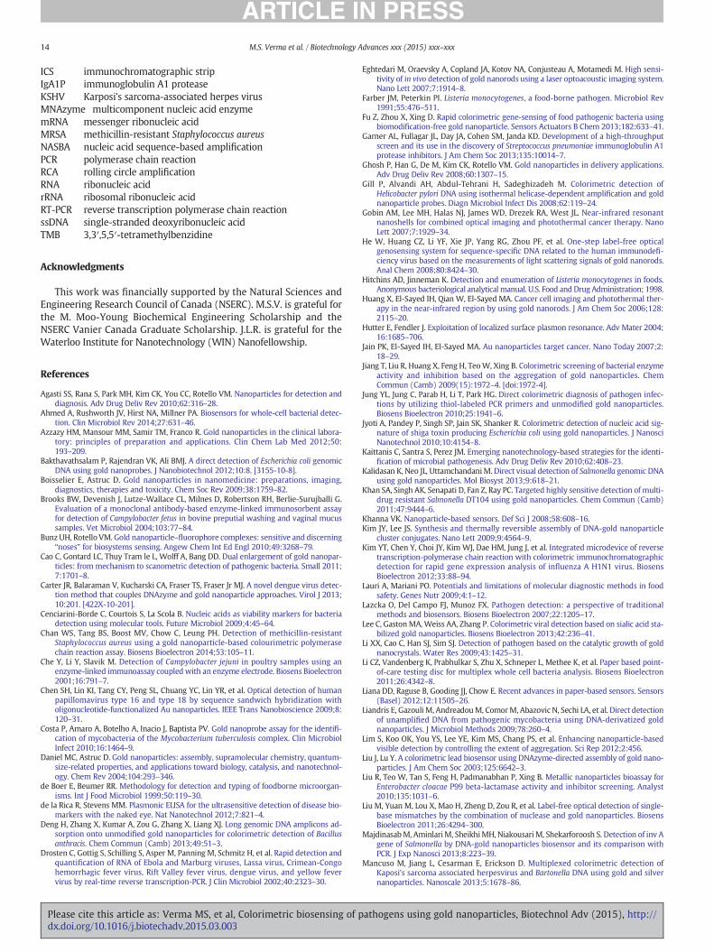
14 M.S. Verma et al. / Biotechnology Advances xxx (2015) xxx–xxx
ICS immunochromatographic stripIgA1P immunoglobulin A1 proteaseKSHV Karposi's sarcoma-associated herpes virusMNAzyme multicomponent nucleic acid enzymemRNA messenger ribonucleic acidMRSA methicillin-resistant Staphylococcus aureusNASBA nucleic acid sequence-based amplificationPCR polymerase chain reactionRCA rolling circle amplificationRNA ribonucleic acidrRNA ribosomal ribonucleic acidRT-PCR reverse transcription polymerase chain reactionssDNA single-stranded deoxyribonucleic acidTMB 3,3′,5,5′-tetramethylbenzidine
Acknowledgments
This work was financially supported by the Natural Sciences andEngineering Research Council of Canada (NSERC). M.S.V. is grateful forthe M. Moo-Young Biochemical Engineering Scholarship and theNSERC Vanier Canada Graduate Scholarship. J.L.R. is grateful for theWaterloo Institute for Nanotechnology (WIN) Nanofellowship.
References
Agasti SS, Rana S, Park MH, Kim CK, You CC, Rotello VM. Nanoparticles for detection anddiagnosis. Adv Drug Deliv Rev 2010;62:316–28.
Ahmed A, Rushworth JV, Hirst NA, Millner PA. Biosensors for whole-cell bacterial detec-tion. Clin Microbiol Rev 2014;27:631–46.
Azzazy HM, Mansour MM, Samir TM, Franco R. Gold nanoparticles in the clinical labora-tory: principles of preparation and applications. Clin Chem Lab Med 2012;50:193–209.
Bakthavathsalam P, Rajendran VK, Ali BMJ. A direct detection of Escherichia coli genomicDNA using gold nanoprobes. J Nanobiotechnol 2012;10:8. [3155-10-8].
Boisselier E, Astruc D. Gold nanoparticles in nanomedicine: preparations, imaging,diagnostics, therapies and toxicity. Chem Soc Rev 2009;38:1759–82.
Brooks BW, Devenish J, Lutze-Wallace CL, Milnes D, Robertson RH, Berlie-Surujballi G.Evaluation of a monoclonal antibody-based enzyme-linked immunosorbent assayfor detection of Campylobacter fetus in bovine preputial washing and vaginal mucussamples. Vet Microbiol 2004;103:77–84.
Bunz UH, Rotello VM. Gold nanoparticle–fluorophore complexes: sensitive and discerning“noses” for biosystems sensing. Angew Chem Int Ed Engl 2010;49:3268–79.
Cao C, Gontard LC, Thuy Tram le L, Wolff A, Bang DD. Dual enlargement of gold nanopar-ticles: from mechanism to scanometric detection of pathogenic bacteria. Small 2011;7:1701–8.
Carter JR, Balaraman V, Kucharski CA, Fraser TS, Fraser Jr MJ. A novel dengue virus detec-tion method that couples DNAzyme and gold nanoparticle approaches. Virol J 2013;10:201. [422X-10-201].
Cenciarini-Borde C, Courtois S, La Scola B. Nucleic acids as viability markers for bacteriadetection using molecular tools. Future Microbiol 2009;4:45–64.
Chan WS, Tang BS, Boost MV, Chow C, Leung PH. Detection of methicillin-resistantStaphylococcus aureus using a gold nanoparticle-based colourimetric polymerasechain reaction assay. Biosens Bioelectron 2014;53:105–11.
Che Y, Li Y, Slavik M. Detection of Campylobacter jejuni in poultry samples using anenzyme-linked immunoassay coupledwith an enzyme electrode. Biosens Bioelectron2001;16:791–7.
Chen SH, Lin KI, Tang CY, Peng SL, Chuang YC, Lin YR, et al. Optical detection of humanpapillomavirus type 16 and type 18 by sequence sandwich hybridization witholigonucleotide-functionalized Au nanoparticles. IEEE Trans Nanobioscience 2009;8:120–31.
Costa P, Amaro A, Botelho A, Inacio J, Baptista PV. Gold nanoprobe assay for the identifi-cation of mycobacteria of the Mycobacterium tuberculosis complex. Clin MicrobiolInfect 2010;16:1464–9.
Daniel MC, Astruc D. Gold nanoparticles: assembly, supramolecular chemistry, quantum-size-related properties, and applications toward biology, catalysis, and nanotechnol-ogy. Chem Rev 2004;104:293–346.
de Boer E, Beumer RR. Methodology for detection and typing of foodborne microorgan-isms. Int J Food Microbiol 1999;50:119–30.
de la Rica R, Stevens MM. Plasmonic ELISA for the ultrasensitive detection of disease bio-markers with the naked eye. Nat Nanotechnol 2012;7:821–4.
Deng H, Zhang X, Kumar A, Zou G, Zhang X, Liang XJ. Long genomic DNA amplicons ad-sorption onto unmodified gold nanoparticles for colorimetric detection of Bacillusanthracis. Chem Commun (Camb) 2013;49:51–3.
Drosten C, Gottig S, Schilling S, Asper M, Panning M, Schmitz H, et al. Rapid detection andquantification of RNA of Ebola and Marburg viruses, Lassa virus, Crimean-Congohemorrhagic fever virus, Rift Valley fever virus, dengue virus, and yellow fevervirus by real-time reverse transcription-PCR. J Clin Microbiol 2002;40:2323–30.
Please cite this article as: Verma MS, et al, Colorimetric biosensing of pdx.doi.org/10.1016/j.biotechadv.2015.03.003
Eghtedari M, Oraevsky A, Copland JA, Kotov NA, Conjusteau A, Motamedi M. High sensi-tivity of in vivo detection of gold nanorods using a laser optoacoustic imaging system.Nano Lett 2007;7:1914–8.
Farber JM, Peterkin PI. Listeria monocytogenes, a food-borne pathogen. Microbiol Rev1991;55:476–511.
Fu Z, Zhou X, Xing D. Rapid colorimetric gene-sensing of food pathogenic bacteria usingbiomodification-free gold nanoparticle. Sensors Actuators B Chem 2013;182:633–41.
Garner AL, Fullagar JL, Day JA, Cohen SM, Janda KD. Development of a high-throughputscreen and its use in the discovery of Streptococcus pneumoniae immunoglobulin A1protease inhibitors. J Am Chem Soc 2013;135:10014–7.
Ghosh P, Han G, De M, Kim CK, Rotello VM. Gold nanoparticles in delivery applications.Adv Drug Deliv Rev 2008;60:1307–15.
Gill P, Alvandi AH, Abdul-Tehrani H, Sadeghizadeh M. Colorimetric detection ofHelicobacter pylori DNA using isothermal helicase-dependent amplification and goldnanoparticle probes. Diagn Microbiol Infect Dis 2008;62:119–24.
Gobin AM, Lee MH, Halas NJ, James WD, Drezek RA, West JL. Near-infrared resonantnanoshells for combined optical imaging and photothermal cancer therapy. NanoLett 2007;7:1929–34.
He W, Huang CZ, Li YF, Xie JP, Yang RG, Zhou PF, et al. One-step label-free opticalgenosensing system for sequence-specific DNA related to the human immunodefi-ciency virus based on the measurements of light scattering signals of gold nanorods.Anal Chem 2008;80:8424–30.
Hitchins AD, Jinneman K. Detection and enumeration of Listeria monocytogenes in foods.Anonymous bacteriological analytical manual. U.S. Food and Drug Administration; 1998.
Huang X, El-Sayed IH, Qian W, El-Sayed MA. Cancer cell imaging and photothermal ther-apy in the near-infrared region by using gold nanorods. J Am Chem Soc 2006;128:2115–20.
Hutter E, Fendler J. Exploitation of localized surface plasmon resonance. Adv Mater 2004;16:1685–706.
Jain PK, El-Sayed IH, El-Sayed MA. Au nanoparticles target cancer. Nano Today 2007;2:18–29.
Jiang T, Liu R, Huang X, Feng H, TeoW, Xing B. Colorimetric screening of bacterial enzymeactivity and inhibition based on the aggregation of gold nanoparticles. ChemCommun (Camb) 2009(15):1972–4. [doi:1972-4].
Jung YL, Jung C, Parab H, Li T, Park HG. Direct colorimetric diagnosis of pathogen infec-tions by utilizing thiol-labeled PCR primers and unmodified gold nanoparticles.Biosens Bioelectron 2010;25:1941–6.
Jyoti A, Pandey P, Singh SP, Jain SK, Shanker R. Colorimetric detection of nucleic acid sig-nature of shiga toxin producing Escherichia coli using gold nanoparticles. J NanosciNanotechnol 2010;10:4154–8.
Kaittanis C, Santra S, Perez JM. Emerging nanotechnology-based strategies for the identi-fication of microbial pathogenesis. Adv Drug Deliv Rev 2010;62:408–23.
Kalidasan K, Neo JL, UttamchandaniM. Direct visual detection of Salmonella genomic DNAusing gold nanoparticles. Mol Biosyst 2013;9:618–21.
Khan SA, Singh AK, Senapati D, Fan Z, Ray PC. Targeted highly sensitive detection of multi-drug resistant Salmonella DT104 using gold nanoparticles. Chem Commun (Camb)2011;47:9444–6.
Khanna VK. Nanoparticle-based sensors. Def Sci J 2008;58:608–16.Kim JY, Lee JS. Synthesis and thermally reversible assembly of DNA-gold nanoparticle
cluster conjugates. Nano Lett 2009;9:4564–9.Kim YT, Chen Y, Choi JY, Kim WJ, Dae HM, Jung J, et al. Integrated microdevice of reverse
transcription-polymerase chain reaction with colorimetric immunochromatographicdetection for rapid gene expression analysis of influenza A H1N1 virus. BiosensBioelectron 2012;33:88–94.
Lauri A, Mariani PO. Potentials and limitations of molecular diagnostic methods in foodsafety. Genes Nutr 2009;4:1–12.
Lazcka O, Del Campo FJ, Munoz FX. Pathogen detection: a perspective of traditionalmethods and biosensors. Biosens Bioelectron 2007;22:1205–17.
Lee C, GastonMA,Weiss AA, Zhang P. Colorimetric viral detection based on sialic acid sta-bilized gold nanoparticles. Biosens Bioelectron 2013;42:236–41.
Li XX, Cao C, Han SJ, Sim SJ. Detection of pathogen based on the catalytic growth of goldnanocrystals. Water Res 2009;43:1425–31.
Li CZ, Vandenberg K, Prabhulkar S, Zhu X, Schneper L, Methee K, et al. Paper based point-of-care testing disc for multiplex whole cell bacteria analysis. Biosens Bioelectron2011;26:4342–8.
Liana DD, Raguse B, Gooding JJ, Chow E. Recent advances in paper-based sensors. Sensors(Basel) 2012;12:11505–26.
Liandris E, Gazouli M, AndreadouM, ComorM, Abazovic N, Sechi LA, et al. Direct detectionof unamplified DNA from pathogenic mycobacteria using DNA-derivatized goldnanoparticles. J Microbiol Methods 2009;78:260–4.
Lim S, Koo OK, You YS, Lee YE, Kim MS, Chang PS, et al. Enhancing nanoparticle-basedvisible detection by controlling the extent of aggregation. Sci Rep 2012;2:456.
Liu J, Lu Y. A colorimetric lead biosensor using DNAzyme-directed assembly of gold nano-particles. J Am Chem Soc 2003;125:6642–3.
Liu R, Teo W, Tan S, Feng H, Padmanabhan P, Xing B. Metallic nanoparticles bioassay forEnterobacter cloacae P99 beta-lactamase activity and inhibitor screening. Analyst2010;135:1031–6.
Liu M, Yuan M, Lou X, Mao H, Zheng D, Zou R, et al. Label-free optical detection of single-base mismatches by the combination of nuclease and gold nanoparticles. BiosensBioelectron 2011;26:4294–300.
MajdinasabM, AminlariM, SheikhiMH, Niakousari M, Shekarforoosh S. Detection of inv Agene of Salmonella by DNA-gold nanoparticles biosensor and its comparison withPCR. J Exp Nanosci 2013;8:223–39.
Mancuso M, Jiang L, Cesarman E, Erickson D. Multiplexed colorimetric detection ofKaposi's sarcoma associated herpesvirus and Bartonella DNA using gold and silvernanoparticles. Nanoscale 2013;5:1678–86.
athogens using gold nanoparticles, Biotechnol Adv (2015), http://

15M.S. Verma et al. / Biotechnology Advances xxx (2015) xxx–xxx
Mao X, Ma Y, Zhang A, Zhang L, Zeng L, Liu G. Disposable nucleic acid biosensors based ongold nanoparticle probes and lateral flow strip. Anal Chem 2009;81:1660–8.
Marin MJ, Rashid A, Rejzek M, Fairhurst SA, Wharton SA, Martin SR, et al.Glyconanoparticles for the plasmonic detection and discrimination between humanand avian influenza virus. Org Biomol Chem 2013;11:7101–7.
Mayer KM, Hafner JH. Localized surface plasmon resonance sensors. Chem Rev 2011;111:3828–57.
Miranda OR, Li X, Garcia-Gonzalez L, Zhu ZJ, Yan B, Bunz UH, et al. Colorimetric bacteriasensing using a supramolecular enzyme-nanoparticle biosensor. J Am Chem Soc2011;133:9650–3.
Mollasalehi H, Yazdanparast R. Non-crosslinking gold nanoprobes for detection of nucleicacid sequence-based amplification products. Anal Biochem 2012;425:91–5.
Monis PT, Giglio S. Nucleic acid amplification-based techniques for pathogen detectionand identification. Infect Genet Evol 2006;6:2–12.
Murphy CJ, Gole AM, Stone JW, Sisco PN, Alkilany AM, Goldsmith EC, et al. Gold nanopar-ticles in biology: beyond toxicity to cellular imaging. Acc ChemRes 2008;41:1721–30.
Nagatani N, Yamanaka K, Ushijima H, Koketsu R, Sasaki T, Ikuta K, et al. Detection ofinfluenza virus using a lateral flow immunoassay for amplified DNA by a microfluidicRT-PCR chip. Analyst 2012;137:3422–6.
Nath N, Chilkoti A. A colorimetric gold nanoparticle biosensor: effect of particle sizeon sensitivity. Second joint EMBS–BMES conference 2002, vols. 1–3, conferenceproceedings: bioengineering — integrative methodologies. New Technologies; 2002.p. 574–5.
Ng LK, Kingombe CI, Yan W, Taylor DE, Hiratsuka K, Malik N, et al. Specific detection andconfirmation of Campylobacter jejuni by DNA hybridization and PCR. Appl EnvironMicrobiol 1997;63:4558–63.
Niazi A, Jorjani ON, Nikbakht H, Gill P. A nanodiagnostic colorimetric assay for 18S rRNA ofLeishmania pathogens using nucleic acid sequence-based amplification and goldnanorods. Mol Diagn Ther 2013;17:363–70.
Paciotti GF, Myer L, Weinreich D, Goia D, Pavel N, McLaughlin RE, et al. Colloidal gold: anovel nanoparticle vector for tumor directed drug delivery. Drug Deliv 2004;11:169–83.
Pandey SK, Suri CR, Chaudhry M, Tiwari RP, Rishi P. A gold nanoparticles based immuno-bioprobe for detection of Vi capsular polysaccharide of Salmonella enterica serovarTyphi. Mol Biosyst 2012;8:1853–60.
Peters RP, van Agtmael MA, Danner SA, Savelkoul PH, Vandenbroucke-Grauls CM. Newdevelopments in the diagnosis of bloodstream infections. Lancet Infect Dis 2004;4:751–60.
Popovtzer R, Agrawal A, Kotov NA, Popovtzer A, Balter J, Carey TE, et al. Targeted goldnanoparticles enable molecular CT imaging of cancer. Nano Lett 2008;8:4593–6.
Prasad D, Shankaracharya, Vidyarthi AS. Gold nanoparticles-based colorimetric assay forrapid detection of Salmonella species in food samples. World J Microbiol Biotechnol2011;27:2227–30.
Pylaev TE, Khanadeev VA, Khlebtsov BN, Dykman LA, Bogatyrev VA, Khlebtsov NG. Color-imetric and dynamic light scattering detection of DNA sequences by using positivelycharged gold nanospheres: a comparative studywith gold nanorods. Nanotechnology2011;22:285501. [4484/22/28/285501. Epub 2011 May 31].
Qi H, Chen S, Hao R, Shi H, ZhangM,Wang S. Introduction of nanogold-DAB as a HRP sub-strate for simplifying detection in visual DNA microarrays. Anal Methods 2012;4:1178–81.
Renuart I, Mertens P, Leclipteux T. One step oligochromatographic device and method ofuse; 2011 [US 10/555,154].
Saha K, Agasti SS, Kim C, Li X, Rotello VM. Gold nanoparticles in chemical and biologicalsensing. Chem Rev 2012;112:2739–79.
Saleh M, Soliman H, Schachner O, El-Matbouli M. Direct detection of unamplified springviraemia of carp virus RNA using unmodified gold nanoparticles. Dis Aquat Organ2012;100:3–10.
Shawky SM, Bald D, Azzazy HM. Direct detection of unamplified hepatitis C virus RNAusing unmodified gold nanoparticles. Clin Biochem 2010;43:1163–8.
Skottrup PD, Nicolaisen M, Justesen AF. Towards on-site pathogen detection usingantibody-based sensors. Biosens Bioelectron 2008;24:339–48.
Smith ZJ, Chu K, Espenson AR, Rahimzadeh M, Gryshuk A, Molinaro M, et al. Cell-phone-based platform for biomedical device development and education applications.PLoS One 2011;6:e17150.
Su H, Ma Q, Shang K, Liu T, Yin H, Ai S. Gold nanoparticles as colorimetric sensor: a casestudy on E. coli O157:H7 as a model for Gram-negative bacteria. Sensors ActuatorsB Chem 2012;161:298–303.
Please cite this article as: Verma MS, et al, Colorimetric biosensing of pdx.doi.org/10.1016/j.biotechadv.2015.03.003
Su H, Zhao H, Qiao F, Chen L, Duan R, Ai S. Colorimetric detection of Escherichia coli O157:H7 using functionalized Au@Pt nanoparticles as peroxidase mimetics. Analyst 2013;138:3026–31.
Sun J, Ge J, Liu W, Wang X, Fan Z, ZhaoW, et al. A facile assay for direct colorimetric visu-alization of lipopolysaccharides at low nanomolar level. Nano Res 2012;5:486–93.
Sung YJ, Suk HJ, Sung HY, Li T, Poo H, KimMG. Novel antibody/gold nanoparticle/magnet-ic nanoparticle nanocomposites for immunomagnetic separation and rapid colori-metric detection of Staphylococcus aureus in milk. Biosens Bioelectron 2013;43:432–9.
Tallury P, Malhotra A, Byrne LM, Santra S. Nanobioimaging and sensing of infectious dis-eases. Adv Drug Deliv Rev 2010;62:424–37.
Tang J, Zhou L, GaoW, Cao X,Wang Y. Visual DNAmicroarrays for simultaneous detectionof human immunodeficiency virus type-1 and Treponema pallidum coupled withmultiplex asymmetric polymerase chain reaction. Diagn Microbiol Infect Dis 2009;65:372–8.
Taton TA, Mirkin CA, Letsinger RL. Scanometric DNA array detection with nanoparticleprobes. Science 2000;289:1757–60.
U.S. Food and Drug Administration. Fish and fishery products hazards and controls guid-ance; 2011.
Upadhyayula VK. Functionalized gold nanoparticle supported sensory mechanisms ap-plied in detection of chemical and biological threat agents: a review. Anal ChimActa 2012;715:1–18.
Urusov AE, Kostenko SN, Sveshnikov PG, Zherdev AV, Dzantiev BB. Immuno-chromatographic assay for the detection of ochratoxin A. J Anal Chem 2011;66:770–6.
Veigas B, Machado D, Perdigao J, Portugal I, Couto I, Viveiros M, et al. Au-nanoprobes fordetection of SNPs associated with antibiotic resistance inMycobacterium tuberculosis.Nanotechnology 2010;21:415101. [4484/21/41/415101. Epub 2010 Sep 16].
Veigas B, Jacob JM, Costa MN, Santos DS, Viveiros M, Inacio J, et al. Gold on paper-paperplatform for Au-nanoprobe TB detection. Lab Chip 2012;12:4802–8.
Velusamy V, Arshak K, Korostynska O, Oliwa K, Adley C. An overview of foodborne path-ogen detection: in the perspective of biosensors. Biotechnol Adv 2010;28:232–54.
Verma MS, Chen PZ, Jones L, Gu FX. Branching and size of CTAB-coated gold nanostarscontrol the colorimetric detection of bacteria. RSC Adv 2014a;4:10660–8.
VermaMS, Chen PZ, Jones L, Gu FX. “Chemical nose” for the visual identification of emerg-ing ocular pathogens using gold nanostars. Biosens Bioelectron 2014b;61:386–90.
Wang C, Chen Y, Wang T, Ma Z, Su Z. Biorecognition-driven self-assembly of gold nano-rods: a rapid and sensitive approach toward antibody sensing. Chem Mater 2007;19:5809–11.
Wang S, Singh AK, Senapati D, Neely A, Yu H, Ray PC. Rapid colorimetric identification andtargeted photothermal lysis of Salmonella bacteria by using bioconjugated oval-shaped gold nanoparticles. Chemistry 2010;16:5600–6.
Wang J, Gao J, Liu D, Han D,Wang Z. Phenylboronic acid functionalized gold nanoparticlesfor highly sensitive detection of Staphylococcus aureus. Nanoscale 2012a;4:451–4.
Wang X, Li Y, Wang J, Wang Q, Xu L, Du J, et al. A broad-range method to detect genomicDNA of multiple pathogenic bacteria based on the aggregation strategy of gold nano-rods. Analyst 2012b;137:4267–73.
Willets KA, Van Duyne RP. Localized surface plasmon resonance spectroscopy and sens-ing. Annu Rev Phys Chem 2007;58:267–97.
WuWH, Li M,Wang Y, Ouyang HX,Wang L, Li CX, et al. Aptasensors for rapid detection ofEscherichia coli O157:H7 and Salmonella Typhimurium. Nanoscale Res Lett 2012;7:658. [276X-7-658].
Xing Y, Wang P, Zang Y, Ge Y, Jin Q, Zhao J, et al. A colorimetric method for H1N1 DNAdetection using rolling circle amplification. Analyst 2013;138:3457–62.
Yaron S, Matthews KR. A reverse transcriptase-polymerase chain reaction assay for detec-tion of viable Escherichia coli O157:H7: investigation of specific target genes. J ApplMicrobiol 2002;92:633–40.
Yeh C, Chang Y, Lin H, Chang T, Lin Y. A newly developed optical biochip for bacteria de-tection hybridization. Sensors Actuators B Chem 2012;161:1168–75.
Zagorovsky K, Chan WC. A plasmonic DNAzyme strategy for point-of-care genetic detec-tion of infectious pathogens. Angew Chem Int Ed Engl 2013;52:3168–71.
ZhaoW, BrookMA, Li Y. Design of gold nanoparticle-based colorimetric biosensing assays.Chembiochem 2008;9:2363–71.
athogens using gold nanoparticles, Biotechnol Adv (2015), http://