Embed Size (px)
Citation preview

Colorectal Cancer Surveillance in Inflammatory Bowel Disease
RENEE MARCHIONI BEERY, DO
Director, Inflammatory Bowel Disease Assistant Professor of MedicineUniversity of South Florida

Disclosures
No disclosures or conflicts of interest

Objectives: Colorectal Cancer Surveillance in IBD Describe current approach for colorectal (CRC)
surveillance in inflammatory bowel disease (IBD)
Outline classification scheme for describing IBD-associated dysplastic lesions
Discuss role of chromoendoscopy in evaluation and detection of dysplasia
Highlight optimal endoscopic surveillance techniques and management strategies in clinical practice

Burden of Colorectal Neoplasia in IBD Colitis-associated colorectal neoplasia, including
dysplasia and malignancy, linked to IBD Initially described by Crohn & Rosenberg (1925)
IBD-associated colorectal cancer (CRC) 1-2% of CRC cases in general population 10-15% of all deaths among IBD patients IBD is third highest risk factor for CRC behind genetic
causes
Distinct from sporadic CRC Molecular, endoscopic and histologic features Associated with increased mortality Crohn BB, Rosenburg H. Am J Med Sci. 1925;170:220–227.
Munkholm P. Aliment Pharmacol Ther. 2003;18(suppl 2):1–5.Kulaylat MN, Dayton MT. J Surg Oncol. 2010;101:706–712. Jewel Samadder N et al. Dig Dis Sci 2017; Jan 3.

Risk of Colorectal Cancer in IBD
Patients with long-standing IBD have higher risk for development of CRC than the general population
2.4-fold increased risk in UC and similar for Crohn’s colitis (relative to general populations)
1.5 to 2 times increased risk in IBD population compared with general population in North America
Jess T et al. Clin. Gastroenterol. Hepatol 2012;10:639–45.Bleday R et l. Dis. Colon Rectum 1993; 36: 908–12.

Risks for Colorectal Cancer in IBD
Ulcerative colitis or Crohn’s colitis IBD Duration > 8-10 years Early age at onset of colitis Extensive colitis or backwash ileitis Histologic disease activity Family history of colon cancer Dysplasia at surveillance Primary sclerosing cholangitis (PSC) Pseudopolyps

PSC
Pseudopolyps

Factors that Decrease CRC Risk in IBD
Colectomy (prophylactic)
Surveillance colonoscopy with appropriate lesion resection and timely surgical intervention
Chemoprevention (observational data) 5-ASAs: potential chemopreventive effect
UDCA & folate- data inconclusive
Thiopurines/Anti-TNFs- data not supportive but effective for IBD management
Bezzio et al. Expert Rev Gastroentero Hepatol 2017 Feb 6.

Endoscopic CRC Surveillance: Random Biopsies
American Gastroenterological Assoc Guidelines (2010)
4-Quadrant Random Biopsies (n=32) from all colon segments (every 10 cm) to identify invisible dysplasia
Samples <0.1% of the surface mucosa
Approximately 1 dysplasia detected per 1000 biopsies
Approximately 9% of patients with dysplasia diagnosed
Farraye FA et al. Gastroenterology 2010; 138: 738–45Farraye FA et al. Gastroenterology 2010; 138: 746–74 774 e1–4

Wang YR et al. Am J Gastroenterol 2013;108:444-9Soetinko et al. Gastrointest Endosc Clin N Am 2014; 4(3):483-520
5.8% 15.1% 15.8% (P<0.001)
High Rates of Interval CRC in Colitic IBD

Soetinko R et al. Gastrointestinal Endoscopy Clinics of North America 2014;24(3):483–520.

Pathway Toward CRC in IBD
INFLAMMATION
**
DYSPLASIA
**
CANCER
DEATH
Current Approach to Colorectal Cancer Surveillance in IBD

Colorectal Dysplasia in IBD
Most dysplasia is endoscopically visible
Random biopsies to detect flat dysplasia
Poor sensitivity with significant risk of missed lesions
Recent international consensus guidelines (SCENIC): major change in practice of colonoscopy surveillance and dysplasia management
Key recommendations & chromoendoscopy (CE) technique
Higgins JP et al. BMJ 2003;327:557–560DerSimonian R, et al. Control Clin Trials 1986;7:177–188Whiting P, et al. BMC Med Res Methodol 2003;3:25Laine L et l. Gastrointest Endosc 2015;81(3):489-50.e26.1

SCENIC: Key Recommendations
High definition (HD) white-light colonoscopy favored over standard definition for surveillance colonoscopy
Narrow band imaging (NBI) not a replacement for HD white-light colonoscopy
Chromoendoscopy used as an adjunct to HD-colonoscopy (conditional recommendation; strong recommendation with standard definition)
Laine L et l. Gastrointest Endosc 2015;81(3):489-50.e26.1

Narrow Band Not Superior to White Light
HD-NBI not superior to HD-white light endoscopy (WLE) in detecting dysplasia
Two studies on performance of surveillance colonoscopy with a HD colonoscope to compare NBI with WLE
NBI versus WLE did not yield significant differences in dysplasia detection
Ignjatovic A et al. Am J Gastroenterol 2012;107: 885–90 van den Broek FJ et al. Endoscopy 2011;43:108–15


Advanced Endoscopic Techniques for Studying Colonic Mucosa in IBD
Various modalities to assess mucosal healing, microscopic inflammation, dysplasia or neoplasiaChromoendoscopy* Narrow band imaging
Confocal laser endomicroscopy i-SCAN
Autofluorescence imaging Fujinon Intelligent Color Enhancement
*Chromoendoscopy superior to white-light endoscopy for detection of dysplasia or neoplasia detection and assessment of mucosal healing or inflammation
Stefanescu D et al. J Intern Med 2016 Jan-Mar;54(1):11-23

Soetinko R et al. Gastrointestinal Endoscopy Clinics of North America 2014;24(3):483–520.

Chromoendoscopy with Targeted Biopsy Yields Over WLE Increase in dysplasia detection/patient:
7% (95% CI 3.3-10.3%)
Likelihood of any dysplasia detection: OR 8.9 (95% CI 3.4-23)
Likelihood of flat dysplasia detection: OR 5.2 (95% CI 1.7-15.9)
To find another patient with >1 dysplasia:NNT 14.3 (range 9.7-30.3)
Soetinko R et al. Gastroenterology 2013;144(7):1349-52

CE More Effective in Dysplasia Detection
Prospective longitudinal study from Mt Sinai Medical Center (2005-2011) of 68 IBD patients comparing standard colonoscopy versus CE in detecting dysplasiaN=68 (UC 55, CD 13), median followup 27.8 mo
Each patient had: Random biopsies, Targeted white light examination (WLE), and CE
Odds for detecting dysplasia (primary outcome)
Marion J et al. Clin Gastroenterol Hepatol 2016;14(5):713-19.

CE More Effective in Dysplasia Detection
208 exams conducted, 44 dysplastic lesions, 24 pts -6 by random biopsy, 11 by WLE, 27 by CE
-10 referred for colectomy: No carcinomas found
Any time during study, CE (OR 5.4, 95% CI 2.9–9.9) and targeted WLE (OR 2.3, 95% CI 1.0–5.3) more likely than random biopsy to detect dysplasia
CE was superior to WLE (OR 2.4, 95% CI 1.4–4.0)
Patents identified with dysplasia more likely to need colectomy (HR 12.1, 95% CI 3.2–46.2)

CE More Effective in Dysplasia Detection
Conclusions: CE was superior to random biopsy or WLE analyses in detecting dysplasia in IBD patients
Negative CE examination was best indicator of a dysplasia-free outcome
Positive CE result was associated with earlier referral for colectomy
Marion J et al. Clin Gastroenterol Hepatol 2016;14(5):713-19.

Nontargeted Biopsies
SCENIC makes no recommendation on performance of random biopsies 60% of panel members disagreed on this practice
when using high-definition white-light colonoscopy with chromoendoscopy
Targeted Biopsies Superior Identify a greater proportion of patients with
neoplasia than random biopsies
Ten Hove JR. Clin Gastroenterol Hepatol 2017; 15(2):222-228.e2.

Classification of Colorectal Dysplasia in IBD

SCENIC: Terms & Characterization
AVOID DALM (Dysplasia-Associated Lesion or Mass)
Adenoma-like
Non-Adenoma-like
ADVOCATE TERMS Endoscopically resectable^
Nonendoscopically resectable
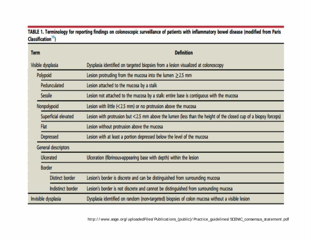
http://www.asge.org/uploadedFiles/Publications_(public)/Practice_guidelines/SCENIC_consensus_statement.pdf

SCENIC: Terms & Characterization

Soetinko R et al. Gastrointestinal Endoscopy Clinics of North America 2014;24(3):483–520

Soetinko R et al. Gastrointestinal Endoscopy Clinics of North America 2014;24(3):483–520

NONPOLYPOID COLORECTAL LESIONS IN IBD

Chromoendoscopy

SCENIC: Chromoendoscopy Technique
Chromoendoscopy (CE) is currently the only technique included IBD surveillance guidelines

Soetinko R et al. Gastrointestinal Endoscopy Clinics of North America 2014;24(3):483–520.

Chromoendoscopy: Dye PreparationTechnique Purpose Delivery Methylene
Blue Dilution(1%, 10mL ampule)
Indigo Carmine Dilution
(0.8%, 5mL ampule)
Panchromoendoscopy Lesion Detection
Water jet channel using auxiliary foot
pump or biopsy
channel using spray
catheter
1 ampule + 240 mL
sterile water (0.04%)
2 ampules + 250 mL
sterile water (0.03%)
Targeted Chromoendoscopy
Lesion characterization
and border delineation
Syringe spray through biopsy
channel
1 ampule + 40 mL sterile water (0.2%)
1 ampule + 25 mL sterile water
(0.13%)

Chromoendoscopy: Technique
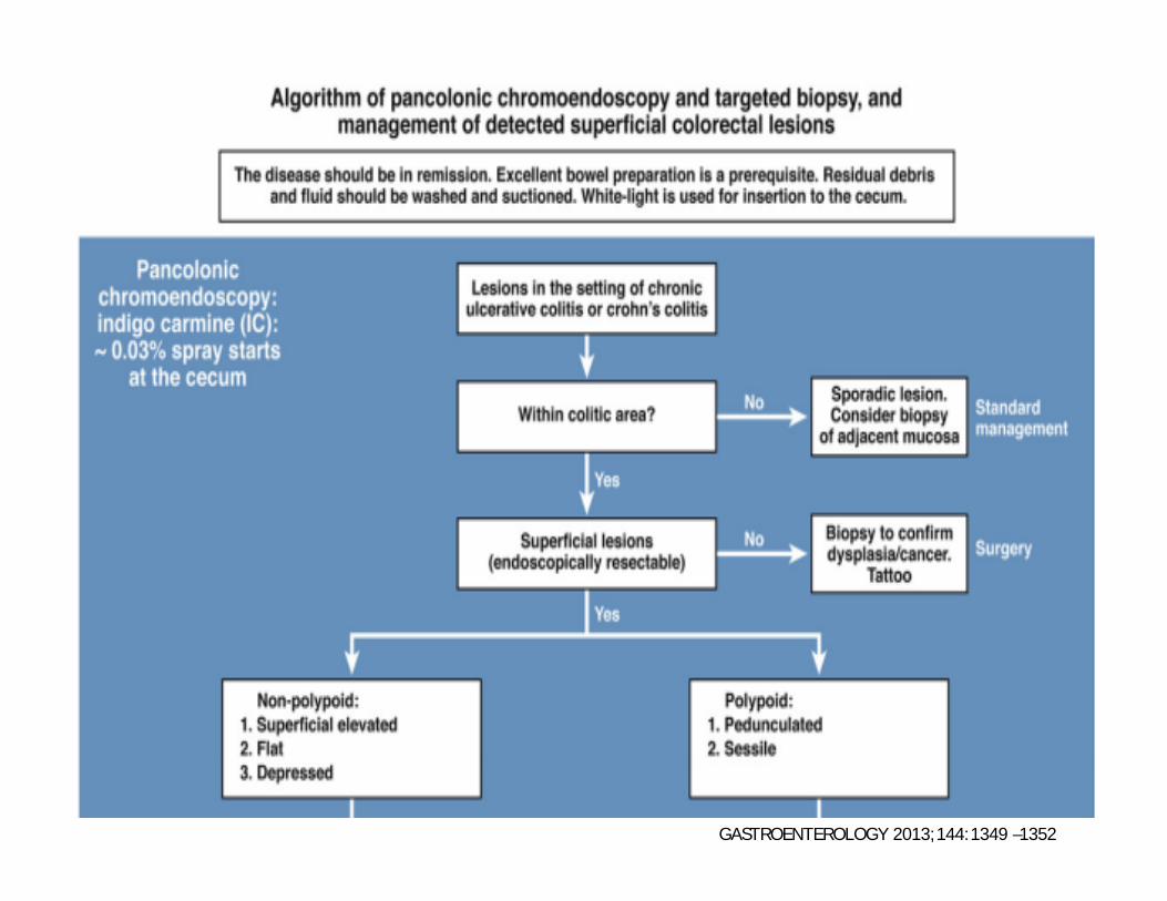
Dilute dye sprayed through water jet during withdrawal
Concentrated dye sprayed through accessory channel to detail suspicious lesions
Lesions examined closely to characterize & determine borders
Biopsies targeted to abnormal-appearing areas

Chromoendoscopy
Superficial lesions with well-defined borders: potentially endoscopically resectable
Resectable lesionRemove en-bloc if possible
Biopsy adjacent mucosa
Unresectable lesionBiopsy and tattoo
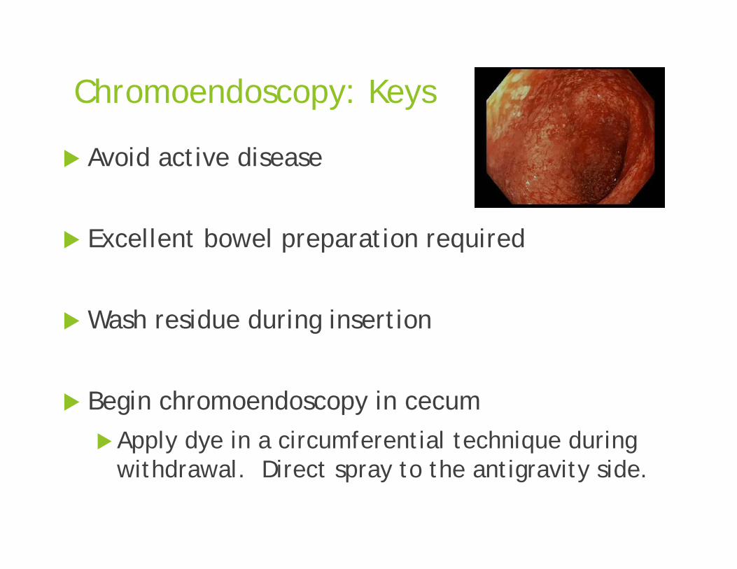
Chromoendoscopy: Keys
Avoid active disease
Excellent bowel preparation required
Wash residue during insertion
Begin chromoendoscopy in cecumApply dye in a circumferential technique during
withdrawal. Direct spray to the antigravity side.

Chromoendoscopy: Keys
Suction excess solution after about 1 minute to aid mucosal visualization
Assess 20-30–cm segments sequentially Reinsert endoscope to the proximal extent of
each segment followed by slow withdrawal and mucosal visualization
Targeted dye spray for suspicious lesions:
Chromoendoscopy Challenges
Time-consuming procedure
Preparation/supplies
Limitations: Poor preparation or active colitis
No minimum competence requirement
Effect on outcomes (cancer/mortality) require further study

Management of Colorectal Dysplasia in IBD

SCENIC: Visible Dysplasia
ENDOSCOPICALLY RESECTABLE SURVEILLANCE COLONOSCOPY rather than
colectomy after removal of endoscopically resectable dysplasia (polypoid or nonpolypoid) Data limited on management of non-polypoid
endoscopically visible dysplasia; surveillance suggested; conditional recommendation
NONENDOSCOPICALLY RESECTABLE Refer to surgery or advanced colonoscopist

SCENIC: Invisible Dysplasia
Confirm with GI pathologist
Referral to endoscopist with expertise in IBD surveillance using chromoendoscopy with high-definition colonoscopy
GASTROENTEROLOGY 2013;144:1349 –1352

GASTROENTEROLOGY 2013;144:1349 –1352

GASTROENTEROLOGY 2013;144:1349 –1352

Objectives: Colorectal Cancer Surveillance in IBD Describe burden of colorectal cancer (CRC) &
current approach for CRC surveillance in IBD
Outline classification scheme for describing IBD-associated dysplastic lesions
Discuss role of chromoendoscopy in evaluation and detection of dysplasia
Highlight optimal endoscopic surveillance techniques and management strategies in clinical practice
Case 1
60 year old female with long-standing ulcerative pancolitis now in remission on 5-ASA therapy presents for second opinion regarding flat dysplasia found on random right colon biopsies. Chromoendoscopy revealed a large depressed lesion in the right colon with ill-defined borders that appeared technically difficult to resect, and biopsy of the lesion showed high-grade dysplasia as confirmed by a second GI pathologist.

Case 1
The recommended next step for this patient is:
A) No immediate action is required. Repeat surveillance colonoscopy in 1 year.
B) Repeat colonoscopy in 3 months.
C) Repeat colonoscopy with chromoendoscopy in 6 months.
D) Refer to colorectal surgeon to discuss proctocolectomy.

Case 1
The recommended next step for this patient is:
A) No immediate action is required. Repeat surveillance colonoscopy in 1 year.
B) Repeat colonoscopy in 3 months.
C) Repeat colonoscopy with chromoendoscopy in 6 months.
D) Refer to colorectal surgeon to discuss proctocolectomy.

Case 2
A 56 year old male with chronic ulcerative pancolitis for 31 years presents for surveillance colonoscopy. You would like to perform the procedure using chromoendoscopy. Patient-related factors that may limit your ability to perform this technique include all but which of the following?

Case 2: Answer
A) Presence of active colon inflammation
B) Presence of pseudopolyps
C) History of Primary Sclerosing Cholangitis
D) Poor bowel preparation

Case 2
A) Presence of active colon inflammation
B) Presence of pseudopolyps
C) History of Primary Sclerosing Cholangitis
D) Poor bowel preparation

Thank You