Embed Size (px)
Citation preview
Journal Pre-proof
Glial Cell Derived Neurotrophic Factor Induces Enteric Neurogenesis and ImprovesColon Structure and Function in Mouse Models of Hirschsprung Disease
Rodolphe Soret, Sabine Schneider, Guillaume Bernas, Briana Christophers,Ouliana Souchkova, Baptiste Charrier, Franziska Righini-Grunder, Ann Aspirot,Mathieu Landry, Steven W. Kembel, Christophe Faure, Robert O. Heuckeroth,Nicolas Pilon
PII: S0016-5085(20)34944-1DOI: https://doi.org/10.1053/j.gastro.2020.07.018Reference: YGAST 63631
To appear in: GastroenterologyAccepted Date: 10 July 2020
Please cite this article as: Soret R, Schneider S, Bernas G, Christophers B, Souchkova O, CharrierB, Righini-Grunder F, Aspirot A, Landry M, Kembel SW, Faure C, Heuckeroth RO, Pilon N, Glial CellDerived Neurotrophic Factor Induces Enteric Neurogenesis and Improves Colon Structure and Functionin Mouse Models of Hirschsprung Disease, Gastroenterology (2020), doi: https://doi.org/10.1053/j.gastro.2020.07.018.
This is a PDF file of an article that has undergone enhancements after acceptance, such as the additionof a cover page and metadata, and formatting for readability, but it is not yet the definitive version ofrecord. This version will undergo additional copyediting, typesetting and review before it is publishedin its final form, but we are providing this version to give early visibility of the article. Please note that,during the production process, errors may be discovered which could affect the content, and all legaldisclaimers that apply to the journal pertain.
© 2020 by the AGA Institute
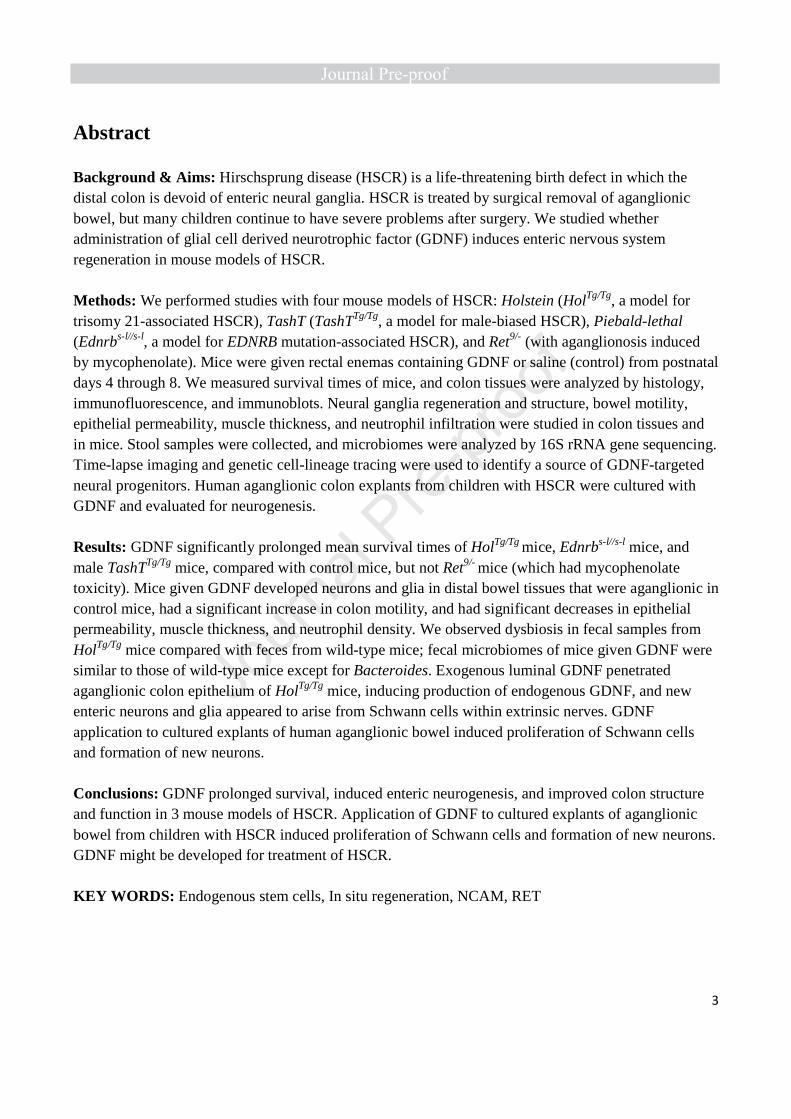
What you need to know: Background and Context: Hirschsprung disease (HSCR) is a life-threatening birth defect in which distal colon is devoid of enteric neural ganglia. HSCR is treated by surgical removal of aganglionic bowel, but many children continue to have severe problems after surgery. New Findings: GDNF prolonged survival, induced intestinal neurogenesis, and improved colon structure and function in 3 mouse models of HSCR. Application of GDNF to cultured explants of aganglionic bowel from children with HSCR also induced formation of new neurons. Limitations: Most of these studies were performed in mice; clinical studies are needed. Impact: GDNF might be developed for treatment of HSCR, but further studies are needed. Lay Summary: The authors identified a neurotrophic factor that promotes regeneration of the intestinal nervous system in mouse models of HSCR and in colon tissues from pediatric patients.
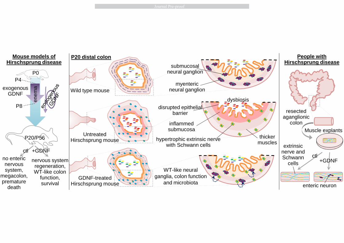
Muscle explants
myentericneural ganglion
WT-like neural ganglia, colon function
and microbiota
inflammedsubmucosa
disrupted epithelialbarrier
thickermuscles
dysbiosis
UntreatedHirschsprung mouse
GDNF-treatedHirschsprung mouse
Wild type mouse
P20 distal colon
no entericnervoussystem,
megacolon,premature
death
Mouse models of Hirschsprung disease
exogenousGDNF
P20/P56
enem
a
P4
P8
P0
hypertrophic extrinsic nerve with Schwann cells
submucosalneural ganglion
People with Hirschsprung disease
resectedaganglionic
colon
enteric neuron
+GDNFctl
extrinsicnerve and Schwann
cells
+GDNFctl
nervous system regeneration, WT-like colon
function, survival
1
Glial Cell Derived Neurotrophic Factor Induces Enteric Neurogenesis and Improves Colon Structure and Function in Mouse Models of Hirschsprung Disease
Short title: Enteric nervous system regeneration Rodolphe Soret1,2, Sabine Schneider3, Guillaume Bernas1,7, Briana Christophers3, Ouliana Souchkova1,2, Baptiste Charrier1,2, Franziska Righini-Grunder4,8, Ann Aspirot5,6, Mathieu Landry1, Steven W. Kembel1,2, Christophe Faure2,4,6, Robert O. Heuckeroth3, Nicolas Pilon1,2,6†
1 Département des Sciences Biologiques, Université du Québec à Montréal (UQAM), Montréal H3C
3P8, Québec, Canada.
2 Centre d’excellence en recherche sur les maladies orphelines – Fondation Courtois (CERMO-FC), Université du Québec à Montréal, Montréal H2X 3Y7, Québec, Canada.
3 Department of Pediatrics, The Perelman School of Medicine at the University of Pennsylvania; and The Children's Hospital of Philadelphia Research Institute, Philadelphia, PA 19104, USA. 4 Division de gastroentérologie, hépatologie et nutrition pédiatrique, Centre hospitalier universitaire Sainte-Justine, Montréal H3T 1C5, Québec, Canada. 5 Division de chirurgie pédiatrique, Centre hospitalier universitaire Sainte-Justine, Montréal H3T 1C5, Québec, Canada. 6 Département de pédiatrie, Université de Montréal, Montréal H3T 1C5, Québec, Canada. 7 Present address: Plateforme de transgenèse, Centre de recherche du centre hospitalier de l’Université de Montréal H2X 0A9, Québec, Canada. 8 Present address: Division of Pediatric Gastroenterology, Hepatology and Nutrition, Children’s Hospital, Lucerne, Switzerland. †Corresponding Author: [email protected] Author contributions: RS and NP conceived the study; ROH and NP supervised the study; RS, SS, ROH and NP designed the experiments; RS, SS, GB, BC, OS, BC and ML performed the experiments and collected data; RS, SS, GB, BC, ML, SWK, ROH and NP analyzed and interpreted data; FRG, AA and CF provided samples; RS, SS, ROH and NP drafted and edited the manuscript; All authors revised the manuscript. Funding: This work was supported by catalyst grants from the Fondation du grand défi Pierre Lavoie to NP and by an operating grant from the Canadian Institutes of Health Research (CIHR #377028) to NP and ROH. RS was supported by a postdoctoral fellowship from the Fonds de la recherche du Québec – Santé (FRQS). SWK holds the Canada Research Chair in Plant Microbiomes. NP is a FRQS Senior Research Scholar and the recipient of the UQAM Research Chair on Rare Genetic Diseases. ROH was also supported by the March of Dimes 6-FY15-235, the Irma and Norman Braman Endowment, the Suzi and Scott Lustgarten Center Endowment, and The Children’s Hospital of Philadelphia Research Institute. The funders had no role in study design, data collection and analysis, decision to publish, or preparation of the manuscript. Competing interests: No conflict of interests exists.
2
Acknowledgements
The authors thank Denis Flipo (UQAM) for assistance with confocal imaging, Dr. Natalie Patey (CHU
Ste-Justine), Ben Wilkins and Archana Shenoy (CHOP) for help with collection of human samples, and
MedGenesis Therapeutix Inc. for generously providing clinical grade human recombinant GDNF.
Data and materials availability: all materials, data and associated protocols will be made promptly
available to readers.
3
Abstract Background & Aims: Hirschsprung disease (HSCR) is a life-threatening birth defect in which the distal colon is devoid of enteric neural ganglia. HSCR is treated by surgical removal of aganglionic bowel, but many children continue to have severe problems after surgery. We studied whether administration of glial cell derived neurotrophic factor (GDNF) induces enteric nervous system regeneration in mouse models of HSCR. Methods: We performed studies with four mouse models of HSCR: Holstein (HolTg/Tg, a model for trisomy 21-associated HSCR), TashT (TashTTg/Tg, a model for male-biased HSCR), Piebald-lethal (Ednrbs-l//s-l, a model for EDNRB mutation-associated HSCR), and Ret9/- (with aganglionosis induced by mycophenolate). Mice were given rectal enemas containing GDNF or saline (control) from postnatal days 4 through 8. We measured survival times of mice, and colon tissues were analyzed by histology, immunofluorescence, and immunoblots. Neural ganglia regeneration and structure, bowel motility, epithelial permeability, muscle thickness, and neutrophil infiltration were studied in colon tissues and in mice. Stool samples were collected, and microbiomes were analyzed by 16S rRNA gene sequencing. Time-lapse imaging and genetic cell-lineage tracing were used to identify a source of GDNF-targeted neural progenitors. Human aganglionic colon explants from children with HSCR were cultured with GDNF and evaluated for neurogenesis. Results: GDNF significantly prolonged mean survival times of HolTg/Tg mice, Ednrbs-l//s-l mice, and male TashTTg/Tg mice, compared with control mice, but not Ret9/- mice (which had mycophenolate toxicity). Mice given GDNF developed neurons and glia in distal bowel tissues that were aganglionic in control mice, had a significant increase in colon motility, and had significant decreases in epithelial permeability, muscle thickness, and neutrophil density. We observed dysbiosis in fecal samples from HolTg/Tg mice compared with feces from wild-type mice; fecal microbiomes of mice given GDNF were similar to those of wild-type mice except for Bacteroides. Exogenous luminal GDNF penetrated aganglionic colon epithelium of HolTg/Tg mice, inducing production of endogenous GDNF, and new enteric neurons and glia appeared to arise from Schwann cells within extrinsic nerves. GDNF application to cultured explants of human aganglionic bowel induced proliferation of Schwann cells and formation of new neurons. Conclusions: GDNF prolonged survival, induced enteric neurogenesis, and improved colon structure and function in 3 mouse models of HSCR. Application of GDNF to cultured explants of aganglionic bowel from children with HSCR induced proliferation of Schwann cells and formation of new neurons. GDNF might be developed for treatment of HSCR. KEY WORDS: Endogenous stem cells, In situ regeneration, NCAM, RET
4
Introduction
The enteric nervous system (ENS) extends along the entire gastrointestinal tract to control bowel
motility and epithelial activity in response to sensory stimuli1. Interconnected enteric ganglia
containing neurons and glia develop from neural crest-derived progenitors before birth. Incomplete
colonization of distal colon by these ENS progenitors causes Hirschsprung disease (HSCR), a condition
affecting 1 in 5000 newborns2. In HSCR, distal colon without ganglia (i.e., aganglionic colon) remains
tonically contracted, causing functional intestinal obstruction. Symptoms also include bowel
inflammation and a high risk of sepsis and premature death2.
HSCR is subdivided into short-segment (S-HSCR) and long-segment forms (L-HSCR)2. In S-HSCR
(>80% of cases), the ENS is absent from rectum and sigmoid colon only. L-HSCR means longer
regions of distal bowel are aganglionic. Many genes influence HSCR risk2, and genetic risk variants
may combine with non-genetic factors3. The major HSCR-associated gene is RET, a transmembrane
tyrosine kinase activated when GDNF binds the co-receptor GFRα1. GDNF-GFRα1-RET signaling is
needed for survival, proliferation, and migration of ENS progenitors4-6. Accordingly, >90% of children
with S-HSCR bear non-coding variants that reduce RET expression7. Protein altering RET variants are
not common in S-HSCR, but occur in ~35% of people with L-HSCR8, 9. Other genes influencing HSCR
risk encode EDNRB signaling pathway molecules, transcription factors, guidance and extra-cellular
matrix molecules, and diverse additional factors7, 9-11. Male sex also increases HSCR risk ~4-fold,
while Down syndrome increases HSCR risk ~100-fold2. Collectively, these observations mean that
most children with HSCR have reduced, but not absent RET signaling, and that diverse additional
factors impact HSCR occurrence.
Since 1948, surgical removal of aganglionic bowel has been life-saving for most children with
HSCR12. However, post-surgical complications are common and can be long-lasting, impacting
5
survival and/or quality of life2. One ideal alternative approach would be to rebuild the ENS and reduce
the need for surgery. This idea prompted many groups to develop cell transplantation-based HSCR
therapies13. However, despite many encouraging results, some difficulties remain14. The optimal source
of stem cells, ideal amplification and/or differentiation strategies prior to transplantation, methods of
cell delivery, and cell fate after transplantation are not yet well defined.
Here we tested the hypothesis that endogenous ENS progenitors could be activated after birth and
generate enteric neurons de novo. Our cell-free strategy is based on the idea that HSCR is due to
incomplete rostrocaudal colonization of distal bowel by the main subpopulation of ENS progenitors,
namely neural crest cells of vagal origin15. Since extracellular matrix in colon becomes refractory to
migration after a certain developmental window16, reactivating vagal-derived ENS progenitor
migration seemed unlikely after birth. However, it seemed possible that ENS progenitors of sacral17 or
Schwann cell lineage18 origin, which are already present in aganglionic colon, could be induced to
proliferate and differentiate into functioning neurons. Schwann cells in particular are abundant in
hypertrophic extrinsic nerve tracts that populate both muscular and submucosal layers of distal
aganglionic bowel19. GDNF appeared as a primary candidate for postnatal reactivation of ENS
progenitors in the aganglionic zone notably because of its ability to stimulate migration and
proliferation of Schwann cells in a RET-independent but GFRα1-dependent manner through its
alternative receptor NCAM20, 21.

6
Materials and Methods
Mice. Details about all mouse lines used are in Supp. Methods. Enema treatments of most mouse lines
were performed at Université du Québec à Montréal, except for Ret9/- that were treated with
mycophenolate22 and GDNF at the Children’s Hospital of Philadelphia Research Institute (as detailed
in Supp. Methods). Where indicated, some HolTg/Tg and Ednrbs-l/s-l were also GDNF-treated at the
Children’s Hospital of Philadelphia Research Institute after reciprocal exchange of mice between
Philadelphia and Montreal. Unless specified otherwise (see Fig.S1), 10µl enemas consisting of a
1µg/µl solution of human recombinant GDNF (Peprotech cat. # 450-10) diluted in PBS were
administered daily between P4 to P8 (see Supp. Methods for more detail). Clinical grade GDNF
(Medgenesis Therapeutix Inc., Canada) and a previously described 6XHis-tagged version23 used for
some experiments had similar efficiency (see Supp. Methods for other tested molecules). For EdU
incorporation assays, mouse pups received 10µl intraperitoneal injections of a 10mM EdU solution
(ThermoFisher Scientific, Cat. # C10337) once a day during the 5-day (P4 to P8) GDNF enema
treatment.
Tissue processing, staining, and imaging. Bowel was cut longitudinally along the mesentery, washed
in PBS, pinned onto Sylgard-coated petri dishes, fixed with 4% PFA at 4°C overnight, and finally
microdissected to separate longitudinal/circular muscles from the submucosa/mucosa layer. For ex vivo
analyses of living tissues, unfixed tissues were microdissected in ice-cold oxygenated Krebs solution.
For histological analyses, fixed full-thickness bowel segments were embedded in paraffin and
transversally sectioned at 10µm. For Western blotting, unfixed organs were weighed and dissolved in
RIPA buffer, using 1mL for every 100mg of tissue (further details are provided in Supp. Methods).
Details about immunofluorescence and imaging can be found in Supp. Methods.
7
Analysis of colonic motility and permeability. In vivo analysis of distal colonic motility in P20 mice
was performed using the bead latency test, as detailed in Supp. Methods. For ex vivo analysis of colonic
motility, strips of living muscles from most distal colon (1cm from the anus) of P20 mice were
prepared as described above, and attached in the longitudinal direction in a Schuler organ bath
(Harvard apparatus) filled with oxygenated Krebs solution. Contraction/relaxation of longitudinal
muscles was then recorded as detailed in Supp. Methods. For ex vivo analysis of mucosal barrier
function, segments of living mucosa from most distal colon of P20 mice were prepared as described
above, mounted in Ussing chambers with 0.5 cm² exposed surface area (Warner Instruments, Model U-
9926), and evaluated for paracellular permeability as described in Supp. Methods.
Microbiome analysis. Stool isolation, microbiome sequencing and data analysis were performed as
previously described 24 (details are provided in Supp. Methods).
Ex vivo time-lapse imaging and culture of murine aganglionic colon. For time-lapse imaging, strips
of living muscles from the last cm of distal colon from P4 HolTg/Tg;G4-RFP double transgenic pups
were prepared as described above, and pinned onto Sylgard-coated 35mm ibidi µ-dishes (ibidi, Cat. #
81156). Muscle strips were then cultured in suspension as detailed in Supp. Methods. For ex vivo
induction of neurogenesis, strips of living muscles from the last cm of distal colon from P4 HolTg/Tg
pups were cultured as for time-lapse imaging in presence of 0.5µM EdU. After 96h of culture, tissues
were fixed with PFA and processed for immunofluorescence and EdU labelling.
Culture of human aganglionic colon tissues. Human sigmoid colon tissue was obtained from 12
HSCR patients undergoing Swenson-type surgical resection of the aganglionic zone. Nine patients (7
boys and 2 girls; aged between 28 and 1638 days at the time of surgery) were recruited at the Centre
hospitalier universitaire Sainte-Justine (Montreal, Canada) while 3 patients were recruited at the
Children’s Hospital of Philadelphia (2 boys and 1 girl; aged between 300 and 1177 days at the time of

8
surgery). After the surgery, full-thickness colon tissues were placed in ice-cold Krebs solution or
Belzer UW Cold Storage Solution (Bridge to Life Ltd.) and immediately brought to the relevant
research laboratory. Muscle strips were then prepared as described above and cut in smaller pieces of
0.5cm X 0.5cm. One piece was kept aside for validation of aganglionosis via immunofluorescence,
while the others were cultured for 96h as described above for inducing neurogenesis in mouse tissues.
Samples from two patients (86 and 1638 days of age at surgery) were in addition cultured for 7 days,
under the same conditions. At the end of culture period, all tissues were fixed with PFA and processed
for immunofluorescence and EdU labelling.
Study approval. All experiments with mice were approved by animal research ethics committees of the
Université du Québec à Montréal (CIPA reference # 878) and the Children's Hospital of Philadelphia
Research Institute (IAC reference # 16-001041). Likewise, experiments with human samples were
approved by human research ethics committees of the Université du Québec à Montréal (CIEREH
protocol # 491), the Centre hospitalier universitaire Sainte-Justine (CER protocol # 4172) and the
Children’s Hospital of Philadelphia (IRB protocol # 13-010357). Informed consent for the collection
and use of human tissues was obtained from all donors, and parents or legal guardian except for one
piece of de-identified human colon.
Statistics. All experiments employed a minimum of three biological replicates. Where relevant, the
exact number of independent replicates (n) and statistical tests used to calculate P values are included
in figures and/or legends. P values were determined using GraphPad Prism 6, with the exception of
microbiome data that were analyzed with R.
9
Results
GDNF enemas rescue aganglionosis in three mouse models of S-HSCR
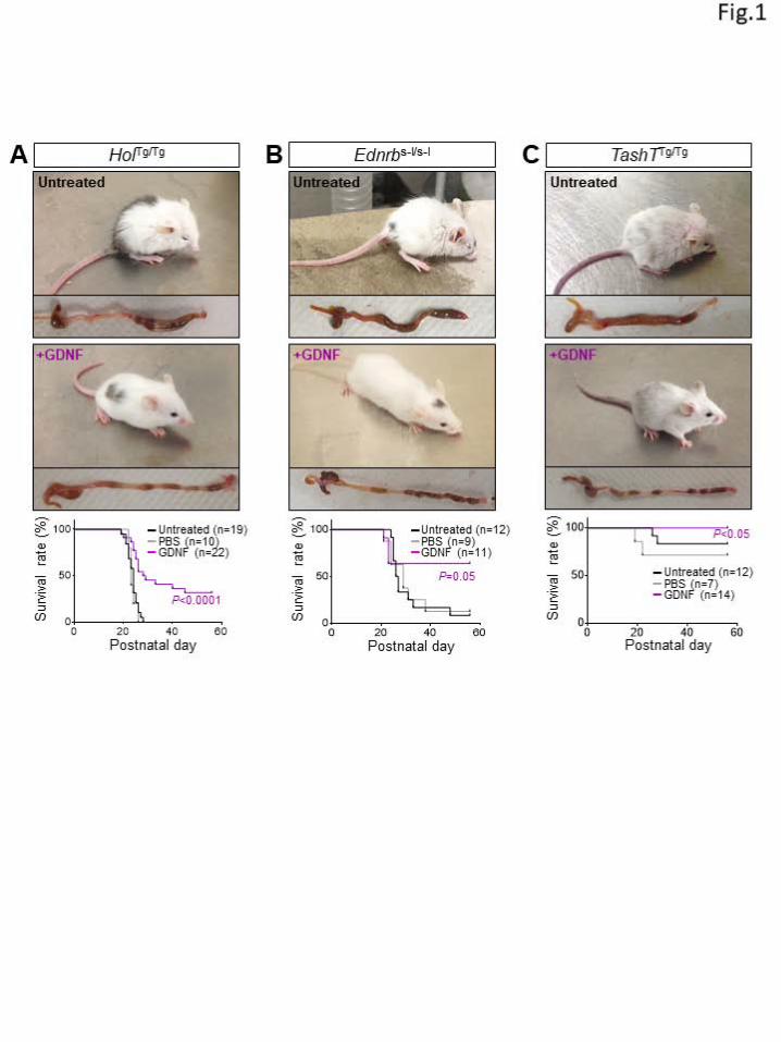
Using rectal enemas, we tested if early postnatal administration of GDNF could enhance survival of
four mouse models of S-HSCR: Holstein (HolTg/Tg; a fully-penetrant model for trisomy 21 [Collagen VI
over-expression]-associated HSCR)25, TashT (TashTTg/Tg; a partially-penetrant model for male-biased
HSCR)26, Piebald-lethal (Ednrbs-l//s-l; a fully-penetrant model for EDNRB mutation-associated HSCR)27
and Ret9/- mutant mice (a hypomorphic model where aganglionosis is induced by mycophenolic acid)22.
The enema volume necessary to fill whole colon, GDNF concentration, treatment time window, as well
as duration and frequency of therapy were first empirically determined with HolTg/Tg pups (Fig.S1A-E).
Remarkably, our selected treatment (i.e., daily administration of 10µg GDNF in PBS as 10µl enemas
for 5 consecutive days between postnatal day [P] 4 to P8) prevented death in about half of HolTg/Tg mice
at P28, the maximum age of survival for control HolTg/Tg mice (Fig.1A). Most animals surviving to P28
reached adult age after GDNF treatment and mice evaluated could reproduce (two tested breeding pairs
were fertile). The few animals that were allowed to survive beyond P56 (our adult reference age)
eventually died from megacolon or dystocia between P68 and P250 (Fig.S1C). Importantly, the same
GDNF enema treatment also prevented premature death for more than 60% of Ednrbs-l//s-l mice
(Fig.1B) and for all male TashTTg/Tg pups (Fig.1C). Nine GDNF-treated male TashTTg/Tg mice kept for
over a year looked healthy without any sign of adverse effects. Enema treatment of HolTg/Tg mice using
Noggin, endothelin-3, or the serotonin receptor (5-HT4R) agonist RS67506 (rationale provided in
Table S1) failed to increase life expectancy, suggesting specific benefit to GDNF (Fig.S1F). We also
failed to further increase the overall survival rate of GDNF-treated HolTg/Tg animals either by replacing
standard chow with a gel diet (Fig.S1G) or by combining GDNF with vitamin C, serotonin or
endothelin-3 (Table S1 and Fig.S1H).

10
Because modest reductions in RET function are common in people with HSCR, we wanted to
determine if GDNF enemas could work in RET hypomorphic mice. Unfortunately, there are no good
mouse models for RET mutation-associated HSCR. Ret-null mice have total intestinal aganglionosis28
whereas Ret heterozygotes are overtly normal4. We therefore decided to use our established protocol to
induce distal bowel aganglionosis in Ret9/- hypomorphic mice using mycophenolate mofetil22.
Surprisingly, far less prenatal mycophenolate was needed to cause dose-dependent aganglionosis in our
novel experimental conditions (with Ret mutants rederived in a new animal facility) (Fig.S2A)
compared to our prior studies22, and postnatal GDNF enemas did not improve survival compared to
PBS alone (Fig.S2B). Instead, many pups died with distended bowel before the end of GDNF
treatment, even at the lowest mycophenolate concentration (Fig.S2C). Moreover, many ill pups had
ganglia throughout the bowel (Fig.S2D), suggesting highly variable efficiency of mycophenolate
treatment and additional toxicity that complicates data interpretation.
To determine how GDNF enemas enhanced survival in the other three HSCR mouse models, we
tested the hypothesis that GDNF induced postnatal neurogenesis in aganglionic distal colon. We
focused on the Holstein line for practical reasons (fertility is low in Piebald-lethal and megacolon
incidence is lower in TashT), and analyzed P20 animals because HolTg/Tg mice generally reach this
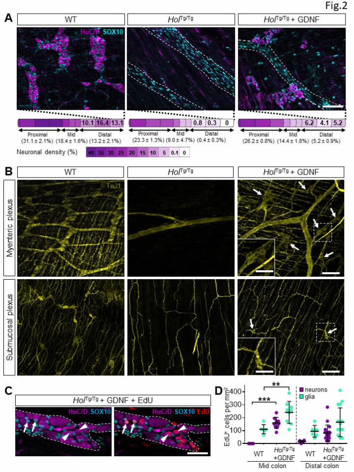
stage even without enema treatment (Fig.1A). As we reported25, myenteric HuC/D+ neurons and
SOX10+ glia were abundant in WT distal colon and absent from the last cm of HolTg/Tg colon (Fig.2A).
In contrast, in HolTg/Tg distal colon, SOX10+ cells were mainly within thick extrinsic nerve fibers
(Fig.2A) where Schwann cells reside18. Remarkably, distal colon from GDNF-treated HolTg/Tg animals
had numerous HuC/D+ neurons and SOX10+ glia organized into ganglia primarily adjacent to extrinsic
nerves (Fig.2A and Movie S1). These GDNF-induced ganglia formed Tuj1+ interconnected networks in
both myenteric and submucosal plexuses (Fig.2B). Quantification of myenteric neuron density in whole
colon of HolTg/Tg and male TashTTg/Tg mice showed GDNF effects are most prominent in distal colon

11
(i.e., final 3 cm), with minor effects in proximal colon (Figs.2A and S3). In the mid-colon of GDNF-
treated HolTg/Tg mice, the increased neuron density (Fig.2A) was mainly due to an enlargement of pre-
existing myenteric ganglia (Fig.S3A). In the most distal colon, where untreated HolTg/Tg mice are
normally devoid of enteric neurons, GDNF-treated HolTg/Tg mice had an average neuron density that
was about 40% that of WT mice (Fig.2A). When neuron density in the distal colon was too low,
GDNF-treated HolTg/Tg mice developed megacolon (Fig.S1I). Remarkably, in GDNF-treated TashTTg/Tg
males, neuron density in the most distal colon was completely restored (Fig.S3B,C).
EdU incorporation assays confirmed that GDNF induced proliferation of neuron and glia progenitors
during the 5-day treatment from P4 to P8. Staining of P20 HolTg/Tg colon from mice that received daily
EdU injections during GDNF treatment revealed many EdU+ HuC/D+ (presumptive neurons) and EdU+
SOX10+ (presumptive glia or neuron/glia progenitors) in both myenteric and submucosal ganglia
(Figs.2C,D and S4). Yet, only a few, very small ganglia (i.e., 3 neurons) were fully populated by EdU+
neurons (Fig.S4C). Collectively these data suggest that GDNF enemas induce proliferation and
differentiation of ENS progenitors in distal colon and that some of induced neurons and glia cluster into
new ganglia.
GDNF-induced ENS is morphologically and functionally similar to WT
Again focusing on the HolTg/Tg model, we next asked to what extent GDNF-induced ENS in the distal
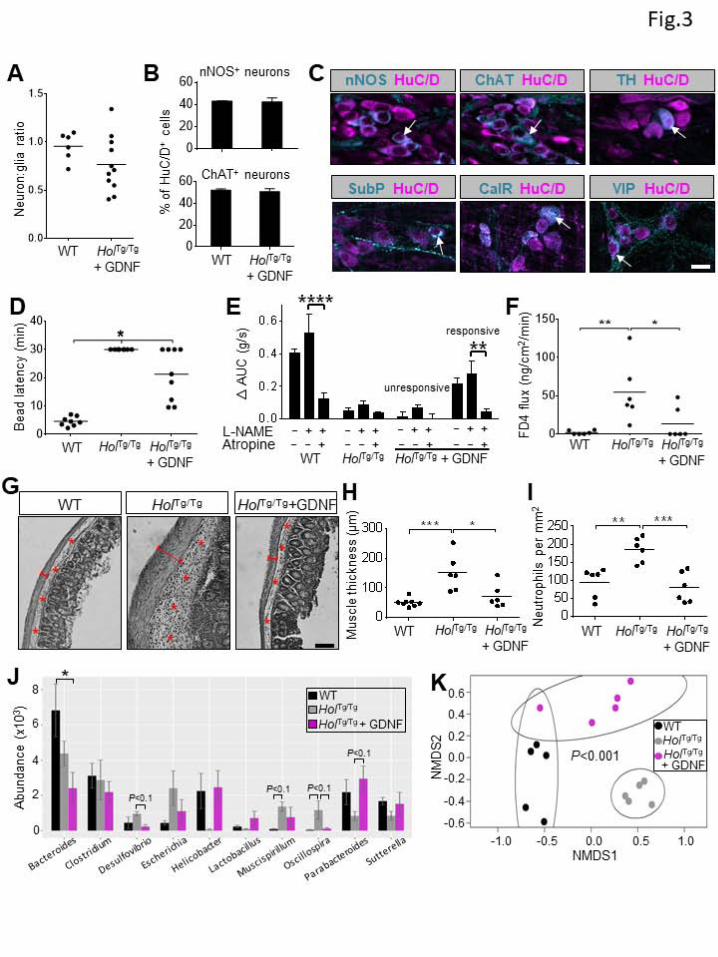
colon resembles WT at P20. Average neuron-to-glia ratio within GDNF-induced myenteric ganglia was
statistically similar to WT (P=0.16) (Fig.3A). Relative proportions of major myenteric neuron
subtypes, including cholinergic (ChAT+) and nitrergic (nNOS+) neurons, were also very similar to WT
(Fig.3B,C). Moreover, many other neuronal subtypes were detected including TH+ dopaminergic
neurons, CalR+ excitatory motor neurons, VIP+ inhibitory motor neurons, and SubP+ excitatory motor
neurons (Fig.3C). Interestingly, in proximal and mid colon of HolTg/Tg mice (Fig.S5), GDNF treatment

12
also corrected the imbalance of nitrergic (increased) and cholinergic (decreased) neuron subtypes that
is observed proximal to the aganglionic segment in both HSCR mouse models and human patients29-32.
To evaluate function of P20 GDNF-induced myenteric ganglia, we analyzed colonic motility in vivo,
using the bead latency test. In contrast to untreated HolTg/Tg mice that never expelled a rectally-inserted
glass bead during our 30 minute observation period, a subset of GDNF-treated HolTg/Tg expelled the
bead in 10-21 min, a bit slower than WT mice (range of 2-8 min) (Fig.3D). Analysis of neuron density
in these GDNF-treated HolTg/Tg mice revealed a robust inverse correlation between time to expel the
bead and neuron density in the distal colon (Fig.S6A). We also evaluated motility ex vivo using muscle
strips from P20 distal colons attached to force transducers in organ baths. This system allows electric
field stimulation-induced contractions of WT colon muscles to be slightly increased by inhibition of
nitric oxide synthase with L-NAME, which can then be robustly counteracted by inhibition of
cholinergic signaling with atropine. Reminiscent of in vivo data, colon muscle strips from GDNF-
treated HolTg/Tg mice displayed one of two distinct response patterns (Figs.3E and S6B), either similar
to WT or similar to untreated HolTg/Tg. To indirectly test function of P20 GDNF-induced submucosal
ganglia, we analyzed epithelial permeability to small fluorescently labeled dextran molecules (FD4) in
Ussing chambers. Once more, distal colonic tissues from GDNF-treated HolTg/Tg mice displayed two
distinct response types, with mucosa either impermeable to FD4 like WT tissues, or permeable to FD4
like control HolTg/Tg tissues from untreated mice (Fig.3F).
To complement ENS analyses, we evaluated other HSCR-associated bowel anomalies. HolTg/Tg
mouse colon had thicker smooth muscles and more neutrophils than WT mice, but GDNF-treated
HolTg/Tg mouse colon was similar to WT (Figs.3G-I and S7). Similarly, stool microbiome profiling
demonstrated dysbiosis in P20 HolTg/Tg mouse colon, but average abundance of several bacterial genera
in HolTg/Tg mouse colon were indistinguishable from WT after GDNF treatment (Fig.3J). A notable
exception was Bacteroides abundance, which was low in HolTg/Tg mice and even lower after GDNF
13
treatment. Accordingly, beta diversity analysis revealed distinct microbial communities among WT,
HolTg/Tg and GDNF-treated HolTg/Tg mice (Fig.3K).
Schwann cells within extrinsic nerves are a target of GDNF in aganglionic colon
To elucidate how GDNF induces enteric neurogenesis, we first evaluated GDNF distribution in P8
bowel via Western blot, taking advantage of size differences between recombinant (15 kDa) and
endogenous (20 kDa, glycosylated) GDNF monomers. Recombinant GDNF was detected in GDNF-
treated HolTg/Tg distal colon but not in proximal colon (Fig.4A). Surprisingly, while endogenous GDNF
is normally only detected in ileum, recombinant GDNF enemas triggered robust increases in
endogenous GDNF throughout the colon (Fig.4A). Recombinant GDNF was no longer detected in
colon nor in any other tissue at P20, suggesting that administered GDNF primarily acts during the
treatment period (Fig.S8). To assess precise locations of recombinant GDNF during treatment, we
treated HolTg/Tg mice using a 6xHis-tagged version of GDNF23 (HisGDNF). Time-course analysis of
distal colon 2h after GDNF treatment on P4, P6 and P8 revealed HisGDNF accumulated over time in
colon submucosa (Fig.4B), smooth muscles, and subsets of enteric neurons (Figs.4C and S9) of HolTg/Tg
mice. Interestingly, RET levels also increased (Figs.4B and S9), supporting the hypothesis that GDNF-
RET auto-regulatory loops are activated in GDNF-treated colon. Remarkably, both HisGDNF and RET
were detected in induced neurons close to extrinsic nerves of HisGDNF-treated animals, not only in
HolTg/Tg mice (Fig.4C) but also in Ednrbs-l/s-l mice (Fig.S10A), and in both of our mouse facilities in
Montreal and Philadelphia (Fig.S10A,B).
Given that induced neurons and glia were often closely associated with extrinsic nerves, we
hypothesized that nerve-associated Schwann cells might be GDNF-targeted ENS progenitors. We first
assessed response of Schwann cells to GDNF using live explants of distal colon muscularis externa
from P4 HolTg/Tg;G4-RFP double transgenic pups. In these mice, neural crest derivatives including

14
Schwann cells are marked by RFP fluorescence 33. Time-lapse imaging of explants after 72h of culture
suggested GDNF (5 µg/ml) stimulates both migration and proliferation of Schwann cells (Fig.4D and
Movies S2-S3). Impact of GDNF on proliferation of these Schwann cells was confirmed via
immunofluorescence after 96h of culture, which revealed a 3-fold increase in Ki67+ SOX10+ double
positive cells upon exposure to GDNF (Fig.4E,F).
To more definitely demonstrate Schwann cells are GDNF targets, we used in vivo genetic cell lineage
tracing with the Schwann lineage-specific Dhh-Cre driver and the R26[Floxed Stop]YFP Cre reporter allele
in the Holstein [FVB/N] mutant background. Analysis of proximal and mid colons from untreated Dhh-
CreTg/+;R26YFP/+ and HolTg/Tg;Dhh-CreTg/+;R26YFP/+ animals at P20 showed that the proportion of
Schwann cell lineage-derived (YFP+) myenteric neurons increased from 5-7% in a pure FVB/N genetic
background to 10-11% in presence of homozygous Holstein mutation (Fig.S11A,B). Remarkably, the
Schwann cell lineage contribution further increased to 34% of myenteric neurons in the distal colon of
GDNF-treated HolTg/Tg;Dhh-CreTg/+;R26YFP/+ animals (Fig.4G,H). By daily EdU administration during
GDNF treatment, we identified four subgroups of induced myenteric neurons based on cellular origin
(YFP fluorescence) and/or EdU incorporation (Figs.4G,H and S11C). While this work supports the
hypothesis that Schwann cells are a source of GDNF-induced neurons and glia in both myenteric
(Fig.4G,H) and submucosal (Fig.S11C) plexus, it also revealed that a majority of induced neurons
(66%) were YFP-negative, suggesting a stronger contribution by non-Dhh-expressing cell type(s).
Regardless of cellular origin, a majority of induced neurons (62%) also did not incorporate EdU,
raising the possibility that neurogenesis might result from transdifferentiation (i.e., direct differentiation
of a post-mitotic cell into another type of specialized cell) instead of requiring proliferating precursor
cells (Fig.4G,H).
GDNF can induce new neurons in human aganglionic colon ex vivo

15
To test if GDNF could induce new enteric neurons in human tissue, we needed an ex vivo model. We
discovered that 96h of ex vivo GDNF treatment induced neurons in all HolTg/Tg distal colon aganglionic
tissues, but neurogenesis was much less efficient than in vivo (Figs.5A and S12A). Induced neurons
rarely clustered into ganglia and such ganglia were always very small (Fig.5B). In marked contrast to
widespread EdU incorporation into Schwann cells (Figs.5C and S12B), EdU incorporation in induced
neurons was also minimal (Fig.5B,C). Although not perfect, we used this ex vivo system to test if
GDNF could induce neurogenesis in aganglionic human colon muscle from children who had pull-
through surgery to resect aganglionic distal bowel. Our cohort consisted of 12 children with
epidemiologic characteristics typical of HSCR (i.e., mostly sporadic, male-biased, short-segment)
(Table 1). Culturing small pieces of freshly-isolated muscularis externa with GDNF for 96h markedly
increased the proportion of EdU+ Schwann cells in 9/9 human tissues where EdU was added to media
(Fig.5D,E). Most importantly, we also detected new neurons expressing HuC/D, βIII-Tubulin (Tuj1),
RET, PGP9.5 and PHOX2B in three HSCR explants (Figs.5F,G and S13; Movie S4). These three
explants were from the youngest children of our cohort (28 to 44 days old) (Fig.5G and Table 1). Two
of these young children had sporadic HSCR with unknown genetic causes. The third child had a
MEN2A syndrome-associated RET mutation (Table 1). For older children (n=2; 86 and 1638 days old),
we found that extending GDNF treatment to 7 days could yield neurons that incorporated EdU
(Fig.5H). Collectively these data suggest our observations in mice may be extended to humans.
16
Discussion
Here we report that GDNF enemas can regenerate a functional ENS in situ and prevent death in three
genetically-distinct mouse models of S-HSCR. Detailed mechanistic studies in HolTg/Tg mice showed
that exogenous GDNF can penetrate the permeable distal aganglionic colon, leading to increased levels
of endogenous GDNF and RET in the whole colon. At least some of the new neurons and glia appear to
arise from extrinsic Dhh-Cre+ lineage Schwann cells, and newly organized enteric ganglia appear to be
self-sustaining until at least P56 (Fig.S14).
One concern for GDNF-based therapy is that RET signaling is often reduced in children with HSCR,
suggesting GDNF responsiveness would also be reduced. However, most children with S-HSCR must
have substantial RET activity in ENS precursors because complete RET absence causes a much more
severe phenotype (i.e., total intestinal aganglionosis) in mice and humans28, 34. Supporting this idea,
recent whole genome sequencing studies of people with S-HSCR found only 4.3% (out of 443
patients)7 and 6.3% (out of 190 patients)9 had RET rare coding variants predicted to be damaging.
Furthermore, rectal GDNF therapy increased levels of RET and endogenous GDNF in mouse colon,
suggesting positive feedback loops that could enhance RET signaling even if initial RET levels were
low. This could be particularly valuable since RET provides trophic support to some enteric neurons in
adults4 and RET is expressed in a subset of GDNF-induced neurons after rectal therapy (Figs.4C,
S10A.B and S11D). Finally, although some of the GDNF-induced neurons that express RET are
derived from Schwann cells (Fig.S11D), RET is most likely not needed to activate these precursors in
aganglionic bowel. GDNF signaling in Schwann cells is instead mediated by NCAM20, and our data
show NCAM but not RET expression in Schwann cells of extrinsic nerves in aganglionic mouse colon
(Fig.S15). Nonetheless, we tried to directly determine if reduced RET levels affected GDNF therapy
using our previously published model of Ret hypomorphic mice exposed to mycophenolate22. However,
17
several problems complicated interpretation in our new experimental conditions, including the
occurrence of HSCR-like dilated bowel even in absence of aganglionosis (Fig.S2C,D). Future studies
would need to test GDNF enema effects in other models where reduced RET activity is associated with
short-segment aganglionosis, but the “ideal” model is not readily apparent.
In contrast to Ret mutants, HolTg/Tg, TashTTg/Tg and Ednrbs-l//s-l mice are all reliable models of S-
HSCR25-27, recapitulating key hallmarks of the human disease in both aganglionic segment and
proximal ENS-containing colonic regions29-32. Although not all GDNF-treated mutant mice have
prolonged survival, the survival advantage after GDNF treatment in Montreal is dramatic and the in
situ generation of new enteric neurons in previously aganglionic bowel, which was observed in both
Montreal and Philadelphia (Fig.S10A,B), is unprecedented. The reason why some mice responded
better to GDNF than others might be due to the degree of aganglionosis-associated inflammation
(Fig.3G, I). Indeed, although GDNF is known to have anti-inflammatory properties35, the inflammatory
microenvironment present in aganglionic bowel before GDNF treatment might help trigger a
neurogenic response as it does in the context of inflammatory bowel disease36. Unfortunately, the exact
inflammatory mediators that enhance enteric neurogenesis are not yet known, but once identified we
could develop adjunct treatments that enhance the effect of GDNF therapy.
Adjunct treatments might also be developed based on a serendipitous finding we made when we tried
to replicate our Montreal survival data (Fig.1A-C) using HolTg/Tg and Ednrbsl/sl mice in Philadelphia.
We unexpectedly discovered that these mouse lines live much longer in Philadelphia than in Montreal
without any specific treatment, even though all our mice originated from the same colonies
(Fig.S10C,D). The prolonged survival in Philadelphia compared to Montreal was especially dramatic
for untreated Ednrbs-l/s-l (Fig.S10C) that lived much longer than previously described in any other
mouse facility27, 30, 37. Intriguingly, the survival advantage for untreated HSCR models in Philadelphia
occurred despite the confirmed presence of distal bowel aganglionosis and/or megacolon (Fig.S10C,D).

18
In fact, Philadelphia-based untreated mice survived as long as GDNF-treated mice in Montreal
(Fig.S10E,F) and survival could not be further enhanced in Philadelphia by GDNF treatment
(Fig.S10E,F) even though GDNF-induced neurogenesis was similar in both Montreal and Philadelphia
(Fig.S10A,B). One especially attractive hypothesis for all these observations is that GDNF treatment in
Montreal and non-genetic factors in Philadelphia might both improve a critical pro-survival bowel
function (e.g., promoting enhanced epithelial barrier function or modulating mucosal immune
responses), either indirectly (via induced ENS ganglia) in Montreal or directly (bypassing the need for
induced ENS ganglia) in Philadelphia. Although we suspect that food- and/or microbiota-based
mechanisms underlie the survival advantage in Philadelphia, there are many variables so defining
mechanisms is complicated.
A potentially more straightforward approach to improve GDNF therapy would be to identify GDNF-
targeted ENS progenitors other than Dhh-lineage Schwann cells that appear to contribute only about a
third of GDNF-induced neurons. This could lead to an improved GDNF-based cocktail that includes
additional trophic factor(s) that bind receptors on these other cells. In this regard, substantial literature
suggests the existence of ENS “stem cells” in postnatal mouse and human bowel38, 39, even in
aganglionic regions40, 41. Interestingly, extrinsic nerve fibers of aganglionic regions were previously
identified as a niche for these ENS stem cells41. Our data confirm this prior observation and further
suggest that at least some of these stem cells are in fact Dhh-lineage Schwann cells. It is possible that
the other ENS progenitors are also Schwann cells that do not express CRE in Dhh-Cre mice. In
accordance with this possibility, we noted that some SOX10+ Schwann cells are not YFP+ in extrinsic
nerves from the aganglionic colon of HolTg/Tg;Dhh-CreTg/+;R26YFP/+ mice at P8 (Fig.S16). The strong
contribution of non-Dhh-expressing cells combined with the location of some GDNF-induced ganglia
away from extrinsic nerves, however, also suggests the involvement of additional cell type(s), which
might include sacral neural crest-derived cells. Sacral-derived ENS progenitors can colonize the
19
aganglionic colon during prenatal development42, and our SOX10 immunofluorescence data suggest
that some of these cells persist in postnatal aganglionic tissues as scattered progenitors and/or enteric
glia, also expressing the alternative GDNF receptor NCAM (Fig.2A and Fig.S15). A contribution by
differentiated enteric glia of sacral origin might also help explain the observation that many GDNF-
induced neurons had not incorporated EdU suggesting they were generated via transdifferentiation
(Fig.4G,H).
In theory, GDNF-based rectal therapy would be easy to implement since normal saline enemas are
already commonly used in children with HSCR both before and after pull-through surgery. If
penetration of GDNF beyond the epithelium was limited, GDNF could be directly injected into the
colon wall with currently available endoscopes or via a specially designed delivery tool. Ideally,
GDNF-based rectal therapy would prevent the need for pull-through surgery. Even if GDNF enemas
did not work as primary HSCR treatment, GDNF therapy might nevertheless improve post-surgical
outcomes by normalizing ENS structure in the retained distal bowel of the “transition zone” (i.e.,
correcting hypoganglionosis and neuronal subtype imbalance). In addition, since ENS stem cell-based
therapies are being considered for the treatment of HSCR, GDNF might be a useful adjunct to these
therapies to promote engraftment. All these considerations make a human clinical trial of GDNF-based
rectal therapy in children with HSCR appealing.

20

21
22
References
1. Furness JB. The enteric nervous system and neurogastroenterology. Nat Rev Gastroenterol Hepatol
2012;9:286-94.
2. Heuckeroth RO. Hirschsprung disease - integrating basic science and clinical medicine to improve
outcomes. Nat Rev Gastroenterol Hepatol 2018;15:152-167.
3. Heuckeroth RO, Schafer KH. Gene-environment interactions and the enteric nervous system: Neural
plasticity and Hirschsprung disease prevention. Dev Biol 2016;417:188-97.
4. Gianino S, Grider JR, Cresswell J, et al. GDNF availability determines enteric neuron number by controlling
precursor proliferation. Development 2003;130:2187-98.
5. Natarajan D, Marcos-Gutierrez C, Pachnis V, et al. Requirement of signalling by receptor tyrosine kinase
RET for the directed migration of enteric nervous system progenitor cells during mammalian
embryogenesis. Development 2002;129:5151-60.
6. Young HM, Hearn CJ, Farlie PG, et al. GDNF is a chemoattractant for enteric neural cells. Dev Biol
2001;229:503-16.
7. Tang CS, Li P, Lai FP, et al. Identification of Genes Associated With Hirschsprung Disease, Based on Whole-
Genome Sequence Analysis, and Potential Effects on Enteric Nervous System Development.
Gastroenterology 2018;155:1908-1922 e5.
8. Emison ES, Garcia-Barcelo M, Grice EA, et al. Differential contributions of rare and common, coding and
noncoding Ret mutations to multifactorial Hirschsprung disease liability. Am J Hum Genet 2010;87:60-74.
9. Tilghman JM, Ling AY, Turner TN, et al. Molecular Genetic Anatomy and Risk Profile of Hirschsprung's
Disease. N Engl J Med 2019;380:1421-1432.
10. Gui H, Schriemer D, Cheng WW, et al. Whole exome sequencing coupled with unbiased functional analysis
reveals new Hirschsprung disease genes. Genome Biol 2017;18:48.
11. Tang CS, Zhuang X, Lam WY, et al. Uncovering the genetic lesions underlying the most severe form of
Hirschsprung disease by whole-genome sequencing. Eur J Hum Genet 2018;26:818-826.
12. Swenson O, Bill AH, Jr. Resection of rectum and rectosigmoid with preservation of the sphincter for benign
spastic lesions producing megacolon; an experimental study. Surgery 1948;24:212-20.
13. Burns AJ, Goldstein AM, Newgreen DF, et al. White paper on guidelines concerning enteric nervous system
stem cell therapy for enteric neuropathies. Dev Biol 2016;417:229-51.
14. McCann CJ, Borrelli O, Thapar N. Stem cell therapy in severe pediatric motility disorders. Curr Opin
Pharmacol 2018;43:145-149.
15. Yntema CL, Hammond WS. The origin of intrinsic ganglia of trunk viscera from vagal neural crest in the
chick embryo. J Comp Neurol 1954;101:515-41.
16. Hotta R, Anderson RB, Kobayashi K, et al. Effects of tissue age, presence of neurones and endothelin-3 on
the ability of enteric neurone precursors to colonize recipient gut: implications for cell-based therapies.
Neurogastroenterol Motil 2010;22:331-e86.
17. Le Douarin NM, Teillet MA. The migration of neural crest cells to the wall of the digestive tract in avian
embryo. J Embryol Exp Morphol 1973;30:31-48.
18. Uesaka T, Nagashimada M, Enomoto H. Neuronal Differentiation in Schwann Cell Lineage Underlies
Postnatal Neurogenesis in the Enteric Nervous System. J Neurosci 2015;35:9879-88.
19. Watanabe Y, Ito F, Ando H, et al. Morphological investigation of the enteric nervous system in
Hirschsprung's disease and hypoganglionosis using whole-mount colon preparation. J Pediatr Surg
1999;34:445-9.
20. Paratcha G, Ledda F, Ibanez CF. The neural cell adhesion molecule NCAM is an alternative signaling
receptor for GDNF family ligands. Cell 2003;113:867-79.
21. Sjostrand D, Ibanez CF. Insights into GFRalpha1 regulation of neural cell adhesion molecule (NCAM)
function from structure-function analysis of the NCAM/GFRalpha1 receptor complex. J Biol Chem
2008;283:13792-8.
23
22. Lake JI, Tusheva OA, Graham BL, et al. Hirschsprung-like disease is exacerbated by reduced de novo GMP
synthesis. J Clin Invest 2013;123:4875-87.
23. Creedon DJ, Tansey MG, Baloh RH, et al. Neurturin shares receptors and signal transduction pathways
with glial cell line-derived neurotrophic factor in sympathetic neurons. Proc Natl Acad Sci U S A
1997;94:7018-23.
24. Toure AM, Landry M, Souchkova O, et al. Gut microbiota-mediated Gene-Environment interaction in the
TashT mouse model of Hirschsprung disease. Sci Rep 2019;9:492.
25. Soret R, Mennetrey M, Bergeron KF, et al. A collagen VI-dependent pathogenic mechanism for
Hirschsprung's disease. J Clin Invest 2015;125:4483-96.
26. Bergeron KF, Cardinal T, Toure AM, et al. Male-Biased Aganglionic Megacolon in the TashT Mouse Line
Due to Perturbation of Silencer Elements in a Large Gene Desert of Chromosome 10. PLoS Genet
2015;11:e1005093.
27. Hosoda K, Hammer RE, Richardson JA, et al. Targeted and natural (piebald-lethal) mutations of endothelin-
B receptor gene produce megacolon associated with spotted coat color in mice. Cell 1994;79:1267-76.
28. Schuchardt A, D'Agati V, Larsson-Blomberg L, et al. Defects in the kidney and enteric nervous system of
mice lacking the tyrosine kinase receptor Ret. Nature 1994;367:380-3.
29. Cheng LS, Schwartz DM, Hotta R, et al. Bowel dysfunction following pullthrough surgery is associated with
an overabundance of nitrergic neurons in Hirschsprung disease. J Pediatr Surg 2016;51:1834-1838.
30. Ro S, Hwang SJ, Muto M, et al. Anatomic modifications in the enteric nervous system of piebald mice and
physiological consequences to colonic motor activity. Am J Physiol Gastrointest Liver Physiol
2006;290:G710-8.
31. Toure AM, Charrier B, Pilon N. Male-specific colon motility dysfunction in the TashT mouse line.
Neurogastroenterol Motil 2016;28:1494-507.
32. Zaitoun I, Erickson CS, Barlow AJ, et al. Altered neuronal density and neurotransmitter expression in the
ganglionated region of Ednrb null mice: implications for Hirschsprung's disease. Neurogastroenterol Motil
2013;25:e233-44.
33. Pilon N, Raiwet D, Viger RS, et al. Novel pre- and post-gastrulation expression of Gata4 within cells of the
inner cell mass and migratory neural crest cells. Dev Dyn 2008;237:1133-43.
34. Shimotake T, Go S, Inoue K, et al. A homozygous missense mutation in the tyrosine E kinase domain of the
RET proto-oncogene in an infant with total intestinal aganglionosis. Am J Gastroenterol 2001;96:1286-91.
35. Zhang DK, He FQ, Li TK, et al. Glial-derived neurotrophic factor regulates intestinal epithelial barrier
function and inflammation and is therapeutic for murine colitis. J Pathol 2010;222:213-22.
36. Belkind-Gerson J, Hotta R, Nagy N, et al. Colitis induces enteric neurogenesis through a 5-HT4-dependent
mechanism. Inflamm Bowel Dis 2015;21:870-8.
37. Fujimoto T. Natural history and pathophysiology of enterocolitis in the piebald lethal mouse model of
Hirschsprung's disease. J Pediatr Surg 1988;23:237-42.
38. Kruger GM, Mosher JT, Bixby S, et al. Neural crest stem cells persist in the adult gut but undergo changes
in self-renewal, neuronal subtype potential, and factor responsiveness. Neuron 2002;35:657-69.
39. Laranjeira C, Sandgren K, Kessaris N, et al. Glial cells in the mouse enteric nervous system can undergo
neurogenesis in response to injury. J Clin Invest 2011;121:3412-24.
40. Badizadegan K, Thomas AR, Nagy N, et al. Presence of intramucosal neuroglial cells in normal and
aganglionic human colon. Am J Physiol Gastrointest Liver Physiol 2014;307:G1002-12.
41. Wilkinson DJ, Bethell GS, Shukla R, et al. Isolation of Enteric Nervous System Progenitor Cells from the
Aganglionic Gut of Patients with Hirschsprung's Disease. PLoS One 2015;10:e0125724.
42. Burns AJ, Champeval D, Le Douarin NM. Sacral neural crest cells colonise aganglionic hindgut in vivo but
fail to compensate for lack of enteric ganglia. Dev Biol 2000;219:30-43.
Author names in bold designate shared co-first authorship

24
Figure legends
Fig.1 GDNF enemas rescue aganglionic megacolon in HSCR mouse models.
(A-C) Daily administration of GDNF enemas to HolTg/Tg (A), EdnrbS-l/s-l (B) and TashTTg/Tg (C) mice
between P4-P8 positively impacts both megacolon symptoms and survival rates (Mantel-Cox statistical
test, GDNF-treated vs PBS-treated groups).
Fig.2 GDNF enemas induce a new ENS in the otherwise aganglionic region of P20 HolTg/Tg mice.
(A) GDNF treatment induces myenteric ganglia containing HuC/D+ neurons and SOX10+ glia. For each
colon subregion (cylinders), average neuronal density (color-coded) is expressed as the percentage of
area occupied by HuC/D+ cells in the myenteric plexus (n=6 mice per group; 3 fields of view per
subregion). (B) Immunofluorescence analysis of TuJ1+ neuronal structures in myenteric and
submucosal plexus, including GDNF-induced ganglia (arrows). Insets are zoomed-in views of dashed
boxes. (C-D) EdU incorporated in GDNF-induced myenteric neurons (arrowheads) and glia (arrows)
during the 5-day treatment. Quantitative results in D are expressed as the number of EdU+ cells per
mm2 (n=3 WT and 3 GDNF-treated HolTg/Tg mice; 2-7 fields of view per animal; ***P<0.001;
**** P<0.0001; one-way ANOVA with post-hoc Sidak’s test). All images show a z-stack projection
representative of observations made from 3 mice. Dashed outlines delineate extrinsic nerve fibers (A)
or a single ganglion (C). Scale bars, 100 µm (A, B) and 50 µm (B insets, C).
Fig.3 Phenotypic and functional characterization of the GDNF-induced ENS in P20 HolTg/Tg mice.
(A-B) WT-like neuron (HuC/D+) to glia (SOX10+) ratio (A), and proportions of nitrergic and
cholinergic neurons (B) in GDNF-induced myenteric ganglia from distal colon of HolTg/Tg mice (n=6
mice per group; 3 fields of view per animal). (C) GDNF-induced myenteric ganglia include many
neuron subtypes (arrows; n=3 mice per marker). Scale bar, 20µm. (D) Bead latency test (n=8-9 mice
25
per group, *P˂0.05; one-way ANOVA with post-hoc Sidak’s test). (E) Electric field-stimulated and
drug-modulated patterns of longitudinal muscle contraction-relaxation (n=6 WT and HolTg/Tg, n=7
HolTg/Tg + GDNF; **P˂0.01; **** P˂0.0001; two-way ANOVA with post-hoc Tukey’s test).
Contractile strength is expressed as the difference from baseline of the area under the curve (AUC)
values obtained after stimulation (see Fig.S6B). Muscle strips from GDNF-treated HolTg/Tg mice are
either unresponsive (i.e., like untreated HolTg/Tg mice; 3/7) or responsive (i.e., similar to WT; 4/7). (F)
Mucosal permeability to FD4 in Ussing chambers (n=6 mice per group; *P˂0.05; **P˂0.01; one-way
ANOVA with post-hoc Sidak’s test). (G-I) H&E staining-based analysis of smooth muscle thickness
(brackets in G and quantification in H) and neutrophil invasion (asterisks in G and quantification in I)
in distal colon sections (n=6 mice per group; Scale bar, 150µm; *P˂0.05; **P˂0.01; ***P˂0.001; one-
way ANOVA with post-hoc Sidak’s test). (J-K) 16S rRNA sequencing-based microbiome profiling
(n=5 mice per group). Bar histograms (J) display the average relative abundance at the genera level
(*P˂0.05; one-way ANOVA with post-hoc Tukey’s test). Beta-diversity comparisons (K) with 95%
confidence interval ellipses are based on non-metric multidimensional scaling (NMDS) of Bray-Curtis
dissimilarity of the relative abundance of operational taxonomic units among samples (P<0.001;
PERMANOVA).
Fig.4 Extrinsic Schwann cells are a source of GDNF-induced neurons and glia in the otherwise
aganglionic colon.
(A) Distribution of recombinant (r)GDNF (asterisks) and endogenous (e)GDNF (dashed boxes) in
different subregions of the GI tract from WT, HolTg/Tg and GDNF-treated HolTg/Tg mice at P8. (B-C)
Accumulation of 6xHis-tagged GDNF (HisGDNF) and RET during enema treatments of HolTg/Tg mice,
in the submucosa between P4-P8 (B) and in induced myenteric neurons at P8 (C). (D) 10-hour long
time-lapse recordings of GDNF-cultured aganglionic colon tissues from P4 HolTg/Tg;G4-RFP mice

26
showing dividing (arrows) and migrating (arrowheads) Schwann cells on extrinsic nerves (50 µm-thick
z-stacks). (E-F) GDNF exposure for 96h increases Schwann cell proliferation (SOX10+ Ki67+) in distal
colon explants from P4 HolTg/Tg mice (**P˂0.01; two-tailed Student’s t-test). (G-H) Myenteric ganglia
from the distal colon of P20 HolTg/Tg;Dhh- CreTg/+;R26YFP/+ mice that were administered GDNF and
EdU between P4-P8. Four categories of induced neuron are detected: 1) EdU-positive Schwann-
derived (filled yellow arrowhead); 2) EdU-negative Schwann-derived (empty yellow arrowhead); 3)
EdU-positive unknown origin (filled white arrowhead); 4) EdU-negative unknown origin (empty white
arrowhead). All blots/images are representative of observations made from 3 mice. Quantifications
were performed using 3 fields of view per mouse. Dashed outlines delineate either an extrinsic nerve
fiber (E), or an extrinsic nerve fiber and an adjacent single ganglion (C and G). Scale bar, 20µm (B-C),
100µm (D), 50µm (E-F).
Fig.5 Ex vivo preclinical testing of GDNF therapy on explants of aganglionic colon from HolTg/Tg
mice and human HSCR patients.
(A-C) Distal colon explants from P4 HolTg/Tg mice cultured for 96h with GDNF and EdU (+GDNF) or
EdU alone (ctl). GDNF-induced HuC/D+ neurons (A) rarely form ganglia (B) and are less likely to
show EdU incorporation than SOX10+ Schwann cells (arrowhead in B and quantification in C) (n=7
explants per condition; *P˂0.05; **P˂0.01; ***P˂0.001; two-tailed Mann-Whitney U test). (D-G)
Aganglionic colon explants from human HSCR patients cultured for 96h with GDNF and EdU
(+GDNF) or EdU alone (ctl). EdU incorporation was detected in SOX10+ Schwann cells but not in
HuC/D+ neurons (D-E). GDNF-induced HuC/D+ neurons were detected in a subset of explants (F), all
originating from patients less than 3 months of age at the time of surgery (G) (n=12 explants per
condition; *P˂0.05; two-tailed Mann-Whitney U test). (H) Extended culture in presence of GDNF for
7 days yielded neurons in explants from older patients, including some that incorporated EdU

27
(arrowhead). All displayed images were taken at myenteric plexus level. Dashed outlines delineate
extrinsic nerve fibers. Scale bars, 50 µm (B and H) and 100 µm (D and F).
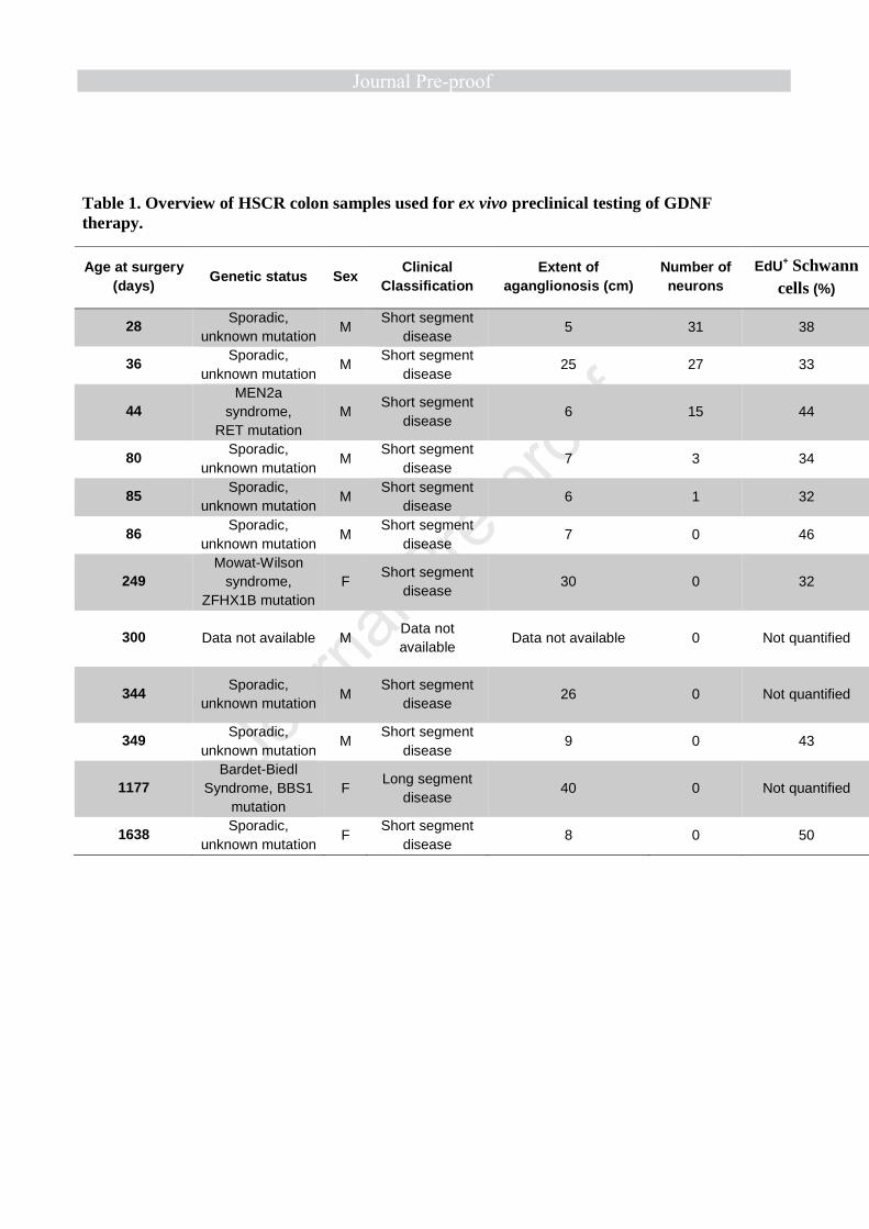
Table 1. Overview of HSCR colon samples used for ex vivo preclinical testing of GDNF therapy.
Age at surgery (days)
Genetic status Sex Clinical
Classification Extent of
aganglionosis (cm) Number of neurons
EdU+ Schwann cells (%)
28 Sporadic,
unknown mutation M
Short segment disease
5 31 38
36 Sporadic,
unknown mutation M
Short segment disease
25 27 33
44 MEN2a
syndrome, RET mutation
M Short segment
disease 6 15 44
80 Sporadic,
unknown mutation M
Short segment disease
7 3 34
85 Sporadic,
unknown mutation M
Short segment disease
6 1 32
86 Sporadic,
unknown mutation M
Short segment disease
7 0 46
249 Mowat-Wilson
syndrome, ZFHX1B mutation
F Short segment
disease 30 0 32
300 Data not available M Data not available
Data not available 0 Not quantified
344 Sporadic,
unknown mutation M
Short segment disease
26 0 Not quantified
349 Sporadic,
unknown mutation M
Short segment disease
9 0 43
1177
Bardet-Biedl Syndrome, BBS1
mutation
F Long segment
disease 40 0 Not quantified
1638 Sporadic,
unknown mutation F
Short segment disease
8 0 50


Supplementary Methods
Mice. Holstein (Tg[Sox3-GFP,Tyr]HolNpln), TashT (Tg[SRY-YFP,Tyr]TashTNpln), and G4-RFP
(Gata4p[5kb]-RFP) lines were as previously described (all maintained on a FVB/N genetic background)1-3,
whereas Piebald-lethal (Ednrbs-l; JAX stock # 000308; C3H/HeJ-C57BL/6 mixed background) and Dhh-Cre
(Tg[Dhh-cre]1Mejr; JAX stock # 012929; FVB/N background) were obtained from The Jackson Laboratory.
Other mouse lines used were R26[Floxed Stop]YFP (Gt[ROSA]26Sortm1(EYFP)Cos; provided by F. Costantini
(Columbia University, USA) and maintained on a FVB/N background)4, RetTGM (here referred to as Ret-null;
Rettm1Jmi; maintained on a C57BL/6 background)5, and Ret9 (Rettm2(RET)Jmi; provided by S. Jain (Washington
University School of Medicine, USA) and maintained on a 129X1/Sv1 background)6.
For mycophenolate mofetil treatments7, timed pregnancies were set up by mating Ret+/- with Ret9/+ or Ret9/9
mice, considering noon of the day of plug detection as E0.5. At E7.5, the drinking water of pregnant dams
was replaced with 0.25X PBS adjusted to pH3.6 with added prodrug mycophenolate mofetil (Accord
Healthcare, NDC Cat. # 16729-094) at varying concentrations (250 µg/mL, 187.5 µg/mL, and 125 µg/mL).
Dams remained on mycophenolate mofetil-supplemented drinking water from E7.5 to E18.5.
GDNF enemas were administered using a 24-gauge gavage needle (Fine Science Tools, Canada) attached to
a micropipette. The head of the gavage needle was introduced in the rectum just beyond the anus, and
enemas were injected over the course of a few seconds. Pups were then placed back with their mother, and
either sacrificed for tissue analysis (at age indicated in relevant figure legends) or checked daily to track
survival. Apart from GDNF, other tested molecules (each at 10µg in 10µl PBS) included the serotonin
receptor (5-HT4R) agonist RS67506 (R&D Systems, Cat. # 0990), Noggin (Sigma, Cat. # SRP4675),
endothelin-3 (Sigma, Cat. # E9137), serotonin (Sigma, Cat. # H9523), and L-ascorbic acid (Sigma, Cat. #
A4403).
Tissue labelling and imaging. For immunofluorescence staining, whole microdissected tissues were
permeabilized for 2 hours in blocking solution (10% FBS and 1% Triton X-100, in PBS) before being
sequentially incubated with specific primary (at 4°C overnight) and relevant secondary (at room temperature
for 2 hours) antibodies, both diluted in blocking solution that was also used to wash tissues between all steps.
All antibodies and dilution factors, are listed in Table S2. EdU was detected using the Invitrogen Click-iT
EdU Imaging Kit (ThermoFisher Scientific, Cat. # C10337) in accordance with the manufacturer’s
instructions. For histological analyses, cross-sections of full-thickness bowel tissues were stained with
hematoxylin and eosin (H&E) as previously described8.
All immunofluorescence images were acquired with either a 20X or a 60X objective on a confocal
microscope (either Nikon A1R or Zeiss 710), with the exception of H&E-stained sections that were imaged
with a 10X objective using an Infinity-2 camera (Lumenera Corporation) mounted on a Leica DM 2000
microscope (Leica Microsystems Canada). Image analysis was performed with ImageJ, using the “Subtract
background” function to decrease non-specific background signal, the “Multi-point” function for cell
counting, and the “Polygon selection” function for calculation of surface area.
Western blot analysis. Organs dissolved in RIPA buffer (containing 1X Roche Complete protease inhibitors)
were sonicated on ice and centrifuged at 14,000 rpm for 15 minutes at 4°C, keeping the supernatants for
western blot analysis. Equal volumes of samples were electrophoretically separated in an 18% sodium
dodecyl sulfate-polyacrylamide gel (SDS-PAGE) and transferred to Immun-blot® PVDF membranes (Bio-
Rad, Cat. # 1620177). Membranes were subsequently incubated in blocking solution (5% skimmed milk and
0.1% Tween 20, in TBS), followed by incubation with either mouse anti-GDNF (Santa Cruz Biotechnology,
Cat. # sc-13147; 1:500 dilution factor) or rabbit anti-αTubulin (Abcam, Cat. # ab176560; 1:70 000 dilution
factor) primary antibodies, and then relevant horseradish peroxydase-conjugated secondary antibodies, all
diluted in blocking solution. Each incubation was for 60 min at room temperature, each time interspersed by
3 washes with blocking solution. Proteins were finally visualized using Immobilon western
chemiluminescent HRP substrate (Millipore Sigma, Cat. # WBKLS0050) and Fusion FX imaging system
(Vilber).
Ex vivo culture for time-lapse imaging. Muscle strips were cultured in suspension as previously described
for time-lapse imaging of embryonic guts3, in DMEM/F12 medium (Wisent, Cat. # 319-085-CL)
supplemented with 10% FBS and 100IU/ml antibiotic-antimycotic with or without 5 µg/ml GDNF under
standard culture conditions (37°C, 5% CO2). After 72h of culture, each petri dish was placed in a microscope
incubation chamber (Okolab) for 10 hours under the same culture conditions, and image stacks (250μm-
thick) of RFP-labelled extrinsic nerves and Schwann cells were acquired every 10 min, using a 20X objective
on a Nikon A1R confocal unit as previously described3.
In vivo and ex vivo analysis of colonic motility. For bead latency test, mice were anesthetized with 2%
isoflurane and a 2mm glass bead (Sigma, Cat. # 1.04014) was inserted into the distal colon with a probe over
a distance of 0.5 cm from the anus. Each mouse was then isolated in its cage without access to food and
water, and monitored for the time required to expel the glass bead. For ex vivo analysis of contractility,
muscle strips were initially stretched with a preload of 2 g of tension for 60 min, and contraction/relaxation
of longitudinal muscles was then continuously recorded with a myograph (Narco Biosystems Inc., Model F-
60) coupled to a computer equipped with the BIOPAC student Lab 4.0.2 software (BIOPAC Systems Inc.).
Electrical field stimulation (EFS) was applied with a voltage stimulator (BIOPAC Systems Inc., Model BSL
MP36/35) connected to electrodes, using parameters that activate enteric neurons without directly activating
muscles (12 V, 20 Hz, 10s train duration, and 300µs stimulus pulse duration). This procedure was repeated 3
times, with 10 min washout periods between stimulations. To characterize the nitrergic and cholinergic
components of EFS-induced contractile responses, N-nitro-L-arginine methyl ester (L-NAME; Sigma, Cat. #
N5751) and atropine (Sigma, Cat. # A01132) were added to Krebs solution at a final concentration of 0.5µM
and 1µM, respectively. The area under the curve (AUC) was measured during each EFS-induced response,
and data were expressed in ΔAUC (corresponding to the difference between the AUC measured 20s after
stimulation minus the AUC measured 20s before stimulation).
Ex vivo analysis of paracellular permeability. Each Ussing chamber contained 5ml of DMEM/F12 medium
(Wisent, Cat. # 319-085-CL), which was maintained at 37ºC and continuously oxygenated (95% O2 / 5%

CO2). After a 30 min equilibration period, 200µl of apical medium was replaced with 200µl of a 1mg/ml
solution of FITC-conjugated dextran 4 kDa (FD4; Sigma, Cat. # 60842-46-8). Fluorescence intensity of
basolateral aliquots of 150ul, reflecting paracellular transit from the luminal surface, was then measured
every 30 min over a period of 3 hours, using a fluorimeter (TECAN, Model Infinite M1000). Fluorescence
intensity was finally converted in amount of FD4 by comparison to a standard curve, and the average value
for the 3 hour period was used to calculate paracellular permeability, which was expressed in ng of FD4 per
surface of mucosa area per min (ng/cm2/min).
Microbiome analysis. Mice were sacrificed at P20 and their feces were directly collected from the colon (3
fecal pellets per mouse). Bacterial DNA was then extracted using the QIAamp® Fast DNA Stool Mini Kit
(QIAGEN, Cat. # 51604), and the V5-V6 region of the 16S rRNA gene was PCR amplified with a collection
of previously described barcoded primers9. Raw sequences generated with an Illumina MiSeq sequencer
were paired and processed using the MOTHUR pipeline10, and the BIOM package11 was subsequently used
to transfer .biom files into R12 for generating graphs of relative taxa abundance and beta diversity.
Supplemental References
1. Bergeron KF, Cardinal T, Toure AM, et al. Male-Biased Aganglionic Megacolon in the TashT Mouse Line Due to Perturbation of Silencer Elements in a Large Gene Desert of Chromosome 10. PLoS Genet 2015;11:e1005093.
2. Pilon N, Raiwet D, Viger RS, et al. Novel pre- and post-gastrulation expression of Gata4 within cells of the inner cell mass and migratory neural crest cells. Dev Dyn 2008;237:1133-43.
3. Soret R, Mennetrey M, Bergeron KF, et al. A collagen VI-dependent pathogenic mechanism for Hirschsprung's disease. J Clin Invest 2015;125:4483-96.
4. Srinivas S, Watanabe T, Lin CS, et al. Cre reporter strains produced by targeted insertion of EYFP and ECFP into the ROSA26 locus. BMC Dev Biol 2001;1:4.
5. Enomoto H, Crawford PA, Gorodinsky A, et al. RET signaling is essential for migration, axonal growth and axon guidance of developing sympathetic neurons. Development 2001;128:3963-74.
6. Jain S, Naughton CK, Yang M, et al. Mice expressing a dominant-negative Ret mutation phenocopy human Hirschsprung disease and delineate a direct role of Ret in spermatogenesis. Development 2004;131:5503-13.
7. Lake JI, Tusheva OA, Graham BL, et al. Hirschsprung-like disease is exacerbated by reduced de novo GMP synthesis. J Clin Invest 2013;123:4875-87.
8. Boulende Sab A, Bouchard MF, Beland M, et al. An Ebox element in the proximal Gata4 promoter is required for Gata4 expression in vivo. PLoS ONE 2011;6:e29038.
9. Laforest-Lapointe I, Paquette A, Messier C, et al. Leaf bacterial diversity mediates plant diversity and ecosystem function relationships. Nature 2017;546:145-147.
10. Schloss PD, Westcott SL, Ryabin T, et al. Introducing mothur: open-source, platform-independent, community-supported software for describing and comparing microbial communities. Appl Environ Microbiol 2009;75:7537-41.
11. McDonald D, Clemente JC, Kuczynski J, et al. The Biological Observation Matrix (BIOM) format or: how I learned to stop worrying and love the ome-ome. Gigascience 2012;1:7.
12. Team RC. R: A language and environment for statistical computing. R Foundation for Statistical Computing, Vienna, Austria. 2014.
13. Chalazonitis A, D'Autreaux F, Guha U, et al. Bone morphogenetic protein-2 and -4 limit the number of enteric neurons but promote development of a TrkC-expressing neurotrophin-3-dependent subset. J Neurosci 2004;24:4266-82.
14. Nagy N, Goldstein AM. Endothelin-3 regulates neural crest cell proliferation and differentiation in the hindgut enteric nervous system. Dev Biol 2006;293:203-17.
15. Liu MT, Kuan YH, Wang J, et al. 5-HT4 receptor-mediated neuroprotection and neurogenesis in the enteric nervous system of adult mice. J Neurosci 2009;29:9683-99.
16. Fiorica-Howells E, Maroteaux L, Gershon MD. Serotonin and the 5-HT(2B) receptor in the development of enteric neurons. J Neurosci 2000;20:294-305.
17. Fattahi F, Steinbeck JA, Kriks S, et al. Deriving human ENS lineages for cell therapy and drug discovery in Hirschsprung disease. Nature 2016.
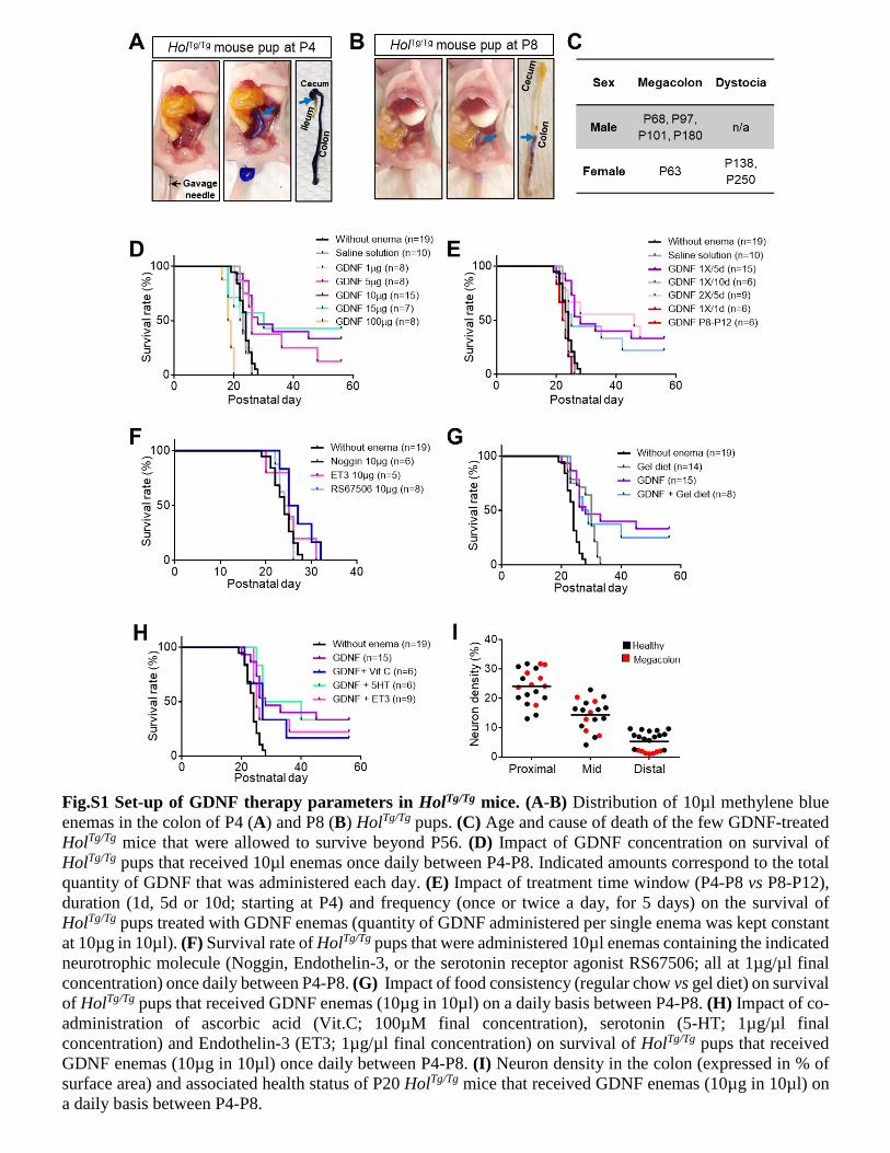
Fig.S1 Set-up of GDNF therapy parameters in HolTg/Tg mice. (A-B) Distribution of 10µl methylene blue enemas in the colon of P4 (A) and P8 (B) HolTg/Tg pups. (C) Age and cause of death of the few GDNF-treated HolTg/Tg mice that were allowed to survive beyond P56. (D) Impact of GDNF concentration on survival of HolTg/Tg pups that received 10µl enemas once daily between P4-P8. Indicated amounts correspond to the total quantity of GDNF that was administered each day. (E) Impact of treatment time window (P4-P8 vs P8-P12), duration (1d, 5d or 10d; starting at P4) and frequency (once or twice a day, for 5 days) on the survival of HolTg/Tg pups treated with GDNF enemas (quantity of GDNF administered per single enema was kept constant at 10µg in 10µl). (F) Survival rate of HolTg/Tg pups that were administered 10µl enemas containing the indicated neurotrophic molecule (Noggin, Endothelin-3, or the serotonin receptor agonist RS67506; all at 1µg/µl final concentration) once daily between P4-P8. (G) Impact of food consistency (regular chow vs gel diet) on survival of HolTg/Tg pups that received GDNF enemas (10µg in 10µl) on a daily basis between P4-P8. (H) Impact of co-administration of ascorbic acid (Vit.C; 100µM final concentration), serotonin (5-HT; 1µg/µl final concentration) and Endothelin-3 (ET3; 1µg/µl final concentration) on survival of HolTg/Tg pups that received GDNF enemas (10µg in 10µl) once daily between P4-P8. (I) Neuron density in the colon (expressed in % of surface area) and associated health status of P20 HolTg/Tg mice that received GDNF enemas (10µg in 10µl) on a daily basis between P4-P8.
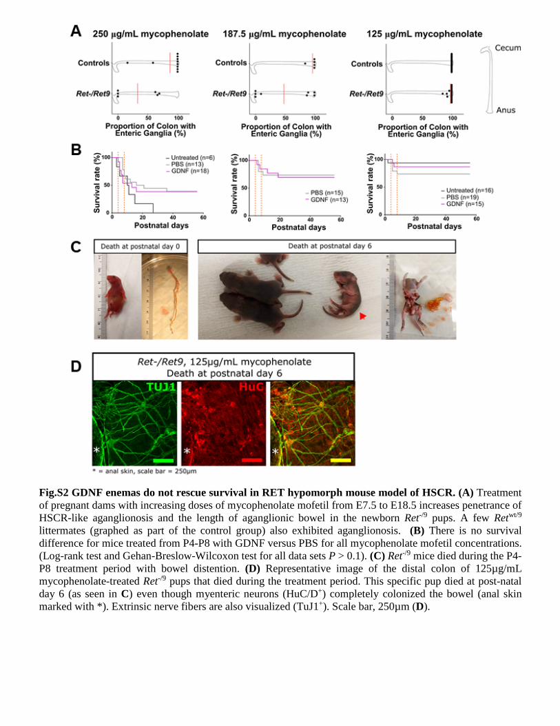
Fig.S2 GDNF enemas do not rescue survival in RET hypomorph mouse model of HSCR. (A) Treatment of pregnant dams with increasing doses of mycophenolate mofetil from E7.5 to E18.5 increases penetrance of HSCR-like aganglionosis and the length of aganglionic bowel in the newborn Ret-/9 pups. A few Retwt/9 littermates (graphed as part of the control group) also exhibited aganglionosis. (B) There is no survival difference for mice treated from P4-P8 with GDNF versus PBS for all mycophenolate mofetil concentrations. (Log-rank test and Gehan-Breslow-Wilcoxon test for all data sets P > 0.1). (C) Ret-/9 mice died during the P4-P8 treatment period with bowel distention. (D) Representative image of the distal colon of 125µg/mL mycophenolate-treated Ret-/9 pups that died during the treatment period. This specific pup died at post-natal day 6 (as seen in C) even though myenteric neurons (HuC/D+) completely colonized the bowel (anal skin marked with *). Extrinsic nerve fibers are also visualized (TuJ1+). Scale bar, 250µm (D).

Fig.S3 Analysis of myenteric ganglion size and neuronal density in the colon of P20 HolTg/Tg and TashTTg/Tg mice that were treated or not with GDNF between P4-P8. (A) Analysis of myenteric ganglion size in HolTg/Tg mice. (B) Analysis of neuronal density in TashTTg/Tg mice. The average neuronal density (color-coded) is indicated for each colon sub-region (represented by cylinders) along the length of the colon. Neuronal density is expressed as the percentage of area occupied by HuC/D+ cells in a single focal plane at the level of the myenteric plexus within the bowel wall (n=6 mice per group; 3 fields of view per sub-region). For each distal colon subregion, the neuronal density is also given as a numerical value. (C) Analysis of myenteric ganglion size in TashTTg/Tg mice.
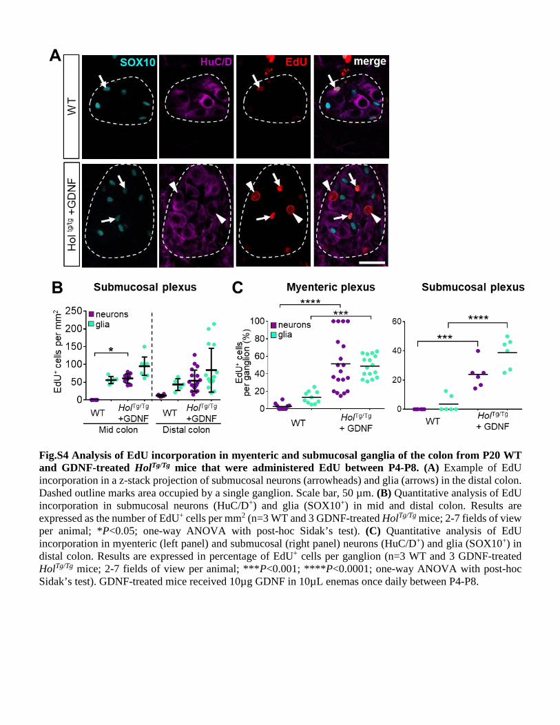
Fig.S4 Analysis of EdU incorporation in myenteric and submucosal ganglia of the colon from P20 WT and GDNF-treated HolTg/Tg mice that were administered EdU between P4-P8. (A) Example of EdU incorporation in a z-stack projection of submucosal neurons (arrowheads) and glia (arrows) in the distal colon. Dashed outline marks area occupied by a single ganglion. Scale bar, 50 µm. (B) Quantitative analysis of EdU incorporation in submucosal neurons (HuC/D+) and glia (SOX10+) in mid and distal colon. Results are expressed as the number of EdU+ cells per mm2 (n=3 WT and 3 GDNF-treated HolTg/Tg mice; 2-7 fields of view per animal; *P<0.05; one-way ANOVA with post-hoc Sidak’s test). (C) Quantitative analysis of EdU incorporation in myenteric (left panel) and submucosal (right panel) neurons (HuC/D+) and glia (SOX10+) in distal colon. Results are expressed in percentage of EdU+ cells per ganglion (n=3 WT and 3 GDNF-treated HolTg/Tg mice; 2-7 fields of view per animal; ***P<0.001; ****P<0.0001; one-way ANOVA with post-hoc Sidak’s test). GDNF-treated mice received 10µg GDNF in 10µL enemas once daily between P4-P8.
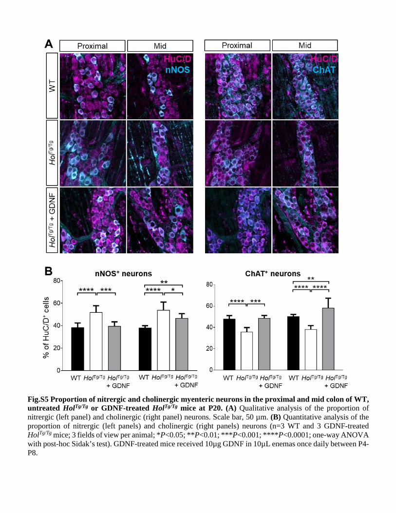
Fig.S5 Proportion of nitrergic and cholinergic myenteric neurons in the proximal and mid colon of WT, untreated HolTg/Tg or GDNF-treated HolTg/Tg mice at P20. (A) Qualitative analysis of the proportion of nitrergic (left panel) and cholinergic (right panel) neurons. Scale bar, 50 µm. (B) Quantitative analysis of the proportion of nitrergic (left panels) and cholinergic (right panels) neurons (n=3 WT and 3 GDNF-treated HolTg/Tg mice; 3 fields of view per animal; *P<0.05; **P<0.01; ***P<0.001; ****P<0.0001; one-way ANOVA with post-hoc Sidak’s test). GDNF-treated mice received 10µg GDNF in 10µL enemas once daily between P4-P8.

Fig.S6 Supporting information for in vivo and ex vivo analyses of motility in the distal colon of WT, untreated HolTg/Tg or GDNF-treated HolTg/Tg mice at P20. (A) Correlation between neuron density in distal colon and time for bead expulsion in GDNF-treated HolTg/Tg mice at P20 (in support of Fig.3D). (B) Examples of electric field-stimulated and drug-modulated patterns of longitudinal smooth muscle contraction-relaxation in an organ bath equipped with a force transducer (in support of Fig.3E). In responsive tissues, electric field stimulation (EFS) triggers contractions of colonic muscles that can be slightly increased by L-NAME-mediated inhibition of nitrergic signaling, and robustly counteracted by atropine-mediated inhibition of cholinergic signaling. GDNF-treated mice received 10µg GDNF in 10µL enemas once daily between P4-P8.
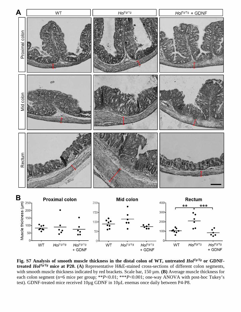
Fig. S7 Analysis of smooth muscle thickness in the distal colon of WT, untreated HolTg/Tg or GDNF-treated HolTg/Tg mice at P20. (A) Representative H&E-stained cross-sections of different colon segments, with smooth muscle thickness indicated by red brackets. Scale bar, 150 µm. (B) Average muscle thickness for each colon segment (n=6 mice per group; **P<0.01; ***P<0.001; one-way ANOVA with post-hoc Tukey’s test). GDNF-treated mice received 10µg GDNF in 10µL enemas once daily between P4-P8.
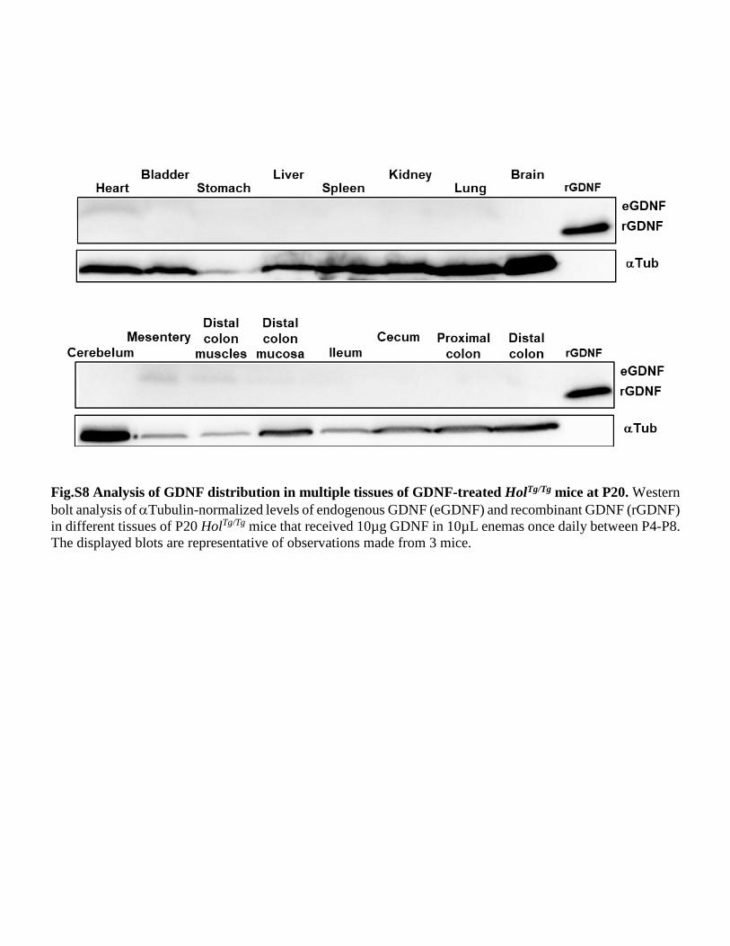
Fig.S8 Analysis of GDNF distribution in multiple tissues of GDNF-treated HolTg/Tg mice at P20. Western bolt analysis of αTubulin-normalized levels of endogenous GDNF (eGDNF) and recombinant GDNF (rGDNF) in different tissues of P20 HolTg/Tg mice that received 10µg GDNF in 10µL enemas once daily between P4-P8. The displayed blots are representative of observations made from 3 mice.

Fig.S9 Time-course analysis of HisGDNF distribution and RET expression in colonic smooth muscles of P4-P8 HolTg/Tg mice treated with HisGDNF. Immunofluorescence analysis of HisGDNF distribution and RET expression in distal colon muscularis of HisGDNF-treated HolTg/Tg mice. White arrowheads point to RET+ neurons that also stain positive for HisGDNF. All images show a single focal plane representative of observations made from 3 mice. Scale bar, 20µm.
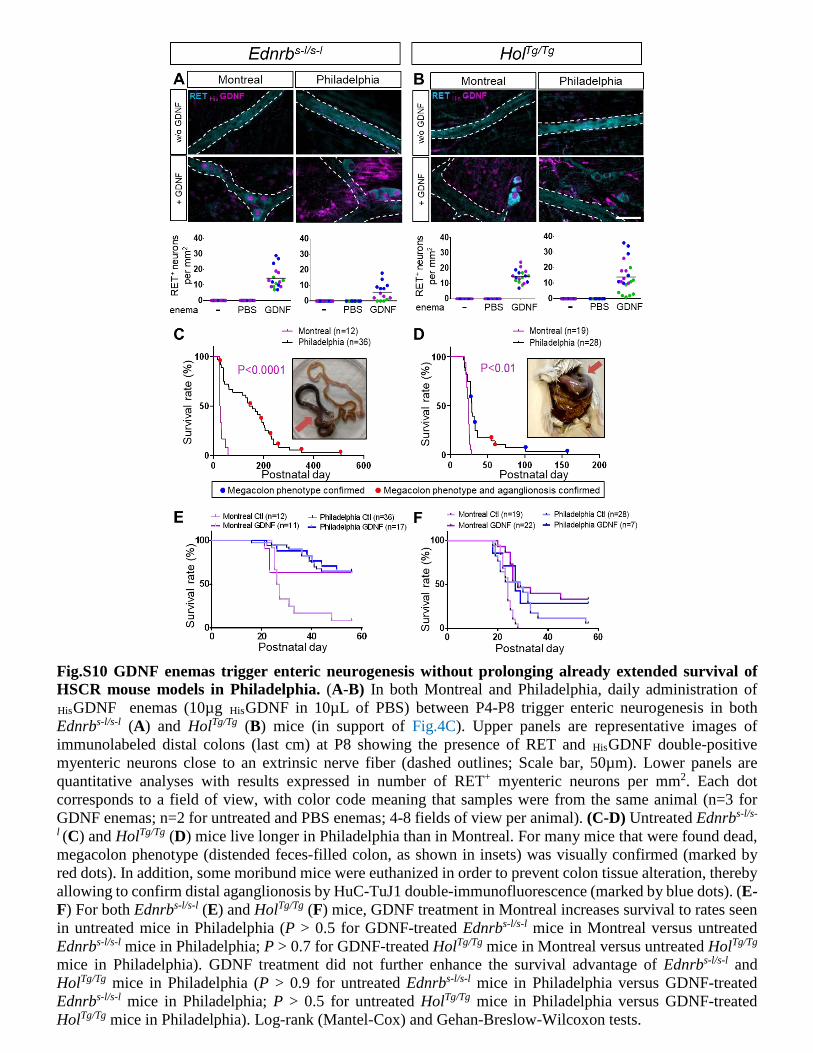
Fig.S10 GDNF enemas trigger enteric neurogenesis without prolonging already extended survival of HSCR mouse models in Philadelphia. (A-B) In both Montreal and Philadelphia, daily administration of HisGDNF enemas (10µg HisGDNF in 10µL of PBS) between P4-P8 trigger enteric neurogenesis in both Ednrbs-l/s-l (A) and HolTg/Tg (B) mice (in support of Fig.4C). Upper panels are representative images of immunolabeled distal colons (last cm) at P8 showing the presence of RET and HisGDNF double-positive myenteric neurons close to an extrinsic nerve fiber (dashed outlines; Scale bar, 50µm). Lower panels are quantitative analyses with results expressed in number of RET+ myenteric neurons per mm2. Each dot corresponds to a field of view, with color code meaning that samples were from the same animal (n=3 for GDNF enemas; n=2 for untreated and PBS enemas; 4-8 fields of view per animal). (C-D) Untreated Ednrbs-l/s-
l (C) and HolTg/Tg (D) mice live longer in Philadelphia than in Montreal. For many mice that were found dead, megacolon phenotype (distended feces-filled colon, as shown in insets) was visually confirmed (marked by red dots). In addition, some moribund mice were euthanized in order to prevent colon tissue alteration, thereby allowing to confirm distal aganglionosis by HuC-TuJ1 double-immunofluorescence (marked by blue dots). (E-F) For both Ednrbs-l/s-l (E) and HolTg/Tg (F) mice, GDNF treatment in Montreal increases survival to rates seen in untreated mice in Philadelphia (P > 0.5 for GDNF-treated Ednrbs-l/s-l mice in Montreal versus untreated Ednrbs-l/s-l mice in Philadelphia; P > 0.7 for GDNF-treated HolTg/Tg mice in Montreal versus untreated HolTg/Tg mice in Philadelphia). GDNF treatment did not further enhance the survival advantage of Ednrbs-l/s-l and HolTg/Tg mice in Philadelphia (P > 0.9 for untreated Ednrbs-l/s-l mice in Philadelphia versus GDNF-treated Ednrbs-l/s-l mice in Philadelphia; P > 0.5 for untreated HolTg/Tg mice in Philadelphia versus GDNF-treated HolTg/Tg mice in Philadelphia). Log-rank (Mantel-Cox) and Gehan-Breslow-Wilcoxon tests.
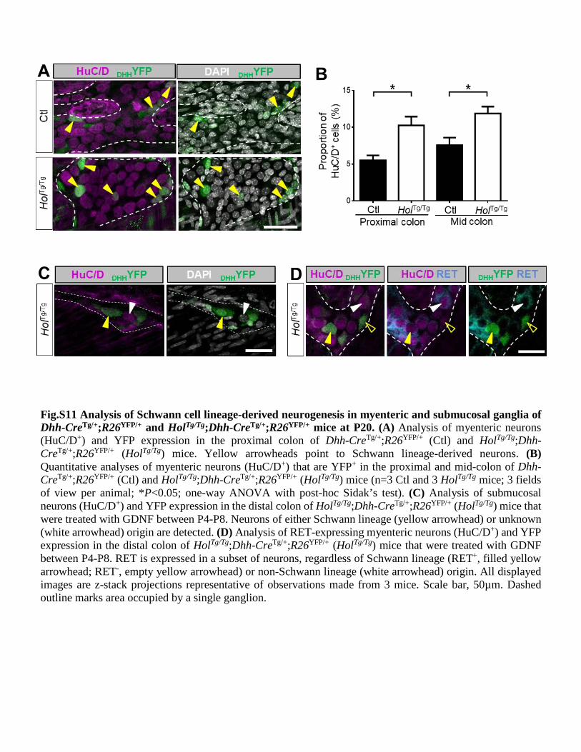
Fig.S11 Analysis of Schwann cell lineage-derived neurogenesis in myenteric and submucosal ganglia of Dhh-CreTg/+;R26YFP/+ and HolTg/Tg;Dhh-CreTg/+;R26YFP/+ mice at P20. (A) Analysis of myenteric neurons (HuC/D+) and YFP expression in the proximal colon of Dhh-CreTg/+;R26YFP/+ (Ctl) and HolTg/Tg;Dhh-CreTg/+;R26YFP/+ (HolTg/Tg) mice. Yellow arrowheads point to Schwann lineage-derived neurons. (B) Quantitative analyses of myenteric neurons (HuC/D+) that are YFP+ in the proximal and mid-colon of Dhh-CreTg/+;R26YFP/+ (Ctl) and HolTg/Tg;Dhh-CreTg/+;R26YFP/+ (HolTg/Tg) mice (n=3 Ctl and 3 HolTg/Tg mice; 3 fields of view per animal; *P<0.05; one-way ANOVA with post-hoc Sidak’s test). (C) Analysis of submucosal neurons (HuC/D+) and YFP expression in the distal colon of HolTg/Tg;Dhh-CreTg/+;R26YFP/+ (HolTg/Tg) mice that were treated with GDNF between P4-P8. Neurons of either Schwann lineage (yellow arrowhead) or unknown (white arrowhead) origin are detected. (D) Analysis of RET-expressing myenteric neurons (HuC/D+) and YFP expression in the distal colon of HolTg/Tg;Dhh-CreTg/+;R26YFP/+ (HolTg/Tg) mice that were treated with GDNF between P4-P8. RET is expressed in a subset of neurons, regardless of Schwann lineage (RET+, filled yellow arrowhead; RET-, empty yellow arrowhead) or non-Schwann lineage (white arrowhead) origin. All displayed images are z-stack projections representative of observations made from 3 mice. Scale bar, 50µm. Dashed outline marks area occupied by a single ganglion.
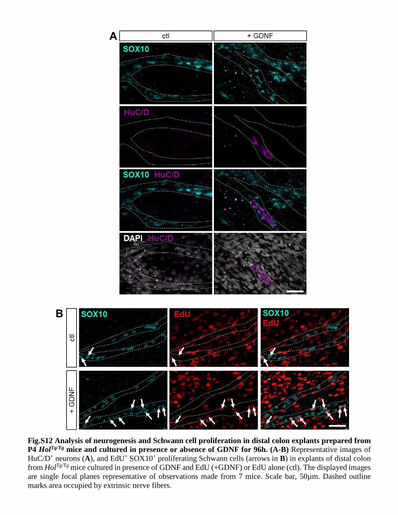
Fig.S12 Analysis of neurogenesis and Schwann cell proliferation in distal colon explants prepared from P4 HolTg/Tg mice and cultured in presence or absence of GDNF for 96h. (A-B) Representative images of HuC/D+ neurons (A), and EdU+ SOX10+ proliferating Schwann cells (arrows in B) in explants of distal colon from HolTg/Tg mice cultured in presence of GDNF and EdU (+GDNF) or EdU alone (ctl). The displayed images are single focal planes representative of observations made from 7 mice. Scale bar, 50µm. Dashed outline marks area occupied by extrinsic nerve fibers.
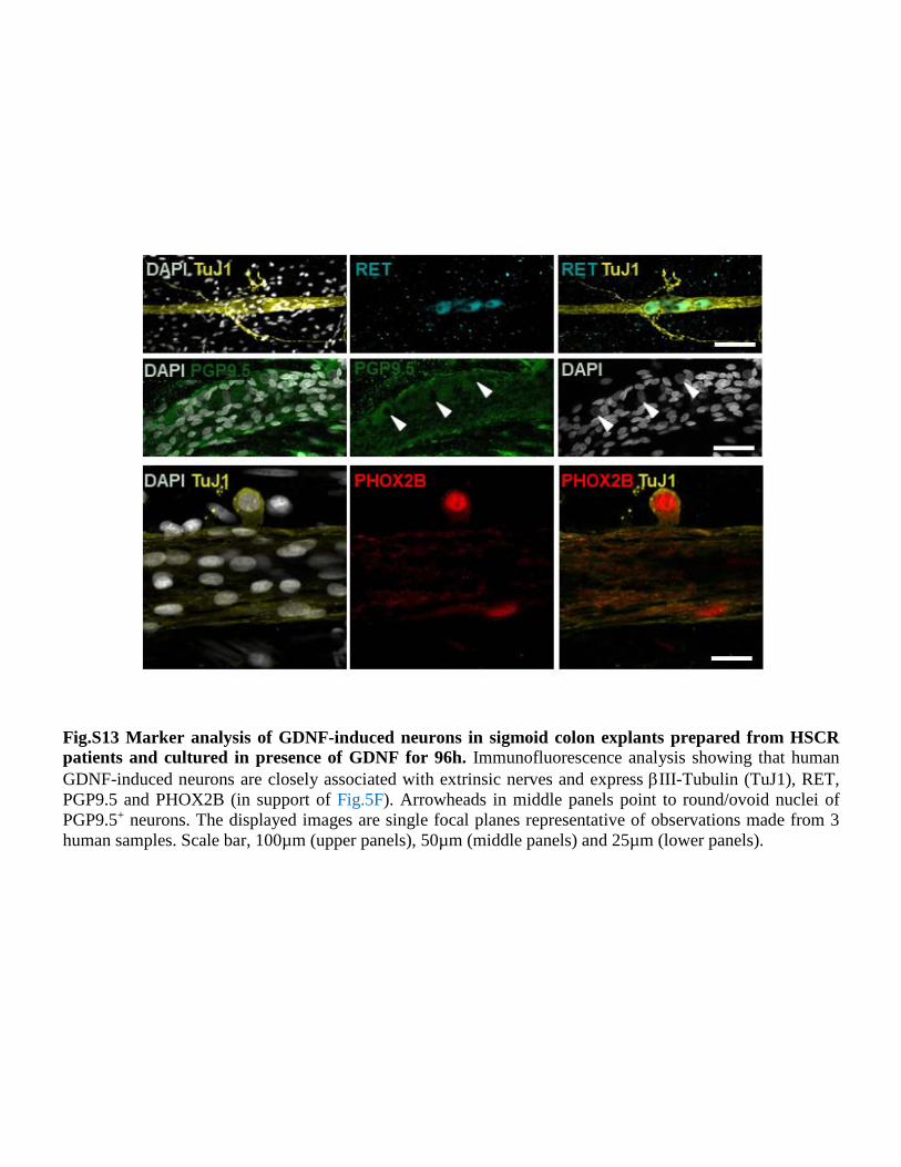
Fig.S13 Marker analysis of GDNF-induced neurons in sigmoid colon explants prepared from HSCR patients and cultured in presence of GDNF for 96h. Immunofluorescence analysis showing that human GDNF-induced neurons are closely associated with extrinsic nerves and express βIII-Tubulin (TuJ1), RET, PGP9.5 and PHOX2B (in support of Fig.5F). Arrowheads in middle panels point to round/ovoid nuclei of PGP9.5+ neurons. The displayed images are single focal planes representative of observations made from 3 human samples. Scale bar, 100µm (upper panels), 50µm (middle panels) and 25µm (lower panels).
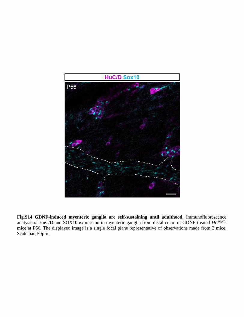
Fig.S14 GDNF-induced myenteric ganglia are self-sustaining until adulthood. Immunofluorescence analysis of HuC/D and SOX10 expression in myenteric ganglia from distal colon of GDNF-treated HolTg/Tg mice at P56. The displayed image is a single focal plane representative of observations made from 3 mice. Scale bar, 50µm.
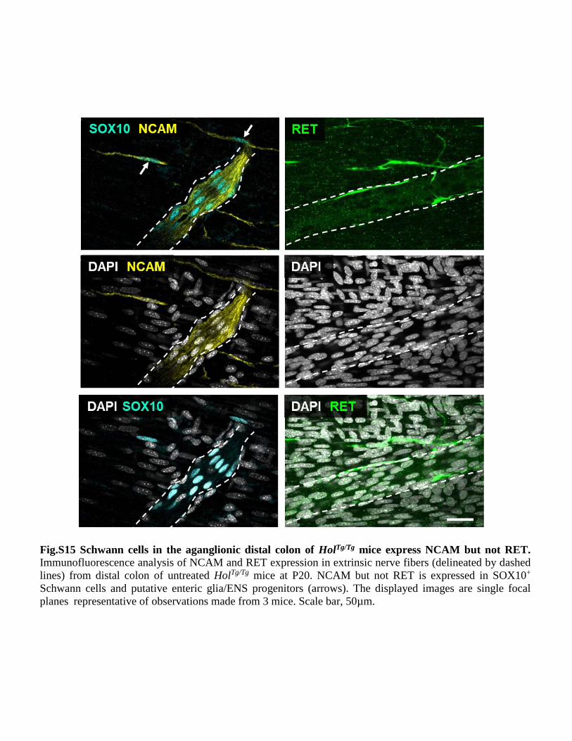
Fig.S15 Schwann cells in the aganglionic distal colon of HolTg/Tg mice express NCAM but not RET. Immunofluorescence analysis of NCAM and RET expression in extrinsic nerve fibers (delineated by dashed lines) from distal colon of untreated HolTg/Tg mice at P20. NCAM but not RET is expressed in SOX10+ Schwann cells and putative enteric glia/ENS progenitors (arrows). The displayed images are single focal planes representative of observations made from 3 mice. Scale bar, 50µm.

Fig.S16 Schwann cells in the aganglionic distal colon of HolTg/Tg;Dhh-CreTg/+;R26YFP/+ mice are not all YFP-labeled. Immunofluorescence analysis of SOX10 and YFP expression in an extrinsic nerve fiber from distal colon of untreated HolTg/Tg;Dhh-CreTg/+;R26YFP/+mice at P8. A subset of SOX10+ Schwann cells are negative for YFP (arrows). The displayed images are single focal planes representative of observations made from 3 mice. Scale bar, 50µm.

Table S1. Rationale for selection of supplemental neurotrophic molecules Molecule Reason for selecting Reference
Noggin NSE promoter-driven Noggin overexpression increases enteric neuron numbers in transgenic mice
Chalazonitis et al., J Neurosci 2004 13
Endothelin-3 Ex vivo culture of avian hindgut in presence of endothelin-3 increases ENS density
Nagy and Goldstein, Dev Biol 2006 14
RS67506 Systemic administration of the serotonin receptor (5-HT4R) agonist RS67506 triggers enteric neurogenesis in adult mice
Liu et al., J Neurosci 2009 15
Serotonin Serotonin (5-HT) enhances in vitro neuronal differentiation of murine enteric neural precursors
Fiorica-Howells et al., J Neurosci 2000 16
Vitamin C Ascorbic acid is needed for in vitro differentiation and maturation of human enteric neural precursors
Fattahi et al., Nature 2016 17
Table S2. List of primary antibodies and dilution factors used for immunofluorescence
Antibody Source Catalog number RRID reference Dilution
6X His R&D systems MAB050 AB_357353 1:500
CalR Swant, CG1 AB_10000342 1:500
ChAT Millipore AB144P AB_2079751 1:100 GFP Abcam Ab290 AB_303395 1:500
HuC/D Molecular Probes A-21271 AB_221448 1:500 HuC/D (ANNA-1) Gift from Vanda Lennon --- AB_2313944 1:2000
Ki67 Abcam ab15580 AB_443209 1:500
NCAM Abcam ab5032 AB_2291692 1:500
NOS1 Santa Cruz Biotechnology sc-648 AB_630935 1:200 PHOX2B R&D systems AF4940 --- 1:250
RET R&D systems MAB718 AB_2232594 1:500
SOX10 Santa Cruz Biotechnology sc-17342 AB_2195374 1:200 SubP Abcam ab67006 AB_1143173 1:500
TH Abcam ab137869 --- 1:500
TUJ1 Covance MRB-435P AB_663339 1:1000 VIP Abcam ab8556 AB_306628 1:500