Embed Size (px)
Citation preview

Pediatrics Grand Rounds 7 May 2010
University of Texas Health Science Center at San Antonio
1
Pediatric Drug EruptionsJohn C. Browning, MD, FAAD, FAAP
Assistant Professor of Pediatrics & Dermatology
• I do not have any relevant relationships with industry.
• Disclosure: I’m older than I look.
1963: College 1967: Medical School
1971: Residency 2009: FacultyBotox, Cosmetic Treatments

Pediatrics Grand Rounds 7 May 2010
University of Texas Health Science Center at San Antonio
2
Objectives
• Understand the pathogenesis of pediatric drug eruptions
• Develop an organized approach to pediatric drug eruptions
• Know when treatment for a drug eruption is indicated
“The desire to take medicine is perhaps the greatest feature which distinguishes man from animals.”
Sir William Osler
What are drug eruptions?
• Better thought of as “drug‐induced” skin diseases
• Names are not universal
• Most are idiosyncratic– Specific to the individual
– Unpredictable
– Hard to study
– Immunologic component
– Not explained by dose alone
Drug Eruptions
• Common– Morbilliform– Urticarial
• Less Common: – Fixed drug, lichenoid, photodrug, vasculitis
• Severe/Less Common– AGEP (acute generalized exanthematous pustulosis)– DRESS (Drug rash with eosinophilia and systemic symptoms)
– Stevens‐Johnson Syndrome/TEN
• 2.5% of children treated with a drug experience a drug reaction
• 12% of children treated with an antibiotic will experience a drug reaction
Timing
• < 2‐weeks– URTICARIA
– AGEP
– MORBILLIFORM DRUG
– LINEAR IgA
• > 2‐weeks– STEVENS‐JOHNSON/TEN
– DRESS

Pediatrics Grand Rounds 7 May 2010
University of Texas Health Science Center at San Antonio
3
R.A.S.H.From Neil Shear “Drug Reactions” presented at 2007 Dermatology Foundation Annual Meeting
• R = Remember– History, timing of eruption in relation to drug administration, previous exposure
• A = Appearance– Exanthematous, Pustular, Bullous, urticarial
• S = Systemic symptoms– Fever, lymphadenopathy, hepatitis, arthritis
• H = Histology (when needed)
Risk Factors
• Female gender (1.5:1)
• Increasing age
• Polypharmacy
• Slow acetylator genotype
• Immunosuppresion (HIV)
• NSAIDS, antibiotics, anticonvulsants, long‐term meds
Risk Factors
• HLA DR4 drug‐induced pemphigus
• HLA B7 insulin allergy
• HLA B22 fixed drug eruption
Pathogenesis
• Drugs or their metabolites act as haptens
• Haptens cell‐mediated or humoral response
• Non‐immunologic mechanisms– cumulative toxicity
– overdose
– drug‐drug interactions
– alterations in metabolism
– direct mast cell degranulation
Immune‐mediated Drug Reactions
• IgE-dependent drug reactions (Type I): urticaria, angioedema and anaphylaxis.
• Cytotoxic drug-induced reactions (Type II): pemphigus, petechiae due to drug-induced thrombocytopenia.
• Immune complex-dependent drug reactions (Type III): vasculitis, serum sickness, urticarial vasculitis.
• Delayed-type, cell-mediated drug reactions (Type IV): morbilliform, fixed, and lichenoid drug eruptions, DRESS, Stevens-Johnson syndrome (SJS), and TEN.
“There is no more difficult art to acquire than the art of observation, and for some men it is quite as difficult to record an observation in brief and plain language.”
Sir William Osler

Pediatrics Grand Rounds 7 May 2010
University of Texas Health Science Center at San Antonio
4
Urticarial Drug Eruption
• Common • Type I hypersensitivity (IgE‐mediated)• IgE antibodies form upon first exposure to drug; Re‐exposure leads to mast cell degranulationwith histamine release and development of urticaria
• Frequently due to penicillin and occurs within minutes of exposure
• RAST test available for penicillin
Urticaria: Characteristics
• Very pruritic• Can be anywhere on the body, including palms, soles, and scalp
• May be figurate an polycyclic with central pallor
• Individual lesions should last < 24 hours (migratory)
• Biopsy: Nonspecific

Pediatrics Grand Rounds 7 May 2010
University of Texas Health Science Center at San Antonio
5
Urticarial Vasculitis
• Persistent urticaria (individual lesions lasting > 24 hours) or purpura may be a sign of vasculitis and/or serum sickness
• Urticarial vasculitis: Involves underlying antigen‐antibody formation with deposition of immune complexes (composed of antibodies directed against drug‐related haptens) within post‐capillary venules.
• PCN, ACE inhibitors, sulfa, fluoxetine, thiazides
Urticarial Vasculitis
Urticaria: treatment
• Discontinue offending drug
• Antihistamines
• May consider systemic corticosteroids
• EpiPen when severe
• Topicals have little benefit
Serum Sickness‐like Reaction
• Fever, rash, arthralgias occuring 1‐3 weeks after start of drug
• Lymphadenopathy and eosinophilia may be present
• Hypocomplementemia, immune complexes, and vasculitis are absent
• Cefaclor classic example

Pediatrics Grand Rounds 7 May 2010
University of Texas Health Science Center at San Antonio
6
Morbilliform Eruption
• AKA exanthematous reaction, maculopapularrash, “drug rash,” scarlatiniform
• Thought to be a type IV delayed hypersensitivity reaction
• Viral infections may increase the incidence of morbilliform eruptions: classically seen with amoxicillin and mononucleosis
Morbilliform Eruption
• Classically begins 7‐14 days after starting a medication, sooner upon re‐challenge
• Blanching, erythematous macules that coalesce to form patches over the entire body, may have some urticarial‐looking papules
• Often pruritic, can progress to erythroderma with desquamation
• Dependent surfaces seem to have greater involvement• Biopsy: Nonspecific changes consisting of a mild
perivascular lymphocytic infiltrate and a few necrotic keratinocytes within the epidermis.
Treatment
• Remove offending drug when possible
• Okay to “treat through” the rash if drug is medically necessary
• Antihistamines, topical steroids, Sarna
Copyright ©2007 American Academy of Pediatrics
Segal, A. R. et al. Pediatrics 2007;120:e1082-e1096

Pediatrics Grand Rounds 7 May 2010
University of Texas Health Science Center at San Antonio
7
Copyright ©2007 American Academy of Pediatrics
Segal, A. R. et al. Pediatrics 2007;120:e1082-e1096
Morbilliform reaction to amoxicillin
Viral Rash
• Symmetric, blanching erythema
• May have prodrome or concurrent fever, malaise with rash
• Commonly seen with enteroviruses, EBV, HHV‐6, HHV‐7, parvovirus B‐19, and respiratory viruses

Pediatrics Grand Rounds 7 May 2010
University of Texas Health Science Center at San Antonio
8
Unilateral Laterothoracic Exanthem
When an exanthematouseruption is associated with fever, lymphadenopathy, and/or edema
of the face, the possibility of DRESS must be considered and
an evaluation for systemic involvement conducted.
DRESS
• Drug Rash with Eosinophilia and Systemic Symptoms
• AKA anticonvulsant hypersensitivity reaction (aromatics: carbamazepine, phenobarbitol, phenytoin) or drug hypersensitivity reaction
• Looks like a morbilliform drug eruption with fever and hand/facial edema but can also look like SJS or TEN
• Biopsy: Non‐specific
DRESS
• Lab findings– Elevated LFT’s– Elevated eosinophils (30% of the time): may be a late finding
– Elevated TSH several months after initial reaction– Elevated HHV‐6 and other HHV titers (sometimes)
• Treatment– Remove offending drug (absolute necessity)– Systemic corticosteroids with slow taper– Alert family: increased risk with sibs and first degree relatives
• 10 year‐old girl
• Vancomycin IV x 4 weeks for osteomyelitis
• h/o rash and fever x 2 days
• Seen in ER
• Elevated AST/ALT

Pediatrics Grand Rounds 7 May 2010
University of Texas Health Science Center at San Antonio
9
• 8 year‐old boy
• On carbamazepine x 5 weeks for sz disorder
• Admitted to hospital with fever and rash
• Elevated AST/ALT
• Treated initially for Kawasaki
Kawasaki Disease
• Fever > 5 days + 4 out of 5 of the following:
• Bilateral non‐purulent conjunctival injection
• Oropharyngeal changes (strawberry tongue, cracked lips, hyperemia)
• Cervical lymphadenopathy
• Peripheral extremity changes (erythema, edema, desquamation)
• Polymorphous exanthem

Pediatrics Grand Rounds 7 May 2010
University of Texas Health Science Center at San Antonio
10
• 6 year‐old male
• h/o multiple medical problems including CHD s/p repair and seizure disorder
• Admitted for fever (low‐grade x 5 days) and 102 x 1 day
• Meds: Keppra & Topamax(> 1 year), Cefzil x 4 months
Scarlet Fever
• Sore throat, headache, malaise, chills, anorexia, nausea and high fevers
• Rash begins 12‐48 hours later sunburn with goose bumps, feels like sandpaper
• Pastia’s lines
• White/red strawberry tongue
• Tender cervical adenopathy
• Superficial desquamation hands/feet

Pediatrics Grand Rounds 7 May 2010
University of Texas Health Science Center at San Antonio
11
Photodrug Reactions
• Photoirritant: sufficient amount of drug + ultraviolet light
• Exaggerated sunburn appearance
• NSAIDS, fluoroquinolones, tetracyclines, topical retinoids
• Methotrexate sunburn recall reaction when given 1‐3 days after sun exposure (not prevented by leucovorin rescue)
Phototoxic reaction in a patient taking methotexate

Pediatrics Grand Rounds 7 May 2010
University of Texas Health Science Center at San Antonio
12
Voriconazole Reaction
Photodrug Reactions
• Photoallergic reactions: allergen activated or produced by the effect of light on a drug with resultant cell‐mediated hypersensitivity
• UV radiation required to convert the drug into an immunologically active compound (photoallergen) that induces the immune response
• Idiosyncratic, often low levels of UV light can trigger the reaction
• Eczematous appearance in sun‐exposed areas• Griseofulvin, statins, celecoxib

Pediatrics Grand Rounds 7 May 2010
University of Texas Health Science Center at San Antonio
13
Lichenoid Drug
• May occur on sun‐exposed or non sun‐exposed areas
• Photo lichenoid drug: only on sun‐exposed areas
• Flat‐topped pruritic, purple papules• Most commonly due to ACE inhibitors, antimalarials, and gold
• Biopsy lichen planus
Gold Photolichenoid drug eruption due to hydrochlorothiazide.Figure 22‐4, Bolognia 2nd Edition
Leukocytoclastic Vasculitis (LCV)
• Small vessels
• Deposition of immune complexes (composed of antibodies directed against drug‐related haptens) in post‐capillary venulesactivation of compliment neutrophils

Pediatrics Grand Rounds 7 May 2010
University of Texas Health Science Center at San Antonio
14
Leukocytoclastic Vasculitis (LCV)
• Non‐blanching purpuric papules, primarily on the lower extremities
• R/O renal and GI disease• Occurs 7‐21 days after starting new med• NSAIDS, antibiotics, anticonvulsants• Treatment: discontinue drug, systemic steroids only if with renal or GI involvement
Erythromycin Penicillin
Fixed Drug Eruption
• One or a few sharply demarcated erythematousor edematous plaques with dusky centers – may be associated with burning or stinging
• Lesions develop 1‐2 weeks after 1st exposure, within 24 hours of subsequent exposures
• Upon re‐administration, lesions recur at exactly the same site
• Men > Women

Pediatrics Grand Rounds 7 May 2010
University of Texas Health Science Center at San Antonio
15
Fixed Drug Eruption
• May occur anywhere on the body but favor the lips, face, hands, feet, and genitalia
• Fade over several days leaving PIH
• Generalized fixed drug eruption may be difficult to distinguish from erythema multiforme
• Seen with sulfonamides, barbiturates, tetracyclines, carbamazepine, NSAIDs, pseudoephedrine, and phenolphthalein

Pediatrics Grand Rounds 7 May 2010
University of Texas Health Science Center at San Antonio
16
Acute Generalized Exanthematous Pustulosis(AGEP)
• Acute febrile drug eruption with neutrophilia• Clinically and histologically looks like pustularpsoriasis
• Numerous small, non‐follicular sterile pustules arising within areas of edematous erythema start on face or intertriginous areas and disseminate over hours
• Seen with antibiotics (Β−lactams, macrolides), calcium channel blockers, and others
Acute Generalized Exanthematous Pustulosis(AGEP)
• Time between drug administration and onset of rash is usually less than 2 days
• Lesions last 1‐2 weeks after drug is discontinued, followed by superficial desquamation
• Bx: pustular psoriasis• Tx: withdraw offending drug, topical steroids, antipyretics

Pediatrics Grand Rounds 7 May 2010
University of Texas Health Science Center at San Antonio
17
Stevens‐Johnson Syndrome/TEN
• < 10% of the body = Stevens‐Johnson Syndrome (SJS)
• > 30 % of the body = toxic epidermal necrolysis (TEN)
• 10‐30% = SJS/TEN overlap
• Occurs 7‐21 days after start of drug
Pediatrics Grand Rounds 7 May 2010
University of Texas Health Science Center at San Antonio
18
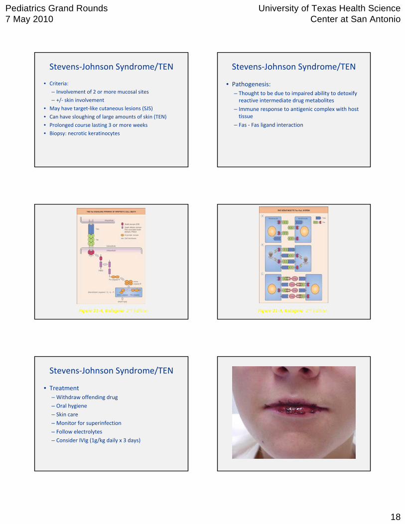
Stevens‐Johnson Syndrome/TEN
• Criteria:
– Involvement of 2 or more mucosal sites
– +/‐ skin involvement
• May have target‐like cutaneous lesions (SJS)
• Can have sloughing of large amounts of skin (TEN)
• Prolonged course lasting 3 or more weeks
• Biopsy: necrotic keratinocytes
Stevens‐Johnson Syndrome/TEN
• Pathogenesis:– Thought to be due to impaired ability to detoxify reactive intermediate drug metabolites
– Immune response to antigenic complex with host tissue
– Fas ‐ Fas ligand interaction
Figure 21‐4, Bolognia 2nd Edition Figure 21‐4, Bolognia 2nd Edition
Stevens‐Johnson Syndrome/TEN
• Treatment– Withdraw offending drug
– Oral hygiene
– Skin care
– Monitor for superinfection
– Follow electrolytes
– Consider IVIg (1g/kg daily x 3 days)

Pediatrics Grand Rounds 7 May 2010
University of Texas Health Science Center at San Antonio
19
Pediatr Dermatol. 2007 Jul‐Aug;24(4):E22‐5.
Bronchiolitis obliterans: a rare chronic pulmonary complication associated with Stevens‐Johnson syndrome.
Pulmonary manifestations are well recognized during the acute phase of Stevens‐Johnson syndrome but persistent pulmonary sequela is rarely reported. We report two boys with bronchiolitis obliterans following the acute phase of Stevens‐Johnson syndrome and discuss the clinical picture and treatment of persistent pulmonary complications with reference to earlier reports.

Pediatrics Grand Rounds 7 May 2010
University of Texas Health Science Center at San Antonio
20
EM: phenytoin
Linear IgA
• Can look like SJS, BP, EM, or DH
• IgA along basement membrane zone
• Occurs within days of starting the drug
• Antibodies to COL7, BPAg1(230kda)
• Vancomycin, lithium, diclofenac, piroxicam, rifampin

Pediatrics Grand Rounds 7 May 2010
University of Texas Health Science Center at San Antonio
21
Drug‐induced lupus
• Rare cutaneous findings
• Can have photosensitivity (NOT malar rash), E. nodosum, and vasculitis
• Arthralgias, fever, weight loss, cough
• ANA and antihistone antibodies
• Minocycline‐induced lupus occurs after 2years of therapy

Pediatrics Grand Rounds 7 May 2010
University of Texas Health Science Center at San Antonio
22
Drug‐induced SCLE
• Papulosquamous annular plaques in a generalized distribution
• Circulating ANA and anti‐Ro (SS‐A) antibodies
• Seen with thiazide diuretics, calcium channel blockers, ACE inhibitors, terbinafine, TNF‐alpha blockers
• 4‐20 wks and even years after start of drug
“One of the first duties of the physician is to educate the masses not to take medicine.”
Sir William Osler
References
• “Drug Reactions,” Presented by Dr. Neil Shear, Dermatology Foundation Annual Meeting, March 2007.
• Dermatology. J Bolognia, J Jorizzo, R Rapini, eds. 2nd Edition
• Segal et al. Cutaneous reactions to drugs in children. Pediatrics 2007;120(4):e1082‐96.
• Nigen et al. Drug eruptions: approaching the diagnosis of drug‐induced skin diseases. J Drugs Derm
The End