Embed Size (px)
Citation preview

COLLAPSE OF THE SEED FOLLOWING THE MATING OF HORDEUM JUBATUMXSECALE CEREALE
D. C. COOPER AND R. A. BRINK University of Wisconsin
Received December 7, 1943
INTRODUCTION
H E impairmint in seed development often associated with wide crossing
the endosperm as a storage tissue and the large size of the normal caryopsis. Cytogeneticists interested in the cereals were early impressed, accordingly, with the importance of this stage in the life cycle of the plant for the general problem of hybrid incompatibility. It is not surprising to find, therefme, that a larger amount of work has been directed toward discovering the basis of hybrid seed failure in these plants than in any other group of angiosperms.
Some of the histological findings on the Gramineae as put forward in their original form, however, are seemingly a t variance with more recent evidence on seed collapse as manifested in certain Dicotyledons. Additional data are desirable, therefore, on the question whether seed failure following hybridiza- tion of distantly related forms follows a common basic pattern or differs sig- nificantly from group to group.
T is readily observable in cereal species hybrids because of the persistence of
MATERIALS AND METHODS
The present study of seed failure in the Hordeum jubatum (squirrel-tail barley) 9 XSecaZe cerede (rye) 8 mating was planned along the lines of our earlier investigations of the crosses Nicotiana rustica XN. glutinosa (COOPER and BRINK 1940) and N . rzcsticaXN. tabacum (BRINK and COOPER 1941). The histological methods employed are described in these earlier papers. Locally occurring plants of H . jubatum (n= 14) were employed. The rye (n= 7) usedas the male parent in the intergeneric cross was of the Imperial variety as cul- tivated commercially in Wisconsin.
H. jubatum and S . cerede cross readily when the former is used as the pis- tillate parent, as QUINCKE (1940) has reported; and the hybrid seeds promptly begin to enlarge. Development continues for varying periods of time. Some seeds break down as early as four days; none has been found alive beyond 13 days. No seeds reach a germinable condition. The cross, therefore, is com- pletely abortive. On the other hand, the normally autogamous H. jubatum, used as the control in these experiments, sets an approximately full comple- ment of plump, viable seeds on artificial self-pollination. Development of the H. jubatumXS. cerede and H. jubatum, selfed, seeds was followed histologically from fertilization to the time of collapse of the hybrid.
Paper No. 326 from the Department of Genetics, Agricultural Experiment Station, Um- VERSITY of WISCONSIN. The writers desire to acknowledge the support provided by a grant-in-aid from the ROCICE~ELLER FOUNDATION.
GENETICS rg: 370 July 1944
COLLAPSE OF BARLEYXRYE HYBRID SEED
2
4
37 f
3
FIG. r.-Hordeum jubatum. Longitudinal section through an ovary showing a mature mega- gametophyte. X32.
FIG. 2.-H. jubatum. Longitudinal section through an ovary four hours after pollination. Fertilization has taken place and the primary endosperm nucleus is in a stage of mitosis. X32.
FIG. 3.-H. jubatum. Longitudinal section of a seed at 28 hours. The nuclei of the endosperm are scattered in the peripheral layer of dense cytoplasm. Large vacuoles occupy the central region of the endosperm, The antipodal cells are large and highly vacuolate. X32.
FIG. 4.-H. jubatumXSecale cerede. Longitudinal section of a seed at 28 hours. Four giant endosperm nuclei are located in the dense cytoplasm between the antipodals and the embryo. The cytoplasm of the antipodal cells is dense and many cells are binucleate. X32.

372 The findings on the H . jubatum XS. cereale cross are in agreement with those
of earlier investigators in showing that mitotic behavior of the endosperm in cereal hybrids may be markedly abnormal. This condition has not been ob- served in any Dicotyledonous mating thus far reported; and the fact, a t first sight, may seem to preclude the possibility of bringing the known cases of seed failure under one point of view. Evidence is forthcoming from the present study, however, that the seeming non-conformity of endosperm behavior in cereal hybrid seeds rests upon a special relation of the antipodals to early development. This aspect of the problem will be discussed in detail in a fol- lowing paper.
D. C. COOPER AND R. A. BRINK
ENDOSPERM DEVELOPMENT I N H. JUBATUM
Fertilization usually occurs within four hours after self-pollination in H. jubatum and is delayed little, if any, in the cross with S. cereale. The frequency with which fertilization takes place following hand-pollination of castrated flowers is high in both matings.
Development of the ET. jubatum endosperm proceeds with great rapidity following fertilization. Stages in division of the primary nucleus are often found in four-hour ovules (fig. 2). Occasionally the endosperm is already two- nucleate a t this time. Further growth continues with a high degree of regular- ity by synchronous free nuclear division up to about 3 2 hours, at which time cell division is initiated; and a t 48 hours a layer of cells completely surrounds the large central lumen (fig. 5 ) . The increase in number of free endosperm nu- clei proceeds geometrically as shown by the data in table I on modal number of nuclei a t successive times during this interval. The modal number of endo- sperm nuclei a t eight and a t ten hours is four. The value has increased to 32 a t 24 hours, the next period a t which observations on selfed H . jubatum seeds were made. Further increases to 64 nuclei a t 28 hours and to 128 nuclei a t 32 hours are found. A few seeds were observed a t 32 hours with a 256-nucleate endosperm. Cell formation is initiated in that portion of the 128-nucleate endosperm adjacent to the embryo and gradually progresses toward the anti- podal region. The endosperm is almost completely cellular a t the 256-nucleate stage, a few free nuclei remaining in the thicker layer of dense cytoplasm immediately adjacent to the antipodals.
The volume of the initial endosperm cell increases approximately six-fold prior to the division of the primary endosperm nucleus. This increase occurs in all dimensions but is greatest a t the chalazal end where this cell expands beyond the antipodals altering the position of the latter from terminal (fig. I) to median lateral (fig. 2). The increase in volume of the endosperm during the period of free-nuclear division is likewise great. Some of the nuclei become distributed peripherally in the thin layer of cytoplasm surrounding the highly vacuolate central region. The remainder are embedded in the denser cytoplasm between the embryo and the antipodals (fig. 3).
The endosperm growth is accompanied by an enlargement of the seed as a whole and a progressive absorption of the nucellus. These changes are il-
COLLAPSE OF BARLEYXRYE HYBRTD SEED 373 lustrated in figure 3 which shows a longitudinal section of a H. jubatum seed a t 2 8 hours.
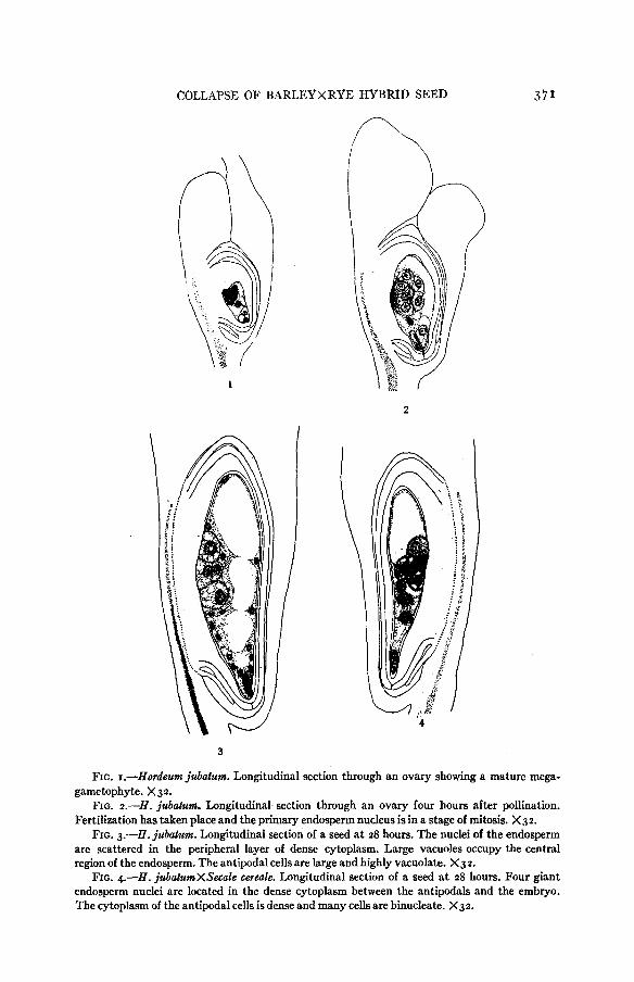
The cellular endosperm in a normal H . jubatum seed a t 48 hours is illustrated in figure 5 . A t this stage the tissue consists of a single layer of cells, except in
5 6
FIG. 5--H. jubatum. Longitudinal section of a young caryopsis a t 48 hours. The cellular endo- sperm surrounds a large central lumen. The apical cells of the nucellus are being digested leaving a cavity between it and the antipodals. X33.
FIG. 6.-H. ju6elumXS. cerede. Longitudinal section of a young caryopsis at 48 hours. Nuclei of varying sizes are present in the multinucleate endosperm. The antipodal cells are large and there isno evidence of digestion of the nucellus. X33.
the pocket region adjacent to the embryo which is multilayered, surrounding a large, elongated, somewhat flattened, central cavity. Further growth occurs by cell division. The lumen of the endosperm becomes entirely filled with large, highly vacuolate cells by three days (fig. 7). Initial stages of food storage are

3 74 D. C. COOPER AND R. A. BNNIC present a t 8 2 hours and a large amount of starch is to be found in the central region of the endosperm a t the four-day interval. By this time the peripheral cells have become differentiated as the aleurone layer. The aleurone cells are smaller than those of the main mass of the endosperm (6g. 8) and are highly vacuolate. The endosperm cells immediately opposite the vascular tissue of the ovary are exceptional. They are densely cytoplasmic and have the appearance of absorbing cells (fig. 9.) Active cell division continues and the endosperm in-
10
9
FIGS. 7 to 10.-H. jubatum. Stages in the development of the endosperm. FIG. 7.-Transverse section through the mid-region of developing caryopsis a t three days.
FIG. &-Highly vacuolate aleurone cells of a fourday endosperm. X 199. FIG. 9.-Epidermal cells of a four-day endosperm from a region opposite the raphe. These
cells are densely cytoplasmic and have the appearance of absorbing cells. X 199. FIG. Io.-Median transverse section of a caryopsis a t five days. The epidermal cells of the
endosperm opposite the raphe are elongate. A viscous substance is present in the space between these cells and the remains of the nucellus. X33.
creases in size (fig. IO). The highly vacuolate cells of the aleurone layer have become densely cytoplasmic and are covered with a thick cuticle a t seven days (fig. 13). The outer layer of endosperm cells opposite the vascular tissue in- creases in size, becomes highly vacuolate and by seven days is becoming packed with storage materials. It does not become cutinized.
x33.
ENDOSPERM DEVELOPMENT IN THE HYBRID SEED
Behavior of the hybrid, endosperm stands in marked contrast to that just outlined for the normal H . jubatum seed. The differences may be summarized as follows: (I) division of the primary endosperm nucleus is somewhat de- layed; (2) mitotic division becomes disorderly, giving rise to irregular numbers

COLLAPSE OF BARLEYXRYE HYBRID SEED 375 of nuclei which vary widely in size and form; and (3) the endosperm fails to become cellular even though it remains alive well beyond the time a t which its counterpart in the H . jubatum seed attains this condition.
The delay in division of the hybrid primary endosperm nucleus is evident from the following facts. A t four hours, two out of eight N. jubatum seeds ex- amined already had binucleate endosperms, whereas the endosperm in all five hybrid seeds observed were still uninucleate. A t six hours, three selfs had ad-
TABLE I
Number of endosperm nuclei in H . jubatum and H . jubatumXS. cerede seeds.
H . jubatum SELFED H . jubalumXS. cerede
TIWE IN NO. ENDOSPERM NUCLEI MEAN NO.
HOURS NO. OF NO. OF SEEDS SEEDS
ENDOSPERM
MODE MEAN NUCLEI
4 6 8
14 16 18
24 28
32
38 42
48 52
56 62
IO
21
8 9 13 I2
I 1.2
2 2.7
4 3.4 4 5.7
32 32.0
64 57.3 I 28 ' 57 .1
cellular
5 6
' 3 5 6
I1
I2
10
9 9 16 8 4 6 4 9 6
I .o
1 . 3
2.9
2.8
2.8
6.5 6.2 7.6 9.5
17.5 31.0
13.0
28.8
2.0
2.2
67.5
43.1
vanced to the four-nucleate condition, six were binucleate. Four out of six hybrid endosperms observed a t this stage were uninucleate, the other two being binucleate. Nine in 13 H . jubatum endosperms were four-nucleate, the others being binucleate a t eight hours. A t this time, five hybrid endosperms in a total of 11 examined still possessed a single nucleus.
The further increase in number of nuclei in the hybrid endosperms is shown in the right hand column in table I. Since the modal number of nuclei becomes progressively less well defined as age of the seeds advances the modal values have little significance and are not given. The means, however, show that in number of nuclei present after a given interval of time the hybrid endosperms fall behind those of H . jubatum, selfed.
Consideration of number of nuclei alone, however, gives a very incomplete picture of the character of the hybrid endosperms. Due to gross disturbances in mitotic behavior the nuclei also become highly variable in size and shape.

376 D. C. COOPER AND R. A. BRINK
In table 2 is shown the proportion of hybrid seeds a t successive 12-hour intervals up to 62 hours whose endosperms contain nuclei obviously abnormal in size. It will be noted that the frequency increases from 17 percent during the initial 12-hour period following pollination to IOO percent during the last in- terval (14 hours) a t which observations were taken, ending a t 62 hours. That
TABLE 2
Profortion of hybrid seeds having endosperms containing nuclei of a b n o r d size.
NUhfBER OF SEEDS
PERCENTAGE TIME INTERVAL
IN HOURS ENDOSPERM NUCLEI ABNORMAL TOTAL
ABNORMAL -
0-1 2
13-24 25-36 3 7-48 49-62
35 42 25 I8 I9
6 I9 16
I9 11
I7 45 64 61
I O 0
is to say, all the hybrid endosperms, sooner or later, become abnormal in this respect.
The diversity in number, shape and size of nuclei arising in the hybrid endo- sperms is very great. Figure 4, based on a 28-hour seed, and figure 6, showing a 48-hour seed, illustrate how wide the departure may be from the normal condition a t these times (4. figs. 3 and 5 ) . All the endosperm nuclei shown in figure 4 are excessively large and instead of being distributed peripherally throughout the whole endosperm they all lie between the embryo and the antipodals. A more uniform distribution is seen in figure 6, but here the nuclei in the vicinity of the embryo are excessively large in contrast with those in the distal portion of the tissue. One nucleus in the former group is dumbbell- shaped. Variously malformed nuclei are frequent.
Abnormal mitotic behavior sometimes occurs in division of the primary endosperm nucleus. An anaphase of such a first division is shown in figure 14. It will be noted that due to several chromosomes bridging the spindle the separation of the two daughter groups of chromosomes is not complete. Such chromosome bridges may lead to dumbbell-shaped interphase nuclei like that shown in figure 15. These compound nuclei may then enter another division cycle (fig. 16) during which a greatly increased number of chromosomes ap- pears. Giant nuclei (fig. 17) thus arise. Figure 18 shows a metaphase plate from a 52-hour endosperm on which 105 chromosomes are distributed. This number is three times the value (35) expected in the endosperm nucleus of a hybrid between H. jubatum (n=14) and S . cereale (n=7). The increase in chromosome number in the abnormal hybrid nuclei, however, does not always proceed in multiple series. Aneuploid numbers also arise frequently as a result of various kinds of mitotic irregularities. These nuclear disturbances are

COLLAPSE OF BARLEYXRYE HYBRID SEED 377 cumulative and, as stated above, eventually appear in the endosperms of all the hybrid seeds.
EMBRYO GROWTH
Development of the young R. jubatumXS. cereale embryo is comparatively regular in sharp contrast to the frequently radical derangement of the accom- panying endosperm. Rate of embryo growth up to 96 hours, in comparison with that of the normal H . jubatum, may be seen from the data given in table 3 and plotted in text figure I. Most of the hybrid zygotes divide between 18
TIME IN HOUR5 TEXT FIG. I.-Early growth of the embryo following the matings H . jubatum, selfed (con-
tinuous line) and H . jubatumXS. cerede (broken line).
and 24 hours. This is apparently a few hours later than in B. jubatum for which, however, the data are less complete over this period. At 24 hours the average number of cells in the embryo of the H . jubatum seed is four as against 1.8 for the hybrid. From this time forward to 96 hours the hybrid embryos attain a given size about one day later than in the control.
When compared with the development of the normal embryo (fig. 11) dif- ferentiation of the hybrid embryo appears to be almost normal, as illustrated in figure 12. There is a tendency from four days onward for the hybrid em- bryos to become somewhat more elongated than the controls probably as a result of less compression in this direction due to the absence of a cellular endosperm.
It is interesting to note also that up to 62 hours, beyond which time the necessary data were not taken, there seems to be little relationship between size of the embryo and the nuclear candition of the endosperm so long as the latter

3 78 D. C. COOPER AND R. A. BRINX
tissue is still alive. For example, a t 56 hours, four of the nine seeds recorded in table 3 for that time had 8-celled embryos. The endosperms associated with these embryos showed the following nuclear conditions, respectively: 32 large; 4 large, 8 small; 42, large and small; 48, large and small; 2 very large. The endosperms in three seeds with Io-celled embryos possessed nuclei as
TABLE 3
Number of cells in H . jubatum and H . jubatumXS. cerede hybrid embryos.
H . jubatum, SELFED El. jubatumX.9. ceteale TIhUC IN
HOURS NO. OF MEAN NO. CELLS NO. OF MEAN NO. CELLS
SEEDS IN EMBRYO SEEDS IN EMBRYO
4 6 8
I4 16 IS
24 28 32 38 42 48 5 2
56 62
72
IO
21
8 I
9 I
'3 I I -
5 I 6 I I1 I
I3 5 6
I2
IO 1 . 7 9 1.8 9 2 . 7
8 3.5
6 5 . 5
16 2.3 ,
4 3.5
4 9 9 9.1 6 I7 I1 35 5 113 - - 96
follows: 8 large, 16 small; 4 large, 8 small; 8 large. One seed with a 12-celled embryo possessed about 80 large and small endosperm nuclei. Possibly beyond this period the number and kind of nuclei present in the endosperm may be- come critical for the growth of the embryo but in the earliest stages of seed development, a t least, this is not the case.
BEHAVIOR OF THE ANTIPODALS
The antipodals are a prominent constituent of the mature embryo sac of H . jubatum, as in other species of Gramineae. The number of antipodals in H . jubatum a t fertilization is usually 15, and they occupy about one-quarter of the space in the embryo sac. The tissue is situated initially a t the chalazal end of the sac (fig. I), but it soon becomes lateral as a result of growth of the endosperm beyond it (fig. 2). In the latter position the antipodals lie on the funicular side of the ovule opposite the vascular bundle. They are, therefore, in the path which nutrient materials would be expected to follow in entering the endosperm.

COLLAPSE OF BARLEYXRYE HYBRID SEED 379
C D
FIG. II.-H. jubutum. Stages in the development of the embryo at A , five; B, six; C, nine and D, 11 days respectively. X33.
Parallel with normal fertilization in H . jubatum the antipodals enlarge about six-fold. A proportionate increase in volume of the embryo sac also occurs a t this time. The antipodal nuclei, likewise, increase greatly in size. The enlarged antipodal cells become increasingly vacuolate (figs. I, 2 and 3) and reach their maximum expansion a t about 28 hours. No increase in cell number occurs.
B
C
D
FIG. 12.-H. jubatumXS. cereele. Stage in the development of the embryo at A , five; B , six; C, nine and D, 11 days respectively. X33.

380 D. C. COOPER AND R. A. BRINK
13
14
18
15 -15
20
21 FIG. 13.-8. jubatum. Median transverse section of a portion of the endosperm opposite
the raphe at 7 days. The epidermal cells in the mid-region (e) are thin walled and contain storage

COLLAPSE OF BAmEE'XkE'E HYBRID SEED
From 2 8 hours onward the antipodals shrink steadily until a t 48 hours the tissue is reduced to a flattened group of cells pressed between the nucellus and the now cellular endosperm (fig. 5 ) . Disintegration continues until, a t 7 2
hours, only fragments remain. Following fertilization in the H . jubatum ovule by S. cereale sperm, on the
other hand, the course of development of the antipodals is very different. The rapid, initial enlargement of the cells and nuclei observed in H . jubatum selfed a t fertilization does not occur. In contrast the cytoplasm remains dense, or only small vacuoles appear in a few cells. A correspondingly limited increase in embryo sac size takes place. From this time onward the antipodals enlarge slowly, although a t 28 hours they are still small and dense relative to their counterparts in the normal H . jubatum seed (fig. 4). At this time some cells undergo mitotic division, and this activity continues to about 7 2 hours. Re- gression of the tissue begins some hours later, and by 96 hours breakdown is clearly apparent. Further data on the antipodals are presented in a following paper in which the relationship of this tissue to endosperm behavior is con- sidered in detail.
381
THE IMMEDIATE CAUSE OF SEED COLLAPSE
The hybrid seed collapses promptly following breakdown of the last traces of endosperm. The immediate cause of failure appears to be starvation associ- ated with hypofunction, and, eventually, disintegration of the endosperm. It was pointed out above that the hybrid embryo, although retarded in growth,
materials. The small aleurone cells (a) on either side are densely cytoplasmic and a dense cuticle (c) covers the outer surface. X69.
FIGS. 14-18.-H. jubetumXS. cereale. Stages of mitosis in the endosperm and the formation of giant nuclei.
FIG. 14.-Anaphase of the division of the primary endosperm nucleus with chromosome bridges at 8 hrs. X p 4 .
FIG. rg.-Dumb-bell shaped interphase nucleus a t 8 hrs. X324. FIG. 16.-Prophase stage of division of a dumb-bell shaped nucleus a t 16 hrs. X p 4 . FIG. 17.-Large nucleus in a gz-hour endosperm. X324. FIG. 18.-Polar view of a metaphase with 105 chromosomes. X648. FIG. 19.-H. jzlbetum. Detailed sketch of that portion of embryo outlined in figure 11 d.
The cells are densely cytoplasmic. Granular materials are stored in the uniformly dense cyto- plasm of the epidermal cells. X I 74.
FIG. 20.-H. jubatumXS. cerede. Detailed sketch of that portion of the embryo outlined in figure 12 d. The cells are large and highly vacuolate. There is no evidence of storage materials in the epidermal layer. X 173.
FIG. ~ I . - H . jubatum. Portion of the raphe and adjacent tissues a t 7 days; r-cells of the raphe with thick walls and gelatinous contents; o.w.--ovary wall with vacuolate cells and normal nuclei; int.-integument with transparent cuticle; nuc.-nucellus with normal resting nuclei. x324.
FIG. 22.-H. jubetumXS. cereale. Portion of raphe and adjacent tissues at 7 days; r-the cells of the raphe have thick walls and are filled with a dense gelatinous material in which is em- bedded granular substances; o.w.--ovary wall-the nuclei of the cells opposite the raphe are disintegrating and the cytoplasm is very dense; int.-integument with dense cuticle. The cells are packed with granular materials; nuc.-nucellus with normal resting nuclei. X324.

382 D. C. COOPER AND R. A. BRINK
differentiates in the usual way. Formative processes in this tissue, therefore, are not visibly impaired by the hybridity. There is, however, a conspicuous difference in the nutrition of H . jubatum and H . jubatumXS. cereale embryos. This difference is illustrated in figures 19 and 2 0 which are based upon 11-day old seeds of the two iespective classes.
The stage of development attained by the 11-day H . jubatum embryo repre- sented in figure 11 D and the H . jubatumXS. cereale embryo drawn in figure 12 D is nearly the same, the hybrid being only slightly less advanced. The cellular condition prevailing in these embryos is shown in figures 19 and 2 0 ,
which represent a t an increased magnification a portion of the scutellum indicated by broken lines in figures 11 D and 12 D, respectively. The cyto-
TABLE 4
Frequency of collapsing hybrid seeds at stated intervals following fertilization.
NUMBER OF SEEDS TIME I N DAYS
TOTAL COLLAPSING
PERCENTAGE
COLLAPSING
9 IO I1
I2
I3
16
23 23 35
8 7 7
2 0
I2
IO
0
2
I
0
5 2
2
4 9 - 1 0
0
1 0
4
I 4 25
29 57 75
0
- I O 0
plasm in the scutellar cells of the control embryo is dense and finely vacuolate. The outer layer of cells is especially rich in cytoplasm and contains consider- able food material in granular form. In contrast, the cells in the corresponding region of the hybrid embryo are larger and much more highly vacuolate (fig. 20). The total mass of cytoplasm in these cells appears to be low relative to that in the H . jubatum cells. The cells of the outer layer, likewise, are highly vacuolate and are entirely lacking in granular reserve material. This starved appearance characterizes the entire hybrid embryo.
Undernourishment of the hybrid embryo is doubtless a result of malfunc- tioning of the associated endosperm. Development of the latter tissue is radi- cally disturbed by mitotic irregularities, as described earlier. In spite of its abnormal condition, the hybrid endosperm may continue to support embryo growth for several days. The plane of nutrition declines, however, until the low level represented in figure 2 0 is reached. This is the prelude to collapse of the seed.
Five hybrid seeds were observed, varying in age from four to 11 days, in which cell division in the embryo had ceased although the embryo was still

COLLAPSE OF BAIUEYXRYE HYBRID SEED
intact. The endosperm had disappeared in two of these seeds and comprised a very small amount of tissue in the remaining three. Endosperm activity in the later cases had apparently dropped below the level a t which growth of the embryo can proceed. The endosperms had entirely broken down in 31 other seeds, four to 13 days of age, and the embryos were disintegrating. These seeds and the five mentioned above were collapsing.
383
THE OVARY AND CERTAIN ACCESSORY TISSUES OF THE SEED
The H . jubatum ovary contains a single vascular bundle which enters a t the base and extends about half-way to the apex in the mid-region of the broad, somewhat flattened ventral wall as shown in figure I. The ovule is at- tached to the inner surface of the ovary wall, its base being directly opposite the apex of the vascular bundle. The basal cells of the ovule which constitute the raphe serve to conduct nutrient materials into the developing seed. This conducting tissue is not conspicuously differentiated as such but is continuous with the nucellus on its inner side and with the ovary wall outwardly.
The antipodal cells in the initial stages of seed development are immediately adjacent to that portion of the nucellus directly opposite the raphe and very probably play an important role in transmitting food materials to the endo- sperm during the period in which the latter tissue is in the free nuclear condi- tion. These cells break down shortly after the endosperm becomes cellular and from that time on the principal absorbing surface of the endosperm appears to be the portion adjacent to the above mentioned conducting tissue. The outer layer of endosperm cells in older seeds comprises the aleurone which is well differentiated a t five days. The aleurone layer in typical form does not extend across the absorbing region but is replaced here by elongated cells (fig. IO) whose main axis is directed toward the interior of the endosperm, doubtless facilitating the passage of nutrients into this tissue.
The nucellus is progressively absorbed as the endosperm develops so that a t five days a very small portion only remains directly opposite the vascular bun- dle (figs. I , 3 , 7 and IO). This part of the nucellus, which functions as conduct- ing tissue, is digested on the side toward the endosperm but is maintained a t a functional level for about five days by cell division in the basal region. Lying between it and the endosperm in five-day seeds is a narrow cell-free gap filled with a viscous substance probably of a nutritive character (fig. IO).
The nuclei of the cells of the raphe in H . jubatum seeds show early signs of disintegration about five days after pollination and a t seven days such nuclei as remain are uniformly dense and there is little evidence of a chromatin- linin network. The cells a t this stage have thick walls and the cytoplasm has a gelatinous appearance with a dense peripheral layer and a less dense mid- region (fig. 21). As the seed approaches maturity these cells become packed with storage materials of a mucilaginous character.
The outer integument becomes flattened between the inner integument and the ovary wall, due to the rapid expansion of the endosperm, collapses and disintegrates within the first four or five days after pollination. The cells of

384 D. C. COOPER AND R. A. BRINK
the inner integument enlarge somewhat, the cytoplasm becomes transparent and the nuclei have a more or less granular appearance (fig. 21). The exposed surface of the outer layer of cells becomes coated with a thin transparent cuti- cle. The cells of the inner integument are much flattened between the single layer of nucellar cells and the ovary wall by the expansion of the endosperm as the seed approaches maturity.
Histologically, the course of early development of the H . jubatzcmXS. cereale hybrid seeds, aside from the antipodals and the endosperm, is not con- spicuously different from that just outlined for H . jubatum. The ovary increases in size following fertilization, although less rapidly than in the control, and probably lags behind the latter in size a t all ages. The nucellus is gradually ab- sorbed, and only in the region opposite the vascular bundle is a difference in its behavior to be noted. As digestion of the tissue proceeds in association with the enlarging endosperm the nucellus is not renewed by cell division in the basal portion of the absorbing region. Throughout the life of the hybrid seed, there- fore, the role of the nucellus is entirely passive. There is no accumulation of nutritive material between the endosperm and the nucellus as occurs in H . jubatum selfed.
The outer integument of hybrid seeds becomes crushed during the early course of seed development. The nuclei of the cells of the inner integument begin to disintegrate and the cytoplasm takes on a uniform gelatinous appear- ance about three days after pollination. A dense cuticle is formed on the ex- posed surface of the integument and the cells become packed with storage materials. The materials in the outer layer of cells a t seven days have the ap- pearance of oil droplets as contrasted with the fine granules present in the inner layer (fig. 2 2 ) .
The nuclei of the cells of the raphe likewise show evidence of breakdown a t three days and have completely disintegrated a t five days, a t which time the cytoplasm has a gelatinous appearance, and there is some accumulation of storage materials. The cell walls become thickened and this accumulation con- tinues so that by seven days the cells are packed with storage materials of various kinds (fig. 2 2 ) . This series of changes is similar to that which occurs in H . jubatum seeds but it takes place earlier. Apparently the alteration of this tissue does not interfere with the movement of nutrients into the endosperm since considerable storage takes place in the normal seed after it is completed. Likewise its premature onset in hybrid seeds does not appear to be directly related to collapse. The early and sudden breakdown of H . sativumXS. cereale seeds may occur in advance of any visible change in the raphe, whereas such transformation is normal in somewhat older common barley (B. vulgare) seeds just as in H . jubatum.
The histological differences just related as characterizing certain of the accessory tissues of the seed and the ovary following the H . jubatumXS. cereale mating probably have little relation to collapse of the seed. They are important, however, in illustrating what appears to be a significant feature of failure in this particular case, namely, the relatively weak development of the

COLLAPSE OF BARLEYXRYE HYBRID SEED
entire caryopsis including the hybrid seed within. No part of the structure, leaving aside the antipodals which become hyperplastic, attains the degree of development following the cross that is found in the corresponding tissue of the normal H . jubatum seed.
385
DISCUSSION
The ultimate failure of the H . jubatumXS. cereale seed may be attributed to starvation. The entire structure appears to be undernourished. The impaired nutritive condition is conspicuously shown in the embryo. This fact is signi- ficant because the embryo is normally formed and potentially functional. That is to say, differentiation occurs in the usual manner during the life of the seed, even though the rate of growth is reduced. Furthermore, the zygote is capable of development beyond the most advanced stage reached in situ. The latter fact is attested by the successful rearing of a hybrid plant from an embryo excised from a seed in advance of collapse and cultivated on a synthetic me- dium (BRINK, COOPER and AUSHERMAN, in press).
It is a noteworthy fact that the embryos resulting from hybrid fertilization, in general, undergo normal differentiation even though other tissues in the seed may depart widely from the normal course of development. The hybrid em- bryo may be relatively undersized for its age, but it remains alive as long as the seed continues growth, and differentiates in a regular manner. BOYES and THOMPSON (1937) have emphasized the contrast in this respect between embryo and endosperm following certain interspecific matings in Triticum. The observations of workers on other species are in agreement. One may con- clude, therefore, that the conditions necessary for development of the embryo into a normal form are not wanting in these hybrid seeds. Growth, as distin- guished from morphallaxis, however, appears to be more or less reduced a t all stages and ceases as the seed approaches collapse. It would appear that the hybrid embryo can utilize the nutrients it receives; but the supply is limited. This leads, in the extreme case, to starvation. On these grounds the search for an explanation of seed collapse in the H . jubatumXS. cereale mating was di- rected to the nutritive mechanism.
The tissue which occupies the central position in nourishment of the seed is the endosperm. The extensive morphological literature bearing on this question has been critically surveyed by SCHNARF (1929), and will not be re- viewed here. The present investigators have shown more recently how the findings on seed failure following inbreeding in Medicago sativa (BRINK and COOPER 1940) and interspecific hybridization in Nicotiana (COOPER AND
BRINK 1940) provide additional support for this view. These latter studies have yielded evidence indicating that not only does the endosperm absorb food materials and transmit them to the embryo but also that certain early post-fertilization structural changes of primary significance for the distribution of nutrients within the seed are under its control.
The endosperm of the Gramineae, unlike that of most other families of flowering plants, persists to maturity of the seed in which it forms a massive
386 D. C. COOPER AND R. A. BRINK
storage tissue. The writers are unaware of any evidence, however, indicating that persistence of the endosperm, in itself, is a factor of any significance for the type o€ seed failure with which we are here concerned. Several students of interspecific hybridization in the cereals have observed that the germinative power of the seed is correlated with plumpness of the mature endosperm ( c j . KIHARA and NISHYAMA 1932; WAKAKUWA 1934). THOMPSON and CAMERON (1928) point out also that diiTerential germination incident upon degree of shrivelling of the seed may play an important part in the non-appearance of certain expected genotypes in segregating families derived from wide cereal crosses. Obviously, therefore, the condition of the endosperm in the ripe seed is a significant aspect of the general problem of hybrid incompatibility in this group of plants. The important point in the present connection, however, is that, as KIHARA and NISHIYAMA (1g32), WAKAKUWA (1934) and BOYES and THOMPSON (1937) have shown, the abnormal condition of the mature hybrid endosperm in the Gramineae has its origin in developmental disturbances which may appear soon after fertilization. Thus, the impairment in seed de- velopment following wide crosses in the cereals, which have a persistent endo- sperm, traces back to the same stages at which disfunction of the endosperm may initiate seed failure in the much more numerous species in which the tissue is of short duration. From this point of view, the fact that the cereal endosperm eventually becomes an important storage organ is incidental. The tissue also has a basic function in the morphogenesis and early growth of the seed; and, in the absence of evidence to the contrary, its significance in the Gramineae in these respects may be assumed to be neither greater nor less than that in other Angiosperms.
A remarkable feature of the endosperm in all H . jubatumXS. cereale seeds observed is the abnormal mitotic behavior of the nuclei. Chromosome distribu- tion becomes highly irregular, leading to widely varying numbers of nuclei of diverse sizes and shapes. Division of the primary endosperm nucleus, some- times occurring within two hours after fertilization, may be affected, or the first evidence of disturbance may appear a t varying times thereafter up to 60 hours. Furthermore, the tissue never becomes cellular, a change which takes place in normal H . juhatu& seeds a t about 48 hours. This extraordinary nuclear behavior in the endosperm was reported by KIHARA and NISRIYAMA (1932) in the cross Avena strigosaXA. jutua and has also been described by BOYES and THOMPSON (1937) in Triticum durumx T . vulgare, T . dicoccum var. KhapliX T . vulgare, T . speltax T . monococcum and reciprocal. We find that it also char- acterizes the endosperm of the short-lived Hordeum vulgareXSecale cereale seed. These data suggest that the phenomenon may be of wide occurrence fol- lowing distant crosses in the Gramineae. It has not been observed following interspecific hybridization in the other families which have been used in studies of this kind, namely, the Onagraceae (RENNER I~IS), Labiatae (MUNTZING 1930), Solanaceae (SATINA and BLAKESLEE 1935; COOPER and BRINK 1940) and Leguminoseae (LEDINGHAM 1940). .
If it is true that the endosperm plays the primary role in early development

COLLAPSE OF BARLEYXRYE HYBRID SEED
of the young seed, as suggested above, it is not difficult to understand why the H . jubatumXS. cereale hybrid fails. The highly unbalanced genic conditions in the endosperm nuclei resulting from irregular chromosome distribution must greatly impair the functional capacity of the tissue. An early result is a reduced supply of food materials to the seed and progressive starvation of the otherwise normal embryo. Observations on seeds approaching collapse show that even a small amount of endosperm tissue suffices to keep the seed alive. But when the last traces of the tissue disintegrate the seed promptly breaks down. These facts indicate that the normal endosperm of H . jabutum provides a consider- able margin of safety for seed formation. That is to say, a hybrid embryo may attain a given degree of early development in association with a considerably weaker endosperm than accompanies a normal embryo a t the same stage.
The abnormal mitotic behavior of the endosperm nuclei, although not known to occur following other species crosses, is such a frequent and positive feature of seed failure in the Gramineae as to constitute a major aspect of the problem in this family. In view of the non-appearance of the phenomenon in other mat- ings which have been studied one might suspect that the observed nuclear irregularity has its basis in some characteristic of the Gramineae not common to flowering plants in general. Such indeed appears to be the case. The evi- dence obtained on H . jubatumX.5. cereale seeds points to the antipodals as the source of the unusual endosperm behavior. The antipodals form a very promin- ent tissue in the mature embryo sac of the Gramineae. They enlarge greatly a t fertilization and undergo a series of morphological changes during the period of free-nucleate endosperm development suggesting intense secretory activity. This behavior, while not unique among the angiosperms, is uncom- mon, the antipodals usually regressing quickly following syngamy (SCHNARF 1929). As described briefly above and considered more fully in a following paper, the H . jubatum antipodals react very differently when S. cereale sperm are substituted for H . jubatum sperm in fertilization; and the difference is readily measurable before the primary endosperm nucleus divides. Advent of rye sperm to the H . jubatum embryo sac fails to arouse the antipodals to normal activity, and they tend to remain small and densely staining. Division of the primary endosperm nucleus, on the other hand, is only slightly delayed after hybrid fertilization, and further development of this tissue proceeds in association with the comparatively quiescent antipodals. It is evident from the location of the antipodals in the nutrient stream between the vascular bundle and the endosperm that the latter may become undersupplied with certain substances essential to nuclear growth and division, and that mitotic irregularities might thus arise.
In the light of these findings a re-examination of the antipodals in other cereal crosses in which abnormal nuclear behavior characterizes the endosperm would be helpful. KIHARA and NISHIYAMA (1932) refer briefly to the tissue in their study of seed development following reciprocal crosses between Avena fatua and A . strigosa and do not note any unusual behavior. It seems not un- likely, however, that differences present in the other hybrid cereals studied
387

388 have been overlooked. The true situation in H . jubatumXS. cereale seeds be- came apparent when the volume changes in the antipodals and embryo sac were measured as development proceeded. These data have not been reported for the other cereal hybrids which have been studied.
We have not found evidence of overgrowth in the maternal tissues of the aborting H . jubatumXS. cereale seed. This, like the abnormal behavior of the endosperm nuclei just discussed, is a departure from the course of development leading to failure in Nicotiana, and probably various other, hybrid seeds (cf. COOPER and BRINK 1940). It raises again the question which THOMPSON (1940) has recently posed, whether impaired functioning of the endosperm is directly responsible for collapse of the hybrid seed rather than indirectly, as we have inferred in Medicago (BRINK and COOPER 1940) and in various Solanaceous species crosses (COOPER and BRINK 1940; BRINK and COOPER 1941). The evi- dence we obtained pointed to a secondary factor being involved in seed failure, namely, overgrowth of maternal tissue adjacent to the endosperm. The hybrid endosperm was found to be inherently weak so that the maternal tissues out- grew and starved it. The term “somatoplastic sterility” was applied to the phenomenon as descriptive of the endosperm-maternal tissue unbalance lead- ing to seed failure.
There is no evidence of such competition in the H . jubatumXS. cereale caryopsis. The nuclear disorganization present is alone sufficient, however, to cause breakdown of the endosperm and death of the seed. Although funda- mentally like the cases described in Nicotiana in that the main factor in failure is disfunction of the endosperm, this example does not conform to the pattern there found so far as overgrowth of the maternal tissue is concerned. What is the basis of the difference? Furthermore, in view of this exception, is there justification for considering that a developmental unbalance, arising primarily from genetic diversity, between endosperm and associated maternal tissue is a general relation in seed failure following distant crosses? Additional data are needed before either of these questions can be answered with confidence. Meanwhile certain considerations which appear important for a further anal- ysis of the problem may be set down.
Basically, collapse of the H . jubatumXS. cereale seed appears to stem from failure of the hybrid endosperm to arouse the H . jubatum antipodals to full functional activity. This fact raises doubt concerning the extent to which the findings on seed failure in the Gramineae may be applicable to other flowering plants in view of the infrequency of species in which the antipodals persist after fertilization. The extent to which the antipodals may occasion excep- tional endosperm behavior in the cereals in general cannot be stated without further study but the possibilities should not be underestimated. Intercalation in a diploid maternal sporophyte-triploid endosperm system of an active hap- loid antipodal (gametophytic) tissue may well give rise to a set of physiological relations differing markedly from that ordinarily obtaining within the seed.
Whether competition between maternal tissues and endosperm ever occurs following species hybridization in the Gramineae, giving rise to the condition we have described as somatoplastic sterility in Medicago and Nicotiana must
D. C. COOPER AND R. A. BRINK

COLLAPSE OF B A R L E Y X R Y E H Y B R I D SEED
remain an open question for the present. Possibly its absence in the H.jubatum X S . cereale seed is due to the relatively week activity of the endosperm from the start. If the hybrid endosperm affects the antipodals only feebly its in- fluence beyond these cells in stimulating development of the maternal tissues of the seed may be even less.
WAKAKUWA’S (1934) data on seed size in Triticum are of interest in this con- nection. He measured length and width (maximum values) of the seeds a t intervals from 15 to 96 hours after pollination following a series of interspecific hybrid matings and compared the results with those from the respective nor- mally fertilized maternal parents a t corresponding ages. We have computed the product of the two values which WAKAKUWA gives, as providing a simple although rough basis for comparison. The value for the normal matings is re- duced to IOO in each case. The size of seed of each hybrid is then expressed as a percentage of the value for the respective maternal parent. The results thus obtained a t 15 hours, which is shortly after fertilization, are as follows:
389
T. spdta T. s p d b X T. polonicum T. spd taX T. aegilopoides
100
127
11.5 ~
T. polonicum I O 0
T. polonicumX T. spdta 1 2 5 T. polonicumX T. aegilopoides
T. aegilopoides 100
T. aegilopoides X T. spelta T . aegilopoides X T. polonicum
I IO
I39 I08
It will be noted that in all cases the hybrids exceed the control seeds in size. WAKAKUWA obtained similar results a t 24 hours. Only a t later stages did cer- tain of the hybrids fall behind their controls. WAKAKUWA’S observations on the rate of endosperm development are somewhat inconsistent. It appears, however, from the data given on time of cell wall formation that this tissue lags behind the control in all hybrids except those in which T . spelta served as the pistillate parent. T . spel tax T. pololzicum and T. spe l tax T . aegilopoides are reported as beginning to form cell walls in the endosperm a t 48 hours, as in normal T. spelta. The hybrid embryos throughout, however, were found to be smaller than their respective controls a t 120 hours.
WAKAKUWA’S data suggest that, with the two possible exceptions noted, endosperm and embryo in the Triticum hybrids are more or less retarded in early development whereas the maternal tissues of the seed increase in size more rapidly than in the controls. Whether this apparent overgrowth of the maternal tissue reacts unfavorably on the endosperm and thus depresses seed development is worthy of further study.
SUMMARY
Failure of the hybrid seed to develop to a germinable condition following the mating Hordeum jubatum 9 XSecale cerealed is a result of starvation aris- ing from irregular development and, eventually, complete breakdown of the

390 endosperm. Division of the hybrid primary endosperm nucleus may be some- what delayed; mitotic division becomes disorderly, giving rise to abnormal numbers of endosperm nuclei varying widely in size and form; and the hybrid endosperm fails to become cellular as in normal H . jzcbatum.
The hybrid embryo is retarded in early 'development but differentiates normally. The embryo shows pronounced evidence of starvation in seeds about to collapse.
The antipodals, comprising about 15 cells lying opposite the vascular bundle, form a massive tissue in the mature A. jubatzcm embryo sac. The antipodals normally persist during the free-nucleate endosperm stage during which they undergo a striking series of changes in size and character.
The advent of S. cereale sperm in fertilization evokes a much less pronounced initial response in the antipodals than follows normal syngamy and the subse- quent behavior of the antipodals is radically changed. The irregular mitotic behavior of the hybrid endosperm is interpreted as a secondary effect proceed- ing from the associated antipodals.
Attention is called to the possibility that seed failure following distant hy- bridization in the Gramineae generally may involve developmental changes not encountered in most other flowering plants because of the intercalation in the nutrient stream between maternal sporophyte and endosperm in this family of a highly developed antipodal tissue which functions during early post-fertilization stages.
D. C. COOPER AND R. A. BRINK
LITERATURE CITED
BO=, J. W., and W. P. THOMPSON, 1937 The development of the endosperm and embryo in reciprocal interspecific crosses in cereals. J. Genet. 34: 203-227.
BRINK, R. A., and D. C. COOPER, 1940 Double fertilization and development of the seed in angiosperms. Bot. Gaz. 102: 1-25.
1941 COOPER, D. C., and R. A. BRINK, I940 Somatoplastic stenlity as a cause of seed failure after
interspecific hybridization. Genetics 25: 593-617. KIHARA, H., and I. NISEIIYAMA, I932 Different compatibility in reciprocal crosses of Avena,
with special reference to tetraploid hybrids between hexaploid and diploid species. Japan J.
LEDINGHAM, G. F., 1940 Cytological and developmental studies of hybiids between Medicago
M ~ Z I N G , A., I930 tfber Chromosomenvehrmehrung bei Galeopis-Kreuzungen und ihre
QUINCKE, F. L., 1940 Interspecific and intergeneric crosses with Hordeum. Canad. J. Res., Sect.
RENNER, O., 1915 Befruchtung und Embryobildung bei Omothera Lamarckiena und einigen
SATINA, S., and A. F. BLAKESLEE, 1935 Fertilization in the incompatible cross Datura stramon-
SCHNARF, K., 1929 Embryologie der Angiospermen. 689 pp. Berlin: Borntraeger. THO~~~PSON, W. P., and D. R. CAMERON, 1928 Chromosome numbers in functioning germ cells
in species hybrids in wheat. Genetics 13: 456-469. THOMFSON, W. P., 1940 The causes of hybrid sterility and incompatibility. Trans. Roy. Soc.
Can. 34: 1-13. WAKAKUWA, SE., 1934 Embryological studies on the different seeddevelopment in reciprocal
interspecific crossesof wheat. Japan J. Bot. 7: 151-185.
Incomplete seed failure as a result of somatoplastic sterility. Genetics 26: 487-505.
Bot. 6: 245-305.
satiuo and a diploid form of M. fdcufu. Genetics25: 1-15.
phylogenetische Bedeutung. Hereditas 14: 153-172.
C., Bot. Sci. 18: 372-373.
verwandten Arten. Flora 107: 115-150.
iumXD. meld. Bull. Torrey Bot. C1.62: 301-312.