Embed Size (px)
Citation preview

Human Bone Marrow Mesenchymal Stem Cells InduceCollagen Production and Tongue Cancer InvasionSirpa Salo1☯, Carolina Bitu1☯, Kalle Merkku1, Pia Nyberg1, Ibrahim O. Bello2, Jussi Vuoristo1, MeeriSutinen1, Hannu Vähänikkilä3, Daniela E. Costea4, Joonas Kauppila5, Petri Lehenkari6, Dan Dayan7,Marilena Vered7,8, Juha Risteli9, Tuula Salo1,10,11,12*
1 Department of Diagnostics and Oral Medicine, Institute of Dentistry, University of Oulu, Oulu, Finland, 2 Department of Oral Medicine and Diagnostic OralSciences, King Saud University, College of Dentistry, Riyadh, Saudi Arabia, 3 Department of Pedodontics, Cardiology and Endodontology, Institute of Dentistry,University of Oulu, Oulu, Finland, 4 Section for Pathology, the Gade Institute University of Bergen Haukeland Hospital, Bergen, Norway, 5 Department ofPathology and Department of Surgery, University of Oulu, , and Oulu University Hospital, Oulu, Finland, 6 Department of Anatomy, University of Oulu, Oulu,Finland, 7 Department of Oral Pathology and Oral Medicine, School of Dentistry, Tel Aviv University, Tel Aviv, Israel, 8 Institute of Pathology, the Chaim ShebaMedical Center, Tel Hashomer, Israel, 9 Institute of Diagnostics, Department of Clinical Chemistry, University of Oulu, and NordLab, Oulu University Hospital,Oulu, Finland. , 10 Institute of Dentistry, University of Helsinki, Helsinki, Finland, 11 Oulu University Hospital, Oulu, Finland, 12 Medical Research Center, Oulu,Finland
Abstract
Tumor microenvironment (TME) is an active player in carcinogenesis and changes in its composition modify cancergrowth. Carcinoma-associated fibroblasts, bone marrow-derived multipotent mesenchymal stem cells (BMMSCs),and inflammatory cells can all affect the composition of TME leading to changes in proliferation, invasion andmetastasis formation of carcinoma cells. In this study, we confirmed an interaction between BMMSCs and oraltongue squamous cell carcinoma (OTSCC) cells by analyzing the invasion progression and gene expression pattern.In a 3-dimensional myoma organotypic invasion model the presence of BMMSCs inhibited the proliferation butincreased the invasion of OTSCC cells. Furthermore, the signals originating from OTSCC cells up-regulated theexpression of inflammatory chemokines by BMMSCs, whereas BMMSC products induced the expression of knowninvasion linked molecules by carcinoma cells. Particularly, after the cell-cell interactions, the chemokine CCL5 wasabundantly secreted from BMMSCs and a function blocking antibody against CCL5 inhibited BMMSC enhancedcancer invasion area. However, CCL5 blocking antibody did not inhibit the depth of invasion. Additionally, afterexposure to BMMSCs, the expression of type I collagen mRNA in OTSCC cells was markedly up-regulated.Interestingly, also high expression of type I collagen N-terminal propeptide (PINP) in vivo correlated with the cancer-specific mortality of OTSCC patients, whereas there was no association between cancer tissue CCL5 levels and theclinical parameters. In conclusion, our results suggest that the interaction between BMMSC and carcinoma cellsinduce cytokine and matrix molecule expression, of which high level of type I collagen production correlates with theprognosis of OTSCC patients.
Citation: Salo S, Bitu C, Merkku K, Nyberg P, Bello IO, et al. (2013) Human Bone Marrow Mesenchymal Stem Cells Induce Collagen Production andTongue Cancer Invasion. PLoS ONE 8(10): e77692. doi:10.1371/journal.pone.0077692
Editor: Dimas Tadeu Covas, University of Sao Paulo - USP, Brazil
Received June 14, 2013; Accepted September 2, 2013; Published October 21, 2013
Copyright: © 2013 Salo et al. This is an open-access article distributed under the terms of the Creative Commons Attribution License, which permitsunrestricted use, distribution, and reproduction in any medium, provided the original author and source are credited.
Funding: This study has been supported by the Academy of Finland, the Finnish Cancer Society, the Sigrid Juselius Foundation, the Finnish DentalSociety Apollonia, and the Emil Aaltonen Foundation grants. The funders had no role in study design, data collection and analysis, decision to publish, orpreparation of the manuscript.
Competing interests: The authors have declared that no competing interests exist.
* E-mail: [email protected]
☯ These authors contributed equally to this work.
Introduction
The tumor microenvironment (TME) undergoes extensivechanges during tumor growth [1] and the progression of atumor is dependent on stromal elements [2]. Cells in themicroenvironment, including carcinoma-associated fibroblasts(CAFs), bone marrow-derived multipotent mesenchymalstromal cells (BMMSCs), tumor associated macrophages
(TAMs) and other inflammatory cells as well as vascular cellsall contribute to varying degrees to the hallmarks of cancer andcancer ecosystem [3] [4]. They produce extracellular matrix,growth factors, cytokines, proteases and their regulators, andthus, provide a microenvironment supporting cancer cellproliferation and immortality, inducing angiogenesis,reprogramming energy metabolism, evading immune
PLOS ONE | www.plosone.org 1 October 2013 | Volume 8 | Issue 10 | e77692

destruction, and favoring invasion and metastasis [5],[1,3,6],[4].
In tongue cancer the components of TME have anelementary role in the invasion and metastasis processes witha direct impact on patients’ clinical outcomes [7]. We haveshown that the high frequency of CAFs is associated with poorprognosis in mobile tongue cancer patients [8], [9]. CAFs havealso been shown to localize at the site of metastatic lymphnode similarly to matched primary tongue tumors suggestingfacilitation of metastasis [10]. Our recent study profiled themolecular cross-talk between oral cancer cells and TME andpresented that the examination of known pro-tumorigeniccomponents of the inflammatory infiltrate, such as regulatory Tcells, TAM2 (i.e. TAM subtype supporting invasion andmetastasis) cells, and regulatory T-cell inducing immune cells,revealed negative impact for patients similar to CAFs [11].
BMMSCs have been shown to incorporate into damaged orinflamed tissue as well as to home at tumors and the site ofmetastasis where they integrate into the TEM and provide asource for cells, such as CAFs [12], [13] [14], [15],, [2].Cytokines and growth factors secreted by tumor cells togetherwith endocrine factors of inflammatory tissues surroundingtumors attract BMMSCs to tumor stroma [16]. BMMSCs havebeen shown to promote invasion and metastasis in variouscancers, such as breast, colon and lymphatic cancers [17],[18], [19]. However, the impact and the role of BMMSCs inTEM and the mechanisms of their potential effects on differenttumors still remain controversial [20], [21].
In addition to various cell types, the extracellular matrix(ECM) proteins in TME can also act as crucial factors indynamic informational system influencing cancer outcome [22].The most abundant protein in TME is type I collagen whichleads to the tumor growth, invasion and spreading of cancer.Particularly, the release of the aminoterminal propeptide of typeI procollagen (PINP) indicates the tumor-induced fibro-proliferative response [22-24].
The objective of this work was to investigate the effect of theBMMSCs and carcinoma cells interactions on OTSCC geneexpression, invasion and clinical outcome of the OTSCCpatients. Here we demonstrated that BMMSCs inducedOTSCC carcinoma cell invasion in vitro partially throughchemokine CCL5 signaling since its inhibition reduced theinvasion area. In OTSCC cells the expression of type I collagenmRNA was up-regulated by signals derived from BMSCC, andthe high expression level of immunoreactive type I procollagencorrelated with the cancer-specific mortality of the OTSCCpatients.
Materials and Methods
Cell cultureHuman tongue squamous cell carcinoma cells HSC-3 (JCRB
0623; Osaka National Institute of Health Sciences, Osaka,Japan), SAS (JCRB 0260; Osaka National Institute of HealthSciences, Osaka, Japan) and human dysplastic oralkeratinocytes DOK (European Collection of Cell Cultures94122104, Salisbury, Wilts., UK) were cultured in 1:1 DMEM/F-12 (Invitrogen) supplemented with 100 U/ml penicillin, 100
µg/ml streptomycin, 50 µg/ml ascorbic acid, 250 ng/mlfungizone, 5 µg/ml insulin (bovine pancreas), 0.4 µg/mlhydrocortisone (all from Sigma-Aldrich), and 10% heat-inactivated fetal bovine serum (FBS). For zymography, fetalbovine serum was replaced by 0.5% lactalbumin (Sigma-Aldrich). Human bone marrow-derived BMMSCs were originallyobtained from patients operated for hip fracture orosteoarthritis. The ethical committee of Oulu UniversityHospital had approved the study protocol (Statements4/2000,58/2009 and 21/2011; Research Diary 180/2001 and12/2004) and the patients had given their informed writtenconsent for participation in the study. The BMMSCs used inthis study were harvested and cultured as described previously[25][26]. All experiments were carried out with cells with lowpassage numbers 3 - 4. Human gingival fibroblasts (GF) usedin this study were obtained from biopsies of healthy gingiva asdescribed earlier [27]. Carcinoma-associated fibroblasts(CaDEC12) [11]derived from a specimen of tongue SCC aswell as normal oral fibroblasts (NOFs) [28] were cultured in thesame media as GFs [27]. All cells were cultured in a humidifiedatmosphere of 5% CO2 at 37°C. In BMMSC or GF co-cultureswith HSC-3, SAS or DOK cells BMMSC or GF culture mediawas used.
Organotypic invasion assayThe organotypic invasion assay and the quantification of
invasion were performed as described previously [29]. Inmonoculture assays 2.0 x 10⁵ - 4 x 10⁵ HSC-3 or SAS cells or 2x 10⁵ DOK cells were cultured on the top of myoma disk for 10- 14 days. In co-culture assays 0.5 - 1.5 x 10⁵ BMMSCs wereadded together with cancer cells on the top of the myoma disks(OTSCC:BMMSC ratio varied from 2:1 to 5:1). The histologicalsections of myoma disks were stained with monoclonalpancytokeratin antibody (DAKO, clone AE1/AE3). The averageof invasion area or depth of HSC-3, SAS, DOK or any othercontrol cells grown in monoculture was set as 100 %. For theinhibition of invasion, 50 µg/ml of monoclonal antibody againsthuman CCL5 (R&D Systems, MAB678), CXCL1 (R&DSystems, MAB275) or isotype matched mouse normal IgG(Jackson Laboratories) were added to cell culture media.
Proliferation assaysQuantification of proliferation in the organotypic invasion
assay was performed from triplicate myoma disks per cell lineused. Histological sections cut from the disks representinginner parts of the disk were immunostained with polyclonalantibody against Ki67 (Abcam, #15580). Cell proliferation ratewas determined as the percentage of Ki67-expressing cellsamong all cells per microscopic field in the cell layer on the topof the myoma disk. Cancer cells were first labeled withVybrant® CM-DiI cell-tracking solution (Life Technologies) forthe discrimination of cancer cells from BMMSCs. Altogetherfour microscopic fields were counted per section (12 fields persample). For the cell culture proliferation assay 2 x 10⁴ HSC-3cells were cultured as a monoculture or as a co-culture with 1 x10⁴ BMMSCs or GFs on four chamber slides (Lab-Tek) per testfor 24 h. The slides were stained with polyclonal antibodyagainst Ki67 and Alexa Fluor® 488 secondary antibody
Mesenchymal Stem Cells in Oral Tongue Carcinoma
PLOS ONE | www.plosone.org 2 October 2013 | Volume 8 | Issue 10 | e77692

(Molecular Probes) and counterstained with DAPI. 10microscopic fields per chamber slide were examined and cellproliferation rate was determined as a percentage of Ki67-expressing cells among all cells per microscopic field.
Scratch assayA total of 1 x 10⁵ HSC-3 cells and 1 x 10⁵ BMMSCs or GFs
were co-cultured in 10% FCS complete culture media in 24-well plates (Costar) in triplicate wells until confluence afterwhich a wound was made by using a pipet tip. The wells werewashed with cell culture media and the wounded areas werephotographed immediately after wounding (0 h) and again inthe end of the study (20 h) when cells were stained with crystalviolet. The size of the wound area and the closure of woundwere analyzed with ImageJ (version 1.45s, http://imagej.nih.gov/ij/). The completely healed wound area was setto a value 100.
ZymographyZymography performed as previously described [29] was
used to detect the expression of metalloproteinases 2 and 9(MMP-2 and -9) in HSC-3, SAS and DOK cell culture mediaafter o/n incubation with 100 ng/ml of recombinant CCL5(rCCL5) (R&D Systems, #278-RN).
MicroarrayHSC-3 cells were cultured as a monoculture or in a co-
culture with BMMSC cells (cell ratio 1:1) on 6-well plates in twoseparate experiments for 24 h. Next HSC-3 cells were sortedout from co-cultures with FACS (FACScan, Becton Dickinson).RNA was extracted and purified from the cells with the QiagenRNeasy kit according to the manufacturer's instructions andpooled for microarray.
In another set of co-culture assay, 80% confluent cultures ofHSC-3 were grown in HSC-3 media with 2% FBS for 24 h. Themedia were collected, centrifuged, transferred to BMMSCcultures and incubated for 24 h, after which the cells werecollected. RNA was extracted as described above from threeseparate co-culture sets.
Affymetrix Human GeneChip Arrays were used forMicroarray analysis. Experimental procedures for GeneChipwere performed according to the Affymetrix GeneChipExpression Analysis Technical Manual. The expression datawas analyzed using the Affymetrix GeneChip OperatingSystem (Affymetrix) and dChip software [30]. The array datawere also deposited in the GEO (accession numberGSE44458).
Determination of chemokine levelsFor the measurement of CCL5 expression 2 x 10⁴ HSC-3,
SAS and DOK cells were cultured as a monoculture or in co-culture with 2 x 10⁴ BMMSCs or GFs (cell ratio 1:1 to 2:1) on24-well plates (Costar). Cell culture media was collected 40 hafter cell plating and filtered. The expression of chemokineCCL5 was determined from cell culture media by QuantikineCCL5/RANTES Immunoassay (R&D Systems).
Patient samples and clinicopathological dataArchival specimens of 105 OTSCC and ten lymph node
metastases (pN1) from patients, surgically treated at the OuluUniversity Hospital between the years 1981-2009, wereretrieved from the Oulu University Hospital, Department ofPathology, Oulu, Finland. Additional nine patients signed out astumor-free lymph nodes (pN0) were retrieved from The ChaimSheba Medical Center, Tel Hashomer, Ramat Gan, Israel. Themedian age of 114 patients was 64 years (range 27-87). Themedian follow-up time was 101 months (range 18-298 months)in the surviving patients (n=53). The median follow-up time forthe study patients was 47 months (range 1-298 months).Patient survival data was acquired from Statistics Finland andother relevant data from patient records (Table 1). We couldnot retrieve treatment data from three of the patients andsurvival data from two of the patients. The retrieval of thepatient data was approved by the local ethics committees andFinnish National Supervisory Authority for Welfare and Health.
ImmunohistochemistryImmunostaining was performed on all of our OTSCC
samples, which were previously selected to be representativeof the tumour mass in the resected specimens. After a hightemperature antigen retrieval method with citrate buffer for
Table 1. Patient clinical data.
N %Age at diagnosis <55 yrs 40 35.1 55-70 yrs 30 26.3 >70 yrs 44 38.6
Sex Male 56 49.1 Female 58 50.9
Tumour grade 1 40 35.1 2 62 54.4 3 12 10.5
Tumour stage 1-2 63 55.3 3-4 51 44.7
Neck lymph nodes Negative 79 69.3 Positive 35 30.7
Neck dissection No 16 14.0 Yes 98 86.0
Adjuvant therapy No 68 59.6 Radiotherapy 34 29.8 Radio- and chemotherapy 9 7.9 Missing data 3 2.6
Total 114
doi: 10.1371/journal.pone.0077692.t001
Mesenchymal Stem Cells in Oral Tongue Carcinoma
PLOS ONE | www.plosone.org 3 October 2013 | Volume 8 | Issue 10 | e77692
CCL5 (REAL Target Retrieval Solution, pH 6; Dako, Glostrup,Denmark) or Tris/EDTA for PINP (10 mmol/L Tris, 1 mmol/LEDTA, pH 9), sections were blocked by incubation with normalgoat serum for 30 min. Slides were incubated overnight at 4°Cwith the polyclonal goat anti-human CCL5 antibody (#AF-278-NA, R&D Systems) at a 1:100 dilution. For the polyclonal rabbitanti-human antibody PINP [31] slides were incubated for onehour at room temperature with the PINP primary antibody at a1:5000 dilution. Samples were incubated with secondaryantibodies specific for each species. For detection we usedDako EnVision kit (Dako) with Diaminobenzidine (Dako basicDAB-kit) as a chromogen. Counterstaining was done in theDako Autostainer (Dako, Copenhagen, Denmark). In negativecontrols IgG of each respective species was used instead ofprimary antibody.
Immunohistochemical assessment of PINP and CCL5expression
Expression of PINP and CCL5 was evaluated throughimmunohistochemistry. Samples were analyzed by threeindependent researchers (K.M., J.H.K., and T.S. or S.S.,J.H.K., and T.S.; lymph nodes free of metastatic disease wereexamined by D.D. and M.V.). Different patterns of classifyingexpression were chosen. At first samples were classified basedon the overall difference of staining between the superficial andinvasive areas of the tumors were categorized (stainingappears more intensive in superficial areas of the tumor orstaining appears more intensive in invasive areas of the tumor).The percentage of positive cells in the stromal and tumor cellsin both the superficial and invasive areas of the tumor wasscored on a four point scale (0 = 0%, 1 = 0-25%, 2 = 25-50%and 3 = >50%, titled no, low, moderate and high expression,accordingly). Additionally, the presence of PINP staining inblood and lymphatic vessels was assessed. The subepithelialstroma of morphologically normal epithelium was also stained.At dysplastic mucosa sites both epithelial cells and thesubepithelial stromal cells were analyzed.
RNA InterferenceThree commercial CCR5 short hairpin RNA (shRNA)
oligonucleotides (Thermo Scientific #VGH5518-98903425,#VGH5518-99159618, #VGH5518-99294601) and non-silencing control (Thermo Scientific #RHS4348) were obtainedfrom Thermo Scientific Open Biosystems GIPZ LentiviralshRNAmir Library (Thermo Scientific). The transductions ofHSC-3 cells with viral and control vectors were performedaccording to manufacturer’s instructions with puromycin(Sigma-Aldrich) selection. The efficiency of knockdown byCCR5-shRNA was determined by flow cytometric analysisusing monoclonal anti-CCR5 antibody (BD Pharmingen,#556903) and isotype matched IgG (BD Pharmingen, #555576)as a control. About 70 % knockdown of CCR5 was obtainedcompared to non-silencing control cells with one of thecommercial oligonucleotides (Thermo Scientific#VGH5518-99159618).
Statistical analysisAll assays were repeated 2 - 4 times, except the 3D invasion
assay with SAS and DOK cells that was performed only oncewith triplicate myoma disks per mono- or co-culture.Differences in cell proliferation and wound healing, invasionarea and depth were evaluated by using Student’s t-test. In allexperiments, a p-value of less than 0.05 was consideredstatistically significant. For statistical analyses of the patientmaterial we used PASW Statistics 18 (IBM corp.) software. Achi-square-test was used to calculate statistically significantdifferences between prognostic and clinicopathologicalvariables. Survival tables were calculated according to theKaplan-Meier method, and were compared with the log-ranktest. Multivariate survival and recurrence analyses were donewith the Cox proportional hazards model using the followingcovariates: gender (male or female), age at the time ofdiagnosis (<55 yrs, 55-70 yrs and >70 yrs), tumour stage (1–4)and tumour histologic grade (well =1, moderately = 2 andpoorly = 3 differentiated carcinomas according to World HealthOrganization head and neck carcinoma classification (2005)together with PINP expression pattern variables as indicated.Cox regression was done using backward stepwise selection ofvariables, and a p value of 0.05 was adopted as the limit forinclusion of a covariate.
Results
BMMSCs induce tongue cancer cell invasion in the 3Din vitro organotypic model
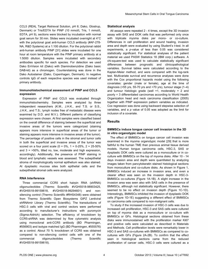
The effect of BMMSCs on tongue cancer cell invasion wasexamined in the myoma organotypic model [29] that is morefaithful to the human TME than previous animal tissue derivedmodels. Human tongue carcinoma cells, HSC-3, SAS ordysplastic DOK cells were cultured as a monoculture or in co-culture with BMMSCs on the top of myoma disks. After 10 - 14days invasion area and depth were quantitated by analyzingimages taken from pancytokeratin stained histological sectionsfrom monoculture and co-culture disks. As shown in Figure 1,BMMSCs induced an increase in invasion area, and even aclearer effect was seen on the invasion depth in HSC-3-BMMSCs co-cultures (Figure 1A-1B). A slight increase in theinvasion area was seen also with SAS cells in the presence ofBMMSCs, although not statistically significant. However, thereseemed to be no effect on invasion depth (Figure 1C-1D).Interestingly, BMMSCs inhibited the invasion of dysplastic DOKcells (Figure 1D-1E) suggesting a different effect of BMMSCson carcinoma cells compared to non-malignant cells.
To study if the increased invasion of HSC-3 cells was due toincreased cell proliferation, HSC-3 and SAS cells were culturedon top of myoma disk as a monoculture or co-culture withBMMSCs or GFs. Histological sections obtained from thesedisks were immunostained with the proliferation marker Ki67and positive cells were calculated as described in Materialsand Methods. Cell proliferation levels were remarkably lower inHSC-3 and SAS co-cultures with BMMSCs as compared to co-cultures with GFs (Figure 2A-2B). To confirm that the effectseen in histological sections came from the reducedproliferation of cancer cells, HSC-3 cells were cultured as a
Mesenchymal Stem Cells in Oral Tongue Carcinoma
PLOS ONE | www.plosone.org 4 October 2013 | Volume 8 | Issue 10 | e77692
mono- or co-culture on cell culture chamber slides withBMMSCs labeled with a fluorescent dye. After co-cultureovernight, cells were stained for Ki67. As shown in Figure 2C,BMMSCs inhibited HSC-3 cell proliferation significantly also inthe cell co-culture assay. The viability of HSC cells remainedstill high and no sign of apoptosis was detected (data notshown).
As proliferation was not increased, to evaluate if theincreased invasion was a consequence of a higher cancer cellmigration a scratch assay was performed with HSC-3 and DOKcells seeded alone or together with BMMSCs or GFs to 24-wellplates. BMMSCs shifted HSC-3 cells, but not dysplastic DOKcells, towards a migratory phenotype with lamellipodiastructures and less cell-cell contacts (Figure 2D). However,GFs promoted faster wound closure than BMMSCs, which maybe partly due to effects of serum on the proliferation capacity ofcancer cells (Figure 2D-2E)
The expression of chemokine CCL5 is up-regulated inBMMSCs after interaction with cancer cells
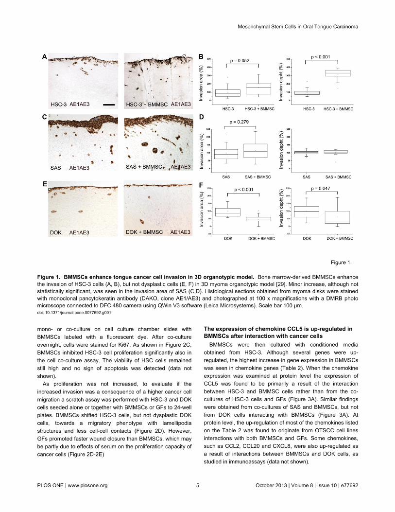
BMMSCs were then cultured with conditioned mediaobtained from HSC-3. Although several genes were up-regulated, the highest increase in gene expression in BMMSCswas seen in chemokine genes (Table 2). When the chemokineexpression was examined at protein level the expression ofCCL5 was found to be primarily a result of the interactionbetween HSC-3 and BMMSC cells rather than from the co-cultures of HSC-3 cells and GFs (Figure 3A). Similar findingswere obtained from co-cultures of SAS and BMMSCs, but notfrom DOK cells interacting with BMMSCs (Figure 3A). Atprotein level, the up-regulation of most of the chemokines listedon the Table 2 was found to originate from OTSCC cell linesinteractions with both BMMSCs and GFs. Some chemokines,such as CCL2, CCL20 and CXCL8, were also up-regulated asa result of interactions between BMMSCs and DOK cells, asstudied in immunoassays (data not shown).
Figure 1. BMMSCs enhance tongue cancer cell invasion in 3D organotypic model. Bone marrow-derived BMMSCs enhancethe invasion of HSC-3 cells (A, B), but not dysplastic cells (E, F) in 3D myoma organotypic model [29]. Minor increase, although notstatistically significant, was seen in the invasion area of SAS (C,D). Histological sections obtained from myoma disks were stainedwith monoclonal pancytokeratin antibody (DAKO, clone AE1/AE3) and photographed at 100 x magnifications with a DMRB photomicroscope connected to DFC 480 camera using QWin V3 software (Leica Microsystems). Scale bar 100 µm.doi: 10.1371/journal.pone.0077692.g001
Mesenchymal Stem Cells in Oral Tongue Carcinoma
PLOS ONE | www.plosone.org 5 October 2013 | Volume 8 | Issue 10 | e77692

Figure 2. BMMSCs inhibit cell proliferation in tongue cancer cells. Histological sections obtained from myoma disks werestained with polyclonal anti-Ki67 antibody. BMMSCs inhibit proliferation of HSC-3 and SAS cells in organotypic invasion model (A,B) as well as in cell culture assays (C), and shift HSC-3, but not dysplastic DOK cells towards a migratory phenotype with less cell-cell contacts (D, arrowheads). Normal gingival fibroblasts, GFs, promote wound closure more than BMMSCs potentially partly dueto the increase in cell proliferation of cancer cells (E). Scale bar 50 µm.doi: 10.1371/journal.pone.0077692.g002
Mesenchymal Stem Cells in Oral Tongue Carcinoma
PLOS ONE | www.plosone.org 6 October 2013 | Volume 8 | Issue 10 | e77692

Function-blocking antibody against CCL5 inhibitsBMMSC increased invasion area
We next explored the importance of CCL5 in the spread oftongue cancer, since CCL5 has been shown to promote themotility of oral cancer cells [32]. CCL5 was also shown to beimportant in tumor progression of several cancers, such asbreast and colorectal carcinoma [17,33]. Therefore, we testedthe effect of CCL5 function-blocking antibody on OTSCC andBMMSC co-cultures in the 3D invasion model. We also testedthe potential effect of function blocking antibody against CXCL1on invasion, since CXCL1 was also expressed from BMMSC-HSC-3 interaction and it has previously been demonstrated tobe present in cancers including breast, lung, colorectal andprostate cancers either supporting or suppressing tumorprogression [34–36]. In oral cancer CXCL1 has beensuggested to have a role in tumor progression since in patientsamples the expression of CXCL1 has been shown beassociated with leukocyte infiltration, lymph node metastasis,and angiogenesis [37]. As shown in Figure 3, the CCL5function–blocking monoclonal antibody was able to significantlyattenuate BMMSC-promoted HSC-3 cell invasion area, but hadno effect on invasion depth (Figure 3B-3C). CXCL1 did nothave an effect on invasion as it did not increase the inhibitoryeffect of CCL5 in the 3D invasion model (Figure 3B-3C).
In patient samples CCL5 is expressed mainly byinflammatory cells and some cancer cells, but onlysparse CAFs are CCL5 positive
Next, to evaluate the importance of CCL5 expression inpatient tumor samples for diagnostics of tongue cancer,histological sections representing different stages of dysplasticlesions and tongue carcinomas from human patients werestained with anti-CCL5 antibody. The expression of CCL5 wasmostly detected in inflammatory cells and in some cancer cells(Figure 4A-4B), but only sparse CAFs were CCL5 positive, asassessed by immunohistochemical co-localization of CCL5 andCAF marker αSMA (data not shown). However, the expressionof CCL5 was not associated with the clinical outcome of theOTSCC patients (not shown). The expression of CCL5 wasfurther studied in normal primary oral fibroblasts (NOF, [28])
Table 2. Up-regulated chemokine genes in BMMSCs afterexposing to conditioned HSC-3 cell culture media (foldchange > 2).
Gene Symbol Fold Changechemokine (C-C motif) ligand 2 CCL2 830.66chemokine (C-C motif) ligand 5 CCL5 194.90chemokine (C-C motif) ligand 20 CCL20 182.23chemokine (C-X-C motif) ligand 1 CXCL1 134.91chemokine (C-X-C motif) ligand 2 CXCL2 80.98chemokine (C-X-C motif) ligand 3 CXCL3 80.14chemokine (C-X-C motif) ligand 5 CXCL5 73.90chemokine (C-X-C motif) ligand 8 (interleukin 8) CXCL8 (IL8) 18.06chemokine (C-C motif) ligand 13 CXCL13 16.86
doi: 10.1371/journal.pone.0077692.t002
and CaDEC12 cells [11]. Immunoassay detecting secretedCCL5 in cell culture media showed very low or no CCL5expression in normal fibroblasts, but considerable higherexpression in CaDEC12 cells (Figure 4C).
The role of CCL5/CCR5 axis is not critical in BMMSCincreased tongue cancer invasion
The CCL5 signaling in cancer cells has been proposed to bemediated mainly, but not necessary exclusively, via CCR5receptor [38]. Both HSC-3 and SAS cells were shown toexpress this receptor (Figure 5A). In co-culture roughly allcancer cells expressed CCR5, however, in monoculturesapproximately only 25 % of cancer cells were positive forCCR5 suggesting that a contact with stromal cells or CCL5induction is needed for higher CCR5 expression (flowcytometry analysis, data not shown). The association of CCL5and CCR5 expression has been proposed recently also byothers [39,40]. As the CCL5-CCR5 axis has been shown topromote the motility of human oral cancer cells in vitro [32] andto induce the expression of MMP9 [32], we also examined theeffect of rCCL5 on MMP9 expression levels in HSC-3 and SASas well as in dysplastic DOK cells. Similarly to previousobservation by Chung and co-workers [32] rCCL5 clearlyincreased the expression of MMP9 in HSC-3 cells and SAScells, but not in dysplastic DOK cells (Figure 5B). However,rCCL5 did not induce epithelial-mesenchymal transition (EMT)as analyzed by Western blotting with anti-vimentin antibody, orproliferation of cancer cells (data not shown).
To explore the importance of CCL5/CCR5 axis in the spreadof tongue cancer, we performed a 3D invasion study with SASand HSC-3 OTSCC cells with reduced CCR5 expression andwith function-blocking antibody against CCL5. First, weinhibited the expression of CCR5 by transducing HSC-3 cellswith CCR5-shRNA and produced a stable cell line with ~70 %inhibition of CCR5 expression and reduced response to rCCL5induction (Figure 5C, 5D). In the 3D co-culture invasion assaythe CCR5 knockdown cells invaded equally or than the wild-type or control HSC-3 cells transduced with non-silencingshRNA (Figure 5E, 5F). Similar results were also obtained witha monoclonal antibody against CCR5 (data not shown).
Expression of collagen and epithelial plasticitycomponents are induced in cancer cells after BMMSCinteraction
Potential mechanisms of the invasion promoting effect ofBMMSCs on cancer cells were further studied by microarrayanalysis. HSC-3 cells were used for the experiments, sincethey responded more clearly to BMMSCs than SAS cells,especially in the 3D invasion model. HSC-3 cells were culturedas a monoculture or co-culture with BMMSC cells on 6-wellplates for 24 h after which HSC-3 cells were sorted out fromco-cultures with FACS (not shown) and RNA was extracted formicroarray analysis. The overall gene profile of HSC-3 cellsafter interaction with BMMSCs showed up-regulated geneexpression of ECM components, with the highest inductionoccurring for type I collagen mRNA expression. In addition tovarious collagens, BMMSC interaction with HSC-3 cells hasalso up-regulated modulators of ECM, such as lysyl oxidase
Mesenchymal Stem Cells in Oral Tongue Carcinoma
PLOS ONE | www.plosone.org 7 October 2013 | Volume 8 | Issue 10 | e77692
Figure 3. The expression of chemokine CCL5 is up-regulated in co-cultures of BMMSCs cells and OTSCC. Co-culture ofOTSCC cells with BMMSCs results in the high expression of chemokine CCL5 in HSC-3 and SAS cells, but not in dysplastic DOKcells (A). Function-blocking monoclonal antibody against CCL5 (Mab CCL5) slightly inhibits BMMSC enhanced invasion area, buthas no effect on the invasion depth, whereas the antibody against CXCL1 does not increase the inhibitory effect in OTSCC invasion(B, C).doi: 10.1371/journal.pone.0077692.g003
Mesenchymal Stem Cells in Oral Tongue Carcinoma
PLOS ONE | www.plosone.org 8 October 2013 | Volume 8 | Issue 10 | e77692

(LOX) and other markers of epithelial plasticity, such ascadherin-11. Molecules associated with cell motility and cancerinvasion, such as α-actinin-4 mRNAs, were also induced inHSC-3 cells by the interaction with BMMSCs (Table 3).
Type I procollagen is highly expressed in the invasionfront in tumor samples obtained from oral tonguecancer patients and correlated with worse prognosis
To further evaluate the relevance of type collagen Iexpression originating from the cancer cell-stromal interactionsto the aggressiveness of OTSCC, we performedimmunohistochemistry for type I procollagen fragment (PINP).PINP has previously been shown to be associated with clinicaloutcome in breast and ovarian cancers, but so far has neverbeen shown in OTSCCs [22-24,31]. In our 114 OTSCC cases,PINP was not found in epithelial cells in histologically normalnor dysplastic peritumoral tissue (Figure 6A-6B). In sometumors PINP was almost absent (Figure 6F), however, most ofthe cases showed high expression by both carcinoma and TMEcells (Figure 6E, 6G, 6H). In general overview, PINPexpression was more intense either in more invasive orsuperficial areas of the tumor (Figure 6C-D). Similar to thepattern of expression found in the primary tumors, PINP was
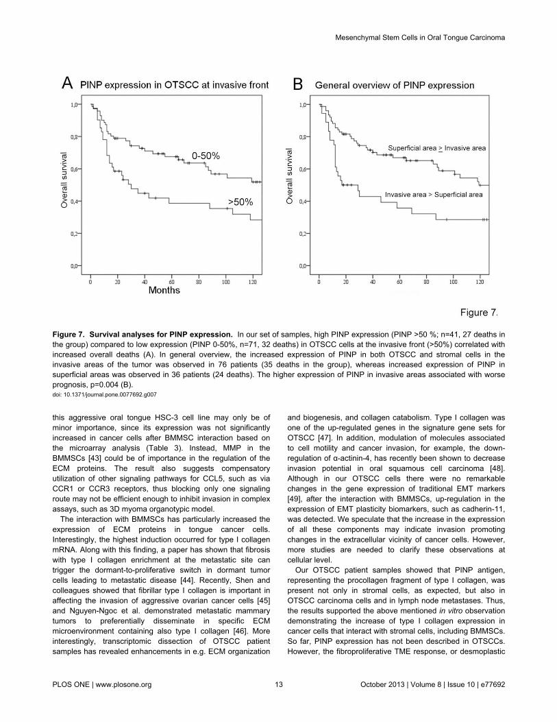
also expressed in OTSCC lymph node metastases (Figure 6I).Furthermore, in a couple of lymph nodes originally signed outas free of metastases (pN0), PINP was expressed in a clusterof cells revealing previously unrecognized occult carcinomametastases which also stained positive for pancytokeratin (notshown). We then compared the pattern of PINP expression andpatient clinical data. In our survival analyses, there was acorrelation between worse prognosis and expression of PINPby OTSCC cells at invasive areas (p=0.018) (Figure 7A).Increased PINP expression by both carcinoma and stromalcells at the invasive areas was also correlated to worseprognosis (p=0.004) (Figure 7B). Both the high expression ofPINP in SCC cells (HR 1.728 95% CI [1.072 - 2.910]) as wellas increased PINP expression by both carcinoma and stromalcells (HR 1.924, 95% CI [1.127 - 3.285]) at invasive areas wereidentified as independent indicators of overall survival alongwith patient age and high tumor stage.
Discussion
We studied here the role of bone marrow-derived BMMSCsin tongue cancer, which had not yet been previouslyaddressed. We could demonstrate that BMMSCs enhanced theaggressiveness of OTSCC cells by inducing their invasion
Figure 4. CCL5 expression in OTSCC patient samples. CCL5 is mainly expressed by inflammatory cells in TME and somecancer cells in patient samples as shown by immunostaining with Pab CCL5, but only sparse CAFs are positive for CCL5 (A, B).CaDEC12 cells, the primary cultures of CAFs obtained from tongue cancer patients, express relative amount of CCL5 compared toNOFs, normal oral fibroblasts (C). Scale bar 100 µm.doi: 10.1371/journal.pone.0077692.g004
Mesenchymal Stem Cells in Oral Tongue Carcinoma
PLOS ONE | www.plosone.org 9 October 2013 | Volume 8 | Issue 10 | e77692
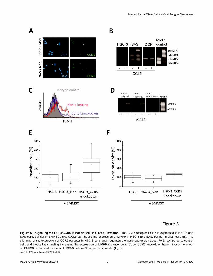
Figure 5. Signaling via CCL5/CCR5 is not critical in OTSCC invasion. The CCL5 receptor CCR5 is expressed in HSC-3 andSAS cells, but not in BMMSCs (A). rCCL5 can induce the expression of MMP9 in HSC-3 and SAS, but not in DOK cells (B). Thesilencing of the expression of CCR5 receptor in HSC-3 cells downregulates the gene expression about 70 % compared to controlcells and blocks the signaling increasing the expression of MMP9 in cancer cells (C, D). CCR5 knockdown have minor or no effecton BMMSC enhanced invasion of HSC-3 cells in 3D organotypic model (E, F).doi: 10.1371/journal.pone.0077692.g005
Mesenchymal Stem Cells in Oral Tongue Carcinoma
PLOS ONE | www.plosone.org 10 October 2013 | Volume 8 | Issue 10 | e77692

capacity, while reducing their proliferation levels. BMMSCsincreased the expression of various chemokines, particularlyCCL5, after interaction with HSC-3 cells. However, interferenceof CCL5 inhibited only the invasion area in our complexorganotypic invasion assay. On the other hand, the interactionbetween cancer and bone marrow derived cells increasedparticularly the expression of type I collagen by carcinomacells. The in vivo results derived from OTSCC patients´samples showed that type I procollagen (PINP) antibodydetected not only stromal mesenchymal cells, as expected, butinterestingly also some of the OTSCC carcinoma cells,confirming our in vitro findings. Surrounding normal epithelialcells were negative for PINP staining. Importantly, the highexpression of PINP in invasive areas of cancer correlated withthe mortality of the patients. We speculate that the molecularcross-talk between BMMSCs and carcinoma cells leads toinvasion promoting TME changes in OTSCC of which PINP,but not CCL5, could be used as prognostic marker for theOTSCC prognosis. In addition, the presence of PINPexpression in cells within metastatic lymph nodes suggests theinvolvement of PINP also in the metastatic spread of theOTSCC.
Table 3. Microarray analysis of up-regulated genes inHSC-3 cells after interacting with BMMSCs.
Gene Symbol Fold Changecollagen, type I, alpha 2 COL1A2 161.55lumican LUM 82.13collagen, type I, alpha 1 COL1A1 67.11periostin, osteoblast specific factor POSTN 43.08fibrillin 1 (Marfan syndrome) FBN1 30.94gremlin 1 homolog, cysteine knot superfamily(Xenopus laevis)
GREM1 30.83
collagen, type VI, alpha 3 COL6A3 30.28platelet-derived growth factor receptor, betapolypeptide
PDGFRB 22.38
gap junction protein, alpha 1, 43kDa (connexin 43) GJA1 21.12collagen, type III, alpha 1 (Ehlers-Danlos syndrometype IV, autosomal dominant)
COL3A1 17.78
ABI gene family, member 3 (NESH) binding protein ABI3BP 13.92transgelin TAGLN 5.61chromosome 5 open reading frame 13 C5orf13 5.30chondroitin sulfate proteoglycan 2 (versican) CSPG2 4.38serine (or cysteine) proteinase inhibitor, clade E(nexin, plasminogen activator inhibitor type 1),member 2
SERPINE2 4.26
angiopoietin-like 4 ANGPTL4 4.06lysyl oxidase LOX 3.67collagen, type V, alpha 2 COL5A2 3.25nicotinamide N-methyltransferase NNMT 2.76prostaglandin-endoperoxide synthase 2(prostaglandin G/H synthase and cyclooxygenase)
PTGS1 2.47
heterogeneous nuclear ribonucleoprotein A1 HNRNPA1 2.41cadherin 11, type 2, OB-cadherin (osteoblast) CDH11 2.33actinin, alpha 4 ACTN4 2.1
doi: 10.1371/journal.pone.0077692.t003
We wished to examine the invasion of aggressive tonguecancer cells using our 3D organotypic model [29] since thismodel represents a fully human, hypoxic tumormicroenvironment [29], [41]. It is ideal for the studies of theearly steps of invasion of tongue cancer, since it representsmore faithfully the structure and composition of human ECM intumors than the traditional invasion models, based on rat tailtype I collagen and mouse EHS tumor derived Matrigel. Weshowed that in co-cultures, BMMSCs stimulated thedissemination of cancer cells and increased the depth ofinvasion, thus enhancing the aggressiveness of tongue cancercells. Several studies have demonstrated the invasion andmetastasis promoting role of BMMSCs in various cancers, suchas breast, colon and lymphatic cancers [17], [18], [19].However, the effect of BMMSCs on different tumor cells hasremained controversial, potentially because of the differentmodels and sources of BMMSCs used. Also, there may bedifferences in the responses of cancer cells and cancersubtypes to BMMSCs [20], [21]. In our experiments, we usedtwo invasive tongue cancer cell lines and found that HSC-3, themost aggressive one, responded more efficiently to signalsoriginating from BMMSCs in the 3D invasion model. However,in monolayer cell culture assays both OTSCC cell linesresponded similarly to the BMMSC interactions. Differences inthe responses in the 3D assays may partially be explained bydifferent genetic background of these cells or by the transienteffect of BMMSCs during the early steps of invasion similarly tobreast cancer [17]. This transient effect is potentially difficult tosee with the fairly aggressive cancer cells in the time-frameused in our 3D assays. In our study, BMMSCs were not able toinduce an invasive phenotype in dysplastic DOK cells,suggesting differences in the capacity of invasive and non-invasive cells to respond to signals originating from BMMSCs.This also could reflect the EMT –process in whichmesenchymal-like cells are more responsive to mesenchymalsignaling.
The roles of chemokines and their receptors in tonguecancer progression are not fully understood. Here we showedthat BMMSCs increased the expression of various chemokines,including CCL5. This effect was more intense in the interactionbetween BMMSCs and tongue cancer cells than betweennormal fibroblasts and cancer cells. The neutralization of CCL5activity with a function-blocking monoclonal antibody resulted inthe inhibition of invasion area, but had no effect on invasiondepth. This suggests that CCL5 might affect more the numberof superficially disseminating invasive cells than the totalinvasive capacity of tongue carcinoma cells. In patientsamples, however, the CCL5 expression was mainly detectedin inflammatory cells in TME, some cancer cells and somesparse CAFs. BMMSCs have been shown to be one source ofCAFs in TME [12,14]. Although we did not examine in detail thedifferentiation capacity of BMMSC, e.g. into CAFs wheninteracting with tongue cancer cells, our preliminary resultsfrom qt-RT-PCR analysis and immunostainings showed a clearincrease in the gene expression of CAF-marker αSMA after theexposure of BMMSC to cancer cell culture media and moreαSMA-positive BMMSCs in co-cultures with cancer cells than inBMMSC mono-culture (not shown). However, similarly to the
Mesenchymal Stem Cells in Oral Tongue Carcinoma
PLOS ONE | www.plosone.org 11 October 2013 | Volume 8 | Issue 10 | e77692

results from other groups [20] we also detected the variability ofthe different batches of patient BMMSCs regarding theexpression of αSMA, even though cell populations with lowpassage numbers were used. It remains to be shown if theeffects seen in this study originate from BMMSCs or fromBMMSCs differentiated to CAFs.
The invasion and metastasis promoting effect of BMMSCson breast cancer cells has been proposed to be essential anddependent on the CCL5-CCR5 axis [17,32,42]. In addition,antibodies against CCL5 have been shown to reduce themetastatic index in breast cancer [17] and CCR5 antagonistshave been demonstrated to block metastasis of basal breast
cancer cells [39]. CCL5/CCR5 axis has also been proposed toaffect the motility of human osteosarcoma [40] and oral cancercells in vitro [32] and CCL5 neutralization was shown to restricttumor progression in colorectal carcinoma [33]. In tonguecancer the CCL5/CCR5 axis stimulated increased cellmigration and the expression of MMP9 has been proposed tobe mediated through NF-kappaB signaling pathways [32].Although HSC-3 cells expressed CCR5 and recombinant CCL5induced MMP9 expression through this receptor, theelimination of CCR5 activity with RNA interference or antibodyagainst CCR5, and the reduction of MMP9 expression did notinhibit invasion in 3D invasion assay. MMP9 expressed here by
Figure 6. OTSCC samples show staining for type I collagen N-terminal propeptide (PINP). The expression of PINP was notfound in any histologically normal epithelial cells of the carcinoma sections, except in a few of the mesenchymal cells within woundbed (arrow heads). The wound epithelial border is marked by dotted line (A). PINP expression was not found in dysplasticepithelium but was present in a few spindle cells in the submucosa (B). In various cases PINP expression was more intense ineither superficial (C) or invasive (D) areas (arrow heads). Some cancers were mainly negative for PINP staining with a few sporaticpositive areas (F), whereas other cases showed intense staining for both cancer and mesenchymal cells (E). In a couple of casesPINP was expressed in the periferal cells of invading islands (G) or mainly in the spindle carcinoma associated mesenchymal cells(H). Lymph node metastases were occasionally positive for PINP (I). Scale bar 200µm .doi: 10.1371/journal.pone.0077692.g006
Mesenchymal Stem Cells in Oral Tongue Carcinoma
PLOS ONE | www.plosone.org 12 October 2013 | Volume 8 | Issue 10 | e77692
this aggressive oral tongue HSC-3 cell line may only be ofminor importance, since its expression was not significantlyincreased in cancer cells after BMMSC interaction based onthe microarray analysis (Table 3). Instead, MMP in theBMMSCs [43] could be of importance in the regulation of theECM proteins. The result also suggests compensatoryutilization of other signaling pathways for CCL5, such as viaCCR1 or CCR3 receptors, thus blocking only one signalingroute may not be efficient enough to inhibit invasion in complexassays, such as 3D myoma organotypic model.
The interaction with BMMSCs has particularly increased theexpression of ECM proteins in tongue cancer cells.Interestingly, the highest induction occurred for type I collagenmRNA. Along with this finding, a paper has shown that fibrosiswith type I collagen enrichment at the metastatic site cantrigger the dormant-to-proliferative switch in dormant tumorcells leading to metastatic disease [44]. Recently, Shen andcolleagues showed that fibrillar type I collagen is important inaffecting the invasion of aggressive ovarian cancer cells [45]and Nguyen-Ngoc et al. demonstrated metastatic mammarytumors to preferentially disseminate in specific ECMmicroenvironment containing also type I collagen [46]. Moreinterestingly, transcriptomic dissection of OTSCC patientsamples has revealed enhancements in e.g. ECM organization
and biogenesis, and collagen catabolism. Type I collagen wasone of the up-regulated genes in the signature gene sets forOTSCC [47]. In addition, modulation of molecules associatedto cell motility and cancer invasion, for example, the down-regulation of α-actinin-4, has recently been shown to decreaseinvasion potential in oral squamous cell carcinoma [48].Although in our OTSCC cells there were no remarkablechanges in the gene expression of traditional EMT markers[49], after the interaction with BMMSCs, up-regulation in theexpression of EMT plasticity biomarkers, such as cadherin-11,was detected. We speculate that the increase in the expressionof all these components may indicate invasion promotingchanges in the extracellular vicinity of cancer cells. However,more studies are needed to clarify these observations atcellular level.
Our OTSCC patient samples showed that PINP antigen,representing the procollagen fragment of type I collagen, waspresent not only in stromal cells, as expected, but also inOTSCC carcinoma cells and in lymph node metastases. Thus,the results supported the above mentioned in vitro observationdemonstrating the increase of type I collagen expression incancer cells that interact with stromal cells, including BMMSCs.So far, PINP expression has not been described in OTSCCs.However, the fibroproliferative TME response, or desmoplastic
Figure 7. Survival analyses for PINP expression. In our set of samples, high PINP expression (PINP >50 %; n=41, 27 deaths inthe group) compared to low expression (PINP 0-50%, n=71, 32 deaths) in OTSCC cells at the invasive front (>50%) correlated withincreased overall deaths (A). In general overview, the increased expression of PINP in both OTSCC and stromal cells in theinvasive areas of the tumor was observed in 76 patients (35 deaths in the group), whereas increased expression of PINP insuperficial areas was observed in 36 patients (24 deaths). The higher expression of PINP in invasive areas associated with worseprognosis, p=0.004 (B).doi: 10.1371/journal.pone.0077692.g007
Mesenchymal Stem Cells in Oral Tongue Carcinoma
PLOS ONE | www.plosone.org 13 October 2013 | Volume 8 | Issue 10 | e77692

reaction, has already been reported as a prognostic predictorof occult metastasis in OTSCCs [50]. The similarity betweendesmoplastic reactions in cancer stroma and wound healingare evident in type I collagen processes [51]. PINP has beendemonstrated to be a prognostic marker in primary andsecondary brain tumors [52] and breast cancer [52] as well asosteosarcoma cells, osteoblasts and proliferating fibroblasts[53,54]. Our results proved that the high PINP expression atthe invasive front serves as a reliable indicator for aggressivegrowth and metastases and could be useful as a prognosticmarker also in OTSCCs.
To conclude, BMMSCs promoted the invasion of tonguecarcinoma cells by inducing the expression of componentsassociated to chemokine signaling, epithelial plasticity, cellmotility and invasion, potentially promoting invasion favoringchanges in cancer cells and in the TME. Although CCL5expression was induced in BMMSCs after interaction withOTSCC cells, signaling through CCL5/CCR5 axis does notseem to be critical for the BMMSC enhanced cancer invasion.Instead, the induction of ECM protein production, particularly
type I collagen, by cancer cells after BMMSC interaction turnedout to be more crucial for OTSCC behavior in vitro. Moreover,based on our clinical data we suggest that PINP antibody,recognizing type I collagen N-terminal propeptide, is anappropriate prognostic marker for OTSCC.
Acknowledgements
The technical assistance in laboratory work of MerjaTyynismaa, Eeva-Maija Kiljander, Minna Savilampi, and HennaEk is acknowledged.
Author Contributions
Conceived and designed the experiments: SS TS PN PL DDMV. Performed the experiments: SS CB KM JV MS. Analyzedthe data: JV HV JK IOB. Contributed reagents/materials/analysis tools: JR DC PL. Wrote the manuscript: SS TS CB PLJR MV DD.
References
1. Allen M, Louise Jones J (2011) Jekyll and hyde: The role of themicroenvironment on the progression of cancer. J Pathol 223: 162-176.PubMed: 21125673.
2. Hogan NM, Dwyer RM, Joyce MR, Kerin MJ (2012) Mesenchymal stemcells in the colorectal tumor microenvironment: Recent progress andimplications. Int J Cancer 131: 1-7. doi:10.1002/ijc.27458. PubMed:22290082.
3. Hanahan D, Coussens LM (2012) Accessories to the crime: Functionsof cells recruited to the tumor microenvironment. Cancer Cell 21:309-322. doi:10.1016/j.ccr.2012.02.022. PubMed: 22439926.
4. Camacho DF, Pienta KJ (2012) Disrupting the networks of cancer. ClinCancer Res 18: 2801-2808. doi:10.1158/1078-0432.CCR-12-0366.PubMed: 22442061.
5. Lorusso G, Rüegg C (2008) The tumor microenvironment and itscontribution to tumor evolution toward metastasis. Histochem Cell Biol130: 1091-1103. doi:10.1007/s00418-008-0530-8. PubMed: 18987874.
6. Hanahan D, Weinberg RA (2011) Hallmarks of cancer: The nextgeneration. Cell 144: 646-674. doi:10.1016/j.cell.2011.02.013. PubMed:21376230.
7. Vered M, Dayan D, Salo T (2011) The role of the tumourmicroenvironment in the biology of head and neck cancer: Lessonsfrom mobile tongue cancer. Nat Rev Cancer 11: 382; author reply:21455256.
8. Vered M, Dobriyan A, Dayan D, Yahalom R, Talmi YP et al. (2010)Tumor-host histopathologic variables, stromal myofibroblasts and riskscore, are significantly associated with recurrent disease in tonguecancer. Cancer Sci 101: 274-280. doi:10.1111/j.1349-7006.2009.01357.x. PubMed: 19804423.
9. Bello IO, Vered M, Dayan D, Dobriyan A, Yahalom R et al. (2011)Cancer-associated fibroblasts, a parameter of the tumormicroenvironment, overcomes carcinoma-associated parameters in theprognosis of patients with mobile tongue cancer. Oral Oncol 47: 33-38.doi:10.1016/j.oraloncology.2010.10.013. PubMed: 21112238.
10. Vered M, Dayan D, Yahalom R, Dobriyan A, Barshack I et al. (2010)Cancer-associated fibroblasts and epithelial-mesenchymal transition inmetastatic oral tongue squamous cell carcinoma. Int J Cancer 127:1356-1362. doi:10.1002/ijc.25358. PubMed: 20340130.
11. Dayan D, Salo T, Salo S, Nyberg P, Nurmenniemi S et al. (2012)Molecular crosstalk between cancer cells and tumor microenvironmentcomponents suggests potential targets for new therapeutic approachesin mobile tongue cancer. Cancer Med. 2: 128-140. PubMed: 23342263.
12. Mishra PJ, Mishra PJ, Humeniuk R, Medina DJ, Alexe G et al. (2008)Carcinoma-associated fibroblast-like differentiation of humanmesenchymal stem cells. Cancer Res 68: 4331-4339. doi:10.1158/0008-5472.CAN-08-0943. PubMed: 18519693.
13. Guo X, Oshima H, Kitmura T, Taketo MM, Oshima M (2008) Stromalfibroblasts activated by tumor cells promote angiogenesis in mouse
gastric cancer. J Biol Chem 283: 19864-19871. doi:10.1074/jbc.M800798200. PubMed: 18495668.
14. Spaeth EL, Dembinski JL, Sasser AK, Watson K, Klopp A et al. (2009)Mesenchymal stem cell transition to tumor-associated fibroblastscontributes to fibrovascular network expansion and tumor progression.PLOS ONE 4: e4992. doi:10.1371/journal.pone.0004992. PubMed:19352430.
15. Koh BI, Kang Y (2012) The pro-metastatic role of bone marrow-derivedcells: A focus on MSCs and regulatory T cells. EMBO Rep 13: 412-422.doi:10.1038/embor.2012.41. PubMed: 22473297.
16. Fox JM, Chamberlain G, Ashton BA, Middleton J (2007) Recentadvances into the understanding of mesenchymal stem cell trafficking.Br J Haematol 137: 491-502. doi:10.1111/j.1365-2141.2007.06610.x.PubMed: 17539772.
17. Karnoub AE, Dash AB, Vo AP, Sullivan A, Brooks MW et al. (2007)Mesenchymal stem cells within tumour stroma promote breast cancermetastasis. Nature 449: 557-563. doi:10.1038/nature06188. PubMed:17914389.
18. Shinagawa K, Kitadai Y, Tanaka M, Sumida T, Kodama M et al. (2010)Mesenchymal stem cells enhance growth and metastasis of coloncancer. Int J Cancer 127: 2323-2333. doi:10.1002/ijc.25440. PubMed:20473928.
19. Amé-Thomas P, Maby-El Hajjami H, Monvoisin C, Jean R, Monnier Det al. (2007) Human mesenchymal stem cells isolated from bonemarrow and lymphoid organs support tumor B-cell growth: Role ofstromal cells in follicular lymphoma pathogenesis. Blood 109: 693-702.doi:10.1182/blood-2006-05-020800. PubMed: 16985173.
20. Klopp AH, Gupta A, Spaeth E, Andreeff M, Marini F 3rd (2011) Concisereview: Dissecting a discrepancy in the literature: Do mesenchymalstem cells support or suppress tumor growth? Stem Cells 29: 11-19.doi:10.1002/stem.559. PubMed: 21280155.
21. Waterman RS, Henkle SL, Betancourt AM (2012) Mesenchymal stemcell 1 (MSC1)-based therapy attenuates tumor growth whereas MSC2-treatment promotes tumor growth and metastasis. PLOS ONE 7:e45590. doi:10.1371/journal.pone.0045590. PubMed: 23029122.
22. Jensen BV, Johansen JS, Skovsgaard T, Brandt J, Teisner B (2002)Extracellular matrix building marked by the N-terminal propeptide ofprotype I collagen reflect aggressiveness of recurrent breast cancer. IntJ Cancer 98: 582-589. doi:10.1002/ijc.10187. PubMed: 11920619.
23. Marin L, Koivula MK, Jukkola-Vuorinen A, Leino A, Risteli J (2011)Comparison of total and intact aminoterminal propeptide of type Iprocollagen assays in patients with breast cancer with or without bonemetastases. Ann Clin Biochem 48: 447-451. doi:10.1258/acb.2011.011040. PubMed: 21733929.
24. Simojoki M, Santala M, Risteli J, Kauppila A (2003) Discrepantexpression of carboxy- and aminoterminal propeptides of type Iprocollagen predicts poor clinical outcome in epithelial ovarian cancer.
Mesenchymal Stem Cells in Oral Tongue Carcinoma
PLOS ONE | www.plosone.org 14 October 2013 | Volume 8 | Issue 10 | e77692

Gynecol Oncol 88: 358-362. doi:10.1016/S0090-8258(02)00139-7.PubMed: 12648587.
25. Nurmenniemi S, Kuvaja P, Lehtonen S, Tiuraniemi S, Alahuhta I et al.(2010) Toll-like receptor 9 ligands enhance mesenchymal stem cellinvasion and expression of matrix metalloprotease-13. Exp Cell Res316: 2676-2682. doi:10.1016/j.yexcr.2010.05.024. PubMed: 20553713.
26. Leskelä HV, Risteli J, Niskanen S, Koivunen J, Ivaska KK et al. (2003)Osteoblast recruitment from stem cells does not decrease by age atlate adulthood. Biochem Biophys Res Commun 311: 1008-1013. doi:10.1016/j.bbrc.2003.10.095. PubMed: 14623282.
27. Kylmäniemi M, Oikarinen A, Oikarinen K, Salo T (1996) Effects ofdexamethasone and cell proliferation on the expression of matrixmetalloproteinases in human mucosal normal and malignant cells. JDent Res 75: 919-926. doi:10.1177/00220345960750030901. PubMed:8675803.
28. Kulasekara KK, Lukandu OM, Neppelberg E, Vintermyr OK,Johannessen AC et al. (2009) Cancer progression is associated withincreased expression of basement membrane proteins in three-dimensional in vitro models of human oral cancer. Arch Oral Biol 54:924-931. doi:10.1016/j.archoralbio.2009.07.004. PubMed: 19674736.
29. Nurmenniemi S, Sinikumpu T, Alahuhta I, Salo S, Sutinen M et al.(2009) A novel organotypic model mimics the tumor microenvironment.Am J Pathol 175: 1281-1291. doi:10.2353/ajpath.2009.081110.PubMed: 19679876.
30. Li C, Wong WH (2001) Model-based analysis of oligonucleotide arrays:Expression index computation and outlier detection. Proc Natl Acad SciU S A 98: 31-36. doi:10.1073/pnas.98.1.31. PubMed: 11134512.
31. Risteli J, Elomaa I, Niemi S, Novamo A, Risteli L (1993)Radioimmunoassay for the pyridinoline cross-linked carboxy-terminaltelopeptide of type I collagen: A new serum marker of bone collagendegradation. Clin Chem 39: 635-640. PubMed: 8472358.
32. Chuang JY, Yang WH, Chen HT, Huang CY, Tan TW et al. (2009)CCL5/CCR5 axis promotes the motility of human oral cancer cells. JCell Physiol 220: 418-426. doi:10.1002/jcp.21783. PubMed: 19334035.
33. Cambien B, Richard-Fiardo P, Karimdjee BF, Martini V, Ferrua B et al.(2011) CCL5 neutralization restricts cancer growth and potentiates thetargeting of PDGFRbeta in colorectal carcinoma. PLOS ONE 6:e28842. doi:10.1371/journal.pone.0028842. PubMed: 22205974.
34. Cheng WL, Wang CS, Huang YH, Tsai MM, Liang Y et al. (2011)Overexpression of CXCL1 and its receptor CXCR2 promote tumorinvasion in gastric cancer. Ann Oncol 22: 2267-2276. doi:10.1093/annonc/mdq739. PubMed: 21343381.
35. Kuo PL, Shen KH, Hung SH, Hsu YL (2012) CXCL1/GROalphaincreases cell migration and invasion of prostate cancer by decreasingfibulin-1 expression through NF-kappaB/HDAC1 epigenetic regulation.Carcinogenesis 33: 2477-2487. doi:10.1093/carcin/bgs299. PubMed:23027620.
36. Benelli R, Stigliani S, Minghelli S, Carlone S, Ferrari N (2013) Impact ofCXCL1 overexpression on growth and invasion of prostate cancer cell.Prostate 73: 941-951. doi:10.1002/pros.22640. PubMed: 23334998.
37. Shintani S, Ishikawa T, Nonaka T, Li C, Nakashiro K et al. (2004)Growth-regulated oncogene-1 expression is associated withangiogenesis and lymph node metastasis in human oral cancer.Oncology 66: 316-322. doi:10.1159/000078333. PubMed: 15218300.
38. Raman D, Baugher PJ, Thu YM, Richmond A (2007) Role ofchemokines in tumor growth. Cancer Lett 256: 137-165. doi:10.1016/j.canlet.2007.05.013. PubMed: 17629396.
39. Velasco-Velázquez M, Jiao X, De La Fuente M, Pestell TG, Ertel A etal. (2012) CCR5 antagonist blocks metastasis of basal breast cancer
cells. Cancer Res 72: 3839-3850. doi:10.1158/1538-7445.AM2012-3839. PubMed: 22637726.
40. Wang SW, Wu HH, Liu SC, Wang PC, Ou WC et al. (2012) CCL5 andCCR5 interaction promotes cell motility in human osteosarcoma. PLOSONE 7: e35101. doi:10.1371/journal.pone.0035101. PubMed:22506069.
41. Teppo S, Sundquist E, Vered M, Holappa H, Parkkisenniemi J et al.(2013) The hypoxic tumor microenvironment regulates invasion ofaggressive oral carcinoma cells. Exp Cell Res 319: 376-389. doi:10.1016/j.yexcr.2012.12.010. PubMed: 23262025.
42. Zhang Y, Yao F, Yao X, Yi C, Tan C et al. (2009) Role of CCL5 ininvasion, proliferation and proportion of CD44+/CD24- phenotype ofMCF-7 cells and correlation of CCL5 and CCR5 expression with breastcancer progression. Oncol Rep 21: 1113-1121. PubMed: 19288016.
43. Parikka V, Väänänen A, Risteli J, Salo T, Sorsa T et al. (2005) Humanmesenchymal stem cell derived osteoblasts degrade organic bonematrix in vitro by matrix metalloproteinases. Matrix Biol 6: 438-447.PubMed: 16098718.
44. Barkan D, El Touny LH, Michalowski AM, Smith JA, Chu I et al. (2010)Metastatic growth from dormant cells induced by a col-I-enrichedfibrotic environment. Cancer Res 70: 5706-5716. doi:10.1158/1538-7445.AM10-5706. PubMed: 20570886.
45. Shen Y, Shen R, Ge L, Zhu Q, Li F (2012) Fibrillar type I collagenmatrices enhance metastasis/invasion of ovarian epithelial cancer viabeta1 integrin and PTEN signals. Int J Gynecol Cancer 22: 1316-1324.doi:10.1097/IGC.0b013e318263ef34. PubMed: 23013730.
46. Nguyen-Ngoc KV, Cheung KJ, Brenot A, Shamir ER, Gray RS et al.(2012) ECM microenvironment regulates collective migration and localdissemination in normal and malignant mammary epithelium. Proc NatlAcad Sci U S A 109: E2595-E2604. doi:10.1073/pnas.1212834109.PubMed: 22923691.
47. Ye H, Yu T, Temam S, Ziober BL, Wang J et al. (2008) Transcriptomicdissection of tongue squamous cell carcinoma. BMC Genomics 9:69-2164-9-69. PubMed: 18254958.
48. Yamada S, Yanamoto S, Yoshida H, Yoshitomi I, Kawasaki G et al.(2010) RNAi-mediated down-regulation of alpha-actinin-4 decreasesinvasion potential in oral squamous cell carcinoma. Int J Oral MaxillofacSurg 39: 61-67. doi:10.1016/j.ijom.2009.10.003. PubMed: 19913389.
49. Zeisberg M, Neilson EG (2009) Biomarkers for epithelial-mesenchymaltransitions. J Clin Invest 119: 1429-1437. doi:10.1172/JCI36183.PubMed: 19487819.
50. Pimenta Amaral TM, Da Silva Freire AR, Carvalho AL, Pinto CA,Kowalski LP (2004) Predictive factors of occult metastasis andprognosis of clinical stages I and II squamous cell carcinoma of thetongue and floor of the mouth. Oral Oncol 40: 780-786. doi:10.1016/j.oraloncology.2003.10.009. PubMed: 15288831.
51. Ohtani H (1998) Stromal reaction in cancer tissue: Pathophysiologicsignificance of the expression of matrix-degrading enzymes in relationto matrix turnover and immune/inflammatory reactions. Pathol Int 48:1-9. doi:10.1111/j.1440-1827.1998.tb03979.x. PubMed: 9589457.
52. Rasmussen HB, Teisner B, Schrøder HD, Yde-Andersen E, AndersenJA (1991) Fetal antigen 2 in primary and secondary brain tumors.Tumour Biol 12: 330-338. doi:10.1159/000217734. PubMed: 1798908.
53. Rasmussen HB, Teisner B, Andersen JA, Yde-Andersen E, Skjødt K etal. (1992) Foetal antigen 2 (FA2) in the stromal reaction induced bybreast carcinoma. APMIS 100: 39-47. doi:10.1111/j.1699-0463.1992.tb00837.x. PubMed: 1536719.
54. Tornehave D, Teisner B, Rasmussen HB, Chemnitz J, Kassem M(1992) Fetal antigen 2 (FA2) in human fetal osteoblasts, culturedosteoblasts and osteogenic osteosarcoma cells. Anat Embryol (Berl)186: 271-274. PubMed: 1416076.
Mesenchymal Stem Cells in Oral Tongue Carcinoma
PLOS ONE | www.plosone.org 15 October 2013 | Volume 8 | Issue 10 | e77692