Embed Size (px)
Citation preview

Collaborative ProgramAVRDC/CIRAD

2
CONTENTSPage
1. ACHIEVEMENTS 3
2. OBTENTION OF DOUBLE HAPLOID OF ONION 4IN THE CONTEXT OF ONION BREEDING
3. POTENTIALITIES OF SOMATIC EMBRYOGENESIS IN THE CONTEXT OF 28THERAPY AND GERMPLASM PRESERVATION OF GARLIC
4. TRAINING IN BIOMOLECULAR TOOLS 56
5. TRAINING IN CRYOPRESERVATION 58
6. PROPOSAL: “CRYOPRESERVATION FOR GERMPLASM CONSERVATION 60
AND SUPPLY OF DISEASE FREE SEEDLINGS OF VEGETATIVELY PROPAGATED
VEGETABLES”.
7. PUBLI 1 ABSTRACT: “ESTABLISHMENT OF EMBRYOGENIC CELL 62
SUSPENSION CULTURES OF GARLIC (ALLIUM SATIVUM L.), PLANT
REGENERATION AND BIOCHEMICAL ANALYSES”
8. PUBLI 2 ABSTRACT: “ EMBRYOGENIC CELL SUSPENSION CULTURES 63
OF GARLIC (ALLIUM SATIVUM L.) AS METHOD FOR MASS PROPAGATION ANDPOTENTIAL MATERIAL FOR GENETIC IMPROVEMENT”
9. POSTER: PRODUCTION DE MASSE DE PLANTS SAINS D’AIL PAR 64EMBRYOGENESE SOMATIQUE
10. ACKNOWLEDGEMENTS 65

3
ACHIEVEMENTS IN THIS COLLABORATIVE PROGRAM
• SET UP OF TECHNIQUE; Obtention of double haploid of onion in the context ofonion breeding
• SET UP OF TECHNIQUE; Process of somatic embryogenesis in the context oftherapy and perspective of germplasm preservation
• TRAINING ; Preparation for initiation of biomolecular tools in onion breeding bytraining for (Cherng Hsin-Chun, Vicky) in CIRAD, France
• TRAINING ; Preparation for initiation of cryopreservation in germplasmconservation by training for (Chang Huan-Jiun, Jessica) in CIRAD, France
• PROPOSAL ; “Cryopreservation for germplasm conservation and supply ofdisease free seedlings of vegetatively propagated vegetables”. (Drs. LeonidasFEREOL; Liwayway M. ENGLE; Sylvia GREEN; Haeng-Hoon KIM)
• PUBLICATION ; Establishment of embryogenic cell suspension cultures of garlic(Allium sativum L.), plant regeneration and biochemical analyses. 2005 “PlantCell Report, Vol. 24, n. 6, p. 319 – 325. L. Fereol, V. Chovelon, S. Causse, D. Triaire,I. Arnault, J. Auger and R. Kahane; AVRDC, World vegetable center, P.O. Box 42,Shanhua, Tainan, Taiwan
• PUBLICATION ; Embryogenic Cell Suspension Cultures of Garlic (Alliumsativum L.) As Method For Mass Propagation and Potential Material ForGenetic Improvement. 2005, “Acta Horticulturae 688, p65-74”L. Fereol, V. Chovelon, S. Causse, M.L. Kalumvueziko and R. Kahane; AVRDC,world vegetable center, P.O. Box 42, Shanhua, Tainan, Taiwan
• POSTER ; Production de masse de plants sains d’ail par embryogénèse somatique.2005. Journées Horticoles CIRAD. L. Fereol; V. Chovelon; S. Causse, M.L.Kalumvueziko and R. Kahane ; AVRDC World Vegetable Center, P.O. Box 42,Shanhua, Tainan, 741 Taiwan R.O.C.
• PROJECT PUBLI ; Potentialities of Garlic somatic embryogenesis for eliminationof OYDV and LYSV. (in progress)
• PROJECT BOOK SECTION ; : Tissue culture and its role in plant biotechnology.(in progress); AVRDC World Vegetable Center, P.O. Box 42, Shanhua, Tainan, 741Taiwan R.O.C.

4
OBTENTION OF DOUBLE HAPLOID OF ONIONIN THE CONTEXT OF ONION BREEDING
1 Background information ....................................................................................................52 Plant material......................................................................................................................53 Methods ..............................................................................................................................7
3.1 Gynogenesis procedure ..............................................................................................73.1.1 Preparation of flowers for culture.......................................................................73.1.2 Aseptisation ........................................................................................................73.1.3 In vitro culture conditions ..................................................................................7
3.2 Determination of ploidy of the gynogenic plants ....................................................103.3 Chromosome doubling .............................................................................................113.4 Transfer and observations in vivo.............................................................................133.5 Maintenance in vivo of the materials........................................................................133.6 Data analysis ............................................................................................................13
4 Results ..............................................................................................................................154.1 Gynogenesis induction .............................................................................................154.2 Obtention of gynogenic plants .................................................................................15
4.2.1 Responses of A. cepa line.................................................................................154.2.2 Responses of A. fistulosum-derived plants .......................................................16
4.3 In vitro culture of the gynogenic plants ...................................................................184.4 Comparisons between generations ...........................................................................184.5 Ploidy of the gynogenic plants .................................................................................194.6 Chromosome doubling .............................................................................................20
5 Discussion ........................................................................................................................205.1 Production of gynogenic interspecific-derived plants ..............................................205.2 Ploidy level ...............................................................................................................215.3 Chromosome doubling .............................................................................................215.4 Use in onion breeding ..............................................................................................21
6 References ........................................................................................................................237 Annexe 1. Stock Solutions of media used for haploid production of onion ....................258 Annexe 2. Flow cytometry protocol .................................................................................27......................................................................................................................................................

5
1 Background information
AVRDC’s breeding program of onion focuses its objectives on increasing yield, improvingstorability, and eliminating diseases. Concerning disease resistance, particularly againstStemphyllium vesicarium, AVRDC is incorporating such traits into short day onion cultivars.Allium fistulosum Stearn (a wild relative of onion) is known to be resistant to Stemphyliumleaf blight (caused by Stemphylium vesicatoria) (Pathak et al., 2001). This wild onion relativeis sexually crossable with common onion (A. cepa L.), providing partially fertile offspring(Pathak et al., 2001). The strategy of onion breeding can help to transfer the resistance gene tostemphyllium and over coming the sexual barrier between the two species. But onion breeding is a slowprocess primarily due to the biennial nature of this outcrossing species. Development of anonion inbred line may require more than 12 years, when the traits of interest are transferredfrom interspecifi c crosses. Doubled haploid (DH) techniques can shorten the time required todevelop onion inbred lines (Alan et al., 2002). To be used effectively, this approach requiressimple methods both for haploid plant production and chromosome doubling to obtain DHlines. The procedure is currently widely used by various breeding companies and has a majorpotential for basic genetic studies. Different techniques have been tried to get haploid in onion.Use of irradiation-inactivated pollen in crosses with male sterile plants was suggested as analternative strategy for production of gynogenic onion plants (Dore, 1987). Currently, cultureof immature flower buds on gynogenesis induction media slightly modified from the originalprotocol (Muren, 1989) is the most common method for the production of haploid onionplants. Many researchers have described improvements of the methodology, use of un-pollinated ovules, ovaries, or whole flowers, and responses of A. cepa lines to gynogenicinduction (Alan et al., 2002; Campion and Alloni, 1990). Although recovery of haploid onionplants through gynogenesis is an established procedure, the low rate of gynogenesis in mostlines, substantial plant to plant variations within lines, the low rate of spontaneouschromosome doubling, and difficulties in inducing chromosome doubling have limited theapplication of gynogenesis and use of DHs in onion breeding.
Along with a traditional backcross breeding program, AVRDC and CIRAD have proposed touse DH techniques to accelerate production of improved inbred lines. Here, we reportacquisition of the technique and production of gynogenic plants from different generations ofA. fistulosum-derived plants.
2 Plant material
Materials are plants generated from the interspecifi c cross of A. cepa and A. fistulosum (Fig.1).They come from AVRDC onion breeding program (Table 1). About 20 plants of eachgenotype were planted in field for flowering. Plants from the initial (F1 and F2) were notavailable for this experiment. But Hybrids F4 and backcross generations (BC1F1 and BC2F1)of A. fistulosum-derived plants (Table ) were selected from the breeding program because oftheir resistance or tolerance to Stemphyllium leaf blight. The plant responses to Stemphylliumleaf blight had been determined by an inoculation Stemphyllium leaf blight-screeningprocedure. All BC2F1 plants were Stemphyllium leaf blight-resistant (R) or highly resistant(HR). A. cepa lines are susceptible to Stemphyllium leaf blight disease. A. cepa lines werefecund whereas the A. fistulosum-derived plants showed differences in numbers of seedsproduced, with most showing lower fecundity than the A. cepa lines. Unlike the A. cepa lines,
6
Fig.1 Interspecific cross Allium cepa X Allium fistulosum and progeny

7
A. fistulosum-derived plants did not produce bulbs and were not dormant. Becausethey did not dry down, A. fistulosum-derived plants were maintained in pots formonths, and so, were available for flower bud collection in successive years.
3 Methods
3.1 Gynogenesis procedure
3.1.1 Preparation of flowers for culture
The whole umbel was excised, when the first flowers start to open (Fig.2). At this time about30% flower buds have reached a three-day-before-anthesis stage (Muren, 1989). They weretaken from donor plants grown in the field, between the months of December and February.The flower buds of other umbels were used on the day of collection. A few umbels were keptin the laboratory at room temperature with the bottom part of flower stalk dipped in water. Inthese conditions they can stay for 4 days in order to reach the stage for sampling. They wereexcised from the umbels. Open flowers were discarded before sterilization.
3.1.2 Aseptisation
The flower buds collected, were surface sterilized by:- washing in ethanol 95° for 1 minute;- sterilizing in a 2% sodium hypochlorite solution + 0.1% Tween-20 for 20 minutes;- rinsing three time with sterilized distilled water.Flower buds were separated into two classes: large (diameter >4 mm), and small (<4 mm).
3.1.3 In vitro culture conditions
Gynogenic induction medium: The medium used for induction of gynogenic embryos wasmodifi ed B5 basal medium (Gamborg et al., 1968) supplemented with 2 mg/l of 2,4-dichlorophenoxyacetic acid (2,4-D), 2 mg/l of 6-benzylaminopurine (BA) and 75 g of sucrose(table ).
Development medium (DM): This medium is a modifi ed (Murashige and Skoog, 1962),containing, no growth regulators, and 40 g/l sucrose (Kahane et al., 1992).All media were adjusted to pH 5.8 and were solidified with 7 g/l agar-agar (Difco-Bacto).
In vitro culture of flowers: Flower buds were placed in 90 mm × 15 mm Petri platescontaining 25 ml of induction medium (20 flowers/plate). Plates were sealed with Plastic film(scello-frais) and cultured at 25 °C under cool white fl uorescent and Gro-Lux lights (45–80µEm−2s−1) with 16 h light: 08 h dark photoperiod.

8
Fig.2 Sum up of the technique of induction and production of Haploid andDH Onion Plants. A-flowerscape; B-Unopened flower excised; C-Gynogenic embryo induction, gynogenic plants with seed coat; D-gynogenic plantlets emerging from ovule; E-gynogenic plantletsgrowing on EM medium
9
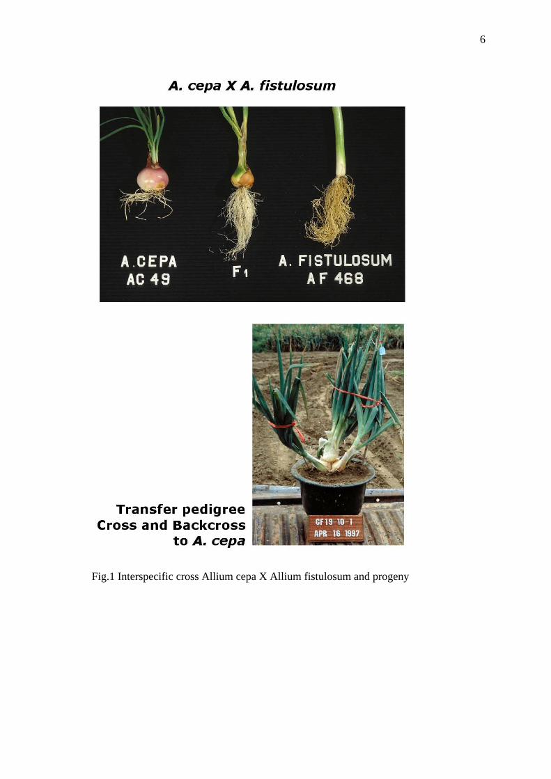
Table 1. Solid media used for onion gynogenesis and embryo regeneration
Nutrients (mg/l) Induction (IM) Development
(DM)
Multiplication (MM)
Macro nutrients X 20 (B5) 50 ml (MS) 50 ml (MS) 50 ml
Micro nutrients X 1000 (B5) 1 ml (MS) 1 ml (MS) 1 ml
Fe- EDTA X 100 10 ml 10 ml 10 ml
Vitamins Morel X 500 5 ml 2.5 ml 5 ml
Growth regulators
-2.4.D (1mg = 10 ml)
-BAP (1mg = 10 ml)
-NAA (1mg = 10 ml)
20 ml
20 ml 20 ml
2 ml
3.1.3.1 Divers (mg/l) Ad SO4 40 mg Ad SO4 40 mg
KH2PO4 300 mg
L-Tyrosin 50 mg
Sucrose (g/l) 75 g 40 g 40 g
Agar-agar, Difco 7 g 7 g 7 g
PH before autoclaving 5.8 5.8 5.8
B5: Gamborg vitamins (Gamborg et al., 1968)
MS: Murashige and Skoog (Murashige and Skoog, 1962)
Vitamins Morel and Martin (1955)
AdSO4: Adenine hemisulfate C5H5N5-1/2H2SO4
Observations of the flowers culture: Observations were performed every two weeks to
monitor fl ower development, basal callus formation and somatic shoot regeneration, and
emergence of gynogenic plantlets. Basal callus development was noted. They developed the
fi rst month of culture. Structures emerging from flower ovaries cultured on induction medium
are called “embryos”. Flowers responding to induction of gynogenesis were noted, and the
rate of response was calculated as numbers of embryos per 100 flowers for each genotype.
10
In vitro sub-culture of emerging embryos : Flowers with emerging embryos were transplanted
in glass tubes of development medium (DM). They grew to the 3–4 leaf stage under the
conditions described above. Plantlets not growing more than one leaf were noted and
eliminated. Plants in tubes were observed for overall growth performance.
3.2 Determination of ploidy of the gynogenic plants
The regenerated materials in vitro, and parents in the interspecific crosses A. fistulosum X A.
cepa grown in soil, were analysed by flow cytometry to measure their ploidy level.
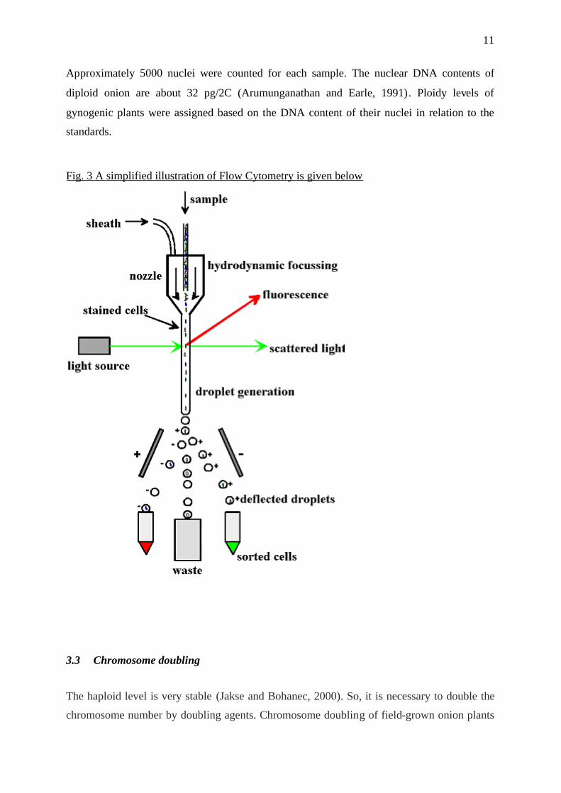
What is Flow Cytometry?In flow cytometry (FCM-techniques) (Fig.3), a suspension of cells is passed through the path
of a laser beam. Light emitted from any fluorescent material within the material transiting the
beam is captured and gives rise to a signal which can be integrated over time to measure the
quantity of fluorescent material within the cell suspension.
FCM is important for applications involving counting cells, or for measuring the size of cell
sub-populations which have been labelled with specific fluorescent probes. FCM-DNA Ploidy
works by integrating the light signal from individual passing nucleii after quantitative
fluorescence labelling of DNA chromatin. In this way a conventional ploidy histogram is
constructed showing population frequency of nucleii having different DNA contents (Fig.4).
We used the protocol described by (Arumunganathan and Earle, 1991). Analyses of A.
fistulosum and A. cepa plants grown in soil used the bottom parts of newly emerging leaves.
Analyses of in vitro grown gynogenic plants (2–3 months old) used pieces of several leaves.
Approximately, 50 mg of leaf tissue was used in each sample. Leaves were cut into fine strips
with a razor blade or a scalpel in 35 mm × 10 mm, polystyrene Petri dishes containing 750 µl
of staining solution. Samples were fi ltered through a 167 µm mesh into a 50 ml Falcon tube.
A second 750 µl of staining solution was added to the plate, and the fi ltered solution was
combined with the previous one.
Samples were incubated at 37◦C for 15 min and were then kept in ice until analyses were
done on a FACS Calibur fl ow cytometer at Dr. Lei’s Laboratory, Chung-Kung University,
Tainan, Taiwan.
11
Approximately 5000 nuclei were counted for each sample. The nuclear DNA contents of
diploid onion are about 32 pg/2C (Arumunganathan and Earle, 1991). Ploidy levels of
gynogenic plants were assigned based on the DNA content of their nuclei in relation to the
standards.
Fig. 3 A simplified illustration of Flow Cytometry is given below
3.3 Chromosome doubling
The haploid level is very stable (Jakse and Bohanec, 2000). So, it is necessary to double the
chromosome number by doubling agents. Chromosome doubling of field-grown onion plants

12
is not possible, because the difficulty of access to the meristem. As a result, all chromosome
doubling procedures in onion are based on various in vitro treatments of explants.
The majority of approaches attempted have used the sliced basal parts of shoots of in vitro-
elongated or micropropagated plantlets (Fig.5), which were subjected to chromosome
doubling agents (colchicine, oryzalin, trifluralin or amiprofos methyl) at various
Fig.4. Flow cytometry histogram of haploid, diploid and mixoploid
gynogenetic plantlets
13
concentrations and for various periods of exposure (Bohanec and Jakse, 1999; Geoffriau et al.,
1997b; Nowak, 2000).
We tested two doubling treatments, which were colchicines and amiprofos methyl (AMP) at
concentrations 25 mM/l and 50 µM/l, respectively, using application in liquid medium. Based
on the results obtained in these preliminary studies APM was used in subsequent experiments.
Therefore, we designed further studies using larger experimental units that included only
APM as the doubling agent at concentration 50µM/l.
3.4 Transfer and observations in vivoThe gynogenic plantlets were acclimatized as follow: They were transplanted to 9 cm pots
containing peat moss soil. They were covered with plastic bags, and transferred to a green
house. The bags were progressively take off 1 week to help plant acclimatization and were
totally removed at the end of 2 weeks. Later the plants must be transferred to field and the
observed for their ability to produce bulbs, seeds and resistance to Stemphyllium leaf blight.
3.5 Maintenance in vivo of the materialsPots of gynogenic plants, obtained from A. fistulosum-derived plants, will be transferred
directly to field. When flowering, anthers collected from fl owers will be macerated with a
droplet of Alexander’s stain (Alexander, 1969) and observed microscopically at 100–400
times to determine the percentage of stainable (fertile) pollen. Flowers will be self-pollinated
by pollinator’s house flies in covered net bags. Seeds will be collected from drying umbels
and stored in paper bags.
3.6 Data analysisConcerning production of gynogenic plantlets, the data from generations BC1F1 and BC2F1
were analyzed to determine whether the ability to produce gynogenic plantlets increased with
backcrossing to A. cepa. Differences in the production of gynogenic embryos or plants
between the BC1F1 and BC2F1 A. fistulosum-derived plants were tested by T-test. T test
analysis was also used to test whether bud size class affected gynogenic plantlet formation
from BC1F1, BC2F1 plants.
Concerning comparison of doubling treatments, shoot survival was visually evaluated during

14
Fig.5 Straegy for double haploid of onion plants. A-anti mitotic (AMP) doubling
treatment on shoot; B-Regeneration of new shoot, ploidy determination; C-Double
hapoid plant; D-Transfer of double haploid to soil; E-Study of progeny
15
the treatment. Estimates of the percentage of shoots surviving to doubling treatment were
made. Surviving plantlets are those in which the apical part of the axis, about 7-10 mm in
length, remained viable and could be used for further reculturing.
4 Results
4.1 Gynogenesis inductionGynogenic embryos formed from non-fertilized flower buds appeared gradually over a 4-
month period, and development of gynogenic plantlets continued until the 16 months of
culture. Hook-shaped gynogenic plants usually broke open the ovaries (Fig.2D). Their colour
was either yellow or green. Many ovaries had empty black seed coats. This is consistent with
the observation that all gynogenic plantlets obtained had a seed coat. Fig.2C shows a
gynogenic plantlet emerging from one ovule like a germinating seedling. Gynogenic plantlets
that continued to grow were transferred to tubes on EM to encourage their further
development (Fig.2E).
Flower buds of A. cepa and A. fistulosum-derived plants showed similar changes in culture.
Most large and small buds opened within several days of culture. Ovaries of all cultured
fl ower buds grew to 4–8 times their original sizes after 1 month. By the end of the month,
cultures initiated from different size buds looked very similar in size. Some of the flower buds
were vitrifi ed, they had a bright green and glassy appearance. Vitrification was observed in
some buds from almost all donor plants. Basal callus also developed on some fl ower buds.
The location and appearance of basal callus was quite different and distinct from that of the
emerging gynogenic plantlets. Basal callus developed more frequently on small fl ower buds
than on large ones.
4.2 Obtention of gynogenic plants
4.2.1 Responses of A. cepa lineMost of the gynogenic plantlets from this A. cepa line was obtained within 3–4 months
although a few emerged later. Only height gynogenic plantlets (2%) were provided by this A.
cepa line. Small A. cepa fl ower buds were less responsive to gynogenesis.
16
4.2.2 Responses of A. fistulosum-derived plants
F1 and F2 plants.Flower buds from other F1 and F2 A.fistulosum-derived plants were not used because of
problem of sterility of interspecific F1 plants.
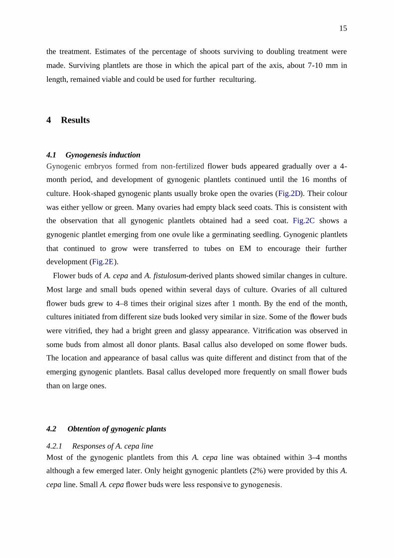
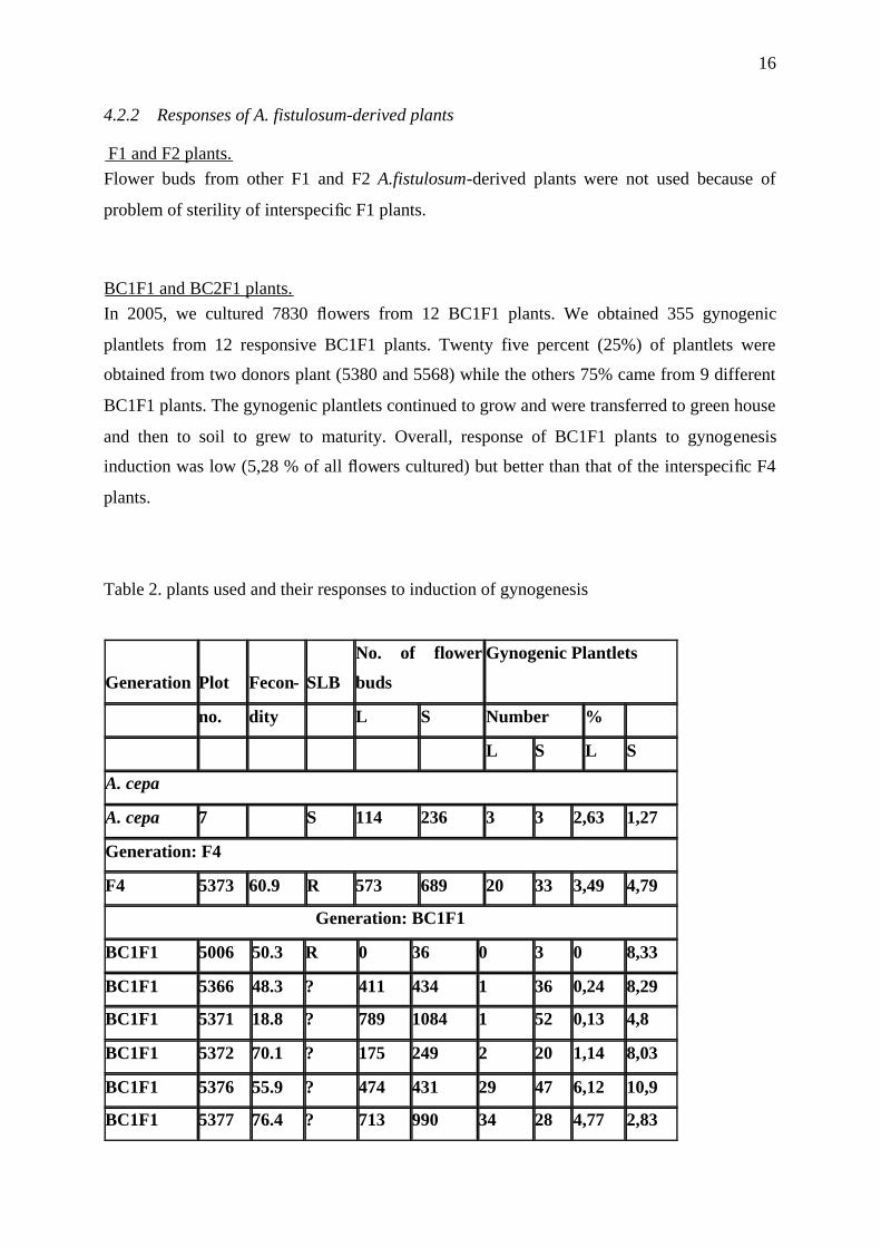
BC1F1 and BC2F1 plants.In 2005, we cultured 7830 fl owers from 12 BC1F1 plants. We obtained 355 gynogenic
plantlets from 12 responsive BC1F1 plants. Twenty five percent (25%) of plantlets were
obtained from two donors plant (5380 and 5568) while the others 75% came from 9 different
BC1F1 plants. The gynogenic plantlets continued to grow and were transferred to green house
and then to soil to grew to maturity. Overall, response of BC1F1 plants to gynogenesis
induction was low (5,28 % of all fl owers cultured) but better than that of the interspecific F4
plants.
Table 2. plants used and their responses to induction of gynogenesis
Generation Plot Fecon- SLB
No. of flower
buds
Gynogenic Plantlets
no. dity L S Number %
L S L S
A. cepa
A. cepa 7 S 114 236 3 3 2,63 1,27
Generation: F4
F4 5373 60.9 R 573 689 20 33 3,49 4,79
Generation: BC1F1
BC1F1 5006 50.3 R 0 36 0 3 0 8,33
BC1F1 5366 48.3 ? 411 434 1 36 0,24 8,29
BC1F1 5371 18.8 ? 789 1084 1 52 0,13 4,8
BC1F1 5372 70.1 ? 175 249 2 20 1,14 8,03
BC1F1 5376 55.9 ? 474 431 29 47 6,12 10,9
BC1F1 5377 76.4 ? 713 990 34 28 4,77 2,83

17
BC1F1 5380 72.2 ? 232 333 24 45 10,3 13,5
BC1F1 5382 79.7 ? 467 413 7 13 1,5 3,15
BC1F1 5563 64.4 ? 23 24 0 2 0 8,33
BC1F1 5566 41.8 ? 181 224 1 1 0,55 0,45
BC1F1 5567 74.8 ? 40 49 1 1 2,5 2,04
BC1F1 5568 55 ? 11 47 2 5 18,2 10,6
Generation: BC2F1
BC2F1 5004 19.9 R 249 272 25 31 10 11,4
BC2F1 5007 42.6 R 20 16 0 8 0 50
BC2F1 5008 26.1 R 19 20 0 3 0 15
BC2F1 5026 54 R 344 515 20 63 5,81 12,2
BC2F1 5029 67.9 R 92 129 2 17 2,17 13,2
BC2F1 5031 34.8 R 332 448 11 85 3,31 19
BC2F1 5034 52.6 R 261 456 6 55 2,3 12,1
BC2F1 5072 60.2 R 191 158 4 0 2,09 0
BC2F1 5074 31.9 R 73 49 1 4 1,37 8,16
BC2F1 5075 37 R 194 111 22 11 11,3 9,91
BC2F1 5077 56.4 R 203 153 16 18 7,88 11,8
BC2F1 5092 98 HR 542 725 23 39 4,24 5,38
BC2F1 5095 58.6 R 1064 1704 30 93 2,82 5,46
BC2F1 5096 98.1 R 396 383 10 29 2,53 7,57
BC2F1 5097 96.5 R 1054 832 49 83 4,65 9,98
BC2F1 5098 56.4 R 810 659 31 75 3,83 11,4
BC2F1 5099 24.6 R 105 125 0 3 0 2,4
BC2F1 5101 97.5 R 309 604 17 76 5,5 12,6
BC2F1 5102 49.7 R 616 762 15 52 2,44 6,82
BC2F1 5103 32.3 R 621 923 20 94 3,22 10,2
Resistance of plants to SLB was scored as follows: S: susceptible; R: resistant; HR: Highly
resistant.

18
Table 3. Analysis of percentage of gynogenic plantlet across generationsGeneration Flowers
Cultured
Gynogenic
Plantlets
Responsive
Flowers (%)
Test
t
A. cepa 350 6 1.7%
F4 1262 53 4.2%
BC1F1 7820 355 4.5%
BC2F1 16550 1141 6.9% 0.02
We cultured about 16539 flowers from 20 BC2F1 plants. We obtained 1141 gynogenic
plantlets from 20 responsive BC2F1 plants. The plantlets obtained in 2005 grew to the plant
level, and the survived transfer to soil. The plants will grow to maturity. Progress with the
BC2F1 plants in 2005, encourage to culture more fl owers from new and previously used
BC2F1 plants in 2006.
In contrast to the results with A. cepa fl ower buds, large fl ower buds of A. fistulosum-derived
BC2F1 showed significantly less response to gynogenesis induction than smaller buds.
Gynogenic plants obtained from the BC2F1s were also more vigorous than those obtained
from the other groups.
4.3 In vitro culture of the gynogenic plants
The gynogenic A. fistulosum-derived plants grew well in vitro on DM medium. Some
contaminations in culture caused losses of many plants, in spite of attempts to rescue the
contaminated plants by transfer to soil. The majority of the gynogenic plants from BC2F1
fl owers showed vigorous growth in vitro.
4.4 Comparisons between generations
The plantlet and plant production of the BC2F1 generation was significantly greater than in
the BC1F1 (Table 3). The improved response of the BC2F1 generation over the BC1F1
generation for gynogenic plant production can also be shown by direct comparison of the data
from these two generations (means significantly different by two sample T-test, in the case of

19
small flowers).
4.5 Ploidy of the gynogenic plants
Leaves of A. fistulosum plants had DNA contents very similar to those of A. cepa. The
majority of the nuclei from the A. fistulosum, A. cepa, and A. fistulosum-derived plants
contained about 32 pg DNA. In the samples, 15–20% of the nuclei contained ca. 55 pg DNA;
these nuclei may originate from cells going through mitotic division.
In this work:
- Some of the samples from gynogenic plants of A. fistulosum-derived plants consisted
of nuclei with about 16 pg DNA, half of the nuclear DNA content of diploid onion;
these were classifi ed as haploids. These samples usually also contained a small
fraction of diploid nuclei (5–25% of the total).
- Some had nuclear DNA contents identical to diploids. These plants were classifi ed as
spontaneous diploids. They represent 50% of the total.
- In other cases, samples contained similar percentages of haploid and diploid nuclei
and were classified as mixoploids.
- A few gynogenic plantlets obtained from A. fistulosum-derived plants had nuclear
DNA content of ca. 55 pg and were classified as spontaneous tetraploids.
Table 4. Flow cytometry analysis of gynogenic plants obtained from A. fistulosum-derived
plantlets
Generation N. Tested n 2n mixo-
ploid.
A. cepa 3 1 1 1
F4 18 1 15 0
BC1F1 32 12 11 (34.3%) 9
BC2F1 114 27 44 (38.5%) 36
Values in paranthesis are percentages of fl owers.
Mixoploid plants containing similar numbers of haploid and diploid nuclei.

20
4.6 Chromosome doubling
We conducted a preliminary experiment to test a method for doubling treatment. An efficient
method for chromosome doubling should take in accounts both survival rate and
chromosome-doubling efficiency. Concerning survival rate: The control was 100% survival,
while the average survival rate for AMP (50 µM/l) or colchicine (25 mMol/l) treatments, was
27.4% and 22.4% respectively (data not shown). Concerning chromosome doubling,
statistical analyses was avoid because the low number of plantlets tested may not have lead to
meaningful conclusions.
Based on the results obtained in these preliminary studies on survival rate of shoots explants,
and according to results by other others on efficiency of these two technique (Hansen and
Andersen, 1998), we decided that, colchicine is slightly more toxic to onion tissues than APM
while the doubling efficiency was similar. For this reason only APM was used in subsequent
experiments following the schema Fig.5. Therefore, we designed further studies using larger
experimental units that included only APM as the doubling agent at concentration 50µM/l.
5 Discussion
5.1 Production of gynogenic interspecific-derived plantsGynogenic plants can be obtained from hybrids between A. cepa and A. fistulosum.
Frequencies of gynogenic responses varied substantially among the A. fistulosum-derived
plants. The increased plantlet production of the BC2F1 generation may indicate segregation
for A. cepa alleles for better response to culture. It is possible that still higher levels of
response would be found in more advanced generations, such as the BC2F2 or BC3F1.
The bud size of the flower is known to be an important factor in the induction of gynogenesis
in onion and shallots, like (Smith et al., 1991) reported concerning leek (Allium porrum L.).
(Campion and Alloni, 1990) re-ported that large buds (2.5–3.5 mm) were the most responsive
ones to gynogenesis induction. As a general trend, culture of unopened large flower buds
(corresponding to a period from several days to just prior to anthesis) is recommended (Cohat,
1994; Michalik et al., 2000). According to (Bohanec and Jakse, 1999; Geoffriau et al., 1997a),
fl ower buds just prior to anthesis contain fully mature embryo sacs, which may be a
contributing factor to the higher frequencies of gynogenesis in large flower buds. But here,
like previous workers, we found no significant difference between large and small flower
buds. So, we can included all size fl ower buds in the culture of A. fistulosum-derived

21
generations.
5.2 Ploidy levelThe majority of gynogenic plants obtained from A. fistulosum-derived plants here were
diploids. Occurrence of various rates of spontaneous mixoploids, diploids, and tetraploids
among gynogenic onion plants has been reported by others (Bohanec and Jakse, 1999;
Geoffriau et al., 1997a). However, the high rate of spontaneous diploids among the gynogenic
plants from A. fistulosum-derived plants is similar to result obtained by Alan et al. (2003) for
A. roylei-derived plants. We assume that gynogenic plants with ploidy levels other than
haploid resulted from spontaneous chromosome doubling in early stages of egg cell division
and/or embryo development, but the possibility of their development from unreduced egg
cells should not be disregarded. Therefore, study of their self-pollinated progenies or
molecular analyses would be desirable to confirm their homozygosity. If truly of haploid
origin, these diploids are advantageous, since they eliminate the need for chromosome
doubling procedures.
A large proportion of plantlets from all treatments expressed mixoploidy, a phenomenon
that was also noted in previous reports (Geoffriau et al., 1997b). It is not yet clear whether
mixoploid regenerants develop into partially fertile diploid plants or remain haploid, as
suggested by (Geoffriau et al., 1997b). In the case of partial diploidy, at least some of them
might produce seeds.
5.3 Chromosome doubling
The choice of APM as the optimal chromosome doubling substance is in agreement to similar
findings in other plants. (Hansen and Andersen, 1998) reported that compared to oryzalin and
trifluralin, APM showed a relatively low toxicity on gynogenic embryo formation in sugar
beet. APM was also shown to be less toxic but similarly efficient when these three substances
were compared in Brassica napus microspore cultures (Hansen and Andersen, 1998) and less
inhibitory to maize haploid callus growth than oryzalin (Wan et al., 1991). Due to its low
toxicity and high-doubling efficiency APM could be the optimal choice among the most
frequently tested chromosome-doubling substances.
5.4 Use in onion breeding
22
In several crops, double-hapoid plants have been of great utility. First for the rapid generation
of inbred lines for hybrid production and then for the creation of recombinant inbred lines
(RILs), so useful for developing molecular markers, mapping genes of interest, and detecting
QTL affecting quantitative traits. When working with a trait that is difficult to screen on a
single plant basis, generation and evaluation of fixed inbred materials is also of considerable
benefi t. Doubled haploids derived from interspecifi c materials, could have additional
advantages as well. Using an effi cient gynogenic system in moderate to advanced interspecifi c
generations would generate RILs fi xed for novel traits but containing relatively little wild
genomic content, signifi cantly accelerating the breeding process.
Breeding to transfer the traits of disease resistance of A. fistulosum to cultivated onion has
been impeded by sexual barriers between the species that reduce fecundity in cross and
backcross generations (Kik, 2002). Nevertheless gynogenic plants can be obtained from
breeding material generated from interspecifi c crosses of onion and A. fistulosum, allowing
development of fecund inbred lines from plants at BC1F1, and BC2F1 stages. There was no
apparent association between the fecundity of the A. fistulosum-derived donor plants and the
ability of the buds to produce gynogenic plantlets (Table 1). DH A. fistulosum-derived plants
could be of material assistance in circumventing the sexual barriers in the transfer of traits
such as disease resistance from A. fistulosum to onion.
The percentage of gynogenic plantlets increase as generations of A. fistulosum-derived
plants were advanced. This indicates the existence of genetic effects in gynogenesis induction.
As the share of A. fistulosum DNA in the resulting plants decreases, their gynogenic response
is likely to improve as well.
The next step in this project will be attempts to recover self-pollinated seed from the spon-
taneous and induced diploids. Seeds of the gynogenic A. fistulosum-derived BC2F1 plant will
be grown in the later season, and the resulting seedlings will be tested for stemphyllium resis-
tance and advanced in the breeding program as appropriate.
Unlike those obtained from A. cepa plants, the majority of the gynogenic plants we obtained
from A. fistulosum-derived plants were spontaneous diploids, not requiring chemical doubling.
Recovery of self-pollinated seed from such plant would suggest that some of the plants may
be fecund. If homozygous and fecund, these plants may speed development of new onion
cultivars with useful agronomical characters such as resistance to stemphyllium. DH plants
currently being obtained from more advanced backcross generations may be even more
valuable to the breeding program.
23
The procedures developed for common onion are also applicable to shallot (A. cepa L. var.
aggregatum) (Cohat, 1994; Sulistyaningsih et al., 2002).
6 References
Alexander, M. 1969. Differential staining of aborted and non-aborted pllen. Stain Technol.44:117-122.
Arumunganathan, K., and E. Earle. 1991. Estimation of nuclear DNA content of plants byflow cytometry. Plant Mol. Biol. Rep. 9:229-241.
Bohanec, B., and M. Jakse. 1999. Variation in gynogenic response among long-day onion(Allium cepa L.) accessions. Plant cell rep. 18:737-742.
Campion, B., and C. Alloni. 1990. Induction of haploid plants in onion (Allium cepa L.) by invitro culture of unpollinated ovules. Plant Cell Tissue and Organ Culture 20:1-6.
Cohat, J. 1994. Obtention chez l'échalote (Allium cepa L. var aggregatum) de planteshaploides gynogenetiques par culture in vitro de boutons floraux. Agronomie 14:299-304.
Dore, C. 1987. Integration of in vitro culture in methods of selection of some vegetablespecies - Integration de la culture in vitro dans les methodes de selection de plusieursespeces potageres. Th.: Sci. naturelles, <University> Paris 11.
Gamborg, O., R. Miller, and K. Ojima. 1968. Nutrient requirements of suspension cultures ofsoybean root. Cells 50:151-158.
Geoffriau, E., R. Kahane, and M. Rancillac. 1997a. Variation of gynogenesis ability in onion(Allium cepa L.). Euphytica 94:37-44.
Geoffriau, E., R. Kahane, C. Bellamy, and M. Rancillac. 1997b. Ploidy stability and in vitrochromosome doubling in gynogenic clones od onion (Allium cepa L.). Plant Sci122:201-208.
Hansen, N., and S. Andersen. 1998. In vitro chromosome doubling with cochicineduingmicrospore culture in wheat (Triticum aestivum L.). Euphytica 102:101-108.
Jakse, M., and B. Bohanec. 2000. Studies of alternative approaches for genome doubling inonion, In B. Bohanec, ed. Biotechnological approach: utilisation of gametic cells.COST 824 final meeting, Bled, Slovania.
Kahane, R., M. Rancillac, and B. Schweisguth. 1992. Bulbing in vitro in Allium species.Allium Improve Newsl 2:18-20.
Kik, C. 2002. Exploitation of wild relatives for the breeding of cultivated Allium species, p.81-100, In H. Rabinowitch and L. Currah, eds. Allium Crop Science: RecentAdvances, Vol. Wallingford, UK. CABI Pubishing.
Michalik, A., E. Adamus, and E. Nowak. 2000. Gynogenesis in polish onion cultivar. J. PlantPhysiol. 156:211-216.
Murashige, T., and F. Skoog. 1962. A revised medium for rapid growth and biosassay withtobacco tissue culture. Physiol. plants 15:473-497.
Muren, P. 1989. Haploid plant induction from unpollinated ovaries in onion. Hortscience24:833-834.
Nowak, E. 2000. Gynogenic onion plant- studies on regeneration and diploidization, p. 95-99,In B. Bohanec, ed. Biotechnological approach: utilisation of gametic cells. COST 824final meeting, Bled, Slovenia.

24
Pathak, C., L. Black, S. Cherng, T. Wang, and S. Ko. 2001. Breeding onions for stemphyliumleaf blight resistance. Acta Horticulturae 555:77-81.
Smith, B., R. Godwin, E. Harvey, and C. Werner. 1991. Gynogenesis from whole flowerbulbs in bulb onions (Allium cepa L.) and leeks (Allium porrum L.). J. Genet. Breed.45:353-358.
Sulistyaningsih, E., K. Yamashita, and Y. Tashiro. 2002. Haploid induction from F1 hybridsbetween CMS shallot with Allium galanthum cytoplasm and common onion byunpollinated flower culture. Euphytica 125:139-144.
Wan, Y., D. Duncan, A. Rayburn, J. Petolino, and J. Widhorm. 1991. The use ofantimicrotubule herbicides for the production of double haploid plants from anther-derived maize callus. Theoretical and Applied Genetics 81:205-211.

25
7 Annexe 1. Stock Solutions of media used for haploid production ofonion
Macro nutrients (B5) (MS)
Concentration Final
Sol mg
X 20 Final
Solmg
X 20
Quantity stock solution per
liter of final solution
50 ml 50 ml
Quantity of stock Solution to do 1 liter 1 liter
-KNO3 2500mg 50 g 2500 50g-(NH4)2SO4 134 2.68g-NH4H2PO4 300 6g
-KH2PO4, 2H2O 170 3.4g-NaH2PO4, 2 H2O 150 3g-CaCl2, 2H2O 150 3g 200 4g-MgSO4, 7H2O 250 5g 400 8g
Micro nutrients (B5) (MS)
Concentration FinalSol
X 500 FinalSol
X 1000Quantity mother Sol for
1 l of final Sol
1 ml 1 ml
Quantity of mother Sol to do 1 liter 1 liter- H3BO3 3mg 3000mg 6,2mg 6200mg- KI 0.75 750 0,83 830- MnSo4, 4H2O 10 10 000 16,7 16700- ZnSo4, 7H20 2 2000 8,6 8600- CoCl2, 6H2O 0,025 25 0,025 25- CuSo4, 5H2O 0,025 25 0,025 25- Na2MoO4, 2H2O 0, 25 250 0, 25 250
Vitamins (Morel)
Concentration FinalSol
X 200Quantity mother Sol for1 liter offinal Sol
5 ml
Quantity mother Sol to do 500 ml- Nic ac 1 100 mg- pyridoxin 1 100- Thiamine HCl 1 100- Calcium panthotenate 1 100- biotin 0.01 1- myo inositol 100 10 000
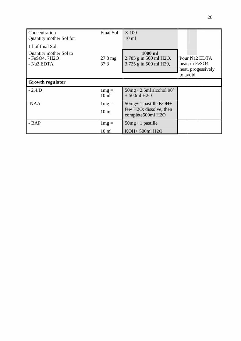
Iron EDTA
26
Concentration Final Sol X 100Quantity mother Sol for
1 l of final Sol
10 ml
Quantity mother Sol to 1000 ml- FeSO4, 7H2O 27.8 mg 2.785 g in 500 ml H2O,- Na2 EDTA 37.3 3.725 g in 500 ml H20,
Pour Na2 EDTAheat, in FeSO4heat, progessivelyto avoidprecipitationGrowth regulator
- 2.4.D 1mg =10ml
50mg+ 2,5ml alcohol 90°+ 500ml H2O
-NAA 1mg =
10 ml
50mg+ 1 pastille KOH+few H2O: dissolve, thencomplete500ml H2O
- BAP 1mg =
10 ml
50mg+ 1 pastille
KOH+ 500ml H2O

27
8 Annexe 2. Flow cytometry protocol
Flow cytometry
Protocol for the extraction and coloration of nuclei,
Adaptation to Allium sp leaves
Extraction of nucleiAbout 250 mg (~ 4 cm2 of leaves) of sample are finely chopped by a razor blade, in a Petridish, in 0.7 ml of buffer PBS (A).The suspension, obtained like this, is filtrated through a 48 µm pore sieve on flow tube.
ColouringThe filtrate is coloured by a solution of Propidium Iodide (B), 200 µl by tube.Stir by Vortex Shaker, and incubate at room temperature for 5 mn.Stir again before taking each measure.
Preparation of solutions
A: Solution of PBSTo this, add:
- Dithiothréitol (DDT), Sigma réf. D5545 1mg per ml of buffer- Triton X100 6 ml in 100 ml of buffer
Dissolve DDT before adding Triton X100Stock at +4°C for stock and during reading of results
B: Propidium IodidePrepare a watery solution 1 mg/ ml of water. (Can be stock 2 months at 4°C).For colouring, dilute this solution to 1/5 (3ml PI at 1mg/ml + 12 ml of buffer A). Maintain at+4°C (for stock and during utilisation).
Caution of use: CHEMICALS DANGEROUSDon’t put in contact with skinLabelled the bottles wellThrow away wastes in a container used for this purpose.

28
POTENTIALITIES OF GARLIC SHOOT TIP AND SOMATIC EMBRYOGENESISIN THE CONTEXT OF THERAPY AND GERMPLASM PRESERVATION
1 BACKGROUND INFORMATION.................................................................................292 PLANT MATERIAL .......................................................................................................30
2.1 Cultivar .....................................................................................................................302.2 Explants ....................................................................................................................31
3 METHODS.......................................................................................................................323.1 Callus production .....................................................................................................323.2 Regeneration from calluses ......................................................................................333.3 Initiation of the suspension cultures .........................................................................333.4 Establishment and maintenance of the cell suspension culture................................33
3.4.1 Initial culture densities .....................................................................................363.4.2 Periodicity of medium renewal ........................................................................363.4.3 Age of the suspension cultures .........................................................................36
3.5 Regeneration of the suspension culture ....................................................................363.6 Adaptation to in vivo Conditions ..............................................................................363.7 ELISA Test ...............................................................................................................373.8 Virological Investigations ........................................................................................373.9 Recording .................................................................................................................393.10 Statistical analysis ....................................................................................................39
4 RESULT ...........................................................................................................................404.1 Callus production .....................................................................................................404.2 Embryo induction from calluses ..............................................................................404.3 Initiation of the suspension cultures .........................................................................414.4 Regeneration of the suspension culture ....................................................................424.5 Conversion of somatic embryos into plants .............................................................424.6 Adapting to in vivo Conditions.................................................................................434.7 Virological investigations ........................................................................................43
5 DISCUSSION ..................................................................................................................446 PERSPECTIVES..............................................................................................................45
6.1 New approach to accelerate multiplication ..............................................................456.2 Cryo-preservation Germplasm management ............................................................466.3 Cryotherapy ..............................................................................................................47
7 REFERENCES.................................................................................................................478 Annexe1. Stock Solutions of media used for garlic embryogenesis ................................52

29
1 BACKGROUND INFORMATION
Dissemination of improved or selected varieties of garlic (Allium sativum L.), to farmers, is
hampered by the presence of two viruses belonging to the potyvirus group, Onion Yellow
Dwarf (OYDV) and Leek Yellow Stripe (LYSV) (Lot et al., 1994; Van Dijk, 1991; Van Dijk,
1993; Walkey, 1990), and in obtaining healthy plants. Moreover, the need for successive
multiplication phases in field in order to provide sufficient material hampers the rapid
dissemination of selected elite material. The establishment of a virus free plant production
system is a prerequisite for seed production and dissemination. The traditional scheme usually
involves virus-free stock material, micro propagation technique, seed production, certification
and distribution of high sanitary quality materials. Some viruses can be effectively eliminated
from infected plants owing to their mode of replication and their mechanism of movement
within the plant.
Three methods are currently used: thermotherapy, meristem tip culture and chemotherapy.
According to the literature, meristem culture is considered to be the reference tool for virus
disease eradication (Faccioli and Marani, 1998). This technique takes advantage of the fact
that many viruses fail to invade the meristem region. Transfer of the meristem dome,
together with one or two leaf primordia, to a culture medium and development into a plantlet
may lead to the elimination of a virus. There are reports of the elimination of sugarcane
yellow leaf virus (SCYLV) using meristem culture (Chatenet et al., 2001; Fitch et al., 2001;
Parmessur and Dookun, 2000). Concerning garlic, eliminating viruses and producing virus-
free material have been developed for the past 30 years, using meristem-tip culture
(Chovelon et al., 1990; Walkey et al., 1987). It was found that a gradient of increasing virus
concentration from the dome to the successive primordia exists (Walkey and Webb, 1968).
This means that the possibility of obtaining virus-free plants is inversely related to the size of
the meristem excised. However, the virus-free state of plants regenerated after meristem
culture alone is not efficient at 100%, particularly when meristems were excised from in vivo
plants comparatively to in vitro plants.
To improve pathogen elimination, thermo and chemotherapy coupled with meristem culture
can be used when meristem culture alone fails. Virus elimination was greatly accelerated at
40°C, but with a subsequent deterioration of the cultured tissue. Chemotherapy has also been
applied to meristem cultures, but this has a negative effect on meristem growth.

30
Therefore, other technique with less negative effect might be attempted. Somatic
embryogenesis, was shown to be highly effective in the elimination of certain viruses from
Vitis vinifera (Goussard et al., 1991), Saccharum officinarum (Parmessur et al., 2002). The
availability of a reliable technique for the production of somatic embryos from garlic (Fereol
et al., 2005a; Fereol et al., 2005b) prompted us to investigate whether the use of this technique
could result in the elimination of viruses OYDV and LYSV from Garlic.
In the present study we report the establishment and regeneration of embryogenic cell
suspension cultures of an infected garlic cultivar. We assess and compare infection at three
steps of this process: embryogenic callus, suspension cultures, and on plantlets regenerated
from these different tissues type.
2 PLANT MATERIAL
2.1 CultivarThe cultivar “29 VFG” (AVRDC, Allium Unit), one part grown in open field and, one part
grown in net house was used in this experiment. It belongs to the tropical group of garlic,
short-day type, expressing short dormancy and bad preservation. It forms medium-size bulb
with white coloured cloves. This variety was issued from clonal selection and meristem-tip
culture. Bulbs, harvested from plants 4-5 months old, were stored 3 weeks at 5°C, in order to
break the dormancy before experiments. Cloves from these bulbs were germinated in vitro
according procedure developed by (Kahane et al., 1992). Plantlets issued of this in vitro
culture were tested by ELISA test for the presence of OYDV and LYSV. On one side, in vitro
cultivated materials coming from net house and tested virus-free, and on other side in vitro
cultivated materials coming from open field and tested infected by both OYDV and LYSV
were used for somatic embryogenesis.

31
Fig. 1 Callus culture; A-clove; B-shoot from clove; C-shoot roll explant; D-primarycallus; E-induction of embryogenic and friable callus; F-regenerated embryo

32
2.2 Explants
Roll shoot sections including young leaf part and basal plate was used as explants for callus
induction (Fig.1A-C).
Cloves were surface sterilised according the following procedure: After removing the outer
protective leaf sheaths, they were dipped in 70% ethanol for 5 minutes, followed by 20
minutes in a sodium hypochlorite solution (2% of active chlorine). They were then rinsed
three times in sterilised water for 5, 10 and 15 minutes. They were dried on a sterile paper
towel, the storage leaf was removed under aseptic conditions. Then the shoot containing the
basal plate and basal young leaf part was cut transversally to produce roll shoot explants 2
mm thick.
3 METHODS
3.1 Callus production
The explants were plated on 90 mm diameter Petri dishes containing 25 ml of semi-solid
medium. They were incubated 1 month on a callus pre-induction medium (CIM1, Tab.1),
which is a modified N6 medium (Chu et al., 1975) containing 0.2 mg l-1 NAA, 0.1 mg l-1 IAA,
1 mg l -1 2,4-D and 0.1 mg l-1 BAP, and solidified with 0.3% agar (Phytagel® ). The explants
were then transferred to a second induction medium (CIM2, Tab.1) for callus production. This
medium differed from the previous one by concentrations of 2,4-D (0.5 mg l-1, Tab. 2). The
callus produced were transferred on a maintenance and Embryogenic medium (CMM, Tab.1)
based on a N6 medium containing cefotaxime (400 mg l-1), and maintained on it before
embryo induction or initiation of suspension cultures.
33
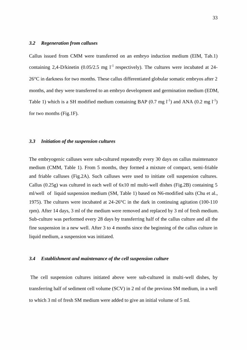
3.2 Regeneration from calluses
Callus issued from CMM were transferred on an embryo induction medium (EIM, Tab.1)
containing 2,4-D/kinetin (0.05/2.5 mg l-1 respectively). The cultures were incubated at 24-
26°C in darkness for two months. These callus differentiated globular somatic embryos after 2
months, and they were transferred to an embryo development and germination medium (EDM,
Table 1) which is a SH modified medium containing BAP (0.7 mg l-1) and ANA (0.2 mg l-1)
for two months (Fig.1F).
3.3 Initiation of the suspension cultures
The embryogenic calluses were sub-cultured repeatedly every 30 days on callus maintenance
medium (CMM, Table 1). From 5 months, they formed a mixture of compact, semi-friable
and friable calluses (Fig.2A). Such calluses were used to initiate cell suspension cultures.
Callus (0.25g) was cultured in each well of 6x10 ml multi-well dishes (Fig.2B) containing 5
ml/well of liquid suspension medium (SM, Table 1) based on N6-modified salts (Chu et al.,
1975). The cultures were incubated at 24-26°C in the dark in continuing agitation (100-110
rpm). After 14 days, 3 ml of the medium were removed and replaced by 3 ml of fresh medium.
Sub-culture was performed every 28 days by transferring half of the callus culture and all the
fine suspension in a new well. After 3 to 4 months since the beginning of the callus culture in
liquid medium, a suspension was initiated.
3.4 Establishment and maintenance of the cell suspension culture
The cell suspension cultures initiated above were sub-cultured in multi-well dishes, by
transferring half of sediment cell volume (SCV) in 2 ml of the previous SM medium, in a well
to which 3 ml of fresh SM medium were added to give an initial volume of 5 ml.

34
Fig.2 Establishment and maintenance of suspension culture A-Friable and
embryogenic callus; B-initiation of suspensionc ulture in multi-well dish; C-
regeneration of embryos after plating on semi-solid medium; D-conversion of
embryos into plantlets.
35
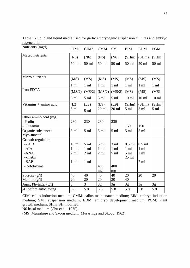
Table 1 - Solid and liquid media used for garlic embryogenic suspension cultures and embryoregeneration.Nutrients (mg/l) CIM1 CIM2 CMM SM EIM EDM PGM
Macro nutrients (N6)
50 ml
(N6)
50 ml
(N6)
50 ml
(N6)
50 ml
(SHm)
50 ml
(SHm)
50 ml
(SHm)
50 ml
Micro nutrients (MS)
1 ml
(MS)
1 ml
(MS)
1 ml
(MS)
1 ml
(MS)
1 ml
(MS)
1 ml
(MS)
1 mlIron EDTA (MS/2)
5 ml
(MS/2)
5 ml
(MS/2)
5 ml
(MS/2)
5 ml
(MS)
10 ml
(MS)
10 ml
(MS)
10 ml
Vitamins + amino acid (L2)5 ml
(L2)
5 ml
(L9)20 ml
(L9)20 ml
(SHm)5 ml
(SHm)5 ml
(SHm)5 ml
Other amino acid (mg)- Prolin- Glutamin
230 230 230 230150 150
Organic substancesMyo-inositol
5 ml 5 ml 5 ml 5 ml 5 ml 5 ml
Growth regulators-2.4.D-AIA-ANA-kinetin-BAP- cefotaxime
10 ml1 ml2 ml
1 ml
5 ml1 ml2 ml
1 ml
5 ml1 ml2 ml
400mg
3 ml1 ml5 ml
400mg
0.5 ml1 ml5 ml25 ml
0.5 ml1 ml2 ml
7 ml
Sucrose (g/l)Manitol (g/l)
4020
4020
4020
4020
2040
20 20
Agar, Phytagel (g/l) 3 3 3g 3g 3g 3g 3gPH before autoclaving 5.8 5.8 5.8 5.8 5.8 5.8 5.8
CIM: callus induction medium; CMM: callus maintenance medium; EIM: embryo inductionmedium; SM : suspension medium; EDM: emlbryo development medium; PGM: Plantgrowth medium; SHm: SH modified.N6 basal medium (Chu et al., 1975).(MS) Murashige and Skoog medium (Murashige and Skoog, 1962).
36
Several parameters must be taken into account for maintaining the suspension cultures and
guarantee the embryo production:
3.4.1 Initial culture densitiesThe quantities of SCV cultured in 5 ml of SM medium. Half of the sediment cell volume(SCV) after 28 days of culture measure about 0.24 ml.
3.4.2 Periodicity of medium renewalThe periodicity of medium renewal was the interval of 14 days between two successive
medium renewals.
3.4.3 Age of the suspension culturesFrom 1 to 15 months after initiation, the medium was renewed every 14 days and sub-culture
was performed every 28 days.
3.5 Regeneration of the suspension culture
The regeneration potential of cell suspension cultures was studied by plating on semi-solid
embryo induction medium (EIM, Table 1), as for regeneration from calluses. Two months
later, these cultures (Fig.2C) were collected from EIM and cultured on an embryo
development and germination medium (EDM, Tab.1) to develop whole plants (Fig.2D). This
SH medium was supplemented with BAP (0.7 mg l-1). Such regenerated plants were
transferred on a plant growth medium (PGM, Tab.1). The in vitro plants were further
developed in vitro according to the protocol reported by (Kahane et al., 1992).
3.6 Adaptation to in Vivo Conditions
Acclimatization was performed at rooted stages. After rinsing with sterile water, the in vitro
plants were established in compost soil and gradually hardened in greenhouse. The adaptation

37
of in vitro propagated and rooted plants to non sterile conditions was done in a steel-glass
greenhouse under air conditioner.
3.7 ELISA Test
The sanitary state of the materials was checked through DAS-ELISA with the DSMZ
(Deutsche Sammlung von Mikrooganismen und Zellkulturen, Gmbh) kit for the detection of
Onion Yellow Dwarf Virus (OYDV) and Leek Yellow Stripped Virus (LYSV), according to
the manufacturer's instructions (annexe 2). The different materials, pieces of leaves from
plantlets, or callus, and or sediment culture of suspension cultures, (0.4— 0.5 g for each) were
homogenized in 10 volumes of extraction buffer containing 0.5 ml Tween-20, 20 g PVP10
(polyvinylpyrolidone) and 2 g bovine serum albumin at pH 7.2. The samples were used
directly. Tissue samples from healthy and infected mother plants provided by this company
were used as negative and positive controls. Absorbance at 405 nm (A405) was read in an
ELISA microplate reader. The A405 values of healthy controls ranged from 0.00 to 0.05 and
values above 0.30 were considered as positive.
3.8 Virological Investigations
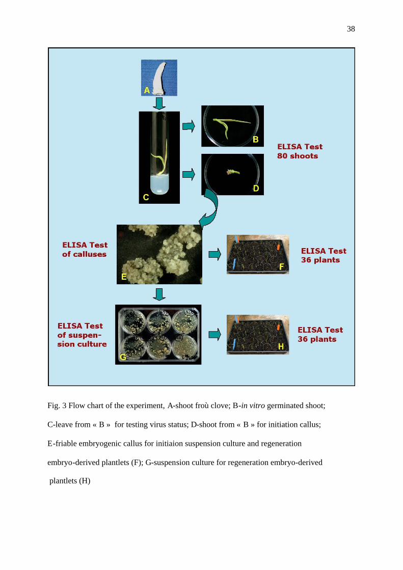
A flow chart (Fig.3) summarizes this experiment. A total of 80 in vitro germinated shoots
(Fig.3-A-D), were tested for the detection of Onion Yellow Dwarf Virus (OYDV) and Leek
Yellow Stripped Virus (LYSV). They came from 40 bulbs harvested in open field and 40
from plants maintaining in net house. Each in vitro germinated shoot correspond to one
distinct bulb and clove per bulb.
To determine the stage from the process for elimination of the virus, the same tests were
perfomed on callus (Fig.3-E), suspension cultures (Fig.3-G) and plantlets in green house
(Fig.3-F and H) issue of these calluses and suspension cultures. We tested 6 samples of
calluses, 6 of suspension culture, 36 plantlets issue of callus-derived somatic embryos, and 36
plantlets issue of suspension cultures-derived somatic embryos.
The putative virus-free plantlets will be tested a second time after a net house transfer phase.
38
Fig. 3 Flow chart of the experiment, A-shoot froù clove; B-in vitro germinated shoot;
C-leave from « B » for testing virus status; D-shoot from « B » for initiation callus;
E-friable embryogenic callus for initiaion suspension culture and regeneration
embryo-derived plantlets (F); G-suspension culture for regeneration embryo-derived
plantlets (H)

39
The eradication rate (ER) on plantlets observed in fine for each phase of the process will be
calculated as follows:
First ELISA test performed on green house plants:
ERi= (negative green house plants/tested green house plants) X 100
Second ELISA test performed on net house plants:
ERf= (negative green house plants/tested green house plants) X (negative net house
plants/tested net house plants) X 100
3.9 Recording
Observations on the following variables were performed:
- number of explants with callus on CIM2,
- number of callus containing embryogenic tissue after 6 months on CMM,
- number of friable and embryogenic calluses
- number of somatic embryo derived plantlets.
- measure of the SCV rate (final SCV / initial SCV), determining the growth of suspension
cultures during maintaining phase
- number of regenerated somatic embryos derived plantlets per aliquot of 0.1ml SCV from
each replicate.
- presence or no of viruses at each stage of the process (callus, suspension cultures,
plantlets regenerated from callus and from suspension cultures).
3.10 Statistical analysis
All data were analysed using SAS statistical package (SAS, 1982) version 8.2 for windows.
The experimental design consisted of 1 cultivar, 2 original virus status, originated virus free
(V-) and originated virus infected (V+), 6 replicates of 10 explants by original virus status.

40
The t-test procedure was performed for the different variables. The means were compared
using the two-sample t-test procedure.
4 RESULT
4.1 Callus production
After one month on CIM1, the explants increased in size but no callus formation was
observed. These explants sub-cultured on CIM2 for one month more, started producing callus
translucent, nodular type, and some time yellowish, also nodular (Fig.1D-E). These calluses
were generally located at leaf edges near a vein. Clumps of callus of both aspects were
transferred on CMM medium. According to t-test, no significant difference was found
between the virus statuses at 5% level (Table 3).
Table 3. Comparative callus induction from two original virus status
original virus statusType of callus induction %
Vrirus (-) Virus (+)
T test
Total number of explant 60 60
Primary callus 49 52 0.45 NS
Callus with embryo-genictissue
29 27 0.71 NS
Friable and embryogeniccallus
14 11 0.55 NS
NS: no significant difference between the original virus statusat 5% level by T test
4.2 Embryo induction from calluses

41
After two months on EIM, the embryogenic callus formed globular somatic embryos (Fig.1F).
The OYDV and LYSV infection of the donor plants had no significant effect on the
production of callus and callus showing embryos (Table 4).
Table 4. Regeneration of callus function of virological status
original virus statusType of callus induction %
Vrirus (-) Virus (+)
T test
Total number of petri dishes 6 6
Mean Nb. of plantletsregenerated per replicate
31.5 29.3 0.49 NS
NS: no significant difference between the virus statusat 5% level
4.3 Initiation of the suspension cultures
The growth of the cell suspension culture was on average quasi similar up to 8 month age.
The OYDV and LYSV infection of the donor plants had no significant effect on the growth
rate of the suspension cultures.

42
4.4 Regeneration of the suspension culture
Aliquots of cell suspension, plated on semi-solid embryo production medium (EIM, Table 1),
could produce numerous somatic embryos in 8 weeks (Fig.2C). They looked like large
structures (2-5 mm) with a smooth surface due to the presence of an epidermis. The best
combinations of growth regulators were 2,4-D/kinetin (0.05/2.5 mg l -1).
4.5 Conversion of somatic embryos into plants
The germination process occurred within 4 weeks. The embryos subsequently developed the
shoot and the root apex, and formed a complete plantlet (Fig.2D). The addition of BAP (0.7
mg l-1) increased it up to 30%. With 0 to 0.5 mg l-1 BAP, the plants presented a normal
development, but higher concentrations (2 mg l-1) promoted abnormalities like curled and
dark green leaves or multiple shooting (Fereol et al., 2005a). According to t-test, no
significant difference was found between the virus statuses at 5% level.
Fig. 4 growth rate of the SCV every month from 8 to 11 months age(subculture every month, start with an initial SCV of 0.24 ml)
4,38
4,58
3,80
4,00
4,20
4,40
4,60
4,80
5,00
5,20
age of the suspension
scv
rate V (-)
V (+)
98 10 11

43
Table 5. Regeneration of suspension culture function of virological statusoriginal virus statusType of callus
induction %
Vrirus (-) Virus (+)
T test
number of containerof culture
6 6
Mean Nb. ofplantletsregenerated (/0.1mlSCV)
109.1 102.1 0.43NS
NS: no significant difference between the virus statusat 5% level by T test
4.6 Adapting to in Vivo Conditions
The in vitro rooted plantlets were successfully transplanted in the glass greenhouse - 95%.
Two to three months after transplanting in green house, the plants were large enough for virustesting or transplanting to the field in net house. The efficiency of regeneration ranged from40 to 50%.
4.7 Virological investigations
Of 80 germinated shoots of garlic tested, 4 were found positive for OYDV, 3 positive to
LYSV, and 1 positive to these both viruses. Somatic embrogenesis strategy through
suspension culture was applied to this infected donor plant to test the possibility of
eliminating OYDV and LYSV along the process of somatic embryogenesis. The scheme
adopted is shown in Fig. 3. Somatic embryogenesis process was also applied to a virus free
donor from plants grown in net house. Although explants source were originally infected, all
of the issued materials, until now proved to be virus-free. No virus could be detected by
ELISA in any sample of neither callus, nor suspension culture and plant regenerated from
these tissue types. The plantlets were transferred to the green house, and will be tested again
for the presence of OYDV and LYSV.

44
5 DISCUSSION
Some viruses can be effectively eliminated from infected plants owing to their mode of
replication and their mechanism of movement within the plant. The most widely used method
for virus elimination is meristem tip culture. This technique takes advantage of the fact that
many viruses fail to invade the meristem region. Transfer of the meristem dome, to a culture
medium and development into a plantlet may lead to the elimination of a virus. Successful
elimination of Sugarcane mosaic virus and Fiji disease virus in sugarcane through apex or
bud culture has been reported (Leu, 1972; Wagih et al., 1995). There are reports of the
elimination of SCYLV using tissue culture (Chatenet et al., 2001; Fitch et al., 2001;
Parmessur and Dookun, 2000). By using meristem culture, only a certain percentage of the
plants developed from the excised meristems of infected plants are really virus free. The
reasons for this includes the failure to eliminate viruses in explants. Consequently it is
necessary to use combination of several techniques to produce virus-free garlic.
Callus proliferation may also lead to the elimination of viruses. Sugarcane yellow leaf virus
(SCYLV) was eliminated from sugar cane, with 100% success when plantlets were derived
from callus culture. A single callus subculture was found to be sufficient for eliminating
SCYLV from infected plant material (Parmessur et al., 2002).
Somatic embryogenesis may get rid of some viruses in different species. These results were
obtained with the grapevine (Goussard et al., 1991) and citrus (Carimi et al., 1994; De Pascale
et al., 1994). Once the results in somatic embryogenesis are satisfactory, it is necessary to
consider the efficiency of this method in the elimination of some viruses.
The results of the present study show that somatic embryogenesis has potentiality to be an
effective method for OYDV and LYSV elimination, since the viruses were eradicated with
100% success. It was possible to eliminate OYDV and LYSV from infected plants by somatic
embryogenesis process from leaf rolls. The uneven distribution of the virus in the different
tissues of the shoot may account for its elimination. Lack of connection between the somatic
embryos and the phloem limits movement of the virus or phytoplasma. Therefore plantlets
regenerated from Embryogenic process are free from both pathogens. This is the first report
using this method in garlic. A final testing 5 months after transplanting to soil might be

45
necessary to know if the plants remain virus-free. Latent infections must also be considered
and consequently, repeated tested of established plants will be necessary to certify that the
propagation material remain virus-free. The possible creation of variants by callus culture
should also be taken into account.
However, the success of this method depends on an effective method of virus detection; a
rigorous test is needed to ensure that the disease has been truly eliminated. The detection of
the virus needs to be effective even at an early stage when the plantlets are still growing in
vitro. At present, for viruses for which antisera are available, ELISA is the most widely used
detection assay (Spiegel et al., 1993). Since it has been shown that the levels of the virus can
fluctuate due to the seasonal variations, the detection with ELISA is not always possible
(Scott et al., 1989; Torrance and Dolby, 1984). Consequently, methods based in polymerase
chain reaction have started to be used (Spiegel et al., 1996). However, the prerequisite of
knowing the viral sequences in order to synthesize specific primers limits the application of
this approach to well-characterized viruses.
6 PERSPECTIVES
6.1 New approach to accelerate multiplication
The slow rate of conventional multiplication hampers the rapid dissemination of selected
elite genotypes. In order to accelerate this process, in vitro multiplication through axillary
shoots proliferation (Kahane et al., 1992) has been attempted and succeed at AVRDC.
However, new approach to accelerating more garlic multiplication has been investigated in
this joint AVRDC-CIRAD project. The method relies in developing embryogenic
suspension cultures from somatic embryogenic calli. Appropriate and efficient methods for
the induction of embryogenic callus, shoot development and suspension cultures have been
developed to make the technique useful for seedlings production (Fereol et al., 2002; Fereol
et al., 2005a; Fereol et al., 2005b). It would shorten the outside phase for base stock
establishment, and would provide a larger quantity of material so that the phase of field
multiplication could be limited to 2 cycles. This change would considerably limit the risk

46
of viral contamination during the cycles of field multiplication, and it would thus reduce
plant production costs.
6.2 Cryo-preservation Germplasm management
Since garlic is vegetatively propagated, genetic resources are traditionally maintained in
the field collections. This approach offers some advantages, because the accessions under
this type of conservation can be readily accessed and observed, allowing detailed evaluation.
However, field conservation risks the build up of various viral, bacterial or fungal
infections, which threatens long term preservation. Other natural hazards such as drought,
weather damage, vandalism, and human error may also undermine the collection.
Germplasm exchange over long distances may be limited due to the risks of disease
transfer through vegetative material. The high cost of maintaining field genebanks may
also limit the number of accessions that can be managed. Such constraints inhibit the
expansion of the collection to increase the diversity of the germplasm, and limit the
possibilities of conducting systematic studies of diversity or crop evolution. Consequently,
researchers are seeking alternative preservation methods, and cryopreservation offers
significant advantages and opportunities to complement other germplasm conservation
strategies (Engelmann, 2004).
Cryopreservation (liquid nitrogen, -196°C) has been applied for storage of different
plant species, many of tropical origin (Engelmann and Takagi, 2000; Keller, 2002;
Makowska et al., 1999; Niwata, 1995), providing cost-effective and long-term (if not
indefinite), stable conservation of genetic resources. It has been feasible also in garlic
(Baek et al., 2003; Keller et al., 2003; Keller et al., 2001; Kim et al., 2004). Various types of
tissues may be subjected to cryopreservation: Seeds, shoot tips or buds are currently
successfully conserved. Somatic embryos or cell suspension cultures might also prove
useful subject tissues, once plant regeneration in all genotypes is possible and off-types are
minimized. Coordinated with other techniques of germplasm management (field collections,
seed archives, etc), cryopreservation of a range of tissue types can add flexibility, reduced
costs and stability to the preservation and exploitation of genetic diversity (Sakai et al.,
2003).
47
6.3 Cryotherapy
Cryo-therapy: In 1997, (Brison et al., 1997) demonstrated for the first time that cryo-
treatment can not only be used for germplasm conservation but also for virus eradication.
Cryopreservation resulted in 50% virus-free in vitro plants from plum shoots infected with
plum pox virus. (Helliot et al., 2002) reported on successful CMV eradication by
cryopreservation of highly proliferating meristems of banana (cv. Williams BSJ, ITC.0570,
AAA). Thirty percent of the regenerated plants were found to be healthy by DAS-ELISA,
and this eradication rate was confirmed after a 6-month acclimatisation of plants in the
greenhouse. This technique might be investigated for garlic.
7 REFERENCES
Baek, H., H. Kim, E. Cho, Y. Chae, and F. Engelmann. 2003. Importance of explant size and
origin and of preconditioning treatments for cryopreservation of garlic shoot apices by
vitrification. CryoLetters 24:381-388.
Brison, M., M.d. Boucaud, A. Pierronnet, and F. Dosba. 1997. Effect of cryopreservation on
the sanitary state of a cv. Prunus rootstock experimentally contaminated with Plum
Pox Potyvirus. Plant Science Limerick 123:189-196.
Carimi, F., F. De Pascale, and G. Grescimanno. 1994. Somatic embryogenesis from styles of
lemon (Citrus limon). Plant Cell Tissue and Organ Culture 37:209-211.
Chatenet, M., C. Delage, M. Ripolles, M. Irey, B. Lockhart, and P. Rott. 2001. Detection of
Sugarcane yellow leaf virus in quarantine and production of virus-free sugarcane by
apical meristem culture. Plant Disease 85:1177-1180.
Chovelon, V., J.P. Leroux, and C. Dore. 1990. Sélection sanitaire de l'ail et de l'échalote:
culture de méristèmes et régénération de variétés. In: Doré C (ed) Cinquantenaire de la
culture in vitro. Colloques de l'INRA 51:142-150.
Chu, C., C. Wang, C. Sun, C. Hsu, K. Yin, C. Chu, and F. Bi. 1975. Establishment of an
efficient medium for anther culture of rice through comparative experiments of the
nitrogen source. Scientia Sinica 118:659-668.
De Pascale, F., F. Carimi, and G. Grescimanno. 1994. Somatic embryogenesis from styles of
different cultivars of Citrus limon (L.). Burm. Aust. J. Bot. 42:587-594.

48
Engelmann, F. 2004. Plant cryopreservation: Progress and prospects. In vitro Cellular &
Developmental Biology - Plant 40:427-433.
Engelmann, F., and H. Takagi. 2000. Cryopreservation of Tropical Plant Germplasm -
Current Research Progress and Applications JIRCAS, Tsukuba & IPGRI, Rome.
Faccioli, G., and F. Marani. 1998. Virus elimination by meristem tip culture and tip
micrografting, p. 346-380, In A. Hadidi, ed. Plant virus diseases control. APS Press,
The American Phytopathological Society, St Paul, MN, USA.
Fereol, L., V. Chovelon, S. Causse, N. Michaux-Ferriere, and R. Kahane. 2002. Evidence of a
somatic embryogenesis process for plant regeneration in garlic (Allium sativum L.).
Plant Cell Reports 21:197-203.
Fereol, L., V. Chovelon, S. Causse, M. Kalumvueziko, and R. Kahane. 2005a. Embryogenic
Cell Suspension Cultures of Garlic (Allium sativum L.) As Method For Mass
Propagation and Potential Material For Genetic Improvement. Acta Horticulturae
688:65-74.
Fereol, L., V. Chovelon, S. Causse, D. Triaire, I. Arnault, J. Auger, and R. Kahane. 2005b.
Establishment of embryogenic cell suspension cultures of garlic (Allium sativum L.),
plant regeneration and biochemical analyses. Plant Cell Reports 24:319-325.
Fitch, M., A. Lehrer, E. Komor, and P. Moore. 2001. Elimination of Sugarcane yellow leaf
virus from infected sugarcane plants by meristem tip culture visualized by tissue blot
immunoassay. Plant Pathology 50:676-680.
Goussard, P., J. Wiid, and G. Kasdorf. 1991. The effectiveness of in vitro Somatic
Embryogenesis in eliminating Fanleaf Virus and Leafroll Associated Viruses from
Grapevines. S. Afr. J. Enol. Vitic. 12:77-81.
Helliot, B., B. Panis, Y. Poumay, R. Swennen, P. Lepoivre, and E. Frison. 2002.
Cryopreservation for the elimination of cucumber mosaic or banana streak viruses
from banana (Musa spp.). Plant Cell Rep. 20:1117-1122.
Kahane, R., M. Rancillac, and B. Schweisguth. 1992. Bulbing in vitro in Allium species.
Allium Improve Newsl 2:18-20.
Keller, E.R. 2002. Cryopreservation of Allium sativum L. (Garlic). TOWILL. L.E., BAJAJ,
Y.P.S. (eds.) Biotechnology in Agriculture and Forestry, Cryopreservation of Plant
Germplasm II, Springer-Verl., Heidelberg 50:37-47.
Keller, E.R., P.L. Senula A <AU2> Forsline, C. Fideghelli, K. Richards, A. Meerow, H.
Knupffer, J. Niens, A. Stoner, E. Thorn, A.F. Tombolato, and D. Williams. 2003.
Germplasm preservation in Allium species: An integrated approach to store

49
morphologically characterised virus-free plant material viacryopreservation. ACTA
HORTICULTURAE:201-208.
Keller, E.R.J., A. Senula, and J. Armstrong. 2001. Progress in structuring and maintaining the
garlic (Allium sativum) diversity for the European genres project. Acta Horticulturae
No.555:189-193.
Kim, H., E. Cho, H. Baek, C. Kim, E. Keller, and F. Engelmann. 2004. Cryopreservation of
garlic shoot tips by vitrification: Effects of dehydration, Rewarming, unloading and
regrowth conditions. CryoLetters 25:59-70.
Leu, L. 1972. Freeing sugarcane from mosaic virus by apical meristem and tissue culture.
Taiwan Sugar Experiment Station Report 57:57-63.
Lot, H., B. Delécolle, G. Bocardo, C. Marzachi, and R. Milne. 1994. Partial characterization
of retrovirus-like particles associated with garlic dwarf disease. Plant Pathol 43:537-
546.
Makowska, Z., J. Keller, and F. Engelmann. 1999. Cryopreservation of apices isolated from
garlic (Allium sativum L.) bulbils and cloves. Cryo-letters. 20:175-182.
Murashige, T., and F. Skoog. 1962. A revised medium for rapid growth and biosassay with
tobacco tissue culture. Physiol. plants 15:473-497.
Niwata, E. 1995. Cryopreservation of apical meristems of garlic (Allium sativum L.) and high
subsequent plant regeneration. Cryo-letters. 16:102-107.
Parmessur and Dookun (ed.) 2000. Proceeding of the 6th ISSCT Sugarcane Pathology
Workshop, Thailand. July 16-23.
Parmessur, Y., S. Aljanabi, S. Saumtally, and A. Dookun-Saumtally. 2002. Sugarcane yellow
leaf virus and sugarcane yellows phytoplasma: elimination by tissue culture. Plant
Pathology 51:561-566.
N. I. o. A. B. Cho EG & Kim HH, Suwon (ed.) 2003. in Proceedings of the International
Workshop on Cryopreservation of Bio-Genetic Resources, Suwon, Korea. 3-5 June
2003. International Technical Cooperation Center, Rural Development Administration.
SAS, I. 1982. SAS user's guide: statistics. SAS Institute, Cary, N.C.
Scott, S., W. Barnett, and P. Burrows. 1989. Prunus necrotic ringspot virus in selected peach
orchards in South Carolina. Plant Disease 73:913-916.
Spiegel, S., E. Frison, and R. Converse. 1993. Recent development in therapy and virus
detection procedures for international movement of clonal plant germplasm. Plant
Disease 77:1176-1180.
50
Spiegel, S., S. Scott, V. Bowman-Vance, Y. Tam, N. Galiakparov, and A. Rosner. 1996.
Improved detection of Prunus necrotic ringspot virus by the polymerase chain reaction.
European Journal of Plant Pathology 102:681-685.
Torrance, L., and C. Dolby. 1984. Sampling conditions for reliable routine detection by
enzyme-linked immunosorbent assay of three ilarviruses in fruit trees. Ann. Appl. Biol.
104:267-276.
Van Dijk, P. 1991. Mite-borne virus isolates from cultivated Allium species and their
classification into two new rymoviruses in the family Potyviridae. Neth J Plant Pathol
97:381-399.
Van Dijk, P. 1993. Survey and characterization of potyviruses and their strains of Allium
species. Neth J Plant Pathol 99:1-48.
Wagih, M., G. Gordon, C. Ryan, and S. Adkins. 1995. Development of an axillary bud culture
technique for Fiji disease virus elimination in sugarcane. Australian Journal of Botany
43:135-143.
Walkey, D. 1990. Virus diseases. In: Rabinovitch HD, Brewster JL (eds) Onions and Allied
Crops 1:191-211.
Walkey, D., and M. Webb. 1968. Virus in plant apical meristem. J. Gen. Virol. 3:311-313.
Walkey, D., M. Webb, C. Bolland, and A. Miller. 1987. Production of virus-free garlic
(Allium sativum L.) and shallot (A. ascalonicum L.) by meristem tip culture. J Hortic
Sci 60:627-634.

51
Abstract
Garlic (Allium sativum L.) is threatened by diseases, including a number of important viral
diseases which represent a constraint on garlic production and on germplasm movement from
country to country. The latter is especially important for farmers who are waiting to benefit
from pest- and disease-resistant varieties, either naturally occurring germplasm or improved
varieties produced by breeding programmes. A significant number of potentially important
and improved varieties from breeding programme are infected with viruses. Most of these
materials are infected with OYDV and LYSV. AVRDC has therefore been very active in
establishing a system for the safe international movement of garlic germplasm.
To make these accessions available, a programme of virus elimination was carried on in the
Plant Virology Unit (Dr. Green). In vitro technique, such meristem tip cultures were used to
produce virus-free plantings of different clones infected with OYDV and LYSV. The health
status of regenerated material was checked on in vitro plants through ELISA. The putative
virus-free material was then tested a second time after greenhouse acclimatisation.
The use of tissue culture was investigated as a means to eliminate both OYDV and LYSV
from infected cultivars. An infected cultivar was induced to form Embryogenic callus in vitro
using leaf rolls as explants, and then suspension cultures. After ten subcultures on one hand of
the callus, and on the other hand of the suspension culture, this cultivar was successfully
regenerated and tested for OYDV and LYSV. No pathogen could be detected in any
regenerated plantlets. All the regenerated plants remained free from both OYDV and LYSV
over a period of 3 months in the glasshouse.
Somatic embryos were obtained from young leaf rolls as explants. Forty 3-month-old plantlets
originating from leaf rolls culture-derived somatic embryos, twenty from callus source, and
twenty from suspension gave negative reactions when assayed by serology for the presence of
the viruses. It was concluded that, similar to other species (e.g. grapevine, sugarcane), somatic
embryogenesis opens new possibilities for the sanitary improvement of garlic.
Key words: somatic embryogenesis, OYDV, LYSV
Abbreviations: 2,4-D, dichlorophenoxy acetic acid; BAP, 6-benzyl aminopurine; N6, salts of
Chu et al. (1975) basal medium; SCV, Sediment Cell Volume; IAA, indol acetic acid; NAA,
naphthalene acetic acid;

52
8 Annexe1. Stock Solutions of media used for garlic embryogenesisMacro nutrients (mg) (N6) (SHm)
Concentration Final
Solmg
X 20 Final
Solmg
X 20
Quantity stock solution per
liter of final solution
50 ml 50ml
Quantity of stock Solution todo
1000ml
1000ml
-KNO3 2850 57 g 2500 50g-(NH4)2SO4 463 9.26g-NH4H2PO4 300 6g-KH2PO4, 2 H2O 400 8g-CaCl2, 2H2O 166 3.32g 440 8.8g-MgSO4, 7H2O 185 3.7g 400 8g
Micro nutrients (mg) (MS)
Concentration FinalSol
X1000
Quantity mother Sol for
1 l of final Sol
1 ml
Quantity of mother Sol to do 1000ml
- H3BO3 6,2mg 6200- KI 0,83 830- MnSo4, 4H2O 16,7 1670- ZnSo4, 7H20 8,6 8600- CoCl2, 6H2O 0,025 25- CuSo4, 5H2O 0,025 25- Na2MoO4, 2H2O 0, 25 250
Vitamins (mg) L2 L9 SHm
Concentration FinalSol
X200
Final
sol
X 50 Final
sol
X 200
Quantity mother Sol for1 literof final Sol
5 ml 20ml
5 ml
Quantity mother Sol to do 100 ml 1000ml
100 ml
- Glycin (mg) 20 400 2 100- Nic ac (mg) 1 50 5 100- pyridoxine (mg) 1 50 0.5 10- Thiamine HCl (mg) 40 800 10 500 5 100- serin 10 500- cystein (mg) 20 400 20 1000

53
Fe- EDTA (mg)
Concentration FinalSol
X 100Quantity mother Sol for
1 l of final Sol
10 ml
Quantity mother Sol to do 1000 ml- FeSO4, 7H2O 27.8 2.785 g in 500 ml- Na2 EDTA 37.3 3.725 g in 500 ml H20,
Pour Na2 EDTAheat, in FeSO4heat, progessivelyto avoidprecipitation
Organic substances
Concentration Final Sol X 200Quantity mother Sol for
1 l of final Sol
5 ml
Quantity mother Sol to do 500 ml- Malt extract 400 mg 40 g- Casein Hydrolysat 100 10 g- Myo-Inositol 400 40g
Growth regulator (mg)
- 2.4.D 1mg =10ml
50mg+ 2,5ml alcohol90° + 500ml H2O
-NAA 1mg =
10 ml
50mg+ 1 tablet KOH+few H2O: dissolve,then complete500mlH2O
- IAA 1mg =
10 ml
50mg+ 2,5ml alcohol90° + 500ml H2O
- BAP 1mg =
10 ml
50mg+ 1 tablet
KOH+ 500ml H2O- Kinetin 1mg =
10 ml
50mg+ 1 tablet
KOH+ 500ml H2O

54
9 Annexe 2.

55

56
TRAINING COURSE ON “THE APPLICATION OF BIO-MOLECULAR TOOLSFOR BREEDING”
IN CIRAD, FRANCE
for Ms Cherng Shin-jiun (Vicky)from September, 1st to October, 29th 2005
Responsible : Dr Marie France Duval
This action is situated in the objective to prepare to bring more tools in the onion breeding
program.
In the framework of the MOU between NSC (ROC) and INRA-CIRAD (France), according
to the MOU between CIRAD and AVRDC, and following the general agreement to
strengthen their scientific collaboration, CIRAD Department Flhor was pleased to confirm
the invitation of an agent of AVRDC (Ms Cherng Shin-jiun, Vicky) to a training on “The
application of bio-molecular tools for breeding”.
CIRAD offered to pay the following costs : boarding and training fees.
NSC paid the travel air ticket.
PROGRAM PROPOSED:
Practical training :
1. Basics on molecular markers and their use. Dr Jean-Louis Noyer
2. DNA extraction and purification
3. SSR (Single Sequence Repeat) markers
4. AFLP (Amplified Fragment Length Polymorphism) markers
5. Methodologies used for data scoring and analysis
6. Fluorescence in situ hybridization (FISH) and genomic in situ hybridization (GISH)
SSR (Single Sequence Repeat) markers
Discussions with the Quality and Safety lab staff

57
Meeting with staffs of CIRAD:
1. Dr. Jean –Marc Lacape, UMR-PIA CIRAD
Marker assistant selection on cotton quality
2. Dr. Norbert Billotte, UMR-PIA CIRAD
Marker assistant selection and maping on palm tree
3. Dr. Jean-Louis Noyer, UMR-PIA CIRAD
Chief of the team on Diversity study by molecular marker on cultivated plants
4. Technetium Magey Rodier-Goud, UMR-PIA
Cytogenetic platform on banana, rubber tree etc.
5. Dr. Marie-France Duval, UPR-MV CIRAD
SSR on pineapple and mongo and its apply to other crops
6. Remi Kahane, Fruit and Horticultural Crops Deportment
Garlic research in CIRAD
Meeting with visitors:
1. Dr. E.R.J Keller, Gene bank IPK, Germany
Introduction the gene bank of Institute of plant genetic and crop plant research
In vitro storage and cryopreservation of Allium crops.
2. Dr. Chris Kik, CGN , Netherlands
Seminar on introduction of the Center for Genetic Resources (CGN)
Garlic and Health project
Allium research at Plant Research International
Field trip to onion production area in Cevennes

58
TRAINING COURSE ON CRYO-PRESERVATION IN CROP PLANTS :TECHNICAL ASPECTS,
CELL BIOLOGY AND PERSPECTIVESIN CIRAD, FRANCE
for Ms Chang, Ching-huan (Jessica)from October, 15th to December, 10th 2005
Responsible CIRAD persons : Dr R. Kahane/ Dr F. Cote and Dr P. Ollitrault
This action is situated in the objective to prepare to set up the technique of cryo-
preservation in the management of germplasm conservation of AVRDC.
In the framework of the MOU between NSC (ROC) and INRA-CIRAD (France), according to
the MOU between CIRAD and AVRDC, and following the general agreement to strengthen
their scientific collaboration, CIRAD Department Flhor was pleased to confirm the invitation
of an agent of AVRDC (Mrs Chang Ching-huan, Jessica) to a training on “The application of
Cryo-preservation in crop plants : technical aspects, cell biology and perspectives”.
CIRAD offered to pay the following costs: boarding and training fees.
NSC paid the travel air ticket.
PROGRAM PROPOSED:
Meeting with Cirad staff
visit of the laboratories “Banana” CIRAD and “Coffee” IRD
Discussions about cryo-preservation technology. Régis Domergue
Cryo-preservation techniques and their use. Régis Domergue
Somatic embryogenesis and embryo rescue in a breeding programme. D. Dambier
Protoplasts fusion and it use in Citrus breeding programme. D. Dambier, P. Ollitrault

59
Bibliography and visit of other laboratories (Vitropic, Biotrop). R. Kahane
Discussions with the Quality and Safety lab staff.
During the training periods has been organized various meetings with researchers involved in
cellular biology programmes and the applied projects (use in breeding, in genetic resources
management, etc… ), according to their availability.

60
PROPOSAL: ABSTRACT
CRYOPRESERVATION FOR GERMPLASM CONSERVATION AND SUPPLY OF
DISEASE FREE SEEDLINGS OF VEGETATIVELY PROPAGATED
VEGETABLES
Dr. Leonidas FEREOL
Dr. Liwayway M. ENGLE
Dr. Sylvia GREEN
Dr. Haeng-Hoon KIM
(DRAFT, December 2005)
Proposal to submit to DED, GTZ, IPGRI …
1 General objectives and expected achievements
1.1 Strategic objectives addressed
The goal of this project is to advance the development of new collected Allium and
improved Allium varieties through the development of optimized genetic resources systems,
thereby ensuring the sustainable cultivation of Allium in different regions of the tropics.
In order to address economic, nutritional and healthy shortfalls in the developing world,
the project brings together major scientific expertise in plant breeding, biotechnology, and
germplasm management. New approaches to vegetable pathology, variety development,
conservation, multiplication and distribution will be developed. These approaches might
serve as a model for development strategies in other vegetable crops.
This proposal presents the rationale, scientific background, technical details, and workplan to
achieve the following objectives:
Objective 1. Improvement of commercial vegetable crops varieties adapted specifically to
tropical regions through multidisciplinary approaches, combining pathology, physiology,
breeding and biotechnology expertise.
Objective 2. Development and efficient use of cryopreservation for germplasm
management and cryo-therapy against some pathogens.

61
1.2 Expected achievements
The objectives of the project will be achieved by combining techniques with innovative
biotechnical approaches, including: in vitro clean up against diseases, embryogenesis and cell
suspension culture techniques, and cryopreservation strategies. We expect to reach the
following achievements:
- Set up of cryopreservation technique to AVRDC germplasm conservation.
- Obtaining new way for virus elimination such as therapy through tissue culture
and cryo-therapy.
- Speed up the distribution of new cultivars by new propagation processes.
2 Exploitation and dissemination results
Introduction of improved vegetables materials particularly garlic and other clonally
propagated vegetable crops for the tropics will strengthen the international competition
force of tropical vegetables growers by giving added value to their product, also by
realistic sustained health claims concerning these species. The programme may lead to the
development of new potent phyto-pharmaceuticals.
The benefits of this project to the human quality of life and management of living
resources programme are manifold:
- Contribute to comprehensive overview of the world gene-pool of Allium and other
vegetable crops.
- The availability of a large documented collection of Allium and other clonally
propagated vegetable crops particularly for tropical regions represents a unique starting point
for future agronomic research in these species.
- Efficient cryopreservation protocols shall be applied in international and national
genebanks
Capacity of the partnership for industrial exploitation
Breeding companies, farmer’s groups and pharmaceutical industries may be
interested in this project.

62
Plant Cell Report (2005), 24: 319-325
PUBLICATION 1: ABSTRACT
ESTABLISHMENT OF EMBRYOGENIC CELL SUSPENSION CULTURES OF
GARLIC (ALLIUM SATIVUM L.), PLANT REGENERATION AND
BIOCHEMICAL ANALYSES
L. Fereol 1,2, V. Chovelon, S. Causse, D. Triaire , I. Arnault, J. Auger and R. Kahane
(1) CIRAD-FLHOR, PMH, Boulevard de la Lironde, 34398 Montpellier cedex 5, France
(2) AVRDC, P.O. Box 42, Shanhua, Tainan, Taiwan 741, R.O.C.
Corresponding author: L. Fereol, E-mail: [email protected], Tel: (+886) 6 583 7801
ext 314; fax: (+886) 6 583 9000
Abstract
Embryogenic cell suspension cultures of garlic were initiated in liquid medium, from friable
embryogenic tissue. The optimal parameters were for culture maintenance: 1) an initial cell
density of 1-4% (vol/vol); 2) medium renewal every 14 days and subculturing every 28 days;
and 3) a low 2,4-D concentration (0.1-0.3 mg/l). Cultures regenerated throughout 14 months.
The cell suspension cultures differentiated embryos after transfer to a semi-solid embryo
induction medium. Histological studies confirmed and characterised the embryogenic nature
of the process. Forty percent of these embryos were converted into plantlets, which produced
micro bulbs in vitro. The composition of sulphur compounds of micro bulbs from cell
suspension embryo derived plantlets differed slightly from those produced by in vitro shoot
proliferation derived plantlets, but after two cycles of multiplication in the field these
difference was not maintained.
Key words: somatic embryogenesis, histology,sulphur compounds

63
Acta Horticulturae, (2005) 688 : 65-74
PUBLICATION 2: ABSTRACT
EMBRYOGENIC CELL SUSPENSION CULTURES OF GARLIC (ALLIUMSATIVUM L.) AS METHOD FOR MASS PROPAGATION AND POTENTIALMATERIAL FOR GENETIC IMPROVEMENT
L. Fereol
AVRDC World Vegetable Center, P.O. Box 42, Shanhua, Tainan, 741 Taiwan R.O.C.
V. Chovelon
INRA, Pathologie végétale, BP 94, 84143 Montfavet, France
S. Causse, M.L. Kalumvueziko and R. Kahane
CIRAD, TA50/PS4, bld de la Lironde, 34398 Montpellier cedex 5, France
Keywords : somatic embryogenesis, histology, plant regeneration, multiplication rate
AbstractEmbryogenic calluses were induced from young leaf explants of garlic (Allium
sativum L). Four cultivars, Rouge de la Réunion, Messidrome, Morasol and Printanor havebeen successfully tested. These calluses expressed up to 90% of embryogenic callusesdifferentiating globular somatic embryos after 2 months on N6 modified mediumsupplemented with 2,4-D (0.1 mg l-1) and kinetin (0.5 mg l-1). Embryogenic calluses wereused to establish cell suspension cultures of the above-mentioned cultivars. Friable calluseswere induced from compact ones, and could give rise the production of cell suspensioncultures composed of small aggregates of embryogenic cells. These suspension cultures weremaintained in liquid medium based on N6 modified macro-nutrients and supplemented with2,4-D/benzyladenine (0.3 mg l-1/0.1mg l-1). The packed cell volume (PCV) of the suspensioncultures increase 2-fold in a 2-week period. These cell suspension cultures led to successfulregeneration of mature embryos and their conversion into plantlets. Optimal embryoregeneration efficiency was obtained after plating on semi-solid medium base on N6 macro-nutrients and a balance in 2,4-D/Kinetin (0.1mg l-1/0.5mg l-1). A large number of somaticembryos (potentially 8 x 10⁹ to 1011) could be produced per year for each cultivar. Theconversion into plantlet was approximately 50%. Plants were successfully acclimatised ingreenhouse. Histological analyses were performed along the suspension cultures andregeneration process, and helped for establishing the sequence of culture media. The somaticembryogenic nature was confirmed by single cell origin and polar development of theregenerants. This protocol was used in a goal of mass propagation of garlic plants true to theoriginal type. It would be a key tool for biotechnologies in genetic improvement of garlic.
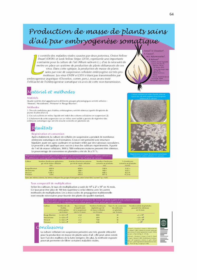
64
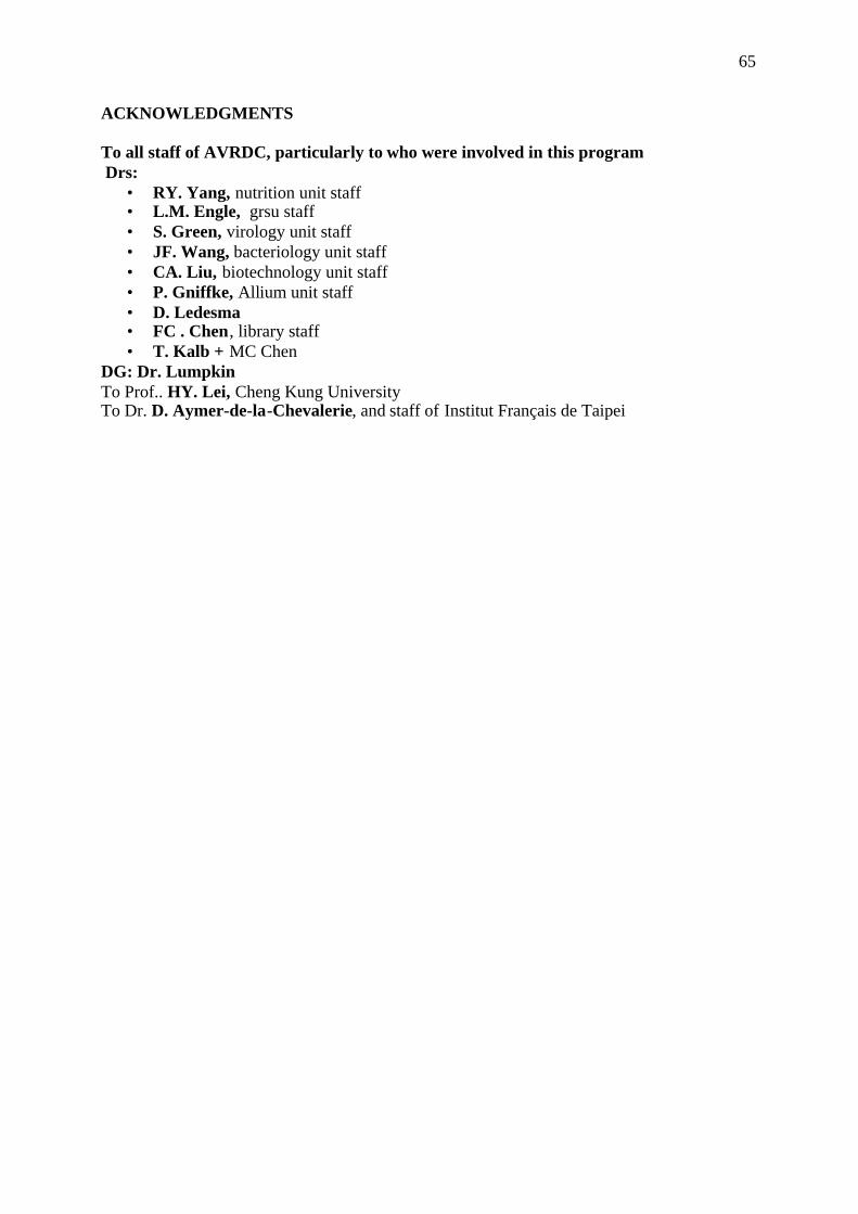
65
ACKNOWLEDGMENTS
To all staff of AVRDC, particularly to who were involved in this programDrs:
• RY. Yang, nutrition unit staff• L.M. Engle, grsu staff• S. Green, virology unit staff• JF. Wang, bacteriology unit staff• CA. Liu, biotechnology unit staff• P. Gniffke, Allium unit staff• D. Ledesma• FC . Chen, library staff• T. Kalb + MC Chen
DG: Dr. LumpkinTo Prof.. HY. Lei, Cheng Kung UniversityTo Dr. D. Aymer-de-la-Chevalerie, and staff of Institut Français de Taipei