Embed Size (px)
Citation preview

Colitis Diagnosis and Therapeutic Strategies
How, what and when to biopsy?Dr Bryan F Warren
Consultant Gastrointestinal Pathologist, Oxford
Falk Symposium 147: Birmingham 2005

!
•Communication and context•Biopsy of normal mucosa •Timing of the biopsy•Biopsy of ulcers•Biopsy of polyps•Distribution of colitis•Biopsies after surgery•Gastroduodenal biopsies•Biopsies in diversion•Biopsies in pouch dysfunction •Biopsies in dysplasia
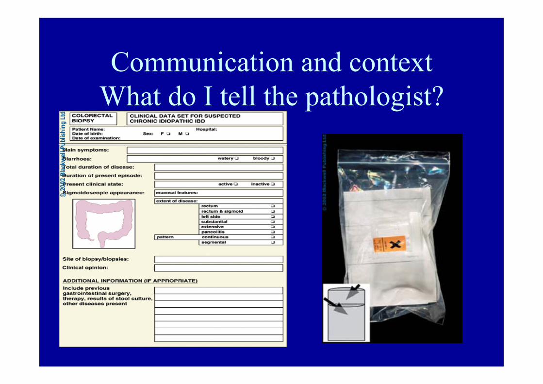
Communication and contextWhat do I tell the pathologist?

s


ABNORMAL APPEARANCESEarly biopsy - IBD vs infection
• 6 weeks to develop crypt architectural distortion
• Basal lymphoid follicles and aggregates• Basal plasmacytosis
• Oedema, superficial neutrophils• Mimics early CD

Biopsy of the “normal” mucosa.Normal racemosely branched
crypt

Normal caecal biopsy

Normal terminal ileal biopsy=“complete colonoscopy”
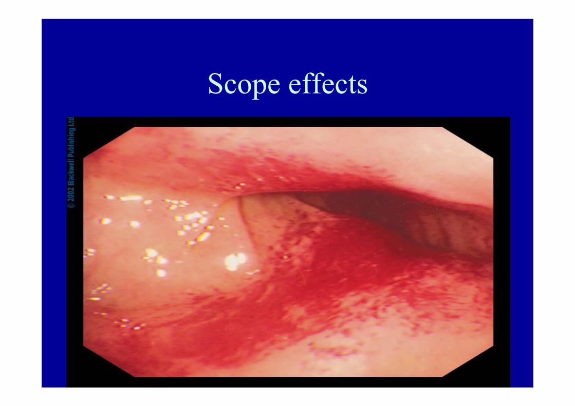
Scope effects
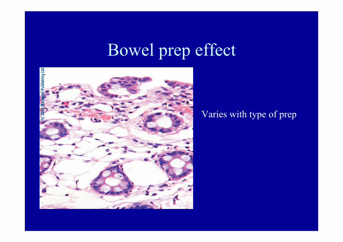
Bowel prep effect
Varies with type of prep

Diarrhoea with normal colonoscopy
Minimal change colitis
Microscopic colitis
Isolated granuloma in Crohn’s di
Non colonic causes

Microscopic colitis - definition and terminology
‘Accepted definition’
Colitis with no endoscopic findings and with ‘specific’ histological features:
CLASSICAL TERMS:
Collagenous colitis and lymphocytic colitis (Lindstrom).
CLASSICAL PATIENT:
Elderly lady with persistent watery, bloodless diarrhoea (as common as UC in this age group - Jarnerot).
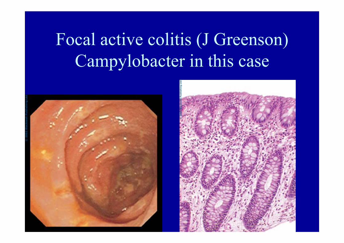
Focal active colitis (J Greenson)Campylobacter in this case
Biopsy of ulcers
• Ulcer• CMV• Amoebae• Solitary ulcer/mucosal prolapse syndrome• Adenocarcinoma
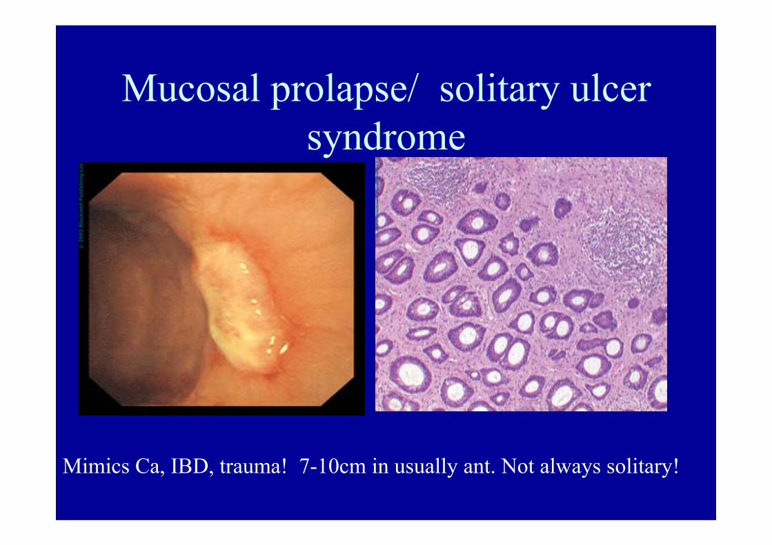
Mucosal prolapse/ solitary ulcer syndrome
Mimics Ca, IBD, trauma! 7-10cm in usually ant. Not always solitary!
Biopsy of polyps
• Benign inflammatory polyps• Lipoma• CMV• Amoebae• Amyloid• Adenoma• DALMs• adenocarcinoma
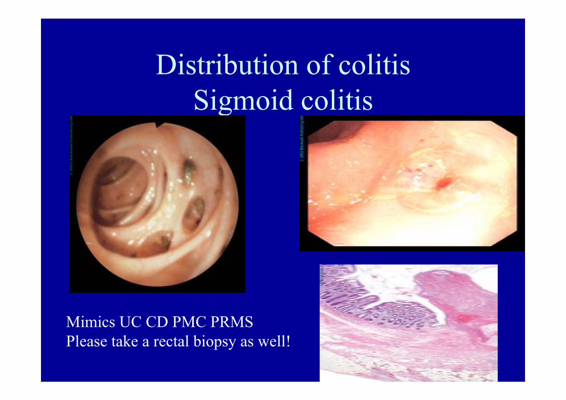
Distribution of colitisSigmoid colitis
Mimics UC CD PMC PRMSPlease take a rectal biopsy as well!

UC


Biopsy –severe UC
Crypts rupture downwards
Villiform surface
?to score activity or not?No except research PMN in lp after Rx=early recurrence

Biopsy pathology UC• Crypt architectural distortion
takes 6 weeks• Diffuse changes-• Architecture, mucin depletion,
chronic inflammation, acute inflammation
• Rectum most severe• Distribution of changes in a
biopsy and in a biopsy series.• Catch-patchiness-post treatment
or at junction of diseased and normal, or in caecal patch.
• IF BIOPSIES ALL IN SAME POT - HARD TO REPORT!!
Early disease-diffuse Chronic inflammationand basal plasma cells
UC after treatment
Skip lesions in UC
Acceptable ones:• Appendix –Davison and Dixon• Caecal patch – D‘Haens
Not contraindications to pouch surgery.

Caecal patch in UC
Courtesy of Dr Axel von Herbay
Tell the pathologist What you sawPlease label biopsy SitesNot all in same pot!
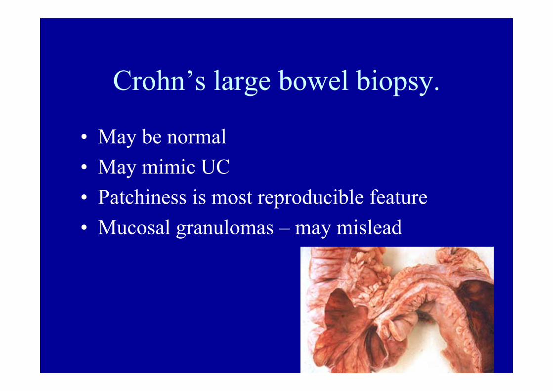
Crohn’s large bowel biopsy.
• May be normal• May mimic UC• Patchiness is most reproducible feature• Mucosal granulomas – may mislead
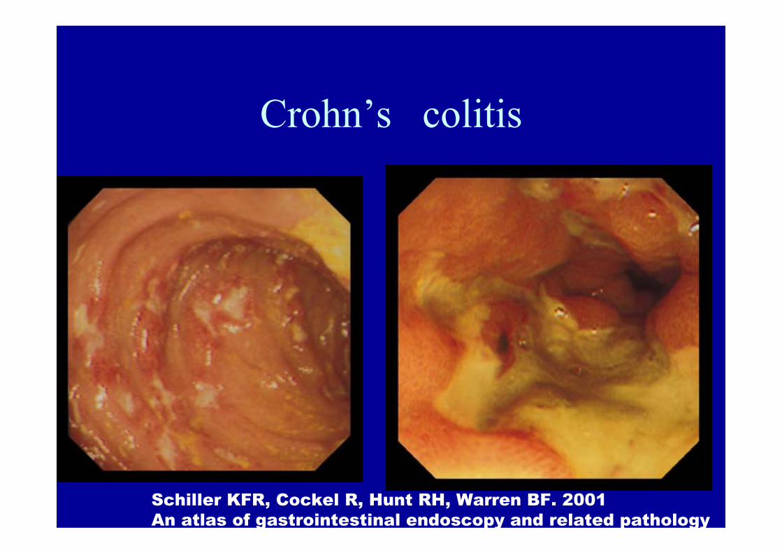
Crohn’s colitis
Schiller KFR, Cockel R, Hunt RH, Warren BF. 2001An atlas of gastrointestinal endoscopy and related pathology

Crohn’s colitis
Focal erosions and Focal inflammation
Perineural chronic inflammationand granuloma.
Aphthous ulcer
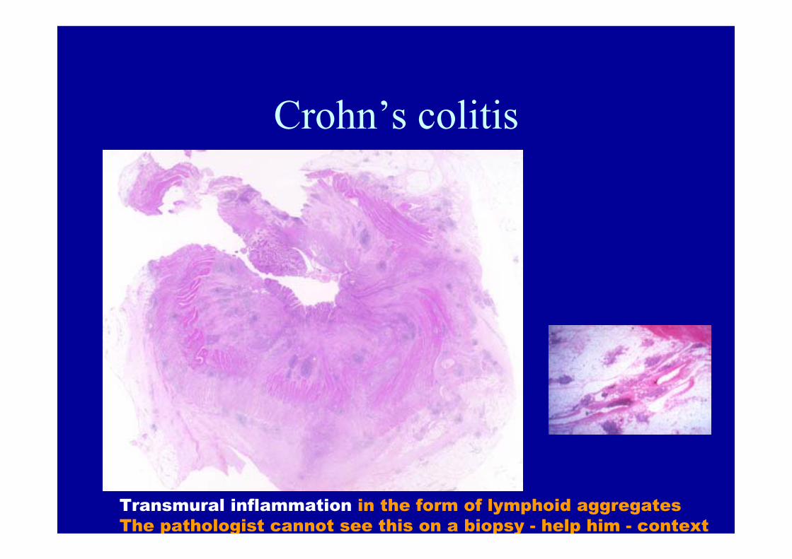
Crohn’s colitis
Transmural inflammation in the form of lymphoid aggregatesThe pathologist cannot see this on a biopsy - help him - context

Crohn’s disease - fat wrapping

Crohn’s colitis-terminal ileal disease.
Backwash ileitis in UC or Crohn’s disease? Ileal biopsies maybe difficult.
When does ulcerative colitis mimic Crohn’s colitis?
• Granulomas in response to crypt damage• Cryptolytic granulomas• Patchiness of disease after treatment• Resolution of histological changes after treatment• Fulminant colitis• Diversion proctitis in UC• SKIP LESIONS
– Caecal patch– Appendix

Granuloma in response to crypt damage-neutrophils and mucin.
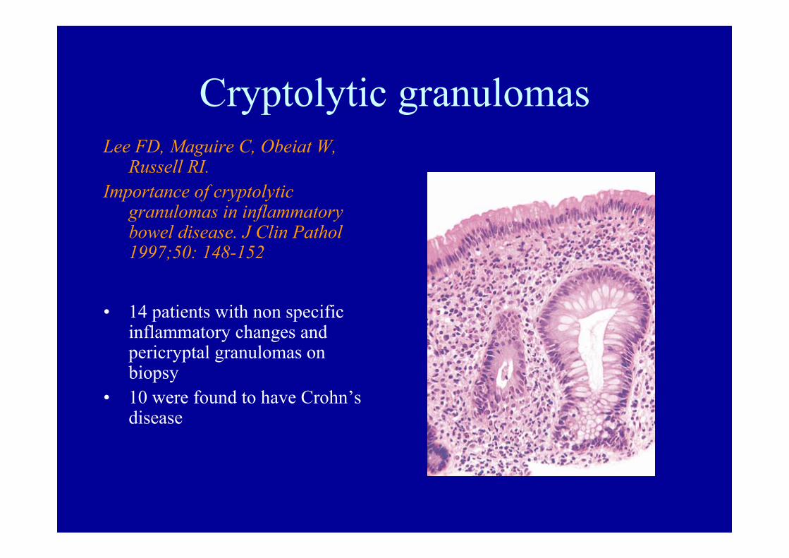
Cryptolytic granulomasLee FD, Maguire C, Obeiat W,
Russell RI.Importance of cryptolytic
granulomas in inflammatory bowel disease. J Clin Pathol 1997;50: 148-152
• 14 patients with non specific inflammatory changes and pericryptal granulomas on biopsy
• 10 were found to have Crohn’s disease
Importance of cryptolytic lesions and pericryptal granulomas in inflammatory bowel disease
• Warren BF, Shepherd NA, Price AB, Williams GT. J Clin Pathol 1997;50:880-881
Price AB.• Cryptolytic granulomas found in infections,
UC, diversion, diverticular disease etc.

Upper GI biopsies
• Biopsy of normal mucosa may reveal focal active gastritis and or focal active duodenitis in Crohn’s disease or granulomas.
• Stolte M.• Riddell RH.
When is it difficult to differentiate CD colitis and UC?
• Fulminant colitis• After treatment of UC• When rare variants of UC are not
recognised.
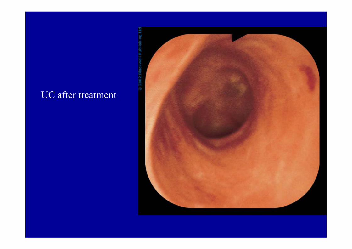
UC after treatment

Follow up/ post treatment biopsies in IBD
• Is it still IBD/UC/Crohn’s disease• Has it got better? Was it IBD after all?• Is it nowcomplicated by infection/PMC?• Go back to the original pretreatment series!

PMC

CMV in UC
Beware of superimposed infectionAfter immunosuppressive treatment.

Quiescent UC
May have only architectural distortion, =/-paneth cells,may return to ‘normal’-review original biopsies ? Infection.
PolypFlat mucosa
DON’T JUST BIOPSY THE POLYP

Biopsies after surgery
• Ileostomy end - non specific changes may misinterpret as Crohn’s disease
• Anastomotic biopsies in Crohn’s• Diversion• Pouch biopsies• Prepouch biopsies• Columnar cuff biopsies

Diversion in UC• Transmural inflammation• Granulomas• PMC like change• Mimics Crohn’s• It is UC and not a contraindication
to pouch surgery.• Seen as part of the three stage
pouch procedure.• Comforting if this occurs-helps
confirm pouch has been made in UC! PUT THE BIOPSIES IN CONTEXT FOR THE PATHOLOGIST!

Diverted Crohn’s colitis

Diverted Crohn’s colitis

Diversion in IBD
• CD often resolves• UC becomes worse and mimics CD
Pouchitis – diagnosis
Clinical, endoscopic and histological• Clinical features – diarrhoea/discharge, systemic
symptoms. • Endoscopic features - diffuse inflammation and
ulceration.• Histological features - ulceration and severe
acute

Diagnosis
Endoscopic and histological evaluation together with symptom assessment are required to diagnose pouchitis.
Shen B, Achkar JP, Lashner BA, Ormsby AH, Remzi FH, Bevins CL, Brzezinski 261-7A, Petras RE, Fazio VW.
Gastroenterology 2001; 121:


Role of histopathology
• Adaptive change or pouchitis?– Colonic metaplasia – Colonic phenotypic change– Other causes of inflammation in the pouch– Cuffitis– Prepouch ileitis– Dysplasia?/ Cancer?
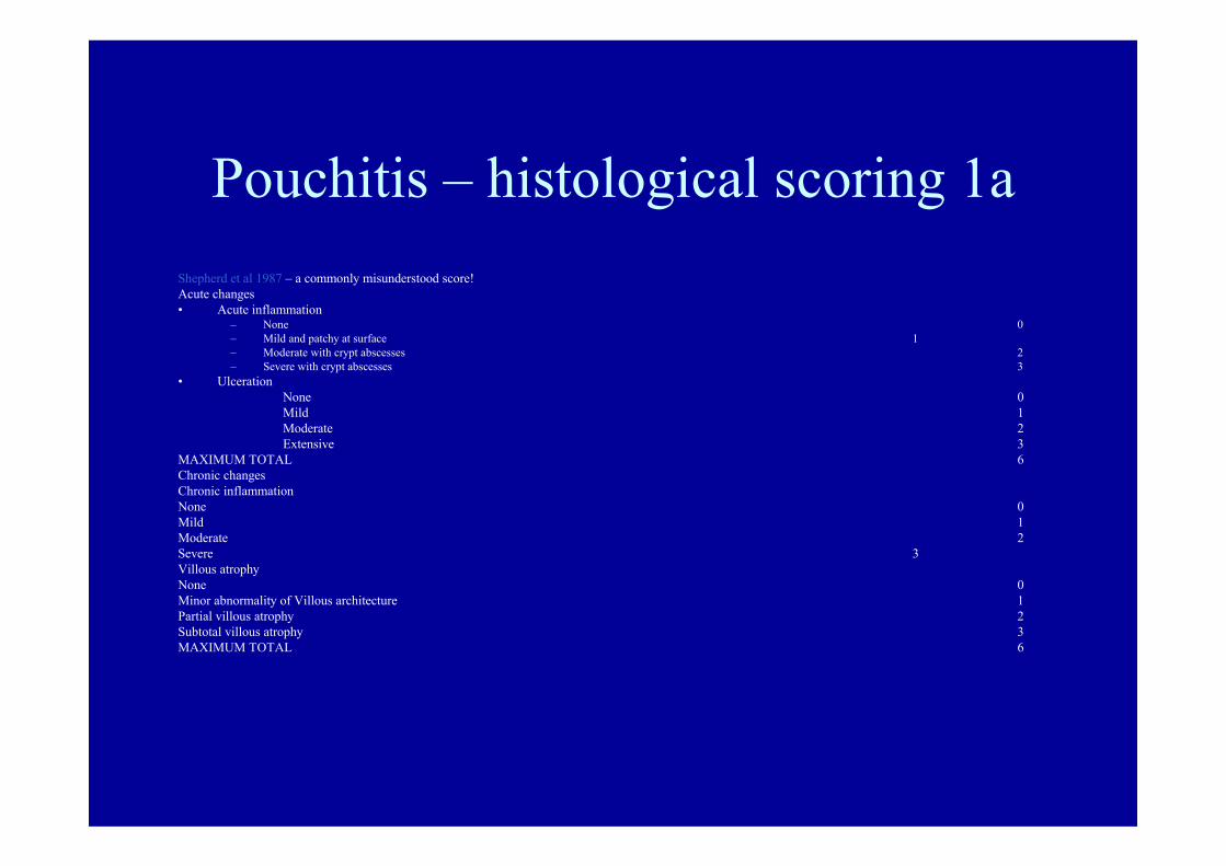
Pouchitis – histological scoring 1aShepherd et al 1987 – a commonly misunderstood score!Acute changes• Acute inflammation
– None 0– Mild and patchy at surface 1– Moderate with crypt abscesses 2– Severe with crypt abscesses 3
• UlcerationNone 0Mild 1Moderate 2Extensive 3
MAXIMUM TOTAL 6Chronic changesChronic inflammationNone 0Mild 1Moderate 2Severe 3Villous atrophyNone 0Minor abnormality of Villous architecture 1Partial villous atrophy 2Subtotal villous atrophy 3MAXIMUM TOTAL 6


Mimics of pouchitis• Secondary pouchitis in response to localised
inflammation outside the pouch • Mucosal prolapse • Cytomegalovirus - induced ulceration • Pouch granulomas• Crohn’s disease

Pouch mucosal prolapse
• Blazeby JM, Warren B F, Bartolo DCC Gut 1994.
• A localised form of inflammation in the pouch mucosa which may be seen as a red patch, an ulcerated area or a polyp (‘the pouch is a neorectum!’).

Cytomegalovirus in the pouch
• CMV in UC – occasional passengers, sometimes cause fulminant colitis.
• CMV in the pouch –irregular discrete ulcers mimic Crohn’s disease.

Pouch granulomas
• UC granulomas – crypt rupture.• Pouch granulomas – crypt rupture,
granulomas within lymphoid follicles. Not all related to pouchitis.
• Not all Crohn’s disease.• May be totally asymptomatic.
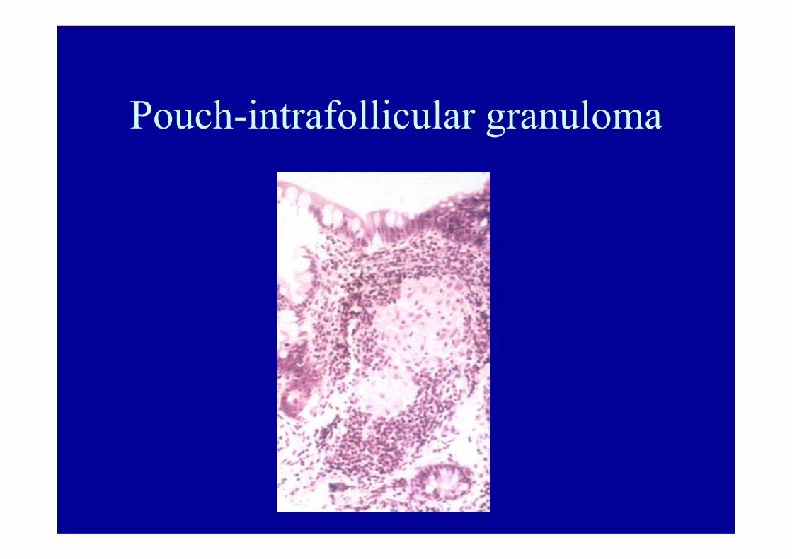
Pouch-intrafollicular granuloma

Ulcer associated cell lineage may be seen following ulceration. This may give an important clue to the presence of previous pouchitis.
The biopsy from the pouch mucosa when the patient’s symptoms have resolved:
Ulcer associated cell lineage


UACL
• UACL produces TFF1,2 in 6 patients with pouchitis/
Pera M, Heppell J, Poulsom R, Teixera FV, Williams J. Gut 2001; 48: 792-6

Symptoms of pouchitis but do not find pouchitis
• Seeing patient when recovered –may see UACL
• Irritable bowel• Secondary pouchitis• Prepouch ileitis • Cuffitis – often missed – needs careful
proctoscopy

UACL
• UACL produces TFF1,2 in 6 patients with pouchitis/
Pera M, Heppell J, Poulsom R, Teixera FV, Williams J. Gut 2001; 48: 792-6

Secondary pouchitis
Localised inflammation in the pouch mucosa related to a focus of inflammation/ abscess outside the pouch.
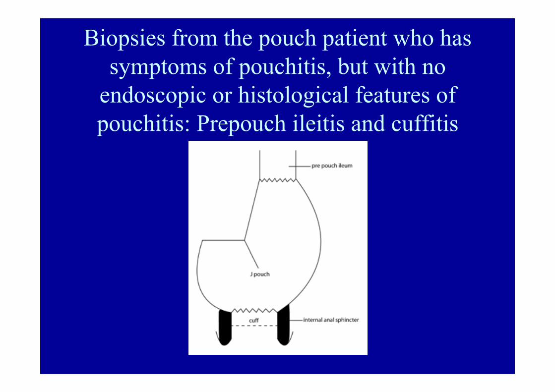
Biopsies from the pouch patient who has symptoms of pouchitis, but with no
endoscopic or histological features of pouchitis: Prepouch ileitis and cuffitis
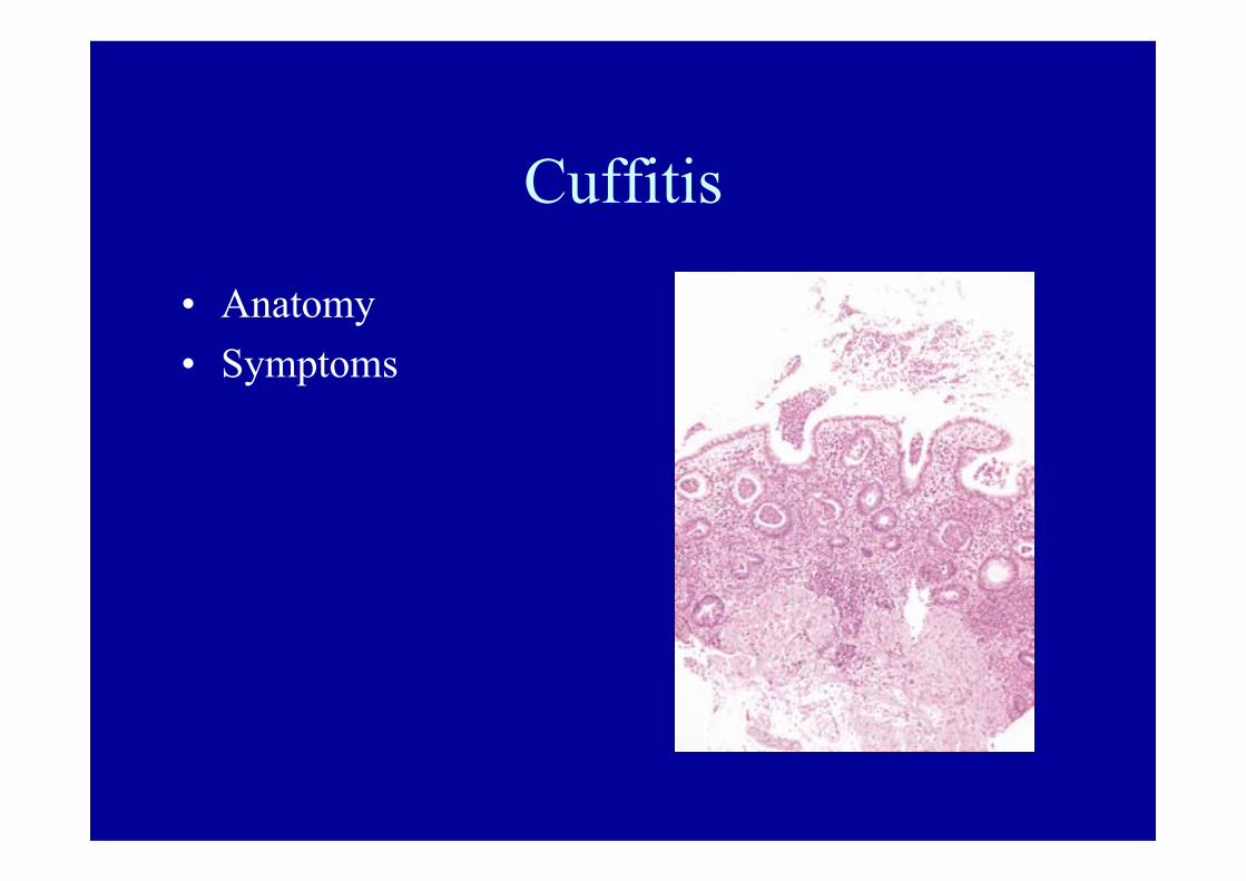
Cuffitis
• Anatomy• Symptoms
Summary-pouch dysfunction
• Other causes of inflammation may mimic pouchitis• Diagnosis is by symptoms, endoscopy with biopsies
from more than one site and accurate histology.• Remember the cuff and the prepouch ileum
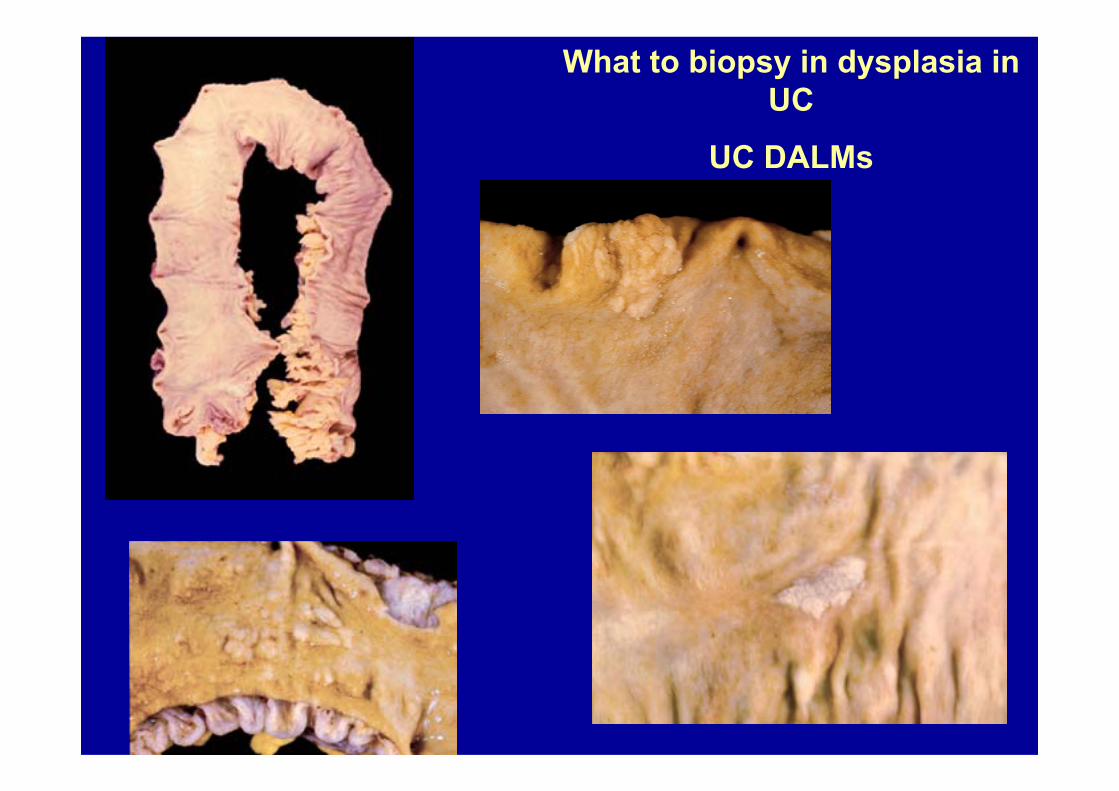
What to biopsy in dysplasia in UC
UC DALMs

Is it a DALM or ADENOMA?
• Age group?• Pedunculated?• Within area of UC?• Is there dysplasia in flat mucosa as well?• Please biopsy flat mucosa around and away
from it
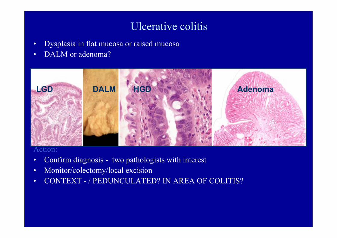
Ulcerative colitis• Dysplasia in flat mucosa or raised mucosa• DALM or adenoma?
Action:• Confirm diagnosis - two pathologists with interest• Monitor/colectomy/local excision• CONTEXT - / PEDUNCULATED? IN AREA OF COLITIS?
LGD DALM HGD Adenoma

Long-term follow up after polypectomy treatment for adenoma-like dysplastic lesions in ulcerative
colitis.Odze R, Farraye FA, Hecht JL, Hornick JL.Clin Gastroenterol Hepatol 2004: 2: 534-541
34 UC patients24 adenoma - like DALMs10 coincidental sporadic adenomas28 had polypectomy; 6 colectomy49 non UC patients with sporadic adenomaFollow up 82 months 20 pts developed more ALMs
1 LGD in flat mucosa1 (PSC) adenocarcinoma 7.5
years post polypectomy.Ns difference from controlsSafe to manage ALMs and adenomas with polypectomy

CYCLOSPORIN CHANGES
CONTEXT PLEASE

Summary
Put the biopsy into context and identify your biopsy sitesThink of iatrogenic disease/ normal variantsWhat will a biopsy from this site at this time tell me?Which site do I need to biopsy to answer my question?Biopsy lesion and apparent “non-lesion”Help the pathologist to put the appearances into.When the patient does not get better consider
a superimposed infection and rebiopsy


![Third European Evidence-based Consensus on Diagnosis and ... · colitis] E3 Extensive Involvement extends proximal to the splenic flexure, including pan-colitis Table 1.2. Disease](https://img.dokumen.tips/doc/110x75/6141c035d64cc55ff0755ece/third-european-evidence-based-consensus-on-diagnosis-and-colitis-e3-extensive.jpg)