Embed Size (px)
Citation preview

Coherent Oscillations in Chlorosome Elucidated by Two-DimensionalElectronic SpectroscopySunhong Jun,†,‡ Cheolhee Yang,†,‡ Megumi Isaji,§ Hitoshi Tamiaki,§ Jeongho Kim,*,∥
and Hyotcherl Ihee*,†,‡
†Center for Nanomaterials and Chemical Reactions, Institute for Basic Science (IBS), Daejeon 305-701, Republic of Korea‡Department of Chemistry, KAIST, Daejeon 305-701, Republic of Korea§Graduate School of Life Sciences, Ritsumeikan University, Kusatsu, Shiga 525-8577, Japan∥Department of Chemistry, Inha University, Incheon 402-751, Republic of Korea
*S Supporting Information
ABSTRACT: Chlorosomes are the most efficient photosynthetic light-harvestingcomplexes found in nature and consist of many bacteriochlorophyll (BChl) moleculesself-assembled into supramolecular aggregates. Here we elucidate the presence and theorigin of coherent oscillations in chlorosome at cryogenic temperature using 2Delectronic spectroscopy. We observe coherent oscillations of multiple frequenciessuperimposed on the ultrafast amplitude decay of 2D spectra. Comparison of oscillatoryfeatures in the rephasing and nonrephasing 2D spectra suggests that an oscillation of 620cm−1 frequency arises from electronic coherence. However, this coherent oscillation canbe enhanced by vibronic coupling with intermolecular vibrations of BChl aggregate, andthus it might originate from vibronic coherence rather than pure electronic coherence.Although the 620 cm−1 oscillation dephases rapidly, the electronic (or vibronic)coherence may still take part in the initial step of energy transfer in chlorosome, which iscomparably fast.
SECTION: Spectroscopy, Photochemistry, and Excited States
Chlorosomes are the largest and the most efficient light-harvesting complexes (LHCs) found in nature. They
consist of hundreds of thousands of bacteriochlorophyll (BChl)molecules self-assembled into supramolecular J-type aggregateswithout the involvement of any protein and exhibit strongexcitonic coupling among constituent BChl c, d, e, or fmolecules.1−3 The molecular architecture of chlorosomes isunique in comparison with other natural LHCs, where severalpigments are held in precise positions by a protein scaffold.This unique composition of chlorosomes allows us to readilysynthesize chlorosomes and their chemically modified ana-logues in vitro.4 Because of the processability and highlyefficient intrachlorosome energy transfer, chlorosomes areenvisioned as potential building blocks of artificial photosyn-thesis.5−8 There have been many efforts to characterize thestructure of chlorosomes, but their large size and the presenceof significant disorder prevents the structural determination atthe molecular level using X-ray crystallography. Fromcryoelectron microscopy and NMR studies, it was proposedthat BChls are organized into curved lamellar structures ormultilayered rolls,9−12 as schematically shown in Figure 1a, butthe exact arrangement of BChls within chlorosomes is still incontroversy.In green sulfur bacteria, solar energy absorbed by the core of
chlorosomes is transferred to the baseplate consisting of BChl amolecules and followed by energy transfer to the reaction
center via the Fenna−Matthews−Olson (FMO) complex. Thetime scales of these excited-state processes in chlorosomes havebeen determined using time-resolved spectroscopic techni-ques.13−20 Although there are some discrepancies on theassignment of the measured time constants to particularprocesses, it was found that a series of energy-transfer processesoccur on various time scales ranging from hundreds offemtoseconds to hundreds of picoseconds, reflecting hierarch-ical structure of chlorosomes. For example, kinetic componentson the time scale of hundreds of femtoseconds were assigned torapid energy relaxation among the exciton states within anindividual layer of BChls, while the layer-to-layer energytransfer in a chlorosome occurs on the time scale of a fewpicoseconds.13−16 Then, the energy transfer from chlorosomesto the baseplate occurs in tens to hundreds of picosecondsdepending on the species of chlorosome.13,15,18 In addition tothese relaxation processes, oscillations of 50−250 cm−1
frequencies superimposed on the time-resolved signals wereobserved and ascribed to coherently excited vibrational modesof the BChl aggregate.13,17−21
Previously, most of the time-resolved studies on chlorosomeswere performed using (pump−probe) transient absorption
Received: February 14, 2014Accepted: March 27, 2014
Letter
pubs.acs.org/JPCL
© XXXX American Chemical Society 1386 dx.doi.org/10.1021/jz500328w | J. Phys. Chem. Lett. 2014, 5, 1386−1392

spectroscopy, whereby the transient changes of the absorptionspectrum after photoexcitation are measured. In transientabsorption spectra, various features arising from short-livedexcited states are often overlapped to one another along asingle energy axis, complicating its interpretation. In contrast,two-dimensional electronic spectroscopy (2D-ES) spreads outthe excited-state absorption features into a 2D energy space(composed of absorption and emission energy axes) so thatthose features can be unambiguously resolved.22,23 As a result, a2D spectrum measured at a specific time delay afterphotoexcitation provides an instantaneous snapshot ofelectronic coupling and energy transfer among different excitedstates. In particular, when multiple excited states are coherentlyexcited by a broadband pulse, complex pathways of energy flowamong those states are obtained. Recently, this powerfultechnique has allowed us to probe the excitation energy flow inphotosynthetic LHCs in real time, elucidating complexpathways and mechanism of energy transfer.24−33 Notably, itwas proposed that long-lived electronic coherence generated
among electronic excited states may play a crucial role inachieving high efficiency of excitation energy transfer in naturalphotosynthetic LHCs,25,26,30−33 thus negating the traditionalmechanism of incoherent hopping (called Forster energytransfer). Although the identity (whether electronic orvibrational) and the exact role of the quantum coherence inphotosynthetic energy transfer are still in dispute, themechanism of energy transfer among the exciton states inLHCs has become a hot topic of investigation in associationwith an emerging field of quantum biology.34,35
Recently, 2D-ES was applied to chlorosomes (consisting ofBChl c molecules) isolated from green sulfur bacteria,Chlorobaculum (Cba.) tepidum and Chlorof lexus (Cf l.)aurantiacus, at room temperature and revealed that the ultrafast(incoherent) diffusion of excitation energy on the sub-100 fstime scale is responsible for the initial energy transfer inchlorosome.36 In particular, any coherent oscillation was notobserved in that study, leading to the conclusion that thechlorosome does not function as a coherent light-harvester. Inthis work, we apply the broadband 2D-ES to a different type ofchlorosome (consisting of BChl e molecules) isolated from agreen sulfur bacterium, Cba. limnaeum (previously calledChlorobium phaeobacteroides), at cryogenic temperature. Incontrast with the previous 2D-ES study, we clearly observedcoherent oscillations superimposed on the decay of 2D-ESsignal. From Fourier analysis, we elucidated the origins of thecoherent oscillations of multiple frequencies. In particular, theoscillation of ∼620 cm−1 frequency originates from electroniccoherence, suggesting that coherent energy transfer mediatedby electronic coherence may be in effect in chlorosome.The steady-state absorption spectrum of the chlorosome
consisting of BChl e molecules measured at room temperatureis shown in Figure 1b. The absorption band at 650−800 nm isascribed to the Qy transition of the chlorosome. The spectrumof the laser pulse used in this experiment covers the entire Qyabsorption band. Figure 1c shows the real (absorptive) part ofthe 2D spectra measured at T = 0 and 150 fs. The principle andthe experimental details of 2D electronic spectroscopy aredescribed in the Supporting Information (SI). Most notably,the spectrum at T = 0 fs contains a positive peak elongatedalong the diagonal. This positive peak corresponds to thecontributions from ground-state bleaching (GSB) and stimu-lated emission (SE) between the ground and the electronicexcited states involved in the Qy transition. As T increases, theshape of the positive peak rapidly changes, becoming moreround in concurrence with the decreasing amplitude along thediagonal. In particular, the amplitude of the positive peak growsprominently below the diagonal, and thus the negative ESApeak below the diagonal disappears, resulting in an asymmetricpeak shape with respect to the diagonal. The line shape changeof the peak occurs mainly on the time scale of 20−30 fs and iscompleted within T = 150 fs with the peak shape staying almostthe same thereafter. Considering that the layer-to-layer energytransfer takes hundreds of femtoseconds to a few picoseconds,this ultrafast change of the 2D line shape must correspond todownhill energy relaxation in a coherent domain or amongcoherent domains within a BChl layer, as was assigned in theprevious 2D-ES study36 and theoretical investigations39,40 onchlorosomes. Here a coherent domain refers to a region whereseveral BChl molecules are strongly coupled to each other andhave excitonic wave functions delocalized over those coupledchromophores (that is, exciton delocalization).
Figure 1. (a) Schematic of the structure of chlorosome. BChls areorganized into either curved lamellar sheets or multilayered tubularstructures.2,10 The size of a single chlorosome is about 100 × 200 × 30nm (width × length × height).37,38 (b) Absorption spectrum (blackline) of chlorosome from Cba. limnaeum measured at roomtemperature and the spectral profile of the laser pulse (red line)used for the 2D-ES measurement. The absorption band at 650−800nm is ascribed to the Qy transition of the BChl e chlorosome. (c) Realpart of 2D spectra of chlorosome at T = 0 and 150 fs, which weremeasured at 77 K. We note that the Qy transition energy slightly shiftsto red at 77 K, as indicated by the position of the major peak in the 2Dspectra. (d) Amplitude decay of 2D-ES signal at (ωτ, ωt) = (1.71, 1.71eV) (black circles) along the T axis shown together with its double-exponential fit (red line). The faster decay component has the timeconstant of 21 fs. The amplitudes at other locations in the 2Dspectrum also change rapidly with similar time constants. It can beclearly seen that oscillations are superimposed on the amplitude decay.
The Journal of Physical Chemistry Letters Letter
dx.doi.org/10.1021/jz500328w | J. Phys. Chem. Lett. 2014, 5, 1386−13921387
We note that the previous 2D-ES study on chlorosomes atroom temperature did not report any oscillatory component inthe population-time evolution of the 2D-ES signal.36 Incontrast, in our 2D spectra for chlorosomes at cryogenictemperature, we clearly see that oscillations are superimposedon the amplitude decays of the 2D-ES signal along T axis andpersist over the time window of 480 fs in our measurement.(See Figure 1d.) In the initial study that proposed theinvolvement of quantum coherence in energy transfer in aphotosynthetic LHC (FMO complex), such long-lastingoscillation of the amplitude in the 2D spectra along thepopulation time T was regarded as the evidence of long-livedelectronic coherence among the exciton states of multiplepigment chromophores.25 However, coherent oscillation of the2D spectra can also arise from another origin, that is, localvibrational modes, which is not directly related to the efficiencyof energy transfer. Since then, there has been much dispute onthe origin of the oscillation, whether it is from purely electronicor vibrational coherence and whether such quantum coherenceplays any important role in efficient energy transfer ofphotosynthetic LHCs.41,42 Unfortunately, the oscillations ofthe two different origins are supposed to appear at the samelocations in the 2D spectrum, making the assignment difficult.Accordingly, several methods of distinguishing the two types ofcoherences,33,43−45 or lack thereof,46 were proposed. Thus far,the most reliable method of distinguishing the coherences oftwo different origins is to decompose a 2D spectrum intorephasing (R) and nonrephasing (NR) components andexamine the presence of oscillations at various locations onthe R and NR components.33,43,44 The oscillation arising fromelectronic coherence appears only (1) at the cross peaks on theR spectrum and (2) at the diagonal peaks on the NR spectrum.In contrast, the oscillation originating from vibrationalcoherence appears at both cross and diagonal peaks on the Rand NR spectra.Following this comparative test between R and NR spectra,
we decomposed our 2D spectra into R and NR components, asshown in Figure S2 in the SI, and examined the origin of thecoherent oscillations. Because a chlorosome consists ofhundreds of thousands of BChl molecules and therefore has amanifold of exciton states, it is impossible to check the presenceof oscillations at specific locations on the 2D spectrum on astate-by-state basis, as was done for other smaller LHCs.Instead, to systematically check the locations where theoscillation is present distinctly, we assembled together a seriesof 2D spectra measured at various population times toconstruct a 3D data set in the (ωτ, T, ωt) domain and thenFourier-transformed the 3D data with respect to T to generatea 3D spectral solid in the (ωτ, ωT, ωt) domain.47 To consideronly the oscillatory components that have small amplitudesrelative to the overall 2D-ES signal, the Fourier transform withrespect to T was performed after subtracting the exponential fitof the amplitude decay at every location in the 2D spectrum.For simplicity, instead of showing the entire 3D spectral solid,we show only the representative slices of the spectral solid, thatis, 2D Fourier transform (FT) maps at selected frequencies ofωT = 140 and 620 cm−1 in Figure S3a in the SI and Figure 2a,respectively. Each 2D FT map demonstrates the distribution ofthe oscillation of a selected ωT frequency over the entire 2Dspectrum.Among coherent oscillations of multiple frequencies
observed in our data, the most pronounced oscillation of 140cm−1 frequency was found to originate from vibrational
coherence according to the comparative test, as described indetail in the SI. Here we focus on the oscillation of 620 cm−1
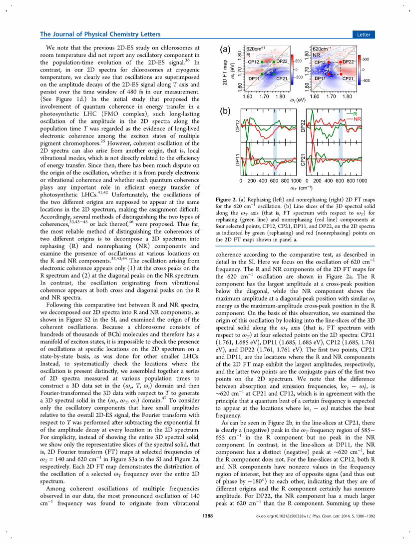
frequency. The R and NR components of the 2D FT maps forthe 620 cm−1 oscillation are shown in Figure 2a. The Rcomponent has the largest amplitude at a cross-peak positionbelow the diagonal, while the NR component shows themaximum amplitude at a diagonal-peak position with similar ωtenergy as the maximum-amplitude cross-peak position in the Rcomponent. On the basis of this observation, we examined theorigin of this oscillation by looking into the line-slices of the 3Dspectral solid along the ωT axis (that is, FT spectrum withrespect to ωT) at four selected points on the 2D spectra: CP21(1.761, 1.685 eV), DP11 (1.685, 1.685 eV), CP12 (1.685, 1.761eV), and DP22 (1.761, 1.761 eV). The first two points, CP21and DP11, are the locations where the R and NR componentsof the 2D FT map exhibit the largest amplitudes, respectively,and the latter two points are the conjugate pairs of the first twopoints on the 2D spectrum. We note that the differencebetween absorption and emission frequencies, |ωτ − ωt|, is∼620 cm−1 at CP21 and CP12, which is in agreement with theprinciple that a quantum beat of a certain frequency is expectedto appear at the locations where |ωτ − ωt| matches the beatfrequency.As can be seen in Figure 2b, in the line-slices at CP21, there
is clearly a (negative) peak in the ωT frequency region of 585−655 cm−1 in the R component but no peak in the NRcomponent. In contrast, in the line-slices at DP11, the NRcomponent has a distinct (negative) peak at ∼620 cm−1, butthe R component does not. For the line-slices at CP12, both Rand NR components have nonzero values in the frequencyregion of interest, but they are of opposite signs (and thus outof phase by ∼180°) to each other, indicating that they are ofdifferent origins and the R component certainly has nonzeroamplitude. For DP22, the NR component has a much largerpeak at 620 cm−1 than the R component. Summing up these
Figure 2. (a) Rephasing (left) and nonrephasing (right) 2D FT mapsfor the 620 cm−1 oscillation. (b) Line slices of the 3D spectral solidalong the ωT axis (that is, FT spectrum with respect to ωT) forrephasing (green line) and nonrephasing (red line) components atfour selected points, CP12, CP21, DP11, and DP22, on the 2D spectraas indicated by green (rephasing) and red (nonrephasing) points onthe 2D FT maps shown in panel a.
The Journal of Physical Chemistry Letters Letter
dx.doi.org/10.1021/jz500328w | J. Phys. Chem. Lett. 2014, 5, 1386−13921388

observations, the oscillation of 620 cm−1 frequency is presentonly at the cross-peak points (CP21 and CP12) in the Rcomponent and at the diagonal-peak points (DP11 and DP22)in the NR component. Therefore, according to the comparativetest, the oscillation of 620 cm−1 frequency presumablyoriginates from electronic coherence.To confirm the origin of these oscillations, we performed the
linear prediction singular value decomposition (LPSVD) andthe global fitting analyses for the time-domain traces at theselected points in Figure 2a. The details of the LPSVD and theglobal fitting analyses are described in Sections 9 and 10,respectively, in the SI, and the complete LPSVD and the globalfitting parameters are listed in Table S2 and S3, respectively, inthe SI. The time traces and their global fits are shown in Figure3. From both analyses, we were able to identify an oscillatorycomponent with the frequency of ∼620 cm−1 (and thedephasing time of 60 fs) only at the CPs of the R spectrumand at the DPs of the NR spectrum. We note that theoscillatory components identified for CP12 (R) and DP22(NR) have smaller amplitudes than the ones found for CP21(R) and DP11 (NR). This difference of the amplitudes seemsto be caused by smaller signal amplitudes at CP12 and DP22,which is the general case in the 2D spectra of LHCs. (SeeSection 8 of the SI for details.) As expected, the frequencies ofthose oscillations extracted from the time-domain traces matchwell with |ωτ − ωt| at CP21 and CP12 within ∼85%. Thus, thecomparative test between R and NR spectra suggests that theoscillation of 620 cm−1 frequency originates from electroniccoherence. Here we note that the 620 cm−1 oscillation hasmuch smaller amplitude compared with the 140 cm−1
oscillation. This comparison of the oscillation amplitudessupports our assignment because, in molecular aggregates, theoscillations induced by vibrational coherence are expected to bemuch stronger than the ones induced by electronic coherenceaccording to a theoretical study.45
According to the LPSVD and global fitting analysis, the 620cm−1 oscillation assigned to electronic coherence in chlorosomeexhibits much shorter lifetime (60 fs) than the electroniccoherence beats observed in other LHCs.25,30,32,33 In general,
such ultrashort lifetime is characteristic of electronic coherence.Specifically, the short-lived beating in chlorosome can beaccounted for by several factors, for example, higher density ofexciton states and relatively larger reorganization energy inchlorosomes than in other smaller LHCs. In addition,dephasing of the electronic coherence in chlorosome must beinfluenced by the supramolecular structure of chlorosome.Unlike other smaller LHCs, a chlorosome has significant staticheterogeneity among coherent domains in a BChl layer as wellas among BChl layers constituting a single chlorosome. Theelectronic coherence in chlorosome will be generated onlywithin each coherent domain, where the delocalized excitonstates of different energies can be coherently excited. Because ofthe heterogeneity among coherent domains, even slightdifferences in the phase of the coherence beats from manycoherent domains will lead to destructive interference byensemble averaging.36,48,49 Therefore, 2D-ES signal measuredfor an ensemble of coherent domains in a chlorosome willexhibit faster dephasing than the signal measured only for asingle coherent domain. Considering this additional artificialdephasing mechanism induced by heterogeneity, there is apossibility that the electronic coherence in individual coherentdomains might be longer-lived than measured by 2D-ES.49
Related to this issue, a recent theoretical study proposed that2D-ES can significantly underestimate the lifetime of electroniccoherences due to the destructive interference with severalpathways that are not important for excitonic energy transfer,for example, interference with vibrational coherences.50
Although the comparative test between R and NRcomponents presented above yielded a conclusion that theoscillation of 620 cm−1 frequency arises from electroniccoherence, we cannot completely rule out the involvement ofvibrational modes that are vibronically coupled to the electronicQy transition. In particular, we note that the oscillationsexamined in this work match well the vibrational frequencies(138, 276, 626 cm−1, ...) of the BChl aggregate obtained fromthe atomistic calculation of spectral density for modelchlorosomes consisting of multilayered rolls.39 (See Figure S7in the SI.) The vibrational frequencies that appear in the
Figure 3. Time-domain traces for the rephasing (green circles) and nonrephasing (red circles) spectra and their global fits (black) at the selectedpoints in Figure 2a. The oscillatory component of 654 cm−1 frequency and 60 fs dephasing time (blue) is identified only at CP21 (R), DP11 (NR),CP12 (R), and DP22 (NR).
The Journal of Physical Chemistry Letters Letter
dx.doi.org/10.1021/jz500328w | J. Phys. Chem. Lett. 2014, 5, 1386−13921389
spectral density correspond to vibrational modes whose nucleardisplacement can modify the excitation energy, that is, thevibrational modes with strong vibronic coupling. In particular,the vibrations in the low-frequency region below 1000 cm−1 areascribed to intermolecular interactions between the BChlmolecules. The 140 cm−1 oscillation, the most pronouncedcoherent oscillation in our 2D-ES data, must arise from theseintermolecular vibrations in a BChl layer, in agreement with ourassignment of this oscillation to vibrational coherence. The 620cm−1 oscillation, which was identified to originate fromelectronic coherence according to the test in the previous,can also be enhanced by borrowing the oscillator strength fromthe intermolecular vibrations of the same frequency via vibroniccoupling, and thus it might originate from vibronic coherencerather than pure electronic coherence. In fact, the role ofvibrations for enhancing the energy transfer in variousbiological and chemical systems has attracted much interestrecently.42,51−57 In particular, this consideration can besupported by recent theoretical developments to account forthe potential role of quantum coherence in photosyntheticene rgy t r an s f e r , f o r e x amp l e , v i b ron i c - e x c i t onmodel.42,51,53−55,57 According to this model, in molecularaggregates and photosynthetic LHCs, strong vibronic couplingbetween excitons and quantized vibrations quasi-resonant withexcitonic energy gaps may significantly enhance the lifetime ofcoherent oscillations as well as the efficiency of energy transfer.In this regard, it would be of much interest to theoreticallyexplore the effect of vibronic coupling on the energy transfer inchlorosome. From this perspective, it is also possible that thevibrational modes of other frequencies assist establishingelectronic coherences. For example, the spectral densitycalculated for BChl aggregate has much larger intensities(and thus stronger vibronic coupling) in the 1500−2000 cm−1
region than below 1000 cm−1. (See Figure S7 in the SI.) Ourmeasurement was not able to resolve such high-frequencyoscillations due to limited signal-to-noise ratio, but it might bepossible to detect the high-frequency oscillations with improvedtemporal stability of the laser system.Despite the rapid dephasing of the coherent oscillation
arising from electronic (or vibronic) coherence, it is plausiblethat the coherence may still contribute to the earliest stage ofenergy transfer in chlorosome. According to the line-shapedynamics of the positive 2D peak presented above, the energytransfer within a coherent domain occurs on a 20−30 fs timescale. This time scale is comparable to or even shorter than thedephasing time of the coherent oscillation at 77 K. Therefore,before the coherent oscillation completely dephases, it has agood chance of completing at least one oscillatory period (= 54fs for 620 cm−1 oscillation). According to theoreticalinvestigations, a complete sampling of all possible energytransfer pathways by a full period of quantum beating andsubsequent excitation trapping at an energetic minimum byrapid dephasing is the optimum condition for unidirectionaland efficient coherent energy transfer.58−60
From the dephasing time of 60 fs at 77 K measured in thiswork, we can estimate the room-temperature dephasing time of∼15 fs, assuming that the dephasing rate is linearly proportionalto the temperature in the Markovian limit.61 With the timeresolution of our experiment (15 fs), it might be challenging tomeasure the oscillation undergoing such rapid dephasing atroom temperature, which was probably the case in the previous2D-ES study of chlorosome at room temperature.36 Theestimated dephasing time at room temperature well-matches a
theoretically predicted value at 300 K by an atomisticcalculation for model chlorosomes consisting of multilayeredrolls.62 Even with ∼15 fs dephasing time (= time constant of anexponential decay), the complete decay of the beating will take∼50 fs. This time scale is long enough to complete one cycle ofbeating and short enough to collapse the coherence forexcitation trapping at an energetic minimum. This scenariobecomes more realistic when considering that the dephasingtime in individual coherent domains can be longer without thepreviously mentioned artificial dephasing induced by hetero-geneity. Therefore, the electronic (or vibronic) coherence maytake part in the initial step of energy transfer in chlorosome,which is comparably fast as the dephasing of electroniccoherence. However, it still needs to be investigated whetherthe electronic coherence enhances the rate and the efficiency ofthe initial ultrafast energy transfer and whether the enhance-ment of energy transfer rate on the ultrashort time scaleinfluences the energy transfer on longer time scales.In conclusion, we elucidated the presence and the origin of
coherent oscillations in chlorosome from Cba. limnaeum atcryogenic temperature by applying 2D-ES. The result of thiswork provides the first evidence of quantum coherence ofelectronic origin observed in chlorosome and may stimulatetheoretical studies that investigate the potential roles ofelectronic and vibrational coherences in ultrafast energy transferin chlorosome at the microscopic level. On the experimentalside, the 2D-ES experiment on single chlorosomes, where theensemble averaging can be alleviated, will give further insightsinto the origin of the coherent oscillations and potential roles ofquantum coherence in photosynthetic energy transfer.
■ ASSOCIATED CONTENT*S Supporting InformationStructure of BChl aggregate in chlorosome. Introduction to 2Delectronic spectroscopy. Experimental methods. FT resolutionof 2D FT spectra. Low-frequency oscillations in 2D spectrum.Locations of low-frequency oscillation in 2D spectrum.Locations of 620 cm−1 oscillation in 2D spectrum. Asymmetricshape of 2D spectra. Linear prediction singular valuedecomposition of the oscillations. Global fitting analysis ofthe oscillations. 2D rephasing and nonrephasing spectra atvarious population times. 2D FT maps for 140 cm−1 oscillationand FT line-slices. Time-domain traces and LPSVD compo-nents at the selected points for low-frequency oscillations. 2DFT maps for 70 cm−1 oscillation. Time-domain traces andLPSVD components at the selected points for 620 cm−1
oscillation. Spectral density calculated for BChl aggregate.Tables listing the parameters from the LPSVD analysis of theoscillations. Table listing the fit parameters from the globalfitting analysis of the oscillations. This material is available freeof charge via the Internet at http://pubs.acs.org.
■ AUTHOR INFORMATIONCorresponding Authors*J.K.: E-mail: [email protected].*H.I.: E-mail: [email protected] authors declare no competing financial interests.
■ ACKNOWLEDGMENTSThis work was supported by Institute for Basic Science (IBS)[CA1401-01]. This work was supported by an Inha University
The Journal of Physical Chemistry Letters Letter
dx.doi.org/10.1021/jz500328w | J. Phys. Chem. Lett. 2014, 5, 1386−13921390

Research Grant (INHA-48581). This work was partiallysupported by Grants-in-Aid for Scientific Research (A) (no.22245030) as well as on Innovative Areas “Artificial Photosyn-thesis (AnApple)” (no. 24107002) from the Japan Society forthe Promotion of Science (JSPS). We thank Prof. Alan Aspuru-Guzik and Dr. Takatoshi Fujita at Harvard University forhelpful discussions and providing the spectral density calculatedfor model chlorosomes.
■ REFERENCES(1) Frigaard, N.-U.; Bryant, D. A. Chlorosomes: Antenna Organellesin Green Photosynthetic Bacteria. In Microbiology Monographs;Shively, J., Ed.; Springer: Berlin, Germany, 2006; Vol. 2, pp 79−114.(2) Oostergetel, G.; Amerongen, H.; Boekema, E. The Chlorosome:A Prototype for Efficient Light Harvesting in Photosynthesis.Photosynth. Res. 2010, 104, 245−255.(3) Harada, J.; Mizoguchi, T.; Tsukatani, Y.; Noguchi, M.; Tamiaki,H. A Seventh Bacterial Chlorophyll Driving a Large Light-HarvestingAntenna. Sci. Rep. 2012, 2, 671.(4) Shoji, S.; Hashishin, T.; Tamiaki, H. Construction ofChlorosomal Rod Self-Aggregates in the Solid State on Any Substratesfrom Synthetic Chlorophyll Derivatives Possessing an OligomethyleneChain at the 17-Propionate Residue. Chem.Eur. J. 2012, 18, 13331−13341.(5) Roger, C.; Miloslavina, Y.; Brunner, D.; Holzwarth, A. R.;Wurthner, F. Self-Assembled Zinc Chlorin Rod Antennae Powered byPeripheral Light-Harvesting Chromophores. J. Am. Chem. Soc. 2008,130, 5929−5939.(6) Miyatake, T.; Tamiaki, H. Self-Aggregates of Natural Chlor-ophylls and Their Synthetic Analogues in Aqueous Media for MakingLight-Harvesting Systems. Coord. Chem. Rev. 2010, 254, 2593−2602.(7) Alster, J.; Polivka, T.; Arellano, J. B.; Chabera, P.; Vacha, F.;Psencík, J. Beta-Carotene to Bacteriochlorophyll c Energy Transfer inSelf-Assembled Aggregates Mimicking Chlorosomes. Chem. Phys.2010, 373, 90−97.(8) Kataoka, Y.; Shibata, Y.; Tamiaki, H. Supramolecular EnergyTransfer from Photoexcited Chlorosomal Zinc Porphyrin Self-Aggregates to a Chlorin or Bacteriochlorin Monomer as Models ofMain Light-Harvesting Antenna Systems in Green PhotosyntheticBacteria. Bioorg. Med. Chem. Lett. 2012, 22, 5218−5221.(9) Hohmann-Marriott, M.; Blankenship, R.; Roberson, R. TheUltrastructure of Chlorobium Tepidum Chlorosomes Revealed byElectron Microscopy. Photosynth. Res. 2005, 86, 145−154.(10) Oostergetel, G. T.; Reus, M.; Gomez Maqueo Chew, A.; Bryant,D. A.; Boekema, E. J.; Holzwarth, A. R. Long-Range Organization ofBacteriochlorophyll in Chlorosomes of Chlorobium tepidum Inves-tigated by Cryo-Electron Microscopy. FEBS Lett. 2007, 581, 5435−5439.(11) Egawa, A.; Fujiwara, T.; Mizoguchi, T.; Kakitani, Y.; Koyama, Y.;Akutsu, H. Structure of the Light-Harvesting Bacteriochlorophyll cAssembly in Chlorosomes from Chlorobium limicola Determined bySolid-State NMR. Proc. Natl. Acad. Sci. U.S.A. 2007, 104, 790−795.(12) Ganapathy, S.; Oostergetel, G. T.; Wawrzyniak, P. K.; Reus, M.;Gomez Maqueo Chew, A.; Buda, F.; Boekema, E. J.; Bryant, D. A.;Holzwarth, A. R.; de Groot, H. J. M. Alternating Syn-AntiBacteriochlorophylls Form Concentric Helical Nanotubes in Chlor-osomes. Proc. Natl. Acad. Sci. U.S.A. 2009, 106, 8525−8530.(13) Prokhorenko, V. I.; Steensgaard, D. B.; Holzwarth, A. R. ExcitonDynamics in the Chlorosomal Antennae of the Green BacteriaChlorof lexus aurantiacus and Chlorobium tepidum. Biophys. J. 2000, 79,2105−2120.(14) Psencík, J.; Polívka, T.; Nemec, P.; Dian, J.; Kudrna, J.; Maly, P.;Hala, J. Fast Energy Transfer and Exciton Dynamics in Chlorosomesof the Green Sulfur Bacterium Chlorobium tepidum. J. Phys. Chem. A1998, 102, 4392−4398.(15) Psencík, J.; Ma, Y.-Z.; Arellano, J. B.; Hala, J.; Gillbro, T.Excitation Energy Transfer Dynamics and Excited-State Structure in
Chlorosomes of Chlorobium phaeobacteroides. Biophys. J. 2003, 84,1161−1179.(16) Martiskainen, J.; Linnanto, J.; Aumanen, V.; Myllyperkio, P.;Korppi-Tommola, J. Excitation Energy Transfer in Isolated Chlor-osomes from Chlorobium tepidum and Prosthecochloris aestuarii.Photochem. Photobiol. 2012, 88, 675−683.(17) Savikhin, S.; Zhu, Y.; Lin, S.; Blankenship, R. E.; Struve, W. S.Femtosecond Spectroscopy of Chlorosome Antennas from the GreenPhotosynthetic Bacterium Chlorof lexus aurantiacus. J. Phys. Chem.1994, 98, 10322−10334.(18) Savikhin, S.; van Noort, P. I.; Zhu, Y.; Lin, S.; Blankenship, R.E.; Struve, W. S. Ultrafast Energy Transfer in Light-HarvestingChlorosomes from the Green Sulfur Bacterium Chlorobium tepidum.Chem. Phys. 1995, 194, 245−258.(19) Cherepy, N. J.; Du, M.; Holzwarth, A. R.; Mathies, R. A. Near-Infrared Resonance Raman Spectra of Chlorosomes: Probing NuclearCoupling in Electronic Energy Transfer. J. Phys. Chem. 1996, 100,4662−4671.(20) Ma, Y. Z.; Aschenbrucker, J.; Miller, M.; Gillbro, T. Ground-State Vibrational Coherence in Chlorosomes of the Green SulfurPhotosynthetic Bacterium Chlorobium phaeobacteroides. Chem. Phys.Lett. 1999, 300, 465−472.(21) Savikhin, S.; van Noort, P. I.; Blankenship, R. E.; Struve, W. S.Femtosecond Probe of Structural Analogies between Chlorosomes andBacteriochlorophyll c Aggregates. Biophys. J. 1995, 69, 1100−1104.(22) Jonas, D. M. Two-Dimensional Femtosecond Spectroscopy.Annu. Rev. Phys. Chem. 2003, 54, 425−463.(23) Cho, M. Two-Dimensional Optical Spectroscopy; CRC Press:Boca Raton, FL, 2009; p 378.(24) Brixner, T.; Stenger, J.; Vaswani, H. M.; Cho, M.; Blankenship,R. E.; Fleming, G. R. Two-Dimensional Spectroscopy of ElectronicCouplings in Photosynthesis. Nature 2005, 434, 625−628.(25) Engel, G. S.; Calhoun, T. R.; Read, E. L.; Ahn, T. K.; Mancal, T.;Cheng, Y. C.; Blankenship, R. E.; Fleming, G. R. Evidence forWavelike Energy Transfer through Quantum Coherence in Photo-synthetic Systems. Nature 2007, 446, 782−786.(26) Collini, E.; Wong, C. Y.; Wilk, K. E.; Curmi, P. M. G.; Brumer,P.; Scholes, G. D. Coherently Wired Light-Harvesting in Photo-synthetic Marine Algae at Ambient Temperature. Nature 2010, 463,644−647.(27) Schlau-Cohen, G. S.; Calhoun, T. R.; Ginsberg, N. S.; Read, E.L.; Ballottari, M.; Bassi, R.; van Grondelle, R.; Fleming, G. R. Pathwaysof Energy Flow in LHCII from Two-Dimensional ElectronicSpectroscopy. J. Phys. Chem. B 2009, 113, 15352−15363.(28) Womick, J. M.; Moran, A. M. Exciton Coherence and EnergyTransport in the Light-Harvesting Dimers of Allophycocyanin. J. Phys.Chem. B 2009, 113, 15747−15759.(29) Myers, J. A.; Lewis, K. L. M.; Fuller, F. D.; Tekavec, P. F.;Yocum, C. F.; Ogilvie, J. P. Two-Dimensional Electronic Spectroscopyof the D1-D2-Cyt B559 Photosystem II Reaction Center Complex. J.Phys. Chem. Lett. 2010, 1, 2774−2780.(30) Panitchayangkoon, G.; Hayes, D.; Fransted, K. A.; Caram, J. R.;Harel, E.; Wen, J.; Blankenship, R. E.; Engel, G. S. Long-LivedQuantum Coherence in Photosynthetic Complexes at PhysiologicalTemperature. Proc. Natl. Acad. Sci. U.S.A. 2010, 107, 12766−12770.(31) Panitchayangkoon, G.; Voronine, D. V.; Abramavicius, D.;Caram, J. R.; Lewis, N. H. C.; Mukamel, S.; Engel, G. S. DirectEvidence of Quantum Transport in Photosynthetic Light-HarvestingComplexes. Proc. Natl. Acad. Sci. U.S.A. 2011, 108, 20908−20912.(32) Harel, E.; Engel, G. S. Quantum Coherence SpectroscopyReveals Complex Dynamics in Bacterial Light-Harvesting Complex 2(LH2). Proc. Natl. Acad. Sci. U.S.A. 2012, 109, 706−711.(33) Turner, D. B.; Dinshaw, R.; Lee, K.-K.; Belsley, M. S.; Wilk, K.E.; Curmi, P. M. G.; Scholes, G. D. Quantitative Investigations ofQuantum Coherence for a Light-Harvesting Protein at ConditionsSimulating Photosynthesis. Phys. Chem. Chem. Phys. 2012, 14, 4857−4874.(34) Fleming, G. R.; Scholes, G. D.; Cheng, Y. C. Quantum Effects inBiology. Proc. Chem. 2011, 3, 38−57.
The Journal of Physical Chemistry Letters Letter
dx.doi.org/10.1021/jz500328w | J. Phys. Chem. Lett. 2014, 5, 1386−13921391

(35) Lambert, N.; Chen, Y.-N.; Cheng, Y.-C.; Li, C.-M.; Chen, G.-Y.;Nori, F. Quantum Biology. Nat. Phys. 2013, 9, 10−18.(36) Dostal, J.; Mancal, T.; Augulis, R.; Vacha, F.; Psencík, J.;Zigmantas, D. Two-Dimensional Electronic Spectroscopy RevealsUltrafast Energy Diffusion in Chlorosomes. J. Am. Chem. Soc. 2012,134, 11611−11617.(37) Tamiaki, H.; Tateishi, S.; Nakabayashi, S.; Shibata, Y.; Itoh, S.Linearly Polarized Light Absorption Spectra of Chlorosomes, Light-Harvesting Antennas of Photosynthetic Green Sulfur Bacteria. Chem.Phys. Lett. 2010, 484, 333−337.(38) Martinez-Planells, A.; Arellano, J.; Borrego, C.; Lopez-Iglesias,C.; Gich, F.; Garcia-Gil, J. Determination of the Topography andBiometry of Chlorosomes by Atomic Force Microscopy. Photosynth.Res. 2002, 71, 83−90.(39) Fujita, T.; Brookes, J. C.; Saikin, S. K.; Aspuru-Guzik, A.Memory-Assisted Exciton Diffusion in the Chlorosome Light-Harvest-ing Antenna of Green Sulfur Bacteria. J. Phys. Chem. Lett. 2012, 3,2357−2361.(40) Linnanto, J. M.; Korppi-Tommola, J. E. I. Exciton Description ofChlorosome to Baseplate Excitation Energy Transfer in FilamentousAnoxygenic Phototrophs and Green Sulfur Bacteria. J. Phys. Chem. B2013, 117, 11144−11161.(41) Christensson, N.; Milota, F.; Hauer, J.; Sperling, J.; Bixner, O.;Nemeth, A.; Kauffmann, H. F. High Frequency VibrationalModulations in Two-Dimensional Electronic Spectra and TheirResemblance to Electronic Coherence Signatures. J. Phys. Chem. B2011, 115, 5383−5391.(42) Christensson, N.; Kauffmann, H. F.; Pullerits, T.; Mancal, T.Origin of Long-Lived Coherences in Light-Harvesting Complexes. J.Phys. Chem. B 2012, 116, 7449−7454.(43) Cheng, Y.-C.; Fleming, G. R. Coherence Quantum Beats inTwo-Dimensional Electronic Spectroscopy. J. Phys. Chem. A 2008,112, 4254−4260.(44) Turner, D. B.; Wilk, K. E.; Curmi, P. M. G.; Scholes, G. D.Comparison of Electronic and Vibrational Coherence Measured byTwo-Dimensional Electronic Spectroscopy. J. Phys. Chem. Lett. 2011,2, 1904−1911.(45) Butkus, V.; Zigmantas, D.; Valkunas, L.; Abramavicius, D.Vibrational vs. Electronic Coherences in 2d Spectrum of MolecularSystems. Chem. Phys. Lett. 2012, 545, 40−43.(46) Mancal, T.; Christensson, N.; Lukes, V.; Milota, F.; Bixner, O.;Kauffmann, H. F.; Hauer, J. System-Dependent Signatures ofElectronic and Vibrational Coherences in Electronic Two-DimensionalSpectra. J. Phys. Chem. Lett. 2012, 3, 1497−1502.(47) Turner, D. B.; Stone, K. W.; Gundogdu, K.; Nelson, K. A.Three-Dimensional Electronic Spectroscopy of Excitons in GaAsQuantum Wells. J. Chem. Phys. 2009, 131, 144510-1−144510-8.(48) Anna, J. M.; Ostroumov, E. E.; Maghlaoui, K.; Barber, J.;Scholes, G. D. Two-Dimensional Electronic Spectroscopy RevealsUltrafast Downhill Energy Transfer in Photosystem I Trimers of theCyanobacterium Thermosynechococcus Elongatus. J. Phys. Chem. Lett.2012, 3, 3677−3684.(49) Ishizaki, A.; Fleming, G. R. On the Interpretation of QuantumCoherent Beats Observed in Two-Dimensional Electronic Spectra ofPhotosynthetic Light Harvesting Complexes. J. Phys. Chem. B 2011,115, 6227−6233.(50) Kreisbeck, C.; Kramer, T.; Aspuru-Guzik, A. DisentanglingElectronic and Vibronic Coherences in Two-Dimensional EchoSpectra. J. Phys. Chem. B 2013, 117, 9380−9385.(51) Womick, J. M.; Moran, A. M. Vibronic Enhancement of ExcitonSizes and Energy Transport in Photosynthetic Complexes. J. Phys.Chem. B 2011, 115, 1347−1356.(52) Jordan, M. W.; Brantley, A. W.; Norbert, F. S.; Andrew, M. M.Vibronic Effects in the Spectroscopy and Dynamics of C-Phycocyanin.J. Phys. B: At., Mol. Opt. Phys. 2012, 45, 154016.(53) Kolli, A.; O’Reilly, E. J.; Scholes, G. D.; Olaya-Castro, A. TheFundamental Role of Quantized Vibrations in Coherent LightHarvesting by Cryptophyte Algae. J. Chem. Phys. 2012, 137, 174109.
(54) Chin, A. W.; Prior, J.; Rosenbach, R.; Caycedo-Soler, F.; Huelga,S. F.; Plenio, M. B. The Role of Non-Equilibrium VibrationalStructures in Electronic Coherence and Recoherence in Pigment-Protein Complexes. Nat. Phys. 2013, 9, 113−118.(55) Chenu, A.; Christensson, N.; Kauffmann, H. F.; Mancal, T.Enhancement of Vibronic and Ground-State Vibrational Coherencesin 2D Spectra of Photosynthetic Complexes. Sci. Rep. 2013, 3, 2029.(56) Richards, G. H.; Wilk, K. E.; Curmi, P. M. G.; Quiney, H. M.;Davis, J. A. Coherent Vibronic Coupling in Light-HarvestingComplexes from Photosynthetic Marine Algae. J. Phys. Chem. Lett.2012, 3, 272−277.(57) Tiwari, V.; Peters, W. K.; Jonas, D. M. Electronic Resonancewith Anticorrelated Pigment Vibrations Drives Photosynthetic EnergyTransfer Outside the Adiabatic Framework. Proc. Natl. Acad. Sci. U.S.A.2013, 110, 1203−1208.(58) Rebentrost, P.; Mohseni, M.; Kassal, I.; Lloyd, S.; Aspuru-Guzik,A. Environment-Assisted Quantum Transport. New J. Phys. 2009, 11,033003.(59) Caruso, F.; Chin, A. W.; Datta, A.; Huelga, S. F.; Plenio, M. B.Highly Efficient Energy Excitation Transfer in Light-HarvestingComplexes: The Fundamental Role of Noise-Assisted Transport. J.Chem. Phys. 2009, 131, 105106.(60) Rebentrost, P.; Mohseni, M.; Aspuru-Guzik, A. Role ofQuantum Coherence and Environmental Fluctuations in Chromo-phoric Energy Transport. J. Phys. Chem. B 2009, 113, 9942−9947.(61) Rebentrost, P.; Chakraborty, R.; Aspuru-Guzik, A. Non-Markovian Quantum Jumps in Excitonic Energy Transfer. J. Chem.Phys. 2009, 131, 184102.(62) Fujita, T.; Huh, J.; Saikin, S.; Brookes, J.; Aspuru-Guzik, A.Theoretical Characterization of Excitation Energy Transfer inChlorosome Light-Harvesting Antennae from Green Sulfur Bacteria.Photosynth. Res. 2014, DOI: 10.1007/s11120-014-9978-7.
The Journal of Physical Chemistry Letters Letter
dx.doi.org/10.1021/jz500328w | J. Phys. Chem. Lett. 2014, 5, 1386−13921392


![Oscillations mécaniques libres non amorties Oscillations ...ww2.cnam.fr/physique/PHR004/04_L08_PHR004.pdf · Leçon n°8 : Oscillations [1] PHR 004 1 Oscillations mécaniques libres](https://img.dokumen.tips/doc/110x75/5b968ab509d3f206218b9064/oscillations-mecaniques-libres-non-amorties-oscillations-ww2cnamfrphysiquephr00404l08.jpg)