Embed Size (px)
Citation preview

Historically, philosophers have subdivided the study of the human mind and behaviour into two broad categories: the cognitive (how we know the world) and the affective (how we feel about it). This division is, how-ever, arbitrary as cognition — a highly complex construct (FIG. 1) — and emotion interact; cognitive status can colour the processing of emotions, and changes in mood affect cognitive function1,2.
It is therefore surprising that changes in emotion are universally recognized as being inherent to psychiatric disorders and their classification, whereas cognitive impairment — which has an equally disabling effect on patients — has been comparatively neglected. Despite this close interrelationship between cognition and mood, the cognitive deficits of psychiatric disorders are not just a secondary consequence of perturbed affect, and their underlying neurobiological substrates differ. Although certain symptoms of psychiatric disorders — such as depression, delusions and anxiety — are allevi-ated by current drugs, cognitive deficits are not usually improved, and may even be worsened3,4. Cognitive dysfunction is, therefore, a poorly controlled and highly
relevant dimension of psychiatric disorders that cuts across traditional diagnostic boundaries, and improved treatment should be a major goal in efforts to enhance quality of life for patients.
Cognitive dysfunction in psychiatric disordersChallenges of defining and characterizing cognitive deficits. Alzheimer’s disease is characterized by poor learning and memory, Parkinson’s disease by motor impairment, depression by melancholy, and schizophre-nia by delusions; however, these and related diagnoses are also accompanied by a range of symptoms involving alterations in mood, motor behaviour, appetite, sleep, diurnal rhythms and, most pertinently, cognitive func-tion. For example, psychosis is common in Alzheimer’s disease, depression can be just as debilitating as motor deficits in Parkinson’s disease, and perturbed cognition is a characteristic of both psychiatric and neurological disorders (TABLE 1).
Defining the precise nature of changes in cognition is challenging. Specificity relative to generalized changes in overall intelligence remains under discussion, in
1Institut de Recherche Servier, 78290 Croissy/Seine, France.Correspondence to M.J.M. e-mail: [email protected]:10.1038/nrd3628
CognitionA suite of interrelated conscious (and unconscious) mental activities, including: pre-attentional sensory gating; attention; learning and memory; problem solving, planning, reasoning and judgment; understanding, knowing and representing; creativity, intuition and insight; ‘spontaneous’ thought; introspection; as well as mental time travel, self-awareness and meta-cognition (thinking and knowledge about cognition).
Cognitive dysfunction in psychiatric disorders: characteristics, causes and the quest for improved therapyMark J. Millan1, Yves Agid2, Martin Brüne3, Edward T. Bullmore4, Cameron S. Carter5, Nicola S. Clayton6, Richard Connor7, Sabrina Davis8, Bill Deakin9, Robert J. DeRubeis10, Bruno Dubois11, Mark A. Geyer12, Guy M. Goodwin13, Philip Gorwood14, Thérèse M. Jay14, Marian Joëls15, Isabelle M. Mansuy16, Andreas Meyer-Lindenberg17, Declan Murphy18, Edmund Rolls19, Bernd Saletu20, Michael Spedding21, John Sweeney22, Miles Whittington23 and Larry J. Young24
Abstract | Studies of psychiatric disorders have traditionally focused on emotional symptoms such as depression, anxiety and hallucinations. However, poorly controlled cognitive deficits are equally prominent and severely compromise quality of life, including social and professional integration. Consequently, intensive efforts are being made to characterize the cellular and cerebral circuits underpinning cognitive function, define the nature and causes of cognitive impairment in psychiatric disorders and identify more effective treatments. Successful development will depend on rigorous validation in animal models as well as in patients, including measures of real-world cognitive functioning. This article critically discusses these issues, highlighting the challenges and opportunities for improving cognition in individuals suffering from psychiatric disorders.
R E V I E W S
NATURE REVIEWS | DRUG DISCOVERY VOLUME 11 | FEBRUARY 2012 | 141
© 2012 Macmillan Publishers Limited. All rights reserved

LearningThe active, experience- and/or training-driven acquisition of information or behaviour. The term ‘conditioning’ is usually used in an experimental context of associative learning. Learning necessitates complementary and distinct processes of encoding and acquisition that can be perturbed and modulated independently.
MemoryPartly separate mechanisms permitting consolidation, retention and retrieval of information from various sensory domains. Short-term memory relates to immediately available information maintained for ~30 seconds. Information retained for longer periods must be consolidated into mechanistically different long-term memory; in principle, this relates to the unlimited (in quantity and in time) capacity to store information.
particular for schizophrenia and autism spectrum dis-orders (ASDs), in which development is abnormal5–8. Furthermore, the precise interrelationship between alterations in cognition and changes in mood, reward, motor performance and effort can be difficult to estab-lish9,10. Finally, apart from treatment, various other factors modify cognitive performance and its measurement in a patient- and disorder-dependent fashion, including: education and age; hormonal status; disease progression; co-morbidity (psychiatric and somatic); whether cog-nitive function is determined in crisis or in remission; motivation; the neuropsychological test used and prac-tice effects; and the means of quantification (self-rating, semi-quantitative scales or informant assessment)6,7,11. Similarities and differences between various disorders are clearly complex — and still being delineated — but several general patterns can be discerned.
Contrasting patterns of cognitive deficits among distinct psychiatric disorders. Cognitive dysfunction does not just signify poor memory — the range of cognitive impairment is broader and more complex (TABLE 1). There are conditions in which a failure to forget or ‘inhibit’ is a characteristic symptom: for example, intrusive thoughts
in obsessive compulsive disorder (OCD)12 and recurrent, unwanted recall (flashbacks) in post-traumatic stress dis-order (PTSD)13,14. The latter state represents a form of ‘hyper-memory’ resulting from defective processes of fear extinction — an active process for suppressing nega-tive emotional memories — rather than just the decay of the mechanisms involved in storage and recall14,15 (FIG. 2). Phobias and social anxiety disorder are likewise typi-fied by blunted fear extinction16,17. Comparatively little cognitive disturbance has been documented for gen-eralized anxiety disorder, despite some subtle changes and a negative cognitive bias to threatening stimuli16,17 (TABLE 1). Cognitive dysfunction in panic disorders is mainly confined to excessive attention and hyperreactivity to threatening — but not emotionally neutral — stimuli. Interestingly, processing speed may actually be acceler-ated in panic disorders16,18. Schizophrenia is charac-terized by a broad pattern of cognitive deficits, from attention and working memory to social cognition and language7,19–22 (BOX 1). Impairments in bipolar disorder, which shares certain genetic risk factors with schizo-phrenia (Supplementary information S1 (figure)), are similar but generally less severe19,23,24 (TABLE 1).
Cognitive impairment is not traditionally associated with depression but it is common, broad-based and often debilitating4,19,25,26. Poor performance in certain tasks reflects reduced reward, low motivation and/or an incapacity for sustained effort — possibly owing to disruption of limbic dopaminergic signalling4,10. This does not, however, provide a satisfactory explanation for overall cognitive impairment. For example, the bias of patients suffering from depression towards affectively negative — and even ambiguous — stimuli (such as facial expressions) involves diminished top-down fronto-cortical cognitive control of emotional processing2,27.
Deficits in attention deficit hyperactivity disorder (ADHD) are not restricted to attention; they affect several other cognitive domains, including an inter-related impairment in working memory and process-ing speed28,29. Among the deficits characterizing OCD, impairment of procedural learning is of particular note12,30. Finally, although disrupted social cognition is a cardi-nal symptom of ASD, several other domains are also affected9,31,32 (TABLE 1).
Changes in specific cognitive domains seen across distinct diagnoses. Determining which cognitive domains are affected in diagnostically discrete disorders is complicated by co-morbidity. Nonetheless, certain cognitive domains can be perturbed in several distinct disorders (TABLE 1).
Most conspicuously, attention is affected in all disor-ders, varying from a cardinal loss of focused attention in ADHD28 to hypervigilance to threatening stimuli in PTSD, panic disorder and even OCD12,13,16–18 (TABLE 1). In ASD, attention to people and their emotions — as well as joint attention with others — is blunted; furthermore, attention towards objects and details is enhanced, while disregarding global aspects (central coherence)8,31.
Perturbed executive function is an additional example of transnosological deficits; however, reflecting their con-trasting integration (FIG. 2), subdimensions are affected
Author addresses2ICM, Pitié-Salpétrière University Hospital, 47 boulevard de l’Hôpital, 75013 Paris, France. 3 Research Department of Cognitive Neuropsychiatry and Psychiatric Preventive Medicine, LWL University Hospital, Ruhr-University Bochum, Alexandrinenstr. 1, 44791 Bochum, Germany.
4 University of Cambridge and GlaxoSmithKline, Cambridge Biomedical Campus, Cambridge CB2 0SZ, UK.
5University of California, Davis, Sacramento, California 95817, USA. 6 Department of Experimental Psychology, University of Cambridge, Cambridge CB2 3EB, UK.
7 Department of Biology, University of Massachusetts Dartmouth, 02747 North Dartmouth, USA.
8 Centre National de la Recherche Scientifique, University of Paris-Sud, 91400 Orsay, France.
9 Neuroscience and Psychiatry Unit, University of Manchester, Manchester M13 9PT, UK. 10University of Pennsylvania, 19104 Philadelphia, USA.11 Institut du Cerveau et de la Moelle Epinière (ICM), Université Pierre et Marie Curie,
Paris 6, UMR-S975 Paris, France. 12University of California San Diego, La Jolla, California 92093-0804, USA.13University of Oxford, Warneford Hospital, Oxford OX3 7JX, UK.14I NSERM; Université Paris, Descartes, Centre de Psychiatrie et Neurosciences U894,
75014 Paris, France.15 Department of Neuroscience and Pharmacology, Rudolf Magnus Institute, University
Medical Center Utrecht, 3584 CG Utrecht, The Netherlands.16Brain Research Institute, University of Zürich and ETHZ, 8057 Zürich, Switzerland.17 Central Institute of Mental Health, Heidelberg University, Medical Faculty Mannheim,
D-68159 Mannheim, Germany.18Institute of Psychiatry, King’s College London, Denmark Hill, London SE5 8AF, UK. 19 Oxford Centre for Computational Neuroscience and Department of Computer
Science, University of Warwick, Coventry CV4 7AL, UK. 20 Department of Psychiatry and Psychotherapy, Medical University of Vienna,
Waehringer Gürtel 18-20, A-1090 Vienna, Austria. 21Les Laboratoires Servier, 50 Rue Carnot, 92284 Suresnes Cedex, France.22 University of Texas Southwestern, Dallas 75235, Texas, USA.23Newcastle University, Newcastle NE2 4HH, UK. 24 Yerkes National Primate Research Center, Emory University, 954 Gatewood Rd,
Atlanta, Georgia 30329, USA.
R E V I E W S
142 | FEBRUARY 2012 | VOLUME 11 www.nature.com/reviews/drugdisc
© 2012 Macmillan Publishers Limited. All rights reserved

Nature Reviews | Drug Discovery
Universal domains:• Attention, working memory, executive function• Procedural learning and memory• Speed of processing• Fear-extinction learning • Semantic memory
Functional and structural disruption in neurons and/or glia of:• Cellular signalling• Gene transcription and mRNA translation• DNA and/or histone epigenetic codes• Firing rate and patterns (LTP and LTD)• Dendritic spines, synaptic plasticity and neurogenesis• Neuromodulator release
Focal and distributed network perturbation:• Interregional dysconnectivity• Local overconnectivity• Collapse of small-world configurations• Disorganization and desynchronization• Disrupted γ-and θ-oscillations
Higher domains:• Episodic memory• Social cognition• Theory of mind• Verbal learning and memory • Language (use and understanding)
Genetic
Multiple spatial scales: molecules to cerebral circuits Multiple time scales: milliseconds to years
Epigenetic Developmental Environmental
Cognitive impairment
SchizophreniaBipolar disorder
Depression
GAD
Panic disorderPTSD
OCD
ADHD
ASD
ExtinctionThe progressive reduction of a response to a stimulus — for example, owing to discontinuation of reinforcement or loss of association between an unconditioned and conditioned stimulus. Extinction does not just refer to forgetting (a loss or weakening of memory) or ‘un-learning’ (a decay of the processes involved in retention and recall); rather, it refers to a special form of learning that involves active processes of suppression. The extinguished response may reappear following a change of context or exposure to stress.
AttentionThe awareness and attendance to a stimulus or set of stimuli. It depends on the perception, selection and filtering of sensory input and information. Sustained attention (vigilance) is the capacity to maintain attention over an extended period. Selective (focused) attention is the ability to preferentially attend to a subset of stimuli, thus avoiding distraction. Divided attention is the capacity to respond to multiple stimuli simultaneously, and may involve executive shifts in focused attention according to the demands of the situation.
Processing speedThe rapidity with which a cognitive operation is undertaken successfully. Although this is usually related to the speed of information processing, it may also apply to the speed of retrieval. Processing speed affects performance in many tasks and is operationally related to reaction time.
Working memoryPermits the transient ‘online’ evaluation, manipulation and synthesis of newly acquired and/or stored information. Working memory operates in short-term memory but the two terms are not synonymous. Working memory is closely interrelated to, and interacts with, attention and executive function.
differently. Children with ADHD have a poor sense of planning28; autistic individuals are inflexible8,31,32; indi-viduals suffering from depression have problems with decision-making and initiating actions16,26; patients with OCD or bipolar disorder display difficulties with response inhibition12,23; and patients with schizophre-nia have generalized deficits in all these aspects6,7,22. Declarative memory is also affected in psychiatric dis-orders. Of its two basic forms, deficits in semantic memory
are mainly restricted to schizophrenia, whereas impair-ment of episodic memory is common to several disorders as well as schizophrenia9,13,22–24,31 (TABLE 1).
A severe disruption in social cognition, including an impaired theory of mind (BOX 1) and empathy, is proto-typical for ASD8,31,32, and deficient social cognition is also seen in bipolar disorder24, major depression25, ADHD29 and OCD30. In schizophrenia, faulty social cognition is a crucial issue: first, it predicts conversion to full psychosis
Figure 1 | A global view of cognition and its disruption in psychiatric disorders. Psychiatric disorders are associated with complex and disease-specific patterns of cognitive impairment (TABLE 1). Certain domains may be considered to be ‘higher’ in terms of their specialized and sophisticated nature. They are all well represented in humans compared with rodents, and some are prominent both in great apes and — reflecting evolutionary convergence — in higher birds, cetaceans and elephants (Supplementary information S3 (box); Supplementary information S5 (box)). Disruption of cognition is provoked — and countered — by various interacting genetic, epigenetic, developmental and environmental factors. Changes are expressed both at the level of neurons and glia (from altered gene transcription to shifts in neuronal firing) and at the level of neural networks (locally and among interlinked cerebral regions). Dysfunction underlying cognitive impairment is hierarchically and spatially diverse, and enacted over a temporal scale running from milliseconds (for example, cellular firing) to hours (for example, protein synthesis) to years (for example, synaptic architecture). Some susceptibility factors, such as germline and epigenetic factors, can be passed on to offspring. Certain causes of cognitive impairment can be rectified or compensated, but network shifts at the molecular to systems level are not necessarily reversible so prevention and early treatment is crucial. ADHD, attention deficit hyperactivity disorder; ASD, autism spectrum disorder: GAD, generalized anxiety disorder; LTD, long-term depression; LTP, long-term potentiation; OCD, obsessive compulsive disorder; PTSD, post-traumatic stress disorder.
R E V I E W S
NATURE REVIEWS | DRUG DISCOVERY VOLUME 11 | FEBRUARY 2012 | 143
© 2012 Macmillan Publishers Limited. All rights reserved
Top-down cortical cognitive controlRelated to executive function. Refers to cortically integrated (in the prefrontal cortex, cingulate cortex and parietal cortex) top-down processes that favour goal-directed behaviours by flexibly investing resources (such as sustained attention) that are needed for goal accomplishment. It also involves the suppression of interference from irrelevant information, habitual actions, negative emotions, and so on.
Procedural learningThe progressive assimilation (learnt association between a stimulus and a response), by practice, of an appropriate behaviour generally involving a motor skill, such as driving a car, which may become an automatic habit. It is closely related to non-declarative (implicit) memory — a form of long-term memory that involves non-conscious recollection of skills, behaviours, habits and preferences such as cycling or one’s favourite colour.
in high-risk, asymptomatic individuals; second, the con-sequent social withdrawal exacerbates negative symp-toms; and third, false attribution to others of harmful intentions aggravates paranoia and delusions9,20 (positive symptoms). Although most studies of the theory of mind have focused on ‘other’ minds, the inability of patients suffering from psychosis and autism to grasp their ‘own’ minds is a major source of distress. Social cognition is related to social learning (from others), shared inten-tionality (BOX 1) and collective cognition (collaborative problem solving)33, which may also be impaired in ASD and schizophrenia8,9,20,31. Finally, social cognition must be intact to appropriately decode verbal language34,35 (BOX 1), which is also compromised in psychiatric dis-orders. Delayed verbal language acquisition as well as impaired prosody and pragmatics are inherent to ASD8,31, whereas disorganization of language, perturbed verbal fluency and a poor grasp of semantics are core features of schizophrenia9,21,22.
Cognitive dysfunction is broad-based and seriously affects real-world functioning. Several conclusions may be drawn from the above discussion. First, cogni-tive deficits are common in psychiatric disorders, and multiple domains are usually affected: this complicates the task of discovering effective treatments. Second, for certain disorders, impairments in social cognition and language are core features, yet such deficits are very hard to model experimentally. Third, assuming that similar
mechanisms are involved, a drug that improves execu-tive function, for example, could be active across several disorders in which this domain is impaired.
Given the panoply of cognitive deficits in schizophre-nia, their deleterious effect on patients’ quality of life is not surprising9,22 (BOX 2), and cognitive dysfunction limits the professional and social options of autistic individu-als8,31. In depression, cognitive impairment is persistent and strongly related to disability, with recovery inversely correlated with the severity of deficits. Even in ostensibly remitted patients, residual cognitive impairment com-promises real-world functioning and socioprofessional efficacy4,36. Cognitive deficits are prominent in even the euthymic phase of bipolar disorder, and persistance during remission predicts poor long-term recovery24,37. Finally, the debilitating cognitive deficits observed in ADHD often continue into adulthood28.
Given the clear need for improved treatment of cog-nitive dysfunction in psychiatric disorders, considerable efforts are being made to better understand the cerebral substrates of cognition, and to determine how their dis-ruption leads to cognitive impairment in psychiatric disorders. These issues are discussed below.
Basis of cognitive impairment in diseaseCerebral circuits and cellular cascades controlling cogni-tion. Cognition can be best understood in terms of com-plex networks operating over multiple temporal scales and incorporating diverse dimensions: from cellular
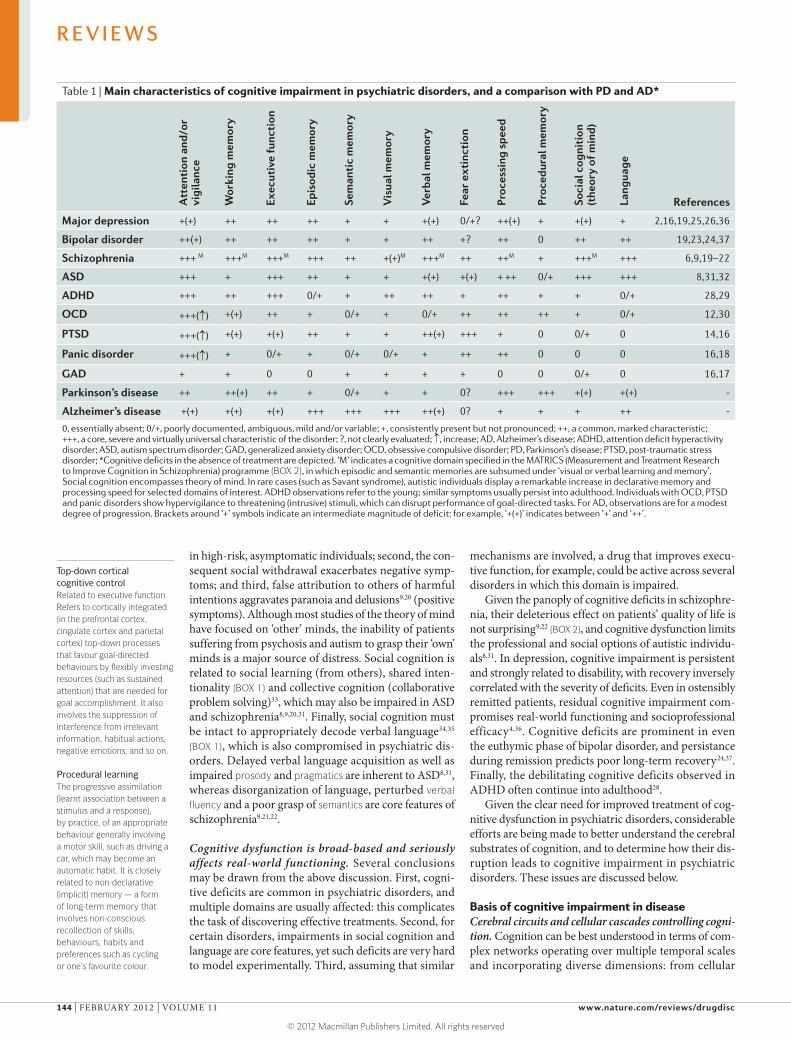
Table 1 | Main characteristics of cognitive impairment in psychiatric disorders, and a comparison with PD and AD*
Att
enti
on a
nd/o
r vi
gila
nce
Wor
king
mem
ory
Exec
utiv
e fu
ncti
on
Epis
odic
mem
ory
Sem
anti
c m
emor
y
Vis
ual m
emor
y
Ver
bal m
emor
y
Fear
ext
inct
ion
Proc
essi
ng s
peed
Proc
edur
al m
emor
y
Soci
al c
ogni
tion
(t
heor
y of
min
d)
Lang
uage
References
Major depression +(+) ++ ++ ++ + + +(+) 0/+? ++(+) + +(+) + 2,16,19,25,26,36
Bipolar disorder ++(+) ++ ++ ++ + + ++ +? ++ 0 ++ ++ 19,23,24,37
Schizophrenia +++ M +++M +++M +++ ++ +(+)M +++M ++ ++M + +++M +++ 6,9,19–22
ASD +++ + +++ ++ + + +(+) +(+) + ++ 0/+ +++ +++ 8,31,32
ADHD +++ ++ +++ 0/+ + ++ ++ + ++ + + 0/+ 28,29
OCD +++(↑) +(+) ++ + 0/+ + 0/+ ++ ++ ++ + 0/+ 12,30
PTSD +++(↑) +(+) +(+) ++ + + ++(+) +++ + 0 0/+ 0 14,16
Panic disorder +++(↑) + 0/+ + 0/+ 0/+ + ++ ++ 0 0 0 16,18
GAD + + 0 0 + + + + 0 0 0/+ 0 16,17
Parkinson’s disease ++ ++(+) ++ + 0/+ + + 0? +++ +++ +(+) +(+) -
Alzheimer’s disease +(+) +(+) +(+) +++ +++ +++ ++(+) 0? + + + ++ -
0, essentially absent; 0/+, poorly documented, ambiguous, mild and/or variable; +, consistently present but not pronounced; ++, a common, marked characteristic; +++, a core, severe and virtually universal characteristic of the disorder; ?, not clearly evaluated; ↑, increase; AD, Alzheimer’s disease; ADHD, attention deficit hyperactivity disorder; ASD, autism spectrum disorder; GAD, generalized anxiety disorder; OCD, obsessive compulsive disorder; PD, Parkinson’s disease; PTSD, post-traumatic stress disorder; *Cognitive deficits in the absence of treatment are depicted. ‘M’ indicates a cognitive domain specified in the MATRICS (Measurement and Treatment Research to Improve Cognition in Schizophrenia) programme (BOX 2), in which episodic and semantic memories are subsumed under ‘visual or verbal learning and memory’. Social cognition encompasses theory of mind. In rare cases (such as Savant syndrome), autistic individuals display a remarkable increase in declarative memory and processing speed for selected domains of interest. ADHD observations refer to the young; similar symptoms usually persist into adulthood. Individuals with OCD, PTSD and panic disorders show hypervigilance to threatening (intrusive) stimuli, which can disrupt performance of goal-directed tasks. For AD, observations are for a modest degree of progression. Brackets around ‘+’ symbols indicate an intermediate magnitude of deficit: for example, ‘+(+)’ indicates between ‘+’ and ‘++’.
R E V I E W S
144 | FEBRUARY 2012 | VOLUME 11 www.nature.com/reviews/drugdisc
© 2012 Macmillan Publishers Limited. All rights reserved

Nature Reviews | Drug Discovery
PRTC PFC Basal ganglia Thalamus
Frontal lobe
PFC OFC
PRHC
PORC
Hippocampus
ACC
DorsolateralPFC
V/VII/II
ERHC
Episodic and semantic memory (space, time and context)
Basal ganglia
Thalamus Parietalcortex
Associationcortex
Amygdala
Temporal lobe(semantic memory:storage and retrieval)
Cerebellum
DGCA3
CA1Sub
Segregation,convergenceand crosstalk
Prelimbic(expression)
Infralimbic(extinction)
Subthalamicnucleus
Striatum
Basal ganglia Cerebellum
Dorsolateral PFC Dorsolateral caudate nucleus
VA and MD
MDVA and MD
ACC Nucleus accumbensOFC Ventromedial caudate
GPi and SNr
a Attention, working memory and executive function
b Conditioned fear memory c Cerebellar modulation of cognition
–
ITCsBasolateral amygdala
Centromedialamygdala
Acquisition and storage
Ventral hippocampus(modulation)
Thalamus PonsBrainstem
Fear responses
FMN PAG
PFC
Amygdala
PFCPRTC
Cortex
Cortex
Dentate
cascades to cerebral circuits and, ultimately, society (FIG. 1). As shown in FIG. 2 and FIG. 3, specific domains such as executive function and social cognition are integrated across broad suites of interlinked and overlapping cerebral regions. Moreover, a diverse palette of neuromodulators
— including acetylcholine38, cytokines39 and brain-derived neurotrophic factor (BDNF)40 — influence cognitive performance. For example, the prefrontal cortex (PFC) and hippocampus receive a rich cholinergic input and are also heavily innervated by serotonergic, dopaminergic,
Figure 2 | Schematic representation of major cerebral circuits underpinning core cognitive domains that are disrupted in psychiatric disorders. Although individual cerebral structures fulfill distinctive roles in the control of core cognitive domains, they operate as coordinated and overlapping networks. a | The frontal lobe, basal ganglia and thalamus comprise loops that integrate attention, working memory and executive function239. The dorsolateral prefrontal cortex (PFC), anterior cingulate cortex (ACC) and orbitofrontal cortex (OFC) differentially contribute to programming and planning, decision-making and response inhibition, respectively41,239. Accordingly, they project to contrasting zones of the basal ganglia: the dorsolateral PFC projects to the dorsolateral caudate nucleus, the ACC projects to the nucleus accumbens and the OFC projects to the ventromedial caudate. Medium spiny neurons in these regions in turn converge onto the internal globus pallidus (GPi) and the substantia nigra pars reticulata (SNr), from which pathways diverge to the ventral anterior (VA) and medial dorsal (MD) thalamic nuclei239. The basal ganglia are also important for procedural learning and memory. The PFC is linked to the parietal cortex (PRTC), which exerts a modulatory influence on attention and working memory. Furthermore, the PFC and parietal cortex form the core of a circuit underpinning intelligence240, and both structures exert a top-down modulatory influence (not shown) on subcortical regions. These include the hippocampal formation (the hippocampus and the entorhinal cortex (ERHC)) and the parahippocampus (the perirhinal cortex (PRHC) and the postrhinal cortex (PORC))241; see main panel. Hippocampal territories are themselves interconnected via several circuits: the perforant pathway projects from the superficial ERHC to the dentate gyrus (DG); Schaffer collaterals project from the DG to CA3 pyramidal neurons, and mossy fibres project from CA3 pyramidal neurons to CA1 pyramidal neurons241. The subiculum (Sub) is the major source of hippocampal output. The hippocampal formation integrates dimensions of space, time and context, and is crucial for declarative learning and memory, although long-term memory may be progressively transferred to regions such as the temporal lobes, PRTC and PFC154,155,241. b | The basolateral amygdala has a key role in conditioned fear learning and extinction15,106. It excites the centromedial amygdala, which in turn projects to the brainstem, periaqueductal grey (PAG) and facial motor nucleus (FMN), where fear responses are expressed. Conditioned stimuli also affect the PFC, which fulfils a dual role: its prelimbic division facilitates the expression of fear memories, whereas its infralimbic division promotes their extinction by recruiting inhibitory GABA (g-aminobutyric acid)-ergic intercalated cells (ITCs)15. A context- dependent influence on fear learning and extinction is exerted by the ventral hippocampus, and by both the OFC and the MD thalamic nuclei, via the PFC (not shown)15. c | The cerebellum modulates cognition by reciprocal interconnections — mainly via the thalamus and the pons — with the basal ganglia and the cortex242. ‘I/II’ and ‘V/VI’ refer to layers of the entorhinal cortex.
R E V I E W S
NATURE REVIEWS | DRUG DISCOVERY VOLUME 11 | FEBRUARY 2012 | 145
© 2012 Macmillan Publishers Limited. All rights reserved

Executive functionA purposeful, goal-directed operation such as planning, decision making, problem solving, reasoning, concept formation, self-monitoring or cognitive flexibility (adaptive alternation between different strategies, responses and behaviours). Executive function reciprocally interacts with attention and working memory. It includes both initiation of appropriate and suppression of inappropriate responses.
Declarative memoryA form of long-term memory that demands conscious learning. It is divided into episodic and semantic memory.
Semantic memoryA form of long-term memory that involves the learning and storing of immutable facts, information, ideas, and so on. In contrast to episodic memory, semantic memory cannot — in principle — be modified by questions and alternative accounts.
noradrenergic and histaminergic neurons. Like the amyg-dala, these key structures contain dense populations of GABA (γ-aminobutyric acid)-ergic interneurons and they communicate with each other — as well as with other territories controlling cognitive function — via glutamatergic projections4,41,42 (FIG. 2).
Pharmacotherapy does not target cerebral circuits per se; rather, it targets G protein-coupled receptors (GPCRs), ion channels, transporters and other pro-teins involved in the actions of neuromodulators. These molecular substrates of cognition43 constitute a vast repertoire of potential drug targets for countering cogni-tive impairment in psychiatric disorders (as discussed below). Mirroring the interlinking of cerebral regions controlling cognition, there is an intricate web of cross-talk among the cellular mediators influencing cognitive processes (Supplementary information S1,S2 (figures)) (FIG. 4), such as the core substrates of neuroplasticity, learning and memory, long-term potentiation (LTP) and long-term depression (LTD)44,45 (BOX 3).
Finally, representing a level of integration that is intermediate between cells and cerebral circuits, neu-rons do not generally act in isolation; rather, they oper-ate as synchronized and rhythmically active assemblies to encode, transmit and modulate information under-pinning cognitive function46,47 (BOX 4).
Disruption of cerebral networks as a cause of cognitive impairment. Networks that modulate cognition display considerable redundancy and pleiotropy at all levels of integration: from intracellular signals, to neurons, to cerebral nuclei4,48,49. The disruption of many elements (known as nodes) can be compensated by others with similar roles; in addition, each element itself has multi-ple functions (Supplementary information S2 (figure)) (FIG. 4). This organization affords considerable resilience to disruption4,48,49. However, the failure of functionally important, highly connected nodes (known as ‘hubs’) has a disruptive effect. For example, a dysfunction in NMDA (N-methyl-d-aspartate) receptors (at the cellu-lar level) and a disruption in frontocortical GABAergic interneurons (at the circuit level) is implicated in the cognitive defects observed in schizophrenia42,50.
Furthermore, multiple ‘hits’ to networks, such as a combination of genetic and developmental or environ-mental factors, are particularly hazardous. For example, when superimposed on a vulnerable genetic background, maternal infection or cannabis use during adolescence increases the risk of schizophrenia and cognitive impair-ment7,11,51,52. Importantly, certain changes in networks (known as phase shifts) may be irreversible, such as the aberrant developmental pruning of neurons in schizo-phrenia7,42,49,53. These network-related concepts can be formally handled by graph theory, which is useful for ana-lysing the perturbation of cognitive circuits in psychiatric disorders4,48,49. For example, information-processing and cognitive performance are enhanced by the small-world features of circuits, which means that key structures are often directly linked to each other, rather than by inter-vening regions. This network attribute is compromised in schizophrenia and ASD4,48,49.
Cognitive deficits observed in schizophrenia have long been ascribed to reduced activation of the dorso-lateral PFC (known as hypofrontality) but many corti-cal and subcortical structures are also affected, with a complex pattern of region-dependent hypo- or hyper-activation9,53–55; increased activity may reflect an attempt to compensate for insufficient performance. Thus, it is arguably more pertinent to consider schizophrenia as a disconnection syndrome55. For example, a disturbance of frontocortical–striatal–thalamic loops (FIG. 2), together with impaired top-down cognitive control from the cortex, contributes to deficits in attention, working memory and executive function54,55. Furthermore, impaired verbal learning and language in schizophrenia can be related to diminished connectivity between the temporal–parietal zone (Wernicke’s area) and frontal lobes (FIG. 3), as well as reduced left hemisphere lateralization of Broca’s area and functionally related regions56.
Altered laterality in language-processing regions is also apparent in ASD57. Altered structure and function of the corpus callosum has been reported in ASD. Although its generality is unclear, a large-scale disconnection among circuits such as frontostriatal, fronto temporal and prefrontal–parietal pathways is a consistent find-ing58,59. Interruption of coupling to the cerebellum has also been reported, together with a disruption of the corticolimbic circuits mediating social and emotional
Box 1 | Social cognition, theory of mind and verbal language
Social cognition refers to processes that are used to acquire and interpret information about others, such as their character, intentions and behaviour. It necessitates: awareness, analysis, choice, sharing and/or avoidance of gaze, recognition of faces, interpretation of facial expressions, as well as scrutiny of head, whole-body and body-part motion34,35,212,213. Social cognition also refers to the understanding (and use) of the rules and concepts governing social interactions by means of gestures, etiquette, touch and proximity (personal space). Social cognition embraces the theory of mind (also known as mental attribution), which is the ability — partly by self-reflection — to infer and internally represent the mental states of others, and hence to attribute and interpret desires, beliefs, intentions and thoughts as determinants and predictors of behaviour20,34,212,214.Cultural context can modify social cognition214, which is indispensable for the full decoding and use of verbal language, especially prosody and pragmatics34,35. Reciprocally, language influences thoughts and feelings related to social cognition215.
Both social cognition and language are disrupted in psychiatric disorders (TABLE 1), and the occurrence of autism spectrum disorder and schizophrenia in humans may be evolutionarily linked to selection for complex social cognition, verbal language, creativity, large brains, an expanded prefrontal cortex and cerebral asymmetry9,20,21,35,216. Sophisticated social cognition is seen in eusocial insects, cetaceans (Supplementary information S3 (box)), some rodents (Supplementary information S4 (box)), great apes, elephants and higher birds102,217–219 (Supplementary information S5 (box)). However, the theory of mind in its fullest expression may be unique to humans, and its unequivocal demonstration in animals is therefore challenging102,214,218. Furthermore, although animals communicate in a sophisticated manner, they lack certain features of human language, such as genuine syntax, full recursion (an infinite palette of meanings generated from a finite set of elements or words) and meta-linguistics (thinking and talking about language)35,103. Hence, it is impossible to fully mimic human language in animals, and to adequately model its disruption in psychiatric disorders. Nonetheless, insights might be gained by studying the communicative role of vocal ultrasonic220, olfactory221 and tactile222 exchanges in rodents and other species, and from both the learning of innate songs and the ‘open-ended’ use of verbal exchanges in birds102,103 (Supplementary information S5 (box)).
R E V I E W S
146 | FEBRUARY 2012 | VOLUME 11 www.nature.com/reviews/drugdisc
© 2012 Macmillan Publishers Limited. All rights reserved

Episodic memoryThe conscious recollection of experiences linked to times and places in the past — what happened, where and when. It may involve mental time- travel back into a situation (known as autobiographical re-experiencing), mirrored by projection into an imagined future (prospective envisioning). As such, it is related to the theory of mind (‘travel into’ or simulation of other minds). Fully-fledged episodic memory may be a uniquely human trait, but there is evidence for its presence in primates, corvids and even some rodents.
ProsodyThe use (and interpretation) of features such as stress, intonation and rhythm that lend additional meaning and emotion to speech.
PragmaticsThe appropriate social use of spoken language.
Verbal fluencyThe ability to use written and spoken language, to choose the right word at the right time and to make appropriate associations.
processing58,59. Some cortical regions may be more strongly linked, and — at least developmentally — local overconnection (that is, excess neurons and increased dendritic spine density) also exemplifies the brain of autistic individuals59.
Somewhat reminiscent of ASD, poor attention in ADHD is related to a disruption of frontostriatal circuits, and networks interlinking temporal and parietal cor-tices with the cerebellum are also affected60. Although perturbed connectivity of the orbitofrontal cortex and subcortical regions has been consistently related to poor inhibitory control and reduced flexibility in OCD, both increases and decreases in connectivity have been observed depending on the experimental conditions61. Finally, PTSD is triggered by exposure to acute and intense stressors that disrupt PFC–amygdala connectivity, resulting in diminished fear-extinction learning14,15 (FIG. 2). Conversely, the accompanying hypervigilance reflects enhanced coupling of the amygdala to structures modulating attention, such as the anterior cingulate cortex and adrenergic projections62.
Thus, cognitive impairment in psychiatric disorders is characterized by a complex pattern of disconnection and overconnection. An important issue, therefore, is whether the circuits controlling cognition can be recon-stituted once they are disrupted, as certain structural perturbations may be irreversible — as implied by the above-mentioned notion of phase shifts4,63.
Genetic risk factors for cognitive deficits in psychiatric disorders. A full discussion of genetic susceptibility factors is beyond the scope of this article but several
points that are relevant to cognitive dysfunction should be highlighted (Supplementary information S1 (figure)).
First, although psychiatric disorders have a moder-ate to high heritability, genetic risk factors are numerous and only have a small effect; they show low penetrance and epistasis, and they do not necessarily adhere to classical nosological boundaries. For example, schizo-phrenia and bipolar disorder share some susceptibility loci7,11,52,64–66, and the same holds for schizophrenia and ASD (Supplementary information S1 (figure)). Hence, it is difficult to identify genetic risk factors for cognitive dysfunction in psychiatric disorders. Compounding the challenge, for specific psychiatric disorders cognitive impairment is heterogeneous among individuals, with regard to both its causes and characteristics8,9,12,18,23,26,28.
Second, ‘correlated’ does not necessarily imply ‘causal’. If a mutation, deletion or other genetic defect is associated with a psychiatric disorder, this does not necessarily indicate a role in the induction of cognitive impairment. Furthermore, the functional significance of single nucleotide polymorphisms is often uncertain, and some risk loci cover numerous genes7,11,52,64–66.
Third, even if a genetic defect is implicated in the pathological mechanisms that lead to cognitive impair-ment, it is not necessarily an appropriate target for their alleviation, as it may trigger anomalous mechanisms that are no longer under its control. For example, mutations in the gene encoding neuregulin 1 contribute to aber-rant patterns of neuronal migration and synaptogenesis in schizophrenia, but neuregulin 1 has a different func-tional role in the adult brain than in the developing brain, so targeting it is unlikely to reverse such anomalies67.
Fourth, some plasticity-related genes predispose individuals to cognitive deficits under adverse devel-opmental conditions but have the opposite effect in a favourable environment. This complicates analyses of their significance64.
Last, the limited success of even genome-wide studies in finding genes that are major risk factors may also be ascribed to additional layers of epigenetic control that can mask the effects of genetic defects.
Despite these hurdles, with the aid of improved experimental models7,11,68,69 several susceptibility genes for psychiatric disorders have been linked to cell-ular mechanisms that control cognitive processing (Supplementary information S1 (figure)). Furthermore, the future identification of genetic risk factors for cogni-tive deficits will be refined by: pathway analyses based on prior knowledge of protein networks65; multivariate statistics for simultaneous analysis of interacting genes66; and studies of gene associations with heritable, stable and co-segregating cognition-related endophenotypes that are likewise (although less markedly) impaired in healthy relatives11. Examples of such endophenotypes include: verbal learning and memory in bipolar disor-der70; sensorimotor gating and social cognition in schiz-ophrenia71,72; and cerebral circuit disruption in OCD and ASD73,74. Some cognitive endophenotypes may, reflecting similar pathological mechanisms, be common to dis-orders like schizophrenia, bipolar disorders or ASD.
Box 2 | The MATRICS initiative
The recognition that poorly treated cognitive deficits contribute to poor functional outcome in schizophrenia led to the establishment of the ‘MATRICS’ (Measurement and Treatment Research to Improve Cognition in Schizophrenia) initiative, which was sponsored by the National Institute of Mental Health (NIMH) in collaboration with the US Food and Drug Administration, academia and industry. The MATRICS initiative had three aims: first, to build a consensus regarding the nature of cognitive impairment in schizophrenia; second, to improve the evaluation of cognitive deficits; and third, to provide a framework for the formal recognition of treatments that specifically address the cognitive deficits associated with schizophrenia independently of an improvement in psychosis6,69,98,178,179,223.
After identifying the cognitive domains that best characterized schizophrenia (TABLE 1), the MATRICS initiative devised a neuropsychological consensus cognitive battery to support the discovery, clinical assessment and registration of new agents6,178,179. Subsequently, the NIMH funded the selection of potential cognition-enhancing agents and set up a group of academic sites to evaluate their efficacy in proof-of-concept trials. Several compounds tested to date (including a GABA
A
(γ-aminobutyric acid type A) receptor α2 subunit agonist and a dopamine D1 receptor agonist) have not proven to be clearly efficacious (TABLE 2), despite having solid conceptual and preclinical support; this highlights the uncertain predictive utility of cognitive tests in animals98,223. Hence, another programme, titled ‘CNTRICS’ (Cognitive Neuroscience Treatment Research to Improve Cognition in Schizophrenia), was established6,179 to build a consensus on two issues: first, the development of new, more reliable and practical translational paradigms for preclinical and early clinical assessment of drug effects on cognitive processes; and second, the development of imaging biomarkers for parallel use in cognitive trials. Particular efforts are being devoted to a more rigorous evaluation of the impact of therapies on real-world function in patients180,181.
R E V I E W S
NATURE REVIEWS | DRUG DISCOVERY VOLUME 11 | FEBRUARY 2012 | 147
© 2012 Macmillan Publishers Limited. All rights reserved

Nature Reviews | Drug Discovery
Amygdala
Cerebellum
Cerebellum
Angular gyrus(mainly visual)
Supramarginalgyrus (mainlyauditory)
Planumtemporale
Primary visual areaof occipital cortexACC
PFC
Broca’s area(sentences and syntax)
Primary auditory cortex
Motor cortex(speech and articulation)
OFC
Inferior frontal gyrus
Inferior parietal cortex*
Meaning, abstractionand prosody
Wernicke’s area (phonemes and words)
ACC
Frontal eye fields
Frontal eye fields Caudate SNr Superior colliculus Thalamus CortexOculomotor loop
PFC
OFC
Medial
Inferior
Temporal gyri(word storageand retrieval)
Medial
Superior
InferiorTem
pora
l gyr
i
Inferior occipital gyrusFusiform face area
Temporal parietal junction
Supplementary motor andpremotor cortex*
Insula
PrecuneusPosterior cingulate
Inferior frontal gyrus*
STS
Superior
STS
Arcuate fasciculus
Gaze and facial processing Theory of mind Verbal language
Facial/gaze processingand theory of mind
SemanticsThe meaning of what is said, written, read or heard.
Epigenetic controlA somatic and/or germline modification of chromatin (DNA plus nuclear proteins) that leads to long-lasting alterations in gene expression but not in the DNA sequence. DNA methylation silences genes and occurs mainly in CpG-rich promoter islands. Histone tails are subject to interacting processes of methylation (lysine and/or arginine residues), acetylation (lysine residues), phosphorylation, sumoylation, ubiquitylation and ADP ribosylation. Acetylation causes decondensation (unwinding), increased access for transcription factors and enhanced gene expression.
Default-mode networkA functionally interconnected network of cortical regions that is active under wakeful, resting conditions in functional magnetic resonance imaging paradigms, yet is consistently deactivated by goal-directed activity such as cognitive tasks. It includes the posterior cingulate cortex, precuneus, medial prefrontal cortex and inferior parietal cortex, and is characterized by synchronised, low-frequency oscillations of less than 1.0 Hz.
Figure 3 | Schematic representation of the principal cerebral circuits integrating social cognition and verbal language, both of which are disrupted in psychiatric disorders. A broad suite of interconnected and overlapping cerebral regions integrate and control social cognition (top panel) and verbal language (bottom panel). Verbal language is generally left-lateralized. However, prosody and the abstract features of language, as well as facial processing and the theory of mind (core elements of social cognition), have a marked implication of the right hemisphere: for example, the temporal–parietal junction34,35,211,212,243,244. The oculomotor loop is modulated by prefrontal and parietal inputs, and guides the direction and speed of voluntary eye movement183–185. Facial processing involves several interrelated dimensions of: facial perception (especially the fusiform face area and the adjacent inferior occipital gyrus); facial recognition and matching (the temporal–parietal junction); gaze tracking (the oculomotor loop, the region around the superior temporal sulcus (STS) and the temporal–parietal junction); and interpretation of facial emotion (the amygdala, the insula, the prefrontal cortex (PFC), the anterior cingulate cortex (ACC) and the orbitofrontal cortex (OFC))183–185,245,246. Some of these regions belong to a network underpinning the theory of mind, in which the medial PFC, the STS, the temporal–parietal junction and the precuneus have prominent roles. This circuit itself overlaps with the task-deactivated default-mode network located in the medial PFC, posterior cingulate, precuneus, angular gyrus and temporal lobes203. Certain structures contain mirror neurons that discharge when observing other people performing relevant behaviours; these neurons may be relevant to the theory of mind, imitation and other forms of social learning, and their dysfunction is possibly implicated in autism spectrum disorder and schizophrenia33,34,212,247. The main role of Wernicke’s area is in the perception, recognition, representation and comprehension of phonemes and words from visual and auditory input243,244. Broca’s area incorporates Brodman’s areas 44 and 45 of the inferior frontal gyrus, as well as the contiguous zones of the frontal lobe and premotor cortex. It is involved in word matching and choice, formation and syntax of sentences, as well as preparation of speech, and has a broader role in motor action preparation, music and sign language243,244,248. The arrows on the figure indicate the principal flow of information involved in the processing and production of language, including the arcuate fasciculus, which projects from Wernicke’s area to Broca’s area. In addition to this dorsal stream, a ventral stream (not shown) runs from the middle temporal lobe to the medial PFC244, which also integrates abstract features of language and prosody, together with the superior temporal gyri and amygdala. The cerebellum has a modulatory influence on social cognition, oculomotor function and language249,250. SNr, substantia nigra pars reticulata. *Contains mirror neurons.
R E V I E W S
148 | FEBRUARY 2012 | VOLUME 11 www.nature.com/reviews/drugdisc
© 2012 Macmillan Publishers Limited. All rights reserved

NeurogenesisThe continuous generation of new neurons from neural precursor cells in humans and other mammals. It is seen mainly in two regions. First, the subventricular zone of the lateral ventricle gives rise to neurons that migrate to become granule neurons and periglomerular neurons mainly in the olfactory bulb. Second, neurogenesis in the subgranular zone of the hippocampal dentate gyrus yields neurons, some of which are integrated into local neural networks once they have matured.
Linking risk genes to network disruption. As empha-sized above, disturbed network synchrony and connec-tivity are implicated in the cognitive deficits observed in psychiatric disorders (BOX 4). From a therapeutic perspective, however, drugs target molecules, so it is crucial to link changes in network operation to events at the cellular and genetic level. Neuroimaging and electrophysiological techniques can help to achieve this goal, and they can be exploited both in humans and in animal models.
One example is the so-called Val158Met polymor-phism (rs4680) in the gene encoding the enzyme cat-echol-O-methyltransferase (COMT), which catabolises dopamine; the Val and Met COMT variants are associ-ated with high and low inactivation of dopamine, respec-tively75,76. In healthy individuals, the Val variant was associated with blunted coupling between hippocampal formation and the PFC during a recognition memory task77. This observation may be related to a role of hippo-campal dopamine D1 receptors in gating hippocampal input to the PFC77. Furthermore, D1 receptor-mediated signalling is modulated by the dopamine- and cyclic AMP-regulated neuronal phosphoprotein (DARPP32; also known as PPP1R1B) (Supplementary information S2 (figure)), and a frequent PPP1R1B haplotype is associated with altered connectivity between the PFC and the stria-tum, as well as cognitive dysfunction and an increased risk of schizophrenia78.
As a second example, a polymorphism (rs1344706)that is associated with the risk of developing schizo-phrenia is located in the gene that encodes zinc finger protein 804A, a transcription factor that affects cogni-tive function72,79. During a working memory procedure, healthy carriers of the polymorphism showed gene dosage-dependent alterations in PFC connectivity across hemispheres, and between the dorsolateral PFC and the hippocampus. Functional anomalies in networks under-pinning theory of mind (FIG. 3) have also been observed72. Interestingly, in patients with schizophrenia this poly-morphism also affects cognition and attention, as well as verbal and/or episodic learning and memory80.
A third example is a rare but penetrant microdele-tion in chromosome 22 (22q11.2) that is associated with learning disabilities, cognitive dysfunction and a 30-fold increased risk of schizophrenia81. Mice with an equiva-lent microdeletion have flawed working memory related to reduced hippocampal–prefrontal synchrony. This in turn reflects a failure of PFC neurons to phase-lock with hippocampal θ-oscillations as a result of aberrant firing of GABAergic interneurons — a deficit seen in psychotic states82.
Stress as a risk factor for cognitive deficits and network disruption. Genetic factors do not fully account for the impaired cognition that is observed in psychiatric disorders. Especially in genetically predisposed individ-uals, exposure to excessive stress is a major risk factor for impaired cognitive function throughout life.
Stress is a familiar but imprecise term for the disrup-tion of homeostasis that occurs following perceived or actual exposure to adverse events, and it harnesses a vast
repertoire of neuromodulators that either promote or counter its effect83,84. An essential feature of pathological stress is hypothalamic–pituitary–adrenal (HPA) axis over-drive: this leads to poorly regulated, sustained and marked increases in levels of corticosterone downstream of the hypophyseal release of corticotropin-releasing hormone. Blockade of forebrain populations of corticotropin- releasing hormone receptor 1 counters the cognitive deficits and dendritic abnormalities elicited by acute stress and early-life adversity85. Nonetheless, most interest has focused on corticosterone. Mirroring the optimal cog-nitive performance seen at moderate levels of arousal, a well-regulated, modest and phasic recruitment of the HPA axis generally favours cognitive performance. However, excessive activation of the HPA axis is detrimental. In other words, there is a bell-shaped curve for the influence of corticosterone on cognition83,84,86,87 (see below).
One explanation for this is that genomic mineralo-corticoid receptors, which are recruited at rest, permit a positive influence over less sensitive glucocorticoid receptors. Conversely, when glucocorticoid receptor stimulation is disproportionate and persistent, cogni-tion is compromised83,88. At least in the hippocampus, this occurs in association with a pronounced release of glutamate and the activation of NMDA receptors mediating LTD45,83,89. However, a diverse pattern of inter actions among corticosterone, mineralocorticoid and glucocorticoid receptors, along with glutamatergic signalling, lead to a complex pattern of influence on cog-nition83,84,86,90,91. Thus, the notion of unitary beneficial and deleterious roles of mineralocorticoid versus gluco-corticoid receptors, respectively, is an oversimplification that complicates their therapeutic exploitation.
Analogous to psychosocial stress in humans, the exposure of adult rodents to adverse events perturbs PFC-derived networks, leading to deficits in LTP, work-ing memory and executive function84,92,93. Chronic stress-induced cognitive deficits are associated with structural remodelling, including dendritic spine retraction and neuronal atrophy in the PFC (BOX 3), reduced LTP and neuro genesis in the hippocampus, and an interference with PFC–hippocampus coupling87,88,90,92,93. Mirroring PTSD in patients14,15, acute stress leads to over-intense encoding of negative emotional memories in PFC–amygdala circuits as well as blunted fear-extinction learning94.
Prenatal and childhood stress triggers long-term changes in adolescents and adults, involving impaired cognitive function and an increased risk of depres-sion and other psychiatric disorders83,84. These delayed effects of stress appear to reflect structural and func-tional changes in corticolimbic circuits. For example, in women suffering from major depression, cognitive impairment was related to a history of early child-hood adversity and reduced hippocampal volume84. Correspondingly, early-life chronic stress in rats is asso-ciated with reductions in hippocampal LTP, dendritic spine complexity, neurogenesis and BDNF expression during adulthood83,95. However, early-life stress is not invariably associated with detrimental consequences. For instance, adult rats that had experienced early-life adversity performed poorly in non-stressful learning
R E V I E W S
NATURE REVIEWS | DRUG DISCOVERY VOLUME 11 | FEBRUARY 2012 | 149
© 2012 Macmillan Publishers Limited. All rights reserved

Agonist
• 5-HT4• D1
Agonist
• mGluR5• M1
Agonist
• GlyB
KAT2
GlyT1
D-AAO
Kynurenine
KYNA
Glycine
D-serine
Pyruvate Agonist
PAM
NMDA
• AMPA
Agonist
• GABA-α2
GABA
Agonist
• BDNF/TRKB
• 5-HT1A• D3• H3• α2C-AR• CB1• GABAB
• 5-HT6• D3
Antagonist
GPCRs
Transactivation
FYN
PKA
PKC
β/γ
Antagonist
Gαq
Gαs
Gαi/o
AC
PLC
ATP
cAMP
PKACalcineurin
cGMP
GTP
PtdIns(4,5)P2
CREB
RAS–RAFcascade
CRE
NO
PKG
NOS1 NO
GC
PDE4
Microtubules
Structural plasticity
Functionalplasticity
mRNAtranslation
Epigeneticmodulation
Synchronization
InterneuronAstrocyte
Gene transcription
PDE10A
PKC
GSK3β
GSK3β
PP1
PP1 PP1
PKC
Calcineurin
ERK
GSK3β
mTOR
AKT
PI3K
MKNK1
CaM
Ca2+
Ca2+
ERK
PP1
P
P
P
• IE genes: ARC• Effector genes: BDNF HMT DNMT HDM and DNDM?
HAT HDAC
PhosphorylationMethylationAcetylationStimulationInhibition
Chromosome
miRNA
+ –
+ –DNA
Histone
Tails
MSK1/MSK2
p90-RSK
CaMKIV
Depolarization
Nature Reviews | Drug Discovery
P P P P
Ca2+
Ins(1,4,5)P3
DAG
Na+
PKA, PKC,ERK, FYN
Cl–Glutamate
Agonist
• α4β2 nAChR• α7 nAChR• VGCC
R E V I E W S
150 | FEBRUARY 2012 | VOLUME 11 www.nature.com/reviews/drugdisc
© 2012 Macmillan Publishers Limited. All rights reserved
tasks yet performed well under stress, suggesting that the brain had been programmed to operate better under challenging conditions64,96.
Nonetheless, uncontrolled stress and HPA axis over-activity can trigger cognitive dysfunction throughout life84,85,90. The risk of middle-age depression, cogni-tive impairment and metabolic disease followed by dementia is exacerbated by stress, possibly as cortico-sterone and corticotropin-releasing hormone aggravate
glutamatergic neurotoxicity. In elderly patients these hormones worsen the harmful actions of β-amyloid and microtubule-associated protein tau — neurotoxic pro-teins that are implicated in Alzheimer’s disease84,87,90,92,93.
Modelling cognitive deficits. Modelling the genetic, developmental and environmental factors that lead to cognitive impairment in psychiatric disorders is clearly challenging. From the drug discovery perspective, the search for animal models of psychiatric disorders neces-sitates a compromise between fidelity to human pathol-ogy and efficient drug validation8,11,52,68,69. A related key issue is whether cognitive procedures in animal models can efficiently predict the efficacy of drugs in patients (BOX 2). This question is underscored by the concern that numerous pro-cognitive agents and mechanisms have been documented in rodents yet little positive feedback has been acquired in patients.
In fact, if one considers animal models to be for — rather than of — psychiatric disorders, and accepts that they can only reproduce specific aspects (such as individual causes, symptoms, responses, and so on) of a disease (not the psychiatric disorder itself), an array of genetic, developmental and environmental rodent mod-els is available for studying cognitive impairment7,52,68,97. Nonetheless, the familiar adage that ‘the best experimen-tal model is man’ is more applicable to psychiatric dis-orders than to any other field of medicine. Hence, animal models clearly need further refinement, and transgenic strategies only partially mimic human pathology and the attendant cognitive deficits7,52,68. Furthermore, no single procedure is adequate alone, gender and age are insuffi-ciently studied, and inter-individual differences deserve greater attention in view of their prominence in humans and their relevance to personalized medicine6,8,11.
Several other areas also require greater focus, particu-larly where there is a mismatch between the experimental evaluation of drugs and their ultimate use in patients. First, more studies should be undertaken with chronic drug administration to establish the delay to onset of action, long-term efficacy and lack of rebound deterioration in cognition following their discontinuation. Second, the pro-cognitive actions of drugs administered alone in rodents may not be reproduced in patients if they are masked by a deleterious cognitive impact of co-administered agents possessing, for example, antagonist properties at mus-carinic receptors and histamine H1 receptors3,97,98. Thus, mirroring their adjunctive use in humans, the effects of co-administration of pro-cognitive drugs with anti-psychotics and antidepressants should be examined in rodents. Third, many studies examine drug effects on baseline cognition. This is very different to the clinical situation, so a greater focus on drug-induced reversal of cognitive deficits in models of psychiatric disorders is desirable7,11,50,68. Last, the influence of drugs on cognition-related parameters other than behavioural outputs should be studied more intensively, as such mechanisms can be translationally monitored in humans (see below).
Despite these potential advances, many problems will remain. Notably, clinical studies are focusing increasingly on real-world function rather than on
Figure 4 | An overview of molecular substrates targeted by drugs that are designed to enhance cognitive performance in psychiatric disorders. The figure illustrates the complex pattern of crosstalk among the cellular mechanisms influencing cognitive function, of which several (in red boxes and listed in TABLE 2) are potential targets for its improvement in psychiatric disorders. Most mechanisms are depicted for simplicity in a postsynaptic element. Although one specific cell type, such as a prefrontal cortex (PFC)-localized pyramidal projection neuron, might not express all elements, these signalling cascades are widespread. The cell is innervated by a glutamatergic terminal (shown in green) adjacent to an astrocyte (shown in beige) that releases the NMDA (N-methyl-d-aspartate) and glycine B receptor co-agonists d-serine and glycine as well as the antagonist kynurenic acid (KYNA), which is cleaved from kynurenine by kynurenine amino transferase II (KAT2). The GABA (γ-aminobutyric acid)-ergic interneuron synchronizes the activity of glutamatergic neurons and other components of neuronal networks controlling cognition (BOX 4). Notably, there is convergence and divergence in signalling pathways emanating from G protein-coupled receptors (GPCRs), ion channels and tyrosine receptor kinases (TRKs) that are either recruited (agonist properties) or blocked (antagonist properties) by pro-cognitive agents. Drugs may act on downstream intracellular targets: for example, kinases (phosphorylation), the phosphatases protein phosphatase 1 (PP1) and PP2B; also known as calcineurin) (dephosphorylation), and cyclic AMP-specific phosphodiesterase 4D (PDE4D) and PDE10A. They may also act through epigenetic mechanisms of DNA and histone methylation, acetylation and phosphorylation (TABLE 2). Moreover, pharmacotherapy may act upstream via the α2 subunit of GABA
A receptors (GABA
A-α2), or it may control
the availability of glycine (reuptake suppression), d-serine (breakdown inhibition) and kynurenine (synthesis suppression) to NMDA receptors located on pyramidal cells and GABAergic interneurons in the PFC. NMDA receptors mediate rapid changes in cellular excitability, and contribute to long-term potentiation (LTP) and long-term depression (LTD) — core substrates of synaptic plasticity (BOX 3). They are permeable to Ca2+, which affects several mediators controlling cognition, including nitric oxide synthase 1 (NOS1). Changes in cognition are ultimately affected by alterations in: key signals such as extracellular-regulated kinase (ERK) and mammalian target of rapamycin (mTOR); transcription of genes pivotal to cognitive processing, such as cyclic AMP-responsive element binding protein (CREB); epigenetic programming of DNA and histones; microRNA (miRNA)-mediated regulation of mRNA translation; LTP, LTD and dendritic spine plasticity (BOX 3); synaptic architecture; and neurotransmitter release (not shown). CREB recruits CREB-responsive element (CRE) to activate immediate-early (IE) genes such as activity-regulated cytoskeleton-associated protein (ARC) and effector genes like brain-derived neurotrophic factor (BDNF). For a more comprehensive view, see Supplementary information S2 (figure). 5-HT
1A, 5-hydroxytryptamine (serotonin)
receptor 1A; α2C
-AR, α2C
-adrenergic receptor; α4β2 nAChR, α4β2 nicotinic acetylcholine receptor; AC, adenylyl cyclase; AMPA, α-amino-3-hydroxy-5-methyl-4-isoxazole propionic acid; CaM, calmodulin; CaMKIV, calcium/calmodulin-dependent protein kinase IV; CB1, cannabinoid receptor 1; D1, dopamine D1 receptor; D-AAO, d-amino acid oxidase; DAG, diacylglycerol; DNDM, DNA demethylase; DNMT, DNA methyltransferase; Gα
q, guanine-nucleotide-binding protein Gα
q; GlyB, glycine B;
GSK3β, glycogen synthase kinase 3β; H3, histamine H
3 receptor; HAT, histone
acetyltransferase; HDAC, histone deacetylase; HDM, histone demethylase; HMT, histone methyltransferase; Ins(1,4,5)P
3, inositol-1,4,5-trisphosphate; M1, muscarinic M1 receptor;
mGluR5, metabotropic glutamate receptor 5; MKNK1, MAP kinase interacting serine/threonine kinase 1; MSK1, mitogen- and stress-activated protein kinase 1; NO, nitric oxide; p90-RSK, 90 kDa ribosomal protein S6 kinase; PAM, positive allosteric modulator; PI3K, phosphoinositide 3-kinase; PtdIns(4,5)P
2, phosphatidylinositol-4,5-bisphosphate;
PKA, protein kinase A; PLC, phospholipase C; TRKB, neurotrophic tyrosine kinase receptor type 2; VGCC, voltage-gated calcium channel.
▶
R E V I E W S
NATURE REVIEWS | DRUG DISCOVERY VOLUME 11 | FEBRUARY 2012 | 151
© 2012 Macmillan Publishers Limited. All rights reserved

neurocognitive test procedures, raising the question of comparability to rodent data (BOX 2). Furthermore, verbal language and human-like social cognition (BOX 1) will presumably remain refractory to study in rodent models.
Non-rodent species may be useful in the search for improved pro-cognitive agents; notable examples include fruitfly models for studying genetics99, Aplysia californica (sea hares)101 for studying synaptic plasticity and Danio rerio (zebrafish)100 for studying developmen-tal processes and behaviour. Moreover, fruitflies and zebrafish are amenable to studies of stress, and to the use of high-throughout protocols99,100. In addition, certain other mammalian species may illuminate the nature and disruption of episodic memory, advanced social cogni-tion and language. These include great apes, elephants, dolphins (Supplementary information S3 (box)), prairie voles (Supplementary information S4 (box)) and higher birds102 (Supplementary information S5 (box)).
Most strikingly, convergent evolution in corvids and parrots has led to alternative neural solutions (including a non-laminar cortex) underpinning genuine episodic memory, sophisticated social cognition and complex vocal communication102. Furthermore, the acquisition of birdsong displays striking parallels to the learning of human language103. Obviously, great apes, elephants and dolphins are unsuitable models for pharmacologi-cal studies, and it remains to be seen whether higher birds will prove to be useful; however, as outlined in
Supplementary information S4 (box), prairie voles are instructive for characterizing the roles of potential drug targets in the control of social cognition.
Strategies to counter cognitive impairmentDirect and indirect modulation of cognitive performance by pharmacotherapy. Increasing awareness of the seri-ousness of cognitive dysfunction in psychiatric disorders, and recent insights into its potential causes, have trig-gered substantial efforts to discover drugs for restor-ing cognitive function104. Studies have focused both on specific domains (such as attention105 and extinction learning106) and on disorders (such as schizophrenia98 and ASD63). The array of concepts under investigation, listed in TABLE 2, is based both on behavioural readouts and on surrogate indexes of cognitive performance, such as cellular signals, LTP and LTD, network synchrony, transmitter release and dendrite spine formation.
As TABLE 2 is limited to targets that directly affect cog-nition, the significance of drug-induced changes in mech-anisms that indirectly modulate cognitive function should be briefly discussed. Agents that enhance sleep quality and architecture, especially slow-wave sleep, should improve hippocampal–cortical mechanisms of consolidation and other components of cognitive processing107. Drugs that normalize disrupted circadian rhythms may favour-ably affect cognitive performance108. Importantly, sleep and diurnal scheduling are often perturbed in psychi-atric disorders4,108. The potential significance of drug-induced changes in appetite and energy balance should also be noted, as glucose is transformed into glutamate and GABA via astrocytes, and diabetes is a risk factor for depression and cognitive impairment90,109. An impact of drugs on immune elements such as cytokines may similarly affect cognitive performance39.
Limited clinical feedback. There has been limited posi-tive clinical feedback so far for many of the putative pro-cognitive drug targets mentioned in TABLE 2. For example, D1 receptor agonists have never been shown to exert pro-cognitive actions in humans98,110, and GABAA recep-tor α2 subunit agonists have yielded mixed findings98,111. Nonetheless, there are some exceptions. Initial clinical studies suggest that α4β2 nicotinic acetylcholine receptor agonists38,112 and 5-hydroxytryptamine (serotonin) recep-tor 6 (5-HT6) antagonists113,114 have positive effects, and substantial data have underscored the role of oxytocin in emotional processing and social cognition115–117. Although its effects may not be entirely unitary, oxytocin consist-ently improves social cognition in volunteers as well as in individuals with ASD or schizophrenia (TABLE 2).
The noradrenaline reuptake inhibitor atomoxetine improves focused attention and executive function in ADHD118. However, noradrenaline reuptake inhibitors have not shown substantial benefits in schizophrenia, and their putative beneficial actions in depression await con-firmation2,4,98. Experimental studies have demonstrated that PFC-localized, pyramidal α2A-adrenergic receptors have a positive influence on working memory. However, the effects of agonists are less robust than those of atomo-xetine in ADHD. Furthermore, a genuine improvement
Box 3 | LTP and LTD: key neuroplastic substrates of cognition
Long-term potentiation (LTP) is the sustained (from hours to months) increase in synaptic strength elicited by a brief period (a few seconds) of patterned, high-frequency (~100 Hz) afferent stimulation. It is a flexible and diverse multiphase mechanism that is involved in many cognitive processes, from declarative learning in the hippocampus to fear-extinction learning in the prefrontal cortex (PFC)15,44. Conversely, long-term depression (LTD) refers to a long-lasting decrease in synaptic response, usually produced by a prolonged sequence (lasting a few minutes) of patterned, low-frequency (~20 Hz) stimulation44,45,134,135. A specific form of LTD (de-potentiation) follows LTP, but LTD does not just serve a homeostatic role as a balancing act for LTP or to improve the signal to noise ratio. Rather, it is also a core mechanism of cognitive plasticity and a legitimate drug target44,45,135. For example, LTD mediated by the NMDA (N-methyl-d-aspartate) receptors and muscarinic M1 receptors in the hippocampus may be implicated in learning45,135. Furthermore, impairment of metabotropic glutamate receptor 5 (mGluR5)-promoted, NMDA-dependent LTP in the PFC and hippocampus may be implicated in the cognitive impairment of schizophrenia50,224–226. Conversely, excessive mGluR5-mediated LTD in the amygdala and other structures contributes to cognitive deficits in fragile X syndrome134,135. The deleterious impact of stress on episodic memory has been related to excessive NMDA receptor-mediated LTD in the hippocampus, possibly as a result of AMPA (α-amino-3-hydroxy-5-methyl-4-isoxazole propionic acid) receptor endocytosis45. Conversely, stress also impairs cognition by disrupting LTP across a hippocampal–PFC-integrated network88,92. Thus, changes in both LTP and LTD are related to the cognitive deficits observed in psychiatric disorders, and numerous drug targets (such as NMDA receptors, M1 receptors and mGluR5) modulate both of these substrates of neuroplasticity44,45,104,112,135,224,226 (TABLE 2).
Importantly, LTP and LTD are associated with the structural plasticity of dendritic spines — that is, their expansion and formation (LTP), and their contraction and loss (LTD)128 — in several classes of neurons that are important for cognition, including pyramidal neurons in the PFC and medium spinal neurons in the basal ganglia (FIGS 2,4). Spines are regulated in an activity-dependent manner by local protein synthesis and mRNA translation, which is itself subject to modulation by microRNAs168,170. Structural spine plasticity is anomalous in disorders such as schizophrenia and autism spectrum disorder128.
R E V I E W S
152 | FEBRUARY 2012 | VOLUME 11 www.nature.com/reviews/drugdisc
© 2012 Macmillan Publishers Limited. All rights reserved

in working memory has yet to be demonstrated and these agonists have a small therapeutic window. Long-term release forms of such agonists may therefore prove to be more useful119,120.
Putative pro-cognitive actions of glycine trans-porter 1 inhibitors in schizophrenia are constrained by motor and autonomic side effects, and results with par-tial agonists at the glycine B co-agonist site on NMDA receptors have been variable98,121. Finally, a vigilance enhancer, modafinil, displayed encouraging effects on cognition (including facial processing and speed of pro-cessing) in patients with schizophrenia, thus supporting studies in volunteers, but the results of more recent, controlled studies have been less compelling98,122,123.
Complex effects on cognition: bell-shaped dose–response curves. Clearly, considerable progress is needed with regard to the clinical profiles of pro-cognitive agents. Their experimental and therapeutic evaluation is com-plicated by the fact that the doses needed to improve cognition depend on several variables, including baseline performance, genotype, test sensitivity and end point. Furthermore, similarly to corticosterone (see above), many agents have ‘inverted U’ dose–response curves in behavioural and mechanistic procedures124–126 (TABLE 2). Biphasic dose–response curves imply a ‘set point’ for optimal performance, such that under- or overactiva-tion of the drug target has a deleterious effect. This is perhaps not surprising, as both deficient and excessive LTP, LTD, ‘plasticity gene’ activity, neurogenesis and dendritic spine generation have a deleterious effect on cognitive processing43–45,64,87,127,128 (BOX 3).
Clinical studies of COMT inhibitors illustrate the significance of this phenonemon75,76. COMT inhibitors enhance extracellular levels of dopamine in the PFC, which improves cognition when basal levels of dopa-mine are low. This has been observed in volunteers and in patients with schizophrenia possessing a Val/Val phe-notype associated with high dopamine metabolism, and also in pathologies in which dopamine levels are reduced in the PFC, such as depression and late-stage Parkinson’s disease. Conversely, cognition deteriorates when basal levels of dopamine are high, as observed in some vol-unteers and in patients with schizophrenia possessing a Met/Met genotype75,76.
Other than off-target, low-potency actions (like muscarinic receptor antagonism) of drugs that perturb cognition, there are several other non-exclusive expla-nations for complex and inverted-U dose–response curves. First, reflecting the homeostatic control of cognition, overactivation of any one mechanism (such as phosphorylation) by a drug may provoke the over-compensatory response of another (such as dephos-phorylation)129 (FIG. 4). Second, a drug may be highly selective for its target but the target itself may exert a complex influence on cognition via spatially distinct receptor populations. For example, activation of post-synaptic α2-adrenergic receptors on PFC pyramidal neurons favours working memory41,119. Conversely, activation of α2-adrenergic receptors that are inhibitory to fronto cortical adrenergic, dopaminergic and cholin-ergic projections is detrimental for working memory and executive function130,131 (TABLE 2). Third, even a single population of sites can mediate a biphasic dose–response curve. Induction of GPCR endocytosis with high concentrations of agonists offers one explanation for this (Supplementary information S2 (figure)), but a more widespread explanation comes from the cou-pling of GPCRs to functionally distinct transduction pathways. For example, 5-HT6 receptors exert a dose-dependent positive and negative effect on cognition via recruitment of cAMP-responsive element binding protein (CREB) and mammalian target of rapamycin (mTOR), respectively114. Accordingly, monotonic, pro-cognitive dose–response relationships could be gen-erated using biased ligands that only recruit specific cellular pathways favouring cognition. A complemen-tary approach would be the exploitation of GPCR-modulatory proteins to direct signalling down specific transduction routes132.
More generally, a prudent approach for enhancing cognition over a broad dose range would be to prior-itize partial rather than full agonists; allosteric modu-lators may also be an option. For intracellular targets such as kinases, partial inhibition is also preferable for safety reasons.
Normalization of pathological processes versus symp-tomatic strategies. There are two complementary ways to restore cognitive performance: first, by countering pathological changes underlying deficits; and second, by recruiting pro-cognitive mechanisms that are inde-pendent of disease aetiology.
Fragile X syndromeA disease that is usually caused by the expansion of a trinucleotide sequence in the 5′-untranslated region of the fragile X mental retardation 1 (FMR1) gene. This leads to FMR1 promoter hyper methylation, transcriptional silencing and loss of the RNA-binding protein FMR1. Abnormal translation of mRNAs, including those regulated by metabotropic glutamate receptor 5, results in excessive long-term depression. Affected individuals have defects in speech, language, attention, working memory and social cognition.
Box 4 | Network synchrony: disruption in psychiatric disorders
Network-coordinated rhythmic activity within and between the regions of the brain controlling cognition is associated with electroencephalographical (EEG) activity that can be quantified non-invasively46,47,88,192,193. Theta (θ; 4–7 Hz) frequencies are related to episodic memory and they are widely used to monitor oscillations driven by hippocampal regions in coordination with the prefrontal cortex (PFC)46,47,88,192,193. Conversely, the activity of GABA (γ-aminobutyric acid)-ergic interneurons in the PFC is reflected in the γ (30–80 Hz) range, which is linked to attention and working memory47,193,195. The synchronization of θ rhythms, γ rhythms and β (12–30 Hz) rhythms across regions of the brain represents a ‘neural code’ that modulates and drives cognitive processes such as top-down cortical control. Accordingly, disruption of EEG-monitored rhythms may reflect cognitive impairment46,47,192,193,227.
Coordinated network activity in the PFC and hippocampus depends on GABAergic interneurons that impose a temporal signature on the firing patterns of neurons controlling cognition42,193,195. Thus, glutamatergic pyramidal cells in the PFC receive trains of fast, inhibitory postsynaptic potentials from parvalbumin-positive GABAergic interneurons that recruit the α2 subunits of GABA
A receptors on their axon hillocks42
(FIG. 4). In schizophrenia, a developmental deficit in excitatory NMDA (N-methyl-d-aspartate) receptors — and possibly AMPA (α-amino-3-hydroxy-5-methyl-4-isoxazole propionic acid) receptors — on these GABAergic interneurons leads to a decrease in their activity, anomalous patterns of pyramidal cell firing, perturbed PFC network synchronicity and cognitive impairment42,50,192,228. Frontocortical GABAergic interneurons and pyramidal cells integrate inputs from many modulators controlling cognitive function, including monoamines, acetylcholine and glutamate. Hence, they are a focal point of strategies for enhancing cognition both in schizophrenia and in other psychiatric disorders (FIG. 4). Finally, GABAergic mechanisms in the amygdala are important for fear memory, and a dysfunction of GABAergic intercalated cells (FIG. 2) is implicated in the weakened fear-extinction learning seen in post-traumatic stress disorder15,106 (BOX 1).
R E V I E W S
NATURE REVIEWS | DRUG DISCOVERY VOLUME 11 | FEBRUARY 2012 | 153
© 2012 Macmillan Publishers Limited. All rights reserved

Table 2 | Overview of drug classes proposed for the treatment of cognitive impairments in psychiatric disorders*
Drug target and molecular action
Disorders to treat
Cellular substrates (sites of action)
Other useful functional properties
Possible MOA-related drawbacks
Clinical feedback on cognitive actions
Other useful tools
Refs
Dopamine D1
receptor
agonist (PAG)
SCZ, PD • ↑PKA, DARPP32, CREB
• ↑ARC• ↑NMDA
signalling • ↑LTP and LTD
(PFC) • ↑ACh (PFC)
• Antiparkinsonian• ↓Drug-seeking
behaviour
• IUDR • Dyskinesia • Desensitization • Hypotension
• Dihydrexidine • ↑PFC activity,
inactive on WM (SCZ)
• SKF81297• ABT-431• A-77636
98,110, 126,198
Dopamine D3 receptor antagonist
SCZ, BPD, PD, ASD, ADHD, AD, NF, FXS, TSC
• ↓mTOR (PFC)• ↑NMDA
signalling (PFC, NACC)
• ↑ACh, d-serine (PFC)
• ↑SMG• Antiparkinsonian• Antipsychotic• ↓Drug-seeking
behaviour• Nephroprotective
• Uncertain • Unavailable • ABT-614• S33138• S33084• SB-277011
97,251, 252
COMT inhibitor
SCZ, PD, ADHD, depression
• ↑Dopamine, noradrenaline (PFC)
• ↑PFC–subcortical connectivity
• ↑SMG• Antiparkinsonian
• IUDR• Genotype
(Val/Met)- dependent
• ↑Sympathetic output
• Tolcapone: ↑WM and PPI (HV)
• ↑ or ↓EXF (Val or Met genotype, HV)
• ↑Cognition (PD)
• Entacapone 75,76
α2A
-AR agonist (PAG)
ADHD, SCZ, Tic disorders
• ↑ERK• ↓HCN activity
(Pyram., PFC)• ↑Connectivity
(PFC)
• ↓Hyperactivity• ↓Impulsivity
(ADHD)• Analgesic• ↓Opioid
withdrawal
• IUDR• ↓LTP (HIPP)• ↓ACh,
noradrenaline, dopamine (PFC, HIPP)
• Mild ↓ in AP/HR
• Somnolence
• Guanfacine XR, Clonidine:↑attention (ADHD)
• Inactive in SCZ
• Guanabenz 119,120
α2A/2C
-AR antagonist
SCZ, PD, depression, PTSD, phobias
• ↑ARC, ACh noradrenaline, dopamine, histamine (PFC, HIPP)
• ↑Neurogenesis (HIPP)
• Antidepressant• Antiparkinsonian• ↑Libido• ↓Erectile
dysfunction
• ↓WM (PFC)• Nervousness• Panicogenic• ↑Sympathetic
output• Over-arousal
• Idazoxan: ↑cognition. (modest) (HV, SCZ)
• Yohimbine: ↑CFE (claustrophobia)
• Atipamezole• BRL-44408
(α2A
-AR)• JP-1302
(α2C
-AR)
104,130, 131
Noradrenaline transporter blocker
ADHD, depression, SCZ, OCD, PD, Korsakoff’s syndrome
• ↑Dopamine, noradrenaline (PFC)
• ↑Noradrenaline (HIPP)
• ↑θ (HIPP) ↑BDNF and neurogenesis (HIPP)
• Antidepressant• Anti-impulsive?
• ↓Sleep• Hypertension
or↑HR• Over-arousal
• Atomoxetine:↑attention, WM and EXF (ADHD); inactive in SCZ, ↑cognition in PD
• Reboxetine:↑attention and SOP, ↓negative bias in depression
• Maprotiline 98,118
5-HT1A
antagonist (PAG)
Depression, SCZ, AD, ASD
• ↑ERK • ↑Pyram.
(PFC, HIPP)• ↑ACh, Glu
(PFC, HIPP)
• Anxiolytic• Antidepressant
(PAG)• ↑Sleep
(antagonist)
• IUDR• Poor
cardiovascular tolerance (PAG)
• Tandospirone: modest ↑WM and VLM (SCZ)
• Buspirone: inactive in SCZ
• S-15535 (PAG)
• Lecozotan• WAY-100635
(antagonists)
104,113
5-HT4 agonist
(PAG)Depression, AD, OCD, SCZ
• ↑PKA, CREB • ↑BDNF, BCL-2,
LTP (HIPP)• ↑ACh (HIPP)
• Antidepressant• Neuroprotective• ↓Aβ accumulation
• Gastrointestinal and cardiac side effects
• Unavailable • PF-4995274• RS-67333• VRX-03011• PRX-03140
104,113, 145
5-HT6
antagonistSCZ, BPD, AD, ASD, FXS, TSC
• ↓mTOR (PFC)• ↑NCAM-PSA• ↑γ (PFC) • ↑ACh, Glu,
dopamine (PFC, HIPP)
• Anxiolytic• Antidepressant• ↓Obesity
• Interference with cognition (via ↓CREB and ERK activity)
• SGS518:↑cognition (SCZ)
• SB742457: ↑cognition (AD)
• PRX07034: ↑cognition (HV)
• SYN-114• SAM-531• R-1485
104,113, 114
R E V I E W S
154 | FEBRUARY 2012 | VOLUME 11 www.nature.com/reviews/drugdisc
© 2012 Macmillan Publishers Limited. All rights reserved

Table 2 (cont.) | Overview of drug classes proposed for the treatment of cognitive impairments in psychiatric disorders*
Drug target and molecular action
Disorders to treat
Cellular substrates (sites of action)
Other useful functional properties
Possible MOA-related drawbacks
Clinical feedback on cognitive actions
Other useful tools
Refs
H3 receptor
antagonist or inverse agonist
ADHD, SCZ, AD, depression, EDS, narcolepsy
• ↑PKA, CREB• ↑NCAM-PSA• ↑θ (HIPP)• ↑ACh, histamine,
noradrenaline (PFC, HIPP)
• ↑SMG• Analgesic• ↓Obesity
• Nervousness• Over-arousal• Poor sleep• Immune and
gastrointestinal side effects
• MK0249: inactive (SCZ)
• JNJ-17216498, PF-03654746:↑attention, inactive in ADHD
• BF2.649• GSK189254• Ciproxifan
139,253
Muscarinic M
1 receptor
agonist (PAM)
SCZ, BPD, AD
• ↓GSK3b• ↑NMDA
signalling • ↑LTP and LTD
(PFC, HIPP) • ↑Pyram. (PFC)• ↑ACh, dopamine
(PFC)
• Antipsychotic• Neuroprotective• ↓Tau hyper-
phosphorylation• ↓Aβ production
and aggregation
• Gastrointestinal side effects
• Depressogenic?• Sweating• Salivation
• Xanomeline: modest• ↑WM and
VLM (SCZ)• Sabcomeline: mild
↑attention, VLM and SOP (SCZ)
• TBPB• BQCA• AC-42• ML-169
98,112, 254,255
α7 nAChR (PAG or PAM)
SCZ, AD, PD, ADHD
• ↑ERK • ↓GSK3b• ↑γ, θ (PFC, HIPP)• ↑ACh, Glu,
noradrenaline (PFC, HIPP)
• ↑BCL-2
• ↑SMG• Antidepressant• Neuroprotective• ↓Tau hyper-
phosphorylation; Aβ neurotoxicity
• Anti-inflammatory
• IUDR• Tachyphylaxis• Dependence?• Gastrointestinal
side effects, especially nausea
• CP810123: inactive (HV)
• DMXB: weak↑attention, WM, EPM and SOP (HV)
• DMXB/MEM3454/EVP6124: ↑SMG, mild ↑cognition (SCZ)
• MEM3454: ↑cognition (AD)
• TC-5619• AZD0328• SEN34625• SSR180711
38,98, 112,140
a4b2 nAChR agonist (PAM)
ADHD, SCZ, AD, PD
• ↑LTP• ↑θ (HIPP) • ↑ACh, Glu,
histamine (PFC, HIPP)
• ↑SMG• ↓Drug-seeking
behaviour• Analgesic• Neuroprotective?
• IUDR• Depressogenic?• Nausea• Tachyphylaxis
• TC1734: ↑attention, EPM, PPI (HV)
• Ispronicline:↑attention, EPM (elderly patients) ABT418: ↑attention, WM (ADHD)
• ABT089: inactive (ADHD)
• Varenicline: ↑attention, WM (nicotine withdrawal)
• S38232• A-85380
38,112
NMDA and/or glycine B receptor agonist (PAG)
Phobias, SAD; panic disorder, PTSD, OCD, SCZ, BPD, ASD
• ↑ARC• ↑NOS1, PKG• ↑PKC• ↑LTP (PFC,
HIPP, STM)• ↑LTD (HIPP,
PFC, BLA)
• ↓Drug-seeking behaviour (for cocaine)
• ↓Deficit symptoms (SCZ)
• IUDR• Tachyphylaxis• Excitotoxicity
• Glycine, d-cycloserine, d-serine: weak ↑cognition or inactive (HV, AD, SCZ)
• d-cycloserine: ↑CFE (SAD, phobias, OCD, panic disorder)
• GLYX-13• S-18841
98,125, 176,256
Glycine reuptake inhibitor
SCZ, BPD, phobias, PTSD?
• ↑Glycine (PFC, HIPP, STM)
• ↑LTP (HIPP)
• ↑SMG• ↓Deficit
symptoms (SCZ)
• IUDR• Motor and
respiratory side-effects (cerebellum, medulla)
• Sarcosine: weak ↑cognition or inactive (SCZ)
• R213129: inactive (HV)
• RF1678 • ALX5407• SSR504734
98,121, 244,256
d-amino acid oxidase blocker
As for glycine B agonists?
• ↑d-serine (PFC, HIPP, CBM)
• ↑LTP (PFC, HIPP)
• As for glycine B agonists?
• As for glycine B agonists?
• Unavailable • AS057278• CBIO• SEP-227900
257,258
AMPA receptor (PAM)
Depression, ADHD, AD, SCZ, PTSD, FXS
• ↑ARC • ↑NMDA
signalling• ↑LTP, BDNF
(HIPP, PFC)• ↑Noradrenaline
(HIPP)
• Antidepressant• Neuroprotective
• LRS• Sensory
dysfunction• Proconvulsant• Excitotoxicity
• CX516/CX717: weak ↑cognition or inactive (HV, SCZ, ADHD, FXS, elderly patients)
• Farampator (NS): ↑EPM (HV)
• LY451395: inactive (AD)
• S-18986• Org-24448• LY392098
98,104, 259,260
R E V I E W S
NATURE REVIEWS | DRUG DISCOVERY VOLUME 11 | FEBRUARY 2012 | 155
© 2012 Macmillan Publishers Limited. All rights reserved

Table 2 (cont.) | Overview of drug classes proposed for the treatment of cognitive impairments in psychiatric disorders*
Drug target and molecular action
Disorders to treat
Cellular substrates (sites of action)
Other useful functional properties
Possible MOA-related drawbacks
Clinical feedback on cognitive actions
Other useful tools
Refs
mGluR5 (PAM and/or PAG)
SCZ, BPD, PD, PTSD, phobias, OCD
• ↑PKC, CREB• ↑ARC• ↑NMDA signalling• ↑LTP (HIPP, PFC)• ↑LTD (BLA, HIPP)
• ↑SMG• Antipsychotic?• ↓Drug seeking
behaviour
• IUDR• Excessive LTD
(HIPP, CBM)• Epileptogenic• Excitotoxic
• Unavailable • CDPPB• ADX-47273• VU1545
225,226
mGluR5
antagonistFXS, depression, PD
• ↓Excessive LTD• Normalization
of dendrite morphology
• Anxiolytic• Antidepressant• Antidyskinetic• ↓GORD
• ↓NMDA signalling, LTP
• ↓CFE• Pro-psychotic
• Fenobam (NS), AFQ056, acamprosate (NS): inactive, modest ↑attention, SOP and PPI (FXS)
• MPEP• MTEP
134,135, 261
GABAAR
α5 subunit inverse agonist
Depression, AD, SCZ, NF, Down syndrome
• ↑LTP• ↑θ (HIPP)• ↑ACh (HIPP)
• ↑Neurogenesis? • ↓CFE and spatial memory
• Proconvulsant?
• α5IA: ↓ethanol-induced amnesia (HV), inactive in AD
• L-655708• PWZ-029• RO4938581• MRK-016
42,133
GABAAR
α2 subunit
agonist (PAG)
SCZ, BPD, NF, OCD, ASD, depression
• ↑Synchrony of pyramidal cells
• ↑γ (PFC)
• ↑SMG• Anxiolytic• Anticonvulsant
• Desensitization • MK0777: ↑VLM, ↑γ but inactive in MATRICS battery (SCZ)
• TPA023B 42,111, 133,262
Adenosine A
2A receptor
antagonist
ADHD, PD, AD, depression
• ↓p38 MAPK• ↑D1 and D2
signalling (STM)• ↑Motivation• ↑dopamine (NACC)
• Antiparkinsonian• Antidepressant• Neuroprotective• ↓Aβ
neurotoxicity
• ↓BDNF and LTP (HIPP)
• Insomnia• Nervousness• Psychostimulant
• Caffeine (NS): ↑arousal, attention and SOP (HV); inactive or modest ↑attention in ADHD
• Preladenant• SCH-58261• ZM241685• Istradefylline
263,264
Cannabinoid receptor 1 (CB1) antagonist
SCZ, BPD, AD
• ↓mTOR (HIPP)• ↑LTP
(HIPP, PFC)• ↑ACh,Glu
(PFC, HIPP)
• ↑SMG• Antipsychotic?• ↓Obesity
• IUDR• ↓LTD (HIPP)• ↓Extinction• Depressogenic• Anxiogenic
• Rimonabant: inactive (SCZ)
• SLV330• AVE1625• AM-251
51,104, 146,265
Oxytocin receptor agonist
ASD, SAD, ADHD, SCZ, BPD, phobias
• ↓Coupling of medial amygdala to brainstem (↓social fear and aversion)
• ↑Dopamine release in nucleus accumbens (↑ approach and reward)
• ↑SMG• Pro-social• ↑Pair-bonding,
parental behaviour
• ↓Anxiety• ↓Aggression• Antidepressant?• ↓HPA stress
activation
• ↓VLM?• Gender-
dependent prosocial actions
• ↑Envy• ↓Trust
(borderline personality disorder?)
• Endocrine side effects
• Oxytocin (intranasal): ↑attention, trust, TOM, facial expression analysis, positive social memory, gaze to eye region, empathy, affective speech comprehension (HV, ASD, SCZ)
• Carbetocin• WAY-267464
(non- peptidergic)
115–117, 201,219
Vasopressin V
1A receptor
agonist
ASD, SCZ, BPD, phobias
• Lateral septum• Centromedial
amygdala• PFC• ↑Noradrenaline
(HIPP)
• ↑SMG• ↓Sexual
dysfunction• ↑Pair-bonding• ↑Partner
preference
• Gender- dependent actions
• ↑Anxiety• ↑Avoidance• ↑Aggression
(male)• Endocrine and
cardiovascular side effects
• Vasopressin (intranasal):↑ability to encode happy and angry faces (males); ↑agonistic versus females (males); ↑friendly attributions (HV)
• NC-1900• VP 4-9• VP 4-8
115,266
BDNF receptor (PAG)
Depression, BPD, PD, AD, PTSD, phobias
• ↑CREB• ↑AMPA and NMDA
signalling• ↑LTP and LTD• ↑Glu• ↑Neurogenesis
(HIPP)
• Antidepressant• Neuroprotective
• IUDR• LRS
• Unavailable • BDNF mimic, 7,8-dihydroxy-flavone
40,104, 158,159
PDE4D inhibitor
SCZ, depression, AD, RTS
• ↑PKA• ↑CREB• ↑LTP and LTD• ↑BDNF (HIPP)
• ↑SMG• Antidepressant• Neuroprotective?
• LRS• Emesis• Vasculitis• Immune side
effects
• HT0712: inactive on WM; ↑VLM (elderly patients)
• MK0952: ↑cognition (AD)
• Rolipram• MEM1091• RO 20-1724
141–143, 145
R E V I E W S
156 | FEBRUARY 2012 | VOLUME 11 www.nature.com/reviews/drugdisc
© 2012 Macmillan Publishers Limited. All rights reserved

Table 2 (cont.) | Overview of drug classes proposed for the treatment of cognitive impairments in psychiatric disorders*
Drug target and molecular action
Disorders to treat
Cellular substrates (sites of action)
Other useful functional properties
Possible MOA-related drawbacks
Clinical feedback on cognitive actions
Other useful tools
Refs
PDE10A inhibitor
SCZ, BPD, PD • ↑PKA and PKG• ↑CREB (STM)• ↑LTP and LTD
• Antipsychotic?• Anti-obesity
• Anxiety• Sedation• Dyskinesia• Side effects in
testes
• Unavailable • Papaverine• TP-10• PQ-10• MP-10
141,142
Protein kinase Cε activator
Depression, AD, stroke
• ↓GSK3β• ↑NMDA
signalling• ↑GAP43• ↑ACh and Glu• ↑Synaptogenesis
• ↓Tau and soluble Aβ levels
• Anti-tumorigenic
• LRS• Desensitization• Pro-manic• ↑Stress-induced
damage (PFC)• Myalgia
• Unavailable • Bryostatin 1c• DCP-LA
144,267
mTOR1 inhibitor
BPD, SCZ, HD, AD, PD, TSC, FXS, NF
• ↓Excessive NMDA signalling, LTP and mGluR5- mediated LTD
• ↓5-HT6, D3
and CB1 overstimulation
• ↓Dyskinesia (PD)• ↓Tau hyperphos-
phorylation and Aβ production
• Neuroprotective• Antitumorigenic
• LRS• IUDR • ↓LTP andLTD• Oncogenic• Immunosuppression
• Unavailable • Rapamycin• Everolimus• Temsirolimus
136,146, 147
GSK3β inhibitor
BPD, SCZ, AD, depression, PD, stroke
• ↑CREB • ↑NMDA and
AMPA signalling• ↑LTP (HIPP)
• Antimanic• Antidepressant• Neuroprotective
(PD, stroke)• ↓Tau hyperphos-
phorylation and ↓Aβ aggregation
• LRS• ↓LTD• Oncogenesis
↑β-catenin activity
• Lithium (NS): inactive or ↓ cognition (BPD)
• AR014418• SB415286• NP031115• NP12
148,149
Calcineurin (protein phosphatase 2B) inhibitor
SCZ, AD, BPD
• ↑D1 signalling • ↑CREB• ↑LTP, ↓LTD
(HIPP/PFC)
• ↑SMG• Anti-apoptotic• Neuroprotective• ↓Aβ deposition and
neurotoxicity
• LRS• ↓CFE• Pro-psychotic• Depressogenic
• Unavailable • FK306• Cyclosporin A• Tacrolimus
129,150, 151,166
Class I histone deacetylase 2 inhibitor
BPD, AD, PD, HD, PTSD, RTS, ASD, FXS,
• ↑CREB, BDNF and LTP (HIPP)
• ↑BCL-2• ↑Synaptogenesis
• Antimanic• Antidepressant• Neuroprotective• Anti-oncogenic
• LRS• Cytoxicity
• Valproate (NS): ↑social cognition (FXS)
• Vorinostat• Trichostatin A• Sodium
butyrate• EVP0334
137,138, 165,167
Microtubule stabilizer
SCZ, BPD, AD, PD, ASD, cerebral palsy
• ↑LTP (HIPP)• ↑Neurite growth• ↑Neurogenesis
• Neuroprotective• ↓Tau hyperphos-
phorylation
• LRS• Cytoxicity
• Davunetide: ↑cognition (SCZ, elderly patients)
• Epothilone D• Darbepoetin α
268
*Major potential domains of therapeutic exploitation are indicated. Several possible neuronal and cellular substrates of pro-cognitive properties are shown but owing to limited data and differences between various cognitive domains, this information is inevitably incomplete; ‘absence of evidence’ must not be misconstrued as ‘evidence of absence’. Many studies have focused on the prefrontal cortex, hippocampus, and as concerns fear-extinction learning, the basolateral amygdala. Some have investigated the cerebellum, striatum and nucleus accumbens. For transmitters like glutamate, changes refer to release in vivo. In most cases sensorimotor gating signifies prepulse inhibition. Apart from cognition, other potential beneficial and undesirable actions related to mechanism of action are summarized. For schizophrenia, cognitive testing in humans has invariably been in association with established antipsychotics, yet the opposite is true for rodents. As clinical data are limited, comparative data for Alzheimer’s disease are provided in some cases. Cognition is indicated when details on cognitive domains tested are unclear. Amyloid-β and tau refer to the load of toxic and/or phosphorylated forms of the proteins. ‘Pyram.’ implies increased firing rate of pyramidal cells. The drugs shown are described as selective for their respective targets, although some agents of limited selectivity are included where clinical data are available. 5-HT
1A, 5-hydroxytryptamine (serotonin) receptor 1A; α
2A-AR, α
2A-adrenergic receptor; γ, increased γ power; θ, increased θ power; Aβ, amyloid-β; ACh,
acetylcholine; AD, Alzheimer’s disease; ADHD, attention-deficit hyperactivity disorder; AMPA, α-amino-3-hydroxy-5-methyl-4-isoxazole propionic acid; AP/HR, arterial pressure and/or heart rate; ARC, activity-regulated cytoskeleton-associated protein; ASD, autistic spectrum disorder; BCL-2, anti-apoptotic protein B cell lymphoma 2; BDNF, brain-derived neurotrophic factor; BLA, basolateral amygdala; BPD, bipolar disorder; BQCA, benzylquinolone carboxylic acid; CBIO, 5-chlorobenzo[d]isoxazol-3-ol; CBM, cerebellum; CBT, cognitive behavioural therapy; CDPBB, 3-cyano-N-(1,3-diphenyl-1H-pyrazol-5-yl)benzamide; CFE, conditioned fear extinction; COMT, catechol-O-methyltransferase; CREB, cyclic AMP-responsive element binding protein; CV, cardiovascular; DA, dopamine; DARPP32, cyclic AMP-regulated neuronal phosphoprotein; DCP-LA, 8-[2-(2-pentyl-cyclopropylmethyl)-cyclopropyl]-octanoic acid; DMXB, 3-2,4-dimethoxy-benzylidene anabaseine; EDS, excessive daytime sleep; EPM, episodic memory; ERK, extracellular signal-regulated kinase; EXF, executive function; FXS, fragile X syndrome; GABA
AR, γ-aminobutyric acid type A receptor; Glu, glutamate; GORD, gastroeosophageal reflux disease; GSK3β, glycogen synthase kinase 3β; HCN,
hyperpolarization-activated cyclic nucleotide-gated channel; HD, Huntington’s disease; HIPP, hippocampus; HPA, hypothalamic–pituitary–adrenal axis; HV, human volunteers; IUDR, inverse ‘U’-shaped dose–response curve; LRS, lack of regional selectivity; LTD, long-term depression; LTP, long-term potentiation; MAPK, mitogen-activated protein kinase; MATRICS, Measurement and Treatment Research to Improve Cognition in Schizophrenia; mGluR5, metabotropic glutamate receptor 5; MOA, mechanism of action; MPEP, 2-methyl-6-(phenylethynyl)pyridine; MTEP, 3-((2-methyl-4-thiazolyl)ethynyl)pyridine; mTOR, mammalian target of rapamycin; NA, noradrenaline; NACC, nucleus accumbens; nAChR, nicotinic acetylcholine receptor; NCAM-PSA, polysialylated form of neuronal cell adhesion molecule; NF, neurofibromatosis; NMDA, N-methyl-d-aspartate; NOS1, nitric oxide synthase 1; NS, not selective; OCD, obsessive compulsive disorder; PAG, partial agonist; PAM, positive allosteric modulator; PD, Parkinson’s disease; PDE4, cyclic AMP-specific phosphodiesterase 4; PFC, prefrontal cortex; PPI, prepulse inhibition; PKA, protein kinase A; PTSD, post-traumatic stress disorder; RTS, Rubinstein–Taybi syndrome; SAD, social anxiety disorder; SCZ, schizophrenia; SMG, sensorimotor gating; SOP, speed of processing; STM, striatum; TBPB, 1-[1′-(2-methylbenzyl)-1,4′-bipiperidin-4-yl]-1,3-dihydro-2H-benzimidazol-2-one; TOM, theory of mind; TRKB, neurotrophic tyrosine kinase receptor type 2; TSC, tuberous sclerosis; VLM, verbal learning memory; WM, working memory; XR, extended release.
R E V I E W S
NATURE REVIEWS | DRUG DISCOVERY VOLUME 11 | FEBRUARY 2012 | 157
© 2012 Macmillan Publishers Limited. All rights reserved

As an example of the former strategy, glycine B ago-nists are designed to treat schizophrenia by stimulating hypoactive NMDA receptors localized on GABAergic interneurons in the PFC42,50,98. PFC-integrated cognition can also be restored by agonists acting downstream at the α2 subunit of GABAA receptors42,98,133 (FIG. 4). Another example of this strategy is provided by the mechanism of action of COMT inhibitors, which compensate for low levels of dopamine in depression75,76. The epigenetic developmental disorder fragile X syndrome is character-ized by excessive metabotropic glutamate receptor 5 (mGluR5)-mediated LTD, which leads to cognitive defi-cits that can be countered by mGluR5 antagonists at these sites134,135. Similarly, cognitive deficits associated with the ASD-related disorder tuberous sclerosis are provoked by mutations in the genes encoding tuberous sclerosis pro-tein 1 and tuberous sclerosis protein 2, which interact with mTOR (Supplementary information S2 (figure)); cognitive impairment may therefore be reversed by the mTOR inhibitor rapamycin63,136. More generally, epige-netic reprogramming raises the hope of reversing the cognitive impairment accompanying monogenic ASD and caused by early-life stress (see below)63,137,138.
Although they are conceptually attractive, pathology-driven approaches have limitations. First — as exempli-fied by rare, genetic forms of ASD — they may only be applicable to a subpopulation of patients. Second, molec-ular substrates underlying cognitive deficits are still not generally well understood. Third, it may be impos-sible to retrospectively normalize certain pathological events, such as neonatal insults that trigger alterations in synaptic architecture, and neural circuits that lead to schizophrenia63,67.
Complementary strategies for symptomatic treat-ment do not attempt to normalize a pathological change, such as NMDA receptor hypofunction. Rather, they engage compensatory pro-cognitive mechanisms spared by the disorder in question. For example, there is no evi-dence for 5-HT6 or histamine H3 receptor hyperactiva-tion in schizophrenia, yet antagonists hold promise for correcting a range of cognitive deficits in schizophrenia as well as in other disorders such as depression113,114,139. Pathology-decoupled mechanisms may actually have a broader application than pathology-driven strategies. In addition, some drugs should have beneficial actions not only against cognitive impairment but also against other symptoms (TABLE 2).
Domain-specific and generalized improvements in cognitive performance. A crucial issue is whether drugs will primarily correct one specific cognitive domain or improve several simultaneously. This obviously depends on the drug’s mechanism of action. If a treatment com-pletely reversed the underlying pathology it might — in theory — correct all deficits: one example, as further evoked below, is the blockade of mTOR overactiva-tion in the monogenic disorder tuberous sclerosis63,136. Conversely, for a complex, multifactorial and hetero-geneous disease like schizophrenia, aiming to normal-ize cognitive performance across all domains and in all patients appears to be overambitious.
There may be greater hope of finding a mechanism that could improve one specific cognitive domain in a transnosological manner across numerous diseases. Examples include oxytocin agonists for promoting social cognition115–117, and α7 nicotinic acetylcholine receptor agonists for reinforcing attention and working memory38,112,140 (Supplementary information S4 (box)) (TABLE 2). Clearly, a balance must be sought between the holy grail of ‘one drug, all domains, all disorders’ versus ‘a separate drug for each domain and each disor-der’. Although a pan-cognitive agent that can restore all domains may be unobtainable, multitarget drugs uniting complementary mechanisms of action appear to be the most promising route towards achieving a broad-based improvement in cognition4,104.
Intracellular targets. Intracellular targets are attracting increasing interest as substrates for improving cognitive deficits in psychiatric disorders (FIG. 4) but they are not easy to exploit43,45,57–60,63,212. Such agents do not neces-sarily sidestep the issue of biphasic dose–response rela-tionships, and they raise serious issues of tolerance and regional specificity because of the ubiquity and the mul-tiple roles of most cellular targets. This is illustrated by the contrast between the restricted cerebral localization of 5-HT6 receptors and the broad organismal distribution of phosphodiesterases catalysing cAMP degradation114,141,142 (FIG. 4).
One solution may be to develop drugs that are selec-tive for protein isoforms such as phosphodiesterase 4D or protein kinase Cε.Although achieving selectivity is a for-midable challenge, exploitation of allosteric rather than catalytic sites may help drug design141,143,144. The influence of an inhibitor should be most prominent where and when the activity of its target is aberrantly high. Hence, another approach involves ‘vectoring’ drugs using dual-acting molecules that act on an intracellular protein and an upstream mechanism. For example, coupling inhi-bition of phosphodiesterase 4D to 5-HT4 receptor ago-nism may restrict the facilitation of cAMP-dependent transmission to regions where 5-HT receptors control cognition145. Association of 5-HT6 receptor antagonism with mTOR inhibition could yield similar benefits114,136. However, the design of drugs acting at two or more useful sites is challenging, and it is important to demonstrate an improved therapeutic window of beneficial versus deleterious actions as well as advantages of these drugs compared with selective drugs used in combination.
mTOR is involved in both LTP and LTD; further-more, it favours and — when hyperactive — counters cognitive processes, so it is a particularly interesting intracellular target136. For example, tuberous sclerosis involves loss of the mTOR inhibitory protein partner tuberculosis sclerosis protein, leading to its overstim-ulation (Supplementary information S2 (figure)). Accordingly, the mTOR inhibitor rapamycin attenuated excessive hippocampal LTP and relieved cognitive defi-cits in a mouse model of tuberous sclerosis, and it may also be effective in other forms of ASD136. Rapamycin antagonises cognitive deficits elicited by the stimulation of cannabinoid receptor 1 on inhibitory GABAergic
Tuberous sclerosisAn autosomal dominant disorder, usually caused by sporadic mutations, leading to inactivation of the tumour suppressor genes tuberous sclerosis 1 (TSC1;also known as hamartin) and TSC2 (also known as tuberin), which normally inhibit RHEB (a GTPase that is an activator of mammalian target of rapamycin). Loss of TSC1 or TSC2 leads to disinhibition of cell growth, cortical tubers and giant astrocytomas in the brain. Patients have deficits in attention, executive function and memory, as well as symptoms resembling autism spectrum disorder and attention deficit hyperactivity disorder.
R E V I E W S
158 | FEBRUARY 2012 | VOLUME 11 www.nature.com/reviews/drugdisc
© 2012 Macmillan Publishers Limited. All rights reserved

interneurons in the hippocampus146, and by the activa-tion of 5-HT6 and D3 receptors in the PFC14 (TABLE 2), which suggests that mTOR overactivation may be rel-evant in schizophrenia. mTOR is also implicated in hippocampal processes sustaining abnormal fear mem-ory147. Despite these indications that mTOR inhibitors might improve cognitive deficits, their therapeutic use could interfere with physiological LTP, and therefore raises issues of safety and specificity. Such concerns also apply to glycogen synthase kinase-3β inhibitors for pro-moting LTP and long-term memory in depression and bipolar disorder148,149 (TABLE 2).
Protein phosphatase 1 and protein phosphatase 2B (also known as calcineurin) are targets for improving long-term memory as they interfere with the activation of CREB and downstream cognition-related genes such as BDNF129 (FIG. 4). Accordingly, calcineurin inhibitors normalize the biphasic dose–response curves of drugs that enhance CREB activity by phosphorylation, and they promote LTP and learning in the hippocampus129,150. However, inhibition of calcineurin strengthens the for-mation of aversive memories in the amygdala, underscor-ing the multiple effects of phosphatases on CREB activity and cognition129,151 (Supplementary information S2 (fig-ure)). As with kinases, the question of specificity must be addressed before phosphatase inhibitors could be thera-peutically exploited for treating cognitive impairment129.
Neurogenesis. In recent years, considerable attention has been devoted to hippocampal neurogenesis, as its suppression is implicated in the impaired episodic memory associated with depression4,127,152. Adult-born, maturing dentate gyrus cells are especially excitable and plastic, sustain prolonged LTP and are rapidly incorporated into and coordinate neural networks, suggesting that increased neurogenesis favours cogni-tion127. This explains the role of these cells in learning, consolidation and updating of new memories, dif-ferentiating separate memories in the dentate gyrus, transferring hippocampal-dependent memories to extra-hippocampal regions and coupling cognition to external context127,153–155. However, neurogenesis is no exception to the inverted-U dose–response rule, as both the generation and suppression of new neurons is required to optimize cognitive function127. Moreover, although antidepressants consistently enhance neuro-genesis in rodents, they do not generally favour cognitive performance4,156. On balance, therefore, facilitation of neurogenesis is not yet a compelling target for improv-ing cognition in psychiatric disorders.
Further insights may be gained by studying the cell ular signals mediating the influence of neurogen-esis on mood and cognition, such as activity-regulated cytoskeleton-associated protein157 and the upstream driver of neurogenesis, BDNF, which acts via neuro-trophic tyrosine kinase receptor type 2 (NTRK2; also known as TRKB)40,158 (FIG. 4). The decreased expression of BDNF seen in chronic stress may be related to the cognitive deficits associated with depression, as BDNF mediates several forms of hippocampal and PFC plas-ticity, including LTP40,152. Interest in BDNF has been
enhanced by reports showing that TRKB activators block the disruptive influence of stress on hippocam-pus-integrated long-term memory40,158, and that BDNF promotes extinction learning159.
Epigenetics. Stress-induced epigenetic changes in ger-mline cells can be passed on to and alter cognition in offspring, suggesting that environmental risks for cog-nitive deficits might be relevant even before concep-tion63,137,138,160–162. Some changes can be sex-specific. For example, epigenetic imprinting of the maternal allele of genes in the 15q11–13 region is implicated in the aetiology of ASD160.
Of perhaps broader relevance to pharmacotherapy, gene-specific alterations in DNA methylation and his-tone acetylation (FIG. 4) may contribute to long-term impairments in cognition resulting from exposure to stress during early life84,137,138. For example, DNA hypo-methylation (leading to enhanced expression) of the gene encoding corticotropin-releasing hormone may account for HPA axis overdrive in adults who have undergone early-life stress137,138,162. Conversely, perinatal stress leads to hypermethylation-induced silencing of the gene encoding BDNF138. Another example is ASD, in which hypermethylation-induced suppression of the oxytocin receptor gene has been reported161.
Although decreases in DNA methylation are hard to counter pharmacologically, increases could be countered by DNA-N-methyltransferase (DNMT) inhibitors. Apart from their potential utility for correcting epigenetic changes provoked by early-life stress, DNMT inhibitors may also be useful in other disorders: for example, for counteracting the consolidation of fear memories seen in PTSD137,138. Furthermore, DNMT inhibitors may improve cognition in Rett’s syndrome, an X-linked ASD characterized by gene hypermethylation138. In addition, in schizophrenia DNMT inhibitors may normalize cog-nitive deficits resulting from hypermethylation-induced suppression of PFC-localized genes synthesizing GABA and reelin (a developmental glycoprotein that controls synaptic plasticity)163,164.
Reflecting interactions between DNMTs and his-tone deacetylases (HDACs), certain effects of DNMT inhibitors can be mimicked by suppressors of overactive HDACs, such as valproate137,138. Valproate is not selective for HDAC isoforms but selective inhibition of HDAC2 could be especially useful as this isoform negatively reg-ulates hippocampal LTP, dendritic spine density and vis-ual learning, as well as BDNF and CREB gene expression. HDAC2 inhibitors could therefore enhance these pro-cognitive processes and also promote fear-extinction learning, suggesting utility in PTSD137,138,165,166. Turning to ASDs, fragile X syndrome is characterized both by the loss of the mRNA translation regulator fragile X mental retardation 1 protein and by hypoacetylation of several functionally interacting classes of histones134,138. Valproate and a class III HDAC inhibitor increased histone acetylation and reactivated silenced fragile X mental retardation 1 protein in lymphoblastoid cells of patients with fragile X syndrome. Furthermore, val-proate also improved social cognition and attention167.
Rett’s syndromeAn X-linked developmental disorder, mainly seen in females, caused by de novo mutations in the gene encoding methyl CpG binding protein 2 (MECP2). MECP2 normally binds to methylated DNA to transcriptionally repress genes, although some are activated. MECP2 also interacts with histone deacetylases, so its loss leads to gene-dependent histone hypo- and hyperacetylation. Patients with Rett’s syndrome suffer from retardation, loss of verbal learning and speech, and impaired social cognition.
R E V I E W S
NATURE REVIEWS | DRUG DISCOVERY VOLUME 11 | FEBRUARY 2012 | 159
© 2012 Macmillan Publishers Limited. All rights reserved

Rubinstein–Taybi syndrome is an epigenetic ASD-related disorder caused by mutations in CREB-binding proteins that act as histone acetyltransferases (Supplementary information S2 (figure)). Hence, HDAC inhibitors are being studied for the correction of cognitive deficits associated with this syndrome137,138.
Finally, blockade of nuclear protein phosphatase 1 (FIG. 4) should, by promoting histone phosphorylation, favour hippocampal-dependent LTP and memory137,166.
Epigenetic control of gene transcription operates around a set point: DNA hyper- or hypomethylation and histone hypo- or hyperacetylation disrupts cogni-tion. Hence, pharmacotherapy must restore the balanced integration of multiple modes of epigenetic control that are requisite for appropriate cognitive performance. Specificity and safety are also vital issues. A particular concern is the risk of tumorigenic side effects, for example, resulting from hypermethylation-induced inactivation of tumour suppressor genes. Nonetheless, the targeting of methylation, acetylation and other epigenetic markers offers a potentially unique route for the correction of cog-nitive deficits in disorders such as ASD and schizophrenia.
Modulation of miRNA-controlled neural circuits. Further potential opportunities for restoring cognitive function are emerging from studies on brain-enriched microRNAs (miRNAs) that fine-tune cellular networks controlling synaptic plasticity and cognition, both developmen-tally and in adults168. For example, miR-134 inhibits hippocampal LTP and synaptic plasticity in mice by repressing CREB and BDNF synthesis169. The relevance to cognitive deficits in psychiatric disorders is supported by the observation that schizophrenia is associated with the abnormal biogenesis of miRNAs170. More specifi-cally, decreased levels of miR-219 may be due to NMDA receptor hypoactivity171. Furthermore, in fragile X syn-drome altered miRNA processing is implicated in the excessive mGluR5 signalling that contributes to cogni-tive impairment134,135,170.
With regard to the therapeutic exploitation of miR-NAs, one possibility could be to modify their biogen-esis via cellular signals that phosphorylate the proteins involved in their maturation81 (Supplementary informa-tion S2 (figure)) (FIG. 4). Another approach could be to recruit HDACs that suppress the synthesis of miR-134, thereby promoting cognition by preventing the inhibi-tion of CREB and BDNF generation169; furthermore, various classes of modified and stabilized oligonucleo-tides, and of mRNA analogues, have been designed to directly interfere with (or mimic) the activity of specific miRNAs170. Even if miRNA-targeted therapy appears to be a distant prospect at present, miRNA profiling may serve as a biomarker both of cognitive dysfunction and of the actions of pro-cognitive agents170.
Coupling pharmacotherapy with alternative strategies. As mentioned above, highly selective agents acting at a single target may be insufficient for the broad-based correction of cognitive deficits across pathogenetically distinct psychiatric disorders; this has encouraged inter-est in the development of multifunctional agents4. More
generally, it might be questioned whether the admin-istration of pro-cognitive agents (even in combina-tions) will always be adequate. Rather, a combination of pharmacotherapy with alternative strategies (BOX 5) may sometimes be a more effective strategy for palliat-ing cognitive deficits in psychiatric disorders, similarly to the combination of cognitive behavioural therapy and pharmacotherapy for improving the treatment of depression7,172–175.
For example, although they are only modestly active when administered alone, the α2-adrenergic receptor antagonist yohimbine and the NMDA receptor partial agonist d-cycloserine enhance the efficacy of behav-ioural extinction techniques for countering cognitive deficits associated with phobias, panic disorder and OCD106,176. Furthermore, d-cycloserine and oxytocin might be especially effective for treating social cogni-tive deficits in ASD when coupled with behavioural therapies that likewise reinforce social learning176,177 (Supplementary information S4 (box)). It may be instructive to extend such studies to cognitive remedia-tion therapy173 (BOX 5), as the effects of novel agents for relieving cognitive impairments in schizophrenia could best be expressed in synergy with this approach, rather than on top of antipsychotics that even interfere with their actions3,97,98,174.
Clinical development of pro-cognitive agentsAs highlighted above, many concepts are under explora-tion for countering cognitive dysfunction in psychiatric disorders. Their successful development depends on the careful translation of information acquired in animal and cellular models into clinical research6,8,11,68,69,97. Clinical studies of pro-cognitive agents can now use various techniques with experimental counterparts (FIG. 5) for: estimating optimal drug doses for efficacy; tracking cognitive actions in a manner complementary to behavioural rating scales; improving stratification (choice of subpopulations) of patients for drug trials; and exploring cerebral mechanisms of pro-cognitive properties.
Neuropsychological tests for evaluating cognitive func-tion in volunteers and patients. Many procedures are available for characterizing the influence of drugs on cognitive performance, and several test batteries have been proposed7,9,11,68,69,98,178,179 (BOX 2). Although these test batteries are instructive, one drawback is the potential loss of a large domain-specific effect in an overall (aver-age) non-effect. Furthermore, patient performance and test sensitivity can be compromised by poor engage-ment during long, tiring and stressful clinical trials6,9,10. Another issue is that separate cognitive domains may not be genuinely independent, as they interact with and depend on common neuronal substrates5,6,178,179 (FIGS 2,3). Notably, sustained, focused attention — together with high processing speed — is important for success in many procedures. These observations — and a com-mon interrelationship with intelligence — explain why measures of cognitive performance are often corre-lated5,6,97,178,179. It might even be suggested that a single
Rubinstein–Taybi syndromeA rare disorder characterized by autistic features, learning difficulties and poor attention. In approximately 50% of cases, it is caused by de novo mutations or deletions in the genes encoding CREB-binding protein or, rarely, histone acetyltransferase p300. These CREB-binding proteins and transcriptional co-activators are also histone acetylases, so patients display histone hypoacetylation and reduced gene transcription.
MicroRNAs(miRNAs). Small, non-protein- coding sequences (22–24 nucleotides) of RNA, mostly derived from intergenic regions, although some are found in introns. An individual species of miRNA can bind to the 3′-untranslated regions of up to hundreds of different species of mRNA. Translation is usually suppressed but it is sometimes enhanced, and in certain cases mRNA may even be degraded.
R E V I E W S
160 | FEBRUARY 2012 | VOLUME 11 www.nature.com/reviews/drugdisc
© 2012 Macmillan Publishers Limited. All rights reserved

Therapeutic approach Influence on emotional symptoms*
Influence on cognitive impairment*
Psychiatric disorders targeted
Currently available pharmacotherapy –/0/++
Schizophrenia, depression, bipolar disorder, anxiety disorders
Deep-brain stimulation or electroconvulsive therapy 0/–+
Major depression
Repetitive transcranial magnetic stimulation 0/+0/+
Mainly depression (autism, schizophrenia)
Cognitive behavioural therapy 0+
Mainly depression (anxiety disorders)
Cognitive remediation therapy +0/+
Mainly schizophrenia (depression)
Exposure therapy for desensitization +0/+
Post-traumatic stress disorder, obsessive compulsive disorder, phobias, social anxiety disorders
Improved drugs (alone and in combination with above strategies)
++Dependent on mechanism of action
*The ‘+’ symbol corresponds to improvement; the ‘–’ symbol corresponds to worsening; and ‘0’ corresponds to no marked change.
dimension, such as working memory, would suffice to predict the overall effect of the drug on cognitive func-tion. However, this is both risky and contentious, so it is best to use multiple procedures to monitor the influence of drugs on baseline cognition and deficits in patients.
The importance of optimizing measurements of cog-nitive performance in volunteers and patients, and of optimizing comparability between preclinical and clini-cal procedures, is exemplified by the establishment of the MATRICS (Measurement and Treatment Research
Savant syndromeA rare syndrome that is closely associated with high- functioning autism spectrum disorder but also found in other developmental disorders and following damage to or disease of the central nervous system. It alludes to ‘islands of genius’ in one or a few cognitive domains such as mathematics despite broader deficits in others, and is usually associated with prodigious memory. Savant-like abilities can partially be reproduced by transcranial magnetic stimulation over the cortex.
Box 5 | Alternative therapeutic strategies to address cognitive impairment
Conventional psychotropic drugs were not specifically designed to target cognitive dysfunction in psychiatric disorders, and they display a range of (usually modest) beneficial, deleterious and neutral actions on cognition (see table). Non-pharmacotherapeutic strategies can be compared in terms of their differential impact on cognitive impairment versus emotional symptoms.
Despite showing efficacy in refractory depression, neither electroconvulsive therapy (which provokes transient retrograde amnesia) nor deep-brain stimulation of the subgenual cortex exerts a long-term, beneficial influence on cognition229,230. Conversely, despite having a less robust influence on depression, repetitive transcranial magnetic stimulation (rTMS) over the left dorsolateral prefrontal cortex (PFC) appears to promote attention, working memory and procedural learning. It probably acts by enhancing GABA (γ-aminobutyric acid)-ergic interneuron activity, γ-oscillations and PFC-driven top-down cognitive control227,230–233. Intriguingly, rTMS of the PFC enhanced γ-activity and cognitive processing in autism spectrum disorder, and elicited Savant syndrome-like cognitive feats in normal individuals233,234. In schizophrenia, enhanced cognition and suppression of auditory hallucinations has been reported230. Beneficial effects are not restricted to the PFC. Application of rTMS to the right parietal cortex favours focused attention, whereas rTMS over Broca’s or Wernickes’s areas improves verbal fluency and language learning; these effects mimic direct transcranial current stimulation — a technique that is used for rehabilitating patients with language and cognitive deficits following brain damage230,235.
Psychotherapy encompasses techniques such as problem solving, behavioural activation and cognitive behavioural therapy (CBT), which encourages patients to shed their negative views on themselves and their life. CBT for depression and anxiety can lead to durable improvements in mood (outlasting treatment), possibly by strengthening PFC-controlled top-down control of emotional processing in dysfunctional limbic structures like the amygdala and the hippocampus172,236. However, CBT has not yet been demonstrated to improve cognitive function. Conversely, cognitive remediation therapy (CRT), which adopts an approach that is broadly similar to cognitive training in elderly patients, can improve cognitive function in schizophrenia173,237 and possibly also in depression238. As for CBT, it is labour-intensive and treatment outcome depends on the therapist, methodology, content and intensity of the programme. Furthermore, CRT cannot be used for all patients and requires a regular commitment despite the low motivation of many patients with schizophrenia. Nonetheless, CRT is instructive as improvement is measured in terms of community functioning, such as salary, time spent at work and normalized social interactions.
Finally, exposure therapy is a cognitive approach for treating post-traumatic stress disorder, phobias, social anxiety disorder and obsessive compulsive disorders. Its application involves repeated contact with an aversive situation or stimulus to encourage desensitization (extinction)106,176,177.
Ideally, a novel pharmacotherapy should target both the emotional and cognitive impairments in psychiatric disorders. Moreover, agents should be developed and evaluated alone, in combination with other pro-cognitive mechanisms as well as in association with alternative therapies as a function of the symptom and patient population concerned. The combination of pharmacotherapy with CBT, CRT or similar approaches may be particularly effective for improving the cognitive impairments associated with psychiatric disorders174,175.
R E V I E W S
NATURE REVIEWS | DRUG DISCOVERY VOLUME 11 | FEBRUARY 2012 | 161
© 2012 Macmillan Publishers Limited. All rights reserved

Nature Reviews | Drug Discovery
EEG of ERPs:• MMN (50–150 milliseconds, pre-attentional)• N170 (170 milliseconds, facial processing)• P300 (300 milliseconds, attentional): amplitude and gating
Electromyography (eye-blink reflex):• Pre-attentional sensorimotor gating• Pre-pulse inhibition
Oculomotor neurophysiology:• Eye movements (saccades and antisaccades)• Smooth pursuit eye movements• Delayed responses
Magnetoencephalography event-related fields:• High spatial and temporal resolution, but mainly sources parallel to skull surface
PET and SPECT imaging:• Cerebral metabolism• Target (for example, GPCR) occupation• Transmitter release
Neuropsychological proceduresand batteries for evaluating drug influence on cognitive function
qEEG: spectral analysis and neuronal synchrony: • γ (30–80 Hz): local, cortical• θ (4–7 Hz): hippocampal and cortico-subcortical
fMRI (BOLD) measures of cerebral activity:• Performance of cognitive tasks• Default mode (resting state: task deactivated)
Magnetic resonance spectroscopy: • Glutamate, ACh and GABA transmission• NAA: energy, neuronal integrity• Fatty acids, neurogenesis
Measures of real-world functioning
to Improve Cognition in Schizophrenia) initiative for schizophrenia6,69,98,178,179 (BOX 2). The insights gained from this initiative could be exploited to set up com-parable programmes dedicated to improving cognition in depression. In an effort to better relate drug actions to everyday living, for both MATRICS and other pro-grammes, co-primary measures of functional capacity are being developed, including patient competence to undertake tasks such as shopping180,181. Improved predic-tion of the social and vocational outcomes of treatment is crucial, as relief of functional disability — not superior performance in test batteries — is the real goal. Notably, however, one tricky question is whether improved real-world functioning can be attributed to the cognitive and/or emotional actions of a specific treatment.
With regard to measures, patient self-assessment is desirable but can be misleading. Furthermore, although informants can reliably assess cognitive abilities of patients, this is not usually practical for drug trials6,180,181.
Finally, the design of long-term studies — for example, from the prodrome to diagnosis in schizophrenia — is far from simple. Shifts from baseline performance rather than changes versus placebo may be preferable, but practice effects following repeated testing must be considered6,182.
All clinical studies in the psychiatric domain com-mence with human volunteers. This population can deliver early feedback on how drugs potentially affect specific cognitive domains, how their effects may best be monitored in subsequent studies, and which under lying cerebral substrates they possibly engage — especially when neurocognitive tests are coupled to electro-encephalography (EEG) and neuroimaging (FIG. 5). Some observations may also be of more direct relevance to patient populations. For example, oxytocin consist-ently enhances social cognition in healthy probands as well as in autistic or schizophrenic individuals115–117. Acute administration of the NMDA receptor antagonist
Figure 5 | Overview of translational models for characterizing and predicting the influence of pharmacological agents on cognitive function in humans. Numerous procedures are available for assessing cognitive domains ranging from attention to social cognition, and several test batteries have been designed, such as the CANTAB (Cambridge Neuropsychological Test Automated Battery) core cognition test and the BACS (Brief Assessment of Cognition in Schizophrenia) test for schizophrenia, as well as the MATRICS (Measurement and Treatment Research to Improve Cognition in Schizophrenia)-derived consensus cognitive battery (BOX 2). Currently, efforts are being directed towards measures that are more closely linked to real-world cognitive functioning. Complementary approaches for exploring the actions of putative pro-cognitive agents in humans, most of which have translational counterparts in rodents, may be broadly classified as follows: quantitative electroencephalographical (qEEG) analysis coupled to spectral analysis of neural circuits, and EEG quantification of auditory event-related potentials (ERPs); magnetoencephalography, which monitors magnetic fields emitted by synchronized neurons with millisecond precision; sensorimotor paradigms, which include measures of eye movement; functional magnetic resonance imaging (fMRI) to estimate neuronal activity in defined brain regions; positron emission tomography (PET) and single-photon emission computed tomography (SPECT) to visualize radioligand binding; and magnetic resonance spectroscopy to quantify the levels of neuromediators. Many informative ERP-based signals can be translationally exploited. Notably, prepulse inhibition refers to the blunting of the startle reflex to an intense auditory stimulus following pre-exposure 100 milliseconds earlier to a subthreshold stimulus. Mismatch negativity (MMN) is a response to an (auditory or visual) oddball stimulus deviating from a regular sequence. Negative deflection (N170) is a 170-millisecond EEG signal that is associated with facial processing, whereas a positive 300-millisecond (P300) signal is associated with attention to a task-relevant, infrequent stimulus. ACh, acetylcholine; BOLD, blood oxygenation level-dependent; GABA, γ-aminobutyric acid; GPCR, G protein-coupled receptor; NAA, N-acetylaspartate.
R E V I E W S
162 | FEBRUARY 2012 | VOLUME 11 www.nature.com/reviews/drugdisc
© 2012 Macmillan Publishers Limited. All rights reserved

ketamine to volunteers mimics the NMDA receptor hypoactivity observed in schizophrenia, so this pro cedure is useful for characterizing the influence of putative pro-cognitive antipsychotics on sensorimotor gating (see below) and cognition50,71,98. Furthermore, abnormalities in cognitive function and its underlying neural sub-strates can be probed in healthy individuals displaying variants of susceptibility genes for psychiatric disorders, such as zinc finger protein 804A (see above)72,79,80.
Nonetheless, several important limitations of Phase I studies should be mentioned. The influence of acute drug administration on baseline cognitive function in volunteers — and even its disruption by risk genes or pharmacological agents — may differ from its actions following chronic administration to patients with more complex pathologies. For example, drugs that are designed to normalize the aberrant epigenetic pro-gramming that causes cognitive dysfunction in ASD would be hard to evaluate in volunteers. Furthermore, many drugs developed for schizophrenia are intended for use in combinations, yet studies with other classes of agents cannot be undertaken in Phase I trials. More generally, the influence of drugs on cognitive per-formance in patients will reflect not only their direct impact on cognitive mechanisms but also their ability to relieve emotional symptoms, a facet largely inacces-sible in volunteers. Finally, studies in healthy individuals are inevitably restricted to neurocognitive procedures rather than genuine measures of real-world function. Without neglecting the importance of early-stage clinical studies, these points illustrate the risk of drawing prema-ture conclusions, and highlight that proof of concept can only come from studies in patients.
Oculomotor studies of the control of cognition. Eye-movement studies (FIG. 5) can be exploited to evaluate the influence of drugs on neural processes related to cogni-tion183–186. As few preclinical models of social cognition are recognized, oculomotor-tracking studies of eye gaze in primates as well as in humans — including attention to (and avoidance of) the gaze of others — are of particular interest184. Oculomotor paradigms have also been pro-posed for evaluating the effect of drugs on neural bases involved in sensorimotor gating, attention, working memory, executive function and procedural memory185. For example, oculomotor techniques in primates have been used to explore the influence of PFC populations of D1 receptors on working memory and top-down con-trol of visual processing185,186. Such observations provide a translational platform for clinical studies showing that oculomotor measures are sensitive to both the favourable and unfavourable effects of drugs such as antipsychotics and benzodiazepines on cognition183,185.
Underpinning interest in eye-moment studies in patients, oculomotor deficits have been reported in schiz-ophrenia and several other psychiatric disorders183–185. Eye-movement paradigms can also be coupled to neuro-imaging to characterize the influence of potential pro-cognitive drugs on cerebral circuits187. Although further work is needed to clarify the relationship between eye-movement measures and cognitive mechanisms, they
deserve further characterization as a distinctive trans-lational approach for exploring the actions of putative pro-cognitive drugs, especially with respect to social cognition.
Sensorimotor gating in relation to cognitive perfor-mance. Information processing and sensory gating — pre-attentional and attentional processes that are required for effective cognitive performance — can be monitored by various procedures7,71,98,188,189 (FIG. 5). These include pre-pulse inhibition (FIG. 5), which has marked dopaminergic, serotonergic and glutamatergic components. Accordingly, deficits in prepulse inhibition in patients with schizo-phrenia, and its disruption by psychotomimetic agents in volunteers, can be countered by antipsychotics7,68,71,189. Evaluation of new drugs, such as nicotinic acetylcholine receptor agonists and histamine H3 receptor antagonists, can be guided by their influence on the perturbation of prepulse inhibition in humans and rodents by psychoto-mimetics and (in the latter) by genetic and developmental models of schizophrenia52,71,97,98,139,140,189 (TABLE 2).
An additional pre-attentional response, ‘mismatch negativity’ (BOX 5), has a marked glutamatergic and NMDA receptor component, and deficits have so far been mainly seen in schizophrenia; moreover, impair-ment in high-risk individuals predicts onset of the disease7,71,190. In patients with schizophrenia mismatch negativity was enhanced by N-acetylcysteine, which elevates extracellular levels of glutamate in the PFC191. These observations highlight the interest in sensorimotor gating paradigms for exploring the effects of pro-cognitive drugs on pre-attentional and attentional function in both animals and in humans (FIG. 5).
Quantitative EEG for probing cognitive circuits. By directly probing large-scale electrical activity in the brain with high temporal resolution, quantitative EEG (qEEG) coupled to spectral analysis provides unique insights into the cortical processes underlying cognitive func-tion and affected both in psychiatric disorders and by pharmacotherapy46,47,188 (FIG. 5). EEG monitoring of cortical–subcortical networks in humans and animals is of particular interest because alterations in neural synchronization and connectivity are strongly related to cognitive deficits and their pharmacotherapeutic modu-lation46,47,192,193 (BOX 4). For example, oxytocin can shift cortical resources in volunteers to regions involved in social cognition and emotional processing194. EEG stud-ies are also instructive for characterizing the influence of drugs on arousal, sleep–wake cycles and sleep architec-ture, which can affect memory consolidation and other cognitive processes107.
The related technique of magnetoencephalography picks up magnetic fields generated by intraneuronal cur-rents to provide fine-grained (millisecond) temporal and spatial (superior to EEG) information on coordinated neuronal activity. It can also be used to characterize the relationship of cortical networks to cognition in psychi-atric disorders188. Supporting its use for drug characteri-zation, diazepam modifies θ- and γ-oscillations with a profile that is consistent with its negative influence on
R E V I E W S
NATURE REVIEWS | DRUG DISCOVERY VOLUME 11 | FEBRUARY 2012 | 163
© 2012 Macmillan Publishers Limited. All rights reserved

Functional magnetic resonance imaging(fMRI). A technique that exploits the differential paramagnetic properties of oxy- and deoxyhaemoglobin to estimate local cerebral blood oxygenation level-dependent (BOLD) activity. Increased oxygen supply compensates for (and transiently exceeds) energy needs, so the BOLD signal is proportional to neuronal activity. Interpretation of data is challenging as BOLD integrates changes both in neurons and in glia, pre- and postsynaptic changes in excitability, as well as local and upstream effects of drugs. Furthermore, BOLD signals can be affected by energy balance and haemodynamic parameters.
Graph theoryA mathematical approach for modelling complex networks whereby individual elements, like cerebral regions, neurons or cellular proteins, are considered as ‘nodes’ linked by ‘edges’. Brain graphs (derived from neuroimaging data) and cellular graphs (derived from studies of protein networks) reveal non-random topological properties such as modularity (clusters of nodes highly connected to each other) and hubs (nodes with numerous connections). These properties help to optimize network function, including cognitive processing.
cognition195. Low-resolution brain electromagnetic tomography provides three-dimensional information on electrical activity, affording further insights into the actions of pro-cognitive drugs and the allocation of resources to cognitive operations. It revealed a positive influence of modafinil on processing speed, and of psychotropic agents on network oscillations in relation to cognition123,196.
Functional magnetic resonance imaging: cognitive task-related and default modes. An important difference between functional magnetic resonance imaging (fMRI) and qEEG is the high spatial (but lower temporal) resolu-tion of fMRI, emphasizing complementary roles in drug characterization48,49,53,188,197. Although it is challenging to perform in animals (which must be anesthetized)7, fMRI is widely used in humans, and there is increasing interest in pharmacological fMRI to explore the actions of new drugs. It has, for example, been used to probe the influ-ence of antipsychotics and D1 receptor agonists on PFC connectivity in relation to cognition in schizophrenia198,199. Furthermore, α7 nicotinic acetylcholine receptor agonists have been shown to recruit hippocampal GABAergic interneurons200. Recent fMRI studies of the influence of oxytocin on social cognition suggest that it suppresses fear responses in the amygdala while enhancing insular medi-ation of empathy201. Interestingly, fMRI has shown that oxytocin and vasopressin influence cognitive and emo-tional processing via contrasting mechanisms of action, yet in each case involving the amygdala115,202. Despite its limitations7,197, pharmacological fMRI holds considerable promise for the prediction of therapeutic (and undesir-able) effects of pro-cognitive drugs, exploration of mecha-nisms of action and estimation of active doses, particularly when used in parallel with qEEG.
Alhough drug actions can be instructively evaluated while performing cognitive procedures, the human brain has an fMRI-accessible default mode of resting-state oper-ation that is deactivated by goal-directed tasks involving attention and executive function203. This default network includes the medial PFC, posterior cingulate, precuneus, angular gyrus and temporal lobes (FIG. 3), although other related circuits may also be involved203. Default-mode networks mirror introspection, mind-wandering and the theory of mind, as well as social and emotional process-ing, and are related to episodic memory and prospective cognition (Supplementary information S5 (box)). Default-mode network function is disrupted in ASD, schizophre-nia and other psychiatric disorders. In pharmacological fMRI studies, an α7 nicotinic acetylcholine receptor ago-nist enhanced default-mode function in schizophrenia, acting differently to the antipsychotic olanzapine204,205. Furthermore, modafinil enhanced task-related deactiva-tion in volunteers, which was consistent with enhanced processing speed and PFC-mediated cognitive control206.
Proton magnetic resonance spectroscopy. Paralleling neurochemical dialysis studies in rodents, proton mag-netic resonance spectroscopy probes glutamatergic, GABAergic and cholinergic transmission in humans, and also evaluates levels of N-acetylasparate, which is an index of neuronal integrity7,53,207 (FIG. 5). Three examples
illustrate its relevance to cognitive dysfunction in psychi-atric disorders and pro-cognitive drug characterization. First, in patients with schizophrenia, decreased levels of glutamate and N-acetylaspartate were correlated with cognitive deficits207,208. Second, the influence of antipsy-chotics on glutamate levels could be related to their mod-ulation of cognition, such as a modest beneficial effect of clozapine on verbal memory208,209. Third, a distinctive spectral peak of fatty acids that is characteristic of pro-genitor neuronal cells may reflect neurogenetic processes. This offers a parameter for clarifying the relationship of neurogenesis with the influence of drugs on cognition210.
PET and single-photon emission computed tomography. Like fMRI, positron emission tomography (PET) can be used to indirectly evaluate, with high (from seconds to minutes) temporal resolution, the influence of drugs on neuronal activity (glucose and/or oxygen utilization) in the PFC and other structures controlling cognition7,53,211. Alhough challenging to perform in animals7, PET is widely used in clinical trials to study the dose- and time-dependent occupation of regionally defined populations of GPCRs and other targets by drugs in relation to their influence on cognition. Appropriate radioligands are available for many sites controlling cognition, including dopamine D3 receptors, noradrenaline transporters and α4β2 nicotinic acetylcholine receptors211. The related technique of single-photon emission computed tomog-raphy generates three-dimensional images of the brain, although spatial resolution is usually less impressive7,53. It has, for example, been used to evaluate the influence of clozapine on cortico–striatal–thalamic pathways in relation to its influence on cognition209.
Translating translational research. Clearly, there is a pan-oply of techniques for guiding the clinical development of pro-cognitive agents and for optimizing the translational link from animals to the clinic. Time, expense and risk are key factors and it is impossible to perform all con-ceivable studies for any individual drug. Choosing the most appropriate and informative studies should help to reduce attrition by refining the choice of clinical indica-tion, cognitive domain and drug doses, thereby enhanc-ing the success rate of clinical trials. However, despite the sophistication of techniques — including PET, mag-netoencephalography, the insights from fMRI and EEG analyses, and the vital role of neurocognitive batteries — these are all essentially surrogate parameters. As noted above, it is crucial to show that pro-cognitive treatments lead to a functionally relevant enhancement of cognitive performance and an improved quality of life. That is, we must rise to the challenge of ‘translating translational research’ for the more effective development and clini-cal exploitation of improved drugs to treat the cognitive deficits associated with psychiatric disorders.
Concluding commentsAlhough historically there has been an emphasis on the motivational, affective and emotional symptoms of psychiatric disorders, cognitive impairment is just as prominent, persistent and disabling. In recent years we
R E V I E W S
164 | FEBRUARY 2012 | VOLUME 11 www.nature.com/reviews/drugdisc
© 2012 Macmillan Publishers Limited. All rights reserved

have witnessed major advances in our understanding of the cellular and neuronal circuits controlling cognition, and of the causes of their perturbation in psychiatric disorders. In this regard, the notion of functional–structural networks and their disruption has emerged to be of particular importance. These insights, along with the improved linking of events integrated at the molecu-lar versus neural level, studies of epigenetic program-ming, identification of novel drug concepts and other advances underpin the hope that it should ultimately be possible to improve the poor cognitive performance in patients with psychiatric disorders. However, rigorous experimental validation of concepts and targets will be required, as well as the imaginative use of translational
procedures to maximize the chances of successful drug development and therapeutic exploitation.
As there is no unitary cause of cognitive impairment, and no single solution for its control, many promising lines of research should be pursued. Furthermore, improved treatment should be articulated around the notions of: uniting complementary mechanisms of pro-cognitive action (for example, with multitarget drugs); combining the benefits of pharmacotherapy with alter-native strategies; and addressing both the emotional and interrelated cognitive deficits associated with psychiatric disorders. Irrespective of the mode of therapy, a focus on genuine improvements in the real-world functioning of patients is essential.
1. Pessoa, L. On the relationship between emotion and cognition. Nature Rev. Neurosci. 9, 148–158 (2008).
2. Harmer, C. J. et al. Effect of acute antidepressant administration on negative affective bias in depressed patients. Am. J. Psychiatry 166, 1178–1184 (2009).
3. Hill, S. K., Bichop, J. R., Palumbo, D. & Sweeney, J. A. Effect of second-generation antipsychotics on cognition: current issues and future challenges. Expert Rev. Neurother. 10, 43–57 (2010).
4. Millan, M. J. Multi-target strategies for the improved treatment of depressive states: conceptual foundations and neuronal substrates, drug discovery and therapeutic application. Pharmacol. Ther. 110, 135–370 (2006).
5. Dickinson, D. & Harvey, P. D. Systemic hypotheses for generalized cognitive deficits in schizophrenia: a new take on an old problem. Schizophr. Bull. 35, 403–414 (2009).
6. Kalkstein, S., Hurford, I. & Gur, R. C. Neurocognition in schizophrenia. Curr. Top. Behav. Neurosci. 4, 373–390 (2010).
7. Millan, M. J. in Animal and Translational Models for CNS Drug Discovery Vol. 1 (eds McArthur, R. A. & Borsini, F.) 1–57 (Academic Press, Burlington, Massachusetts, 2008).
8. Baron-Cohen, S. & Belmonte, M. K. Autism: a window onto the development of the social and the analytic brain. Annu. Rev. Neurosci. 28, 109–126 (2005).
9. Barnett, J. H. et al. Assessing cognitive function in clinical trials of schizophrenia. Neurosci. Biobehav. Rev. 34, 1161–1177 (2010).
10. Hauber, W. & Sommer, S. Prefrontostriatal circuitry regulates effort-related decision making. Cereb. Cortex 19, 2240–2247 (2009).
11. Kas, M. J. H. et al. Advances in multidisciplinary and cross-species approaches to examine the neurobiology of psychiatric disorders. Eur. Neuropsychopharmacol. 21, 532–544 (2011).
12. Burdick, K. E., Robinson, D. G., Malhotra, A. K. & Szeszko, P. R. Neurocognitive profile analysis in obsessive-compulsive disorder. J. Int. Neuropsychol. Soc. 14, 640–645 (2008).
13. McNally, R. J. Cognitive abnormalities in post-traumatic stress disorder. Trends Cogn. Sci. 10, 271–277 (2006).
14. Liberzon, I. & Sripada, C. S. The functional neuroanatomy of PTSD: a critical review. Prog. Brain Res. 167, 151–169 (2008).
15. Quirk, G. J. & Mueller, D. Neural mechanisms of extinction learning and retrieval. Neuropsychopharmacology 33, 56–72 (2008).
16. Castaneda, A. E., Tuulio-Henriksson, A., Marttunen, M., Suvisaari, J. & Lönnqvist, J. A review on cognitive impairments in depressive and anxiety disorders with a focus on young adults. J. Affect. Disord. 106, 1–27 (2008).
17. Coles, M. E., Turks, C. L. & Heimberg, R. G. Memory bias for threat in generalized anxiety disorder: the potential importance of stimulus relevance. Cogn. Behav. Ther. 36, 65–73 (2007).
18. Gordeev, S. A. Cognitive functions and the state of nonspecific brain systems in panic disorders. Neurosci. Behav. Physiol. 38, 707–714 (2008).
19. Dere, E., Pause, B. M. & Pietrowsky, R. Emotion and episodic memory in neuropsychiatric disorders. Behav. Brain Res. 215, 162–171 (2010).
20. Brüne, M. Theory of mind in schizophrenia: a review of the literature. Schizophr. Bull. 31, 21–42 (2005).
21. Crow, T. J. The big bang theory of the origin of psychosis and the faculty of language. Schizophr. Res. 102, 31–52 (2008).
22. Galderisi, S. et al. Correlates of cognitive impairment in first episode schizophrenia: the EUFEST study. Schizophr. Res. 115, 104–114 (2009).
23. Kurtz, M. M. & Gerraty, R. T. A meta-analytic investigation of neurocognitive deficits in bipolar illness: profile and effects of clinical state. Neuropsychology 23, 551–562 (2009).
24. Wolf, F., Brüne, M. & Assion, H. J. Theory of mind and neurocognitive functioning in patients with bipolar disorder. Bipolar Disord. 12, 657–666 (2010).
25. Zobel, I. et al. Theory of mind deficits in chronically depressed patients. Depress. Anxiety 27, 821–828 (2010).
26. Marazziti, D., Consoli, G., Picchetti, M., Carlini, M. & Faravelli, L. Cognitive impairment in major depression. Eur. J. Pharmacol. 626, 83–86 (2010).
27. Beevers, C. G., Clasen, P., Stice, E. & Schnyer, D. Depression symptoms and cognitive control of emotion cues: a functional magnetic resonance imaging study. Neuroscience 167, 97–103 (2010).
28. Vaidya, C. J. & Stollstorff, M. Cognitive neuroscience of attention deficit hyperactivity disorder: current status and working hypotheses. Dev. Disabil. Res. Rev. 14, 261–267 (2008).
29. Uekermann, J. et al. Social cognition in attention-deficit hyperactivity disorder (ADHD). Neurosci. Biobehav. Rev. 34, 734–743 (2010).
30. Sayin, A., Oral, N., Utku, C. Baysak, E. & Candansayar, S. Theory of mind in obsessive-compulsive disorder: comparison with healthy controls. Eur. Psychiatry 25, 116–122 (2010).
31. Hill, E. L. & Frith, U. Understanding autism: insights from mind and brain. Phil. Trans. R. Soc. Lond. B Biol. Sci. 358, 281–289 (2003).
32. Robinson, S., Goddard, L. Dritschel, B., Wisley, M. & Howlin, P. Executive functions in children with autism spectrum disorders. Brain Cogn. 71, 362–368 (2009).
33. Krause, J., Ruxton, G. D. & Krause, S. Swarm intelligence in animals and humans. Trends Ecol. Evol. 25, 28–34 (2010).
34. Adolphs, R. The social brain: neural basis of social knowledge. Annu. Rev. Psychol. 60, 693–716 (2009).
35. Fitch, W. T., Huber, L. & Bugnyar, T. Social cognition and the evolution of language: constructing cognitive phylogenies. Neuron 65, 795–814 (2010).
36. Gorwood, P., Corruble, E., Falissard, B. & Goodwin, G. M. Toxic effects of depression on brain function: impairment of delayed recall and the cumulative length of depressive disorder in a large sample of depressed outpatients. Am. J. Psychiatry 165, 731–739 (2008).
37. Goodwin, G. M., Martinez-Aran, A., Glahn, D. C. & Vieta, E. Cognitive impairment in bipolar disorder: neurodevelopment of neurodegeneration? An ECNP expert meeting report. Eur. Neuropsychopharmacol. 18, 787–793 (2008).
38. Sarter, M., Parikh, V. & Howe, W. M. nAChR agonist-induced cognition enhancement: integration of cognitive and neuronal mechanisms. Biochem. Pharmacol. 10, 658–667 (2009).
39. McAfoose, J. & Baune, B. T. Evidence for a cytokine model of cognitive function. Neurosci. Biobehav. Rev. 33, 355–366 (2009).
40. Cunha, C., Brambilla, R. & Thomas, K. L. A simple role for BDNF in learning and memory? Front. Mol. Neurosci. 3, 1 (2010).
41. Robbins, T. W. & Arnsten, A. F. T. The neuropsycho-pharmacology of fronto-executive function: monoaminergic modulation. Annu. Rev. Neurosci. 32, 267–287 (2009).
42. Lewis, D. A., Fish, K. N., Arion, D. & Gonzalez-Burgos, G. Perisomatic inhibition and cortical circuit dysfunction in schizophrenia. Curr. Neurobiol. 21, 866–872 (2011).
43. Lee, Y. S. & Silva, A. J. The molecular and cell biology of enhanced cognition. Nature Rev. Neurosci. 10, 126–140 (2009).
44. Neves, G., Cooke, S. F. & Bliss, T. V. P. Synaptic plasticity, memory and the hippocampus: a neural network approach to causality. Nature Rev. Neurosci. 9, 65–75 (2008).
45. Collingridge, G. L., Peineau, S., Howland, J. G. & Wang, Y. T. Long-term depression in the CNS. Nature Rev. Neurosci. 11, 459–473 (2010).
46. Buzsaki, G. Neural syntax: cell assemblies, synapsembles, and readers. Neuron 68, 362–385 (2010).
47. Wang, X. J. Neurophysiological and computational principles of cortical rhythms in cognition. Physiol. Rev. 90, 1195–1268 (2010).
48. Bullmore, E. & Sporns, O. Complex brain networks: graph theoretical analysis of structural and functional systems. Nature Rev. Neurosci. 10, 186–198 (2009).
49. Lynall, M. E. et al. Functional connectivity and brain networks in schizophrenia. J. Neurosci. 30, 9477–9487 (2010).
50. Gilmour, G. et al. NMDA receptors, cognition and schizophrenia — testing the validity of the NMDA receptor hypofunction hypothesis. Neuropharmacology 21 Mar 2011 (doi:10.1016/j.neuropharm.2011.03.015).
51. Heifets, B. D. & Castillo, P. E. Endocannabinoid signaling and long-term synaptic plasticity. Annu. Rev. Physiol. 71, 283–306 (2009).
52. Papaleo, F., Lipska, B. K. & Weinberger, D. R. Mouse models of genetic effects on cognition: relevance to schizophrenia. Neuropharmacology 5 May 2011 (doi:10.1016/j.neuropharm.2011.04.025).
53. McGuire, P., Howes, O. D., Stone, J. & Fusar-Poli, P. Functional neuroimaging in schizophrenia: diagnosis and drug discovery. Trends Pharmacol. Sci. 29, 91–98 (2008).
54. Minzenberg, M. J. et al. Meta-analysis of 41 functional neuroimaging studies of executive function in schizo-phrenia. Arch. Gen. Psychiatry 66, 811–822 (2009).
55. Pettersson-Yeo, W., Allen, P., Benetti, S., McGuire, P. & Mechelli, A. Dysconnectivity in schizophrenia: where are we now? Neurosci. Biobehav. Rev. 35, 1110–1124 (2011).
56. Li, X., Branch, C. A. & DeLisi, L. E. Language pathway abnormalities in schizophrenia: a review of fMRI and other imaging studies. Curr. Opin. Psychiatry 22, 131–139 (2009).
57. Knaus, T. A. et al. Language laterality in autism spectrum disorder and typical controls: a functional, volumetric, and diffusion tensor MRI study. Brain Lang. 112, 113–120 (2010).
R E V I E W S
NATURE REVIEWS | DRUG DISCOVERY VOLUME 11 | FEBRUARY 2012 | 165
© 2012 Macmillan Publishers Limited. All rights reserved
58. Wass, S. Distortions and disconnections: disrupted brain connectivity in autism. Brain Cogn. 75, 18–28 (2011).
59. Kennedy, D. P. & Courchesne, E. The intrinsic functional organization of the brain is altered in autism. Neuroimage 39, 1877–1885 (2008).
60. Cubillo, A. & Rubia, K. Structural and functional brain imaging in adult attention-deficit/hyperactivity disorder. Expert Rev. Neurother. 10, 603–620 (2010).
61. Harrison, B. J. et al. Altered corticostriatal functional connectivity in obsessive-compulsive disorder. Arch. Gen. Psychiatry 66, 1189–1200 (2009).
62. Van Marle, H. J. F., Hermans, E. J., Qin, S. & Fernandez, G. Enhanced resting-state connectivity of amygdala in the immediate aftermath of acute psychological stress. Neuroimage 53, 348–354 (2010).
63. Ehninger, D., Li, W., Fox, K., Stryker, M. P. & Silva, A. J. Reversing neurodevelopmental disorders in adults. Neuron 60, 950–960 (2008).
64. Belsky, J. et al. Vulnerability genes or plasticity genes? Mol. Psychiatry 14, 746–754 (2009).
65. Penrod, N. M., Cowper-Sallari, R. & Moore, J. H. Systems genetics for drug target discovery. Trends Pharmacol. Sci. 32, 623–630 (2011).
66. Liu, J. et al. Combining fMRI and SNP data to investigate connections between brain function and genetics using parallel ICA. Hum. Brain Mapp. 30, 241–255 (2009).
67. Jaaro-Peled, H. J. et al. Neurodevelopmental mechanisms of schizophrenia: understanding disturbed postnatal brain maturation through neuregulin-1-ErbB4 and DISC1. Trends Neurosci. 32, 485–495 (2009).
68. Markou, A., Chiamulera, C., Geyer, M. A., Tricklebank, M. & Steckler, T. Removing obstacles in neuroscience drug discovery: the future path for animal models. Neuropsychopharmacology 34, 74–89 (2009).
69. Young, J. W., Powell. S. B., Risbrough, V., Marston, H. M. & Geyer, M. A. Using the MATRICS to guide development of a preclinical cognitive test battery for research in schizophrenia. Pharmacol. Ther. 122, 150–202 (2009).
70. Balanzá-Martínez, V. et al. Neurocognitive endophenotypes (endophenocognotypes) from studies of relatives of bipolar disorder subjects: a systematic review. Neurosci. Biobehav. Rev. 32, 1426–1438 (2008).
71. Turetsky, B. I. et al. Neurophysiological endophenotypes of schizophrenia: the viability of selected candidate measures. Schizophr. Bull. 33, 64–94 (2007).
72. Walter, H. et al., Effects of a genome-wide supported psychosis risk variant on neural activation during a theory-of-mind task. Mol. Psychiatry 16, 462–470 (2011).
73. Mosconi, M. M. et al. Neurobehavioral abnormalities in first-degree relatives of individuals with autism. Arch. Gen. Psychiatry 67, 830–840 (2010).
74. Chamberlain, S. R. & Menzies, L. Endophenotypes of obsessive-compulsive disorder: rationale, evidence and future potential. Expert. Rev. Neurother. 9, 1133–1146 (2009).
75. Apud, J. A. & Weinberger, D. R. Treatment of cognitive deficits associated with schizophrenia: potential role of catechol-O-methyltransferase inhibitors. CNS Drugs 21, 535–557 (2007).
76. Roussos, P., Giakoumaki, S. G. & Bitsios, P. Tolcapone effects on gating, working memory, and mood interact with the synonymous catechol-O-methyltransferase rs4818C/G polymorphism. Biol. Psychiatry 66, 997–1004 (2009).
77. Bertolino, A. et al. Prefrontal–hippocampal coupling during memory processing is modulated by COMT Val158Met genotype. Biol. Psychiatry 60, 1250–1258 (2006).
78. Meyer-Lindenberg, A. et al. Genetic evidence implicating DARPP-32 in human frontostriatal structure, function, and cognition. J. Clin. Invest. 117, 672–682 (2007).
79. Esslinger, C. et al. Cognitive state and connectivity effects of the genome-wide significant psychosis variant in ZNF804A. Neuroimage 54, 2514–2523 (2011).
80. Hashimoto, R. et al. The impact of a genome-wide supported psychosis variant in the ZNF804A gene on memory function in schizophrenia. Am. J. Med. Genet. B Neuropsychiatr. Genet. 5, 153B, 1459–1464 (2010).
81. Karayiorgou, M., Simon, T. J. & Gogos, J. A. 22q11.2 microdeletions: linking DNA structural variation to brain dysfunction and schizophrenia. Nature Rev. Neurosci. 11, 402–416 (2010).
82. Sigurdsson, T., Stark, K. L., Karayiorgou, M., Gogos, J. A. & Gordon, J. A. Impaired hippocampal–prefrontal synchrony in a genetic mouse model of schizophrenia. Nature 464, 763–767 (2010).
83. Joëls, M. & Baram, T. Z. The neuro-symphony of stress. Nature Rev. Neurosci. 10, 459–466 (2009).
84. Lupien, S. J., McEwen, B. S., Gunnar, M. R. & Heim, C. Effects of stress throughout the lifespan on the brain, behaviour and cognition. Nature Rev. Neurosci. 10, 434–445 (2009).
85. Wang, X. D. et al. Forebrain CRF1 modulates early-life stress-programmed cognitive deficits. J. Neurosci. 21, 13625–13634 (2011).
86. Schwabe, L., Wolf, O. T. & Oitzl, M. S. Memory formation under stress: quantity and quality. Neurosci. Biobehav. Rev. 34, 584–591 (2010).
87. Howland, J. G. & Wang, Y. T. Synaptic plasticity in learning and memory: stress effects in the hippocampus. Prog. Brain Res. 169, 145–158 (2008).
88. Mailliet, F. et al. Protection of stress-induced impairment of hippocampal/prefrontal LTP through blockade of glucocorticoid receptors: implication of MEK signalling. Exp. Neurol. 211, 593–596 (2008).
89. Sandi, C. Glucocorticoids act on glutamatergic pathways to affect memory processes. Trends Neurosci. 34, 165–171 (2011).
90. Sotiropoulos, I. et al. Stress and glucocorticoid footprints in the brain — the path from depression to Alzheimer’s disease. Neurosci. Biobehav. Rev. 32, 1161–1173 (2008).
91. Dorey, R. et al. Membrane mineralocorticoid but not glucocorticoid receptors of the dorsal hippocampus mediate the rapid effects of corticosterone on memory retrieval. Neuropsychopharmacology 36, 2639–2649 (2011).
92. Cerqueira, J. J., Maillet, F., Almeida, O. F., Jay, T. M. & Sousa, N. The prefrontal cortex as a key target of the maladaptive response to stress, J. Neurosci. 27, 2781–2787 (2007).
93. Holmes, A. & Wellman, C. L. Stress-induced prefrontal reorganization and executive dysfunction in rodents. Neurosci. Biobehav. Rev. 33, 773–783 (2009).
94. Roozendaal, B., McEwen, B. S. & Chattarji, S. Stress, memory and the amygdala. Nature Rev. Neurosci. 10, 423–433 (2009).
95. Oomen, C. A. et al. Early maternal deprivation affects dentate gyrus structure and emotional learning in adult female rats. Psychopharmacology 214, 249–260 (2011).
96. Champagne, D. L. et al. Maternal care and hippocampal plasticity: evidence for experience-dependent structural plasticity, altered synaptic functioning, and differential responsiveness to glucocorticoids and stress. J. Neurosci. 28, 6037–6045 (2008).
97. Millan, M. J. & Brocco, M. Cognitive impairment in schizophrenia: a review of developmental and genetic models, and pro-cognitive profile of the optimized D3 > D2 antagonist, S33138. Thérapie 63, 187–229 (2008).
98. Barch, D. M. Pharmacological strategies for enhancing cognition in schizophrenia. Curr. Top. Behav. Neurosci. 4, 43–96 (2010).
99. Margulies, C., Tully, T. & Dubnau, J. Deconstructing memory in Drosphila. Curr. Biol. 15, R700–R713 (2005).
100. Hawkins, R. D., Kandel, E. R. & Bailey, C. H. Molecular mechanisms of memory storage in Aplysia. Biol. Bull. 210, 174–191 (2006).
101. Champagne, D. L., Hoefnagels, C. C. M., de Kloet, R. E. & Richardson, M. K. Translating rodent behavioral repertoire to zebrafish (Danio rerio); relevance for stress research. Behav. Brain Res. 214, 332–342 (2010).
102. Emery, N. J. & Clayton, N. S. Comparative social cognition. Annu. Rev. Psychol. 60, 87–113 (2009).
103. Bolhuis, J. J., Okanoya, K. & Scharff, C. Twitter evolution: converging mechanisms in birdsong and human speech. Nature Rev. Neurosci. 11, 747–759 (2010).
104. Wallace, T., Ballard, T. M., Pouzet, B., Riedel, W. J. & Wettstein, J. G. Drug targets for cognitive enhancement in neuropsychiatric disorders. Pharmacol. Biochem. Behav. 99, 130–145 (2011).
105. Levin, E. D., Bushnell, P. J. & Rezvani, A. H. Attention-modulating effects of cognitive enhancers. Pharmacol. Biochem. Behav. 99, 146–154 (2011).
106. Kaplan, G. B. & Moore, K. A. The use of cognitive enhancers in animal models of fear extinction. Pharmacol. Biochem. Behav. 99, 217–228 (2011).
107. Poe, G. R., Walsh, C. M. & Bjorness, T. E. Cognitive neuroscience of sleep. Prog. Brain Res. 185, 1–19 (2010).
108. Kyriacou, C. P. & Hastings, M. H. Circadian clocks: gene, sleep, and cognition. Trends Cogn. Sci. 14, 259–267 (2010).
109. Solas, M. et al. Interactions between age, stress and insulin on cognition: implications for Alzheimer’s disease. Neuropsychopharmacology 35, 1664–1673 (2010).
110. El-Ghundi, M., O’Dowd, B. F. & George, S. R. Insights into the role of dopamine receptor systems in learning and memory. Rev. Neurosci. 18, 37–66 (2007).
111. Buchanan, R. W. et al. A randomized clinical trial of MK-0777 for the treatment of cognitive impairments in people with schizophrenia. Biol. Psychiatry 69, 442–449 (2010).
112. Graef, S., Schönknecht, P., Sabri, O. & Hegerl, U. Cholinergic receptor subtypes and their role in cognition, emotion, and vigilance control: an overview of preclinical and clinical findings. Psychopharmacology, 215, 205–229 (2011).
113. King, M. V., Marsden, C. A. & Fone, K. C. A role for the 5-HT1A, 5-HT4 and 5-HT6 receptors in learning and memory. Trends Pharmacol. Sci. 29, 482–492 (2008).
114. Codony, X., Vela, J. M. & Ramirez, M. J. 5-HT6 receptors and cognition. Curr. Opin. Pharmacol. 11, 94–100 (2011).
115. Goodson, J. L. & Thompson, R. R. Nonapeptide mechanisms of social cognition, behavior and species-specific social systems Curr. Opin. Neurobiol. 20, 784–794 (2010).
116. Insel, T. R. The challenge of translation in social neuroscience: a review of oxytocin, vasopressin, and affiliative behaviour. Neuron 65, 768–779 (2010).
117. Meyer-Lindenberg, A., Domes, G., Kirsch, P. & Heinrichs, M. Oxytocin and vasopressin in the human brain: social neuropeptides for translational medicine. Nature Rev. Neurosci. 12, 524–538 (2011).
118. Garnock-Jones, K. P. & Keating, G. M. Atomoxetine: a review of its use in attention-deficit hyperactivity disorder in children and adolescents. Paediatr. Drugs 11, 203–226 (2011).
119. Ramos, B. P. & Arnsten, A. F. Adrenergic pharmacology and cognition: focus on the prefrontal cortex. Pharmacol. Ther. 113, 523–536 (2007).
120. Sallee, F. R. The role of α2-adrenergic agonists in attention-deficit/hyperactivity disorder. Postgrad. Med. 122, 78–87 (2010).
121. Liem-Moolenaar, M. et al. The effects of the glycine reuptake inhibitor R213129 on the central nervous system and on scopolamine-induced impairments in psychomotor and cognitive function in healthy subjects. J. Psychopharmacol. 24, 1671–1679 (2010).
122. Saavedra-Velez, C., Yusim, A., Anbarasan, D. & Lindenmayer, J. P. Modafinil as an adjunctive treatment of sedation, negative symptoms, and cognition in schizophrenia: a critical review. J. Clin. Psychiatry 70, 104–112 (2009).
123. Saletu, M. et al. Modafinil improves information processing speed and increases energetic resources for orientation of attention in narcoleptics: double-blind, placebo-controlled ERP studies with low-resolution brain electromagnetic tomography (LORETA). Sleep Med. 10, 850–858 (2009).
124. Uslaner, J. M. et al. Dose-dependent effect of CDPPB, the mGluR5 positive allosteric modulator, on recognition memory is associated with GluR1 and CREB phosphorylation in the prefrontal cortex and hippocampus. Neuropharmacology 57, 531–538 (2009).
125. Zhang, Z., Gong, N., Wang, W., Xu, L. & Xu, T. L. Bell-shaped d-serine actions on hippocampal long-term depression and spatial memory retrieval. Cereb. Cortex 18, 2391–2401 (2008).
126. Williams, G. V. & Castner, S. A. Under the curve: critical issues for elucidating D1 receptor function in working memory. Neuroscience 139, 263–276 (2006).
127. Deng, W., Aimone, J. B. & Gage, F. H. New neurons and new memories: how does adult hippocampal neurogenesis affect learning and memory? Nature Rev. Neurosci. 11, 339–350 (2010).
128. Kasai, H., Fukuda, M., Watanabe, S., Hayashi-Takagi, A. & Noguchi, J. Structural dynamics of dendritic spines in memory and cognition. Trends Neurosci. 33, 121–129 (2010).
129. Mansuy, I. M. & Shenolikar, S. Protein serine/threonine phosphatases in neuronal plasticity and disorders of learning and memory. Trends Neurosci. 29, 689–696 (2006).
R E V I E W S
166 | FEBRUARY 2012 | VOLUME 11 www.nature.com/reviews/drugdisc
© 2012 Macmillan Publishers Limited. All rights reserved

130. Pertovaara, A., Haapalinna, A., Sirviö, J. & Virtanen, R. Pharmacological properties, central nervous system effects, and potential therapeutic applications of atipamezole, a selective α2-adrenoceptor antagonist. CNS Drug Rev. 11, 273–288 (2005).
131. Lapiz, M. D. S. & Morilak, D. A. Noradrenergic modulation of cognitive function in rat medial prefrontal cortex as measured by attentional set shifting capability. Neuroscience 137, 1039–1049 (2006).
132. Vellano, C. P., Lee, A. E., Dudek, S. M. & Hepler, J. R. RGS14 at the interface of hippocampal signaling and synaptic plasticity. Trends Pharmacol. Sci. 32, 666–674 (2011).
133. Vinkers, C. H. et al. The inhibitory GABA system as a therapeutic target for cognitive symptoms in schizophrenia: investigational agents in the pipeline. Expert Opin. Investig. Drugs 19, 1217–1233 (2010).
134. Dölen, G., Carpenter, R. L., Ocain, T. D. & Bear, M. F. Mechanism-based approaches to treating fragile X. Pharmacol. Ther. 127, 78–93 (2010).
135. Lüscher, C. & Huber, K. M. Group 1 mGluR-dependent synaptic long-term depression: mechanisms and implications for circuitry and disease. Neuron 65, 445–459 (2010).
136. Hoeffer, C. A. & Klann, E. mTOR signaling: at the crossroads of plasticity, memory and disease. Trends Neurosci. 33, 67–75 (2010).
137. Franklin, T. & Mansuy, I. M. Epigenetic inheritance in mammals: evidence for the impact of adverse environmental effects. Neurobiol. Dis. 39, 61–65 (2010).
138. Day, J. J. & Sweatt, J. D. Epigenetic treatments for cognitive impairments. Neuropsychopharmacology 18 May 2011 (doi:10.1038/npp.2011.85).
139. Raddatz, R., Tao, M. & Hudkins, R. L. Histamine H3 antagonists for treatment of cognitive deficits in CNS diseases. Curr. Top. Med. Chem. 10, 153–169 (2010).
140. Leiser, S. C., Bowlby, M. R., Comery, T. A. & Dunlop, J. A cog in cognition: how the α7 nicotinic acetylcholine receptor is geared towards improving cognitive deficits. Pharmacol. Ther. 122, 302–311 (2009).
141. Reneerkens, O. A. H., Rutten, K., Steinbusch, H. W. M., Blokland, A. & Prickaerts, J. Selective phosphodiesterase inhibitors: a promising target for cognition enhancement. Psychopharmacology 202, 419–443 (2009).
142. Schmidt, C. J. Phosphodiesterase inhibitors as potential cognition enhancing agents. Curr. Top. Med. Chem. 10, 222–230 (2010).
143. Burgin, A. B. et al. Design of phosphodiesterase 4D (PDE4D) allosteric modulators for enhancing cognition with improved safety. Nature Biotech. 28, 63–70 (2010).
144. Sun, M. K. & Alkon, D. L. Pharmacology of protein kinase C activators: cognition-enhancing and antidementic therapeutics. Pharmacol. Ther. 127, 66–77 (2010).
145. Levallet, G., Hotte, M., Boulouard, M. & Dauphin, F. Increased particulate phosphodiesterase 4 in the prefrontal cortex supports 5-HT4 receptor-induced improvement of object recognition memory in the rat. Psychopharmacology 202, 125–139 (2009).
146. Puighemanal, E. et al. Cannabinoid modulation of hippocampal long-term memory is mediated by mTOR signaling. Nature Neurosci. 12, 1152–1158 (2009).
147. Gafford, G. M., Parsons, R. G. & Helmstette, F. J. Consolidation and reconsolidation of contextual fear memory requires mammalian target of rapamycin-dependent translation in the dorsal hippocampus. Neuroscience 182, 98–104 (2011).
148. Dewachter, I. et al. GSK3β, a centre-staged kinase in neuropsychiatric disorders, modulates long term memory by inhibitory phosphorylation at serine-9. Neurobiol. Dis. 35, 193–200 (2009).
149. Hooper, C. et al. Glycogen synthase kinase-3 inhibition is integral to long-term potentiation. Eur. J. Neurosci. 25, 81–86 (2007).
150. Taglialatela, G., Hogan, D., Zhang, W. R. & Dineley, K. T. Intermediate- and long-term recognition memory deficits in Tg2576 mice are reversed with acute calcineurin inhibition. Behav. Brain Res. 200, 95–99 (2009).
151. Baumgärtel, K. et al. Control of the establishment of aversive memory by calcineurin and Zif268. Nature Neurosci. 11, 572–578 (2009).
152. Pittenger, C. & Duman, R. S. Stress, depression, and neuroplasticity: a convergence of mechanisms. Neuropsychopharmacology 33, 88–109 (2008).
153. Lacefield, C. O., Itskov, V., Reardon, T., Hen, R. & Gordon, J. A. Effects of adult-generated granule cells on coordinated network activity in the dentate gyrus. Hippocampus 29 Sep 2010 (doi:10.1002/hipo.20860).
154. Rolls, E. T. A computational theory of episodic memory formation in the hippocampus. Behav. Brain Res. 215, 180–196 (2010).
155. Wixted, J. T. & Squire, L. R. The medial temporal lobe and the attributes of memory. Trends Cogn. Sci. 15, 210–217 (2011).
156. Carolis, N. A. & Eisch, A. J. Hippocampal neurogenesis as a target for the treatment of mental illness: a critical evaluation. Neuropharmacology 58, 884–893 (2011).
157. Bramham, C. R. et al. The Arc of synaptic memory. Exp. Brain Res. 200, 125–140 (2010).
158. Minichiello, L. TrkB signalling pathways in LTP and learning. Nature Rev. Neurosci. 10, 850–860 (2009).
159. Peters, J., Dieppa-Perea, L. M., Melendez, L. M. & Quirk, G. J. Induction of fear extinction with hippocampal-infralimbic BDNF. Science 328, 1288–1290 (2010).
160. Wilkinson, L. S., Davies, W. & Isles, A. R. Genomic imprinting effects on brain development and function. Nature Rev. Neurosci. 8, 832–843 (2007).
161. Gregory, S. G. et al. Genomic and epigenetic evidence for oxytocin receptor deficiency in autism. BMC Med. 7, 62 (2009).
162. Zhang, T. Y. & Meaney, M. Epigenetics and the environmental regulation of the genome and its function. Annu. Rev. Psychol. 61, 439–466 (2010).
163. Guidotti, A. et al. Epigenetic GABAergic targets in schizophrenia and bipolar disorder. Neuropharmacology, 60, 1007–1016 (2010).
164. Kundakovic, M., Chen. Y., Guidotti, A. & Grayson, D. R. The reelin of GAD67 promoters are activated by epigenetic drugs that facilitate the disruption of local repressor complexes. Mol. Pharmacol. 75, 342–354 (2009).
165. Guan, J. S. et al. HDAC2 negatively regulates memory formation and synaptic plasticity. Nature 459, 55–60 (2009).
166. Koshibu, K., Gräff, J. & Mansuy, I. M. Nuclear protein phosphate-1: an epigenetic regulator of fear memory and amygdala long-term potentiation. Neuroscience 173, 30–36 (2011).
167. Torrioli, M. et al. Treatment with valproic acid ameliorates ADHD symptoms in fragile X syndrome boys. Am. J. Med. Genet. A 152A, 1420–1427 (2010).
168. Fischbach, S. J. & Carew, T. J. MiRNAs in memory processing. Neuron 63, 714–716 (2009).
169. Gao, J. et al. A novel pathway regulates memory and plasticity via SIRT1 and miR-134. Nature 466, 1105–1109 (2010).
170. Hunsberger, J. G., Austin, D. R., Chen, G. & Manji, H. K. MiRNAs in mental health: from biological under-pinnings to potential therapies. Neuromol. Med. 11, 173–182 (2009).
171. Kocerha, J. et al. MicroRNA-219 modulates NMDA receptor-mediated neurobehavioral dysfunction. Proc. Natl Acad. Sci. USA 106, 3507–3512 (2009).
172. DeRubeis, R. J., Siegle, G. J. & Hollon, S. D. Cognitive therapy versus medication for depression: treatment outcomes and neural mechanisms. Nature Rev. Neurosci. 9, 788–796 (2008).
173. Medalia, A. & Choi, J. Cognitive remediation in schizophrenia. Neuropsychol. Rev. 19, 353–364 (2009).
174. Swerdlow, N. R. Are we studying and treating schizophrenia correctly? Schizophr. Res. 130, 1–10 (2011).
175. Cuijpers, P., van Staten, A., Hollon, S. D. & Andersson, G. The contribution of active medication to combined treatments of psychotherapy and pharmacotherapy for adult depression: a meta-analysis. Acta Psychiatr. Scand. 19, 1–9 (2009).
176. Myers, K. M., Carlezon, W. A. & Davis, M. Glutamate receptors in extinction and extinction-based therapies for psychiatric illness. Neuropsychopharmacology 36, 274–293 (2011).
177. Modi, M. E. & Young, L. J. d-cycloserine facilitates socially reinforced learning in an animal model relevant to autism spectrum disorders. Biol. Psychiatry 70, 298–304 (2011).
178. Keefe, R. S. et al. Characteristics of the MATRICS consensus cognitive battery in a 29-site antipsychotic schizophrenia clinical trial. Schizophr. Res. 125, 161–168 (2010).
179. Carter, C. S. & Barch, D. M. Cognitive neuroscience-based approaches to measuring and improving treatment effects on cognition in schizophrenia: the CNTRICS initiative. Schizophr. Bull. 33, 1131–1137 (2007).
180. Heinrichs, R. W., Ammari, N., Miles, A. A. & McDermid Vaz, S. Cognitive performance and functional competence as predictors of community independence in schizophrenia. Schizophr. Bull. 36, 381–387 (2010).
181. Leifker, F. R., Patterson, T. L., Heaton, R. K. & Harvey, P. D. Validating measures of real-world outcome: the results of the VALERO expert survey and RAND panel. Schizophr. Bull. 37, 334–343 (2009).
182. Green, M. F., Kerns, S. R. & Heaton, R. K. Longitudinal studies of cognition and functional outcome in schizophrenia: implications for MATRICS. Schizophr. Res. 72, 45–51 (2004).
183. Hill, S. K., Reilly, J. L., Harris, M. S. H., Khine, T. & Sweeney, J. A. Oculomotor and neuropsychological effects of antipsychotic treatment for schizophrenia. Schizophr. Bull. 34, 494–506 (2008).
184. Luna, B., Velanova, K. & Geier, C. F. Development of eye-movement control. Brain Cogn. 68, 293–308 (2008).
185. Reilly, J. L., Lencer, R., Bishop, J., Keedy, S. & Sweeney, J. A. Pharmacological studies of eye movement control. Brain Cogn. 68, 415–435 (2008).
186. Noudoost, B. & Moore, T. Control of visual cortical signals by prefrontal dopamine. Nature 474, 375–379 (2011).
187. Keedy, S. K. et al. An fMRI study of visual attention and sensorimotor function before and after antipsychotic treatment in first episode schizophrenia. Psychiatry Res. 172, 16–23 (2009).
188. Sakkalis, V. Applied strategies towards EEG/MEG biomarker identification in clinical and cognitive research. Biomark. Med. 5, 93–105 (2011).
189. Braff, D. L. & Light, G. A. Preattentional and attentional cognitive deficits as targets for treating schizophrenia. Psychopharmacology 174, 175–185 (2004).
190. Bodatsch, M. et al. Prediction of psychosis by mismatch negativity. Biol. Psychiatry 69, 959–966 (2011).
191. Lavoie, S. et al. Glutathione precursor, N-acetyl-cysteine, improved mismatch negativity in schizophrenia patients. Neuropsychopharmacology 33, 2187–2199 (2008).
192. Fell, J. & Axmacher, N. The role of phase synchronisation in memory processes. Nature Rev. Neurosci. 12, 105–118 (2011).
193. Whittington, M. A., Cunningham, M. O., LeBeau, F. E. N., Racca, C. & Traub, R. D. Multiple origins of the cortical γ rhythm. Dev. Neurobiol. 71, 92–106 (2010).
194. Perry, A. et al. Intranasal oxytocin modulates EEG mu/alpha and beta rhythms during perception of biological motion. Psychoneuroendocrinology 35, 1446–1453 (2010).
195. Hali, S. D., Barnes, G. R., Furlong, P. L., Seri, S. & Hillebrand, A. Neuronal network pharmacodynamics of GABAergic modulation in the human cortex determined using pharmacomagnetoencephalography. Hum. Brain Mapp. 31, 581–594 (2010).
196. Higuchi, Y. et al. Electrophysiological basis for the ability of olanzapine to improve verbal memory and functional outcome in patients with schizophrenia: a LORETA analysis of P300. Schizophr. Res. 101, 320–330 (2008).
197. Murphy, S. E. & Mackay, C. E. Using MRI to measure drug action: caveats and new directions. J. Psychopharmacol. 25, 1168–1174 (2011).
198. Mu, Q. et al. A single 20 mg dose of the full D1 dopamine agonist dihydrexidine (DAR-0100) increases prefrontal perfusion in schizophrenia. Schizophr. Res. 94, 332–341 (2007).
199. Lui, S. et al. Short-term effects of antipsychotic treatment on cerebral function in drug-naive first-episode schizophrenia revealed by “resting state” functional magnetic resonance imaging. Arch. Gen. Psychiatry 67, 783–792 (2010).
200. Tregellas, J. R. et al. Functional magnetic resonance imaging of effects of a nicotinic agonist in schizophrenia. Neuropsychopharmacology 35, 938–942 (2010).
201. Riem, M. M. et al. Oxytocin modulates amygdala, insula, and inferior frontal gyrus responses to infant crying: a randomized controlled trial. Biol. Psychiatry 70, 291–297 (2011).
202. Garner, M., Zurowski, B. & Büchel, C. Different amygdala subregions mediate valence-related and attentional effects of oxytocin in humans. Proc. Natl Acad. Sci. USA 107, 9400–9405 (2010).
R E V I E W S
NATURE REVIEWS | DRUG DISCOVERY VOLUME 11 | FEBRUARY 2012 | 167
© 2012 Macmillan Publishers Limited. All rights reserved

203. Broyd, S. J. et al. Default-mode brain dysfunction in mental disorders: a systematic review. Neurosci. Biobehav. Rev. 33, 279–296 (2009).
204. Sambataro, P. et al. Treatment with olanzapine is associated with modulation of the default mode network in patients with schizophrenia. Neuropsychopharmacology 35, 904–912 (2010).
205. Tregellas, J. R. et al. Effects of an α7-nicotinic agonist on default network activity in schizophrenia. Biol. Psychiatry 69, 7–11 (2011).
206. Minzenberg, M. J., Yoon, J. H. & Carter, C. S. Modafinil modulation of the default mode network. Psychopharmacology 215, 23–31 (2011).
207. Szulc, A. et al. Proton magnetic resonance spectroscopy study of brain metabolite changes after antipsychotic treatment. Pharmacopsychiatry 44, 148–157 (2011).
208. Bustillo, J. R. Glutamate as a marker of cognitive function in schizophrenia: a proton spectroscopic imaging study at 4 Tesla. Biol. Psychiatry 69, 19–27 (2011).
209. Ertugrul, A. et al. The effect of clozapine on regional cerebral blood flow and brain metabolite ratios in schizophrenia: relationship with treatment response. Psychiatry Res. 174, 121–129 (2009).
210. Manganas, L. N. et al. Magnetic resonance spectroscopy identifies neural progenitor cells in the live human brain. Science 318, 980–985 (2007).
211. Vyas, N. S., Patel, N. H., Nijran, K. S., Al-Nahhas, A. & Puri, B. K. The use of PET imaging in studying cognition, genetics and pharmacotherapeutic interventions in schizophrenia. Expert Rev. Neurother. 11, 37–51 (2011).
212. Van Overwalle, F. & Baetens, K. Understanding others’ actions and goals by mirror and mentalizing systems: a meta-analysis. Neuroimage 48, 564–584 (2009).
213. Young, L. J. & Wang, Z. The neurobiology of pair bonding. Nature Neurosci. 7, 1048–1054 (2004).
214. Hermann, E., Call, J., Hernandez-Lloreda, M. V., Hare, B. & Tomasello, M. Humans have evolved specialized skills to social cognition: the cultural intelligence hypothesis. Science 317, 1360–1366 (2007).
215. Carruthers, P. The cognitive functions of language. Behav. Brain Sci. 25, 657–674 (2002).
216. Nelson, B. & Rawlings, D. Relating schizotypy and personality to the phenomenology of creativity. Schizophr. Bull. 36, 388–399 (2010).
217. Hart, B. L., Hart, L. A. & Pinter-Wollman, N. Large brains and cognition: where do elephants fit in? Neurosci. Biobehav. Rev. 32, 86–98 (2008).
218. Premack, D. Human and animal cognition: continuity and discontinuity. Proc. Natl Acad. Sci. USA 104, 13861–13867 (2007).
219. McGraw, L. A. & Young, L. J. The prairie vole: an emerging model organism for understanding the social brain. Trends Neurosci. 33, 106–109 (2009).
220. Scattoni, M. L., Crawley, J. & Ricceri, L. Ultrasonic vocalizations: a tool for behavioural phenotyping of mouse models of neurodevelopmental disorders. Neurosci. Biobehav. Rev. 33, 508–515 (2009).
221. Arakawa, H., Blanchard, D. C., Arakawa, K., Dunlap, C. & Blanchard, R. J. Scent marking behavior as an odorant communication in mice. Neurosci. Biobehav. Rev. 32, 1236–1248 (2008).
222. Dunbar, R. I. The social role of touch in humans and primates: behavioural function and neurobiological mechanisms. Neurosci. Biobehav. Rev. 34, 260–268 (2010).
223. Buchanan, R. W. et al. The FDA-NIMH-MATRICS guidelines for clinical trial design of cognitive-enhancing drugs: what do we know 5 years later? Schizophr. Bull. 37, 1209–1217 (2010).
224. Manahan-Vaughan, D., Widlfôrster, V. & Thomsen, C. Rescue of hippocampal LTP and learning deficits in a rat model of psychosis by inhibition of glycine transporter-1 (GlyT1). Eur. J. Neurosci. 28, 1342–1350 (2008).
225. Stefani, M. R. & Moghaddam, B. Activation of type 5 metabotropic glutamate receptors attenuates deficits in cognitive flexibility induced by NMDA receptor blockade. Eur. J. Pharmacol. 639, 26–32 (2010).
226. Ayala, J. E. et al. mGluR5 positive allosteric modulators facilitate both hippocampal LTP and LTD and enhance spatial learning. Neuropsychopharmacology 34, 2057–2071 (2009).
227. Zanto, F. P., Rubens, M. T., Thangavel, A. & Gazzaley, A. Causal role of the prefrontal cortex in top-down modulation of visual processing and working memory Nature Neurosci. 14, 656–662 (2011).
228. Belforte, J. E. et al. Postnatal NMDA receptor ablation in corticolimbic interneurons confers schizophrenia-like phenotypes. Nature Neurosci. 13, 76–83 (2010).
229. Moreines, J. L., McClintock, S. M. & Holtzheimer, P. E. Neuropsychologic effects of neuromodulation technique for treatment-resistant depression: a review. Brain Stimul. 4, 17–27 (2010).
230. Matheson, S. L., Green, M. J., Loo, C. & Carr, V. J. Quality assessment and comparison of evidence for electroconvulsive therapy and repetitive transcranial magnetic stimulation for schizophrenia: a systematic meta-review. Schizophr. Res. 118, 201–210 (2010).
231. Vanderhasselt, M. A., De Raedt, R., Baeken, C., Leyman, L. & D’Haanen, H. A single session of rTMS over the left dorsolateral prefrontal cortex influences attentional control in depressed patients. World J. Biol. Psychiatry 10, 34–42 (2009).
232. Barr, M. S. et al. Potentiation of γ oscillatory activity through repetitive transcranial magnetic stimulation of the dorsolateral prefrontal cortex. Neuropsychopharmacology 34, 2359–2367 (2009).
233. Sokhadze, E. M. et al. Effects of low frequency repetitive transcranial magnetic stimulation (rTMS) on γ frequency oscillations and event-related potentials during processing of illusory figures in autism. J. Autism Dev. Disord. 39, 619–634 (2009).
234. Treffart, D. A. The savant syndrome: an extraordinary condition. A synopsis: past, present, future. Philos. Trans. R. Soc. Lond. B Biol. Sci. 364, 1351–1358 (2009).
235. Cattaneo, Z., Pisoni, A. & Papagno, C. Transcranial direct current stimulation over Broca’s region improves phonemic and semantic fluency in healthy individuals. Neuroscience 183, 64–70 (2011).
236. De Carvalho, M. R., Rozenthal, M. & Nardi, A. E. The fear circuitry in panic disorder and its modulation by cognitive-behaviour therapy interventions. World J. Biol. Psychiatry 11, 188–198 (2009).
237. Wykes, T., Huddy, V., Cellard, C., McGurk, S. R. & Czobor, P. A meta-analysis of cognitive remediation for schizophrenia: methodology and effect sizes. Am. J. Psychiatry 168, 472–485 (2011).
238. Naismith, S. L. Enhancing memory in late-life depression: the effects of a combined psychoeducation and cognitive training program. Am. J. Psychiatry 19, 240–248 (2011).
239. Alexander, G. E., DeLong, M. R. & Strick, P. L. Parallel organization of functionally segregated circuits linking basal ganglia and cortex. Annu. Rev. Neurosci. 9, 357–381 (1986).
240. Jung, R. E. & Haier, R. J. The parieto-frontal integration theory (P-FIT) of intelligence: converging neuroimaging evidence. Behav. Brain Sci. 30, 135–187 (2007).
241. Van Strien, N. M., Cappaert, N. L. M. & Witter, M. P. The anatomy of memory: an interactive overview of the parahippocampal–hippocampal network. Nature Rev. Neurosci. 10, 272–282 (2009).
242. Strick, P. L., Dum, R. P. & Fiez, J. A. Cerebellum and non-motor function. Annu. Rev. Neurosci. 32, 413–434 (2009).
243. Price, C. J. The anatomy of language: a review of 100 fMRI studies published in 2009. Ann. NY Acad. Sci. 1191, 62–88 (2010).
244. Saur, D. et al. Ventral and dorsal pathways for language. Proc. Natl Acad. Sci. USA 105, 18035–18040 (2008).
245. Fusar-Poli, P. et al. Functional atlas of emotional faces processing: a voxel-based meta-analysis of 105 functional magnetic resonance imaging studies. J. Psychiatry Neurosci. 34, 418–432 (2009).
246. Ishai, A., Schmidt, C. F. & Boesiger, P. Face perception is mediated by a distributed cortical network. Brain Res. Bull. 67, 87–93 (2005).
247. Cattaneo, L. & Rizzolatti, G. The mirror neuron system. Arch. Neurol. 66, 557–560 (2009).
248. Fadiga, L., Craighero, L. & D’Ausilio, A. Broca’s area in language, action, and music. Ann. NY Acad. Sci. 1169, 448–458 (2009).
249. Doron, K. W., Funk, C. M. & Glickstein, M. Fronto-cerebellar circuits and eye movement control: a diffusion imaging tractography study on human cortico-pontine projections. Brain Res. 1307, 63–71 (2010).
250. Beaton, A. & Mariën P. Language, cognition and the cerebellum: grappling with an enigma. Cortex 46, 811–820 (2010).
251. Watson, D. J. G., Marsden, M. A., Millan, M. J. & Fone, K. F. C. Blockade of dopamine D3 but not D2 receptors reverses the novel object discrimination impairment produced by post-weaning social isolation: implications for schizophrenia and its treatment. Int. J. Neuropsychopharmacol. 18 Mar 2011 (doi:10.1017/S1461145711000435).
252. Loiseau F. & Millan M. J. Blockade of dopamine D3 receptors in frontal cortex, but not in sub-cortical structures, enhances social recognition in rats: similar actions of D1 receptor agonists, but not of D2 antagonists. Eur. Neuropsychopharmacol. 19, 23–33 (2009).
253. Brioni, J. D., Esbenshade, T. A., Garrison, T. R., Bitner, S. R. & Cowart, M. D. Discovery of histamine H3 antagonists for the treatment of cognitive disorders and Alzheimer’s disease. J. Pharmacol. Exp. Ther. 336, 38–46 (2011).
254. Sellin, A. K., Shad, M. & Tamminga, C. Muscarinic agonists for the treatment of cognition in schizophrenia. CNS Spectr. 13, 985–996 (2008).
255. McArthur, R. A., Gray, J. & Schreiber, R. Cognitive effects of muscarinic M1 functional agonists in non-human primates and clinical trials. Curr. Opin. Invest. Drugs 11, 740–760 (2011).
256. Shimazaki, T., Kaku, A. & Chaki, S. d-serine and a glycine transporter-1 inhibitor enhance social memory in rats. Psychopharmacology 20, 263–270 (2010).
257. Smith, S. M., Uslaner, J. M. & Hutson, P. H. The therapeutic potential of d-amino acid oxidase (DAAO) inhibitors. Open Med. Chem. J. 4, 3–9 (2010).
258. Labrie, V. et al. Genetic inactivation of d-amino acid oxidase enhances extinction and reversal learning in mice. Learn. Mem. 16, 28–37 (2009).
259. Roberts, B. M. et al. Prevention of ketamine-induced working memory impairments by AMPA potentiators in a nonhuman primate model of cognitive dysfunction. Behav. Brain Res. 212, 41–48 (2010).
260. O’Neill, M. J. & Dix, S. AMPA receptor potentiatiors as cognitive enhancers. IDrugs 10, 185–192 (2007).
261. Simonyi, A., Schachtman, T. R. & Christoffersen, G. R. J. Metabotropic glutamate receptor subtype 5 antagonism in learning and memory. Eur. J. Pharmacol. 639, 17–25 (2010).
262. Castner, S. A. et al. Reversal of ketamine-induced working memory impairments by the GABAAα2/3 agonist TPA023. Biol. Psychiatry 15, 998–1001 (2010).
263. Takahashi, R. N., Pamplona, F. A. & Prediger, R. D. Adeno sine receptor antagonists for cognitive dysfunction: a review of animal studies. Front. Biosci. 13, 2614–2632 (2008).
264. Wei, C. J. et al. Selective inactivation of adenosine A2A receptors in striatal neurons enhances working memory and reversal learning. Learn. Mem. 21, 459–474 (2011).
265. de Bruin, N. M. W. et al. SVL330, a cannabinoid CB1 receptor antagonist, ameliorates deficits in the T-maze, object recognition and social recognition tasks in rodents. Neurobiol. Learn. Mem. 217, 408–415 (2010).
266. Egashira, N., Mishima, K., Iwasaki, K., Oishi, R. & Fujiwara, M. New topics in vasopressin receptors and approach to novel drugs: role of the vasopressin receptor in psychological and cognitive functions. J. Pharmacol. Sci. 109, 44–49 (2009).
267. Hongpaisan, J., Sun, M. K. & Alkon, D. L. PKCε activation prevents synaptic loss, Aβ elevation, and cognitive deficits in Alzheimer’s disease transgenic mice. J. Neurosci. 12, 630–643 (2011).
268. Gozes, I. Microtubules, schizophrenia and cognitive behaviour: preclinical development of davunetide (NAP) as a peptide-drug candidate. Peptides 32, 428–431 (2010).
AcknowledgementsM. Soubeyran is thanked for excellent secretarial assistance, S.-M. Rivet for the excellent graphics, and A. Gobert and A. Dekeyne are likewise thanked for their logistical help. We would like to thank three anonymous reviewers for their insightful comments that helped to improve the manuscript. This paper emerged from a Congress that took place in France, 2009, organized by ‘Advances in Neuroscience for Medical Innovation’ and supported by an educational grant from Institut de Recherche Servier.
Competing interests statementThe authors declare competing financial interests: see Web version for details.
SUPPLEMENTARY INFORMATIONSee online article: S1 (figure) | S2 (figure) | S3 (box) | S4 (box) | S5 (box)
ALL LINKS ARE ACTIVE IN THE ONLINE PDF
R E V I E W S
168 | FEBRUARY 2012 | VOLUME 11 www.nature.com/reviews/drugdisc
© 2012 Macmillan Publishers Limited. All rights reserved


![Cognitive dysfunction in sleep disorders · COGNITIVE DYSFUNCTION IN SLEEP DISORDERS 425 Table 1 Neuropsychological tasks Perception (1–6) 1- Graphesthesia [12], 2- Hooper Visual](https://img.dokumen.tips/doc/110x75/5e0c9b08b929f624b50d2189/cognitive-dysfunction-in-sleep-disorders-cognitive-dysfunction-in-sleep-disorders.jpg)