Embed Size (px)
Citation preview

International Journal of
Molecular Sciences
Review
Cognitive Decline in Rheumatoid Arthritis: Insight into theMolecular Pathogenetic Mechanisms
Maria Sofia Basile 1 , Rosella Ciurleo 1, Alessia Bramanti 1, Maria Cristina Petralia 1, Paolo Fagone 2 ,Ferdinando Nicoletti 2,* and Eugenio Cavalli 2
�����������������
Citation: Basile, M.S.; Ciurleo, R.;
Bramanti, A.; Petralia, M.C.; Fagone,
P.; Nicoletti, F.; Cavalli, E. Cognitive
Decline in Rheumatoid Arthritis:
Insight into the Molecular
Pathogenetic Mechanisms. Int. J. Mol.
Sci. 2021, 22, 1185. https://doi.org/
10.3390/ijms22031185
Academic Editor: Francesca
Romana Spinelli
Received: 28 December 2020
Accepted: 22 January 2021
Published: 26 January 2021
Publisher’s Note: MDPI stays neutral
with regard to jurisdictional claims in
published maps and institutional affil-
iations.
Copyright: © 2021 by the authors.
Licensee MDPI, Basel, Switzerland.
This article is an open access article
distributed under the terms and
conditions of the Creative Commons
Attribution (CC BY) license (https://
creativecommons.org/licenses/by/
4.0/).
1 IRCCS Centro Neurolesi “Bonino-Pulejo”, Via Provinciale Palermo, Contrada Casazza, 98124 Messina, Italy;[email protected] (M.S.B.); [email protected] (R.C.); [email protected] (A.B.);[email protected] (M.C.P.)
2 Department of Biomedical and Biotechnological Sciences, University of Catania, Via S. Sofia 89,95123 Catania, Italy; [email protected] (P.F.); [email protected] (E.C.)
* Correspondence: [email protected]
Abstract: Cognitive decline refers to a deterioration of intellectual and learning abilities and relatedmemory problems, and is often associated with behavioral alterations, which prevents sufferers fromcarrying out the most common daily activities, such as maintaining normal productive interpersonalrelationships, communicating, and leading an autonomous life. Numerous studies have highlightedthe association between cognitive decline and autoimmune disorders, including rheumatoid arthritis(RA). RA is a chronic, inflammatory, autoimmune disease that involves systems and organs other thanthe bones and joints, with varying severity among patients. Here, we review the studies investigatingthe link between cognitive decline and RA, focusing on the main molecular pathogenetic mechanismsinvolved. The emerging body of data suggests that clinical, psychological, and biological factors maycontribute to the pathogenesis of cognitive decline in RA, including cardiovascular complications,chronic pain, depression, inflammatory factors, changes in hormone levels, drug side effects, andgenetics. Further studies are warranted in order to fully clarify the basis underlying the associationbetween cognitive decline and RA and to find new possible diagnostic strategies and therapeutictargets for RA patients.
Keywords: rheumatoid arthritis; cognitive decline; pathogenesis
1. Rheumatoid Arthritis (RA)
RA is a chronic, inflammatory, autoimmune disease that involves systems and or-gans other than the bones and joints, with varying severity between patients [1]. RA ischaracterized by symmetrical joint pain associated with morning stiffness (joints are af-fected for >30 min), hyperplasia (swelling), and cartilage and bone destruction that causesrheumatoid nodules under the skin (deformity) [2,3]. However, RA not only affects thejoints but is also associated with secondary amyloidosis, lymphomas, cardiovascular andpulmonary disease, vasculitis, and psychological and skeletal disorders that may causepermanent disability in many instances [2]. The presence of RA is relatively constant in theglobal population, with a prevalence between 0.5% and 1.0% in the European and NorthAmerican populations [4]. Twice as many women are affected than men, and although it ismore common in people in their fifties, it can appear at any age [4].
A complex interaction between genotype and exposure to environmental factors(cigarette smoking, air pollutants, and occupational dust) is likely to determine the onsetand development of RA [5]. Genetic studies have found an association of more than100 polymorphisms with RA, mainly in the major histocompatibility complex (MHC) locus,but also in genes encoding for cytokines/chemokines and their receptors, components ofintracellular signaling pathways, and costimulatory factors [6]. The implication of geneticfactors in the RA pathogenesis is demonstrated by a positive family history, which increases
Int. J. Mol. Sci. 2021, 22, 1185. https://doi.org/10.3390/ijms22031185 https://www.mdpi.com/journal/ijms

Int. J. Mol. Sci. 2021, 22, 1185 2 of 17
the risk of RA by about three to five times [4,7]. Along the same lines, a concordance rateof 15% to 30% is observed among monozygotic twins, while only a 5% concordance canbe observed among dizygotic twins [1]. The characteristic chronic inflammation of thejoints suggests an autoimmune origin of the pathology [8]. Typical histological findingsare the symmetrical synovial proliferation, with the destruction of cartilage and bonedamage induced by the activation of self-reactive T and B lymphocytes, which produceproinflammatory cytokines and autoantibodies [9] (Figure 1). Evidence obtained frompreclinical and clinical studies from animal models of RA and RA patients demonstratesthat CD4+ T cells belonging to the proinflammatory subgroups Th1 and Th17, together withM1 macrophages, contribute to the development and maintenance of RA and counteractthe action of Th2 and Th3 anti-inflammatory cells and M2 macrophages [8].
Int. J. Mol. Sci. 2021, 22, x FOR PEER REVIEW 2 of 18
factors in the RA pathogenesis is demonstrated by a positive family history, which in-
creases the risk of RA by about three to five times [4,7]. Along the same lines, a concord-
ance rate of 15 to 30% is observed among monozygotic twins, while only a 5% concord-
ance can be observed among dizygotic twins [1]. The characteristic chronic inflammation
of the joints suggests an autoimmune origin of the pathology [8]. Typical histological find-
ings are the symmetrical synovial proliferation, with the destruction of cartilage and bone
damage induced by the activation of self-reactive T and B lymphocytes, which produce
proinflammatory cytokines and autoantibodies [9] (Figure 1). Evidence obtained from
preclinical and clinical studies from animal models of RA and RA patients demonstrates
that CD4+ T cells belonging to the proinflammatory subgroups Th1 and Th17, together
with M1 macrophages, contribute to the development and maintenance of RA and coun-
teract the action of Th2 and Th3 anti-inflammatory cells and M2 macrophages [8].
Figure 1. Pathogenic role of immune cells in rheumatoid arthritis (RA). The immune cells mainly
involved in the pathogenesis of RA are B cells, T cells, and macrophages. These cells are normally
present in the synovial tissue. B cells release proteins, such as rheumatoid factor (RF), protein anti-
bodies (ACPAs), and proinflammatory cytokines, that support the establishment of RA. B cells
also mediate the activation of T lymphocytes through the expression of costimulatory molecules.
In RA, the main function of T lymphocytes is to activate macrophages. Activated T lymphocytes
and macrophages release proinflammatory molecules, such as cytokines and chemokines, which
keep the osteoarticular tissue inflamed. This condition favors the activation of synoviocytes and
osteoclasts, with consequent damage to the osteoarticular tissue and pannus formation [1]. APCs:
antigen-presenting cells, ROS: reactive oxygen species.
2. Diagnosis and Treatment of RA
RA is a symmetrical polyarthritis with a gradual and persistent chronic course that
primarily involves the joints of the hands and feet [4]. The key features of inflammatory
arthritis are the presence of early morning stiffness, joint swelling affecting more than
three joints, and tenderness across the metacarpo- and metatarso-phalangeal joints, as
evaluated by the “squeeze test.” Axial joint involvement is less common, with cervical
spine involvement occurring in 30–50% of cases, but rarely in isolation. Temporo-mandib-
ular and crico-arytenoid joints may also be affected [10]. However, the clinical presenta-
tion and the course of the disease varies among patients, with some having very acute
onset polyarthritis [4].
The European League Against Rheumatism (EULAR) Disease Activity Score (DAS-
28) includes the evaluation of the number of affected joints, the level of acute inflamma-
tory markers, and the patient global well-being score [10]. RA is characterized by the pres-
ence of rheumatoid factor (RF) and anti-citrullinated protein antibodies (ACPAs). Positive
Figure 1. Pathogenic role of immune cells in rheumatoid arthritis (RA). The immune cells mainly involved in the patho-genesis of RA are B cells, T cells, and macrophages. These cells are normally present in the synovial tissue. B cells releaseproteins, such as rheumatoid factor (RF), protein antibodies (ACPAs), and proinflammatory cytokines, that support theestablishment of RA. B cells also mediate the activation of T lymphocytes through the expression of costimulatory molecules.In RA, the main function of T lymphocytes is to activate macrophages. Activated T lymphocytes and macrophages releaseproinflammatory molecules, such as cytokines and chemokines, which keep the osteoarticular tissue inflamed. This condi-tion favors the activation of synoviocytes and osteoclasts, with consequent damage to the osteoarticular tissue and pannusformation [1]. APCs: antigen-presenting cells, ROS: reactive oxygen species.
2. Diagnosis and Treatment of RA
RA is a symmetrical polyarthritis with a gradual and persistent chronic course thatprimarily involves the joints of the hands and feet [4]. The key features of inflammatoryarthritis are the presence of early morning stiffness, joint swelling affecting more thanthree joints, and tenderness across the metacarpo- and metatarso-phalangeal joints, asevaluated by the “squeeze test.” Axial joint involvement is less common, with cervical spineinvolvement occurring in 30–50% of cases, but rarely in isolation. Temporo-mandibularand crico-arytenoid joints may also be affected [10]. However, the clinical presentationand the course of the disease varies among patients, with some having very acute onsetpolyarthritis [4].
The European League Against Rheumatism (EULAR) Disease Activity Score (DAS-28)includes the evaluation of the number of affected joints, the level of acute inflammatorymarkers, and the patient global well-being score [10]. RA is characterized by the presence ofrheumatoid factor (RF) and anti-citrullinated protein antibodies (ACPAs). Positive serologyfor RF can be found in 60–80% of patients with established RA, but it is less frequently

Int. J. Mol. Sci. 2021, 22, 1185 3 of 17
present in early RA (<50%). ACPAs have a specificity of 94–97% and a sensitivity of62–72% in early RA. ACPAs can be detected early in the course of RA, often appearingbefore RF, and may be observed before clinical manifestations of the disease. In ACPAs-negative patients, other autoantibodies have been observed, including anti-carbamylatedproteins [11], anti-malondialdehyde acetaldehyde [12], anti BRAF [13], 14-3-3eta, anti-CarP,anti-Sa [14], and anti PAD3/PAD4 [15] antibodies.
Non-steroidal anti-inflammatory drugs (NSAIDs), such as ibuprofen, naproxen, ke-toprofen, piroxicam, diclofenac, and Celecoxib, are widely used as symptomatic thera-pies [16]. Disease-modifying antirheumatic drugs (DMARDs) are a heterogeneous col-lection of agents that represent the mainstay of treatment for RA [16]. DMARDs can beclassified as conventional synthetic DMARDs (csDMARDs, which include methotrexate,sulfasalazine, leflunomide, and hydroxychloroquine), targeted synthetic DMARDs (tsD-MARDs, which include tofacitinib and baricitinib), and biological DMARDs (bDMARDs,such as adalimumab, tocilizumab, secukinumab, abatacept, and anakinra). Methotrexaterepresents the first choice csDMARD. Leflunomide or sulfasalazine is used in the caseof contraindication to methotrexate. In patients not responding to treatment, or in thepresence of poor prognostic factors, it is recommended to add a bDMARD or tsDMARD.Steroids, although effective in reducing pain and disease progression, are used temporarilyas an adjunctive treatment because of their side effects [17].
3. Cognitive Decline
Cognitive decline can be defined as a psychophysical condition that is characterizedby an alteration in the orientation, attention, problem-solving abilities, memory, andexecutive functions [18]. Cognitive impairment may range from a subtle decline in a singlecognitive domain to impairment in multiple cognitive domains (mild cognitive impairment(MCI)) to frank dementia, which is characterized by cognitive decline and the loss offunction. To diagnose MCI, the following parameters should be met: complaint of a declinein cognitive function, impairment of one or more cognitive domains, and independentfunction preserved, with no alteration in social and work skills [19].
Impaired episodic memory is typically seen in patients with MCI, who may laterprogress to dementia. Alzheimer’s disease (AD) is the most common form of dementiaand accounts for 50–70% of dementia cases [20].
The growth of the aging population has been associated with an increased burdenfrom cognitive disorders. The prevalence of MCI ranges from 12% to 18% among the olderadults (≥65 years old) with an annual 10–15% conversion rate to AD [20].
Cognitive impairment can arise from many chronic diseases, such as hypertension,dyslipidemia, vascular disease, diabetes, chronic obstructive lung disease, depression,anxiety, autoimmune diseases, epilepsy, and drug dependency. Head injuries can lead toimpaired cognition. Furthermore, anti-depressants, anticonvulsants, and antipsychoticsare associated with cognitive decline. However, in most of these conditions, cognitivedisorders are treatable, particularly when they are detected early through monitoring.
For the diagnosis of cognitive decline, different standardized tests have been de-veloped, including the Mini-Mental State Examination (MMSE), the Montreal CognitiveAssessment (MoCA), the Trail-Making Test (TMT), the Victoria Stroop Test (VST), theWechsler Adult Intelligence Scale (WAIS), and the Benton Visual Retention Test (BVRT) [21].The Beck Depression Inventory (BDI) and the State-Trait Anxiety Inventory (STAIT/S) areused to assess the presence of depression and anxiety, which are commonly found in RApatients [21].
4. RA and Cognitive Decline
An increased risk of cognitive decline has recently been associated with the presenceof rheumatic diseases [22,23]. It is hypothesized that the triggering cause could be rep-resented by the systemic inflammation that is associated with a chronic rheumatological

Int. J. Mol. Sci. 2021, 22, 1185 4 of 17
condition [22,23]. In particular, numerous studies have shown the presence of a cognitivedecline in RA patients [18].
To date, the molecular pathogenetic mechanisms that underlie the association of cog-nitive decline and RA are not fully clarified. However, during the last few years, a growingnumber of studies have investigated the link between these conditions, highlighting thepotential pathogenic role of several clinical, psychological, and biological factors (Figure 2).These include cardiovascular complications and chronic pain, along with the involvementof autoimmune and inflammatory factors, changes in hormone levels, drug side effects,genetic factors, and psychiatric disorders [18,22,24–26].
Int. J. Mol. Sci. 2021, 22, x FOR PEER REVIEW 5 of 18
Figure 2. Multistep progression of rheumatoid arthritis to cognitive dysfunction.
5. Clinical and Psychological Factors
Among the clinical and psychological factors that may be involved, the presence of
cardiovascular complications, chronic pain, and depression seems to be particularly rele-
vant in the onset and development of cognitive decline in RA patients.
5.1. Cardiovascular Complications
It is known that RA patients have a higher risk of cardiovascular diseases (e.g., stroke
and myocardial infarctions) [39]. The common molecular mechanisms that relate RA and
cardiovascular diseases are inflammatory mediators, post-translational modifications of
peptides and/or proteins and the consequent immunological response, changes in lipo-
protein levels, higher oxidative stress, and endothelial dysfunction [45]. Interestingly, it
has been suggested that the increased risk of cardiovascular diseases in RA patients could
be involved in cognitive decline via mechanisms associated with metabolic syndrome and
inflammatory proteins [18]. On the other hand, it is known that the risk of dementia is
higher in the presence of cerebrovascular dysfunctions and that cerebral small vessel dis-
order may impair cognitive and cerebral functions, thus suggesting other possible links
between dementia and RA [39]. However, it has also been shown that there is no correla-
tion between cognitive function and carotid atherosclerotic changes in patients with RA,
thus suggesting the need for more studies in this field [36].
5.2. Chronic Pain
Previous studies have shown that cognitive functions may be affected by pain in RA
patients [46]. Although pain may be considered a useful signal of present or possible in-
jury, chronic pain could damage attention and memory and might impair the capability
to work, sleep, and carry out daily life activities, which usually worsens over time [46].
Even though it is known that chronic pain negatively influences cognitive functions, such
as memory, attention, and mental flexibility, the exact mechanism underlying pain-related
cognitive impairment should be clarified [47]. In particular, it has been suggested that two
possible factors could be involved. On the one hand, this might be due to some overlaps
between the brain regions involved in cognition and pain modulation, such as the pre-
frontal cortex and the anterior cingulate cortex [39,47]. On the other hand, this could
Figure 2. Multistep progression of rheumatoid arthritis to cognitive dysfunction.
Among the psychiatric conditions, depression and anxiety are mostly associated withRA patients [27]. Usually, the peak of onset of the disease occurs in individuals duringtheir professional and social life, thus compromising the social sphere [28,29]. It has beenobserved that depression affects up to 66% of patients, anxiety affects 70% of patients, andnearly 17% of RA patients have a major depressive disorder [30–33]. Depression is usuallyassociated with higher levels of pain and disability, resulting in a lower health-relatedquality of life and increased mortality [34,35]. Moreover, RA disease activity has beenassociated with cognitive decline [36,37].
Several studies have reported that the association between cognitive decline and RAis more evident in patients of advanced age [38–41]. A debilitating condition leading tocognitive decline is associated with the recorded increase in accelerated inflammatoryatherosclerosis with a consequent risk of stroke, especially in elderly RA patients withlong-standing illness [42]. However, cognitive decline has also been observed in young RApatients, in particular during the early stages of the disease [43].
Other risk factors for cognitive decline in RA include cardiovascular risk and the useof certain drugs, such as glucocorticoids [38]. However, there are some studies that foundno association between cardiovascular risk or medication intake and cognitive declinein RA patients [36,44]. On the other hand, treatment with biological anti-tumor necrosisfactor (TNF) therapies in RA patients may be protective, showing a lower risk percentageof developing cognitive decline [39].
In this review, we analyzed and explored the correlation between cognitive decline andRA from a pathogenic point of view, focusing on the main molecular mechanisms involved.

Int. J. Mol. Sci. 2021, 22, 1185 5 of 17
5. Clinical and Psychological Factors
Among the clinical and psychological factors that may be involved, the presenceof cardiovascular complications, chronic pain, and depression seems to be particularlyrelevant in the onset and development of cognitive decline in RA patients.
5.1. Cardiovascular Complications
It is known that RA patients have a higher risk of cardiovascular diseases (e.g., strokeand myocardial infarctions) [39]. The common molecular mechanisms that relate RAand cardiovascular diseases are inflammatory mediators, post-translational modificationsof peptides and/or proteins and the consequent immunological response, changes inlipoprotein levels, higher oxidative stress, and endothelial dysfunction [45]. Interestingly, ithas been suggested that the increased risk of cardiovascular diseases in RA patients couldbe involved in cognitive decline via mechanisms associated with metabolic syndromeand inflammatory proteins [18]. On the other hand, it is known that the risk of dementiais higher in the presence of cerebrovascular dysfunctions and that cerebral small vesseldisorder may impair cognitive and cerebral functions, thus suggesting other possible linksbetween dementia and RA [39]. However, it has also been shown that there is no correlationbetween cognitive function and carotid atherosclerotic changes in patients with RA, thussuggesting the need for more studies in this field [36].
5.2. Chronic Pain
Previous studies have shown that cognitive functions may be affected by pain in RApatients [46]. Although pain may be considered a useful signal of present or possible injury,chronic pain could damage attention and memory and might impair the capability to work,sleep, and carry out daily life activities, which usually worsens over time [46]. Even thoughit is known that chronic pain negatively influences cognitive functions, such as memory,attention, and mental flexibility, the exact mechanism underlying pain-related cognitiveimpairment should be clarified [47]. In particular, it has been suggested that two possiblefactors could be involved. On the one hand, this might be due to some overlaps betweenthe brain regions involved in cognition and pain modulation, such as the prefrontal cortexand the anterior cingulate cortex [39,47]. On the other hand, this could largely derive fromhypervigilance to pain [39,47]. Indeed, patients focus their attention on coping with pain,thus decreasing their cognitive task performance [47].
5.3. Depression
RA patients also have a higher prevalence of mood disorders than the general popula-tion and, in particular, depression is the psychiatric condition most frequently associatedwith RA [21,48]. Of note, cognitive decline could arise either along with depression orindependently from this factor [21]. It is known that depression may cause problems withconcentration and executive functions and that cognitive decline could be a feature ofdepression [18]. The close relationship between peripheral and brain immune responsessuggests that immune-mediated inflammatory disorders and depression, which often coex-ist, could share different pathophysiological mechanisms [49]. Indeed, it has been proposedthat proinflammatory mediators may negatively impact monoaminergic neurotransmissionand the maintenance of synaptic plasticity [49]. In addition, as we reviewed elsewhere,increasing evidence indicates that the augmented levels of proinflammatory cytokines,such as interleukin (IL)-6, IL-1β, TNF-α, and macrophage migration inhibitory factors thatare found in RA patients, may play a role in the induction of depression through multiplebiological mechanisms [49–52]. That proinflammatory cytokines may be implicated in thepathogenesis of depression concurs with the observation that other immunoinflammatoryand autoimmune diseases associated with upregulated production of these cytokines, suchas psoriasis and multiple sclerosis, are associated with depression [53–56].
Note, however, that the polarization toward the upregulated production of proinflam-matory cytokines may not entirely account for the induction of depression, as autoimmune
Int. J. Mol. Sci. 2021, 22, 1185 6 of 17
diseases characterized by combined upregulated production of type 1 (proinflammatory)and type 2 (anti-inflammatory) cytokines, such as systemic lupus erythematosus (SLE) [57],are also associated with depression. In particular, in SLE patients, depression has beenshown to correlate with the levels of the anti-inflammatory cytokine IL-10 [58].
Hence, it is likely that the abnormal functioning of the immune system may dictatethe outcome of depression acting in concert with different genetic, environmental, andpharmacological factors. Nonetheless, and of particular relevance for the topic of thisreview, not only there is a consensus that the augmented production of proinflammatorycytokines plays a role in the pathogenesis of depression in RA but it is also emerging thattheir augmented levels may be used as a personalized parameter to predict the risk ofdepression and aid the design of tailored therapeutic approaches. Along this line, it is ofinterest that RA patients that received TNF-α inhibitors as a standard-of-care treatment forRA also exhibited an improvement regarding their depression [59].
These observations could not only contribute to clarifying the increased prevalence ofdepression among RA patients but could also shed new light on the possible biologicalmechanisms involved in the link between depression, cognitive decline, and RA.
6. Biological Factors
The cognitive dysfunctions that are associated with RA are often regarded as a conse-quence of functional disabilities, drug-related effects, and chronic pain. This is supportedby electroencephalography and brain MRI data that show increased responses to nocicep-tion in RA patients as compared to healthy people [60,61]. However, although no globaldifferences in intracranial volume were observed between RA patients and healthy subjects,a significant increase in the volume of basal ganglia was documented [62]. In particular,Wartolowska et al. found that the caudate nucleus had the most significant increase, whichled to the conclusion that these structural changes may be due to pain processing ratherthan a direct consequence of the disease. Despite this data, other studies have shownboth augmented and reduced gray matter structure volumes in patients suffering fromchronic pain, suggesting that several factors could be involved, such as neurodegeneration,adaptive changes to the disease, pharmacological treatment, and changes in lifestyle [62].Bekkelund and colleagues observed that only RA patients with a long-established disease(>15 years) have a decrease in brain volume, which could be related to neurodegenerativechanges [63].
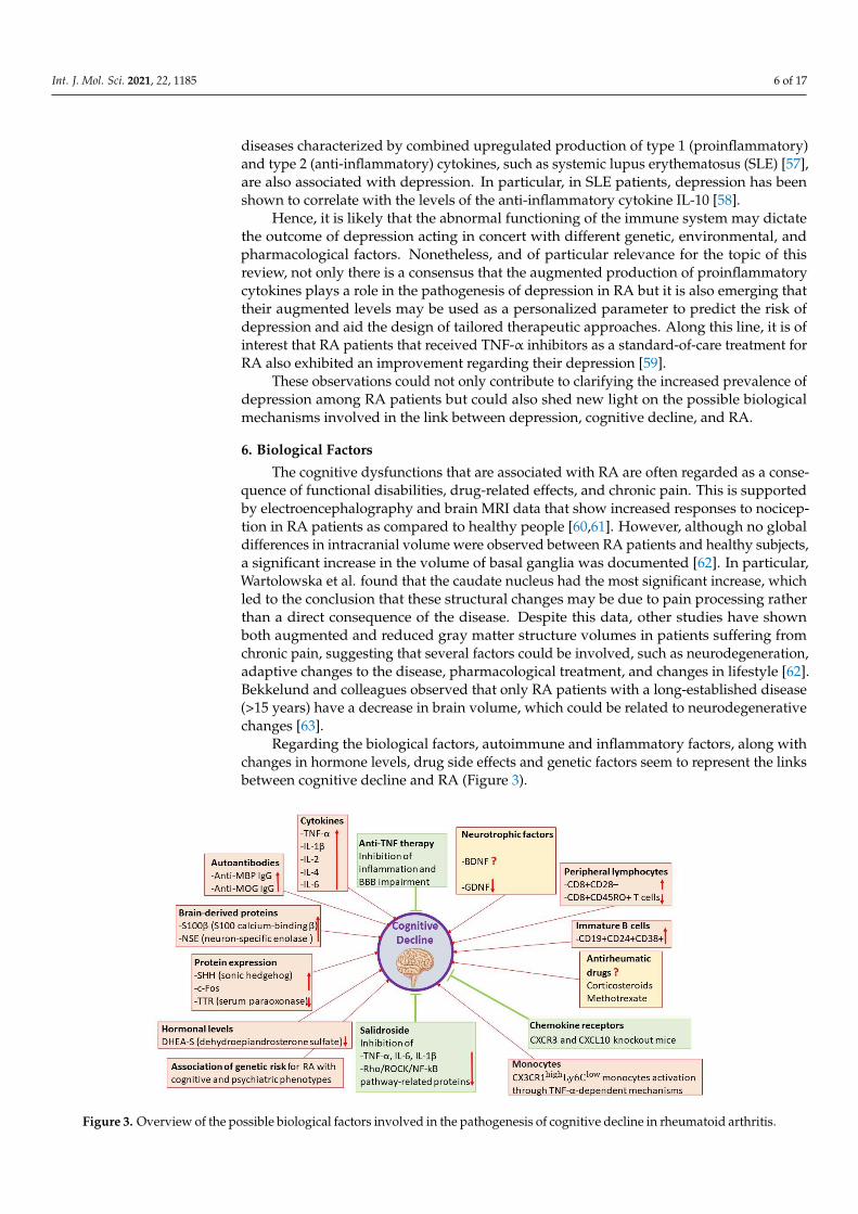
Regarding the biological factors, autoimmune and inflammatory factors, along withchanges in hormone levels, drug side effects and genetic factors seem to represent the linksbetween cognitive decline and RA (Figure 3).
Int. J. Mol. Sci. 2021, 22, x FOR PEER REVIEW 7 of 18
have shown both augmented and reduced gray matter structure volumes in patients suf-
fering from chronic pain, suggesting that several factors could be involved, such as neu-
rodegeneration, adaptive changes to the disease, pharmacological treatment, and changes
in lifestyle [62]. Bekkelund and colleagues observed that only RA patients with a long-
established disease (>15 years) have a decrease in brain volume, which could be related to
neurodegenerative changes [63].
Regarding the biological factors, autoimmune and inflammatory factors, along with
changes in hormone levels, drug side effects and genetic factors seem to represent the
links between cognitive decline and RA (Figure 3).
Figure 3. Overview of the possible biological factors involved in the pathogenesis of cognitive decline in rheumatoid ar-
thritis.
6.1. Autoimmune and Inflammatory Factors
6.1.1. Premature Immunosenescence
Petersen and colleagues studied the link between cognitive functions and peripheral
lymphocyte subsets in RA patients [64]. In comparison to controls, patients showed an
expansion of CD8+CD28− cells and a reduction of memory CD8+CD45RO+ T cells [64]. Of
note, CD8+CD28− and CD8+CD45RO+ T cells were associated with memory decline [64].
In light of these results, accelerated immunosenescence could be involved in the relation-
ship between memory dysfunction and RA [64].
6.1.2. Autoantibodies and Brain-Derived Proteins
Baptista et al. investigated the circulating levels of autoantibodies in order to evaluate
whether there was a link between these parameters and cognitive functions in RA patients
with active disease [25]. Interestingly, they found that cognitive performance was nega-
tively associated with the levels of the anti-myelin basic protein (MBP) IgG, the anti-my-
elin oligodendrocyte glycoprotein (MOG) IgG, and S100 calcium-binding β (S100β) [25].
The authors hypothesized that the augmented permeability of the blood–brain barrier
(BBB) might represent the trigger of the pathophysiological mechanisms of cognitive de-
cline in RA [25]. Indeed, antibodies and inflammatory mediators might be able to reach
the cerebral parenchyma, leading to an exaggerated release of neurotoxic factors, thus
promoting neuroinflammation, along with demyelination processes that are induced by
anti-MOG and anti-MBP antibodies [25]. Overall, these factors could negatively alter the
Figure 3. Overview of the possible biological factors involved in the pathogenesis of cognitive decline in rheumatoid arthritis.
Int. J. Mol. Sci. 2021, 22, 1185 7 of 17
6.1. Autoimmune and Inflammatory Factors6.1.1. Premature Immunosenescence
Petersen and colleagues studied the link between cognitive functions and peripherallymphocyte subsets in RA patients [64]. In comparison to controls, patients showed anexpansion of CD8+CD28− cells and a reduction of memory CD8+CD45RO+ T cells [64]. Ofnote, CD8+CD28− and CD8+CD45RO+ T cells were associated with memory decline [64].In light of these results, accelerated immunosenescence could be involved in the relation-ship between memory dysfunction and RA [64].
6.1.2. Autoantibodies and Brain-Derived Proteins
Baptista et al. investigated the circulating levels of autoantibodies in order to evaluatewhether there was a link between these parameters and cognitive functions in RA patientswith active disease [25]. Interestingly, they found that cognitive performance was negativelyassociated with the levels of the anti-myelin basic protein (MBP) IgG, the anti-myelinoligodendrocyte glycoprotein (MOG) IgG, and S100 calcium-binding β (S100β) [25]. Theauthors hypothesized that the augmented permeability of the blood–brain barrier (BBB)might represent the trigger of the pathophysiological mechanisms of cognitive decline inRA [25]. Indeed, antibodies and inflammatory mediators might be able to reach the cerebralparenchyma, leading to an exaggerated release of neurotoxic factors, thus promotingneuroinflammation, along with demyelination processes that are induced by anti-MOGand anti-MBP antibodies [25]. Overall, these factors could negatively alter the number ofneurons and synapses, as well as information processing speed and neurogenesis, leadingto cognitive decline [25].
Considering that the central nervous system’s involvement in RA might derive fromBBB damage associated with chronic inflammation, Sag et al. examined the potential roleof BBB damage and evaluated the action of TNF blocker therapy on BBB function in RApatients [65]. They found that S100β and glial fibrillary acidic protein (GFAP) levels weresignificantly increased in patients compared to controls [65]. Interestingly, the group treatedwith TNF blocker therapy showed significantly reduced levels of S100β and GFAP after6 months from the start of the treatment [65]. The S100β levels increased in RA patients,along with lesions in the deep white matter examined with cranial magnetic resonanceimaging (MRI) [65]. Overall, the authors suggested that the anti-TNF therapy in RA couldboth reduce disease activity and joint erosions by inhibiting inflammation and block thepossible involvement of the central nervous system in BBB impairment [65].
Furthermore, Hamed et al. evaluated the serum levels of S100β and neuron-specificenolase (NSE) in female RA patients [66]. Of note, they found that, compared to controls,patients showed increased concentrations of S100β and that increased concentrations ofS100β were associated with worse cognitive performance and increased concentrationsof NSE [66]. These data are in line with previous studies suggesting that increased levelsof NSE may enhance neuroinflammatory processes, oxidative stress, and neuronal apop-tosis [66]. These results highlight the potential diagnostic importance of the assessmentof the serum levels of certain specific brain-derived proteins, such as S100β and NSE, inRA patients in order to identify the cognitive dysfunction that is associated with the braininjury subsequent to inflammation [66].
6.1.3. Proinflammatory Cytokines
Chronic inflammation with high circulating levels of proinflammatory cytokines andsustained brain cytokine production is considered to be the leading cause of cognitiveimpairment [67,68]. In addition to cytokines, autoantibodies, including the RF [69,70] andimmune complexes, can also induce neuroinflammatory responses in the brain [71]. Indeed,in a murine model, it was shown that immune complexes in the brain parenchyma elicitedinflammation, along with augmented microglial expressions of CD11b, CD68, and FcRII/III,as well as consequent neuronal damage, in the absence of neutrophil recruitment [71].

Int. J. Mol. Sci. 2021, 22, 1185 8 of 17
Importantly, these effects were dependent on the Fcγ receptors but not on the complementsystem [71].
In the cerebrospinal fluid (CSF) of RA patients, IL-1β levels have been found tobe significantly increased when compared with controls. Interestingly, CSF IL-1β levelswere higher in comparison to the serum levels, indicating that RA is associated withbrain immune activation, despite the lack of other markers of systemic inflammation.Concordantly, Lampa et al. observed a decrease in IL-1R antagonist (IL1Ra) in RA CSF. Onthe other hand, no significant differences were found in CSF TNF-α levels between RApatients and healthy controls, while a trend toward an increase was observed for IL-6 [72].
It has been suggested that cytokines, such as IL-1β and TNF-α, can modulate the ex-citability of neurons, not only via interaction with their receptors but also via noncanonicalsignaling pathways. In particular, IL-1β and TNF-α are able to modulate the main types ofvoltage- and ligand-dependent membrane channels in brain cells (reviewed by [73]). Atthe cellular level, IL-1β is able to inhibit the activity of glucose-sensitive neurons of thelateral hypothalamus to promote the production of vasopressin in the hypothalamus, todiminish the GABA-mediated inhibition of Purkinje cells in the cerebellum, to hinder theglutamatergic transmission in the hippocampus, and to inhibit the N-type voltage-gatedCa2+ channels. Moreover, IL-1β favors astrocyte and microglial proliferation, stimulatesangiogenesis in the brain, and increases blood vessel permeability (reviewed by [73]). Onthe other hand, TNF-α is able to increase the expression of AMPA glutamate receptorsand to reduce the expression of GABA-A receptors in the hippocampus, consequentlycontrolling the plastic changes in the neural networks of this region (reviewed by [73]).Moreover, TNF-α can enhance the outward K+ current in cortical neurons and reduceglutamate-induced currents in hippocampal neurons via the NF-κB pathway (reviewedby [73]).
These data support the notion that cytokines have a significant role in the modulationof synaptic plasticity and could regulate memory formation and cognitive function. How-ever, the effect of TNF-α in learning and memory could be age-dependent. Indeed, oldermice chronically overexpressing neuronal TNF-α display spatial memory impairments,while no such deficits are observed in young (30-day-old) mice [74,75].
A negative role for IL-1β regarding learning and memory has been demonstrated inrodent models of chronic elevated IL-1β levels in the brain. The chronic injection of IL-1βinto the lateral ventricles has been reported to induce spatial memory deficits in rats [76].Moreover, chronic hippocampal overexpression of IL-1β in mice caused an increase inglial inflammatory markers, increased the production of cytokines and chemokines in thehippocampus, and lowered the levels of the Arc gene, which is associated with neuronplasticity [77,78]. Accordingly, these mice showed decreased retention of spatial memoryand fear memory [77,78].
Furthermore, a detrimental effect of IL-6 on cognition has been described [79]. Ascompared to wild-type (WT) mice, IL-6 knockout (KO) animals did not display workingmemory impairment and lacked the expected LPS-induced increase in TNF-α and IL-1β inthe hippocampus [80], suggesting that IL-6 is required for the LPS-induced production ofTNF-α and IL-1β in the brain and the development of behavioral impairments. Further-more, mice chronically expressing astrocytic IL-6 display a progressive age-related declinein learning performance that correlates with presynaptic loss [81] and a decrease in corticaland hippocampal neuronal calbindin [82].
Interestingly, Chou et al. found that RA patients receiving an anti-TNF treatment(infliximab, etanercept, and adalimumab) had a reduced risk of developing AD as com-pared to controls [83]. Contrarily, the risk of developing AD was not changed by treatmentwith other DMARDs. More importantly, the impact of anti-TNF treatment on cognitionseems to occur before its anti-inflammatory effects become clinically apparent in the joints.In a functional MRI (fMRI) study [84], upon anti-TNF-α therapy in patients with activeRA, brain activation was significantly decreased within 3 days after treatment, while thedisease activity score was significantly reduced only by day 28. Hence, the improvement

Int. J. Mol. Sci. 2021, 22, 1185 9 of 17
in cognitive decline observed upon anti-TNF-α therapy may be a direct central effect andmay not be subsequent to the better management of pain in RA patients.
6.1.4. Lymphocyte Subsets, Chemokines, and Neurotrophic Factors
Petersen et al. evaluated the cognitive functions of RA patients with controlled andactive disease and investigated whether cognitive decline was associated with immune andneurotrophic markers, such as lymphocyte subsets, cytokines, and neurotrophic factors,in RA patients [85]. They found an overall cognitive impairment in RA patients andthe cognitive performance was worse in RA patients with active disease than in thosewith controlled disease [85]. Moreover, in comparison to controls, RA patients showedan expansion of some lymphocyte subsets, such as natural killer T cell (NKT) cells andCD4+IL-17+ T cells, as well as a reduction of regulatory T cells [85]. Furthermore, RApatients were characterized by an expansion of immature B cells (CD19+CD24+CD38+)and plasma cells (CD19+CD27+CD38+) compared to controls, and low cognitive scoreswere correlated with bigger proportions of immature B cells [85]. Furthermore, RA patientsrevealed higher TNF-α, interleukin IL-2, IL-4, and IL-6 plasma levels, which was negativelycorrelated with cognitive functions [85].
Moreover, chemokines and the complement system have been found to be importantmediators that are involved in CNS homeostasis [86,87]. Garré et al. showed signifi-cant impairments in dendritic spine formation and in learning abilities upon treatmentwith polyinosinic:polycytidilic acid (poly I:C), and that activation of CX3CR1highLy6Clow
monocytes impaired motor learning and learning-related dendritic spine plasticity viaTNF-α-dependent mechanisms [88]. Moreover, Blank et al. showed that the productionof CXCL10 by brain endothelial and epithelial cells was associated with diminished hip-pocampal plasticity, and that CXCR3−/− and CXCL10−/− mice retained better memoryand learning functions [89], supporting the notion that inflammatory factors could have arole in cognitive decline.
Moreover, increased brain-derived neurotrophic factor (BDNF) levels and decreasedglial-cell-line-derived neurotrophic factor (GDNF) levels were found in RA patients whencompared to controls [85].
Overall, these data demonstrated a global cognitive decline in RA patients, which wasassociated with disease activity and immune differences, thus suggesting that peripheralimmune imbalance, along with a proinflammatory milieu, could predict the cognitivedeficits in RA. Of note, although the presence of higher BDNF plasma levels could seemunusual in patients with chronic inflammation, such as those with RA, it should be consid-ered that the circulating BDNF might largely be derived from leukocytes in inflammatorydiseases [90]. Interestingly, it has been shown that BDNF is constitutively expressed byperipheral blood mononuclear cells (PBMCs) and synovial cells [90]. Instead, GDNF, whosedecreased plasma levels were found to be associated with cognitive dysfunctions in RApatients, is produced only in the central nervous system [90]. Therefore, decreased levels ofGDNF could be a better predictor of worse cognitive performance compared to BDNF [90].
On the other hand, Pedard et al. explored the role of the cerebral BDNF pathway ina preclinical rat model of RA [91]. This study demonstrated that arthritis was negativelyassociated with the cerebral BDNF/tropomyosin-related kinase B (TrkB) pathway both atthe endothelial and neuronal levels, without correlation with the severity of inflammatorysymptoms, but they were dependent on endothelial nitric oxide (NO) production [91]. Inparticular, it is suggested that reduced BDNF production by the cerebral endothelium,deriving from reduced endothelial NO synthesis, could explain the arthritis-associatedreduced activation of neuronal TrkB activation [91]. These results might shed new light onclarifying the relationship between cognitive and endothelial dysfunctions, which are bothpresent in RA [91].

Int. J. Mol. Sci. 2021, 22, 1185 10 of 17
6.1.5. Differential Protein Expression
Yang et al. investigated the differentially expressed proteins that might be possiblebiomarkers for a differential diagnosis of MCI in RA patients [26]. The authors com-pared plasma protein levels from RA patients with and without MCI and from healthycontrols [26]. Interestingly, 14 differentially expressed proteins, 6 upregulated and 8 down-regulated, were identified in RA patients with MCI [26]. These dysregulated proteins areimplicated in numerous biological processes and pathways, such as immunity, inflamma-tion, and coagulation [26]. In particular, sonic hedgehog (SHH) and serum paraoxonase(TTR), which were respectively upregulated and downregulated in RA patients withMCI, seem to be promising potential plasma biomarkers for the diagnosis of MCI in RApatients [26].
Carter et al. investigated the levels of c-Fos expression in the hippocampus, which is abrain region that is crucially involved in cognitive function, in a preclinical rodent modelof RA, namely, the adjuvant-induced arthritis Lewis rat model [92]. A persistent dose-and subfield-dependent expression of c-Fos was found in the arthritis group, whereas atransient expression was found in groups without arthritis [92]. The mechanisms that causec-Fos expression in the hippocampus were not identified in this study [92]. Nonetheless,other studies have previously shown that immunization with different immunogens, suchas lipopolysaccharide or proinflammatory cytokines, including TNF-α or IL-1, may inducea sustained c-Fos immunoreactivity in the hypothalamic, limbic, and autonomic brainareas [92].
The reason for the persistent increase in c-Fos expression in the hippocampus ofadjuvant-induced arthritis rats is not yet fully understood [92]. It is known that c-Fosaccumulates when its C-terminus is phosphorylated in the presence of sustained ERKactivation, thus indicating that hippocampal pyramidal cells in adjuvant-induced arthritisrats may show a chronic increase in ERK activity [92]. Moreover, the higher stability ofc-Fos by phosphorylation increases ERK phosphorylation at its C-terminus [92].
Changes in c-Fos subsequent to sustained ERK signaling in adjuvant-induced arthritismight chronically modify pyramidal cell functional processes; thus, this might favor diseaseprogression and could modify behavior and cognitive functions in adjuvant-inducedarthritis [92].
Overall, these results suggest that the chronic expression of c-Fos in the hippocampusof the adjuvant-induced arthritis rats might influence several cell functions, such as synap-togenesis, electrical activity, and neurotransmitters, and that sustained genomic alterationsin RA could be involved in different processes associated with RA, including cognitivedecline [92].
6.1.6. Rho/ROCK/NF-κB Pathway
Considering the increasing prevalence of cognitive impairment in RA patients andthe increasing lines of evidence about the role of inflammation in arthritis-induced cog-nitive deficits, Zhu et al. investigated the effects of Salidroside (Sal, p-hydroxyphenethyl-b-D-glucoside), which is an effective extracted ingredient of Rhodiola rosea L with anti-inflammatory properties, on the arthritis-induced cognitive dysfunction in a preclinicalrat model of collagen-induced arthritis [93]. The results of this study showed that Salexerted a protective action on arthritis-induced cognitive dysfunction through the inhi-bition of proinflammatory cytokines (TNF-α, IL-6, and IL-1β) and the regulation of theRho/ROCK/NF-κB pathway [93]. Systemic proinflammatory cytokines may pass throughthe damaged BBB and reach the central nervous system, thus stimulating neurodegen-erative processes [93]. Moreover, according to previous studies, the Rho/ROCK/NF-κBpathway may be implicated in cognitive impairment [93]. The Rho/ROCK pathway isusually involved in the production of proinflammatory factors [93]. Even though the exactcellular processes of Rho signaling in the central nervous system have yet to be clarified, itis known that changes in Rho signaling derived from mutations cause anomalous neuronalconnectivity and cognitive deficits in humans [93]. Furthermore, small G proteins of the

Int. J. Mol. Sci. 2021, 22, 1185 11 of 17
Rho family are involved in different biological processes and in the regulation of several sig-naling pathways that are correlated with inflammation, including the NF-κB pathway [93].Of note, Rho proteins may regulate cell adhesion via transmembrane proteins, such ascadherins and integrins [93]. In particular, cadherins are involved in synaptic plastic-ity, thus suggesting that Rho proteins could be implicated in the regulation of synapseformation and plasticity [93]. It is known that synapses provide the structural basis that reg-ulates higher brain functions, including learning and memory, and that damaged synapticplasticity and neurotransmission due to inflammation may alter cognitive functions [93].
Overall, these results highlight the role of inflammation in affecting brain functionsand suggest the involvement of the Rho/ROCK/NF-κB signaling and its potential as apossible novel molecular target [93].
6.2. Changes in Hormonal Levels
Kozora et al. found that RA patients with mild levels of disease activity showed sig-nificantly decreased plasma levels of dehydroepiandrosterone sulfate (DHEA-S) comparedto controls and that reduced plasma levels of DHEA-S were marginally associated withreduced scores on measures of attention [94]. Interestingly, previous studies have shownan association between dehydroepiandrosterone (DHEA) and DHEA-S and cognitive func-tions, both in animal models and in humans [94]. Moreover, it is known that metabolites ofDHEA may play an important role in immune regulation [94]. However, further studiesare needed in order to clarify the mediators of these cognitive differences and to furtherconfirm these data [94]. Overall, the results of this study shed light on the potential valueof hormones as predictors of cognitive function in RA patients [94].
6.3. Drug Side Effects
It has been shown that some drugs that are commonly used to treat RA, includ-ing methotrexate and corticosteroids, could be associated with cognitive dysfunction inRA [18,22]. However, the question seems to be controversial and the possible mechanismsinvolved are not clear. Indeed, it is known that the anti-inflammatory action of methotrex-ate and corticosteroids may exert positive effects on cognitive functions [22]. Nonetheless,methotrexate could be associated with cognitive decline, confusion, and mood changes,whereas corticosteroids could influence memory and hippocampal function [18,22]. More-over, cognitive decline has been associated with current or long-term steroid use in RApatients, probably due to its vascular side effects [24]. On the other hand, other studies haveshown that there is no significant correlation between cognitive decline and glucocorticoiduse for RA [44,90].
6.4. Genetic Factors
Jones et al. explored whether genetics could be related to cognitive and psychiatricphenotypes in children and adolescents before the clinical onset of RA [95]. The authorsidentified a polygenic risk score for RA, which was associated with decreased scoreson some measures of cognition, such as total IQ, performance IQ, and verbal IQ, alongwith significantly higher associations with hyperactive and inattentive symptoms [95].Moreover, trends toward negative associations with working memory (p = 0.058) andverbal learning (p = 0.174) were also found. The results of this study highlight the presenceof a relationship between genetic risk for RA and neural phenotypes, thus indicating thatcognitive decline in RA is not just a result of disease-related processes or drug effects, but itdepends on more complex interactions that also involve genetic susceptibility and immunefactors [95].
7. Conclusions
To date, numerous studies have highlighted the association between cognitive declineand RA and have investigated the underlying potential mechanisms. In this review, weexamined the link between cognitive decline and RA from a pathogenic point of view, fo-

Int. J. Mol. Sci. 2021, 22, 1185 12 of 17
cusing on the main molecular mechanisms involved. Although the molecular pathogeneticmechanisms underlying the association of these conditions are not still fully clarified, theemerging results from the preclinical and clinical studies in this field suggest that differentclinical, psychological, and biological variables may contribute to the pathogenesis ofcognitive decline in RA. Regarding the clinical and psychological variables involved, thepresence of cardiovascular complications, chronic pain, and depression seem to be partic-ularly relevant. Among the biological variables, several autoimmune and inflammatoryfactors, along with changes in hormone levels, drug side effects, and genetic risk, appearto be involved. Overall, inflammation seems to be the main actor in this scenario. Inter-estingly, premature immunosenescence, autoantibodies, and brain-derived proteins, aswell as alterations in signaling pathways, lymphocyte subsets, cytokines, and neurotrophicfactors, might be contributing mechanisms.
Regarding the potential diagnostic and prognostic strategies and the identification ofmolecular targets for cognitive decline in RA, the emerging results from the reviewed stud-ies suggest different possibilities. These include the potential diagnostic importance of theassessment of the serum levels of certain specific brain-derived proteins, such as S100β andNSE, in order to identify the cognitive dysfunction that is associated with brain injury [66];the decreased levels of GDNF as a predictor of worse cognitive performance [90]; theevaluation of SHH and TTR as potential plasma biomarkers for the diagnosis of MCI [26];the Rho/ROCK/NF-κB signaling as a novel molecular target [93]; the determination ofhormones as predictors of cognitive function [94].
Since inflammation, which characterizes RA, could be considered the most importantmolecular pathogenetic mechanism involved in the cognitive decline associated withRA, the use of different anti-inflammatory drugs in RA patients might acquire addedvalue. Indeed, the anti-inflammatory drugs, most of which are commonly used in RAtreatment, could exert a therapeutic action not only on the progression of RA but also onthe development of cognitive decline in RA patients. Therefore, the possibility to evaluatenovel pharmacological classes in order to select the most effective ones, and eventuallyfinding out new possible synergistic co-treatments, could be evaluated. However, itshould be noted that certain drugs, such as methotrexate and corticosteroids, might have acontroversial role in this field. Indeed, despite their anti-inflammatory action, which mayexert positive effects on cognitive functions, certain studies have suggested that these drugsmight be associated with cognitive dysfunction in RA [18,22]. However, other studies onthis topic, in particular on glucocorticoids, have found no correlation [44,90].
Among the anti-inflammatory therapies, the anti-TNF-α therapies seem to be partic-ularly promising. Interestingly, it has recently been shown that RA patients treated withTNF-α-inhibiting biological therapies showed a 50% decreased risk of developing cognitivedecline, where this may be due to the fact that TNF-α is involved in the physiopathologyof dementia, as well as in that of RA [39].
Along this line of research, a recent clinical trial (NCT04378621) aimed to examinehow RA influences the brain structures in RA patients and whether anti-inflammatorytreatments targeting TNF-α or JAK signaling, as compared to the physical training ofhands, exert a positive effect on neuropsychiatric symptoms, including cognitive decline,and on morphological changes in the brain derived from the disease. The TNF-α inhibitorsconsidered for this study were Etanercept, Infliximab, Adalimumab, Certolizumab pegol,and Golimumab, whereas the JAK inhibitors were Baricitinib and Tofacitinib.
Understanding the molecular basis underlying the link between cognitive declineand RA is of fundamental importance to find out new possible diagnostic, prognostic, andtherapeutic strategies; this can be done by focusing on the discovery of novel potentialbiomarkers, therapeutic targets, and treatments for RA patients. Of note, targeting immunepathways could be a potentially valuable therapeutic approach. Considering the stronginteraction between mental and physical dysfunctions, a multidisciplinary approach thataims to target all the variables involved seems to be promising. Further studies are highlywarranted in order to fully clarify the association between cognitive decline and RA.

Int. J. Mol. Sci. 2021, 22, 1185 13 of 17
Author Contributions: Conceptualization, M.S.B., P.F., F.N., and E.C.; writing—original draft prepa-ration, M.S.B. and E.C.; writing—review and editing, R.C., A.B., M.C.P., P.F., and F.N.; visualization,R.C., A.B., and M.C.P. All authors significantly contributed to the preparation of the manuscript. Allauthors have read and agreed to the published version of the manuscript.
Funding: This study was supported by research funds from the IRCCS Centro Neurolesi “Bonino-Pulejo,” Messina, Italy (2020).
Conflicts of Interest: The authors declare no conflict of interest. The funders had no role in the designof the study; in the collection, analyses, or interpretation of data; in the writing of the manuscript, orin the decision to publish the results.
Abbreviations
ACPAs anti-citrullinated peptide antibodiesAD Alzheimer’s diseaseBBB blood–brain barrierBDI Beck Depression InventorybDMARDs biological DMARDsBDNF brain-derived neurotrophic factorBVRT Benton Visual Retention TestcsDMARDs conventional synthetic DMARDsCSF cerebrospinal fluidDAS-28 Disease Activity ScoreDHEADHEA-S dehydroepiandrosterone dehydroepiandrosterone sulfateEULAR European League Against RheumatismfMRI functional MRIGDNF glial-cell-line-derived neurotrophic factorGFAP glial fibrillary acidic proteinMBP myelin basic proteinMCI mild cognitive impairmentMHC major histocompatibility complexMMSE Mini-Mental State ExaminationMoCA Montreal Cognitive AssessmentMOG myelin oligodendrocyte glycoproteinMRI magnetic resonance imagingNKT natural killer T cellNO nitric oxideNSAIDs non-steroidal anti-inflammatory drugsPBMCs peripheral blood mononuclear cellspoly I:C polyinosinic:polycytidilic acidRA rheumatoid arthritisRF rheumatoid factorS100β S100 calcium-binding β
Sal salidrosideSHH sonic hedgehogSLE systemic lupus erythematosusSTAIT/STNF State-Trait Anxiety InventoryTumor necrosis factorTMT Trail-Making TestTrkB tropomyosin-related kinase BtsDMARDs targeted synthetic DMARDsTTR serum paraoxonaseVST Victoria Stroop TestWAIS Wechsler Adult Intelligence Scale
References1. McInnes, I.B.; Schett, G. The Pathogenesis of Rheumatoid Arthritis. N. Engl. J. Med. 2011, 365, 2205–2219. [CrossRef]2. Smolen, J.S.; Aletaha, D.; McInnes, I.B. Rheumatoid arthritis. Lancet 2016, 388, 2023–2038. [CrossRef]
Int. J. Mol. Sci. 2021, 22, 1185 14 of 17
3. Bullock, J.; Rizvi, S.A.A.; Saleh, A.M.; Ahmed, S.S.; Do, D.P.; Ansari, R.A.; Ahmed, J. Rheumatoid Arthritis: A Brief Overview ofthe Treatment. Med. Princ. Pract. 2018, 27, 501–507. [CrossRef]
4. Van der Woude, D.; van der Helm-van Mil, A.H.M. Update on the epidemiology, risk factors, and disease outcomes of rheumatoidarthritis. Best Pract. Res. Clin. Rheumatol. 2018, 32, 174–187. [CrossRef]
5. Deane, K.D.; Demoruelle, M.K.; Kelmenson, L.B.; Kuhn, K.A.; Norris, J.M.; Holers, V.M. Genetic and environmental risk factorsfor rheumatoid arthritis. Best Pract. Res. Clin. Rheumatol. 2017, 31, 3–18. [CrossRef]
6. Mikhaylenko, D.S.; Nemtsova, M.V.; Bure, I.V.; Kuznetsova, E.B.; Alekseeva, E.A.; Tarasov, V.V.; Lukashev, A.N.; Beloukhova, M.I.;Deviatkin, A.A.; Zamyatnin, A.A. Genetic polymorphisms associated with rheumatoid arthritis development and antirheumatictherapy response. Int. J. Mol. Sci. 2020, 21, 4911. [CrossRef]
7. Silman, A.J.; Pearson, J.E. Epidemiology and genetics of rheumatoid arthritis. Arthritis Res. 2002, 4, S265–S272. [CrossRef]8. Petralia, M.C.; Mazzon, E.; Basile, M.S.; Cutuli, M.; Di Marco, R.; Scandurra, F.; Saraceno, A.; Fagone, P.; Nicoletti, F.; Mangano,
K. Effects of Treatment with the Hypomethylating Agent 5-aza-2’-deoxycytidine in Murine Type II Collagen-Induced Arthritis.Pharmaceuticals 2019, 12, 174. [CrossRef]
9. Brennan, F.M.; McInnes, I.B. Evidence that cytokines play a role in rheumatoid arthritis. J. Clin. Investig. 2008, 118, 3537–3545.[CrossRef]
10. Jeffery, R.C. Clinical features of rheumatoid arthritis. Medicine (UK) 2014, 42, 231–236. [CrossRef]11. Trouw, L.A.; Mahler, M. Closing the serological gap: Promising novel biomarkers for the early diagnosis of rheumatoid arthritis.
Autoimmun. Rev. 2012, 12, 318–322. [CrossRef] [PubMed]12. Thiele, G.M.; Duryee, M.J.; Anderson, D.R.; Klassen, L.W.; Mohring, S.M.; Young, K.A.; Benissan-Messan, D.; Sayles, H.; Dusad, A.;
Hunter, C.D.; et al. Malondialdehyde-acetaldehyde adducts and anti-malondialdehyde-acetaldehyde antibodies in rheumatoidarthritis. Arthritis Rheumatol. 2015, 67, 645–655. [CrossRef] [PubMed]
13. Auger, I.; Charpin, C.; Balandraud, N.; Martin, M.; Roudier, J. Autoantibodies to PAD4 and BRAF in rheumatoid arthritis.Autoimmun. Rev. 2012, 11, 801–803. [CrossRef] [PubMed]
14. Salman, E.; Çetiner, S.; Boral, B.; Kibar, F.; Erken, E.; Ersözlü, E.D.; Badak, S.Ö.; Bilici Salman, R.; Sertdemir, Y.; Çetin Duran, A.;et al. Importance of 14-3-3eta, anti-carp, and anti-sa in the diagnosis of seronegative rheumatoid arthritis. Turkish J. Med. Sci.2019, 49, 1498–1502. [CrossRef] [PubMed]
15. Zhao, J.; Zhao, Y.; He, J.; Jia, R.; Li, Z. Prevalence and Significance of Anti-Peptidylarginine Deiminase 4 Antibodies in RheumatoidArthritis. J. Rheumatol. 2008, 35, 969–974. [PubMed]
16. Alam, J.; Jantan, I.; Bukhari, S.N.A. Rheumatoid arthritis: Recent advances on its etiology, role of cytokines and pharmacotherapy.Biomed. Pharmacother. 2017, 92, 615–633. [CrossRef] [PubMed]
17. Smolen, J.S.; Landewé, R.B.M.; Bijlsma, J.W.J.; Burmester, G.R.; Dougados, M.; Kerschbaumer, A.; McInnes, I.B.; Sepriano, A.; VanVollenhoven, R.F.; De Wit, M.; et al. EULAR recommendations for the management of rheumatoid arthritis with synthetic andbiological disease-modifying antirheumatic drugs: 2019 update. Ann. Rheum. Dis. 2020, 79, S685–S699. [CrossRef] [PubMed]
18. Meade, T.; Manolios, N.; Cumming, S.R.; Conaghan, P.G.; Katz, P. Cognitive Impairment in Rheumatoid Arthritis: A SystematicReview. Arthritis Care Res. 2018, 70, 39–52. [CrossRef]
19. Albert, M.S.; DeKosky, S.T.; Dickson, D.; Dubois, B.; Feldman, H.H.; Fox, N.C.; Gamst, A.; Holtzman, D.M.; Jagust, W.J.; Petersen,R.C.; et al. The diagnosis of mild cognitive impairment due to Alzheimer’s disease: Recommendations from the National Instituteon Aging-Alzheimer’s Association workgroups on diagnostic guidelines for Alzheimer’s disease. Alzheimer’s Dement. 2011,7, 270–279. [CrossRef]
20. Tavares-Júnior, J.W.L.; De Souza, A.C.C.; Alves, G.S.; de Carvalho Bonfadini, J.; Siqueira-Neto, J.I.; Braga-Neto, P. CognitiveAssessment Tools for Screening Older Adults with Low Levels of Education: A Critical Review. Front. Psychiatry 2019, 10, 878.[CrossRef]
21. Oláh, C.; Kardos, Z.; Andrejkovics, M.; Szarka, E.; Hodosi, K.; Domján, A.; Sepsi, M.; Sas, A.; Kostyál, L.; Fazekas, K.; et al.Assessment of cognitive function in female rheumatoid arthritis patients: Associations with cerebrovascular pathology, depressionand anxiety. Rheumatol. Int. 2020, 40, 529–540. [CrossRef] [PubMed]
22. Oláh, C.; Schwartz, N.; Denton, C.; Kardos, Z.; Putterman, C.; Szekanecz, Z. Cognitive dysfunction in autoimmune rheumaticdiseases. Arthritis Res. Ther. 2020, 22, 78. [CrossRef] [PubMed]
23. Sood, A.; Raji, M.A. Cognitive impairment in elderly patients with rheumatic disease and the effect of disease-modifyinganti-rheumatic drugs. Clin. Rheumatol. 2020. [CrossRef] [PubMed]
24. Lwin, M.N.; Serhal, L.; Holroyd, C.; Edwards, C.J. Rheumatoid Arthritis: The Impact of Mental Health on Disease: A NarrativeReview. Rheumatol. Ther. 2020, 7, 457–471. [CrossRef] [PubMed]
25. Baptista, T.S.A.; Petersen, L.E.; Molina, J.K.; de Nardi, T.; Wieck, A.; do Prado, A.; Piovesan, D.M.; Keisermann, M.; Grassi-Oliveira,R.; Bauer, M.E. Autoantibodies against myelin sheath and S100β are associated with cognitive dysfunction in patients withrheumatoid arthritis. Clin. Rheumatol. 2017, 36, 1959–1968. [CrossRef] [PubMed]
26. Yang, L.; Zou, Q.H.; Zhang, Y.; Shi, Y.; Hu, C.R.; Hui, C.X.; Liu, X.F.; Fang, Y.F. Proteomic analysis of plasma from rheumatoidarthritis patients with mild cognitive impairment. Clin. Rheumatol. 2018, 37, 1773–1782. [CrossRef]
27. Fiest, K.M.; Hitchon, C.A.; Bernstein, C.N.; Peschken, C.A.; Walker, J.R.; Graff, L.A.; Zarychanski, R.; Abou-Setta, A.; Patten, S.B.;Sareen, J.; et al. Systematic review and meta-analysis of interventions for depression and anxiety in persons with rheumatoidarthritis. J. Clin. Rheumatol. 2017, 23, 425–434. [CrossRef]
Int. J. Mol. Sci. 2021, 22, 1185 15 of 17
28. Eberhardt, K.; Larsson, B.M.; Nived, K.; Lindqvist, E. Work disability in rheumatoid arthritis—Development over 15 years andevaluation of predictive factors over time. J. Rheumatol. 2007, 34, 481–487.
29. Pollard, L.; Choy, E.H.; Scott, D.L. The consequences of rheumatoid arthritis: Quality of life measures in the individual patient.Clin. Exp. Rheumatol. 2005, 23, S43–S52.
30. Isik, A.; Koca, S.S.; Ozturk, A.; Mermi, O. Anxiety and depression in patients with rheumatoid arthritis. Clin. Rheumatol. 2007,26, 872–878. [CrossRef]
31. Lok, E.Y.C.; Mok, C.C.; Cheng, C.W.; Cheung, E.F.C. Prevalence and Determinants of Psychiatric Disorders in Patients withRheumatoid Arthritis. Psychosomatics 2010, 51, 338. [CrossRef] [PubMed]
32. VanDyke, M.M.; Parker, J.C.; Smarr, K.L.; Hewett, J.E.; Johnson, G.E.; Slaughter, J.R.; Walker, S.E. Anxiety in rheumatoid arthritis.Arthritis Care Res. (Hoboken) 2004, 51, 408–412. [CrossRef] [PubMed]
33. Matcham, F.; Rayner, L.; Steer, S.; Hotopf, M. The prevalence of depression in rheumatoid arthritis: A systematic review andmeta-analysis. Rheumatology 2013, 52, 2136–2148. [CrossRef] [PubMed]
34. Mok, C.; Lok, E.; Cheung, E. Concurrent psychiatric disorders are associated with significantly poorer quality of life in patientswith rheumatoid arthritis. Scand. J. Rheumatol. 2012, 41, 253–259. [CrossRef] [PubMed]
35. van den Hoek, J.; Boshuizen, H.C.; Roorda, L.D.; Tijhuis, G.J.; Nurmohamed, M.T.; Dekker, J.; van den Bos, G.A.M. Association ofSomatic Comorbidities and Comorbid Depression with Mortality in Patients with Rheumatoid Arthritis: A 14-Year ProspectiveCohort Study. Arthritis Care Res. (Hoboken) 2016, 68, 1055–1060. [CrossRef] [PubMed]
36. Lee, J.H.; Kim, G.T.; Kim, Y.K.; Lee, S.G. Cognitive function of patients with rheumatoid arthritis is associated with diseaseactivity but not carotid atherosclerotic changes. Clin. Exp. Rheumatol. 2018, 36, 856–861. [CrossRef]
37. Katchamart, W.; Narongroeknawin, P.; Phutthinart, N.; Srinonprasert, V.; Muangpaisan, W.; Chaiamnauy, S. Disease activity isassociated with cognitive impairment in patients with rheumatoid arthritis. Clin. Rheumatol. 2019, 38. [CrossRef]
38. Shin, S.Y.; Katz, P.; Wallhagen, M.; Julian, L. Cognitive impairment in persons with rheumatoid arthritis. Arthritis Care Res.(Hoboken) 2012, 64, 1144–1150. [CrossRef]
39. Vitturi, B.K.; Nascimento, B.A.C.; Alves, B.R.; de Campos, F.S.C.; Torigoe, D.Y. Cognitive impairment in patients with rheumatoidarthritis. J. Clin. Neurosci. 2019, 69, 81–87. [CrossRef]
40. Wallin, K.; Solomon, A.; Kreholt, I.; Tuomilehto, J.; Soininen, H.; Walin, K. Midlife rheumatoid arthritis increases the risk ofcognitive impairment two decades later: A population-based study. J. Alzheimer’s Dis. 2012, 31, 669–676. [CrossRef]
41. Min, C.; Bang, W.J.; Kim, M.; Oh, D.J.; Choi, H.G. Rheumatoid arthritis and neurodegenerative dementia: A nested case-controlstudy and a follow-up study using a national sample cohort. Clin. Rheumatol. 2020, 39, 159–166. [CrossRef] [PubMed]
42. Joaquim, A.F.; Appenzeller, S. Neuropsychiatric manifestations in rheumatoid arthritis. Autoimmun. Rev. 2015, 14, 1116–1122.[CrossRef] [PubMed]
43. Simos, P.; Ktistaki, G.; Dimitraki, G.; Papastefanakis, E.; Kougkas, N.; Fanouriakis, A.; Gergianaki, I.; Bertsias, G.; Sidiropoulos,P.; Karademas, E.C. Cognitive deficits early in the course of rheumatoid arthritis. J. Clin. Exp. Neuropsychol. 2016, 38, 820–829.[CrossRef] [PubMed]
44. Said, F.A.; Betoni, T.B.; Magalhaes, V.; Nisihara, R.; Skare, T.L. Rheumatoid arthritis and cognition dysfunction: Lack of associationwith cumulative glucocorticoid use. Immunopharmacol. Immunotoxicol. 2019, 41, 565–567. [CrossRef]
45. England, B.R.; Thiele, G.M.; Anderson, D.R.; Mikuls, T.R. Increased cardiovascular risk in rheumatoid arthritis: Mechanisms andimplications. BMJ 2018, 361. [CrossRef]
46. Chaurasia, N.; Singh, A.; Singh, I.; Singh, T.; Tiwari, T. Cognitive dysfunction in patients of rheumatoid arthritis. J. Fam. Med.Prim. Care 2020, 9, 2219. [CrossRef]
47. Attal, N.; Masselin-Dubois, A.; Martinez, V.; Jayr, C.; Albi, A.; Fermanian, J.; Bouhassira, D.; Baudic, S. Does cognitive functioningpredict chronic pain? Results from a prospective surgical cohort. Brain 2014, 137, 904–917. [CrossRef]
48. Sturgeon, J.A.; Finan, P.H.; Zautra, A.J. Affective disturbance in rheumatoid arthritis: Psychological and disease-related pathways.Nat. Rev. Rheumatol. 2016, 12, 532–542. [CrossRef]
49. Nerurkar, L.; Siebert, S.; McInnes, I.B.; Cavanagh, J. Rheumatoid arthritis and depression: An inflammatory perspective. TheLancet Psychiatry 2019, 6, 164–173. [CrossRef]
50. Günther, S.; Fagone, P.; Jalce, G.; Atanasov, A.G.; Guignabert, C.; Nicoletti, F. Role of MIF and D-DT in immune-inflammatory,autoimmune, and chronic respiratory diseases: From pathogenic factors to therapeutic targets. Drug Discov. Today 2019,24, 428–439. [CrossRef]
51. Petralia, M.C.; Mazzon, E.; Fagone, P.; Basile, M.S.; Lenzo, V.; Quattropani, M.C.; Di Nuovo, S.; Bendtzen, K.; Nicoletti, F. Thecytokine network in the pathogenesis of major depressive disorder. Close to translation? Autoimmun. Rev. 2020, 19, 102504.[CrossRef] [PubMed]
52. Petralia, M.C.; Mazzon, E.; Fagone, P.; Basile, M.S.; Lenzo, V.; Quattropani, M.C.; Bendtzen, K.; Nicoletti, F. Pathogenic contributionof the Macrophage migration inhibitory factor family to major depressive disorder and emerging tailored therapeutic approaches.J. Affect. Disord. 2020, 263, 15–24. [CrossRef] [PubMed]
53. Koo, J.; Marangell, L.B.; Nakamura, M.; Armstrong, A.; Jeon, C.; Bhutani, T.; Wu, J.J. Depression and suicidality in psoriasis:Review of the literature including the cytokine theory of depression. J. Eur. Acad. Dermatol. Venereol. 2017, 31, 1999–2009.[CrossRef] [PubMed]

Int. J. Mol. Sci. 2021, 22, 1185 16 of 17
54. Patel, N.; Nadkarni, A.; Cardwell, L.A.; Vera, N.; Frey, C.; Patel, N.; Feldman, S.R. Psoriasis, Depression, and InflammatoryOverlap: A Review. Am. J. Clin. Dermatol. 2017, 18, 613–620. [CrossRef]
55. Rossi, S.; Studer, V.; Motta, C.; Polidoro, S.; Perugini, J.; Macchiarulo, G.; Giovannetti, A.M.; Pareja-Gutierrez, L.; Calò, A.;Colonna, I.; et al. Neuroinflammation drives anxiety and depression in relapsing-remitting multiple sclerosis. Neurology 2017,89, 1338–1347. [CrossRef]
56. Ziemssen, T. Multiple sclerosis beyond EDSS: Depression and fatigue. J. Neurol. Sci. 2009, 277. [CrossRef]57. Barcellini, W.; Rizzardi, G.P.; Borghi, M.O.; Nicoletti, F.; Fain, C.; Del Papa, N.; Meroni, P.L. In vitro type-1 and type-2 cytokine
production in systemic lupus erythematosus: Lack of relationship with clinical disease activity. Lupus 1996, 5, 139–145. [CrossRef]58. Figueiredo-Braga, M.; Cornaby, C.; Cortez, A.; Bernardes, M.; Terroso, G.; Figueiredo, M.; Dos Santos Mesquita, C.; Costa,
L.; Poole, B.D. Depression and anxiety in systemic lupus erythematosus: The crosstalk between immunological, clinical, andpsychosocial factors. Medicine (United States) 2018, 97. [CrossRef]
59. Uguz, F.; Akman, C.; Kucuksarac, S.; Tufekci, O. Anti-tumor necrosis factor-α therapy is associated with less frequent mood andanxiety disorders in patients with rheumatoid arthritis. Psychiatry Clin. Neurosci. 2009, 63, 50–55. [CrossRef]
60. Hummel, T.; Schiessl, C.; Wendler, J.; Kobal, G. Peripheral and central nervous changes in patients with rheumatoid arthritis inresponse to repetitive painful stimulation. Int. J. Psychophysiol. 2000, 37, 177–183. [CrossRef]
61. Flodin, P.; Martinsen, S.; Altawil, R.; Waldheim, E.; Lampa, J.; Kosek, E.; Fransson, P. Intrinsic brain connectivity in chronic pain:A resting-state fMRI study in patients with rheumatoid arthritis. Front. Hum. Neurosci. 2016, 10. [CrossRef] [PubMed]
62. Wartolowska, K.; Hough, M.G.; Jenkinson, M.; Andersson, J.; Wordsworth, B.P.; Tracey, I. Structural changes of the brain inrheumatoid arthritis. Arthritis Rheum. 2012, 64, 371–379. [CrossRef] [PubMed]
63. Bekkelund, S.I.; Pierre-Jerome, C.; Husby, G.; Mellgren, S.I. Quantitative cerebral MR in rheumatoid arthritis. Am. J. Neuroradiol.1995, 16, 767–772. [PubMed]
64. Petersen, L.E.; Grassi-Oliveira, R.; Siara, T.; Dos Santos, S.G.R.; Ilha, M.; De Nardi, T.; Keisermann, M.; Bauer, M.E. Prematureimmunosenescence is associated with memory dysfunction in rheumatoid arthritis. Neuroimmunomodulation 2014, 22, 130–137.[CrossRef] [PubMed]
65. Sag, S.; Sag, M.S.; Tekeoglu, I.; Kamanlı, A.; Nas, K.; Acar, B.A. Central nervous system involvement in rheumatoid arthritis:Possible role of chronic inflammation and tnf blocker therapy. Acta Neurol. Belg. 2020, 120, 25–31. [CrossRef]
66. Hamed, S.A.; Selim, Z.I.; Elattar, A.M.; Elserogy, Y.M.; Ahmed, E.A.; Mohamed, H.O. Assessment of biocorrelates for braininvolvement in female patients with rheumatoid arthritis. Clin. Rheumatol. 2012, 31, 123–132. [CrossRef]
67. Boche, D.; Perry, V.H.; Nicoll, J.A.R. Review: Activation patterns of microglia and their identification in the human brain.Neuropathol. Appl. Neurobiol. 2013, 39, 3–18. [CrossRef]
68. Felger, J.C.; Lotrich, F.E. Inflammatory cytokines in depression: Neurobiological mechanisms and therapeutic implications.Neuroscience 2013, 246, 199–229. [CrossRef]
69. Markenson, J.A.; McDougal, J.S.; Tsairis, P.; Lockshin, M.D.; Christian, C.L. Rheumatoid meningitis: A localized immune process.Ann. Intern. Med. 1979, 90, 786–789. [CrossRef]
70. Inan, A.S.; Masatlıoglu, S.; Ozyurek, S.C.; Engin, D.; Erdem, I. Unusual central nervous system involvement of rheumatoidarthritis: Successful treatment with steroid and azathioprine. Rheumatol. Int. 2011, 31, 1383–1385. [CrossRef]
71. Teeling, J.L.; Carare, R.O.; Glennie, M.J.; Perry, V.H. Intracerebral immune complex formation induces inflammation in the brainthat depends on Fc receptor interaction. Acta Neuropathol. 2012, 124, 479–490. [CrossRef]
72. Lampa, J.; Westman, M.; Kadetoff, D.; Agréus, A.N.; Le Maître, E.; Gillis-Haegerstrand, C.; Andersson, M.; Khademi, M.; Corr, M.;Christianson, C.A.; et al. Peripheral inflammatory disease associated with centrally activated IL-1 system in humans and mice.Proc. Natl. Acad. Sci. USA 2012, 109, 12728–12733. [CrossRef] [PubMed]
73. Vezzani, A.; Viviani, B. Neuromodulatory properties of inflammatory cytokines and their impact on neuronal excitability.Neuropharmacology 2015, 96, 70–82. [CrossRef] [PubMed]
74. Aloe, L.; Properzi, F.; Probert, L.; Akassoglou, K.; Kassiotis, G.; Micera, A.; Fiore, M. Learning abilities, NGF and BDNF brainlevels in two lines of TNF-α transgenic mice, one characterized by neurological disorders, the other phenotypically normal. BrainRes. 1999, 840, 125–137. [CrossRef]
75. Fiore, M.; Probert, L.; Kollias, G.; Akassoglou, K.; Alleva, E.; Aloe, L. Neurobehavioral alterations in developing transgenic miceexpressing TNF-α in the brain. Brain. Behav. Immun. 1996, 10, 126–138. [CrossRef] [PubMed]
76. Taepavarapruk, P.; Song, C. Reductions of acetylcholine release and nerve growth factor expression are correlated with memoryimpairment induced by interleukin-1β administrations: Effects of omega-3 fatty acid EPA treatment. J. Neurochem. 2010,112, 1054–1064. [CrossRef]
77. Moore, A.H.; Wu, M.; Shaftel, S.S.; Graham, K.A.; O’Banion, M.K. Sustained expression of interleukin-1β in mouse hippocampusimpairs spatial memory. Neuroscience 2009, 164, 1484–1495. [CrossRef] [PubMed]
78. Hein, A.M.; Stasko, M.R.; Matousek, S.B.; Scott-McKean, J.J.; Maier, S.F.; Olschowka, J.A.; Costa, A.C.S.; O’Banion, M.K. Sustainedhippocampal IL-1β overexpression impairs contextual and spatial memory in transgenic mice. Brain. Behav. Immun. 2010,24, 243–253. [CrossRef]
79. Weaver, J.D.; Huang, M.H.; Albert, M.; Harris, T.; Rowe, J.W.; Seeman, T.E. Interleukin-6 and risk of cognitive decline: Macarthurstudies of successful aging. Neurology 2002, 59, 371–378. [CrossRef]

Int. J. Mol. Sci. 2021, 22, 1185 17 of 17
80. Sparkman, N.L.; Buchanan, J.B.; Heyen, J.R.R.; Chen, J.; Beverly, J.L.; Johnson, R.W. Interleukin-6 facilitates lipopolysaccharide-induced disruption in working memory and expression of other proinflammatory cytokines in hippocampal neuronal cell layers.J. Neurosci. 2006, 26, 10709–10716. [CrossRef]
81. Cunningham, C.; Deacon, R.; Wells, H.; Boche, D.; Waters, S.; Picanco Diniz, C.; Scott, H.; Rawlins, J.N.P.; Perry, V.H. Synapticchanges characterize early behavioural signs in the ME7 model of murine prion disease. Eur. J. Neurosci. 2003, 17, 2147–2155.[CrossRef] [PubMed]
82. Kook, S.Y.; Jeong, H.; Kang, M.J.; Park, R.; Shin, H.J.; Han, S.H.; Son, S.M.; Song, H.; Baik, S.H.; Moon, M.; et al. Crucial roleof calbindin-D 28k in the pathogenesis of Alzheimer’s disease mouse model. Cell Death Differ. 2014, 21, 1575–1587. [CrossRef][PubMed]
83. Fuggle, N.R.; Howe, F.A.; Allen, R.; Sofat, N. New insights into the impact of neuro-inflammation in rheumatoid arthritis. Front.Neurosci. 2014, 8, 357. [CrossRef] [PubMed]
84. Rech, J.; Hess, A.; Finzel, S.; Kreitz, S.; Sergeeva, M.; Englbrecht, M.; Doerfler, A.; Saake, M.; Schett, G. Association of brainfunctional magnetic resonance activity with response to tumor necrosis factor inhibition in rheumatoid arthritis. Arthritis Rheum.2013, 65, 325–333. [CrossRef]
85. Petersen, L.E.; Baptista, T.S.A.; Molina, J.K.; Motta, J.G.; do Prado, A.; Piovesan, D.M.; de Nardi, T.; Viola, T.W.; Vieira, É.L.M.;Teixeira, A.L.; et al. Cognitive impairment in rheumatoid arthritis: Role of lymphocyte subsets, cytokines and neurotrophicfactors. Clin. Rheumatol. 2018, 37, 1171–1181. [CrossRef] [PubMed]
86. Tran, P.B.; Miller, R.J. Chemokine receptors: Signposts to brain development and disease. Nat. Rev. Neurosci. 2003, 4, 444–455.[CrossRef]
87. Veerhuis, R.; Nielsen, H.M.; Tenner, A.J. Complement in the brain. Mol. Immunol. 2011, 48, 1592–1603. [CrossRef]88. Garré, J.M.; Silva, H.M.; Lafaille, J.J.; Yang, G. CX3CR1+ monocytes modulate learning and learning-dependent dendritic spine
remodeling via TNF-α. Nat. Med. 2017, 23, 714–722. [CrossRef]89. Blank, T.; Detje, C.N.; Spieß, A.; Hagemeyer, N.; Brendecke, S.M.; Wolfart, J.; Staszewski, O.; Zöller, T.; Papageorgiou, I.; Schneider,
J.; et al. Brain Endothelial- and Epithelial-Specific Interferon Receptor Chain 1 Drives Virus-Induced Sickness Behavior andCognitive Impairment. Immunity 2016, 44, 901–912. [CrossRef]
90. Bauer, M.E. Accelerated immunosenescence in rheumatoid arthritis: Impact on clinical progression. Immun. Ageing 2020, 17.[CrossRef]
91. Pedard, M.; Quirié, A.; Garnier, P.; Tessier, A.; Demougeot, C.; Marie, C. The cerebral brain-derived neurotrophic factor pathway,either neuronal or endothelial, is impaired in rats with adjuvant-induced arthritis. Connection with endothelial dysfunction.Front. Physiol. 2018, 8. [CrossRef] [PubMed]
92. Carter, J.L.; Lubahn, C.; Lorton, D.; Osredkar, T.; Der, T.C.; Schaller, J.; Evelsizer, S.; Flowers, S.; Ruff, N.; Reese, B.; et al. Adjuvant-induced arthritis induces c-Fos chronically in neurons in the hippocampus. J. Neuroimmunol. 2011, 230, 85–94. [CrossRef][PubMed]
93. Zhu, L.; Chen, T.; Chang, X.; Zhou, R.; Luo, F.; Liu, J.; Zhang, K.; Wang, Y.; Yang, Y.; Long, H.; et al. Salidroside amelioratesarthritis-induced brain cognition deficits by regulating Rho/ROCK/NF-κB pathway. Neuropharmacology 2016, 103, 134–142.[CrossRef] [PubMed]
94. Kozora, E.; Laudenslager, M.; Lemieux, A.; West, S.G. Inflammatory and hormonal measures predict neuropsychologicalfunctioning in systemic lupus erythematosus and rheumatoid arthritis patients. J. Int. Neuropsychol. Soc. 2001, 7, 745–754.[CrossRef] [PubMed]
95. Jones, H.J.; Hubbard, L.; Mitchell, R.E.; Jones, S.A.; Williams, N.M.; Zammit, S.; Hall, J. Association of Genetic Risk for RheumatoidArthritis with Cognitive and Psychiatric Phenotypes Across Childhood and Adolescence. JAMA Netw. Open 2019, 2. [CrossRef]


