Embed Size (px)
Citation preview

Matthew W McDonald, Sandra Black, Dale Corbett, Rick M Dijkhuizen, Tracy D Farr, Matthew S Jeffers, Rajesh N Kalaria, Alexander P Leff, Jess Nithianantharajah, Sarah Pendlebury, Terence J Quinn, Andrew N Clarkson’ Michael J O’Sullivan
Cognition in Stroke Rehabilitation and Recovery Research:
Consensus-based Core Recommendations from the Stroke Recovery and
Rehabilitation Roundtable
Matthew W McDonald1,3*
Sandra Black2,3
Dale Corbett1,3
Rick M Dijkhuizen4
Tracy D Farr5
Matthew S Jeffers1,3*
Rajesh N Kalaria6
Alexander P Leff7
Jess Nithianantharajah8
Sarah Pendlebury9
Terence J Quinn10
Andrew N Clarkson11,‡
Michael J O’Sullivan12,‡
(NOT YET FINALISED – PLEASE ADD A SINGLE AFFILIATIONS BELOW (double
affiliations limited to recognition of funders of SRRR, as I understand it))
1. Department of Cellular and Molecular Medicine, University of Ottawa, Canada
2.

3. Canadian Partnership for Stroke Recovery, University of Ottawa, Canada
4. Center for Image Sciences, University Medical Center Utrecht and Utrecht
University, Utrecht, Netherlands
5.
6. Institute of Neuroscience, Newcastle University, Newcastle upon Tyne, UK
7. Department of Brain Repair and Rehabilitation, UCL Queen Square Institute
of Neurology, London, UK.
8.
9.
10. Institute of Cardiovascular and Medical Sciences, University of Glasgow,
Glasgow, UK
11. The Department of Anatomy, Brain Health Research Centre and Brain
Research New Zealand, University of Otago, Dunedin, New Zealand
12. University of Queensland Centre for Clinical Research, Faculty of Medicine,
University of Queensland, Brisbane, Australia
Keywords
cognitive function; animal models; stroke; rehabilitation; recovery; consensus

Abstract
Cognitive impairment is common and associated with poor quality of life after stroke
and therefore, an important target for rehabilitation. Cognitive impairment can also
be an obstacle to rehabilitation of movement and other functions, given that current
approaches often engage learning mechanisms. Cognitive function was identified as
an important, but relatively neglected target during the first Stroke Recovery and
Rehabilitation Roundtable (SRRR I) and a Cognition Working Group was convened
as part of SRRR II. There is currently insufficient evidence to build consensus on
specific approaches to cognitive rehabilitation. However, consideration of cognition in
recovery studies more broadly is important. We present recommendations on the
integration of cognition into stroke rehabilitation studies generally and define
priorities for ongoing and future research in the field of stroke recovery and
rehabilitation. A number of promising interventions are ready to be taken forward to
trials to tackle the gap in evidence for cognitive rehabilitation and some of the most
promising of these approaches are discussed as part of mapping future directions for
cognitive recovery research.
Background
Epidemiology and Importance
SRRR I1 focussed on motor recovery since it was most developed in terms of
mechanistic understanding and readiness for clinical trials. Cognitive function
was not considered in SRRR I but was identified as a future priority. The definition
of post-stroke cognitive impairment that we use here is a new cognitive deficit that
develops in the first 3 months following stroke onset and persists for a minimum
duration of six months from outset, which is not explained by any other condition or
disease, e.g. metabolic and endocrine disorders, or depression13.
Neuropsychological testing 3 weeks after stroke reveals cognitive deficits in 30-
40% of individuals2, across a broad range of domains such as executive function,
visuospatial cognition, episodic and working memory3. Furthermore, cognitive,
affective and behavioural consequences of stroke are more strongly associated
with quality of life than measures of physical disability4. The risk of dementia after
stroke is high, with a post-event incidence of 34% one year after either stroke or
TIA5. Stroke survivors highlight cognitive disturbance as an area of unmet need:
difficulties with memory, concentration and fatigue were the leading areas
reported as unmet needs in the UK Stroke Survivor Survey6. Existing
international guidelines for stroke rehabilitation highlight the lack of evidence for
specific approaches for rehabilitation of cognitive function7, so defining priorities
to fill gaps in existing evidence was a major element of the SRRR II effort to
generate research alignment.
Mechanisms of Impairment and Recovery

Cognitive function relies on effective signalling between cortical and subcortical brain
regions. Lesions may disrupt network structure and function by direct or indirect
injury to grey matter regions or white matter connections that form cognitive
networks. Secondary mechanisms of injury include degeneration of connected
structures and secondary injury that results from the cascade of pathological events
triggered by strokes such as, reactive astrogliosis, infiltration of immune cells, and
activation of programmed cell death. Alterations in structural connectivity can be
evaluated non-invasively by diffusion MRI, or invasively (in animal models) using
neuroanatomical tracing. Functional connectivity can be measured with
electrophysiological approaches including EEG/MEG and functional MRI. Cognitive
impairments are in part mediated by changes in functional connectivity, as has been
shown for frontal brain regions that are involved in executive function8. Whilst there
are available methods to assess structural and functional connectivity changes after
stroke, longitudinal studies are needed to fully understand the evolution of post-
stroke cognitive impairment.
Mechanisms of recovery are likely to include synaptic and experience-dependent
plasticity of damaged and undamaged circuits11, which supports the acquisition of
new cognitive skills. Neurogenesis is now thought to occur throughout adult life and
is also implicated in some aspects of cognition9. The role of neurogenesis in
cognitive recovery after stroke remains unknown, but immature neurons migrate to
sites of ischaemic injury10. The potential role of immune and glial cells in recovery of
function is also an area of active interest, with chronic reactive astrogliosis in white
matter tracts linked to delayed impairment in memory.

Challenges in Recovery and Rehabilitation Research
Cognition is multi-dimensional. The DSM-5 approach to neurocognitive disorders
recognises six major domains, each having multiple subdomains12. A proper account
of the consequences of damage to specific domains requires an understanding of
the distributed neural networks that span cortical and subcortical structures that
underpin the neurocognitive domains (Figure 1). As a consequence, at the typical
spatial scale of stroke, multiple networks are affected to varying degrees. This
heterogeneity, along with a vast range of approaches for testing cognition, creates
difficulties in defining consistent measurement approaches for use in rehabilitation
trials. The vast array of cognitive testing approaches is replicated in behavioural
paradigms for animal model research. Additionally, cognitive impairments can be
delayed and, acutely, apparent impairments can be exaggerated by systemic factors,
such as acute infection and delirium. Another complicating factor is the lack of
information on pre-morbid cognitive status.
Context and Scope
The SRRR meetings have been firmly anchored around recovery from stroke, as a
discrete clinical event defining the start of a period for rehabilitation. Previous studies
adopt a design that included stroke as an index event, such as studies of cognition in
hospital-based cohorts or studies in a stroke rehabilitation setting.
There have been a number of initiatives to develop consistency of approach and
consensus in relation to clinical entities that overlap or can co-exist with acute stroke,
such as cerebral small vessel disease and vascular cognitive impairment (VCI)14.

More recently, the Vascular Impairment of Cognition Classification Consensus
Study15 agreed on guidelines for diagnosis and reiterated support for standardised
neuropsychological and imaging approaches, previously proposed by the National
Institute of Neurological Disorders and Canadian Stroke Network (the VCI
Harmonization Standards16). A recent UK initiative is seeking to build consensus
around functional assessment for animal models of VCI17. Efforts to drive research
alignment in VCI are relevant to the Cognition theme of SRRR II and share features,
notably the emphasis on translation and new therapies. The SRRR II Cognition
Working Group was focussed primarily on the setting of stroke rehabilitation, and
rehabilitation trials. Aspects of VCI other than acute stroke, such as silent infarction
or insidious small vessel disease, were not within the group’s scope.
Developing interventions that interfere with mechanisms of delayed injury or
enhance intrinsic mechanisms of neural adaptation is at the heart of rehabilitation
research. Animal models provide a means to explore basic mechanisms and
possible interventions. The two-way interaction between preclinical and clinical
research was therefore viewed as central to cognitive recovery research and a core
component of the working group’s mission. The group sought to make
recommendations that span preclinical and clinical research and that will foster more
effective translation.
Methods and Participants
The Cognition Working Group gathered experts from a diverse range of fields. A
group (n=7) met in person at the SRRR II meeting in Saint-Sauveur, Canada in

October 2018. A wider advisory group was also established to provide additional
expertise. Overall, expertise in clinical stroke, rodent models of stroke, neuroimaging
of humans and animal models, neuropsychology, including cross-species
approaches, the neurobiology of language and cognitive rehabilitation were
represented.
In advance of SRRR II, a structured survey was sent to the participants and from
this, a list of the major challenges in cognition in relation to stroke recovery and
rehabilitation was defined, and an agenda formed for the working group meeting. In
a number of areas, it was recognized that there was inadequate evidence to support
alternative approaches to develop consensus. In these areas, the methodology
shifted to definition of the major priorities for research in post-stroke cognition.
Cognition in Rehabilitation Research: Generic Recommendations
An essential aspect of function to be incorporated into the design of a stroke
rehabilitation study might be defined as one that: is likely affected by stroke; is
sensitive to therapy; and has importance relative to overall outcome. Therapeutic
approaches are often not specific to motor function (e.g. systemically administered
drugs) so that cognitive improvement may be part of a therapeutic effect.
Furthermore, cognitive impairment is an important determinant of quality of life after
stroke. Therefore, the collective view was that cognitive function meets the criteria to
be evaluated in all trials and observational studies of stroke recovery. This should
include assessment of cognition at entry and as an outcome measure. The need to
develop recommendations for outcome measurements of cognition for stroke trials is
something that was recognised during the SRRR in 201618. The Montreal Cognitive
Assessment is currently the most extensively evaluated, in terms of sensitivity and
cultural validity, but alternatives are needed. A separate working group is currently
undertaking a prioritisation method using Value Focused Thinking Methodology to
rank the psychometric properties of cognitive screening tools against pre-determined
desirable properties of measurement tools, across stroke recovery time points
(acute, sub-acute and chronic).
The systematic exclusion of patients with aphasia from recovery studies was
identified as a major concern, both in terms of generalisability and equitable access
to treatments that are shown to work in non-aphasic individuals. A consensus
recommendation of the group is that a scientifically robust approach is required to
participant selection on this basis (Table 1).
Research Priorities: Enhancing the translational potential of preclinical
cognitive recovery research
Most preclinical stroke recovery research has focussed on the motor system. This
work has identified critical periods of sensorimotor recovery and cellular mechanisms
that govern neural repair following stroke10, 19. Previous consensus
recommendations for the alignment of preclinical and clinical stroke recovery
research from SRRR I focussed on sensorimotor recovery, although many of these
guidelines could be applied to cognitive recovery. The recommendations
emphasised the importance of using sensitive outcome measures that are in close

association with human stroke to best capitalise on the potential of experimental
models.
Traditionally, research has utilised models such as the middle cerebral artery
occlusion (MCAo) model, which have limited value for studying cognition as the
injury primarily impacts sensorimotor circuits. Greater priority should be given to
replicating behavioural impairments observed in the clinical setting using
photothrombosis, endothelin-1, and microvascular emboli models that can produce
targeted damage to brain regions, with no (or limited) motor deficits. Additionally,
efforts should be made to employ models that exhibit cognitive deficits in a variety of
domains, particularly higher-order processes commonly impacted in human stroke
(e.g. attention, executive function, speed of processing, dual-tasking, and cognitive
flexibility). These domains can be examined in the rodent; however, few preclinical
stroke models have employed such measures, with studies largely focussing on
using relatively easy to study spatial memory deficits (e.g. Morris water maze, Radial
arm Maze, etc.), which are not dramatically impacted by MCAo. While traditional
paradigms to assess cognition still have merit, greater emphasis should be placed
on cognitive tasks that directly translate across species (Table 2). This may be
achieved through utilisation of new technologies, such as touch-screen tablets, that
allow the delivery of testing paradigms in rodents that mirror the conditions that
humans would also be tested under20, 21. It can be argued that a significant
component of stroke rehabilitation is relearning of many tasks of daily life; therefore,
preclinical studies should consider the addition of relearning paradigms in their
experiments. We also know that cognitive deficits following a discrete and identifiable
stroke do not occur in isolation and may be modulated by underlying cognitive risk

factors. Therefore, preclinical studies should also incorporate increasing age,
cardiovascular and metabolic comorbidities (diabetes, diet, etc.), and microvascular
injury.
There are a number of ways in which preclinical research can significantly contribute
to our understanding of post-stroke cognitive recovery. Effort should be made to
monitor longer-term behavioural changes to reflect chronicity and progressive
decline in cognitive function, in combination with structural and functional changes in
neural networks (histology, in vivo neuroanatomical tracing, MRI, electrophysiology),
in an attempt to identify important epochs and markers of cognitive recovery. In
addition, the preclinical environment can serve as a fertile testing ground for
validation of novel therapeutic strategies prior to translation to the clinical setting. To
maximise the chance of translation, adoption of a level of rigour equivalent to clinical
trials in humans is required, including randomisation, blinding and reporting
standards. Specific to cognitive function, this includes rigorous standards for
selection, execution and reporting of cognitive test paradigms. Preclinical
researchers require training and expertise to properly conduct cognitive tests, and
experimental procedures should be reported in detail20.
Research Priorities: Translational and Clinical Research
Recovery epochs, Therapeutic Windows and Biomarkers
The cellular and biochemical changes triggered by stroke include both early and late
events that occur both proximal and distal to the site of injury. This heterogeneity
suggests that there may be distinct epochs of recovery. The notion of recovery

epochs dominated by one or several cellular or biochemical mechanisms
emphasizes the challenge of correct timing of interventions in trials.
One consensus conclusion was that epochs need to be defined mechanistically
because mechanistic understanding defines candidate therapies. However, much
more data is needed. Major gaps include the lack of detailed longitudinal studies
using imaging (structural and functional MRI) and other biomarkers in humans, and a
relative paucity of long-term follow up data in animals after stroke. Biomarkers
provide promising avenues for the definition of epochs, with the potential to span
clinical and preclinical models. For example, PET ligands can track microglial
activation along white matter pathways after stroke and label biochemical hallmarks
of late neurodegeneration, such as amyloid and tau deposition. Refining definitions
of post-stroke cognitive impairment based on biomarkers would parallel the use of
biomarkers in recent approaches to the diagnosis of Alzheimer’s Disease.
Premorbid Function and Functional Reserve
Pre-stroke cognitive performance is thought to influence cognitive outcome after
stroke, including the risk of developing future dementia. However, accurate
ascertainment of premorbid ability is challenging and contemporary approaches are
limited to methods to infer past performance based on patient and carer responses
to questions asked after stroke. There is no validated method to assess cognitive
reserve and predict its influence on rehabilitation trajectory. Multivariate approaches
to neuroimaging data are beginning to reveal information about overall brain status,
such as methods to infer “brain age”. Application in a stroke rehabilitation setting is,

however, complicated by the fact that structural and functional alterations after stroke
are known to extend well beyond sites of visible infarction.
Integration of Patient and Carer-reported Outcome Measures
Much attention has focussed on defining optimal objective testing approaches to
measure post-stroke cognitive impairment, primarily based on trying to capture
typical patterns of cognitive impairment. However, more work is required to link this
approach to stroke outcome as defined by patients, relatives and carers.
Understanding the associations of cognitive function with quality of life after stroke is
essential both in setting research priorities – across the translational spectrum – and
defining the health economics benefits of new interventions to enhance cognitive
recovery after stroke. Technology provides new opportunities for integration of
objective, patient and carer-reported outcome measures. For example, tablets and
smartphones can deliver cognitive tests and prompt reporting of status by patients
and carers. Wearable devices can provide information on natural behaviour
(locomotion) and information about factors that modulate cognitive performance,
such as sleep. The integration of patient- and carer-reported and technology-derived
information with more traditional evaluation of cognition presents a major opportunity
for recovery research, with particular relevance for cognition.
Candidate Therapies for Cognitive Rehabilitation
The approaches we are interested in directly target cognitive impairments
themselves (e.g. executive functions) and not aids (e.g. pagers, reminders) that
improve patients’ real-world functioning, but not by directly changing cognitive

processing (when the aid is removed, its therapeutic effects are suddenly lost).
There are several detailed reviews relating to this topic22, 23, so here we confine
ourselves to outlining some of the key issues. Firstly, it is difficult to isolate individual
cognitive functions in terms of measuring outcomes (e.g. working memory and
attention frequently modulate tests of executive function). One promising approach
that also does away with the issue of correcting for multiple comparisons across
tests that are somewhat collinear, is to perform an omnibus test on two different sets
of outcomes; those that represent a range of executive functions and those that do
not (e.g. are more sensitive to posterior cortical functions). Love et al. did just this
using a Bayesian approach24. Secondly, interventions need to be delivered in high
enough doses to maximise the likelihood of clinically meaningful gains. A way to do
this is to augment therapist-delivered, face-to-face training with digital therapies. This
has been carried out successfully in studies designed to improve: working memory25;
goal processing and sustained attention26; and, real-world problem solving, with
promising effect sizes27. Thirdly, with respect to generating evidence from animal
models that will be relevant for human rehabilitation, some aspects of cognition are
easier to study than others. For example, there are good measures of working
memory and attention (Table 2) but less analogous ones for complex decision
making. While not all cognitive interventions and tests currently have direct
equivalents in animal models, there are synergies in cellular and genetic
mechanisms that mediate higher cognitive functions across species. For example,
changes in the tonic GABA inhibitory pathway have been implicated in age-related
decline of human memory function, including spatial reference and working
memory28. Stroke induces an elevation in tonic GABA signaling and compounds that
dampen this response have shown promise in animal models for motor recovery and

are currently being tested in a Phase II trial (ClinicalTrials.gov ID; Servier RESTORE
BRAIN Study - NCT02877615). These compounds have also been recently tested in
a preclinical model of VCI and shown to improve both reference and working
memory. and there is evidence that GABAergic drug therapy improves some of the
attentional and cognitive symptoms of Fragile X-syndrome29. Similarly, Brain Derived
Neurotrophic Factor (BDNF) has been implicated as mediating improved spatial
memory in a trial of aerobic exercise training in older adults, where training increased
hippocampal volume, effectively reversing age-related loss by 1 to 2 years30. A
recent phase II clinical trial in patients with post-stroke cognitive impairment, showed
that exercise paired with cognitive training did improve fluid intelligence, but the
relationship to BDNF was less clear31. It is here that animal models provide a much
more fine-grained approach to understanding the intricacies of the cellular and
genetic substrates that underpin human cognition32. Armed with such an
understanding, we will be in a better position to test interventions in rehabilitative
studies in patients.
Conclusions
Research on cognitive recovery after stroke is at an earlier stage of evolution than
research in motor recovery. Nevertheless, international consensus is possible in a
number of areas. All stroke recovery studies should consider cognition and integrate
cognitive evaluation and outcome into their design. Basic neuroscience is essential
to develop new interventions to enhance recovery. In order to achieve this, greater
alignment between preclinical and clinical research – and the development of an
agenda of shared priorities – is required to accelerate progress towards novel
therapies. This is best achieved using a bedside to bench to bedside approach.
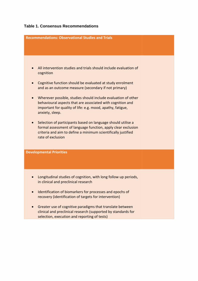
Table 1. Consensus Recommendations
Recommendations: Observational Studies and Trials
All intervention studies and trials should include evaluation of cognition
Cognitive function should be evaluated at study enrolment and as an outcome measure (secondary if not primary)
Wherever possible, studies should include evaluation of other behavioural aspects that are associated with cognition and important for quality of life: e.g. mood, apathy, fatigue, anxiety, sleep.
Selection of participants based on language should utilise a formal assessment of language function, apply clear exclusion criteria and aim to define a minimum scientifically justified rate of exclusion
Developmental Priorities
Longitudinal studies of cognition, with long follow up periods, in clinical and preclinical research
Identification of biomarkers for processes and epochs of recovery (identification of targets for intervention)
Greater use of cognitive paradigms that translate between clinical and preclinical research (supported by standards for selection, execution and reporting of tests)

Table 2. Cognitive paradigms and translation from models to humans
Neurocognitive Domain (DSM-5)
Subdomain Human Paradigm Preclinical Paradigms Comments
Executive function
Cognitive flexibility Wisconsin Card Sorting Test Digit Symbol Substitution33
Attention set-shift
Inhibition/impulsivity Go no-go tasks Operant conditioning Working memory Digit Span
Spatial Span T-maze (Delayed alternation task) Y-maze Radial 8-arm maze Morris water maze Trial-unique delayed non-matching-to-location (TUNL) task
Tasks of spatial working memory translate more easily between humans and models. There are many paradigms that have been studied extensively in humans. Despite this, clinical studies currently often opt for digit or letter span tasks.
Complex Attention
Sustained Attention Choice Reaction Time34, 35
5-choice serial reaction time task36 5-choice continuous performance test Signal detection task Cross-modal stimulus presentations
Speed of processing Reaction time tasks 5-choice serial reaction time task
Divided attention Walking while counting backward
Unclear whether tested in preclinical models
Neglect Cancellation tasks37 Line bisection
Adhesive strip removal While the adhesive removal test can assess sensory neglect it is

a combination of cognitive and motor. While cancellation tasks have used letters, other versions use simple objects, such as stars, would more easily translate between models and humans.
Language Likely cannot be addressed in preclinical animal models
Perceptual-Motor Function
Object recognition (agnosia)
Visual discrimination Pattern recognition
Pairwise/Visual Discrimination/Reversal tasks
Learning and Memory
Recognition Delayed non-matching to sample Scene Recognition38, 39
Delayed non-matching to sample
Spatial Memory Morris Water Maze (human adaptation)*
Morris Water Maze Tests of verbal recall are common in human studies, but less well-suited than spatial, object-based or perceptual tasks to translation
Associative Learning Paired Associate Learning Object-in-Scene memory
Paired Associate Learning Object-in-Scene memory
Emotional Memory Fear conditioning

Social Cognition Little studied in post
stroke setting Not tested in preclinical stroke models

References
1. Bernhardt J, Hayward KS, Kwakkel G, Ward NS, Wolf SL, Borschmann K, et al. Agreed definitions and a shared vision for new standards in stroke recovery research: The stroke recovery and rehabilitation roundtable taskforce. Int. J. Stroke. 2017;12:444-450
2. Nys GM, van Zandvoort MJ, de Kort PL, Jansen BP, de Haan EH, Kappelle LJ. Cognitive disorders in acute stroke: Prevalence and clinical determinants. Cerebrovasc. Dis. 2007;23:408-416
3. Nys GM, van Zandvoort MJ, de Kort PL, van der Worp HB, Jansen BP, Algra A, et al. The prognostic value of domain-specific cognitive abilities in acute first-ever stroke. Neurology. 2005;64:821-827
4. Brookes RL, Willis TA, Patel B, Morris RG, Markus HS. Depressive symptoms as a predictor of quality of life in cerebral small vessel disease, acting independently of disability; a study in both sporadic small vessel disease and cadasil. Int. J. Stroke. 2013;8:510-517
5. Pendlebury ST, Rothwell PM, Oxford Vascular S. Incidence and prevalence of dementia associated with transient ischaemic attack and stroke: Analysis of the population-based oxford vascular study. Lancet Neurol. 2019;18:248-258
6. McKevitt C, Fudge N, Redfern J, Sheldenkar A, Crichton S, Rudd AR, et al. Self-reported long-term needs after stroke. Stroke. 2011;42:1398-1403
7. Quinn TJ, Paolucci S, Sunnerhagen KS, Sivenius J, Walker MF, Toni D, et al. Evidence-based stroke r-ehabilitation: An expanded guidance document from the european stroke organisation (eso) guidelines for management of ischaemic stroke and transient ischaemic attack 2008. J. Rehabil. Med. 2009;41:99-111
8. Veldsman M, Brodtmann A. Disconnectomics: Stroke-related disconnection and dysfunction in distributed brain networks. Int. J. Stroke. 2019;14:6-8
9. Clelland CD, Choi M, Romberg C, Clemenson GD, Fragniere A, Tyers P, et al. A functional role for adult hippocampal neurogenesis in spatial pattern separation. Science. 2009;325:210-213
10. Carmichael ST. Cellular and molecular mechanisms of neural repair after stroke: Making waves. Ann. Neurol. 2006;59:735-742
11. Ray NJ, Metzler-Baddeley C, Khondoker MR, Grothe MJ, Teipel S, Wright P, et al. Cholinergic basal forebrain structure influences the reconfiguration of white matter connections to support residual memory in mild cognitive impairment. J. Neurosci. 2015;35:739-747
12. Sachdev PS, Blacker D, Blazer DG, Ganguli M, Jeste DV, Paulsen JS, et al. Classifying neurocognitive disorders: The dsm-5 approach. Nat. Rev. Neurol. 2014;10:634-642
13. Hachinski V. Vascular dementia: A radical redefinition. Dementia. 1994;5:130-132 14. Sachdev P, Kalaria R, O'Brien J, Skoog I, Alladi S, Black SE, et al. Diagnostic criteria for
vascular cognitive disorders: A vascog statement. Alzheimer Dis. Assoc. Disord. 2014;28:206-218
15. Skrobot OA, Black SE, Chen C, DeCarli C, Erkinjuntti T, Ford GA, et al. Progress toward standardized diagnosis of vascular cognitive impairment: Guidelines from the

vascular impairment of cognition classification consensus study. Alzheimers Dement. 2018;14:280-292
16. Hachinski V, Iadecola C, Petersen RC, Breteler MM, Nyenhuis DL, Black SE, et al. National institute of neurological disorders and stroke-canadian stroke network vascular cognitive impairment harmonization standards. Stroke. 2006;37:2220-2241
17. Horsburgh K, Wardlaw JM, van Agtmael T, Allan SM, Ashford MLJ, Bath PM, et al. Small vessels, dementia and chronic diseases - molecular mechanisms and pathophysiology. Clin. Sci. (Lond.). 2018;132:851-868
18. Kwakkel G, Lannin NA, Borschmann K, English C, Ali M, Churilov L, et al. Standardized measurement of sensorimotor recovery in stroke trials: Consensus-based core recommendations from the stroke recovery and rehabilitation roundtable. Int. J. Stroke. 2017;12:451-461
19. Murphy TH, Corbett D. Plasticity during stroke recovery: From synapse to behaviour. Nat. Rev. Neurosci. 2009;10:861-872
20. Tanila H. Testing cognitive functions in rodent disease models: Present pitfalls and future perspectives. Behav. Brain Res. 2018;352:23-27
21. Nithianantharajah J, McKechanie AG, Stewart TJ, Johnstone M, Blackwood DH, St Clair D, et al. Bridging the translational divide: Identical cognitive touchscreen testing in mice and humans carrying mutations in a disease-relevant homologous gene. Sci. Rep. 2015;5:14613
22. Weicker J, Villringer A, Thone-Otto A. Can impaired working memory functioning be improved by training? A meta-analysis with a special focus on brain injured patients. Neuropsychology. 2016;30:190-212
23. Bogdanova Y, Yee MK, Ho VT, Cicerone KD. Computerized cognitive rehabilitation of attention and executive function in acquired brain injury: A systematic review. J. Head Trauma Rehabil. 2016;31:419-433
24. Glass BD, Maddox WT, Love BC. Real-time strategy game training: Emergence of a cognitive flexibility trait. PLoS One. 2013;8:e70350
25. Westerberg H, Jacobaeus H, Hirvikoski T, Clevberger P, Ostensson ML, Bartfai A, et al. Computerized working memory training after stroke--a pilot study. Brain Inj. 2007;21:21-29
26. Levine B, Schweizer TA, O'Connor C, Turner G, Gillingham S, Stuss DT, et al. Rehabilitation of executive functioning in patients with frontal lobe brain damage with goal management training. Front. Hum. Neurosci. 2011;5:9
27. Man DW, Soong WY, Tam SF, Hui-Chan CW. A randomized clinical trial study on the effectiveness of a tele-analogy-based problem-solving programme for people with acquired brain injury (abi). NeuroRehabilitation. 2006;21:205-217
28. McQuail JA, Frazier CJ, Bizon JL. Molecular aspects of age-related cognitive decline: The role of gaba signaling. Trends Mol. Med. 2015;21:450-460
29. Lozano R, Hare EB, Hagerman RJ. Modulation of the gabaergic pathway for the treatment of fragile x syndrome. Neuropsychiatr. Dis. Treat. 2014;10:1769-1779
30. Erickson KI, Voss MW, Prakash RS, Basak C, Szabo A, Chaddock L, et al. Exercise training increases size of hippocampus and improves memory. Proc. Natl. Acad. Sci. U. S. A. 2011;108:3017-3022
31. Ploughman M, Eskes GA, Kelly LP, Kirkland MC, Devasahayam AJ, Wallack EM, et al. Synergistic benefits of combined aerobic and cognitive training on fluid intelligence and the role of igf-1 in chronic stroke. Neurorehabil. Neural Repair. 2019;33:199-212
32. Schmidt-Wilcke T, Fuchs E, Funke K, Vlachos A, Muller-Dahlhaus F, Puts NAJ, et al. Gaba-from inhibition to cognition: Emerging concepts. Neuroscientist. 2018;24:501-515
33. O'Sullivan M, Morris RG, Markus HS. Brief cognitive assessment for patients with cerebral small vessel disease. J. Neurol. Neurosurg. Psychiatry. 2005;76:1140-1145
34. Bonnelle V, Leech R, Kinnunen KM, Ham TE, Beckmann CF, De Boissezon X, et al. Default mode network connectivity predicts sustained attention deficits after traumatic brain injury. J. Neurosci. 2011;31:13442-13451
35. Langner R, Willmes K, Chatterjee A, Eickhoff SB, Sturm W. Energetic effects of stimulus intensity on prolonged simple reaction-time performance. Psychol. Res. 2010;74:499-512
36. Grottick AJ, Higgins GA. Assessing a vigilance decrement in aged rats: Effects of pre-feeding, task manipulation, and psychostimulants. Psychopharmacology (Berl.). 2002;164:33-41
37. Li K, Malhotra PA. Spatial neglect. Pract. Neurol. 2015;15:333-339 38. Mundy ME, Downing PE, Dwyer DM, Honey RC, Graham KS. A critical role for the
hippocampus and perirhinal cortex in perceptual learning of scenes and faces: Complementary findings from amnesia and fmri. J. Neurosci. 2013;33:10490-10502
39. Moodley K, Minati L, Contarino V, Prioni S, Wood R, Cooper R, et al. Diagnostic differentiation of mild cognitive impairment due to alzheimer's disease using a hippocampus-dependent test of spatial memory. Hippocampus. 2015;25:939-951