Embed Size (px)
Citation preview

Journal of Pharmacy and Pharmacology 7 (2019) 98-109
doi: 10.17265/2328-2150/2019.03.002
Coconut Oil Reduces Visceral Adipocyte Size and
Improves the Metabolic Profile of Rats Fed a
High-Carbohydrate Diet
Valéria Schoffen Romão-Carrascoza1, Rosângela Fernandes Garcia
1, Lorena Lima Gargaro
2, Maria Montserrat Diaz
Pedrosa1, Nilton de Almeida Brito
1, Clairce Luzia Salgueiro-Pagadigorria
1 and Márcia do Nascimento Brito
1
1. Department of Physiological Sciences, State University of Maringá, Maringá 87020900, PR, Brazil
2. Department of Medicine, State University of Maringá, Maringá 87020900, PR, Brazil
Abstracts: This work aimed to evaluate the effects of the food supplementation with coconut oil (CO) on biometric and metabolic
parameters in an animal model of obesity. Wistar rats fed a high-carbohydrate diet (HCD rats) develop metabolic disorders very
similar to those found in human metabolic syndrome, including increased adiposity, insulin resistance (IR), non-alcoholic fatty liver
disease (NAFLD) and dyslipidemia. Although HCD rats had normal fasting glycemia, they developed IR as revealed by Homa-IR
index and, after glucose overload, exhibited higher peaks of insulinemia and glycemia. These alterations were accompanied by a
higher formation of advanced glycated end products (AGEs). The supplementation with CO improved IR, reduced AGEs formation
and reverted NAFLD. These effects could be due to CO action of reducing visceral adipocyte size and take place without undesirable
collateral effects on lipid profile, placing CO as an adjuvant in the treatment of the metabolic syndrome.
Key words: Adipocyte hypertrophy, metabolic syndrome, insulin resistance, NAFLD, coconut oil.
1. Introduction
Metabolic syndrome (MSyn) is a multifactorial
condition characterized by visceral obesity,
hypertension, hyperglycemia, insulin resistance (IR)
and dyslipidemia that can increase the risk of type 2
diabetes, cardiovascular diseases and non-alcoholic
fatty liver disease (NAFLD) [1, 2].
The link between the individual MSyn components
is not totally known, but the frequent combination of
these metabolic abnormalities suggests that they are
indeed interrelated, and that just a few factors may
underlie such combination.
Over 4 decades ago, adipocyte size was shown to
vary inversely with adipocyte insulin sensitivity [3].
Studies have shown functional differences between
large and small adipocytes [4-6]. In general, the
hypertrophy of visceral, in comparison with
Corresponding author: Rosângela Fernandes Garcia, Ph.D.,
professor, research fields: liver metabolism and nutrition.
subcutaneous, adipocytes leads to more detrimental
metabolic effects [7, 8], first because the enlarged
visceral adipocytes exhibit more remarkable
physiological dysfunctions [9, 10] and second because
of their close proximity to hepatic and visceral
immune cells [11]. For instance, enlarged visceral
adipocytes secrete large amounts of TNF-α, which
induces IR in both adipocytes and myocytes [12].
Thus, the enlarged visceral adipocytes display
increased lipolysis, releasing fatty acids (FA) directly
into the portal vein, overloading the liver and leading
to liver fat accumulation [13]. In fact, IR has been
considered the “first-level” contributing event for the
pathogenesis of NAFLD and other metabolic and
cardiovascular complications of obesity, the so-called
IR-syndromes [14, 15].
It is a reasonable thought that the reduction of
visceral fat depots should improve life expectancies.
This is not always possible simply by changing
lifestyle or eating habits. One of the alternative
D DAVID PUBLISHING

Coconut Oil Reduces Visceral Adipocyte Size and Improves the Metabolic Profile of Rats Fed a High-Carbohydrate Diet
99
strategies has been to replace some of the
conventional dietary long-chain triglyceride-rich
(LCT, > 12 carbons FA) by medium-chain
triglyceride-rich (MCT, 6-12 carbons) oil [16]. Lipids
varying in FA chain lengths are metabolized
differently. MCTs are absorbed directly into the portal
circulation and transported to the liver for rapid
oxidation [16, 17]. LCTs, on the other hand, are
transported via chylomicrons into the lymphatic
system, allowing for extensive uptake into adipose
tissue. Therefore, the hypothesis has been put forward
that the rapid metabolism of MCTs may increase
energy expenditure and decrease their storage in
adipose tissue [18, 19]. All these results are suggestive
that MCT could potentially prevent or control obesity
in humans.
In this context, the intake of coconut oil (Cocos
nucifera) has increased over the years. Cold-pressed
coconut oil (CO) is composed of about 93%
medium-chain saturated FA, predominantly lauric and
myristic acids. Studies aimed at investigating the
effects of CO and MCT oil in body weight (BW) have
demonstrated that it was effective in reducing central
obesity without undesirable effects on plasma lipid
profile [20-22]. However, the effects of CO on the
size of adipocytes—an indirect indicator of IR—were
not assessed in these studies.
Previous studies have reported that rats fed with
high-carbohydrate diet (HCD) develop metabolic,
cardiovascular and hepatic complications [23, 24] that
are similar to human MSyn [25]. In this way, the
purpose of this work was to evaluate the effects of CO
on an animal model of obesity induced by HCD. The
parameters evaluated included BW gain, adiposity
index, adipocyte diameter, liver lipid contents, plasma
lipid profile, glycemia, insulinemia and the formation
of advanced glycated end-products (AGEs).
2. Material and Methods
2.1 Materials
Kits from Gold Analisa®
(Belo Horizonte, Brazil)
were used to measure blood levels of lipids, glucose
and fructosamine. Sodium heparin was obtained from
Roche (São Paulo, Brazil). The other reagents were
from Merck (Darmstadt, FRG), Carlo Erba (São Paulo,
Brazil) and Reagen (Rio de Janeiro, Brazil). Plasma
insulin levels were measured by radioimmunoassay
(Wizard2 Automatic Gamma Counter®, TM-2470,
PerkinElmer, Shelton-CT, EUA). A commercially
available extra-virgin coconut oil was used and its
composition is presented in Table 1.
2.2 Animals, Diets and Treatment
This study was approved by the Ethics Committee
for the Use of Animals of the State University of
Maringá (Certificate No. 018/2014).
Male Wistar rats (n = 35) aging 21 days (weaned)
were kept under controlled temperature (23 ºC) and
photoperiod (12 h light/12 h dark) in polypropylene
cages. Eleven rats were fed with a standard rodent
chow (Nuvilab CR1®, Nuvital, Brazil) (CON rats, n = 11)
Table 1 Fatty acid composition of coconut oil (CO).
Fatty acid %/100 g CO
Caproic acid C6:0 0.38
Caprylic acid C8:0 5.56
Capric acid C10:0 4.99
Lauric acid C12:0 45.78
Myristic acid C14:0 18.56
Palmitic acid C16:0 8.85
Stearic acid C18:0 3.39
Oleic acid C18:1 5.65
Linoleic acid C18:2 0.94

Coconut Oil Reduces Visceral Adipocyte Size and Improves the Metabolic Profile of Rats Fed a High-Carbohydrate Diet
100
and twenty-four rats were fed with high-carbohydrate
diet (HCD rats). The HCD was composed of 33%
standard chow, 33% condensed milk (Nestlé®, Brazil),
7% crystalized sugar (União®
, Brazil) and 8.6% water.
The nutritional values of the standard and HC diets
were, respectively: 402 and 428 kcal/100 g; 57.5 and
68% carbohydrate; 30 and 16% protein; 12.5 and 16%
fat [23]. Diets were offered in pellets and ad libitum.
After 10 weeks, twelve HCD rats were treated with
extra-virgin coconut oil (HCD + CO rats) for 4 weeks,
at the dose of 6 mg/100 g body weight (BW) by
esophageal gavage (final volume of 0.1 mL/100 g BW)
divided into two administrations, which were done at
10:00 AM and 5:00 PM. The other twelve untreated
HCD rats and the CON rats were given the same
volume of saline. The dose of CO was chosen based
on Assunção et al. [20].
The entire experimental period lasted 14 weeks,
during which the animals were recorded for their BW
once a week and food ingestion every two days to
calculate total food intake. Weight gain was calculated
as the difference between the final weight and the
initial weight.
At the age of 120 days, the animals were used for
the experiments described below.
2.3 Intravenous Glucose Tolerance Test (ivGTT)
To perform these tests, a polyethylene cannula was
inserted in the right jugular vein [26] under anesthesia
(thionembutal 40 mg/kg BW after lidocaine 5 mg/kg
BW) on the day before the experiments. The cannula
was used for glucose infusion as a “bolus” (1 g
glucose/kg BW) and collection of blood samples at
the times 0, 5, 15, 30, 45 and 60 min, time 0 min
being immediately before glucose infusion. The
ivGTT was performed at 8:00 AM after an overnight
fasting. The blood samples collected were transferred
to heparin-coated tubes and centrifuged for glucose
determination. Plasma samples were kept at -20 °C for
insulin measurements. The fasting insulinemia and
glycemia were used to calculate the IR, expressed in
terms of the homeostasis model assessment of IR
(HOMA-IR Index) [27]. Plasma glucose was
expressed as mg/dL and insulinemia as ng/mL.
2.4 Removal of Tissues and Collection of Blood
After the glucose tolerance test, euthanasia was
carried out with a lethal dose of thionembutal (100
mg/kg BW, i.p.). Blood samples were collected by
cardiac puncture and the serum was used to measure
fructosamine, total cholesterol (TC), high-density
lipoprotein (HDL) and triglycerides (TG). Samples of
liver were removed, clamped in liquid nitrogen and
stored at -80 °C for further determination of lipid
content. In addition, fat depots (epididymal,
mesenteric, retroperitoneal and inguinal) were
removed and weighed. The adiposity index was
defined as the sum of fat weight/100 g BW [28]. The
retroperitoneal adipose tissue was used to determine
adipocyte size. The respective protocols are described
below.
2.5 Serum Biochemical Analyses
Total cholesterol, HDL and TG were analyzed in
serum by standard methods using assay kits (Gold
Analisa®). Very low-density lipoprotein (VLDL)
levels were calculated using the Friedewald’s equation
[29] and low-density lipoprotein (LDL) levels were
determined by subtracting HDL and VLDL from TC.
The values were expressed as mg/dL. Fructosamine
was used as a marker of long-term glycemic control
and formation of AGEs. The levels of fructosamine
were evaluated using commercial kits (Gold Analisa®
,
Brazil) and expressed as mmol/L.
2.6 Determination of Hepatic Lipid Content
The liver total lipid content was determined using
the gravimetric method [30], which is based on the
extraction of lipids from homogenized liver samples
(approximately 1.0 g) in a chloroform-methanol
mixture (2:1). Lipids were resuspended in 2% Triton,
followed by agitation and heating to 55 °C. TC and

Coconut Oil Reduces Visceral Adipocyte Size and Improves the Metabolic Profile of Rats Fed a High-Carbohydrate Diet
101
TG contents were measured using commercial kits
(Gold Analisa®, Brazil). The results were expressed as
g/g of liver wet weight.
2.7 Adipocyte Isolation and Adipocyte Cell Size
Determination
Adipocytes were isolated using a modified version
of the method described by Rodbell [31]. Briefly, the
retroperitoneal adipose tissue (approximately 1.0 g)
was cut into fine pieces and put into 4.0 mL digestive
buffer pH 7.5 containing 1.0 g% glucose-free DMEM
(Dulbecco’s Modified Eagle Medium Base), 1 mM
sodium pyruvate, 4.2 mM sodium bicarbonate, 25 mM
HEPES, 4.0 g% bovine serum albumin, 1 mg/mL type
II collagenase, 1 mg% phenol red. The samples were
incubated in an orbital agitation water bath at 37 ºC
for 60 min. The cell suspension was filtered through a
double layer of nylon cloth and then washed three
times by flotation in a medium containing: 0.81 mM
magnesium sulfate, 5.4 mM potassium chloride, 0.11
M sodium chloride, 1.2 mM sodium phosphate
monobasic, 1.4 mM calcium chloride, 25 mM HEPES,
1 mM sodium pyruvate, 1 mg% phenol red and 1 g%
bovine serum albumin, pH 7.5.
The washed cell suspension was placed on a glass
slide and the images of the adipocytes were captured
at 40× original magnification with a high-resolution
digital camera (Pro-Media Series, Cibertecnics®)
coupled to an optical microscope Olympus BX40®
,
Japan) with 50-µm scale bar. Adipocyte diameters (50
cells per animal) were measured using a computerized
image analysis system (Image Pro-Plus®, Media
Cybernetics, USA).
2.8 Statistical Analyses
The results were expressed as mean ± standard error
(SE). Prism 5.0®
(GraphPad, USA) was used to
calculate the area under curve (AUC) and perform the
statistical analyses (One-way ANOVA followed by
the Tukey’s test). The level of significance was set at
5%.
3. Results
3.1 Progression of BW
Fig. 1 shows the time course of BW gain of the three
animal groups, from weaning (21 days old, week zero)
until the end of the experimental period (week 14).
At 21 days, the BW of the animals of the three groups
was similar (CON = 39.4 ± 1.0 g; HCD = 39.8 ± 1.1 g;
HCD + CO = 39.0 ± 1.0 g). Even after the animals
started to receive different diets this tendency did not
Weeks
Fig. 1 Body weight of CON, HCD and HCD + CO rats from weaning (Week 0) until the end of the experimental period
(Week 14). The arrow shows the beginning of the supplementation with CO (Week 10, 6 mg/100 g BW). The columns are the
BW gain and are expressed as the mean ± SE (n = 11-12/group). Different letters represent statistical difference (p < 0.05).

Coconut Oil Reduces Visceral Adipocyte Size and Improves the Metabolic Profile of Rats Fed a High-Carbohydrate Diet
102
change until Week 10. Coincidentally, this was the
onset of the supplementation with CO. However, all
HCD rats exhibited greater weight, regardless of
supplementation with CO or not. At the end of Week
14, HCD and HCD + CO rats were heavier than CON
rats by about 13%.
The animals were evaluated for adipose tissue mass
and adiposity index. As shown in Fig. 2, HCD and
HCD + CO rats exhibited significant and similar
increases in their epididymal (+49%) and
retroperitoneal (+64%) fat depots. Although no
alterations were seen in the inguinal or mesenteric fats
(> 0.05 compared with CON rats), there was a
remarkable increase in the adiposity index in these
groups (+33%) compared with CON rats.
The changes in BW and adiposity index were
correlated with changes in food intake, since all the
animals ingested similar amounts of chow (p > 0.05)
along the entire experimental period (CON rats
2,274.80 ± 38.9 g; HCD rats 2,236.10 ± 25.9 g; HCD
+ CO rats 2,234.9 ± 32.9 g).
3.2 Determination of Adipocyte Diameter
The results of retroperitoneal adipocyte diameters
are presented in Fig. 3. The HCD rats had larger
adipocytes compared with CON rats; the
supplementation with CO reversed this hypertrophy.
3.3 Biochemical Blood Analysis
Table 2 shows the blood metabolic profile of the
three groups after overnight fasting at the end of the
14 weeks. All animals had normal and similar fasting
glycemia (p > 0.05), but HCD and HCD-CO rats had
much higher insulin levels than CON rats. The
supplementation with CO, however, significantly
reduced this level so that insulin level in HCD + CO
rats was intermediary between HCD and CON rats but
different from both. The insulin sensitivity, as
assessed by Homa-IR index, matched the insulin level
of each group—it was much greater in HCD rats,
intermediary in HCD + CO rats and lower in CON
rats.
As for the lipid profile, although TC, HDL and
LDL did not differ between the groups,
VLDL-cholesterol and TG levels were significantly
and similarly increased in both HCD and HCD + CO
rats, by approximately 26%, compared with CON rats.
3.4 Glucose Tolerance Test
The values of glycemia and insulinemia obtained
Fig. 2 Adipose tissue weight and adiposity index of CON, HCD and HCD + CO rats. The results are expressed as mean ± SE
(n = 11-12/group). Different letters represent statistical difference (p < 0.05).

Coconut Oil Reduces Visceral Adipocyte Size and Improves the Metabolic Profile of Rats Fed a High-Carbohydrate Diet
103
Fig. 3 Isolated retroperitoneal adipocytes of CON (A), HCD (B) and HCD + CO (C) rats and adipocyte diameters (D). The
results are expressed as mean ± SE (n = 8/group). Different letters represent statistical difference (p < 0.05).
Table 2 Biochemical blood parameters of CON, HCD and HCD + CO rats.
Parameters CON HCD HCD+CO
Glycemia (mg/dL) 79.89 ± 1.70a 80.42 ± 2.77a 82.90 ± 3.53a
Insulinemia (ng/mL) 0.051 ± 0.26a 0.209 ± 0.06b 0.159 ± 0.04c
Homa-IR index 0.165 ± 0.03a 0.671 ± 0.14b 0.311 ± 0.05c
TG (mg/gL) 89.14 ± 7.26a 118.96 ± 9.07b 119.71 ± 7.35b
TC (mg/dL) 76.48 ± 3.23a 85.66 ± 1.49a 86.33 ± 1.86a
HDL (mg/dL) 32.86 ± 2.27a 34.21 ± 1.01a 35.00 ± 1.66a
LDL (mg/dL) 25.79 ± 2.83a 27.65 ± 1.94a 27.38 ± 1.94a
VLDL (mg/dL) 17.83 ± 1.45a 23.79 ± 1.81b 23.94 ± 1.47b
The results are expressed as mean ± SE (n = 10-12 /group). Different letters represent statistical difference (p < 0.05).
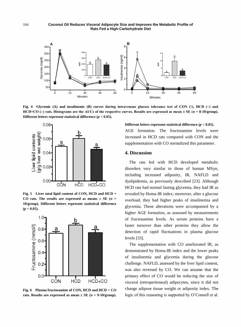
during the ivGTT are given in Figs. 4A and 4B,
respectively. The histograms are AUCs of both
measures. HCD rats had higher glycemic and
insulinemic indices than CON rats. The
supplementation with CO could restore these values,
as shown by the AUC values.
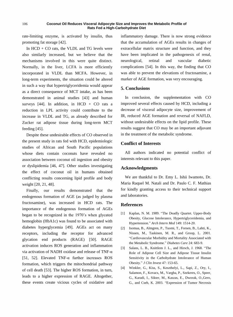
3.5 Liver Total Lipid Content
Fig. 5 shows the results of liver total lipid content.
The livers of CON rats had normal lipid content of
about 5% [32]. However, in HCD rats, it increased
21%, characterizing extensive NAFLD. The
supplementation with CO totally reversed this
condition.
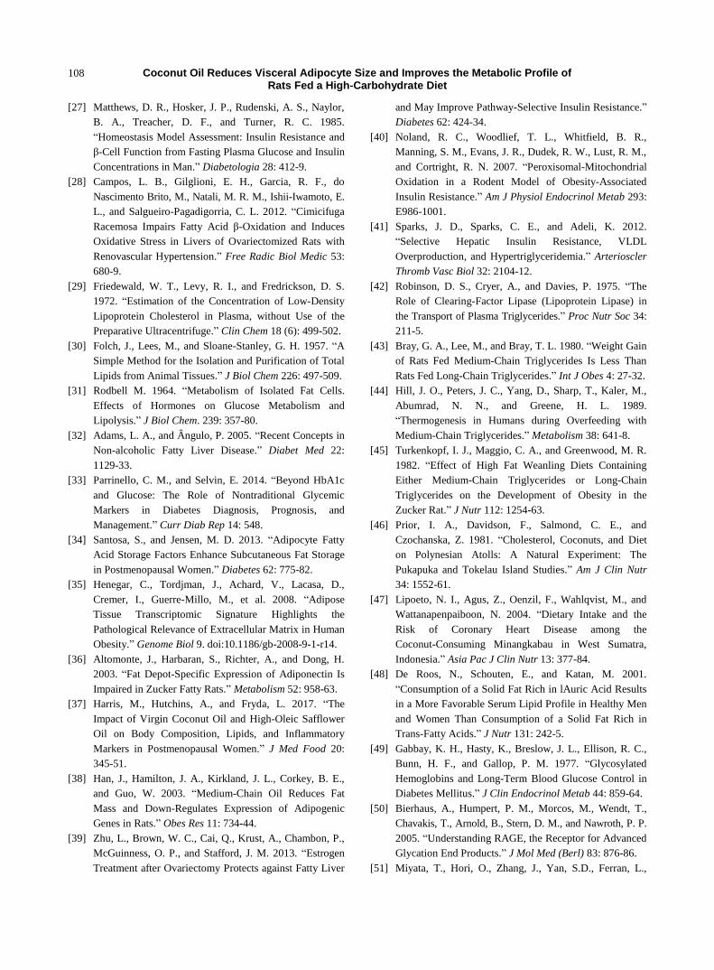
3.6 Determination of Fructosamine
Plasma fructosamine concentrations (Fig. 6) were
used as an index of long-term glycemic control and
Coconut Oil Reduces Visceral Adipocyte Size and Improves the Metabolic Profile of Rats Fed a High-Carbohydrate Diet
104
0 15 30 45 60
50
100
150
200
250
300
350A
Minutes
Gly
ce
mia
(m
g/d
l)
CON HCD HCD+CO
0
1000
2000
3000
4000
a
b
a
AU
C
0 15 30 45 600
2
4
6
8
Minutes
B
Insulin
em
ia (
ng/m
l)
CON HCD HCD+CO
0
5
10
15
b
aa
AU
C
Fig. 4 Glycemic (A) and insulinemic (B) curves during intravenous glucose tolerance test of CON (◊), HCD (○) and
HCD+CO (□) rats. Histograms are the AUCs of the respective curves. Results are expressed as mean ± SE (n = 8-10/group).
Different letters represent statistical difference (p < 0.05).
Fig. 5 Liver total lipid content of CON, HCD and HCD +
CO rats. The results are expressed as means ± SE (n =
10/group). Different letters represent statistical difference
(p < 0.05).
Fig. 6 Plasma fructosamine of CON, HCD and HCD + CO
rats. Results are expressed as mean ± SE (n = 9-10/group).
Different letters represent statistical difference (p < 0.05).
AGE formation. The fructosamine levels were
increased in HCD rats compared with CON and the
supplementation with CO normalized this parameter.
4. Discussion
The rats fed with HCD developed metabolic
disorders very similar to those of human MSyn,
including increased adiposity, IR, NAFLD and
dyslipidemia, as previously described [23]. Although
HCD rats had normal fasting glycemia, they had IR as
revealed by Homa-IR index; moreover, after a glucose
overload, they had higher peaks of insulinemia and
glycemia. These alterations were accompanied by a
higher AGE formation, as assessed by measurements
of fructosamine levels. As serum proteins have a
faster turnover than other proteins they allow the
detection of rapid fluctuations in plasma glucose
levels [33].
The supplementation with CO ameliorated IR, as
demonstrated by Homa-IR index and the lower peaks
of insulinemia and glycemia during the glucose
challenge. NAFLD, assessed by the liver lipid content,
was also reversed by CO. We can assume that the
primary effect of CO would be reducing the size of
visceral (retroperitoneal) adipocytes, since it did not
change adipose tissue weight or adiposity index. The
logic of this reasoning is supported by O’Connell et al.

Coconut Oil Reduces Visceral Adipocyte Size and Improves the Metabolic Profile of Rats Fed a High-Carbohydrate Diet
105
[8], who compared healthy and unhealthy severely
obese men and observed that omental, but not
subcutaneous, adipocyte sizes are correlated with
metabolic disturbances, and that the individual’s
adipocyte size is more important than his body weight.
These same observations were made by comparing
lean and obese humans [10, 34]. The decreased
adipocyte size not accompanied by decreased
retroperitoneal weight can be ascribed to a persistent
fibrosis of the extracellular matrix, which is enhanced
by body weight gain [35].
Enlarged visceral adipocytes exhibit the most
pronounced diameter-related alterations, including
high levels of tumor necrosis factor (TNF-α) [4] and
low adiponectin release [36], which were shown to be
related with lower adipocyte responsiveness to insulin
[12], increased lipolysis [13] and lower glucose uptake
[5]. In addition to inducing IR TNF-α, together with
other cytokines, such as interleukin (IL) 1β, are
pro-inflammatory [11]. Recent studies performed with
obese, postmenopausal women have revealed that, by
replacing common vegetal oil by CO, the plasma
levels of TNF-α and IL-1β decreased, as well as the
inflammation [37]. It has also been demonstrated that
MCTs inhibit the production of inflammatory
cytokines [38], once again supporting our hypothesis.
The increased lipolysis by the insulin resistant
visceral adipocytes overloads the liver with FA [13].
Additionally, insulin robustly stimulates liver de novo
lipogenesis, decreases FA oxidation [38] and
promotes autophagic degradation of apolipoprotein
B100 (apoB100), thus limiting the release of VLDL
from the liver [39]. Together, these events trigger
NAFLD, as observed here for HCD rats. Our results
also demonstrated that the improvement of IR by CO
was accompanied by a reversal of NAFLD in HCD +
CO rats.
Beyond hyperinsulinemia, another factor in HCD
rats that could impair the lipid disposal pathways is
the inhibition of the enzyme carnitine palmitoyl
transferase I (CPT-I) by malonyl-CoA, in this
condition of dietary carbohydrate abundance. This
first limiting enzyme for the oxidation of long chain
fatty acids (LCFA) is located at the mitochondrial
outer membrane, where it controls the conversion of
acyl-CoA into acyl-carnitine, its access to the
mitochondrial matrix and, therefore, the rate of
mitochondrial LCFA oxidation. CPT-I is
physiologically inhibited by malonyl-CoA, which is
formed from acetyl-CoA in the first (limiting) step of
the de novo FA synthesis [40]. Thus, high rates of
fatty acid synthesis result in low rates of fatty acid
oxidation, and vice versa.
As medium chain fatty acids (MCFA) enter
mitochondria independently of CPT-I, excess
carbohydrates (which are converted to malonyl-CoA)
may not inhibit their oxidation [40]. In this way, it is
expected that the supplementation with CO during
HCD would bring about a novel situation to the liver:
β-oxidation (of MCFA) as well as the production of
malonyl-CoA and subsequent de novo FA synthesis
(from carbohydrates) should be accelerated. As the
oxidation of MCFA has been associated with
increased oxygen consumption and energy
expenditure compared to LCFA [18], the reduction of
fat liver deposits, as observed in HCD + CO rats, was
not surprising.
Some of the lipids synthesized within the
hepatocytes are released into the bloodstream as
VLDL. The fasting VLDL levels were increased in
HCD rats, probably as a consequence of the
physiological impairment of hepatic insulin regulation
of apoB100 as a result of liver fat accumulation [39].
Insulin stimulation of de novo lipogenesis occurs
independent of its effects on apoB100. Thus,
increased release of VLDL and hypertriglyceridemia
are common features in the setting of
hyperinsulinemia and IR [41].
In this way, in HCD rats, the increase of fasting
VLDL and TG levels should reflect VLDL
hyper-secretion rather than reduced clearance of TG
by adipose tissue lipoprotein lipase (LPL). In fact, this
Coconut Oil Reduces Visceral Adipocyte Size and Improves the Metabolic Profile of Rats Fed a High-Carbohydrate Diet
106
rate-limiting enzyme, is activated by insulin, thus
promoting fat storage [42].
In HCD + CO rats, the VLDL and TG levels were
also similarly increased, but we believe that the
mechanisms involved in this were quite distinct.
Normally, in the liver, LCFA is more efficiently
incorporated in VLDL than MCFA. However, in
long-term experiments, the situation could be altered
in such a way that hypertriglyceridemia would appear
as a direct consequence of MCT intake, as has been
demonstrated in animal studies [43] and human
surveys [44]. In addition, in HCD + CO rats a
reduction in LPL activity could contribute to the
increase in VLDL and TG, as already described for
Zucker rat adipose tissue during long-term MCT
feeding [45].
Despite these undesirable effects of CO observed in
the present study in rats fed with HCD, epidemiologic
studies of African and South Pacific populations
whose diets contain coconuts have revealed no
association between coconut oil ingestion and obesity
or dyslipidemia [46, 47]. Other studies investigating
the effect of coconut oil in humans obtained
conflicting results concerning lipid profile and body
weight [20, 21, 48].
Finally, our results demonstrated that the
endogenous formation of AGE (as judged by plasma
fructosamine), was increased in HCD rats. The
importance of the endogenous formation of AGEs
began to be recognized in the 1970’s when glycated
hemoglobin (HbA1c) was found to be associated with
diabetes hyperglycemia [49]. AGEs act on many
receptors, including the receptor for advanced
glycation end products (RAGE) [50]. RAGE
activation induces ROS generation and inflammation
via activation of NADH oxidase and release of TNF-α
[51, 52]. Elevated TNF-α further increases ROS
formation, which triggers the mitochondrial pathway
of cell death [53]. The higher ROS formation, in turn,
leads to a higher expression of RAGE. Altogether,
these events create vicious cycles of oxidative and
inflammatory damage. There is now strong evidence
that the accumulation of AGEs results in changes of
extracellular matrix structure and function, and they
have been implicated in the pathogenesis of renal,
neurological, retinal and vascular diabetic
complications [54]. In this way, the finding that CO
was able to prevent the elevations of fructosamine, a
marker of AGE formation, was very encouraging.
5. Conclusions
In conclusion, the supplementation with CO
improved several effects caused by HCD, including a
decrease of visceral adipocyte size, improvement of
IR, reduced AGE formation and reversal of NAFLD,
without undesirable effects on the lipid profile. These
results suggest that CO may be an important adjuvant
in the treatment of the metabolic syndrome.
Conflict of Interests
All authors indicated no potential conflict of
interests relevant to this paper.
Acknowledgments
We are thankful to Dr. Emy L. Ishii Iwamoto, Dr.
Maria Raquel M. Natali and Dr. Paulo C. F. Mathias
for kindly granting access to their technical support
and laboratories.
References
[1] Kaplan, N. M. 1989. “The Deadly Quartet. Upper-Body
Obesity, Glucose Intolerance, Hypertriglyceridemia, and
Hypertension.” Arch Intern Med 149: 1514-20.
[2] Isomaa, B., Almgren, P., Tuomi, T., Forsen, B., Lahti, K.,
Nissen, M., Taskinen, M. R., and Groop, L. 2001.
“Cardiovascular Morbidity and Mortality Associated with
the Metabolic Syndrome.” Diabetes Care 24: 683-9.
[3] Salans, L. B., Knittlem J. L., and Hirsch, J. 1968. “The
Role of Adipose Cell Size and Adipose Tissue Insulin
Sensitivity in the Carbohydrate Intolerance of Human
Obesity.” J Clin Invest 47: 153-65.
[4] Winkler, G., Kiss, S., Keszthelyi, L., Sapi, Z., Ory, I.,
Salamon, F., Kovacs, M., Vargha, P., Szekeres, O., Speer,
G., Karadi, I., Sikter, M., Kaszas, E., Dworak, O.,Gero,
G., and Cseh, K. 2003. “Expression of Tumor Necrosis

Coconut Oil Reduces Visceral Adipocyte Size and Improves the Metabolic Profile of Rats Fed a High-Carbohydrate Diet
107
Factor (TNF)-Alpha Protein in the Subcutaneous and
Visceral Adipose Tissue in Correlation with Adipocyte
Cell Volume, Serum TNF-Alpha, Soluble Serum TNF-
receptor-2 Concentrations and C-Peptide Level.” Eur J
Endocrinol 149: 129-35.
[5] Franck, N., Stenkula, K. G., Ost, A., Lindstrom, T.,
Stralfors, P., and Nystrom, F. H. 2007. “Insulin-Induced
GLUT4 Translocation to the Plasma Membrane Is
Blunted in Large Compared with Small Primary Fat Cells
Isolated from the Same Individual.” Diabetologia 50:
1716-22.
[6] Skurk, T., Alberti-Huber, C., Herder, C., and Hauner, H.
2007. “Relationship between Adipocyte Size and
Adipokine Expression and Secretion.” J Clin Endocrinol
Metab 92: 1023-33.
[7] Liu, K. H., Chan, Y. L., Chan, W. B., Chan, J. C., and
Chu, C. W. 2006. “Mesenteric Fat Thickness Is an
Independent Determinant of Metabolic Syndrome and
Identifies Subjects with Increased Carotid Intima-Media
Thickness.” Diabetes Care 29: 379-84.
[8] O'Connell, J., Lynch, L., Cawood, T. J., Kwasnik, A.,
Nolan, N., Geoghegan, J., McCormick, A., O'Farrelly, C.,
and O'Shea D. 2010. “The Relationship of Omental and
Subcutaneous Adipocyte Size to Metabolic Disease in
Severe Obesity.” PLoS One 5: e9997.
[9] Drolet, R., Belanger, C., Fortier, M., Huot, C., Mailloux,
J., Legare, D., and Tchernof, A. 2009. “Fat
Depot-Specific Impact of Visceral Obesity on Adipocyte
Adiponectin Release in Women.” Obesity (Silver Spring)
17: 424-30.
[10] Yang, Y. K., Chen, M., Clements, R. H., Abrams, G. A.,
Aprahamian, C. J., and Harmon, C. M. 2008. “Human
Mesenteric Adipose Tissue Plays Unique Role versus
Subcutaneous and Omental Fat in Obesity Related
Diabetes.” Cell Physiol Biochem 22: 531-8.
[11] Wajchenberg, B. L. 2000. “Subcutaneous and Visceral
Adipose Tissue: Their Relation to the Metabolic
Syndrome.” Endocr Rev 21: 697-738.
[12] Diehl, A. M. 2004. “Tumor Necrosis Factor and Its
Potential Role in Insulin Resistance and Nonalcoholic
Fatty Liver Disease.” Clin Liver Dis 8: 619-38.
[13] Petta, S., Gastaldelli, A., Rebelos, E., Bugianesi, E.,
Messa, P., Mieli, L. 2016. “Pathophysiology of
Non-alcoholic Fatty Liver Disease.” Int J Mol Sci 17 (12):
E2082. doi:10.3390/ijms17122082.
[14] Wellen, K. E., and Hotamisligil, G. S. 2005.
“Inflammation, Stress, and Diabetes.” J Clin Invest. 15:
1111-9.
[15] Grattagliano, I., Palmieri, V. O., Portincasa, P.,
Moschetta, A., and Palasciano, G. 2008. “Oxidative
Stress-Induced Risk Factors as Sociated with the
Metabolic Syndrome: A Unifying Hypothesis.” J Nutr
Biochem 19: 491-504.
[16] St-Onge, M. P., and Jones, P. J. 2002. “Physiological
Effects of Medium-Chain Triglycerides: Potential Agents
in the Prevention of Obesity.” J Nutr 132: 329-32.
[17] Sung, M. H., Liao, F. H., and Chien, Y. W. 2018.
“Medium Chain Triglycerides Lower Blood Lipids and
Body Weight in Streptozotocin-Induced Type 2 Diabetes
Rats.” Nutrientes 26: 10. doi: 10.3390/nu10080963.
[18] St-Onge, M. P., and Jones, P. J. 2003. “Greater Rise in
Fat Oxidation with Medium-Chain Triglyceride
Consumption Relative to Long-Chain Triglyceride Is
Associated with Lower Initial Body Weight and Greater
Loss of Subcutaneous Adipose Tissue.” Int J Obes Relat
Metab Disord 27 (12): 1565-71.
[19] St-Onge, M., Ross, R., Parsons, W. D., and Jones, P. J.
2003. “Medium-Chain Triglycerides Increase Energy
Expenditure and Decrease Adiposity in Overweight
Men.” Obes Res 11: 395-402. doi:10.1038/oby.2003. 53.
[20] Assunção, M. L., Ferreira, H. S., Santos, A. F., Cabral Jr,
C. R., and Florencio, T. M. 2009. “Effects of Dietary
Coconut Oil on the Biochemical and Anthropometric
Profiles of Women Presenting Abdominal Obesity.”
Lipids 44: 593-601.
[21] Liau, K. M., Lee, Y. Y., Chen, C. K., amd Rasool, A. H.
2011. “An Open-Label Pilot Study to Assess the Efficacy
and Safety of Virgin Coconut Oil in Reducing Visceral
Adiposity.” ISRN Pharmacol 2011: 949686. doi:
10.5402/2011/949686
[22] St-Onge, M. P., Bosarge, A., Goree, L. L., and Darnell, B.
2008. “Medium Chain Triglyceride Oil Consumption as
Part of a Weight Loss Diet Does Not Lead to an Adverse
Metabolic Profile when Compared to Olive Oil.” J Am
Coll Nutr 27: 547-52.
[23] Crepaldi, L. D., Mariano, I. R., Trondoli, A. J. P. C.,
Moreno, F. N., Piovan, S., Formigoni, M.,
Salgueiro-Pagadigorria, C. L., Godoi, V. A. F., Brito, M.
N., and Garcia, R. F. 2018. “Goji Berry (Lycium
barbarum) Extract Improves Biometric, Plasmatic and
Hepatic Parameters of Rats Fed a High-Carbohydrate
Diet.” Journal of Pharmacy and Pharmacology 6. doi:
10.17265/2328-2150/2018-10.000
[24] Panchal, S. K., Poudyal, H., Iyer, A., Nazer, R., Alam, A.,
Diwan, V., Kauter, K., Sernia, C., Campbell, F., Ward, L.,
Gobe, G., Fenning, A., and Brown, L. 2011.
“High-Carbohydrate High-Fat Diet-Induced Metabolic
Syndrome and Cardiovascular Remodeling in Rats.” J
Cardiovasc Pharmacol 57: 51-64.
[25] Grundy, S. M. 2016. “Metabolic Syndrome Update.”
Trends Cardiovasc Med 26: 364-73.
[26] Harms, P. G., and Ojeda, S. R. 1974. “A Rapid and
Simple Procedure for Chronic Cannulation of the Rat
Jugular vein.”J Appl Physiol 36 (3): 391-2.
Coconut Oil Reduces Visceral Adipocyte Size and Improves the Metabolic Profile of Rats Fed a High-Carbohydrate Diet
108
[27] Matthews, D. R., Hosker, J. P., Rudenski, A. S., Naylor,
B. A., Treacher, D. F., and Turner, R. C. 1985.
“Homeostasis Model Assessment: Insulin Resistance and
β-Cell Function from Fasting Plasma Glucose and Insulin
Concentrations in Man.” Diabetologia 28: 412-9.
[28] Campos, L. B., Gilglioni, E. H., Garcia, R. F., do
Nascimento Brito, M., Natali, M. R. M., Ishii-Iwamoto, E.
L., and Salgueiro-Pagadigorria, C. L. 2012. “Cimicifuga
Racemosa Impairs Fatty Acid β-Oxidation and Induces
Oxidative Stress in Livers of Ovariectomized Rats with
Renovascular Hypertension.” Free Radic Biol Medic 53:
680-9.
[29] Friedewald, W. T., Levy, R. I., and Fredrickson, D. S.
1972. “Estimation of the Concentration of Low-Density
Lipoprotein Cholesterol in Plasma, without Use of the
Preparative Ultracentrifuge.” Clin Chem 18 (6): 499-502.
[30] Folch, J., Lees, M., and Sloane-Stanley, G. H. 1957. “A
Simple Method for the Isolation and Purification of Total
Lipids from Animal Tissues.” J Biol Chem 226: 497-509.
[31] Rodbell M. 1964. “Metabolism of Isolated Fat Cells.
Effects of Hormones on Glucose Metabolism and
Lipolysis.” J Biol Chem. 239: 357-80.
[32] Adams, L. A., and Ângulo, P. 2005. “Recent Concepts in
Non-alcoholic Fatty Liver Disease.” Diabet Med 22:
1129-33.
[33] Parrinello, C. M., and Selvin, E. 2014. “Beyond HbA1c
and Glucose: The Role of Nontraditional Glycemic
Markers in Diabetes Diagnosis, Prognosis, and
Management.” Curr Diab Rep 14: 548.
[34] Santosa, S., and Jensen, M. D. 2013. “Adipocyte Fatty
Acid Storage Factors Enhance Subcutaneous Fat Storage
in Postmenopausal Women.” Diabetes 62: 775-82.
[35] Henegar, C., Tordjman, J., Achard, V., Lacasa, D.,
Cremer, I., Guerre-Millo, M., et al. 2008. “Adipose
Tissue Transcriptomic Signature Highlights the
Pathological Relevance of Extracellular Matrix in Human
Obesity.” Genome Biol 9. doi:10.1186/gb-2008-9-1-r14.
[36] Altomonte, J., Harbaran, S., Richter, A., and Dong, H.
2003. “Fat Depot-Specific Expression of Adiponectin Is
Impaired in Zucker Fatty Rats.” Metabolism 52: 958-63.
[37] Harris, M., Hutchins, A., and Fryda, L. 2017. “The
Impact of Virgin Coconut Oil and High-Oleic Safflower
Oil on Body Composition, Lipids, and Inflammatory
Markers in Postmenopausal Women.” J Med Food 20:
345-51.
[38] Han, J., Hamilton, J. A., Kirkland, J. L., Corkey, B. E.,
and Guo, W. 2003. “Medium-Chain Oil Reduces Fat
Mass and Down-Regulates Expression of Adipogenic
Genes in Rats.” Obes Res 11: 734-44.
[39] Zhu, L., Brown, W. C., Cai, Q., Krust, A., Chambon, P.,
McGuinness, O. P., and Stafford, J. M. 2013. “Estrogen
Treatment after Ovariectomy Protects against Fatty Liver
and May Improve Pathway-Selective Insulin Resistance.”
Diabetes 62: 424-34.
[40] Noland, R. C., Woodlief, T. L., Whitfield, B. R.,
Manning, S. M., Evans, J. R., Dudek, R. W., Lust, R. M.,
and Cortright, R. N. 2007. “Peroxisomal-Mitochondrial
Oxidation in a Rodent Model of Obesity-Associated
Insulin Resistance.” Am J Physiol Endocrinol Metab 293:
E986-1001.
[41] Sparks, J. D., Sparks, C. E., and Adeli, K. 2012.
“Selective Hepatic Insulin Resistance, VLDL
Overproduction, and Hypertriglyceridemia.” Arterioscler
Thromb Vasc Biol 32: 2104-12.
[42] Robinson, D. S., Cryer, A., and Davies, P. 1975. “The
Role of Clearing-Factor Lipase (Lipoprotein Lipase) in
the Transport of Plasma Triglycerides.” Proc Nutr Soc 34:
211-5.
[43] Bray, G. A., Lee, M., and Bray, T. L. 1980. “Weight Gain
of Rats Fed Medium-Chain Triglycerides Is Less Than
Rats Fed Long-Chain Triglycerides.” Int J Obes 4: 27-32.
[44] Hill, J. O., Peters, J. C., Yang, D., Sharp, T., Kaler, M.,
Abumrad, N. N., and Greene, H. L. 1989.
“Thermogenesis in Humans during Overfeeding with
Medium-Chain Triglycerides.” Metabolism 38: 641-8.
[45] Turkenkopf, I. J., Maggio, C. A., and Greenwood, M. R.
1982. “Effect of High Fat Weanling Diets Containing
Either Medium-Chain Triglycerides or Long-Chain
Triglycerides on the Development of Obesity in the
Zucker Rat.” J Nutr 112: 1254-63.
[46] Prior, I. A., Davidson, F., Salmond, C. E., and
Czochanska, Z. 1981. “Cholesterol, Coconuts, and Diet
on Polynesian Atolls: A Natural Experiment: The
Pukapuka and Tokelau Island Studies.” Am J Clin Nutr
34: 1552-61.
[47] Lipoeto, N. I., Agus, Z., Oenzil, F., Wahlqvist, M., and
Wattanapenpaiboon, N. 2004. “Dietary Intake and the
Risk of Coronary Heart Disease among the
Coconut-Consuming Minangkabau in West Sumatra,
Indonesia.” Asia Pac J Clin Nutr 13: 377-84.
[48] De Roos, N., Schouten, E., and Katan, M. 2001.
“Consumption of a Solid Fat Rich in lAuric Acid Results
in a More Favorable Serum Lipid Profile in Healthy Men
and Women Than Consumption of a Solid Fat Rich in
Trans-Fatty Acids.” J Nutr 131: 242-5.
[49] Gabbay, K. H., Hasty, K., Breslow, J. L., Ellison, R. C.,
Bunn, H. F., and Gallop, P. M. 1977. “Glycosylated
Hemoglobins and Long-Term Blood Glucose Control in
Diabetes Mellitus.” J Clin Endocrinol Metab 44: 859-64.
[50] Bierhaus, A., Humpert, P. M., Morcos, M., Wendt, T.,
Chavakis, T., Arnold, B., Stern, D. M., and Nawroth, P. P.
2005. “Understanding RAGE, the Receptor for Advanced
Glycation End Products.” J Mol Med (Berl) 83: 876-86.
[51] Miyata, T., Hori, O., Zhang, J., Yan, S.D., Ferran, L.,

Coconut Oil Reduces Visceral Adipocyte Size and Improves the Metabolic Profile of Rats Fed a High-Carbohydrate Diet
109
Lida, Y., and Schmidt, A. M. 1996. “The Receptor for
Advanced Glycation End Products (RAGE) Is a Central
Mediator of the Interaction of AGE-beta2 Microglobulin
with Human Mononuclear Phagocytes via an
Oxidant-Sensitive Pathway. Implications for the
Pathogenesis of Dialysis-Related Amyloidosis.” J Clin
Invest 98: 1088-94.
[52] Inoguchi, T., Li, P., Umeda, F., Yu, H. Y., Kakimoto, M.,
Imamura, M., Aoki, T., Etoh, T., Hashimoto, T., Naruse,
M., Sano, H., Utsumi, H., and Nawata, H. 2000. “High
Glucose Level and Free Fatty Acid Stimulate Reactive
Oxygen Species Production through Protein Kinase
C-Dependent Activation of NAD(P)H Oxidase in
Cultured Vascular Cells.” Diabetes 49: 1939-45.
[53] Crompton, M., Virji, S., Doyle, V., Johnson, N., and
Ward, J. M. 1999. “The Mitochondrial Permeability
Transition Pore.” Biochem Soc Symp 66: 167-79.
[54] Aso, Y., Inukai, T., Tayama, K., and Takemura, Y. 2000.
“Serum Concentrations of Advanced Glycation
Endproducts Are Associated with the Development of
Atherosclerosis as well as Diabetic Microangiopathy in
Patients with Type 2 Diabetes.” Acta Diabetol 37: 87-92.